J Orthop Sci (2007) 12:385–389
DOI 10.1007/s00776-007-1146-x
Case report
Mechanical failure of external fi xator during hip joint distraction for
Perthes disease
Sanjeev Sabharwal and David Van Why
Department of Orthopedics, UMDNJ–New Jersey Medical School, Doctor’s Offi ce Center, 90 Bergen Street, Suite 7300, Newark, NJ 07103,
USA
Introduction
Legg-Calve-Perthes (Perthes) disease is a pediatric dis-
order of the hip joint of unknown etiology, predomi-
nantly affecting young boys ages 4–10 years. The
disorder is characterized by an insidious onset of necro-
sis with fragmentation and collapse of the proximal
femoral epiphyses followed by reossifi cation and
repair.
1,2
Subchondral fracture and collapse of the
femoral head can occur during the repair process.
1,2
In
severe cases, fl attening of the femoral head with joint
incongruity can lead to “hinged abduction”
2
and prema-
ture osteoarthritis of the hip.
3,4
For these high-risk
patients, a variety of surgical treatment methods have
been proposed with the hope of restoring articular con-
gruity and thus delaying onset of arthritis and loss of hip
joint mobility.
2,5
Results of traditional techniques such
as bed rest, containment treatment using a hip abduc-
tion brace or Petrie cast, and femoral or acetabular
osteotomy have been mixed.
2,5
More recently, the tech-
nique of articulated joint distraction or arthrodiastasis
using an external fi xator has been utilized to achieve
controlled distraction across the hip joint.
6–9
The goal of
such treatment is prevention of femoral head fl attening
while maintaining mobility of the involved hip joint.
Early reports have been encouraging,
6–9
but there is
limited information about the surgical pitfalls and com-
plications associated with this technique.
The purpose of this study is to report a case of
mechanical failure of a monolateral external fi xator
during arthrodiastasis treatment in a child with severe
Perthes disease. The specifi c mode of failure, a method
to correct this problem, and possible preventive mea-
sures are discussed. Our patient’s family was informed
that data concerning the case would be submitted for
publication.
Case report
An 8-year-old boy presented with a 7-month history of
insidious onset of left groin discomfort and a limp.
There was no history of any trauma or systemic illness
including sickle cell disease. Clinical examination
revealed a thinly built, healthy-appearing child with an
antalgic gait and 1.5 cm shortening of the left lower
extremity. The range of motion of the hips revealed
limited fl exion of 85° on the left side compared to 135°
on the right side. He had a 15° fi xed fl exion deformity
of the involved hip. He had no internal rotation com-
pared to 30° on the right side, and external rotation was
5° on the left and 55° on the right side. Hip abduction
was 5° on the left and 45° on the right side.
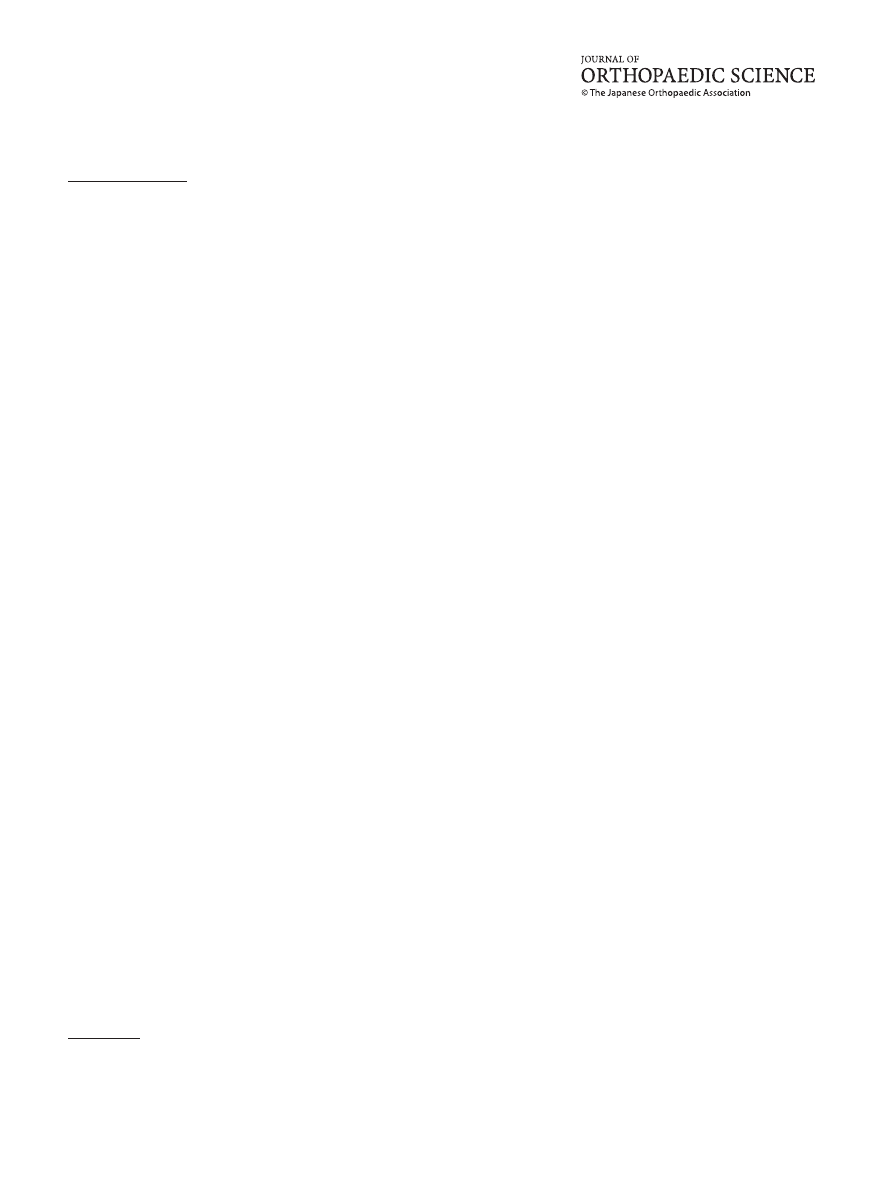
Radiographs, including an anteroposterior (AP) view
of the pelvis (Fig. 1) and a lateral view of the hips,
revealed total head involvement of the left hip consis-
tent with a diagnosis of Perthes disease. In addition to
more than 50% collapse of the lateral pillar (Herring
type C),
10
this patient had other radiographic signs,
including lateral extrusion of the epiphysis, metaphy-
seal cyst, horizontal appearance of proximal femoral
physis, a break in Shenton’s line, lateral subluxation of
the hip, and possible hinged abduction, suggesting a
poor prognosis.
A trial of outpatient physical therapy and a home
exercise program failed to alleviate his symptoms and
the clinical fi ndings. A hip arthrogram revealed fl atten-
ing of the superolateral portion of the femoral head
with proximal migration of the femur.
The patient underwent adductor tenotomy and appli-
cation of a previously unused EBI (Parsipanny, NJ,
USA) hinged external fi xator (Fig. 2) for arthrodiasta-
sis. Three hydroxyapatite-coated pins were placed in
the supraacetabular area and two in the femoral shaft.
The uniplanar hinge was placed at the level of the center
of the femoral head, and the left lower extremity was
kept in approximately 15° of abduction and 10° of inter-
Offprint requests to: S. Sabharwal
Received: November 14, 2006 / Accepted: March 30, 2007
386
S. Sabharwal and D. Van Why: Mechanical failure of external fi xator
nal rotation. Acute distraction (5 mm) at the fi xator was
carried out under general anesthesia. Satisfactory place-
ment of the external fi xator and free mobility of the hip
in the fl exion-extension arc were confi rmed intraopera-
tively (Fig. 3). All connectors and bolts were fi rmly
hand-tightened with a wrench, based on the manufac-
turer’s recommendation.
11
The patient was discharged
home the following day with instructions for no weight
bearing on the affected extremity.
Outpatient physical therapy, including fl exion and
extension range of motion exercises of the left hip, was
initiated. The patient’s family was instructed to start
Fig. 1. Preoperative anteroposterior (AP) radiograph of the
pelvis showing total head involvement of the left hip with
metaphyseal cysts and lateral subluxation secondary to Perthes
disease in an 8-year-old boy
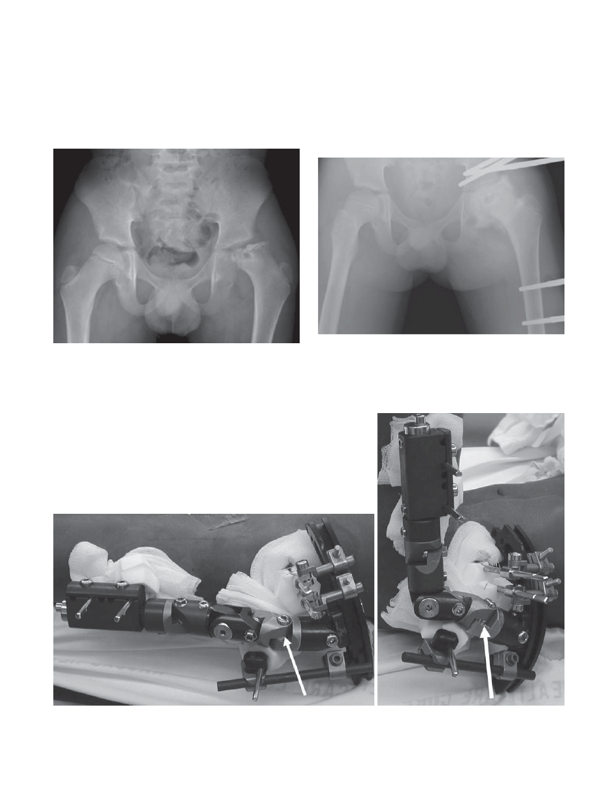
Fig. 2. Immediate postoperative radiograph following a hip
arthrogram, adductor tenotomy, and placement of a monolat-
eral hinged external fi xator for arthrodiastasis across the hip
joint. Note the mild abduction positioning of the left lower
extremity
Fig. 3. Clinical photograph of the hinged external fi xator, allowing passive extension (A) and fl exion (B) of the hip joint. The
arrow indicates the dual locking connector between the pelvic and femoral portions of the fi xator
A
B
S. Sabharwal and D. Van Why: Mechanical failure of external fi xator 387
distraction at the rate of 1 mm a day in two installments,
starting the third day following surgery. The goal of
distraction was slight overcorrection of the break in
Shenton’s line, as seen on the AP radiograph of the
hip.
Two weeks later, the AP radiograph of the left hip
revealed a persistent break in Shenton’s line. The family
was instructed to continue distraction at the same rate.
The patient was compliant with physical therapy
and non-weight-bearing instructions. Approximately 6
weeks postoperatively, despite several millimeters of
distraction of the external fi xator, Shenton’s line
remained disrupted on radiographs, and the left lower
extremity was noted to be in 15° of adduction (Fig. 4).
Pin sites were dry and clean with no change in position
of the half-pins on radiographs. Loss of serrations of the
large bolt connecting the pelvic and femoral portions of
the external fi xator was noted (Fig. 5). This mechanical
failure of the external fi xator had allowed the left leg to
adduct at the hip.
An examination was performed under anesthesia,
and the left lower extremity was repositioned in 15°
of abduction and 10° of internal rotation. Improved
seating of the femoral head was confi rmed with an
arthrogram, and the dual locking connector and
bolt between the pelvic and femoral segments were
replaced and cemented with polymethylmethacrylate
(PMMA) (Fig. 6). Acute distraction (15 mm) was per-
formed under anesthesia, and adequate repositioning
of the femoral head with restoration of Shenton’s line
was achieved. No further distraction was done post-
operatively, and the physical therapy regimen was
reinstituted.
Follow-up radiographs showed no further change in
the position of the hip. Eight weeks following fi xator
adjustment, the patient was brought back to the operat-
ing room. A left hip arthrogram revealed restoration of
Shenton’s line with residual fl attening of the weight-
bearing portion of the femoral head. The external
fi xator was removed. Under general anesthesia, left hip
abduction was noted to be 35°. The patient was placed
in a customized hinged hip abduction orthosis, and
his weight-bearing status was gradually advanced with
physical therapy.
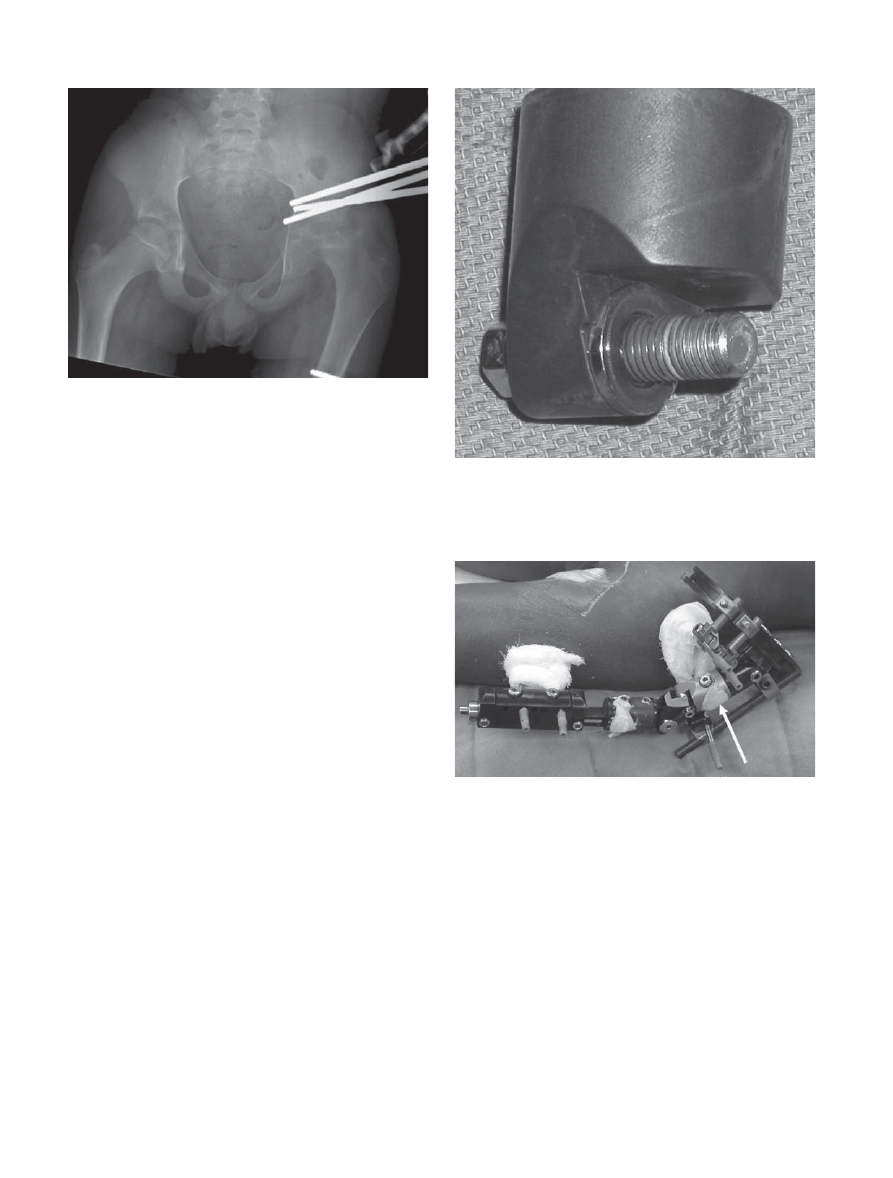
Fig. 4. Follow-up radiograph 6 weeks postoperatively demon-
strating an adduction deformity of the left lower extremity
with no signifi cant distraction at the hip joint
Fig. 5. Retrieved dual locking connector, demonstrating loss
of serrations and damage to the threads of the connector
bolt
Fig. 6. Intraoperative photograph following revision of the
external fi xator with polymethylmethacrylate (PMMA) sup-
plementation at the dual locking connector (arrow)
388
S. Sabharwal and D. Van Why: Mechanical failure of external fi xator
On a recent examination, done 2 years postopera-
tively, hip fl exion was 95° on the left and 130° on the
right; abduction was 20° compared to 50°; external rota-
tion was 15° compared to 60°; and internal rotation was
15° compared to 30°, respectively. He has remained
asymptomatic and resumed regular activities despite a
mild abductor lurch on the affected left side. Follow-up
radiographs reveal slight disruption of Shenton’s line,
although it had improved compared to the preoperative
imaging studies. Reossifi cation of the femoral head with
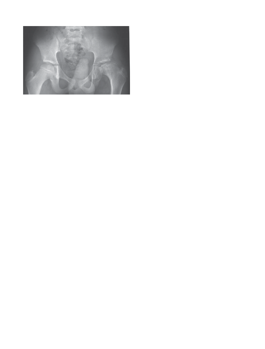
residual fl attening was noted (Fig. 7). A scanogram
revealed 5-mm leg-length discrepancy, with the left side
being shorter.
Discussion
Articulated joint distraction treatment has been reported
for various stages of osteoarthritis and chondrolysis
affecting a variety of joints including the hip.
12–14
Unlike
femoral and pelvic osteotomies, arthrodiastasis treat-
ment is minimally invasive, does not involve any iatro-
genic alteration of the local bony anatomy, and avoids
cast immobilization. During joint distraction treatment
for Perthes disease, the goal of treatment is to improve
hip mobility and favorably alter the natural history. This
is likely accomplished by reducing the mechanical
stresses across the hip joint, which may facilitate carti-
lage proliferation and endochondral ossifi cation of the
proximal femoral epiphysis.
13,14
It appears to be a viable
surgical alternative in older children who would other-
wise have a high likelihood of poor radiographic and
functional outcome.
2
Few authors have reported adverse events other than
pin-tract infections related to arthrodiastasis treat-
ment.
6–8,12
Maxwell et al.,
8
using the Orthofi x external
fi xator, reported two patients who had advanced col-
lapse of the femoral head secondary to Perthes disease
and sustained pin breakage. Although no fi rm recom-
mendations were made, they suggested that advanced
age and weight should be further investigated as a
potential cause for this failure, as these factors may
infl uence the amount of force that can be safely toler-
ated by the external fi xator pins. Segev et al.
9
reported
on 16 patients with late-onset severe Perthes disease
who were treated with arthrodiastasis in combination
with limited soft tissue release. They mentioned that
one clamp broke during treatment and required replace-
ment. However, no details of the cause or potential
preventive measures were provided. Interestingly, none
of the potential factors that can contribute to hardware
failure, such as obesity, application of a previously used
external fi xator, noncompliance with weight-bearing
status, or attempts at forceful hip abduction-adduction
exercises, was present in our patient.
Although no study has measured the forces gener-
ated during joint distraction, few investigators have
tried to measure them during limb lengthening. Simpson
et al.,
15
using precalibrated load cells incorporated into
the lengthening mechanism of monolateral external fi x-
ators, reported generation of axial forces of 300–1000 N
in patients undergoing femoral lengthening. Angular
deformity at the osteotomy site and mechanical failure
of the external fi xator was noted in some patients
with congenital shortening, who also demonstrated the
highest axial forces. They cautioned that with the high
distraction forces recorded during limb lengthening safe
levels for many unilateral fi xators might be exceeded.
Younger et al.
16
found similar values for axial forces on
the external fi xator frame in their analysis of three
patients undergoing femoral lengthening.
Chao and Hein
17
performed mechanical testing on the
Orthofi x (Verona, Italy) external fi xator and found that
the cam positioning of the ball joint gradually migrated
as the forces were incrementally increased. Repetitive
manual tightening and loosening of the ball joint caused
abrasive wear on the cam and bushing surfaces. Modi-
fi cation of the fi xator design was recommended to
improve its mechanical performance. Moroz et al.
18
also
reported on mechanical testing of the Orthofi x device
and found the ball joint to be the most common site of
mechanical failure. Marsh et al.
19
reported on the use
of the Orthofi x external fi xator for treatment of adult
supracondylar femur fractures. They encountered
similar failures at the ball joint and suggested reinforce-
ment with PMMA.
Dirschl and Obremskey
20
reported on mechanical
testing of previously used monolateral external fi xators
and compared their mechanical strength with previ-
ously unused fi xators. They found that a mean load 721
±
70 N caused failure of previously used standard fi x-
ators, which was not signifi cantly different from the 749
Fig. 7. Final AP pelvis radiograph of the patient in the remod-
eling phase, showing evidence of persistent fl attening of the
femoral head and mild superolateral hip subluxation

S. Sabharwal and D. Van Why: Mechanical failure of external fi xator 389
±
81 N for the unused fi xators. However, they did report
major damage in 14% of the 120 serrated joints tested,
and the remaining 86% of the serrated joints also exhib-
ited minor damage. The damage included deformation
or loss of material across serrations, which resulted in
the removed material being forced into the trough
between the teeth, limiting complete interdigitation of
the components. This mode of failure and fi ndings at
the serrated joints are similar to observations seen in
our case. We were unable to fi nd a biomechanical study
reporting on the increased load to failure following rein-
forcement with PMMA of either a ball joint or dual
locking connector of an external fi xator.
The treating surgeon must be aware of mechanical
failure as a potential cause for lack of anticipated hip
joint distraction during arthrodiastasis treatment for
Perthes disease. There is a lack of biomechanical studies
investigating the forces generated at the external fi xator
during articulated hip joint distraction and whether use
of PMMA decreases such forces at the various connec-
tors of the external fi xator. Based on the information
available, we recommend routine cementing of the dual
locking connector mechanism or ball joints of monolat-
eral external fi xators in patients who undergo articu-
lated joint distraction of the hip.
None of the authors received fi nancial support for this
study.
References
1. Caterrall A. Legg-Calve-Perthes syndrome. Clin Orthop 1981;58:
41–52.
2. Herring JA. The treatment of Legg-Calve-Perthes disease: a criti-
cal review of the literature. J Bone Joint Surg Am 1994;76:
448–58.
3. Stulberg SD, Cooperman DR, Wallensten R. The natural history
of Legg-Calve-Perthes disease. J Bone Joint Surg Am 1981;63:
1095–108.
4. Ismail AM, Macnicol MF. Prognosis in Perthes’ disease: a com-
parison of radiological predictors. J Bone Joint Surg Br 1998;80:
310–4.
5. Grzegorzewski A, Bowen JR, Guille JT, Glutting J. Treatment of
collapsed femoral head by containment in Legg-Calve-Perthes
disease. J Pediatr Orthop 2003;23:15–9.
6. Kocaoglu M, Kilicoglu OI, Goksan SB, Cakmak M. Ilizarov
fi xator for the treatment of Legg-Calve-Perthes disease. J Pediatr
Orthop B 1999;8:276–81.
7. Kucukkaya M, Kabukcuoglu Y, Ozturk I, Kuzgun U. Avascular
necrosis of the femoral head in childhood: the results of treatment
with articulated distraction method. J Pediatr Orthop 2000;20:
722–8.
8. Maxwell SL, Lappin KJ, Kealey WD, McDowell BC, Cosgrove
AP. Arthrodiastasis in Perthes’ disease: preliminary results. J
Bone Joint Surg Br 2004;86:244–50.
9. Segev E, Ezra E, Wientroub S, Yaniv M. Treatment of severe late
onset Perthes’ disease with soft tissue release and articulated hip
distraction: early results. J Pediatr Orthop B 2004;13:158–65.
10. Herring JA, Neustadt JB, Williams JJ, Early JS, Browne RH. The
lateral pillar classifi cation of Legg-Calve-Perthes disease. J Pediatr
Orthop 1992;12:143–50.
11. EBI website: http://www.ebimedical.com/products/detail.cfm/.
12. Aldegheri R, Trivella G, Saleh M. Articulated distraction of the
hip: conservative surgery for arthritis in young patients. Clin
Orthop 1994;301:94–101.
13. Van Roermund PM, van Valburg AA, Duivemann E, van Melke-
beek J, Lafeber FPJ, Bijisma JWJ, et al. Function of stiff joints
may be restored by Ilizarov joint distraction. Clin Orthop 1998;348:
220–7.
14. Van Valburg AA, van Roermund PM, Marijnissen AC, Wenting
MJ, Verbout AJ, Lafeber FPJ, et al. Joint distraction in treatment
of osteoarthritis: effects on cartilage in a canine model. Osteoar-
thritis Cartilage 2000;8:1–8.
15. Simpson AH, Cunningham JL, Kenwright J. The forces which
develop in the tissues during leg lengthening: a clinical study. J
Bone Joint Surg Br 1996;78:979–83.
16. Younger ASE, Mackenzie WG, Morrison JB. Femoral forces
during limb lengthening in children. Clin Orthop 1994;301:
55–63.
17. Chao EY, Hein TJ. Mechanical performance of the standard
Orthofi x external fi xator. Orthopaedics 1988;11:1057–69.
18. Moroz TK, Finlay JB, Rorabeck CH, Bourne RB. External skel-
etal fi xation: choosing a system based on biomechanical stability.
J Orthop Trauma 1988;2:284–96.
19. Marsh JL, Jansen H, Yoong HK, Found EM Jr. Supracondylar
fractures of the femur treated by external fi xation. J Orthop
Trauma 1997;11:405–11.
20. Dirschl DR, Obremskey WT. Mechanical strength and wear of
used EBI external fi xators. Orthopedics 2002;25:1059–62.
Wyszukiwarka
Podobne podstrony:
17 209 221 Mechanical Study of Sheet Metal Forming Dies Wear
Resuscitation- The use of intraosseous devices during cardiopulmonary resuscitation, MEDYCYNA, RATOW
52 737 754 Relationship Between Microstructure and Mechanical Properts of a 5%Cr Hot Works
32 425 436 Ifluence of Vacuum HT on Microstructure and Mechanical Properties of HSS
Pair Creation of Black Holes During Inflation
03 Electrophysiology of myocardium Myocardial Mechanics Assessment of cardiac function PL
W Borek Mechanical properties of high manganese austenitic TWIP type steel
Mechanical Properties of Native and Cross linked Type I Collagen Fibrils Yang
Changes in Brain Function of Depressed Subjects During
Mechanical Performance of Plastics
Fibrillar Structure and Mechanical Properties of Collagen
Effect of heat treatment on microstructure and mechanical properties of cold rolled C Mn Si TRIP
95 1373 1389 A new Investigation on Mechanical Properties of Ferro Titanit
Effects of preoperative physiotherapy in hip osteoarthritis patients awaiting total hip replacement
[Mises org]Grams,Mart Failure of The New Economics Study Guide
Failure of Gun Control Laws
MECHANICAL PROPERTIES OF METALS
Adolf Hitler vs Henry Ford; The Volkswagen, the Role of America as a Model, and the Failure of a Naz
Microstructure and mechanical properties of plasma sprayed H
więcej podobnych podstron