
CHAPTER
13
Tumors of the
Spinal Canal
Martin Greenberg
Dennis E. McDonnell
Herman F. Flanigin
Mass lesions that affect the function of the spinal cord are
divided into: (1) intramedullary, those that originate within
the spinal cord; (2) intradural-extramedullary, which is
self-explanatory; and (3) extradural, those that arise outside
the dura, most of which involve the vertebral column.
Symptoms usually begin with local pain, which may be
exacerbated at night and accompanied by a significant
radicular component.
4
-
7
-
10
'
11
'
13
Paresis may become promi-
nent. The rate of progression of the paresis varies greatly,
depending on the degree of compromise of blood supply to
the spinal cord, but paralysis below the level of involve-
ment is associated with a grave prognosis—even with ade-
quate decompression.
Virtually all neoplastic lesions located in or behind me
spinal cord can be approached by unroofing the spinal canal
(laminectomy), as can most neoplastic lesions located lat-
eral to the spinal cord and most cystic lesions wherever
they are located in the spinal canal.
1
"
13
Many lesions origi-
nating in the vertebral column are located anterior to the
spinal cord and are best approached anteriorly or antero-
laterally, depending on the vertical level of the lesion.
For instance, lesions at the cervicomedullary junction
may be attacked through a transoral or transcervical retro-
parapharyngeal approach,
14
-
16
while those lower in the
neck are approached through an anterolateral cervical ap-
proach.'-
3
'
8
'
10
'
11
Lesions located in the thoracic and abdomi-
nal regions may be approached through a thoracotomy and
retroperitoneal approach, although the costotransversec-
tomy approach has also been popularized.
1
'
8
'
10
'
11
Generally,
at least a part of, if not an entire, vertebral body must be
removed to approach the spinal canal and, if so, it must be
replaced with a graft or prosthesis. Malignant sacral tumors
may require simultaneous anterior and posterior approaches
in order to effect a cure.
10
'
11
Risks to neurological function during surgery may be
reduced by intraoperative monitoring, which at present is
most commonly using somatosensory evoked potentials.
Monitoring motor potentials may be more rewarding and is
being developed with the use of magnetic field stimulators.
CLINICAL PRESENTATION
1
-"
Most spinal tumors present with the onset of localized pain,
often with a radicular component. Pain is often present at
rest and severe at night, unrelieved by narcotics or analge-
sics. It may not be brought on by exertion and may also be
exacerbated by vigorous exercise. Patients with intramedul-
lary tumors may complain of burning dysesthesias in the
hands or legs, often for months to years.
4
'
5
-
12
-
13
Metastases
must be considered in patients with a history of malignancy.
Patients with metastatic disease may have focal regions of
tenderness with muscle cramps. Palpation often reproduces
pain. Focal kyphoscoliosis or lordosis may be secondary to
instability caused by a tumor infiltrating a vertebral
body(ies). Patients may have a previous or current history of
incidental trauma with a pathological compression frac-
ture(s), associated with occult spinal tumor(s).
Along with pain, metastatic tumors present a variable
course, with an abrupt onset below the lesion, of a demon-
strable level of sensation to pinprick corresponding to the
spinal segment involved and variable urinary retention with
hyperreflexia and clonus corresponding to an upper motor
neuronal deficit.
4
-
5
-
8
Metastatic tumors can present insidi-
ously. When located posterior to the spinal cord, they cause
proprioceptive deficits with early myelopathic signs.
Intramedullary tumors classically present with a dissocia-
tive sensory loss because of damage to crossing central
commisural fibers of the spinothalamic tracts, sometimes
secondary to a syrinx.
4
-
5
There is a marked disturbance of
pain and temperature sensation typically at the level of the
lesion but preservation of touch and position sense. There
may be a history of accidental burns or a shoulder-cape
distribution of loss to pinprick sensation.
229

230
CHAPTER
Intradural-extramedullary tumors classically present with
predominant motor and radicular symptoms. A neuroma or
neurofibroma arising from a sensory nerve root sheath typi-
cally causes ipsilateral pain and weakness in the distribution
of a root, as well as early spastic hemiparesis with hyperre-
flexia below the level of the lesion. As the neuroma expands,
a Brown-Sequard syndrome may become evident. Menin-
giomas arising near the nerve root sheath from the dura-
arachnoid present in a lateral or ventrolateral anatomic posi-
tion with a combination of long-standing neurological signs
and symptoms, particularly motor deficits.
9
Both intramedullary and intradural-extramedullary
tumors can present initially with signs and symptoms of
increased intracranial pressure (ICP), particularly hydro-
cephalus, headaches, nausea and vomiting, papilledema,
visual loss, obtundation, and gait apraxia.
65
Neurinomas
and ependymomas secrete large amounts of proteins into
the cerebral spinal fluid (CSF), which block or impede the
flow in the spinal subarachnoid compartments. This block
of CSF absorption results in increased ICP. Gardner hy-
pothesized that such blockage accounts for hydrocephalus
occulta, often seen as the presenting sign of primary intra-
spinal neoplasms.
65
DIAGNOSTIC STUDIES
1
-
16
RADIOGRAPHY
Preliminary diagnostic examinations should include frontal
(AP) and lateral x-rays, as well as oblique or swimmer's
views to visualize the cervicothoracic region.
13
Absence,
asymmetry, or overt destruction of a pedicle is suggestive of
metastatic cancer. Extensive metastatic disease may present
as a large paraspinal mass of soft tissue seen on plain
radiographs. Malignant lesions have a predilection for verte-
bral bodies with consequent pathological compression frac-
tures seen clearly on the lateral x-rays. Plain radiographs are
positive in 80 to 90 percent of patients with spinal tumors,
both extrinsic and intrinsic.
2
-
13
Osteolytic lesions are most common with metastatic
cancer, especially when the primary lesion is in the breast,
lung, kidney, or colon.
4
'
5
'
10
-
11
Prostatic cancer produces an
"ivory" vertebra or osteoblastic lesion with a sclerotic bone
edge.
Primary bone tumors can be diagnosed by plain radiog-
raphy .2,6,7,10,11 Hemangiomas produce coarse vertical striations
or trabeculae ("corduroy cloth" impression) while aneurysmal
bone cysts and giant cell tumors produce multiloculated, lytic
lesions. Osteoid osteomas and osteoblastomas are typically
sclerotic. Osteosarcomas, chondrosarcomas, and multiple mye-
lomas which are malignant (primary neoplasms) present with
extensive bone destruction and paraspinal soft tissue masses.
Chordomas produce gross destruction of the bone elements and
amorphous, peripheral calcification, as well as a large soft
tissue mass with epidural extension.
Intramedullary tumors, like ependymomas and astrocyta- fr
mas, can attain considerable size and cause widening of the f
interpedicular distance, with enlargement of the canal on AP
films or even kyphoscoliosis or lordosis in the later*
views.
4
'
5
Intradural-extramedullary neurofibromas can cauv
marked widening of the neural foramina and scalloping c;
the vertebral bodies. Dumbbell thoracic neurofibromas can
be seen as masses on chest x-rays. Rarely, meningiomas are
sufficiently calcified to be seen on plain radiographs, due to
psammoma bodies commonly seen in intracranial tumors/
Bony hyperostoses are rare with spinal meningiomas.
9
MAGNETIC RESONANCE IMAGING (MRI)
17
f
Magnetic resonance imaging (MRI) has nearly supplanted
computerized tomography (CT) in the diagnostic evaluatioe
of spinal tumors.
17
MRI images the spine in three dimen-
sions, (axial, sagittal, and coronal) and highlights the soft
tissue and intraspinal changes.
17
It can be diagnostic for
intrinsic cord lesions when gadolinium (Gd-DTPA) is ad-
ministered.
17
Vascular tumors can be visualized as lesions
with varying signals on MRI. Magnetic resonance angiogra-
phy (MRA) further enhances the diagnostic capabilities of
this imaging modality.
Metastatic cancer to vertebrae is visualized by MRI. As
bone marrow is replaced by tumor, the tumor appears hy-
pointense on the Tl image, hyperintense on the T2 image,
and enhanced with gadolinium (Gd-DTPA).
17
MRI reveals
soft tissue changes and epidural compression by lymphoma.
multiple myeloma, and chordoma.
Intramedullary tumors are best imaged with MRI as the
solid and cystic components of an astrocytoma or ependy-
moma can be defined on the Tl, T2, and gadolinium-en-
hanced images.
17
Hemangioblastomas can be diagnosed by
MRI since they demonstrate a vascular nodule and the tumor
is associated with a syrinx or cystic mass. There are asso-
ciated surface vessels, including arteries and veins leading to
the nodule.
Intradural tumors are outlined by MRI.
17
Neurinomas and
neurofibromas are hyperintense on Tl and T2 images, are
enhanced with gadolinium, and often can be visualized aris-
ing from nerve sheaths in the neural foramina. Lipomas have
a hyperintense signal on the Tl image. Meningiomas en-
hance with gadolinium, allowing definition of their ventral
or ventrolateral orientation to the adjacent, compressed spi-
nal cord.
A deficiency in MRI technology is that high sensitivity
relies on direct imaging of the protons in body water, which
is generally lacking in bone. Hence, tumors of bone and
structural abnormalities are not imaged well by the MRI.
There continues to be need for CT alone or in conjunction
with myelography for diagnostic capabilities.
COMPUTERIZED TOMOGRAPHY (CT)
Although MRI has supplanted computerized tomography
(CT) as the primary imaging technique for spinal tumors,

TUMORS OF THE SPINAL CANAL
231
There is still a key role for CT alone or in conjunction with
myelography.
7
-
13
CT delineates involvement of pedicles, la-
minae, spinous processes, and vertebral bodies by primary
bone tumors, i.e., multiple myeloma, chondrosarcoma, os-
eosarcoma, and chordoma.
7
-
13
It may demonstrate tumors or
calcifications not seen by plain x-rays. Osteomas, osteohlas-
omas, giant cell tumors, meningiomas, and aneurysmal
bone cysts or bone tumors are best visualized by thin-section
MYELOGRAPHY
10
'
11
'
13
Myelography followed by CT remains a useful imaging
modality for spinal tumors. CT helps to differentiate intra-
medullary, intradural-extramedullary, and extradural lesions.
Further, CT-myelography accurately delineates the ventral
vs. dorsal or lateral spinal cord compression by outlining the
contrast media in CSF or subarachnoid spaces. In clinical
situations where the MRI indicates multiple levels of metas-
tatic cancer, CT-myelography is helpful in determining the
level producing deficits, especially if the neurological exam-
ination does not correlate closely with the MRI.
ANGIOGRAPHY
Spinal angiography can be helpful in localizing the artery of
Adamkiewicz, typically on the left side between T8 and
L3.
13
However, this procedure is not routinely necessary for
preoperative evaluation. Vascular tumors such as metastatic
renal cell carcinomas, hemangiomas, and aneurysmal bone
cysts can be delineated by angiography, and preoperative
embohV.ation is helpful in reducing blood loss during sur-
gery. Hemangioblastomas can be accurately diagnosed by
spinal angiography. Arteriovenous (AV) shunting and highly
vascular tumor nodules with large, draining veins are char-
acteristically visualized.
17
Embolization may be helpful in
the management of large or multiple hemangioblastomas.
At the cervicomedullary junction, vertebral angiography is
warranted for meningiomas or neurofibromas that may encase
a major vessel, signaling caution during tumor removal. It is
important to know whether a dominant vertebral artery can be
sacrificed. Extrinsic tumors like lymphomas, chordomas, and
renal cell carcinomas which can present as extensive soft tissue
masses palpable in the neck may have multiple tumor vessels
arising from the muscular branches of the vertebral artery.
Preoperative embolization may be useful.
In primary benign bone tumors—e.g., osteoid osteomas
and osteoblastomas—the bone scan is helpful in diagnosis,
since it shows increased uptake in areas of active bone
growth. With an aneurysmal bone cyst or hemangioma, there
may be minimal, if any, uptake in the bone. In malignant
bone tumors, particularly multiple myelomas, there may be
decreased uptake or "cold" spots on radionuclide bone scan-
ning, reducing the value of this modality in identifying sites
of involvement in the spinal axis. A skeletal survey with
plain x-rays is more helpful in myeloma for detecting occult
lesions.
LABORATORY INVESTIGATIONS
Several hematological investigations may be pertinent with
suspected spinal tumors. Patients with metastatic lesions of
the vertebrae may present with anemia, leukopenia, or
thrombocytopenia due to involvement of the bone marrow.
Widespread bony metastases may lead to hypercalcemia
and elevated serum alkaline phosphatase. Multiple myeloma
can be diagnosed by the presence of the Bence Jones mono-
clonal antibody protein by urine or serum protein electro-
phoresis, a screening test that may be positive even with
solitary plasmacytomas. Tumor markers can be diagnostic:
prostatic specific antigen (PSA) with prostate cancer, CA
125 with ovarian cancer, carcinoembryonic antigen (CEA)
with colon cancer, and vanillylmandelic acid (VMA) with
neuroblastoma.
13
Elevated levels of alpha fetoprotein (a-FP)
and beta human chorionic gonadotrophin (b-HCG) are diag-
nostic for tumors of germ cell origin, including seminomas,
germinomas, embryonal carcinomas, endodermal sinus
tumors, and mixed teratomas.
49
Microscopic examination of the centrifuged sediment
from CSF can be diagnostic for extramedullary tumors,
especially lymphoma, leukemia (ALL), and in cases of
"drop-metastases," including pinealoblastomas, medullo-
blastomas, ependymomas, germinomas, and other germ cell
tumors.
The CSF should be routinely obtained at myelography and
examined for protein, glucose, cell count, and cytology. The
protein value is elevated with blocks of the subarachnoid
space and is usually elevated in association with neurinomas
and neurofibromas because of secretion of protein by the
tumor. The elevated protein can distinguish neurinomas
from meningiomas. CSF pleocytosis is a harbinger of lepto-
meningeal infiltration with metastatic or primary neoplasms.
BONE SCANS
Radioisotope bone scans are useful in locating sites of
metastases.
7
'
10
-
11
'
13
Multiple metastases may affect the indi-
cations for, or type of, neurosurgical procedure. However,
the isotope scan is nonspecific, and other radiological mo-
dalities are frequently required.
ADJUNCTIVE OPERATIVE
MANAGEMENT
1
-
16
STEROID ADMINISTRATION
18
'
19
Glucocorticoids should be given to patients at least 24 h
prior to surgery and should be continued postoperatively

232
CHAPTER 13
with tapering dosages beginning 3 to 5 days after surgery to
decrease overall spinal cord edema.
18
-
19
Steroid tapering can
be adjusted as the patient's neurobiological function is mon-
itored. Ordinarily dexamethasone is administered at 4 mg q
6 h, but this dosage can be increased to 10 mg q 4 h.
Protection of the gastrointestinal (GI) tract may be accom-
plished with ranitidine HCL [Zantac (H
2
blocker)] or an
antacid. For patients with metastatic cancer to the spine and
sudden, dramatic paraparesis or quadriparesis, an initial dose
of 100 mg dexamethasone IV can be given, followed by 20
mg q 4 h. Glucocorticoids appear to have a beneficial effect
on spinal cord edema from tumor cells, although it is unclear
whether the mechanism of action is inhibition of cell growth
or actual cytolysis.
18
-
19
Interestingly, there is evidence that
glucocorticoids may have a direct oncolytic effect on lym-
phomas and leukemias through cell lysis.
19
Regardless of the
mechanism, the administration of glucocorticoids to patients
with spinal tumors appears to have a beneficial effect on
neurological function.
18
'
19
INTRAOPERATIVE (FROZEN) PATHOLOGY
Tumor specimens are usually examined microscopically dur-
ing surgery. In patients with metastatic tumors with an
unknown primary, the intraoperative frozen tissue will often
be diagnostic. The biology of the metastatic tumor provides
the opportunity to develop a rational management scheme. It
may direct the type of surgery required. For example, in
patients with metastatic lung cancer, it is important to know
whether the tumor is undifferentiated carcinoma or oat-cell
carcinoma as opposed to large cell or squamous cell carci-
noma, since undifferentiated and oat cell tumors are asso-
ciated with a very poor prognosis. Certain meta^tatic spinal
neoplasms are quite radiosensitive, particularly lymphomas
and seminomas, and postoperative irradiation will suppress
or possibly eradicate residual tumor. Craniospinal irradiation
is effective for intradural, radiosensitive tumors like medul-
loblastomas, germinomas, and pinealoblastomas, which may
present as "drop-metastases."
Frozen tissue pathology is helpful during surgery for
intramedullary tumors, particularly when differentiating as-
trocytomas from ependymomas. The neurosurgeon may be
hesitant to attempt complete removal of an astrocytoma due
to indistinct tumor margins, whereas ependymomas more
readily peel away from normal spinal cord.
INTRAOPERATIVE SONOGRAPHY
The recent application of real-time ultrasonography to surgi-
cal explorations allows localization of spinal tumors by
imaging the structural details.
13
The ultrasonic probe is ap-
plied to the epidural or intradural spaces. Then the full
extent of an epidural mass, including ventral and ventrola-
teral extensions, is determined. In intramedullary tumors, the
depth and extent of the tumor is ascertained, and any asso-
ciated syringes identified. Adequate decompression of a syr-
inx and division of septations can be followed by resolution
or disappearance of an echogenic signal. After microsurgic,
resection, the spinal cord is examined by ultrasound 1\
residual tumor, or the placement of a shunt is verifie^
Spinal cord pulsations demonstrating adequate decompres-
sion are monitored.
13
INTRINSIC TUMORS
4
'
5
'
8
'
11
'
12
A recent retrospective review of primary intraspinal neo-
plasms in a denned population base imparts reasonable
unbiased data and sheds light on the overall tumor biology.
1
Meningioma is the most-common spinal tumor (46.7 per
cent), followed by ependymoma (18.6 percent), neurinoin^
(10.8 percent), astrocytoma (6.0 percent), mixed glioma (5.5
percent), and glioblastoma (1.4 percent).
20
Oligodendro-
glioma was 0.7 percent and other miscellaneous tumors 6.3
percent.
The gender distribution of meningiomas was 6:1 female/
male, and ependymomas were 2:1 male/female. Remaining
types of spinal tumor were almost evenly distributed among
males and females. The incidence of intraspinal tumor is 0.3
to 0.5 per 100,000 population per year, thus it is rare.
20
By
comparison, this is the same percentage as that seen for male
breast cancer in Norway.
20
In males, there is a peak of
intraspinal tumors in the 15- to 29-year-old age range, wher-
eas in females a similar peak is seen in the 60- to 74-year-
old age group. Intraspinal meningiomas are associated with a
very high 5-year survival rate of 99.5 percent, whereas the
intracranial survival rate for meningiomas is significantly
less, at 84 percent.
20
Similarly, the 5-year survival rate for
intraspinal ependymomas is 88.9 percent, compared to 24.4
percent for the intracranial tumors.
20
Interestingly, there is
an equivalent 5-year survival rate of 48 percent vs. 44.9
percent for intraspinal and intracranial astrocytomas. How-
ever, the intracranial mixed glioma has a survival rate of
28.1 percent, compared to 50.7 percent in the intraspinal
mixed gliomas. In general, the prognosis is better in patients
with primary intraspinal glial tumors as compared with
intracranial tumors.
20
INTRAMEDULLARY TUMORS
EPENDYMOMAS
21
-
29
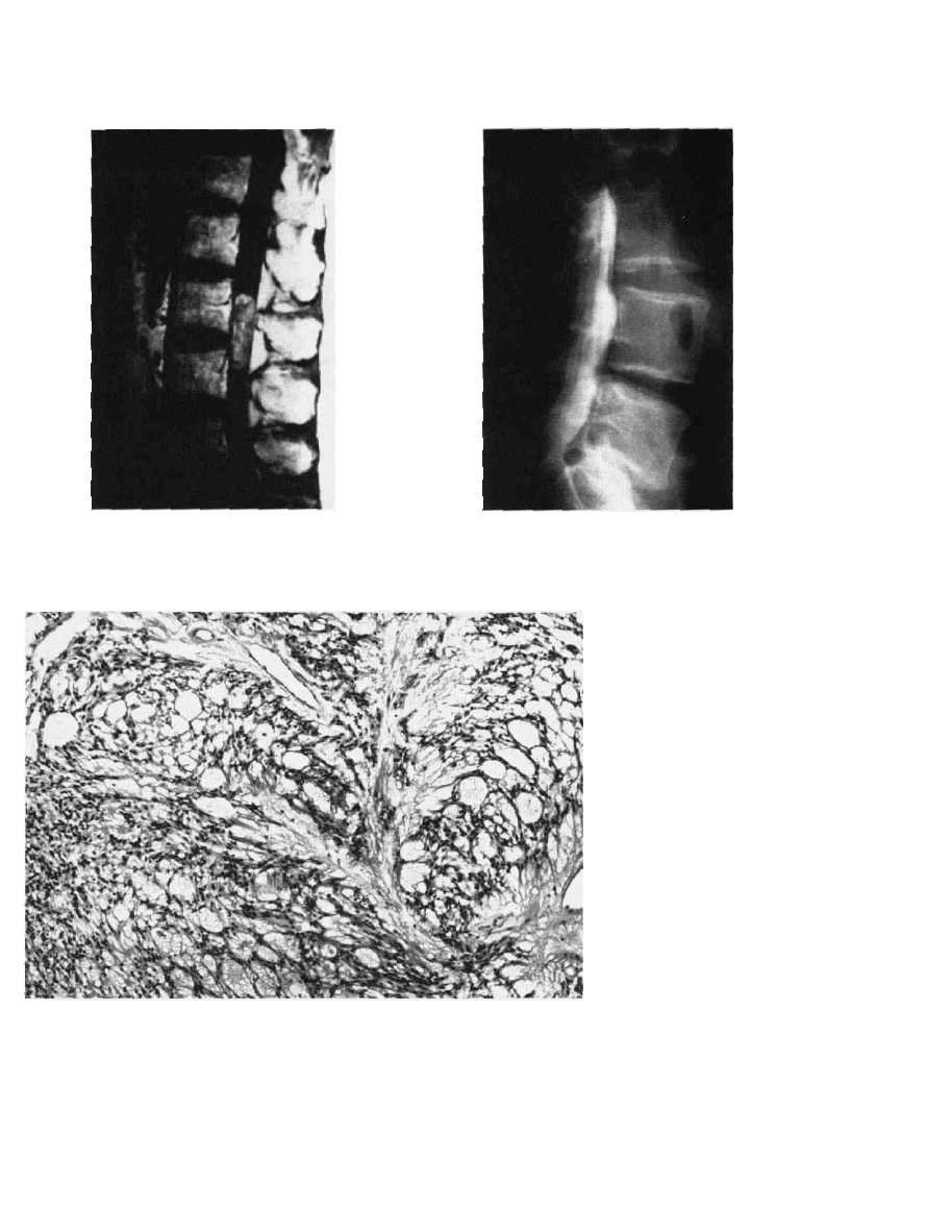
Ependymoma is the most common intramedullary tumor of
the spinal cord (Fig. 13-1, Plate 1). It is commonly found in
cervical or cervicothoracic regions, but it is frequently found
with a special predilection for the conus medullaris or filum
terminale (56 percent).
21
'
23
Fusiform tumors extend from the
medulla oblongata to the conus medullaris. Often the mean
XIMORS OF THE SPINAL CANAL
233
Figure 13-L4 Myxopapillary ependymoma. A T2-weighted MRI
in the sagittal plane with Gd-DTPA contrast shows a globular,
inhomogeneous mass that enhances very intensely. It is adjacent to
and spans the L2 and L3 vertebral bodies.
Figure 13-B Myelographic image in the lateral view shows a
spinal block at L2-L3 with a "capping" defect, or meniscus, due
to an intradural-extramedullary mass. By comparison, an
intramedullary ependymoma produces a fusiform enlargement of
the entire spinal cord.
Figure 13-1C Microscopic features
characteristic of the myxopapillary
ependymoma usually found in the
area of the filum terminate. Notice
presence of streaming vessels with
arrangements of tumor cells around
them. The cystoplastic round
vacuoles are filled with mutinous
contents.
length of solid tumor is three to five spinal segments. Pain,
sensory disturbance, and weakness are common symptoms,
often preceding the diagnosis by 2 to 3 years. The age of
presentation is typically 30 to 40 years old. The cauda
equina ependymoma is particularly common in males. The
tumor histology is 40 percent cellular, 2 percent epithelial,
21 percent myxopapillary, and 37 percent mixed.
4
A rare
histologically malignant type of ependymoma is termed
ependymoblastoma.
The cauda equina ependymoma is characterized by perin-
eal pain; bowel, bladder, and sexual dysfunction; and para-
paresis. Rarely, there is an associated symptom complex of
acute subarachnoid hemorrhage with sciatica, termed
Fincher's syndrome, which may be precipitated by preg-
nancy or trauma. Ependymomas arise from ependymal cell
nests within the ventriculus terminalis and filum terminale.
Very rarely, the myxopapillary ependymomas originate in
ectopic sites outside the spinal canal, particularly in the!

234
CHAPTER 13
sacrum and presacral tissues. They can even present as
widespread metastases in the lymph nodes, lung, liver, and
bone.
Syringomyelia and cystic fluid-filled cavitations are fre-
quently found with intramedullary tumors.
24
'
26
The cystic
fluid is yellow and proteinaceous when compared to the
clear, colorless CSF obtained from syringomyelia cavities
associated with the Arnold-Chiari malformation. There may
be objective signs and symptoms of syringomyelia due to
central canal destruction: amyotrophy in the upper extremi-
ties, "main-en-griffe," with concurrent hyporeflexia, and
dissociated sensory loss in the face, neck ("Balaclava hel-
met"), shoulders, and arms, extending in a capelike distribu-
tion due to interruption of the crossing spinothalamic tracts.
There may be scars from painless accidental burns, and
Horner's syndrome from interruption of the central, antero-
mediolateral sympathetic fiber column at Tl.
In the lower extremities, there is rigidity, hyperreflexia,
clonus, and occasionally spasticity. In cervicomedullary
tumors, there may be downbeat and vertical nystagmus;
dizziness; vertigo; cough-syncope; occipital headaches; nu-
chal rigidity; hoarseness, dysphagia, and other bulbar signs;
ataxia and dysmetria from cerebellar dysfunction; and spas-
tic tetraparesis from bilateral involvement of the cortico-
spinal tracts.
The neurosurgical rationale for treating intramedullary
tumors was summarized by Elsberg's treatise in 1925.
25
Operative techniques including bipolar coagulation and use
of the operating microscope were advanced by Greenwood,
Kurze, and Malis in the period 1950-1970.
24
'
26
'
27
Several recent series on the operative removal of the
intramedullary and cauda equina ependymomas report com-
plete removal in the majority of cases.21,23,26,28,29 jjjg intra-
medullary variant is a discrete tumor with a cleavage plane.
It is well demarcated, allowing total removal, and there is a
high incidence of cure evidenced by no recurrence of low-
grade ependymomas 5 to 10 years postoperatively.
21
'
22
'
28
-
29
However, the quality of the recovery depends on the
degree of preoperative neurological impairment since pa-
tients who are paraplegic preoperatively usually do not re-
gain motor function postoperatively. Further, dorsal column
deficits due to midline myelotomy and dysesthetic pain
syndromes are common complications. Overall, the sensori-
motor deficit stabilizes after tumor removal without signifi-
cant deterioration in the majority of cases.
21
'
22
The role of radiation therapy in the treatment of ependy-
momas is relegated to infiltrative lesions or the malignant
variant, ependymoblastoma, as radiation myelopathy is a
serious complication. Reoperation is indicated for recurrent
ependymomas; however, it may be complicated by a CSF
fistula from an incompetent dural closure, with complicating
meningitis and arachnoiditis.
A recent series of ependymomas of the cauda equina
mirrors results similar to those seen in the intramedullary
variant. Total tumor removal is the rule.
23
However, bowel
and bladder dysfunction, present preoperatively, did not re-
cover. Early surgery results in an excellent outcome in those
patients presenting with pain only, as compared to pain with
deficits and disturbance of sphincters. The role of chemo-
therapy in radioresistant tumors and the management of
incompletely resected tumors are variable. There is no effec-
tive therapeutic regimen for infiltrating ependymomas or
ependymoblastomas.
ASTROCYTOMA
30
-
33
The astrocytoma is the second most common primary intra-
medullary tumor, followed by malignant astrocytoma and
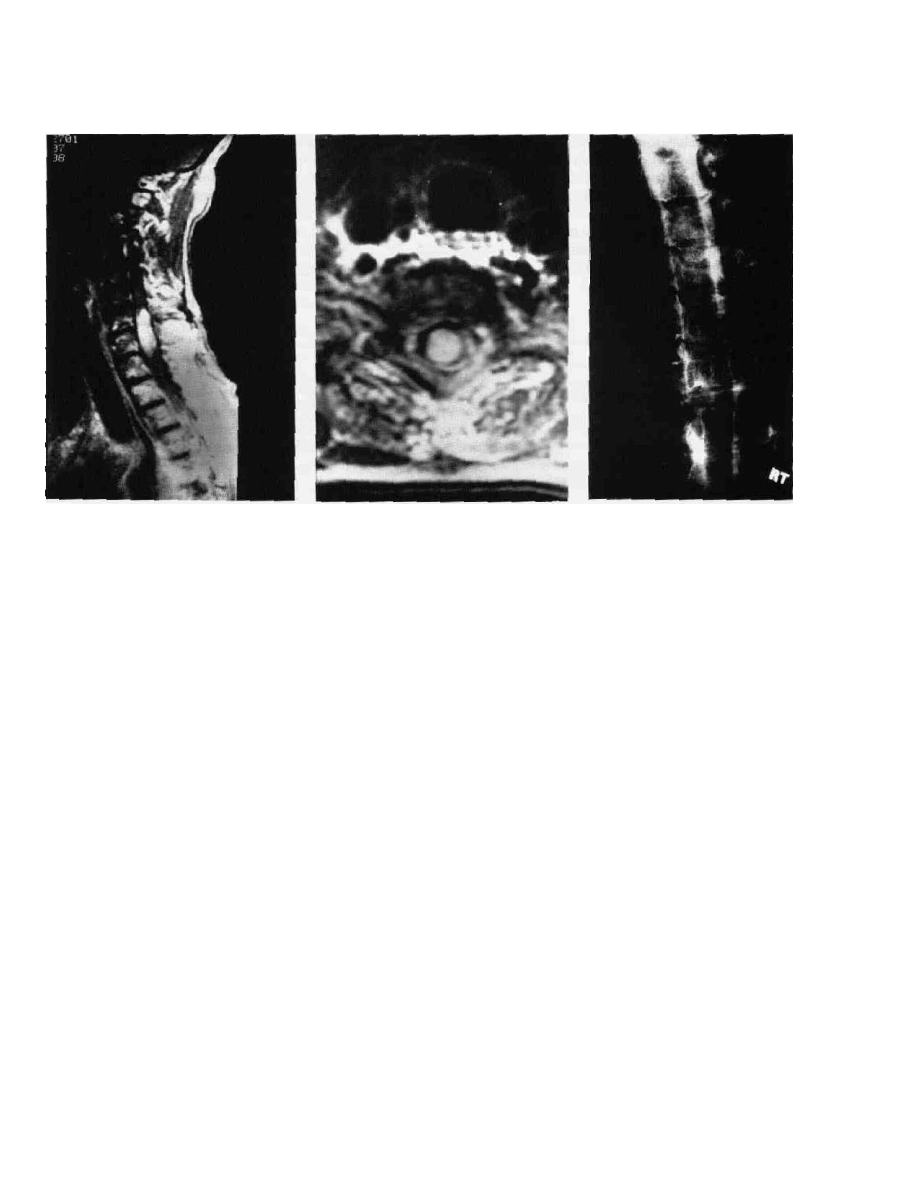
glioblastoma multiforme (Fig. 13-2, Plates 2 and 3).
20
It
presents throughout the cord or as a fusiform tumor at the
cervicomedullary junction in children.
33
Similar to the epen-
dymoma, the astrocytoma occurs most commonly in the
cervical and cervicothoracic regions and less commonly in
the thoracolumbar region. The neurological signs and symp-
toms mirror those in the ependymomas, including cortico-
spinal and spinothalamic tract involvement, paresis, and
dysesthetic pain.
Also, cysts associated with tumors or large syringes are
quite common, and they define the upper and lower extents
of the tumor. They are readily identified by MRI. Cysts
contain xanthochromic fluid, which is rich in protein, and
they have a gliotic wall that is demarcated from normal
spinal cord tissue. Classically, Schlesinger
4
-
5
'
8
advocated
percutaneous drainage of these tumor cysts to alleviate neu-
rological signs and symptoms, and this was performed both
diagnostically and therapeutically at the time of myelotomy.
However, the biology of the astrocytoma lends itself more
readily to an open definitive resection.
Most spinal astrocytomas are low-grade. The mean dura-
tion of symptoms can go to up to 10 years, whereas less-
common gliomas have a more-rapid course of 6 to 12 mo.
30
Benign astrocytomas are fibrillary or pilocytic and may
contain Rosenthal fibers and microcysts. Gliomas show evi-
dence of hypercellularity, vascular proliferation, necrosis,
and hyperchromatic nuclei as seen in intracranial tumors.
Older series of surgically removed astrocytomas have
stressed that radical, complete excision is rarely possible
because of an absence of cleavage planes.
27
-
30
Few patients
improved neurologically after subtotal excision or biopsy,
and the lesions resumed their clinical course in 50 percent of
cases after a period of as much as 5 years postoperatively.
Recent reports, however, have indicated complete tumor
removal in most instances, with improvement or stabiliza-
tion of motor deficits in 70 to 80 percent of the pa-
tients. 28,29,33 The more extensive removal of intramedullary
tumors has been attributed to advances in the microneuro-
surgical techniques.
28
-
29
Unfortunately, radical resection of
intramedullary gliomas has but a transient effect on the
natural history of the disease process.
29
In striking contrast to ependymomas, recurrence is com-
mon despite near-complete tumor removal of astrocytomas,
and a 50 percent recurrence rate is seen at 5 years.
29
This is
accompanied by an increased mortality by 5 years postoper-
TUMORS OF THE SPINAL CANAL
235
Figure 13-2A Astrocytoma. T2-weightcd
MR1 in the sagittal plane with Gd-DTPA
administration reveals an intrinsic,
homogeneously enhancing tumor involving
the entire spinal cord spanning the C6 and C7
vertebral levels.
Figure 13-2B Same case as 13-2A. T2-
weighted MR1 in the axial plane with Gd-
DTPA administration reveals an intrinsic,
enhancing tumor involving the entire spinal
cord.
Figure 13-2C Myelographic image
in the AP view showing a large
intramedullary tumor that expands
and nearly obliterates the spinal
subarachnoid space.
atively. Several authors have stated that the outcome after
treatment for astrocytomas is much poorer than that seen in
the ependymomas, since ependymomas may be completely
removed.
26
'
29
Patients with malignant tumors succumb
within 5 to 6 months of onset.
In pediatric patients, intramedullary astrocytomas appear
to be amenable to complete surgical excision, especially
tumors localized at the cervicomedullary junction.
33
It ap-
pears that these tumors displace rather than invade normal
neural tissue, and they may be clinically similar to cystic
cerebellar astrocytomas, also seen in the pediatric popula-
tion.
29
-
33
The role of radiation therapy for treatment of astrocyto-
mas is still controversial. To date, there has been a natural
bias to treat those patients whose tumors have been subto-
tally resected or who have had recurrences.
29
'
31
'
32
So far
there is no clear benefit to adjuvant radiation therapy, al-
though the long natural history of astrocytomas and Ihe
rarity of such tumors make it difficult to answer this ques-
tion definitively. Postoperative radiation therapy should be
indicated in the glioma patient as a palliative treatment
regimen.
29
Unfortunately, there is no specific chemotherapy
for intramedullary astrocytomas or mixed gliomas.
HEMANGIOBLASTOMA
34
-
35
Hemangioblastomas are rare, vascular, intramedullary be-
nign tumors with a peak incidence in the fourth decade of
life, an equal male-to-female ratio, and a preferential loca-
tion in the cervical and cervicomedullary regions.
4
-
5
'
34
-
35
His-
tologically, the stromal cell may be endothelial in origin
with positive staining for factor VIII, thus accounting for the
vascular mural nodules.
Hemangioblastoma has a high association with syringo-
myelia and tumor-associated cysts and a less common asso-
ciation (22 percent) with Lindau's disease or cystic cerebel-
lar hemangioblastomas.
4
'
5
'
34
'
35
Von Hippel-Lindau's disease
can include both retinal angiomas and cerebellar-spinal he-
mangioblastomas, indicating a wide genetic overlap for this
clinical entity originally described as an autosomal dominant
trait.
Sixty to seventy percent of hemangioblastomas are intra-
medullary and located preferentially on the dorsal surface of
the spinal cord, whereas 20 to 30 percent are intradural,
extramedullary, and present near the nerve root sheath, typi-
cally in the thoracic area.
4
'
534
-
35
These tumors are readily
diagnosed by MRI and spinal angiography and have in-
tensely shiny mural nodules that are highly vascular with
rapid AV shunting.
17
The clinical presentation is like that of
other intramedullary tumors, although subarachnoid hemor-
rhage with or without focal neurological deficits is a classic
presentation and should be thought of in a young patient
with new-onset of suboccipital headaches and nuchal rigid-
ity.
Microneurosurgical techniques facilitate the complete ex-
cision of spinal hemangioblastomas, as seen in a series of
twelve patients by Yasargil.
34
The CUSA, LASER, Malis
CMC-Ill Bipolar, cautery, and cardiopulmonary bypass
under hypothermia
35
are useful adjuncts in tumor removal.

236
CHAPTER 13
Paradoxically, Stein
26
found a uniform enlargement of the
spinal cord adjacent but caudal to the hemangioblastoma in
his two cases, which resolved within several months after
complete tumor removal. Edema of the spinal cord is postu-
lated to be secondary to vascular shunting by the tumor.
26
Overall, surgical principles are similar to those used in
treating arteriovenous malformations (AVMs). The arterial
supply is secured first, and draining veins are preserved until
the end of the resection for a total tumor removal.
26
OLIGODENDROGLIOMA
36
-
37
Oligodendrogliomas are very rare intramedullary tumors that
are often calcified and can be intermixed with glial and
cystic elements.
12
Occasionally, an intracranial oligoden-
droglioma is implicated as the origin of an intraspinal tumor
by a "drop metastasis" throughout the spinal subarach-
noid space.
37
Because of its rarity, the overall natural history
of the intramedullary oligodendroglioma is poorly under-
stood.
36
-
37
LIPOMA, DERMOID, EPIDERMOID,
TERATOMA
38
-
1
*
These rare tumors are congenital lesions which typically
present in the midline of the spinal cord in children, adoles-
cents, and young adults, but they are also seen in the mature
adult population. (These tumors are reported in Chap. 9, but
a brief discussion is indicated here.)
In adults, the lipoma is most common in the cervical and
thoracic regions, whereas in children the lumbosacral area is
usually affected.
38
-
39
-
41
There is a high association with over-
lying cutaneous abnormalities, including nevi, dimples, skin
hyperpigmentation, hypertrichosis, capillary angiomas, mid-
line hairy patches, and subcutaneous lipomas—all indicative
of an occult intraspinal tumor. There is a high incidence of
underlying dysraphia. MRI is diagnostic for lipoma, with a
very hyperintense signal on Tl imaging and a hypointense
signal on T2 consistent with adipose tissue. Surgical exci-
sion is rarely complete, however, as the lipoma is often
embedded within the pial substance of the spinal cord,
making complete removal difficult.
38
^
11
In children, the lumbosacral lipoma associated with spina
bifida occulta is usually attached to the caudally displaced
conus medullaris and adherent to the cauda equina rootlets.
41
There is no distinct cleavage plane between lipoma and
spinal cord, prohibiting complete tumor removal.
41
The dermoid is frequently associated with a fistulous
sinus tract and occult spinal dysrhaphism, often with overly-
ing skin hyperpigmentation or hypertrichosis.
42
'
43
The lesion
contains skin with dermal appendages. It is most common in
the lumbar and lumbosacral regions, and it can present with
clinical evidence of meningitis due to rupture of the dermoid
cyst into the subarachnoid space, with resultant chemical
arachnoiditis. In contrast, the dermoid tumor presents classi-
cally as a midline cerebellar tumor in children, with a
clinical history of repeated episodes of bacterial, or occa-
sionally aseptic, meningitis. Total excision is often pre-
cluded by a diaphanous tumor capsule adherent to the spinai
cord and with abundant through and through grumous
hairs.
26
'
43
Epidermoids are also associated with spina bifida occulta.
but they predominate in the thoracolumbar region.
42
Epider-
moid tumors contain four layers of normal skin. The epider-
moid can be caused iatrogenically from repeated lumbar
punctures or may be a remnant from a meningomyelocek
repair. It has been produced experimentally in a rat model.—
The teratoma is a rare congenital tumor with a predilection
for the conus medullaris.
45
It contains skin and dermal
appendages with abundant hair and cartilage, representing
mesoderm and endodermal appendages. There is a tendency
for malignant degeneration with occasional systemic metas-
tases. This is a feature of teratomas in the sacrococcygeal
region.
CANCER METASTASES
4(M8
These are rare intramedullary tumors with rapid clinical
onset of signs and symptoms, typically in the cervical and
thoracic spinal regions, usually presenting with progressive
myelopathy of short temporal duration.
46
Lung cancer, fol-
lowed by breast cancer and melanoma, are the most-
common primary tumors, and spinal metastases may be the
presenting feature of the occult cancer.
47
-
48
Most patients
with intramedullary tumors have a previously diagnosed and
widely metastatic malignancy at the time of presentation.
47
'
48
MRI will reveal an enhancing intramedullary metastatic
nodule with surrounding edema not unlike that seen in
astrocytoma or ependymoma. The intramedullary tumor can
be completely resected through a definitive cleavage plane
by microneurosurgical techniques, and surgery is recom-
mended in patients with discrete solitary metastases and
limited cancer.
46
Unfortunately, the long-term prognosis and
outcome is still poor in patients who have metastatic cancer
to the spine, despite surgery, palliative radiotherapy, and
corticosteroid treatment.
47
^*
9
SPINAL METASTASES FROM
INTRACRANIAL TUMORS
Several primary intracranial tumors have high rates of me-
tastasis throughout the spinal subarachnoid space, producing
drop metastases which present clinically with paraparesis or
quadriparesis. Tumors in the pineal region—including pin-
ealoblastoma, pinealocytoma, germinoma, and the malignant
germ cell tumor (embryonal carcinoma, yolk sac tumor or
endodermal sinus tumor, and choriocarcinoma)—can seed
the entire neuraxis, prompting surveillance by panspinal
MRI or myelography, CSF cytologic examination, and cra-
niospinal irradiation with chemotherapy for chemosensitive

TUMORS OF THE SPINAL CANAL
237
tumors.
49
Medulloblastomas with spinal metastases can dif-
fusely coat and expand the spinal cord, producing a desmo-
plastic reaction, or they present as multiple discrete tumor
nodules on the nerve roots or on the surface of the cord.
17
Ependymomas of the fourth ventricle can spread through
the subarachnoid spaces of the adjacent upper cervical cord,
lending a "plastic" appearance by direct examination.
Rarely, drop-metastases from an occult intracranial tumor
can be the initial clinical presentation of the disease. It may
be necessary to obtain an MRI of the brain in addition to an
MRI of the spine.
diagnostic. Surprisingly, the neurenteric cyst has a predilec-
tion for the ventral cervicomedullary junction. The bran-
chiogenic cyst has an associated respiratory epithelial lining
and congenital vertebral anomalies in the thoracic spine as
well.
Definitive treatment of intraspinal cysts includes micro-
surgical excision and/or fenestration of the intramedullary or
extramedullary cyst, and occasional cystosubarachnoid, cys-
topleural, or cystoperitoneal shunting to divert the cystic
fluid.
51
'
55
Recurrence is rare after definitive surgical treat-
ment, and pain relief is common.
51
'
55
PARAGANGLIOMA
7
-
50
Paragangliomas are rare tumors of the cauda equina derived
from the sympathetic ganglia and adrenal medulla, related
phylogenetically to pheochromocytomas and carotid body
tumors.
7
'
50
The tumors are intradural, intraarachnoid, hyper-
vascular, and inherently benign, with "Zellballen" clusters
histologically.
7
'
50
The incidence of paragangliomas is highest
in the fifth decade of life, and there is a 2:1 male preference.
A recent screening test includes radioactive metaiodo-ben-
zyl-guanidine (MIBG) to image occult paragangliomas, car-
otid body tumors, and pheochromocytomas.
7
ARACHNOID, EPENDYMAL, EPITHELIAL,
ENTEROGENOUS, AND BRANCHIOGENIC
CYSTS OF THE LEPTOMENINGES
51
-
55
These rare congenital, developmental lesions are found pre-
dominantly in the cervical and thoracic regions, and they
present as intramedullary or extramedullary intradural mass
lesions. The most prominent symptom is pain with variable
radiculopathies. Progressive myelopathy can result in a typi-
cally protracted clinical course over years.
5
'-
55
These cysts
usually present clinically by the fourth or fifth decade of life
and show no gender predilection. MRI is diagnostic for the
cystic mass, but histological examination is required to es-
tablish a definitive diagnosis. By myelography, cysts may or
may not communicate with the CSF subarachnoid space, but
the fluid will appear clear to colorless, similar to normal
CSF.
The arachnoid cyst is the most-common intraspinal cyst.
It has a single-layered arachnoid cell lining, without epithe-
lium or cilia. It has a peak incidence in the fifth decade of
life. It is typically located dorsal to the thoracic spinal cord
but is less commonly ventral.
51
'
55
The ependymal cyst has a
ciliated, cuboidal, or columnar epithelial lining and is com-
mon in children in the ventral, cervical spinal cord.
52
The
enterogenous cyst, derived from the neurenteric canal or
primitive endoderm, is common in the ventral cervicothora-
cic and thoracic canal, and it may be associated with dupli-
cation of the GI tract and dysraphic bony abnormalities of
the vertebral body(ies). This cyst is lined by cilated, secre-
tory columnar epithelium and can produce mucin, which is
EXTRAMEDULLARY TUMORS
56
-
60
MENINGIOMAS
Meningiomas are the most-common intradural spinal tumors,
with 60 to 70 percent occurring most frequently at thoracic
levels and 10 to 20 percent at cervical levels.
4
-
5
'
9
-
56
Lumbo-
sacral and craniovertebral meningiomas are rare. Meningio-
mas have a 5:1 female-to-male predilection, and they are
diagnosed at a mean age of 50 to 60 years.
4
-
5
-
9
-
56
They are
typically intradural, extramedullary.
4
-
5
-
9
-
56
'
60
Over half are
located laterally, the remainder being divided between dorsal
and ventral segments of the canal (Fig. 13-3, Plate 4).
Between 5 and 10 percent of spinal meningiomas have
extradural components. Multiple meningiomas are asso-
ciated with neurofibromatosis. Rarely, spinal meningiomato-
sis occurs in association with intracranial meningiomas.
Meningiomas arise from the arachnoid, near a nerve root
sheath, and they are slow-growing, with a 1- to 2-year
history of symptoms. The histology is typically of the syn-
cytial or transitional type with whorls.
4
'
5
Angioblastic or
hemangiopericytic types are rare. Calcification, en plaque
growth, and hyperostosis are also rare.
9
Long-tract signs, including paraparesis and quadriparesis,
are common presentations, and because of the laterally posi-
tioned meningioma, a Brown-Sequard syndrome with a dis-
tinct sensory level to pinprick is frequent. Radicular pain is
common and is often girdlelike in distribution near the
involved root(s). Radicular pain is most prominent at night
and may be exacerbated by Valsalva maneuvers. Foramen
magnum meningiomas are uniquely associated with cold
dysesthesias and clumsiness of the hands, as well as marked
wasting of the intrinsic muscles. Suboccipital pain and nu-
chal rigidity are referable to involvement of the second
cervical nerve root (C2), and eleventh nerve compression
may cause weakness of the trapezius and sternocleidomas-
toid muscles.
9
-
58
-
59
Surgically, meningiomas should be debulked anteriorly
and laterally away from the compressed, displaced spinal
cord. Sectioning of the dentate ligament ensures access to
the tumor.
9
Meningiomas are frequently found attached to an
insertion of the dentate ligament. Dorsal or sensory nerve
238
CHAPTER 13
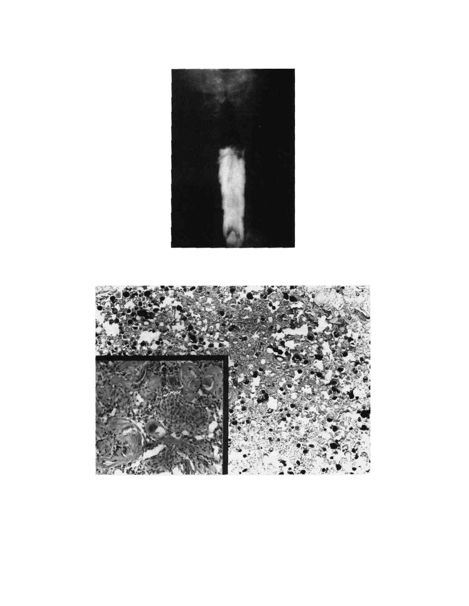
Figure 13-3A M e n i n g i o m a . AP myelographic image reveals a
complete block at C7 with classic "capping" or meniscus defect
with the cord displaced to the left.
Figure 13-3B Spinal meningioma showing overwhelming abundance of psammomatous bodies
forming the architecture of the tumor. Some are hyalimzed (lighter spherules), and some are
calcified (darker spherules). The inset represents rare areas of meningothelial tumor cells, revealing
the true nature of the tumor as a meningioma.
roots can be sectioned, and traction sutures can be placed
laterally in the dura mater for greater access to the tumor.
Microsurgical excision of meningiomas is successful in 90
to 95 percent of patients.
56
'
57
A recurrence rate of only 6
percent after a period of 4 to 17 years has been reported.
56
The need for resection of the dural base is not clear, but the
dura should be resected if it is easily accessible. When it is
not feasible to resect the dura, the dura should be cauterized
and scraped with microdissectors to reduce the risk of recur-
rence. Over 80 percent of patients treated for spinal menin-
giomas regain neurological normality, and only 5 percent
have increased neurological deficits.
56
-
57
Even paraplegic
patients may recover sufficiently to ambulate without assis-
tance after surgery. Infrequent complications include CSF
leak, meningitis, and arachnoiditis.
Ventrally placed meningiomas can be approached and
TUMORS OF THE SPINAL CANAL
239
excised anteriorly or anterolaterally. This approach is partic-
ularly helpful for tumors in the region of the neck and at the
cervicomedullary junction.
14
-
16
'
60
Crockard
14
has advocated
a transoral approach to intradural tumors ventral to the
cervicomedullary junction, particularly meningiomas and
neurofibromas, whereas Stevenson
15
and, later, McDonnell
16
have favored a transcervical approach. Both techniques give
similar results and represent methods for reaching formerly
inaccessible lesions.
NEURINOMA, NEUROFIBROMA
61
-*
5
Neurinomas (schwannomas) and neurofibromas are the sec-
ond most common intradural-extramedullary tumors. They
occur most frequently at the thoracic level, followed by the
cervical level, less commonly in the lumbosacral region, and
rarely at the cervicomedullary junctions. About 70 to 80
percent are intradural extramedullary.
4
-
5
>
61
Ten to 20 percent
are solely extradural.
4
'
5
'
61
Also, 10 to 20 percent are classi-
cally dumbbell, or hourglass, tumors.
4
'
5
'
61
Over 1 percent are
wholly intramedullary. The male-to-female ratio is equal.
The average duration of symptoms is almost 2 years before
diagnosis (Fig. 13-4, Plates 5 and 6).
The sensory nerve root is the usual site of origin of the
neurinoma or neurofibroma, but the ventral or motor root
can be involved by local compression. A large cervical
neurinoma or neurofibroma may be palpated in the neck by
physical examination. Radicular pain and, occasionally, dy-
sesthesias are reported in over 80 percent of patients. Motor
and bladder dysfunction and sensory levels to pinprick are
seen in less than 50 percent of patients. Rarely, neurofibro-
mas present clinically with subarachnoid hemorrhage, caus-
ing sudden pain, fever, and meningismus. •
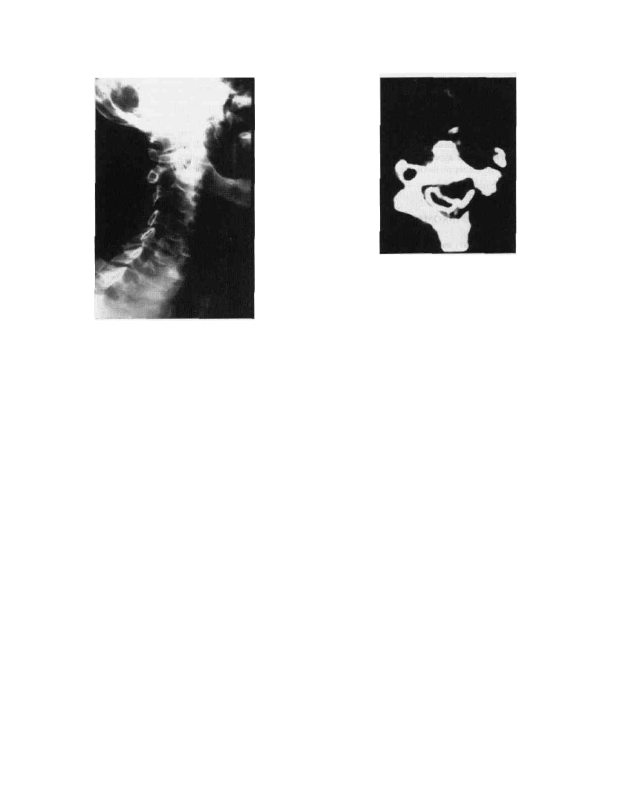
Plain x-rays of the spine are abnormal in nearly half of
patients, in marked contrast to meningiomas where changes
are seen in only 15 to 20 percent of patients.
4
'
5
-
9
Common
abnormalities include erosion and scalloping of pedicles and
vertebral bodies (Fig. 13-5, Plates 7 and 8). Enlarged fora-
mina may accompany dumbbell masses and, rarely, dural
ectasia is seen by myelography in patients with von Reck-
linghausen's disease, with or without neurofibromas. In the
case of a dumbbell neurofibroma, with intra- and extradural
components, the extradural component may be large and
readily visible as a soft tissue mass on chest or abdominal
x-rays.
CSF may have markedly elevated levels of proteins,
sometimes greater than 400 mg/100 ml, whereas meningio-
mas are associated with protein levels of 100 mg/100 ml.
Nerve sheath tumors are markedly hyperintense on T2-
weighted images compared to neural tissue. Tumor exten-
sion through an enlarged neural foramen is a characteristic
feature.
Pathologically, schwannomas are typically limited to one
nerve fascicle or bundle. The perineurium remains intact.
Grossly, neurinomas may be cystic with nerve fibers absent,
but Schwann cells grow out by tissue cultures. In contrast,
neurofibromas have extensive amounts of collagen or fi-
brous tissue with axons dispersed throughout the tumor,
making tumor excision impossible without sacrificing
nerve(s). Grossly, neurofibromas are firm and lobulated
rather than cystic. They have an estimated 13 to 15 percent
incidence of malignant degeneration to sarcoma. Like neur-
inomas, neurofibromas grow as Schwann cells in tissue
cultures, identifying a common cellular type.
Gardner has postulated that intraspinal tumors can cause
hydrocephalus and CNS symptoms by obstructing the spinal
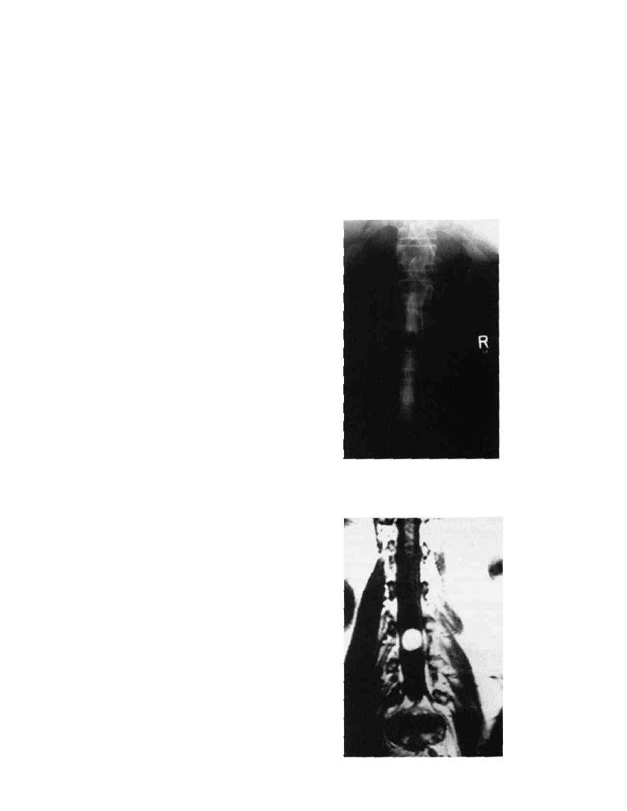
Figure 13-44 Schwanoma. AP myelographic image reveals an
intradural tumor at L3 between the cauda equina roots and below
the conus medullaris.
Figure 13-4B Same case as 13-4A. Coronal MRI with Gd-DTPA
showing intradural tumor at L3 between the cauda equina roots.
240
CHAPTER
Figure 13-5 Neurofibroma. Right oblique x-ray showing
extensive Cl to C2 bone erosion and foraminal enlargement with
scalloping and a "silhouette" from a tumor mass.
subarachnoid space with large amounts of secreted protein.
65
This putative mechanism of altering the CSF dynamics
postoperatively may cause the development of subdural he-
matomas.
Results following excision of neurinomas or neurofibro-
mas are rewarding. In Levy's series of 66 neurofibromas, 80
percent had resolution of pain while 60 percent had full
neurological recovery postoperatively and returned to
work.
61
Only 5% experienced worsening of neurological
deficits after surgery. No tumors recurred during follow-up
of 1 to 7 years. In Kirn's series,
62
in 86 cases where the
nerve root was resected to achieve complete tumor removal,
only 23 percent of patients developed detectable sensory or
motor deficits, and these deficits were minimal. They con-
cluded that the spinal nerve roots giving rise to the schwan-
noma, typically sensory, are frequently nonfunctional at the
time of surgery. Risks of incurring disabling neurological
deficits are minimal. The studies indicate that radical resec-
tion of a neurinoma or neurofibroma is indicated for an
excellent outcome.
SARCOIDOSIS
66
'*"
Sarcoidosis is a rare manifestation of systemic disease, char-
acterized by a noncaseating, granulomatous infiltration. In-
volvement of the spinal cord including meninges is about
1 percent clinically and can present as three entities: multi-
ple intramedullary lesions with focal arachnoiditis; large
intradural-extramedullary tumors with marked mass effects
and focal neurologic deficits or myelopathy; or as an extra-
dural mass from sarcoid infiltration of the spinal cord and
dura (Fig. 13-6, Plate 9).
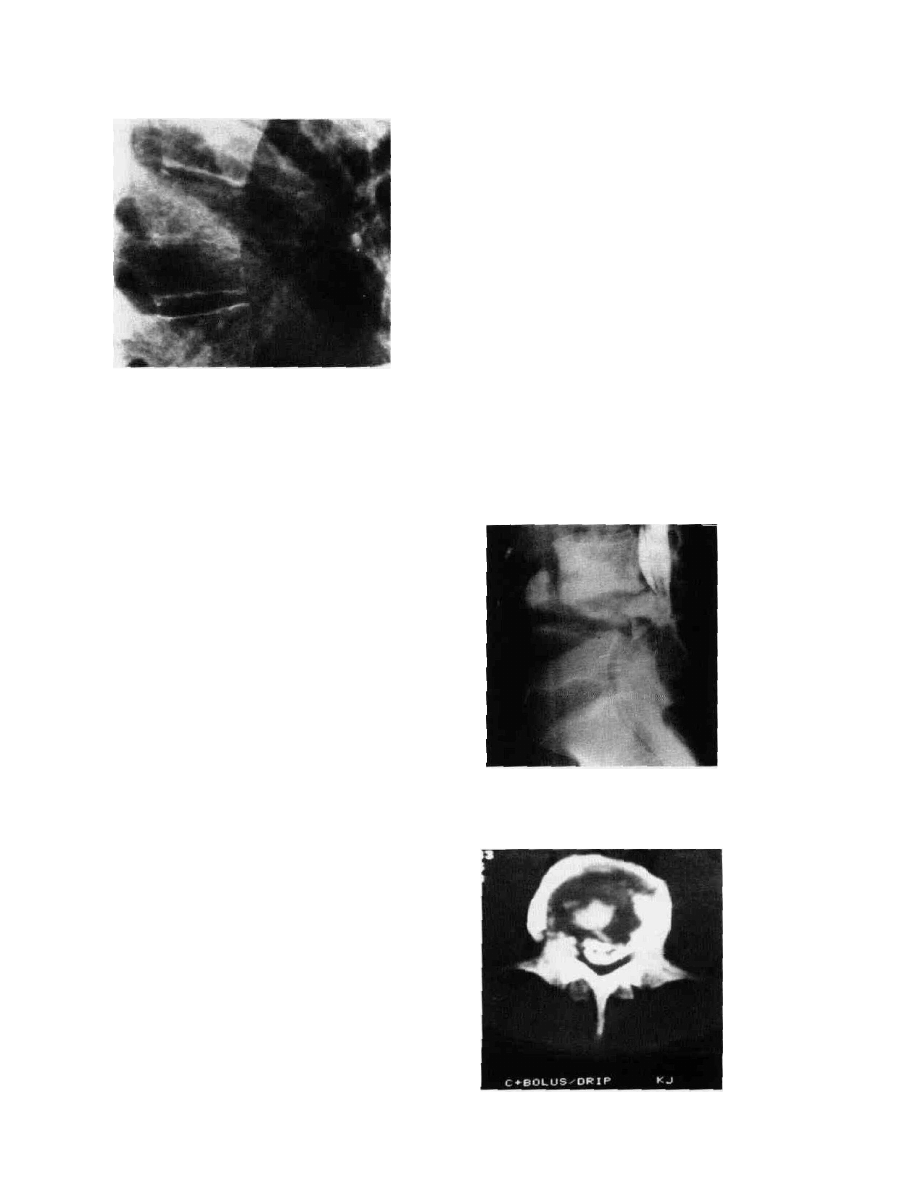
Figure 13-6 Ssrcoidosis.Myelographic CT axial image reveals
an intradural-extramedullar mass compressing and displacing the
cord to the right. There is also infiltration of the vertebral
body and dura.
The typical presentation is progressive, painless parapare-
sis. The thoracic spine is the most common site of involve-
ment. Surgical treatment is laminectomy, biopsy and, if
indicated, decompression of the granuloma coupled with the
administration of corticosteroids, known to be an effective
medical treatment in this disease. Serum and CSF levels of
angiotensin converting enzyme (ACE) can be followed to
assess the progression of the disease.
66
'
67
The natural history
of spinal sarcoidosis is remission and relapse, and cortico-
steroids are the cornerstone of continuing medical treatment.
EPIDURAL TUMORS
CANCER METASTASES
68
"
72
About 5 percent of cancer patients develop clinical signs of
compression of the spinal cord or a nerve root due to
metastases.
2
'
4
'
5
'
7
'
10
'
11
'
13
'
68
-
72
In nearly 10 percent of patients
presenting for the first time with spinal metastases, the
primary site is unknown and a surgical resection is under-
taken to establish a tissue diagnosis.2,4,5,7,10.11.13,68-72
The majority of spinal tumors, i.e., greater than 80 per-
cent, are cancer metastases most commonly from lung,
breast, kidney, prostate, colon, thyroid, melanoma, lympho-
mas, or sarcoma.
2
'
4
'
5
-
7
'
10
'
11
'
13
'
68
-
72
Postmortem studies of
cancer victims show that 50 to 70 percent have clear-cut
evidence of vertebral metastases. A smaller percentage have
dural encroachment and spinal cord compression.
The vertebral body is often involved first in metastasis.
Posterior elements are affected only one-fifth to one-seventh
as often as vertebral bodies.
4
'
5
'
7
'
10
'
11
-
13
'
68
^
72
Many metastases
are believed to be spread through Batson's venous plexus.
Even more than vertebral bodies, pedicles appear to be
infiltrated first as they are composed of cortical bone only,
TUMORS OF THE SPINAL CANAL
241
Figure 13-7 Metastatic hypernepiuotna. LUIL-UU inmate x-ray
shows a 25 percent compression fracture of the T8 vertebral body.
and metastatic disease is manifested by pedicle erosion or
enlargement on AP x-ray films of the spine. Nearly 50
percent of the vertebral bodies, which are primarily cancel-
lous bone, are infiltrated by metastases by the time abnor-
malities are seen on plain x-rays of the spine,
4
-
5
'
7
-
10
'
11
-
13
'
68
-
72
i.e., collapse, of a vertebral body or a compression fracture
Fig. 13-7). Usually, lung, breast, and colon metastases
affect the thoracic spine, whereas prostate, testicular, and
ovarian or uterine carcinoma affect the lumbosacral spine.
Metastasis to the cervical spine is slightly less common.
Spinal cord compression is most likely to occur at the
thoracic level; here the diameter of the canal is, at most,
1 cm, making little room for tumor mass.
11
The lumbosacral
canal typically spans 1.5 to 3.0 cm, allowing room for
metastatic deposits that cause subtle lumbosacral radiculo-
pathy or symptoms of cauda equina compression.
10
'
11
Mye-
lopathic changes are acutely apparent in metastases at thora-
cic levels. The cervical cord averages 1.5 to 2.0 cm in
diameter.
2
'
3
It is also the site of progressive myelopathic
changes due to metastatic disease.
Symptoms of metastatic disease may begin with sharp,
unremitting pain, localized and occasionally radicular, ex-
acerbated by deep direct palpation. The localized site of pain
will be associated with focal abnormalities on x-ray in 60
percent of patients, including pedicular erosion and a "wink-
ing owl" sign on AP films, collapse of vertebral bodies,
wedge compression fracture and subluxation, kyphoscoliosis
and/or a paraspinal soft tissue shadow.4,5,10,11,13 Devastating
myelopathy is seen in over 50 percent of patients, and
bowel/bladder dysfunction occurs in 25 percent.
4
-
5
'
10
'
1
u
3
-
68
-
72
The level of motor loss is a more-dependable diagnostic
indicator than the sensory level. Subtle neurological findings
include hyperreflexia, Hoffman, or Babinski signs, and pro-
prioceptive or dorsal column deficits provide additional
diagnostic indications.
MRI imaging with Gd-DTPA enhancement reveals more
than 95 percent of all spinal metastases, and it is the diag-
nostic test of choice after plain x-rays of the spine. MRI
delineates the extent of spinal cord compression, detects any
multilevel involvement, differentiates tumor and infection if
the clinical history is unclear, and identifies contiguous
organ or tissue involvement, including lung or uterine
cancer. If the MRI does not coincide with the clinical history
and examination, a myelogram followed by CT, above and
below a presumed level of spinal block, is the gold standard
of radiographic examinations (Fig. 13-8).
The surgical indications are manifold and should consist
of a thorough analysis and evaluation of the cancer biology,
including prognosis, life expectancy, and extent of disease as
assessed in concert with the oncologist and radiation thera-
pist. Spinal instability or compression fractures with com-
pression of the neural elements should prompt urgent de-
compression and stabilization in a combined or staged
procedure.
The presence of a radiosensitive metastatic tumor—e.g.,
lymphoma, leukemia, seminoma, plasmacytoma, myeloma,
or neuroblastoma, which has progressed rapidly with marked
neurological deterioration despite emergency radiation ther-
apy—should prompt early surgical intervention. The imme-
diate salutary effect of radiation therapy may be seen in 24
to 48 h, at best. However, many metastatic tumors are
Figure 13-8A Metastatic seminoma. Lateral lumbosacral
myelogram showing a complete extradural block at L4 with
the classic "paintbrush" tapering of the contrast seen in
extradural lesions.
Figure 13-8B Axial CT image through pathological fracture at
L4. There is bone, disk, and tumor extruded within the canal
producing a complete block.

242
CHAPTER 13
radioresistant, unfortunately. Commonly the neurosurgeon
will be consulted and presented with a case of a patient with
known mgastatic disease who has deteriorated neurologi-
cally despite emergent radiation therapy as a palliative mea-
sure. It is less common for a metastatic tumor to present
with an unknown or occult primary, and surgery is indicated
for decompression and definitive diagnosis.
Postoperative radiation therapy and the administration of
corticosteroids may be palliative, adjunctive treatments
for metastatic tumors. The neurosurgeon's zeal to provide
decompression should be tempered in any patient who has a
life expectancy limited to a few months because of wide-
spread metastases.
In older surgical series of metastatic cancer to the spine,
laminectomy was the procedure of choice. But there was
little difference in outcome between decompressive laminec-
tomy and conservative radiation therapy, and the surgical
approach was decried, particularly by Posner.
72
However,
more recent advanced surgical techniques to the vertebral
body—including the transthoracic, transpedicular, retroperi-
toneal, and lateral, extracavitary approaches to anterior spi-
nal metastatic disease—have revealed excellent results and
overall outcomes.
68
-
71
Sundaresan treated 54 patients with documented spinal
metastases in a prospective study,
68
using anterior resection
of the vertebral body in 45 patients and laminectomy in 7
patients, and all patients became ambulatory after surgery,
with the majority of patients surviving after 2 years and
remaining ambulatory. This is significant since 24 patients
were nonambulatory before surgical treatment. Primary
tumors were soft tissue sarcoma, kidney, breast, and lung.
Unfortunately, there was a 25 percent recurrence rate at the
site of surgery, precluding a long-term cure. Nevertheless,
pain and motor deficits were markedly improve^, and Sie-
gal,
70
Overby,
69
and Harrington
71
reported excellent surgical
results in extensive series.
It might be concluded that de novo surgery should be
considered for selected patients with cancer metastases to the
spine, while external beam radiation therapy is reserved as
the second phase of treatment after extensive surgical resec-
tion of the tumor.
LIPOMATOSIS
73
-
74
Epidural lipomatosis is a rare disease, characterized by
excessive fatty accumulation with spinal cord compres-
sion.
73
-
74
The symptoms and signs are those of acute pain
and progressive myelopathy. Lipomatosis is typically seen in
the thoracic spine and described in patients with a history of
chronic exogenous steroid usage, for various clinical dis-
orders, particularly Gushing's syndrome, morbid obesity,
and hypothyroidism.
7
-
73
-
74
MRI is diagnostic with a very
high intensity signal on T2-weighted images in the posterior,
epidural space, consistent with fat accumulation. Treatment
is wide decompressive laminectomy and debulking of the
adipose tissue, with or without significant weight loss in the
morbidly obese patient.
7
-
73
-
74
The surgical results
good.
73
-
74
ANGIOLIPOMA, ANGIOMYOLIPOMA
75
Angiolipoma is a rare tumor composed of mature lipocytes
and angiomatous proliferation, with or without other mesen-
chymal elements (e.g., muscles, cartilage), and it is found
predominantly in the thoracic spine with no male or female
predilection.
75
The neurological presentation is slowly pro-
gressive paraparesis.
75
Commonly, the angiolipomas are
multiple, cystic, and encapsulated; less commonly, they in-
filtrate the entire vertebral body and epidural space and recur
after excision.
75
Since infiltrating angiolipomas do not un-
dergo malignant transformation, there is no role for postop-
erative radiation therapy (Fig. 13-9, Plate 10).
Anterior vertebrectomy or posterior laminectomy is nec-
essary to obtain total excision.
75
Differential diagnosis in-
cludes vertebral hemangioma, since the angiolipoma also
presents as a coarse trabecular pattern on plain x-ray and CT
scan. However, MRI reveals a high-signal intensity in the
vertebral body, consistent with fatty infiltration from the
angiolipoma.
75
MALIGNANT OSSEOUS TUMORS
CHORDOMA
76
-
78
Chordomas are rare malignant tumors arising from primitive
notochord with a predilection for the clivus, specifically the
spheno-occipital synchondrosis and sacrococcygeum, and.
less commonly, the cervical spine.
2
-
5
-
7
'
10
-
11
-
76
-
78
Chordoma is
slightly more common in males, from 1.5:1 to 2:1, with a
peak incidence at about 50 to 60 years of age. The tumor is
locally invasive and slow-growing. Local pain is seen in
over 70 percent of patients.2-5,7,10,11,76-78
Cervical chordomas present classically as a palpable pre-
vertebral or retropharyngeal soft tissue mass with dysphagia
and neck pain, whereas sacrococcygeal chordomas present
as a presacral or pelvic mass with lower back and rectal pain
and dysfunction that involves the bowel and bladder. Consti-
pation is common. Clivus chordomas present with localized
pain, headache, dysfunction of multiple lower cranial nerves,
and a foramen magnum syndrome with gait ataxia.
The classic radiological features are expansile, destructive
tumors with significant osteolytic destruction of bone, cou-
pled with focal calcification. The presence of a large soft
tissue mass is diagnostic, and chordomas can extend locally
to the epidural space, but they rarely extend intradurally to
cause compression of the spinal cord. MRI reveals a high-
signal, soft tissue extradural mass, whereas CT and plain
films of the spine highlight the extensive osteolytic effects
and scattered focal calcification.
17
All chordomas demon-
TUMORS OF THE SPINAL CANAL
243
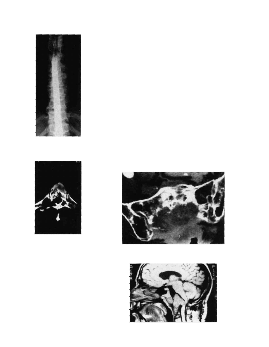
Figure 13-9A Angiolipoma. Thoracic myelogram shows a
complete extradural block at T5 with the classic tapering or
paintbrush" and highlights the right T5 pedicle erosion.
Figure 13-9B Axial CT image cut uirougn the block at T5 shows
no contrast visible secondary to marked bone destruction of the
vertebral body, pedicles, and lamina on the right with cord
compression.
strate high-signal images on T2-weighted MRIs (Fig. 13-10,
Plate 11).
Pathologically, chordomas form soft tissue masses with
pseudocapsules. They are composed of two cell types: (1)
compact stellate cells and (2) physaliphorous cells, which
are jellylike, vacuolated, with characteristic "signet-ring"
nuclei displaced eccentrically. Tumors of the physaliphorous
type have a tendency toward recurrence. They infiltrate
locally and have distant metastases, with a poor prognosis
despite palliative radiation therapy, investigational chemo-
therapy, and even experimental interstitial brachytherapy.
The surgical management of chordomas is still difficult
as there is an 80 to 90 percent recurrence rate despite
grossly complete tumor removal.
76
"
78
Unfortunately, com-
plete tumor excision is often impossible, and debulking
procedures with spinal stabilization are necessary. Chordo-
mas are radioresistant, although there are still preliminary
attempts with interstitial brachytherapy using radioactive
iodine seeds to halt tumor growth.
77
Overall, there is a 15
percent 10-year survival despite radiation therapy,
76
-
78
and,
unfortunately, chemotherapy is not promising to date.
There is a significant tendency to metastatic spread.
MULTIPLE MYELOMA, PLASMACYTOMA
Multiple myeloma is the most common primary malignant
bone tumor of the spine, with a peak incidence in the sixth
to eighth decade.
2
"
5
'
7
'
10
-
11
There is a slight predominance in
males. The vertebral bodies are replaced by malignant
plasma cells, a B-cell lymphoproliferative disorder, resulting
in local pain and systemic symptoms including weight loss,
anorexia, and malaise. There is resultant anemia, hypercalce-
mia, and an elevated sedimentation rate (ESR).
Multiple myeloma can be detected by urine or serum
protein electrophoresis as a monoclonal gamma-spike pat-
tern, the Bence Jones protein. Plain x-rays of the spine and
CT reveal multiple round, "punched-out" or "moth-eaten"
appearances secondary to widespread osteolysis, with patho-
Figure 13-10A Chordoma. Axial bone window CT image of the
sacrum shows the large mass with extensive bone destruction and
focal sites of calcification characteristic of chordoma.
Figure 13-10B MRI in the sagittal plane showing a large
chordoma indenting the pons and cervicomedullary junction.

244
CHAPTER 11
logical fractures and dislocation. MRI reveals decreased
signal intensity in multiple vertebral bodies secondary to
myeloma infiltration, although these features are also com-
monly seen in cancer metastases. The bone scan is often
negative in contradistinction to scans of patients with metas-
tases, which show multiple "hot" spots.
The primary treatment of multiple myeloma is medical,
specifically, multiregimen chemotherapy and radiation ther-
apy to the affected spine. Pain can be alleviated dramatically
by local radiotherapy and corticosteroids. If patients develop
spinal cord compression, surgery is indicated by an anterior
or a posterior approach, with spinal reconstruction. Despite
adjunctive treatment, the prognosis for survival in patients
with disseminated multiple myeloma is only about 30 per-
cent at 5 years after diagnosis.2-5,7,10,11
Less commonly, a single vertebral body is infiltrated by
malignant plasma cells, a plasmacytoma.
19
Radiologically,
the plasmacytoma presents as a single lytic lesion. In these
cases the disease is self-limited and has a better prognosis,
but 10 to 20 percent of patients progress to multiple mye-
loma, with systemic effects and multilevel spinal involve-
ment. The treatment is vertebrectomy, spinal reconstruction,
and local radiation therapy, with careful follow-up for signs
of multilevel malignancy while maintaining routine spine
x-rays, MRI, CT, and laboratory studies.
79
With aggressive
treatment, the plasmacytoma has a 5-year survival of greater
than 60 percent, since tumors are radiosensitive and unlikely
to dedifferentiate to myeloma.
79
OSTEOSARCOMA (OSTEOGENIC
SARCOMA)
7
'
10
'
11
-
80
Osteogenic sarcoma is a rare, bone-forming tumor, which
can arise de novo in 50 percent of cases, or secondarily, as
metastases from a limb extremity.
7
-
10
-
11
'
80
It occurs at a site
of earlier irradiation but is commonly associated with
Paget's disease. The median age of presentation is 40 years,
with a slight male preponderance and an equal distribution
among spinal segments. Intractable pain is a uniformly omi-
nous symptom, and neurological deficits are seen in 70
percent of patients.
7
-
10
-
11
-
80
Osteosarcoma usually involves
the vertebral body primarily, with areas of additional lysis
and dense sclerosis with calcification seen radiographically
by plain x-rays and CT scan. Despite aggressive surgical
debulking, focal radiation therapy, and multiregimen chemo-
therapy, the overall prognosis is poor. Less than 10 percent
of patients survive at 5 years.
7
-
10
-
11
-
80
CHONDROSARCOMA
7
'
10
'
11
'
81
'
82
Chondrosarcomas are rare malignant tumors that form carti-
lage. They are commonly found in long bones, pelvis, and
skull base with 6 percent distributed throughout the spinal
column.
7
-
10
-
11
-
81
-
82
Chondrosarcomas typically involve verte-
bral bodies, and the age distribution is 40 to 60 years. Pain is
a frequent and constant finding, as in the case of osteosar-
coma. Radiographically, there are characteristic "fluffy"
globular areas of sclerotic tumor, combined with lucent area*
secondary to bone lysis; angiography reveals a very vascular
tumor. As with osteosarcoma, despite surgery and en bl:>
radical excision, radiation therapy, and chemotherapy, loc*
recurrence and metastases are common, leading to a dismal
overall prognosis.
BENIGN BONE TUMORS
7
-
10
'
11
-
83
OSTEOCHONDROMA
Osteochondromas are common benign single or multiple
bone tumors.
7
-
10
-
11
-
83
Osteochondromas rarely occur in the
spine, but when they do occur they present as mass lesion-
that cause spinal cord compression or radiculopathy. The>
occur typically in males under 30 years of age and are found
predominantly in the spinous processes and, particularly, the
posterior neural arches. Pain is not a predominant feature,
but slowly progressive myelopathy has been described re-
peatedly. Tumors have a characteristic appearance radio-
graphically, showing a round bony exostosis with a radiolu-
cent, cartilagenous cap. They are found predominantly in the
cervical spine and less commonly in the thoracic spine.
Surgical decompression by laminectomy is curative, with
recovery of neurological function and rare recurrences. Ma-
lignant degeneration occurs in at least 10 percent of patients
with the genetically inherited Oilier's disease or Maffucci's
syndrome, multiple chondromatosis, or exostoses, with a
distinct tendency toward the chondrosarcoma and, very
rarely, the osteosarcoma.
7
'
I0
'
n
-
83
OSTEOBLASTOMA, OSTEOID OSTEOMA
7
-
10
'
11
'
84
-
86
Osteoblastomas are common benign tumors seen in males,
with a peak incidence below 30 years of age.
7
-
10
-
11
-
84
-
86
They
have a predilection for the posterior spinal elements, specifi-
cally spinous and transverse processes and lamina. Osteo-
blastoma is commonly accompanied by scoliosis or spinal
stiffness, and it has an equal distribution throughout the
spinal column. Classically, symptoms described are dull
aching pains, neither nocturnal nor relieved by aspirin.
By plain x-rays and CT, the tumors have a central, radio-
lucent, or lytic, area surrounded by a sclerotic rim, and its
size is usually greater than 1.5 cm in diameter. Although
most frequently in the femur and tibia, 35 percent of osteo-
blastomas occur in the spine.
7
-
10
-
11
-
84
^
86
MRIs of osteoblasto-
mas show enhancement with Gd-DTPA on Tl images and
present a high signal on T2 images. The technetium bone
scan has intense, or "hot," uptake with radioisotopes.
Osteoblastoma is locally aggressive and can attain consid-
erable size, causing spinal cord compression associated with

TUMORS OF THE SPINAL CANAL
245
local scoliosis. Treatment consists of en bloc resection and
spine stabilization, and recurrence is common because of
aggressive tumor infiltration and inability to achieve com-
plete removal surgically. Both anterior and posterior proce-
dures may be required for total tumor removal; curettage and
bone grafting are very successful.
7
'
10
'
11
-
84
"
86
Local irradiation
is ineffective and may cause malignant transformation to
sarcoma.
Osteoid osteoma is histologically identical to osteoblas-
toma, yet it is characterized classically by nocturnal pain
well-alleviated by aspirin and arbitrarily defined in size as
less than 1.5 cm in diameter. Although frequently located in
the femur and tibia, approximately 10 percent of osteoid
osteomas are located in the spine.
7
'
10
-
11
'
84
-
86
The technetium
bone scan is very sensitive, as the central lucent nidus is
"cold" and the sclerotic rim "hot," allowing confirmation of
complete removal of th nidus intraoperatively by testing the
surrounding bone with the radioactive counter for any resid-
ual "hot" spots. MRIs seem to enhance vividly the nidus
with Gd-DTPA contrast agent.
Osteoid osteoma is seen in even younger patients, particu-
larly males, with an average age of 19 years, and it has a
preference for the lumbar spine, followed by the cervical
spine. As with osteoblastoma, the osteoid osteoma involves
preferentially the posterior spinal elements, i.e., lamina and
pedicles. Painful scoliosis is a common symptom (>60
percent) in adolescents with osteoid osteoma.
7
'
10
'
11
-
84
-
86
Surgical excision of osteoid osteomas is often complete
and curative without recurrence due to its small size; scolio-
sis will resolve spontaneously with time. However, long-
standing lesions may require spinal instrumentation to
correct the scoliosis. Pain resolution is a gratifying end-re-
GIANT CELL TUMORS OR
OSTEOCLASTOMAS
7
W'
11
-*
7
-
88
These are rare benign bone tumors composed of multinu-
cleated giant cells with a fibrous stroma consisting of mon-
onuclear cells.
7
-
10
-
11
-
87
-
88
They are most common in long
bones, epiphyses, and metaphyses, occurring in young adults
and adolescents, with a female preponderance, and they are
found less commonly, but preferentially, in the thoracic
spine with a predilection for the vertebral body. Giant cell
tumors grow rather large and are highly vascular. They
cause localized or radicular pain with slowly progressive
Spinal cord compression. Radiographically, these tumors are
large, osteolytic masses termed soap bubbles, with evidence
of destruction on plain x-rays or CT. MRI delineates the
extensive vertebral bone and soft tissue components of the
tumor, with a low signal intensity by T2 image due to
marrow replacement.
Surgery is aimed at radical en bloc excision or curettage
with reconstruction of the involved vertebra and tumor mass,
but the rate of local recurrence is very high, nearly 50
percent,
87
-
88
and giant cell tumors can metastasize at a rate of
15 percent.
87
'
88
Postoperative radiotherapy is routinely indi-
cated to halt metastases.
ANEURYSMAL BONE CYSTS
7
-
10
'
11
-
89
-
90
These are benign lesions of bone. Approximately 10 to 20
percent of aneurysmal bone cysts occur in the spine, most
commonly in the lumbar region.
7
'
10
-
11
-
89
-
90
The cyst presents
in the 10- to 30-year-age range, and there is a slight prefer-
ence for females, with predilection for the posterior neural
arches and pedicles. Less commonly, these cysts involve
several vertebral bodies. Painful scoliosis in the back is
commonly seen, and tumors enlarge as soft tissue masses
into the spinal canal, causing neurological deficits in 60
percent of cases.
7
-
10
-
11
-
89
-
90
Histologically, aneurysmal bone cysts contain cystic,
fluid-filled spaces with fibrous septae. Radiographically,
there is a honeycomb trabeculated pattern with an eggshell-
thin cortical margin by plain x-ray and CT. Angiography
reveals multiple vascular "lakes." MRI confirms the expan-
sile soft tissue mass with internal septations and lakes con-
taining fluid levels.
Complete surgical excision should be attempted, but a
recurrence rate of 15 to 25 percent is reported despite
extensive curettage and reconstruction.
7
-
10
-
11
'
89
-
90
Low-dose
radiation therapy (20 to 30 cGy) prevents recurrence and
should be considered in cases where complete excision is
not possible.
7
-
10
-
11
-
89
-
90
HEMANGIOMAS
7
'
10
'
11
'
91
'
92
Hemangiomas are the most common benign bone tumor
with asymptomatic lesions in autopsy series, ranging from 9
to 13 percent.
7
'
10
'
11
'
91
-
92
Hemangiomas are vascular tumors
arising from newly formed blood vessels of diverse sizes. In
about 33 percent of cases, multiple vertebra are involved,
and the thoracic and thoracolumbar regions are most often
affected.
7
-
10
-
11
-
91
-
92
Hemangiomas are frequently incidental
findings on x-ray, and the majority of patients are asympto-
matic. There is a slight female preponderance of 1.5:1, and
the age at presentation is typically 30 to 50 years.
7 ltu 19192
Hemangiomas become symptomatic, with progressive
vertebral body collapse, fracture, and, rarely, epidural hem-
orrhage. They cause focal pain and tenderness with muscle
spasms, with or without neurological deterioration, and
myelopathy is commonly seen.
Plain radiographs or CT scans show coarse vertical stria-
tions, or trabeculae. Dilated vascular spaces are easily seen
by CT. and they give a characteristic spotted appearance.
Typically, hemangiomas are confined to the vertebral bodies
but can extend into the neural arches.
MRI is very sensitive and shows bright high-intensity
signals on both Tl- and T2-weighted images. MRI readily
shows the paravertebral and epidural extension of the tumor,
and it outlines spinal cord compression. Hemangiomas have

246
CHAPTER 13
vascular lacunae on angiography and multiple tumor vessels,
which are readily amenable to embolization.
91
'
92
Most hemangiomas can be treated symptomatically with
low-dose radiation therapy and external bracing, alleviating
local pain and tenderness. Vertebrectomy and stabilization of
the spine are considered in cases of spinal cord compression
from an enlarging hemangioma, but intraoperative hemor-
rhage is a concern, and preoperative embolization of tumor
feeders is recommended. Asymptomatic hemangiomas are
left untreated, but they are followed since there is a low
incidence of new-onset signs or symptoms in these patients.
Early surgical intervention in a neurologically compromised
patient leads to a good outcome postoperatively and a suc-
cessful result, with rare recurrence.
7
'
10
-
11
'
91
'
92
EOSINOPHILIC GRANULOMAS
7
'
10
-
11
-
93
Eosinophilic granulomas are the most common benign bone
tumors of children, particularly males, with a peak incidence
at 5 to 10 years of age.
7
'
10
-
11
'
93
The bone is infiltrated and
destroyed by histocytes and eosinophils. The lesion can be
representative of one end of a diagnostic spectrum of sys-
temic diseases, including Letterer-Siwe and Hand-Schiler-
Christian disease, the last being a malignant reticuloendothe-
liosis akin to cancer. Solitary eosinophilic granulomas are
described commonly in the skull as "punched-out" lesions,
but vertebral involvement occurs in 10 to 15 percent of
cases.
7
-
10
'
11
-
93
Eosinophilic granuloma occurs frequently in the thoracic
spine, with acute onset of chest or back pain, focal sprain
torticollis, kyphoscoliosis, and, rarely, neurological compro-
mise. The classic radiographic appearance is the "vertebra
plana," or vertebral body, which is symmetrically flattened
and thinned compared with the adjacent vertebrae.
7
'
10
-
11
-
93
Treatment includes a diagnostic needle biopsy and curet-
tage. Patients with a solitary eosinophilic granuloma are
immobilized in a spinal orthosis and given low-dose radia-
tion therapy, 5 to 10 cGy. Systemic eosinophilic granulomas
are treated with multiregimen chemotherapy.
7
'
10
-
n
'
93
Rarely
eosinophilic granulomas cause myelopathy or radiculopathy,
necessitating spinal decompression and/or stabilization.
93
REFERENCES
1. Seeger W: Microsurgery of the Spinal Cord and Surrounding
Structures. New York, Springer-Verlag, 1982.
2. Bailey RW, Sherk HH: The Cervical Spine. The Cervical
Spine Research Society, Philadelphia, Lippincott
k
1983.
3. Dunsker SB: Cervical Spondylosis. New York, Raven, 1981.
4. Youmans JR: Neurological Surgery, vols 1 to 6. Philadelphia,
Saunders, 1982.
5. Wilkins RH, Rengachary SS: Neurosurgery, vols 1 to 3. New
York, McGraw-Hill, 1985.
6. Wilkins RH, Rengachary SS: Neurosurgery, Update I. New
York, McGraw-Hill, 1991.
7. Wilkins RH, Rengachary SS: Neurosurgery, Update II. New
York, McGraw-Hill, 1991.
8. Schmidek HH, Sweet WH: Operative Neurosurgical Tech-
niques, vols 1 and 2. New York, Grime and Stratton, 1982.
9. AI-Mefty O: Meningiomas. New York, Raven, 1991.
10. Rothman RH, Simeone FA: The Spine. Philadelphia, Saunders,
1982.
11. Fryrnoyer JW: The Adult Spine. New York, Raven, 1991.
12. Rand RW, Rand CW: Intraspinal Tumors of Childhood,
Springfield, Charles C. Thomas, 1960.
13. Congress of Neurological Surgeons: Clin Neurosurg Proc, vols
30, 37, and 38. Baltimore, Williams & Wilkins, 1983, 1991,
1992.
14. Crockard HA, Sen CN: The transoral approach for the manage-
ment of intradural lesions at the craniovertebral junction: Re-
view of 7 cases. Neurosurgery 28:88-97, 1991.
15. Stevenson GC, Stoney RJ, Perkins RK, Adams JE: A trans-
cervical transclival approach to the ventral surface of the brain
stem for removal of a clivus chordoma. J Neurosurg 24:544-
551, 1966.
16. McDonnell DE: Anterolateral cervical approach to the cranio-
vertebral junction, in Rengachary SS, Wilkins RH (eds): Neur-
osurgical Operative Atlas, vol 1, no 3. Baltimore, Williams &.
Wilkins, pp 147-164, 1991.
17. Kucharczyk W: MRI: Central Nervous System, Philadelphia.
LippincoU, 1990.
18. Gutin P: Corticosteroid therapy in patients with cerebral
tumors: Benefits, mechanisms, problems, practicalities. Semi-
nars in Oncology 2:49-56, 1975.
19. Thompson EB, Srivistava D, Johnson BH: Interaction of the
phenylpyrazolo steroid cortivazol with glucocorticoid receptors
in steroid-sensitive and steroid-resistant human leukemia cells.
Cancer Res 49:2253-2258, 1989.
20. Helseth A, Mork S: Primary intraspinal neoplasms in Norway.
J Neurosurg 11:842-845, 1989.
21. McCormick PC, Torres R, Post KD, Stein BM: Intramcdullary
ependymoma of the spinal cord. J Neurosurg 72:523-532,
1990.
22. Fischer G, Mansuy L: Total removal of intramedullary ependy-
moma: Follow-up study of 16 cases. Surg Neural 14:243-249.
1980.
23. Schweitzer JS, Batzdorf U: Ependymoma of the cauda equina
region: Diagnosis, treatment, and outcome in 15 patients.
Neurosurgery 30:202-207, 1992.
24. Greenwood J Jr: Total removal of intramedullary tumors. J
Neurosurg 11:616-621, 1954.
25. Elsberg CA: Tumors of the Spinal Cord. New York, Paul B.
Hoeber, 1925.
26. Stein BM: Surgery of intramedullary spinal cord tumors. Clin
Neurosurg 26:529-542, 1979.

248
CHAPTER 13
75. Kuroda S, Abe H, Akino M, et al: Infiltrating spinal angioli-
poma causing myelopathy: Case report. Neurosurgery 27:315-
318, 1990.
76. Sundaresan N, Galicich JH, Chu FCH, Huvos AG: Spinal
chordomas. J Neurosurg 50:312-319, 1979.
77. Gutin PH, Leibel SA, Hosobuchi Y, et al: Brachytherapy of
recurrent tumors of the skull base and spine with Iodine-125
sources. Neurosurgery 20:938-945, 1987.
78. O'Neill P, Bell BA, Miller JD, et al: Fifty years of experience
with chordoma in southeast Scotland. Neurosurgery 16:166-
170, 1985.
79. Loftus CM, Michelsen CB, Rapoport F, Antunes JL: Manage-
ment of plasmacytoma of the spine. Neurosurgery 13:30-36,
1983.
80. Sundaresan N, Rosen G, Huvos AG, Krol G: Combined treat-
ment of osteosarcoma of the spine. Neurosurgery 23:714—719,
1988.
81. Camins MB, Duncan AW, Smith J, Marcovic RC: Chondrosar-
coma of the spine. Spine 3:202-209, 1978.
82. Hirsch LF, Thanki A, Spector HB: Primary spinal chordosar-
coma with eighteen-year followup: Case report and literature
review. Neurosurgery 14:747-749, 1984.
83. Palmer FJ, Blum PW: Osteochondroma with spinal cord com-
pression: A report of three cases. J Neurosurg 52:842-845,
1980.
84. Janin Y, Epstein JA, Carras R: Osteoid osteomas and osteo-
blastomas of the spine. Neurosurgery 8:31-38, 1981.
85. Shikata J, Yamamuro T, lida H, Kotoura Y: Benign osteo
blastema of the cervical vertebra. Surg Neurol 27:381-385
1987.
86. Bucci MN, Feldenzer JA, Phillips WA, et al: Atlantoaxi_
rotational limitations secondary to osteoid osteoma of the axis
J Neurosurg 70:129-131, 1989.
87. Dilorenzo N, Spallone A, Nolletti A, Nardi P: Giant eel;
tumors of the spine. A clinical study of six cases, with empha-
sis on the radiological features, treatment and follow-up. Neur-
osurgery 6:29-34, 1980.
88. Dahlin DC: Giant cell tumor of the vertebra above the sacruir
A review of 31 cases. Cancer 39:1350-1356, 1977.
89. Ohry A, Lipschitz M, Shemesh Y, et al: Disappearance c
quadriparesis due to a huge cervicothoracic aneurysmal bone
cyst. Surg Neurol 29:307-310, 1988.
90. Podos PN Jr, White RJ: Aneurysmal bone cyst of the cervicau
spine: A twelve-year followup after surgical treatment. Sure
Neurol 14:259-262, 1980.
91. Feuerman T, Dwan PS, Young RF: Vertebrectomy for treat-
ment of vertebral hemangioma without preoperative emboliza-
tion: Case report. J Neurosurg 65:404-406, 1986.
92. Healy M, Herz DA, Pearl L: Spinal hemangioma. Neurosur-
gery 13:689-691, 1983.
93. Acciarri N, Paganini M, Fonda C, et al: Langerhans' eel"
histiocytosis of the spine causing cord compression: Case re-
port. Neurosurgery 31:965-968, 1992.
STUDY QUESTIONS
1. A 38-year-old female is referred because of localized inter-
scapular pain with radiation in a band around to the right
breast. This is worse at night. There is a strip of hyperesthesia
along the distribution of the right fifth intercostal nerve. There
are hyperactive reflexes in the right lower extremity and de-
creased sensation to pin prick on the left. X-rays show a
enlarged neural foramen at T4-L5 on the right.
1. What is the most likely diagnosis? 2. What imaging
structures might be considered to outline the lesion? 3. What
image might be produced by myelography? 4. How might
the lesion be approached? 5. Would the lesion most likely be
dorsal or ventral to the dentate ligament? Why?
n. A 60-year-old male has a long history of upper lumbar
back pain and progressive weakness of his lower extremities.
X-rays and CT scan reveal a sharp kyphosis centered at T10.
Disk spaces are preserved. There is compression of the tenth
thoracic vertebra. There are multiple lesions throughout the
chest. An endotracheal biopsy shows evidence of squamous
cell carcinoma.
1. What is the most likely cause of the paraparesis?
2. How might the spinal cord be decompressed? 3. What is
the objective of corpectomy? 4. How can the spine be
stabilized surgically? 5. What part might irradiation therapy
play in the treatment of this patient?
III. A 52-year-old female is seen because of progressive
weakness and ataxia in all four extremities, but most severe
2. What is the microscopic appearance? 3. How might this
appearance differ if the lesions involved the upper spinal in
the upper extremities. She has difficulty eating, and there is
paresis and severe atrophy of the right side of the tongue.
MRI shows a mass lesion on the anterior lip of the foramen
magnum.
1. What is the most likely diagnosis? 2. From what
approach might the lesion be attacked surgically, anteriorly
or posteriorly? 3. What are the variations to the anterior
approach that might be used? 4. What adjunctive therapy
might be necessary? 5. What form of radiation therapy might
be effective?
IV. A 43-year-old male is experiencing weakness of his
hands and lower extremities. He has decreased motor func-
tion and pinprick in the hands but fast reflexes in lower
extremities. He is spastic in the lower extremities and has
intrinsic muscle wasting of the hands. An MRI shows intrin-
sic mass effect in the lower cervical area. The spinal cord is
enhanced only moderately. There is no cystic component.

TUMORS OF THE SPINAL CANAL
249
1. What is the differential diagnosis? 2. How might this
patient obtain definitive diagnosis? 3. What surgical therapy
might be considered? 4. How could radiation play a part in
the treatment of this lesion? 5. How might his course change
if syringomyelia became a problem?
V. A 30-year-old male complains of severe lumbar back
pain radiating into both buttocks. Sphincter control has been
lost. MRI reveals an intraspinal mass fanning out into the
paraspinal area. Microscopically, the lesion is myxomatous.
It has infiltrated the cauda equina.
1. What intrinsic spinal tumor fits this description?
2. What is the microscopic appearance? 3. How might this
appearance differ if the lesions involved the upper spinal
cord? 4. What forms of therapy might be considered?
5. What is the prognosis?
Wyszukiwarka
Podobne podstrony:
Kenyon, Sherrilyn Dark Hunter 13 Sins of the Night rtf
13 One of the most serious problems facing young people today 2
Kenyon, Sherrilyn Dark Hunter 13 Sins of the Night
13 Grammar of the Estonian language tables (Russian)
Enid Blyton Mystery 13 Mystery of the Missing Man
Robert Adams Horseclans 13 Horses of the North (v1 0) [Undead]
[13]Role of oxidative stress and protein oxidation in the aging process
13 examples of good and?d manners around the World
13 WoW Arthas Rise of The Lich King (2010 01)
ashel 13 Total Eclipse of the Heart
Wisner H 12th Conference of the Commission for Lithuanian Studies 2004 89 APH 13
Erle Stanley Gardner [Mason 13] The Case of the Shoplifter s Shoe (rtf)
Hall, C S , Van de Castle, R L (1966) The content analysis of dreams r 13 Reliability of scoring, s
Edmond Hamilton Captain Future 13 The Face of the Deep
The 13 Principles of Witchcraft
Khalek Unified Octonionic Repr of the 10 13 D Clifford Algebra (1997) [sharethefiles com]
The law of the European Union
więcej podobnych podstron