
Asian Pacific Journal of Cancer Prevention, Vol 13, 2012
5131
DOI:http://dx.doi.org/10.7314/APJCP.2012.13.10.5131
Anticancer Activity of Gonothalamin in a Cervical Cancer Cell Line
Asian Pacific J Cancer Prev, 13 (10), 5131-5136
Introduction
Cancer is uncontrolled growth of cells. It can affect
almost any part of the body. The growths often invade
surrounding tissue and can metastasize to distant sites
(WHO, 2011). Cancer is caused by mutations in the
DNA. Normal cells repair the mutation or simply
die when a mutation occurs whereas cancerous cells
continue to survive with the mutations and they grow
in an uncontrolled manner until a mass of cells known
as tumor is created. Often the tumor interferes with the
normal functioning of healthy tissues and can spread to
other parts of the body (Tompa, 2007).
Cervical cancer is a malignant neoplasm of the
cervical area. It is an important women’s health problem
in developing countries, killing 270,000 women each year.
It is the third most common cancer overall and the leading
cause of death from cancer among women in developing
countries. At least 370,000 new cases are identified each
year (WHO, 2010). Current cancer chemotherapy can
damage or kill the rapid dividing and healthy cell but
causes serious side effects such as nausea, anemia, and hair
loss. In addition, the cost of chemotherapy drug is high as
compared to the natural compound from medicinal plants.
Goniothalamin, a natural occurring styryl-lactone and
1
Department of Biotechnology,
3
Department of Chemistry, Universiti Sultan Zainal Abidin (UniSZA), Kuala Terengganu,
2
Faculty
of Dentistry, Universiti of Malaya,
4
Community Medicine Department, International Medical University, Kuala Lumpur, Malaysia,
5
Faculty of Pharmacy, Sana’a University, Yemen *For correspondence: aied_absi@yahoo.com
Abstract
Cancer is one of the major health problems worldwide and its current treatments have a number of undesired
adverse side effects. Natural compounds may reduce these. Currently, a few plant products are being used to
treat cancer. In this study, goniothalamin, a natural occurring styryl-lactone extracted from Goniothalamus
macrophyllus, was investigated for cytotoxic properties against cervical cancer (HeLa), breast carcinoma
(MCF-7) and colon cancer (HT29) cells as well as normal mouse fibroblast (3T3) using MTT assay. Fluorescence
microscopy showed that GTN is able to induce apoptosis in HeLa cells in a time dependent manner. Flow cytometry
further revealed HeLa cells treated with GTN to be arrested in the S phase. Phosphatidyl serine properties
present during apoptosis enable early detection of the apoptosis in the cells. Using annexin V/PI double staining
it could be shown that GTN induces early apoptosis on HeLa cells after 24, 48 and 72 h. It could be concluded
that goniothalamin showing a promising cytotoxicity effect against several cancer cell lines including cervical
cancer cells (HeLa) with apoptosis as the mode of cell death induced on HeLa cells by Goniothalamin was.
Keywords: Goniothalamin - HeLa cervical cancer cell line - fluorescence microscopy - cellular DNA content - apoptosis.
RESEARCH ARTICLE
Apoptosis Induction, Cell Cycle Arrest and in Vitro Anticancer
Activity of Gonothalamin in a Cancer Cell Lines
Aied M Alabsi
1, 2
*, Rola Ali
1
, Abdul Manaf Ali
1
, Sami Abdo Radman Al-Dubai
4
,
Hazlan Harun
3
, Noor H Abu Kasim
2
, Abdulsamad Alsalahi
5
extract from Goniothalamus SPP. it is a novel compound
with putative anti-cancer properties (Lin and Pihie, 2003;
Chen et al., 2005; Al-Qubaisi et al., 2011). Goniothalamin
extracted from Goniothalamus andersonii had been able
to induce cytotoxicity in a variety of cancer cell lines
including cervical (HeLa), gastric (HGC-27), kidney (768-
0), breast carcinomas (MCF-7, T47D and MDA-MB-231)
and leukemia (HL-60, Jurkat and CEM-SS) (Rajab et al.,
2005; Inayat-Hussain et al., 2010). Goniothalamin has
been proved to be only cytotoxic to ovarian cancer cell line
(Caov-3) without causing cell death in normal kidney cell
(MDBK) as happened in tamoxifen or taxol treated cells
(Lin and Pihie, 2003). In addition, goniothalamin showed
lower toxicity to normal liver Chang cell line as compared
to doxorubicin (chemotherapy drug) (Al-Qubaisi et
al., 2011). Goniothalamin is a promising antitumor
agent against cancerous cell lines (Wattanapiromsakul
et al., 2005). Cytotoxicity of goniothalamin in human
leukemia (HL-60 and Jurkat) and human breast carcinoma
(MDA-MB-231) occurs via apoptosis after treated with
goniothalamin (Chen et al., 2005; Inayat-Hussain et al.,
2010).
In this study, goniothalamin, a natural occurring
styryl-lactone and extract from root of Goniothalamus
macrophyllus is used to investigate the cytotoxic

Aied M. Alabsi1 et al
Asian Pacific Journal of Cancer Prevention, Vol 13, 2012
5132
properties against several cancer cell lines. Furthermore,
this study carried out to study the mechanism of apoptosis
induction of goniothalamin on HeLa cells by determines
the DNA content and Phosphatidyl Serine properties.
Materials and Methods
Goniothalamin extract
Dried and powdered root (500g) of Goniothalamus
macrophyllus were extracted with dichloromethane and
concentrated. Fifty g of brown resin was subjected to silica
gel chromatography with gradient of hexane/ethyl acetate
(8:2) which gave goniothalamin 5g (colorless crystal),
structurally confirmed by comparing 1H and 13C-NMR
data with those reported. 13C NMR: δ 29.87, 77.95,
121.60, 125.72, 126.72, 128.71, 133.10, 135.80, 144.76
and 163.90.
Cells and cell culture
cervical cancer (HeLa), breast carcinoma (MCF-7),
colon cancer (HT29) and Normal mouse fibroblast cells
(3T3) obtained from animal tissue culture laboratory,
UniSZA. cell lines were grown in 25 cm² tissue culture
flasks (Nunclon TM, Nunc) at 37°C, 5%CO
2
and 90%
humidity in RPMI -1640 medium (Sigma Chemical
Company), containing 10% fetal bovine serum (Culture
lab), penicillin (100 IU/ml) and streptomycin (100 µg/ml).
The cells were grown confluence, which could be observed
under an inverted microscope and sub – cultured at three
to four days interval.
MTT Cytotoxicity Assay
All cell lines were trypsinized and counted using
hemocytometer then were seeded in 96-well micro plate
at 5×10
5
cells/ml and then incubated at 37
o
C in 5%CO
2
to allow cells attachment. The medium was removed
and replaced with fresh medium containing various
concentrations of goniothalamin starting with the highest
concentration of 60 µg /ml (two folded dilution). Cells
were incubated at 37
o
C, 5%CO
2
for 72 hours. Each
concentration was assayed in triplicates (n=3). Seventy-
two hours later, 20 µl of MTT (5 mg/ml) solution was
added to each well and then the plate was further incubated
for 4 h. All remaining supernatant were removed and 150
µl of DMSO was added to dissolve the formed crystal
formazan. MTT assay reading was performed using
ELISA plate reader (Tecan 200, USA).
The MTT Cell Proliferation Assay
To confirm anti-proliferative effects of goniothalamin
on HeLa cells, MTT cell proliferation assay was carried
out. In this assay, two different concentrations of compound
with cells were prepared together with control. The
concentration chosen were IC
25
and IC
50
concentrations
(3.2 and 1.2 µg/ml). Each sample was assayed in triplicate,
and control samples include cells without goniothalamin.
The cells were treated by goniothalamin for 24, 48, and
72 hours. At the end of incubation periods, 20μl of MTT
solution (5 mg/ml MTT dissolved in PBS) were added to
each well containing cells and the plate was incubated at
37ºC in an atmosphere of 5%CO
2
for 4 hours. After that,
most of the medium was removed, then a volume 100 µl
of DMSO (dimethyl sulfoxide) was added into the wells to
soluble the crystals. Finally the absorbance was measured
by ELISA reader at a wavelength of 570 nm. Graphs (OD
of samples against time) were plotted to determine the
growth rates of cells in a given values.
Acridine Orange (AO) and Propodium Iodide (PI) Double
Staining using Fluorescent Microscopy
HeLa cells were quantified using propidium iodide
(PI) and acridine-orange (AO) double staining according
to standard procedures and examine under fluorescence
microscope (Lieca attached with Q-Floro Software)
(Mishell et al., 1980; Ali, 2011).
Cells suspension was mixed with an equal volume
of staining solution (1 : 1) containing 10 μg/mL acridine
orange and 10 μg/mL propium iodide (dissolved in PBS)
and observed under fluorescence microscope within 30
minutes. The viable (green intact cells), apoptotic (green
shrinking cells with condensed of fragmented nucleus),
and necrotic (red cells) were the morphological changes
that were examined under fluorescence microscope.
HeLa cells were seeded in six-well plate and incubated
at 37
o
C in 5%CO
2
atmosphere. Twenty-four hours later,
the medium in each well was removed and replaced with
Goniothalamin at IC50 concentration dissolved in medium
and incubated at 37
o
C in 5%CO
2
atmosphere for 24, 48,
and 72 h. After incubation period, Cells suspension was
mixed with an equal volume of staining solution (1 :
1) containing 10 μg/mL acridine orange and 10 μg/ml
propium iodide (dissolved in PBS) and observed under
fluorescence microscope within 30 minutes. The viable
(green intact cells), apoptotic (green shrinking cells with
condensed of fragmented nucleus), and necrotic (red cells)
were the morphological changes that were examined
under fluorescence microscope (Leica, Germany). Each
experiment was assayed three times (n=3) to provide
a useful quantitative evaluation. Viable, apoptotic and
necrotic cells was quantified in a population of 200 cells.
The results were expressed as a proportion of the total
number of the cells examined.
Analysis of Cellular DNA Content Using Propidium Iodide
HeLa cells at a concentration of 1x10
6
cells/ml were
seeded into 6-well plate in 2 ml culture medium with a
concentration of IC
50
value of goniothalamin and were
incubated at 37ºC in an atmosphere of 5%CO
2
for 24,48
and 72 hours. Some wells were left with no treatment to be
used as a control. After the incubation period, the cultured
cells were harvested using trypsin and centrifuged. After
incubation, the cells were detached and stained by using
the Cycle TEST TM PLUS DNA Reagent Kit. Cell cycle
was read using the Cell Quest software within 3 hours.
Flow cytometry (Annexin V/PI double staining): HeLa
cells at a concentration of 1 X 10
6
cell/ml were seeded
into the 6-well plate and treated with IC
50
concentration
of Goniothalamin. After 24, 48 and 72 h incubation, the
cells were detached and stained by using PE Annexin V
Apoptosis Detection Kit I. All samples were read by the
flow cytometer.
Asian Pacific Journal of Cancer Prevention, Vol 13, 2012
5133
DOI:http://dx.doi.org/10.7314/APJCP.2012.13.10.5131
Anticancer Activity of Gonothalamin in a Cervical Cancer Cell Line
Statistical Analysis
Data was expressed as mean±SD. Statistical analysis
was performed with Student’s t-test using the independent
t-test (SPSS version 15). Differences were considered
significant at P=0.05.
Results
MTT Cytotoxicity Assay
Cytotoxicity of goniothalamin was evaluated
using MTT assay. The IC
50
values of gonoitahalamin
concentrations that kill 50% of treated cell lines compared
to untreated cells were 3.2±0.72, 6.6±0.92, 3.8±1.10 and
>10 µl/ml for (HeLa), breast carcinoma (MCF-7), colon
cancer (HT29) and Normal mouse fibroblast cells (3T3),
respectively (Table 1).
The MTT Cell Proliferation Assay
The effect of goniothalamin on cells proliferation was
studied in vitro, by using the MTT proliferation assay
with HeLa cell lines. In the assay, both concentrations of
GTN, IC
50
and IC
25
, were used. Untreated cells were used
as control. To determine the changes in the numbers of
cells in the wells during the experiment, cells proliferation
had to be measured 24, 48 and 72 hours after the start
of the incubation period. GTN treatment on HeLa cells
showed that the optical density was lower in both
concentrations, IC
50
and IC
25
, than controls. Whereas
GTN treatment on HeLa cells with the IC
50
values showed
that the optical density was lower than inoculation with
the IC
25
values. This optical density is in proportion to
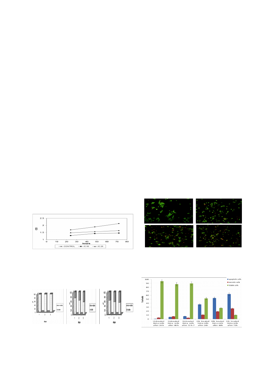
the number of variable cells. Figure 1 shows that the
growth rates decreased in the treated cells as compared
with the untreated cells whereas inoculation with a higher
concentration (IC
50
) decreased the growth rate more than
low concentration (IC
25
). On the other hand, the percentage
of non-viable cells treated with IC
25
value was 28% (day
1), 38% (day 2) and 45% (day 3). But the percentage of
non-viable cells treated with IC50 values were 38% (day
1), 45% (day 2) and 48% (day 3) (Figure 2).
Acridine Orange (AO) and Propodium Iodide (PI) Double
Staining using Fluorescent Microscopy
fluorescent microscope was conducted to study of
morphological changes of cell death mode induced by
goniothalamin after 24, 48 and 72 h. Acridine orange
(AO) and propidium iodide (PI) staining was used. Viable
cells displayed green fluorescence with the appearance of
circular cell; intact DNA and nucleus give a round and
green nuclei. The early apoptotic cells have fragmented
DNA which gives several green colored nuclei and cell
blebbing. Late apoptotic and necrotic cell’s DNA would
be fragmented and stained orange and red (Figure 3)
Besides the study of morphological changes, the
percentage of viable, apoptotic and necrotic cells also
recorded in Table 2 and plotted as a graph in Figure 4. The
percentage of apoptotic cells in untreated cells slightly
increased from 0.67% after 24 h to 5.33% and 7% after
48 and 72h, respectively (Figure 5). Whereas, cells treated
with goniothalamin at IC
50
concentration, the percentage
of apoptotic cells increased rapidly from 37% after 24h
to 53% and 63% after 48 and 72h, respectively.
Analysis of Cellular DNA Content Using Propidium Iodide
The DNA Content of HeLa cells were monitored by
Figure 1. MTT Proliferation Assay for IC
50
and IC
25
Goniothalamin Concentrations (3.2 and 1.2 µg/ml)
Against HeLa Cells at 24, 48 and 72 Hours Post-
Treatment. The growth rates decreased in the treated cells
as compared with the untreated cells whereas
inoculation
with a higher concentration of virus (IC
50
) decreased the growth
rate more than low concentration (IC
25
)
0
25.0
50.0
75.0
100.0
Newl
y
di
agnosed
wi
thout
tr
eatment
Newl
y
di
agnosed
wi
th
tr
eatment
Persi
stence
or
recurr
ence
Remi
ssi
on
None
Chemother
ap
y
Radi
other
ap
y
Concurr
ent
chemor
adi
ati
on
10.3
0
12.8
30.0
25.0
20.3
10.1
6.3
51.7
75.0
51.1
30.0
31.3
54.2
46.8
56.3
27.6
25.0
33.1
30.0
31.3
23.7
38.0
31.3
A
B
C
Figure 2. The Percentage of Viable and Non-
viable HeLa Cells in Population after Treated with
Goniothalamin after 24, 48 and 72 h.
A) untreated cells,
B) HeLa cells treated with IC
25
Goniothalamin concentration
(1.2 µg/ml), C) HeLa cells treated with IC
50
Goniothalamin
concentration (3.2 µg/ml)
A)
B)
C)
A
B
C
D
Figure 3. Fluorescence Microscopy Examination of
HeLa Cell Line (Magnification 200X).
A) Untreated HeLa
cells, B) HeLa cells treated with Goniothalamin after 24 h, C)
HeLa cells treated with Goniothalamin after 48 h, D) HeLa cells
treated with Goniothalamin after 72 h.
Figure 4. Flourecent Microscopy Examination.
Percentage of apoptotic cells, necrotic cells and viable cells in
HeLa cell population with gonithalamin treatment after 24 48
and 72 h. HeLa cell death via apoptosis increased significantly
(*P < 0.05) in time-dependent manner
Aied M. Alabsi1 et al
Asian Pacific Journal of Cancer Prevention, Vol 13, 2012
5134
Table 1. Cytotoxicity of Goniothalamin against Various
Cell Lines
Cell line
IC
50
value (μg/ml)
Cervical cancer (HeLa)
3.2±0.72
Breast carcinoma (MCF-7)
6.6±0.92
Colon cancer (HT29)
3.8±1.10
Normal mouse fibroblast cells (3T3)
>10
Table 2. Percentages of Apoptotic, Necrotic and Viable
HeLa Cells after 24 and 48 h
HeLa cells and Treatment
Apoptotic Necrotic Viable
cells % cells % cells %
Untreated HeLa cells after 24 h
0.67
3.67
95.66
Untreated HeLa cells after 48 h
5.33
6.67
88.00
Untreated HeLa cells after 72 h
7.00
3.33
89.67
GN treated HeLa cells after 24h 37.00
11.00
52.00
GN treated HeLa cells after 48h 53.00
19.20
27.80
GN treated HeLa cells after 72h 63.00
26.67
10.33
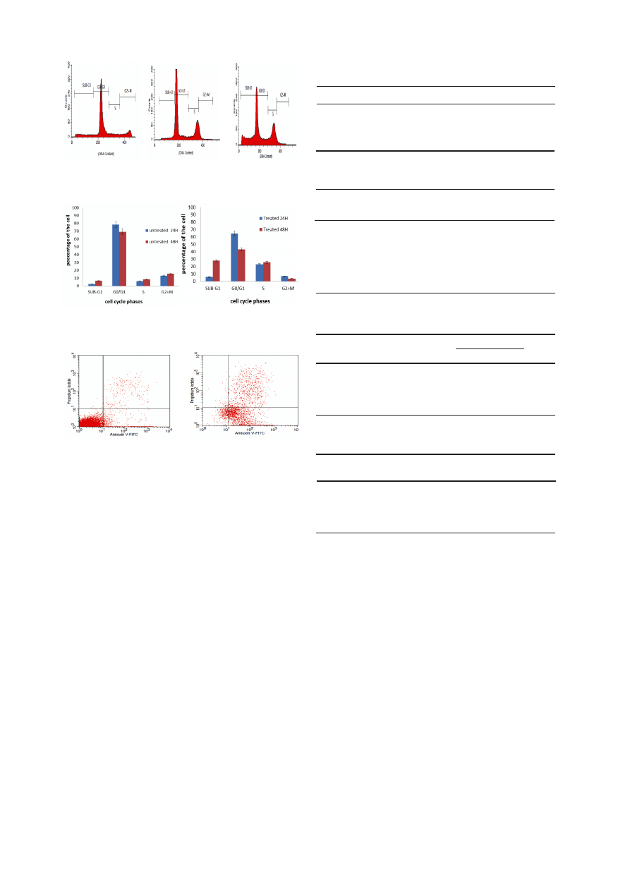
Table 4. Cell Cycle Analysis of Cervical Cancer Cells
(HeLa) at 24h and 48h Treated with Goniothalamin
Cell cycle phase
Treated with IC
50
Goniothalamin (%)
24H 48H
SUB-G1
5.91
27.99
G1
64.66 43.10
S
23.17
25.90
G2/M
7.08 3.76
flow cytometry after propidium iodide staining nuclei.
Goniothalamin induced a significant time-dependent
increase in the proportion of sub-G1 in HeLa cell
population. However, a slight increase was observed at
Sub G1 phase of untreated cervical cell (HeLa) over time.
Tables 3 and 4 show that the percentages of the treated
cells in Sub-G1 increased from 5.91% at 24 hours to
27.99% at 48 h while the percentages in untreated cells
increased from 2.44% at 24 hours to 6.62% at 48 h
(p<0.005).
On the other hand DNA histogram showed that
goniothalamin increased the population of cells at S phase
in a time-dependent manner (Figure 6). The S population
increased significantly from 6.17% and 8.53% in the
untreated cells to 23.17% and 25.92% in cells treated with
IC50 goniothalamin for 24 and 48h, respectively Tables 3
and 4. While concomitantly the G1 population decreased
from 78.38 %and 69.27% in the untreated cells to 64.66%
and 43.10% in the treated cells for 24 and 48h, respectively
Tables 3 and 4. Similarly the G2/M population decreased
from 12.98% and 15.58% in the untreated cells to 7.08%
and 3.76% in the treated cells for 24 and 48h, respectively
Tables 3 and 4.
0
25.0
50.0
75.0
100.0
Newl
y
di
agnosed
wi
thout
tr
eatment
Newl
y
di
agnosed
wi
th
tr
eatment
Persi
stence
or
recurr
ence
Remi
ssi
on
None
Chemother
ap
y
Radi
other
ap
y
Concurr
ent
chemor
adi
ati
on
10.3
0
12.8
30.0
25.0
20.3
10.1
6.3
51.7
75.0
51.1
30.0
31.3
54.2
46.8
56.3
27.6
25.0
33.1
30.0
31.3
23.7
38.0
31.3
A
B
C
Figure 5. Cell Cycle Analysis of Cervical Cell Cancer
Treated with Goniothalamin at IC
50
Concentration.
A)
Untreated cells, B) Treatment after 24h, C) Treatment after 48 h.
A
B
Figure 6. Analysis of the Cell Cycle on Cervical Cancer
Cells (HeLa) after 24h and 48h.
A) Untreated HeLa cells,
B) HeLa cells treated with Goniothalamin.
A
B
A)
B)
A
B
Figure 7. Contour Diagram of Annexin V/PI Flow
Cytometry.
A) untreated HeLa cells, B) HeLa cells at 24
h post-inoculation of IC
50
value of Goniothalamin. Lower left
quadrants show viable cells, excluding PI and negative for
Annexin V binding. The upper right quadrants contain the non-
viable, necrotic cells, positive for Annexin V and PI uptake.
Lower right quadrants represent the apoptotic cells, Annexin V
positive and PI negative.
Flow cytometry (Annexin V/PI double staining)
Apoptotic cells exclude all dyes which are in use for
cell viability assays, such as PI, while necrotic cells do
not. In cells with a damaged cell membrane PI induces a
red fluorescence on the DNA, whilst it is excluded by cells
with a preserved cytoplasm membrane. Hence during the
initial phase of apoptosis, the cells are still able to exclude
PI and therefore do not show any red fluorescence signal,
similar to that of living cells. Figure 7 showed the results of
Annexin V/PI flow cytometry of HeLa cells after treatment
with IC
50
value of goniothalamin. Untreated cell was
found in the lower left quadrant of the cytograms, these
viable cells excluded PI and were negative for Annexin
V binding. The upper right quadrant represents the non-
viable, necrotic cells, positive for Annexin V binding and
showing PI uptake. The lower right quadrant represents
the apoptotic cells, Annexin V positive and PI negative,
demonstrating Annexin V binding and cytoplasmic
membrane integrity (Figure. 11). The Annexin V/PI –
apoptotic cell population for HeLa cell line increased from
6.4%in untreated cells, to 26.45% in treated cells at 24 h
post-infection.
Table 3. Percentages of Untreated Cervical Cancer
Cells at 24h and 48 h
Cell cycle phase
HeLa cells (%)
24H 48H
SUB-G1
2.44 6.62
G1
78.38
69.27
S
6.17 8.53
G2/M
12.98
15.58

Asian Pacific Journal of Cancer Prevention, Vol 13, 2012
5135
DOI:http://dx.doi.org/10.7314/APJCP.2012.13.10.5131
Anticancer Activity of Gonothalamin in a Cervical Cancer Cell Line
Discussion
Cell death in mammalian cells are divided into two
morphologically and biochemically distinct modes
namely apoptosis and necrosis (Doyle and Griffiths,
1998). Apoptosis is an organized, pre-programmed
response of cell to shifting of environmental conditions.
Characteristics of apoptotic cell include cell shrinkage,
nuclear and DNA fragmentation and breaking up of the
cell into membrane-bounded vesicles, termed ‘apoptotic
bodies’, which are subsequently ingested by macrophages
(Doyle and Griffiths, 1998). Apoptosis plays a vital role
in regulating growth, development and immune response,
and also clearing abnormal cells (Fan et al., 2005).
This apoptosis program becomes important in medical
study in order to cure the cancerous cell without give
the inflammatory effect. Aberrant cell death processes
may underlie many human diseases including cancers,
autoimmune, neurodegenerative and immunodeficiency
disorders (Baehrecke, 2002).
Cytotoxic has been defined as the cell killing property
of a chemical compound independent from the mechanism
of death (Graham-Evans et al., 2003). Cytotoxicity assay
is an appropriate method for screening new substances
within a short time in order to determine cytotoxicity on
cancer cells (Alley et al., 1988). The effective dose for a
50% reduction in cell number for plants products to be
considered cytotoxic should be less than 20 µg/ml (Geran
et al., 1972 ).
MTT cytotoxicity assay used to measure the cytotoxic
effect of goniothalamin (GTN) on cervical cancer (HeLa),
breast carcinoma (MCF-7), colon cancer (HT29) and
Normal mouse fibroblast cells (3T3) measure of cytotoxic
effect and The IC
50
concentration that kill 50% of the cells
was determined graphically after 72 h. In screening result,
GNT has shown broad spectrum cytotoxicity and It had
most active cytotoxic activity cervical cancer (HeLa) but
not on Normal mouse fibroblast cells (3T3). These results
conducted to other studies investigated the cytotoxic effect
of Goniothalamin towards human breast cancer, vascular
smooth muscle cells (VSMCs), Jurkat leukemia cells,
HL-60 leukemia cells, Chinese hamster ovary (CHO)
and hepatoblastoma HepG2 cells (Ali et al., 1997; Pihie
et al., 1998; Inayat-Hussain et al., 1999; Inayat-Hussain
et al., 2003; Nasir et al., 2004; Chen et al., 2005; Chan et
al., 2006; Al-Qubaisi et al., 2011).
In this study, GTN have indicated significant growth
inhibition in HeLa cell line at low concentration of
IC
50
values. MTT proliferation assay was carried out to
determine the growth rate of cells. A linear relationship
between the formazan generated and the number of viable
cells was demonstrated, together with time-dependent
growth characteristics for HeLa cells (Ferrari et al., 1990).
GTN treatment on HeLa cells cell lines showed significant
decrease in growth rate compared with control. Whereas
treatment with high concentration (IC
50
value) showed that
the growth rates of the cells were more decreased than of
low concentration (IC
25
values). On the other hand the
percentage of non-viable cells on both cell lines increased
with the increasing period of treatment.
However, MTT cytotoxic results of GTN on HeLa
cells have been further supported with morpgological
study using fluorescent microscopy Acridine Orange
Propidium Iodide staining assay and flow cytometric
analysis of cell cycle.
The apoptotic features were confirmed and the
percentage of apoptotic cells was determined from
at least 300 counted cells observed under fluorescent
microscope. The calculation of apoptotic cells is described
as the percentage of apoptotic cells and apoptotic bodies
within the overall population of cells. The percentage of
apoptotic cells and the graph showed that the percentage of
apoptotic cells treated with goniothalamin was increasing
among the time. These distinctive morphological features
form the basis of some of the most widely used techniques
for the identification and quantification of apoptosis,
and thus morphologic description using Phase Contrast
microscopy and fluorescence microscopy remains one of
the best ways to define apoptosis (Doonan and Cotter ,
2008).
The quantitative analysis of cell cycle is very important
in the study of molecular mechanism of cell death and
cell cycle progression (Tao et al., 2004). Untreated
and treated HeLa cells were evaluated for apoptosis by
measuring the amount of apoptotic cells using of DNA
flow cytometry (FCM). Flow cytometric analysis of cell
cycle measures the apoptotic changes in cells by staining
them with DNA dyes (Telford et al., 1994). Apoptotic
cells, due to a change in membrane permeability, showed
an increased up-take of the vital dye, PI, compared to
live cells (Nicoletti et al., 1991; Telford et al., 1994).
This method is useful for quantitative estimates of the
fractions of cells in the different phases of the cell cycle
(Ali et al., 2011). In this study goniothalamin treatment
on HeLa cells produced S phase cell cycle accumulation
with a large increase in the sub-G1 which mean there was
a relationship between goniothalamin-induced S phase
arrest and apoptosis(p<0.001). A study of cell cycle pattern
has been documented that goniothalamin treatment causes
cell cycle arrest and cell death maximally at G2/M phase
(Chen et al., 2005). Another study demonstrates that GTN
arrested cell cycle at G0/G1 in SK-Hep1, and at G2/M in
Hep-3B cells (Cheng-Hui , 2008). These results concurred
with the previous results to suggest that goniothalamin
induce apoptosis on HeLa cells more extensively with
increasing in time.
Change in plasma membranes is the earliest features of
apoptosis. In apoptotic cells, the membrane phospholopid,
phosphotidylserine (PS) is translocated from the inner to
the outer leaflet of the plasma membrane thereby exposing
PS to the external cellular activity (Lawen, 2003).
Annexin binding assay is a method permits the detection
of the early phases of apoptosis before the loss of cell
membrane integrity (Vermes et al., 1995; Aubry et al.,
1999). The principle of Annexin V staining method used
is the conjugation of Annexin V to phosphotidylserine of
the apoptosis cells and in conjunction of dye Propodium
Iodide which binds to cells at different stage and
distinguishes apoptosis cells with necrotic cells (Tao et al.,
2004). Apparently, the results indicate that the percentage
of cells in early apoptosis of the cervical cancer cell
(HeLa) treated with Goniothalamin appeared after 24 hr.

Aied M. Alabsi1 et al
Asian Pacific Journal of Cancer Prevention, Vol 13, 2012
5136
References
WHO, World Health Organization (2011). Cervical Cancer.
http://www.who.int/reproductivehealth/topics/cancers/en/
index.html.
Tompa A (2007). Theory and practice of primary cancer
prevention. MagyarOnkológia, 51, 7-21.
World d Health Organization (WHO)/Institut Català d’Oncologia
(ICO) (2010). Malaysia: Human Papillomavirus and Related
Cancers, Summary Report. Third Edition. WHO/ICO
Information Centre.
Lin TP, Pihie AHL ( 2003). Goniothalamin-induced apoptosis
in human ovarian cancer cell line, Caov-3 through the
regulation of Bcl-2 and Bax. Borneo Sci, 14, 9-14.
Chen WY, Wu CC, Lan YH, et al (2005). Goniothalamin induces
cell cycle-specific apoptosis by modulating the redox status
in MDA-MB-231 cells. Eur J. Pharmacol, 522, 20-29.
Al-Qubaisi M, Rozita R, Yeap SK, et al (2011). Selective
cytotoxicity of Goniothalamin against Hepatoblastoma
HepG2 Cells. Molecules, 16, 2944-59.
Rajab N F, Hamid Z A, Hassan H, Ali A M, Din L B and Inayat-
Hussain S H (2005).Evaluation of the cytotoxicity and
genotoxicity effects of goniothalamin in leukemic cell lines.
Environ. Mutagen Res, 27, 161-4.
Inayat-Hussain SH, Chan KM, Rajab NF, et al (2010).
Goniothalamin-induced oxidative stress, DNA damage and
apoptosis via caspase-2 independent and Bcl-2 independent
pathways in Jurkat T-cells. Toxicol Lett, 193, 108-14.
Wattanapiromsakul C, Wangsintaweekul B, Sangprapan P,
Itharat A, Keawpradub N (2005). Goniothalamin, a cytotoxic
compound, isolated from Goniothalamus macrophyllus
(Blume) Hook. f. & Thomson var. macrophyllus.
Songklanakarin. J. Sci. Technol, 27, 479-87.
Mishell BB, Shiiqi SM, Henry C (1980). Selected methods. In:
Mishell BB, Shiiqi SM (eds) Cellular Immunology. Freeman,
San Francisco, 21–22,
Ali R, Alabsi AM, Ali AM, et al (2011). Cytolytic effects and
apoptosis induction of newcastle disease virus strain AF2240
on anaplastic astrocytoma brain tumor cell line. Neurochem
Res, 36, 2051-62.
Doyle A, Griffiths JB (1998). Cell and Tissue Culture:
Laboratory Procedures in Biotechnology. Second Edition.
John Wiley & Sons Ltd, 201.
Fan T J, Han L H, Cong R S, Liang J (2005). Caspase Family
Proteases and Apoptosis. Acta Biochimica et Biophysica
Sinica, 37, 719-27.
Baehrecke EH (2002). How death shapes life during development.
Nat Rev Molecular Cell Biol, 3, 779-87.
Graham-Evans B, Tchounwou PB, Cohly HH ( 2003).
Cytotoxicity and proliferation studies with arsenic in
established human cell lines: keratinocytes, melanocytes,
dendritic cells, dermal fibroblasts, microvascular endothelial
cells, monocytes and T-cells. Int J Mol Sci, 4, 13-21.
Alley M C, Scudiro D A, Monks A, Hursey M L, Czerwinski M
J, Fine D L (1988). Feasibility of drug screening with panels
of human tumor cell lines using a microculture tetrazolium
assay. Cancer Res, 48, 589-601.
Geran RI, Greenberg N H, Macdonald MM, Schumacher AM,
Abbott BJ (1972). Protocols for screening chemical agents
and natural products against animal tumors and other
biological systems. Cancer Chemother Rep, 3, 1-104.
Ali AM, Mackeen MM, Hamid M, et al (1997). Cytotoxicity
and electron micreactive oxygen speciescopy of cell death
induced by goniothalamin, Planta Med, 63, 81–83.
Chan KM, Rajab NF, Ishak MHA, et al (2006) Goniothalamin
induces apoptosis in vascular smooth muscle cells, Chem-
Biol Interact, 159, 129–140.
Pihie A H, Stanslas J, Din L B (1998). Non-steroid receptor
mediated antiproliferative activity ofstyrylpyrone derivative
(SPD) in human breast cancer cell lines. Anticancer Res,
18, 1739-44.
Inayat-Hussain SH, Annuar O, Laily D, et al (1999). Caspases-3
and -7 are activated in goniothalamin-induced apoptosis in
human Jurkat Tcells. FEBS Letters, 456, 379-83.
Inayat-Hussain S H, Annuar BO, Din LB, Ali AM, Ross D
(2003). Loss of mitochondrial transmembrane potential
and caspase-9 activation during apoptosis induced by the
novel styryllactone goniothalamin in HL-60 leukemia cells,
Toxicol. In Vitro, 17, 433-439.
Nasir U T, Saifulaman M, Rozita R, Laily BD, Leslie CL (2004).
Genotoxicity of goniothalamin in CHO cell line. Mutat Res,
562, 91-102.
Ferrari M, Fornasiero MC, Isetta AM (1990). MTT colorimetric
assay for testing macrophage cytotoxic activity in vitro. J.
Immunological Methods, 131, 165-70.
Doonan F, Cotter TG (2008). Morphological assessment of
apoptosis. Methods, 44, 200-4.
Tao D, Wu J, Feng, F, et al (2004). New method for the analysis
of cell cycle–specific apoptosis. Cytometry Part A, 57, 70-74.
Telford WG, King LE, Fraker PJ (1994). Rapid quantitation of
apoptosis in pure and heterogeneous cell populations using
flow cytometry. J Immunol Methods, 172, 1-16.
Nicoletti I, Migliorati G, Pagliacci M C, Grignani F, Riccardi C
(1991). A rapid and simple method for measuring thymocyte
apoptosis by propidium iodide staining and flow cytometry.
J Immunol Methods, 139, 271-9.
Cheng-Hui T (2008). Goniothalamin induces TP53-dependent
and -independent apoptosis in hepatocellular carcinoma
derived cells, Master’s Thesis. Institute of Biomedical
Sciences, Sun Yat-Sen University.
Lawen A (2003). Apoptosis; an introduction. BioEssays, 25,
888-96.
Aubry J, Blaecke A, Lecoanet-Henchoz S, et al (1999). Annexin
V used for measuring apoptosis in the early events of cellular
cytotoxicity. Cytometry, 37, 197–204.
Vermes I, Haanen C, Steffens-Nakken H, Reutelingsperger
C (1995). A novel assay for apoptosis flow cytometric
detection of phosphatidylserine early apoptotic cells using
fluorescein labeled expression on Annexin V. J Immunol
Methods, 184, 39–51.
The percentage of the cells treated with Goniothalamin
were decreased in early apoptosis phase and increased in
late apoptosis over time.
In summary, goniothalamin (GTN) showed selective
cytotoxic towards cervical cancer (HeLa), breast
carcinoma (MCF-7), and colon cancer (HT29) but is not
normal mouse fibroblast cells (3T3). The compound is
potentially a good anti-cancer drug since it is non-toxic
towards healthy cells. Our results indicate that GTN
inhibits HeLa cell proliferation via apoptosis and causes
cell cycle arrest and cell death at S phase.
Wyszukiwarka
Podobne podstrony:
Chemical Composition and in Vitro Antifungal Activity Screening
In Vitro Anticancer Activity of Ethanolic Extract
Evaluation of in vitro anticancer activities
Mechanical and in vitro
In vitro cytotoxicity activity
Morphogenesis and cell cycle progression in Candida albicans
Cytolytic Effects and Apoptosis Induction
2001 In vitro fermentation characteristics of native and processed cereal grains and potato
Wheat bread enriched with green coffee – In vitro bioaccessibility and
The pathogenesis of Sh flexneri infection lessons from in vitro and in vivo studies
2000 Glucose Based Oligosaccharides Exhibit Different In Vitro Fermentation Patterns and Affect In V
in vitro, studia rolnictwo, rok IV
Kultury in vitro roslin rozmnazanie klonalne
więcej podobnych podstron