
1
AI Pearce et al.
Animal models for implant biomaterial research
European Cells and Materials Vol. 13. 2007 (pages 1-10) ISSN 1473-2262
Abstract
Development of an optimal interface between bone and
orthopaedic or dental implants has taken place for many
years. In order to determine whether a newly developed
implant material conforms to the requirements of
biocompatibility, mechanical stability and safety, it must
undergo rigorous testing both in vitro and in vivo. Results
from in vitro studies can be difficult to extrapolate to the in
vivo situation. For this reason the use of animal models is
often an essential step in the testing of orthopaedic and
dental implants prior to clinical use in humans. This review
discusses some of the more commonly available and
frequently used animal models such as the dog, sheep, goat,
pig and rabbit models for the evaluation of bone-implant
interactions. Factors for consideration when choosing an
animal model and implant design are discussed. Various
bone specific features are discussed including the usage of
the species, bone macrostructure and microstructure and
bone composition and remodelling, with emphasis being
placed on the similarity between the animal model and the
human clinical situation. While the rabbit was the most
commonly used of the species discussed in this review, it
is clear that this species shows the least similarities to human
bone. There were only minor differences in bone
composition between the various species and humans. The
pig demonstrates a good likeness with human bone,
however difficulties may be encountered in relation to their
size and ease of handling. In this respect the dog and sheep/
goat show more promise as animal models for the testing
of bone implant materials. While no species fulfils all of
the requirements of an ideal model, an understanding of
the differences in bone architecture and remodelling will
assist in the selection of a suitable model for a defined
research question.
Key Words: Animal-models, biomaterials, osseointegration,
bone, implant, dog, pig, sheep, goat, rabbit.
Address for correspondence*
A.I. Pearce
AO Research Institute, AO Foundation
Clavadelerstrasse 8, CH-7270 Davos Platz
Switzerland
Telephone Number: +41 (0)814142311
E-mail: alexandra.pearce@aofoundation.org
Introduction
The development and modification of orthopaedic and
dental implants has taken place for many years in an effort
to create an optimal interaction between the body and the
implanted material. The goal of achieving an optimal
bone-implant interface has been approached by the
alteration of implant surface topography, chemistry,
energy and charge as well as bulk material composition.
Schmidt et al. (2001) defines an ideal bone implant
material as having a biocompatible chemical composition
to avoid adverse tissue reaction, excellent corrosion
resistance in the physiologic milieu, acceptable strength,
a high resistance to wear and a modulus of elasticity
similar to that of bone to minimise bone resorption around
the implant. The features relating to implant safety such
as avoidance of adverse tissue reaction and resistance to
wear and corrosion are of high clinical significance for
implants used in long-term clinical situations in both
human and veterinary medicine as there have been some
links between prolonged exposure to non-biocompatible
materials and neoplastic tissue responses. In order to
determine whether a new material conforms to the
requirements of biocompatibility and mechanical stability
prior to clinical use, it must undergo rigorous testing under
both initial in vitro and then in vivo conditions.
In vitro testing is popular for the characterisation of
bone-contacting materials, particularly as medical
researchers embrace the principles of animal reduction.
It is accepted that in vitro testing be used primarily as a
first stage test for acute toxicity and cytocompatibility to
avoid the unnecessary use of animals in the testing of
cytologically inappropriate materials. The term
biocompatibility is often incorrectly used with in vitro
tests, as biocompatibility can only be used in the case of
animals or humans (in vivo), with the correct term being
cytocompatibility for in vitro tests (Richards et al., 2001).
In vitro cell culture is centred upon the growth of cells
no longer organised into tissues, where cells are collected
either through enzyme digestion or mechanically from
native tissue and proliferate in a suspension or attached
to a substrate surface as a monolayer. In vitro testing gives
information regarding cytotoxicity, genotoxicity, cell
proliferation and differentiation (Hanks et al., 1996; Nahid
and Bottenberg, 2003) and is more easily standardised
and quantifiable than in vivo testing (Nahid and
Bottenberg, 2003). In vitro studies are also useful for
screening new materials for product quality and the release
of potentially harmful additives incorporated during the
manufacturing process (Pizzoferrato et al., 1994).
However, in vitro characterisation is not able to
demonstrate the tissue response to materials, instead being
ANIMAL MODELS FOR IMPLANT BIOMATERIAL RESEARCH IN BONE:
A REVIEW
AI Pearce*, RG Richards, S Milz, E Schneider and SG Pearce
AO Research Institute, AO Foundation, Clavadelerstrasse 8, Davos, Switzerland

2
AI Pearce et al.
Animal models for implant biomaterial research
confined to the response of individual cell lines or primary
cells taken from animals. In addition, cellular responses,
such as cytotoxicity due to the presence of metal ions, can
vary between cell lines and passage number (Wataha et
al., 1994). In vitro tests may also overestimate the level of
material toxicity and are limited to acute studies of the
effects of toxicity due to the relatively short lifespan of
cultured cells (Pizzoferrato et al., 1994).
In vitro tissue culture maintains small tissue fragments
of tissue, but does not necessarily preserve architecture.
In vitro organ culture maintains tissue or organs (in part or
whole), which may allow some differentiation and
preservation of architecture and function (though systemic
factors are absent, the lack of vascularisation limits nutrient
and oxygen supply and waste removal and therefore
extrapolation of results to the in vivo situation limits the
model). In vitro cells may suffer from phenotypic drift,
which may be due to dissociation of cells from their three
dimensional geometry and/or growth on a two dimensional
surface. The dynamic properties of cell culture are difficult
to control in vitro and it is difficult to recreate the
appropriate cell interactions found in vivo. One major
limitation to bone culture is the lack of controlled
physiological loading since without load bone will increase
resorption, as is seen in patients after prolonged bed rest
(Vico et al., 1987). No in vitro cell culture system is able
to produce loading that simulates the in vivo situation and
currently very few ex vivo systems are able to approach
such physiological loading (and usually only with small
tissue samples) (Davies et al., 2006). For these reasons
animal models are essential for evaluating biocompatibility,
tissue response and mechanical function of an orthopaedic
or dental material prior to clinical use in the human.
Animal models allow the evaluation of materials in
loaded or unloaded situations over potentially long time
durations and in different tissue qualities (e.g. normal
healthy or osteopenic bone) and ages. Not only can the
tissues in the immediate vicinity of the implant be assessed,
tissues in remote locations can be studied, which is
particularly relevant to the study of wear particle debris.
In human patients, such debris has been reported to travel
into different distant organs such as liver and spleen (Urban
et al., 2000). While animal models may closely represent
the mechanical and physiological human clinical situation,
it must be remembered that it is only an approximation,
with each animal model having unique advantages and
disadvantages. Currently there are numerous models for
testing implant materials in vivo, ranging in purpose from
the assessment of protein adsorption and soft tissue
adherence to the integration of bone and the dissemination
of implant wear particles. This review examined the
literature relating to animal models used in the evaluation
of bone-implant interactions.
Implant Design
For testing orthopaedic and dental implants, it is necessary
to use a model which is reproducible and in which implant
dimensions are comparable to those used in humans. The
number and size of implants to be tested will influence
directly the species of animal chosen for a study. The most
common implant designs used in animal models are either
screw type (threaded) or cylindrical (rod shaped) and less
commonly coin, disc, plate or irregular shaped. Regardless
of the design, implants should have an appropriate size
for the species chosen and for the bone implantation site.
Screw type implants have the advantage of producing good
initial stability, whereas cylindrical implants are dependent
on exact fit in order to be stable within the bone and give
accurate results regarding their effect on bone integration
(Carlsson et al., 1988). However, analysis of rod or
cylindrical shaped implants may be less complicated due
to their more simple geometry.
Guidelines are provided for the dimensions of implants
for in vivo studies, based on the size of animal and bone
chosen and on the implant design, in order to avoid
pathological fracture of the test site. Cylindrical implants
placed into rabbit tibial and femoral diaphyseal bone should
be no larger than 2mm in diameter and 6mm in length. For
larger animals such as sheep, goats and dogs the ISO
recommended dimensions of cylindrical implants are 4mm
in diameter and 12mm in length for implantation into the
femur and tibia. Orthopaedic bone screw-type implants
may range from 2-4.5mm depending on the species chosen,
with the 4.5mm screws generally being reserved for the
larger species such as the dog, sheep and pig. The breed of
animal used in a study must be considered when choosing
the exact implant dimensions as for example, large breeds
of sheep may allow the use of implant materials up to 5mm
in diameter in certain locations such as the tibia and
metatarsus (Huffer et al., 2006). It is extremely important
that control implants are included in the study design. These
implants should be of a material already in clinical use
(International Standard ISO 10993-6, 1994) and should
allow the outcome data to be related to existing products.
The chosen implant design will determine the experimental
techniques used to evaluate the material, in particular the
mechanical testing techniques. Common mechanical
testing used on tissues harvested from in vivo studies
include torque removal tests (screw-type implants), pull-
out tests and push-out tests (screw, cylindrical implants).
These tests are used to evaluate the strength of the
interaction between the bone and implant surface. High
forces encountered during these tests indicate a good
integration between the bone and implant surface or in the
case of porous materials, a high degree of bone in-growth
into the pores of the implant.
Many studies aim to evaluate the effect of implant
surface modification on alteration of the bone-implant
interaction. In order to draw accurate conclusions regarding
the effects of implant modification, one must first
accurately determine the implant surface characteristics
with regard to the chemical composition of the material
and the surface topography. This should be performed both
visually (e.g. light microscopy, scanning electron
microscopy) and numerically (e.g. profilometry, contact
angle, X-ray photoelectron spectroscopy, energy dispersive
X-ray microscopy) thus including both qualitative and
quantitative data. There are numerous studies that draw
conclusions regarding the effect of surface topography on
bone formation without having properly characterised the
surfaces used, which unfortunately give varying results

3
AI Pearce et al.
Animal models for implant biomaterial research
leading to confusion (e.g. describing a surface as rough or
smooth, without having measured this numerically).
Animal Selection
Kirkpatrick et al. (2002) outlines three types of studies
which yield data on factors influencing the biological
response to materials implanted in bone. These are studies
on explanted biomaterials, in vitro techniques and animal
models. Desirable attributes of an animal model include
demonstration of similarities with humans, both in terms
of physiological and pathological considerations as well
as being able to observe numerous subjects over a relatively
short time frame (Egermann et al., 2005; Liebschner, 2004;
Schimandle and Boden, 1994).
When deciding on the species of animal for a particular
model there are several factors that should be considered.
One must define clearly the research question being
addressed prior to selecting the species of animal to be
used in the study. According to Schimandle and Boden
(1994), animal selection factors include: cost to aquire and
care for animals, availability, acceptability to society,
tolerance to captivity and ease of housing. A detailed
discussion on handling and care of the individual species,
listed in this review, will not be made. The welfare and
housing of animals is usually covered by a Federal Animal
Protection Act and may vary slightly between countries.
The Animal Protection acts outline the minimum
requirements in terms of housing dimensions, lighting,
flooring etc. and must be complied with when undertaking
an animal study. Specific features will vary according to
species. Other factors include low maintenance care, ease
of handling, resistance to infection and disease, inter-
animal uniformity, biological characteristics analogous to
humans, tolerance to surgery, adequate facilities and
support staff and an existing database of biological
information for the species. In addition to this, the lifespan
of the species chosen should be suitable for the duration
of the study. More specifically, for studies investigating
bone-implant interactions, an understanding of the species
specific bone characteristics – such as bone microstructure
and composition, as well as bone modelling and
remodelling properties, are important when later
extrapolating the results to the human situation. Finally
the size of the animal must be considered to ensure that it
is appropriate for the number and size of implants chosen
(Schimandle and Boden, 1994); (International Standard
ISO 10993-6, 1994). Hazzard et al. (1992) comment that
within a field of study, no single animal model will be
appropriate for all purposes, nor can a model be dismissed
as inappropriate for all purposes. Furthermore, multiple
model systems are likely required to establish a broad body
of knowledge (Hazzard et al., 1992).
International standards established regarding the
species suitable for testing implantation of materials in
bone, state that dogs, sheep, goats, pigs or rabbits are
suitable. At least four rabbits and at least two of each of
the other species mentioned above should be used for each
treatment at each implantation period, though appropriate
power calculations should be performed. Long term
implantation periods for these species are given as 12, 26,
52 and 78 weeks and in certain instances (with the
exception of rabbits) 104 weeks (International Standard
ISO 10993-6, 1994). Although the rat is one of the most
commonly used species in medical research, it will not be
discussed here due to significant dissimilarities between
rat and human bone and the limitations of size making
rats unsuitable for testing multiple implants simultaneously.
Canine
Usage
The dog is one of the more frequently used large animal
species for musculoskeletal and dental research. Unlike
other animal species, there is a considerable amount of
literature comparing canine and human bone with regard
to the usefulness of the dog as a model for human
orthopaedic conditions. A review by Neyt et al. (1998)
finds that dogs and cats were used in 11% of
musculoskeletal research between 1991 and 1995. This is
confirmed by Martini et al.(2001) who reports that between
1970 and 2001, 9% of orthopaedic studies utilised dogs as
an animal model. The highly tractable nature of dogs can
be beneficial during the post operative healing phase where
they may be trained to take an active part in recuperative
protocols. However, there are increasing ethical issues
relating to the use of dogs in medical research due to their
status as companion animals.
Macrostructure
Depending on the size and breed of dog, there may be
some discrepancy in the size and shape of canine bones in
comparison to human bones; however, commercially
available implants and surgical equipment is available for
canine surgery. There are also obvious differences in bone
loading with the quadrupedal gait of the dog.
Microstructure
Wang et al. (1998) investigate the differences in fracture
properties between bovine, baboon, rabbit and canine bone
and the correlation of compositional and microstructural
properties with these differences. While adult human bone
has a secondary osteonal structure (osteons greater than
100µm containing blood vessels and with cement lines
forming a boundary between adjacent lamellae), canine
bone is found to have a mixed microstructure comprising
predominantly secondary osteonal bone in the centre of
cortical bone, but with, what is called plexiform bone in
the areas adjacent to the periosteum and endosteum (Wang
et al., 1998). Plexiform or laminar bone as it is also called
(Jee et al., 1970) is found predominantly in large, rapidly
growing animals and occasionally in children during
periods of rapid growth. It is formed more rapidly than
secondary osteonal tissue, but provides greater mechanical
support than woven bone. It has a brick-like appearance
and vascular plexuses within the lamellar bone tissue. It
is also concluded from this study that, despite similarities
in organic composition, canine bone had significantly
higher mineral density than human bone (Wang et al.,
1998).
Earlier findings by Kuhn et al. (1989), indicate that
while trabecular bone from the distal femur of humans

4
AI Pearce et al.
Animal models for implant biomaterial research
and dogs is qualitatively similar in terms of mechanical
and mass properties, differences in the coefficients relating
to ultimate strain resistance, indicate that canine trabecular
bone is able to withstand higher compressive strains before
failure than human bone (Kuhn et al., 1989).
Bone composition
A study by Aerssens et al. (1998) examines the differences
in bone composition, density and quality between various
species (human, dog, sheep, pig, cow and chicken). It is
found that there is most similarity in bone composition
(ash weight, hydroxyproline, extractable proteins and IGF-
1 content) between the dog and human. In terms of bone
density the dog and pig most closely represent the human
situation. The authors conclude that of the components
tested, the characteristics of human bone are best
approximated by the properties of canine bone (Aerssens
et al., 1998). These results are also supported by earlier
findings of Gong et al. (1964), where human and dog
cortical and cancellous bone are found to be similar in
terms of water fraction, organic fraction, volatile inorganic
fraction and ash fraction.
Bone remodelling
Another difference between human and canine bone which
may be of importance when assessing the effect of implant
modifications, is the difference in the rate of bone
remodelling between the species. This is an important
consideration as implant associated changes evident in a
canine model may not be as apparent in the human situation
where there is a lower rate of remodelling (Bloebaum et
al., 1991; Bloebaum et al., 1993). While there are
structural similarities in trabecular bone turnover between
dogs and humans (Kimmel and Jee, 1982), it is difficult to
make an exact comparison of bone turnover between these
species from data presented in the literature. Trabecular
bone turnover rates in dogs is found to be highly variable
between bone sites. For example, the lumbar vertebral body
has a bone turnover rate of close to 200% per year in young
adult male beagles. In the talus the turnover is 12% per
year. The average whole body trabecular bone turn over is
calculated as approximately 100% (Kimmel and Jee, 1982).
Not only is trabecular bone turnover variable between bone
sites within the one individual, there is also large variation
in trabecular bone turnover between individuals, for
example the mean turnover rate of bone taken by transilial
biopsy from young adult female beagles varies from 16%
per year to over 300% per year (Kimmel and Jee, 1982).
Remodelling of the total bone mass per year for humans is
given as 5-15%, with estimates of the whole body
trabecular bone turnover rate ranging from 10-15% per
year to 40-55% (Fernandez-Tresguerres-Hernandez-Gil
et al., 2006; Kimmel and Jee, 1982).
With regard to cortical bone, variation in bone turnover
at different sites is also demonstrated, with rib cortical bone
in young adult beagles having an annual turnover rate of
approximately 18% while midshaft cortical bone of long
bones is less than 1% (Polig and Jee, 1989). In addition to
these differences in bone turnover, age does not only affect
normal bone turnover (Jee et al., 1970) but may also affect
the bone response in relation to implant materials. Magee
et al. (1989) demonstrate that there is significantly higher
bone-implant interface strength in young greyhounds
compared with older greyhounds. It is concluded that this
is due to an age related decrease in bone remodelling ability
(Magee et al., 1989).
Sheep
Usage
While the use of dogs for orthopaedic research still
outnumbers sheep, over the last decade sheep numbers
are increasing. In the period of 1990-2001, sheep were
used in 9-12% of orthopaedic research involving fractures,
osteoporosis, bone-lengthening and osteoarthritis, in
comparison with just over 5% in the period from 1980-
1989 (Martini et al., 2001). This increase in usage may be
related to the ethical issues and negative public perception
of using companion animals for medical research.
Macrostructure
Most of the literature reports that the dog is more suitable
as a model for human bone from a biological standpoint
than the sheep; however, adult sheep offer the advantage
of being of a more similar body weight to humans and
having long bones of dimensions suitable for the
implantation of human implants and prostheses (Newman
et al., 1995), which is not possible in smaller species such
as rabbits or smaller breeds of dog.
Microstructure
While macroscopically, sheep bones may represent human
bones relatively closely, histologically, the bone structure
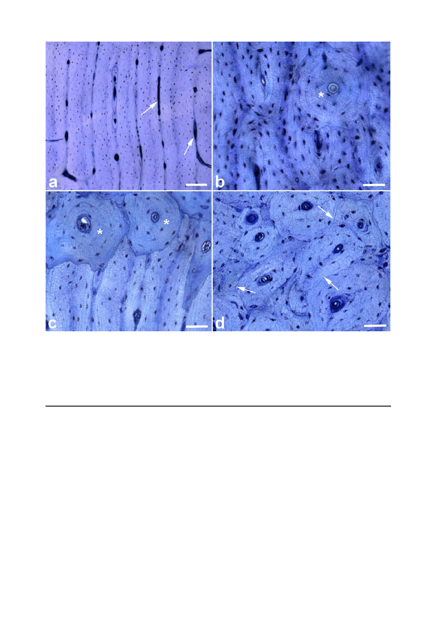
of the sheep is quite different (Fig. 1a-c). Sheep are
described as having a predominantly primary bone
structure (osteons less than 100µm diameter containing at
least two central blood vessels and the absence of a cement
line (deKleer, 2006)) in comparison with the largely
secondary bone of humans (Eitel et al., 1981). Age related
changes in bone structure are also described, whereby
sheep up to 3-4 years of age have a plexiform bone structure
comprising a combination of woven and lamellar bone
within which vascular plexuses are sandwiched (Newman
et al., 1995, Fig. 1a). Secondary, Haversian (osteonal)
remodelling in sheep (Fig. 1d) becomes more prevalent
with age (Liebschner, 2004) and has been seen at 7-9 years
of age (Newman et al., 1995). The location of onset of
haversian remodelling also seems to vary with bone type,
with the caudal femur and diaphysis of the radius and
humerus showing the earliest signs of this type of
remodelling (Newman et al., 1995).
Bone composition
Differences in bone density exist between the human and
sheep, whereby sheep bone shows a significantly higher
density and subsequently greater strength. Nafei et al.
(2000) reports the apparent density (mass/ volume,
reflecting the degree of porosity of bone) of sheep
trabecular bone taken from the proximal tibia of adult sheep
as being 0.61g/cm
3
with an apparent ash density of 0.41g/
cm
3
(ash mass/volume, reflecting the degree of bone
mineralisation) (Nafei et al., 2000). This is higher than
5
AI Pearce et al.
Animal models for implant biomaterial research
reported values for human femoral trabecular bone which
has an apparent density and an apparent ash density of
0.43g/cm
3
and 0.26g/cm
3
respectively, in other words the
sheep femur has a trabecular bone density 1.5-2 times
greater than that of humans (Liebschner, 2004). However,
differences may change with location. For example,
Liebschner (2004) reports that the apparent density of the
trabecular bone from sheep vertebrae has an apparent
density of 60+0.16g/ cm
3
– in contrast to the human
vertebral body, in which the apparent density is
0.14+0.06g/cm
3
. This demonstrates that location must be
considered when contemplating differences between
human and sheep bones. It should be noted that
unfortunately no age information is given in the above
study and it remains unclear as to whether this could also
influence the result. However it seems justified to assume
that sheep have significantly greater trabecular bone
density compared with humans.
In terms of mineral composition, Ravaglioli et al.
(1996) performed an evaluation of bone from humans,
cattle, sheep and dogs. The findings from this study
conclude that, apart from the early stages of physiological
growth in which there is partial substitution of Mg
2+
for
Ca
2+
in tricalcium magnesium phosphate (TCMP), the
mineral composition of humans and animals does not show
significant differences (Ravaglioli et al., 1996).
Bone remodelling
While differences in bone structure are recognised, several
studies argue that the sheep is still a valuable model for
human bone turnover and remodelling activity
(Chavassieux et al., 1987; den Boer et al., 1999; Pastoureau
et al., 1989). In support of this theory, a study observing
bone ingrowth into porous implants placed into the distal
femur of sheep (a weight-bearing model), show that sheep
and humans have a similar pattern of bone in-growth into
Figure 1. Ground and polished sections of MMA embedded sheep tibia stained with Toluidine blue. a) Plexiform or,
as it is also called, laminar appearance of cortical bone with longitudinally arranged vessels (arrows) between the
bone lamellae. Note the absence of a clearly visible cementline between adjacent lamellae. Scale bar = 200µm. b)
Remodelling of an area with originally plexiform bone which has been replaced by secondary osteons(*). Scale bar
= 50µm. c) Remodelling of plexiform bone in the immediate neigbourhood of an implant. During healing and
subsequent remodelling new bone is deposited in form of secondary osteons, seen in the upper part of the image (*).
Scale bar = 50µm. d) Transversely cut secondary bone with numerous osteons that can be clearly distinguished.
Note the cementlines that separate neighbouring osteons (arrows). Scale bar = 50µm.

6
AI Pearce et al.
Animal models for implant biomaterial research
porous implants over time. Although sheep are shown to
have a larger amount of bone in-growth than humans, it is
proposed that this is due to the greater amount of cancellous
bone in the distal femur of sheep, in comparison with
humans (Willie et al., 2004). Turner and Villanueva (1993)
find that measurements of bone volume, osteoid volume
and mineral apposition rate of 9-10 year old ewes are
comparable with those of men and post-menopausal
women in their 6-7th decade of life, suggesting that aged
sheep may make suitable models for human osteopenic
and osteoporotic bone.
As found in humans and dogs (Aerssens et al., 1997;
Kimmel and Jee, 1982), it is likely that bone location may
also alter bone composition and turnover in the sheep.
Other
Age has been found to play a significant role in determining
the amount of bone remodelling. The mechanical and
physical properties of ovine bone and interestingly, bone
from skeletally immature sheep showed a similar apparent
density and apparent ash density to humans (0.43g/cm
3
and 0.28g/cm
3
respectively) (Nafei et al., 2000). Trabecular
bone of skeletally immature sheep is weaker, less stiff,
more deformable before failure, has higher shock
absorptive qualities, contains more collagen and is less
dense and more porous than that of skeletally mature sheep
(Nafei et al., 2000). Thus it is essential to maintain a
consistent age of sheep within a study and to be aware
that age differences may make comparisons between
studies difficult.
Goats
Usage
While goats are the chosen species for 8.2% of animal
studies published in the Journal of Orthopaedic Research
(John Wiley & Sons, Hoboken, NJ) between 1992 and
1996, their predominant use is in studies of cartilage,
meniscal and ligamentous repair (An and Friedman, 1999).
Like sheep, goats are considered food producing animals
and thus also have the advantage of less critical public
perception when used for research, than companion
animals such as dogs. In comparison with sheep, goats
tend to have a more inquisitive and interactive nature which
may make confinement for long durations more
challenging than for sheep. In certain regions such as
south-east Asia where there is often a high temperature
and humidity, goats are reported to be more tolerant to
ambient conditions than other species such as sheep (Leung
et al., 2001).
Macrostructure
Like sheep, goats also have a body size suitable for the
implantation of multiple implants per goat or of larger,
human implants and prostheses (Anderson et al., 1999;
van der Donk et al., 2001).
Microstructure
Histologically, Qin et al. (1999) demonstrate that the tibial
cortical bone of goats does not have homogeneously
distributed Haversian systems (concentrically oriented
lamellar bone containing a centrally located blood vessel,
also known as osteons). Similar to the sheep, where the
Haversian systems are non-uniformly distributed
throughout individual bones, in the goat the Haversian
systems are located primarily in the cranial, cranio-lateral
and medial sectors of the tibial diaphysis, while the caudal
sector is mainly comprised of lamellar bone (where the
collagen fibres are arranged in sheets and do not contain
a central blood vessel) (Qin et al., 1999).
Bone composition
In a paper by Liebschner (2004) discussing the
biomechanical considerations of animal models used in
bone tissue engineering, it is shown that while there are
small differences in the apparent and ash density between
the goat and humans, these differences are probably not
as significant as the differences found between anatomic
sites of the same species. As mentioned previously, the
mineral composition of bone does not vary significantly
across species and therefore one could conclude that this
also holds true for the goat.
Bone remodelling
The literature reports that the goat is a suitable animal
model for testing human implants and materials as they
are considered to have a metabolic rate and bone
remodelling rate similar to that of humans (Anderson et
al., 1999; Spaargaren, 1994). Dai et al. (2005) also
supported the use of goats for studies related to bone
healing due to their comparable bone healing capacity and
tibial blood supply with that of humans.
In a study of the incorporation of morsellised bone
grafts under controlled loading conditions in goats and
humans, Lamerigts et al. (2000) found that the goat is a
suitable model to study bone graft incorporation, as the
sequence of events occurring during incorporation of bone
grafts is similar in humans and goats. However, the rate at
which a bone graft is revascularised and converted into a
vital trabecular structure is found to be faster in the goat,
occurring at approximately 3 months in comparison to 8
months in humans.
There is little information comparing the utility of goats
versus sheep for implant-related studies. Therefore the
choice of which small ruminant to use most likely depends
on availability and other factors.
Pigs
Usage
Pigs are reported as the subject of choice in a variety of
studies including studies of osteonecrosis of the femoral
head, fractures of cartilage and bone, bone ingrowth studies
and studies evaluating new dental implant designs (An and
Friedman, 1999; Buser et al., 1991; Sun et al., 1999).
Commercial breeds of pig are generally considered
undesirable for orthopaedic research due to their large
growth rates and excessive final body weight. However,
the development of miniature and micropigs has overcome
this problem to some extent. Nevertheless, pigs are often

7
AI Pearce et al.
Animal models for implant biomaterial research
considered difficult to handle, noisy and aggressive and
are therefore overlooked in favour of more amenable
species such as the sheep and goat ( Newman et al., 1995;
Swindle et al., 1988).
Macrostructure
With regard to bone anatomy, morphology, healing and
remodelling, the pig is considered to be closely
representative of human bone and therefore a suitable
species of choice (Thorwarth et al., 2005). Similarities have
been found in the femoral cross-sectional diameter and
area between humans and pigs (Raab et al., 1991).
However, pigs have a denser trabecular network
(Mosekilde et al., 1993).
Microstructure
While having a denser trabecular network, the pig is
described as having a lamellar bone structure which is
similar to that of humans (Mosekilde et al., 1987).
Bone composition
When comparing the bone composition of various species,
Aerssens et al. find that while canine bone most closely
resembles human bone, porcine bone also shows
similarities in bone mineral density and bone mineral
concentration to human bone (Aerssens et al., 1998).
Bone remodelling
The literature describes the pig as having bone remodelling
processes similar to humans, comprising both trabecular
and intra-cortical BMU based remodelling (Mosekilde et
al., 1987; Mosekilde et al., 1993). Laiblin and Jaeschke
(1979) compare the bone regeneration rate of dogs, pigs
and humans and find that pigs have a more similar rate of
bone regeneration to humans than do dogs (dog, 1.5-
2.0mm/day; pig, 1.2-1.5mm per day; human, 1.0-1.5mm
per day). In addition, in a study of the effects of fluoride
on cortical bone remodelling in growing pigs the results
show that control animals have a similar cortical bone
mineralization rate to humans (Kragstrup et al., 1989).
Rabbits
Usage
The rabbit is one of the most commonly used animals for
medical research, being used in approximately 35% of
musculoskeletal research studies (Neyt et al., 1998). This
is in part due to ease of handling and size. The rabbit is
also convenient in that it reaches skeletal maturity shortly
after sexual maturity at around 6 months of age (Gilsanz
et al., 1988).
A drawback with the rabbit as an animal model for the
assessment of multiple implant materials is its size
limitation. The international standard for the biological
evaluation of medical devices recommends a maximum
of 6 implants (3 test and 3 control implants) per rabbit
(International Standard ISO 10993-6, 1994). This is half
the maximum number of implants recommended for sheep,
dogs, goats and pigs. Also, the size of the implant which
may be inserted is limited. Cylindrical implants are not
recommended to be larger than 2mm in diameter and 6mm
in length, again this is half that recommended for the other
larger species mentioned (International Standard ISO
10993-6, 1994). Despite this, the rabbit remains a very
popular choice of species for the testing of implant
materials in bone.
Macrostructure
Clearly there are gross differences in the bone anatomy
between the rabbit and human both in the size and shape
of the bones and also in loading due to the differences in
stance between the two species.
Microstructure
Histologically, rabbit long bones have a very different
microstructure from humans (Wang et al., 1998). In
comparison to the secondary bone structure of mature
human bone, rabbits have a primary vascular longitudinal
tissue structure, comprising vascular canals of osteons
running parallel with the long axis of the bone, surrounding
the medullary canal as well as the periosteal surface. The
bone between these layers is comprised of dense haversian
bone (Martiniakova et al., 2005). The maximum mean
osteon diameter described by Martiniakova et al. (2005)
was 223.79+47.69µm with a mean minimum diameter of
50.79+9.71µm.
Bone composition
While there is minimal literature on the differences between
human and rabbit bone composition and density, some
similarities are reported in the bone mineral density (BMD)
and subsequently the fracture toughness of mid-diaphyseal
bone between rabbits and humans (Wang et al., 1998).
Bone remodelling
In comparison to other species, such as primates and some
rodents, the rabbit has faster skeletal change and bone
turnover (significant intracortical, Haversian remodelling)
(Castaneda et al., 2006; Newman et al., 1995) (Gilsanz et
al., 1988). This may make it difficult to extrapolate results
from studies performed in rabbits onto the likely human
clinical response. However, rabbits are commonly used
for screening implant materials prior to testing in a larger
animal model.
Conclusion
It is clear that each of the species discussed here
demonstrate unique advantages and disadvantages in terms
of their appropriateness as a model for demonstrating the
response of bone tissue to an implant material. While non-
human primates are often considered as the most
appropriate model for human bone (Wang et al., 1998;
Turner, 2001), there are clear ethical implications in using
this species for medical research as well as cost, zoonotic
disease risks and handling difficulties.
Of the species mentioned in this discussion, the dog is
described as perhaps having the most similar bone structure
to humans; however, there are ethical implications of using

8
AI Pearce et al.
Animal models for implant biomaterial research
companion animals for medical research. While species
such as the sheep and pig are not as ethically emotive,
they may pose housing, handling and availability issues
which may not be as critical with rabbits, even though
rabbits may be the least similar in bone structure and
properties to the human.
While no species fulfils the requirements of an ideal
animal model, an understanding of the differences in bone
macroscopic, microscopic and remodelling attributes is
likely to improve the choice of animal species and
interpretation of results from these in vivo studies.
References
Aerssens J, Boonen S, Joly J, Dequeker J (1997)
Variations in trabecular bone composition with anatomical
site and age: potential implications for bone quality
assessment. J Endocrinol 155: 411-421.
Aerssens J, Boonen S, Lowet G, Dequeker J (1998)
Interspecies differences in bone composition, density, and
quality: potential implications for in vivo bone research.
Endocrinology 139: 663-670.
An YH, Friedman RJ (1999). Animal selections in
orthopaedic research. In: Animal Models in Orthopaedic
Research (An YH, Friedman RJ, eds) CRC Press LLC,
Boca Raton, FL, pp 39-57
Anderson ML, Dhert WJ, de Bruijn JD, Dalmeijer RA,
Leenders H, van Blitterswijk CA, Verbout AJ (1999)
Critical size defect in the goat’s os ilium. A model to
evaluate bone grafts and substitutes. Clin Orthop Relat
Res 364: 231-239.
Bloebaum RD, Merrell M, Gustke K, Simmons M
(1991) Retrieval analysis of a hydroxyapatite-coated hip
prosthesis. Clin Orthop Relat Res 267: 97-102.
Bloebaum RD, Ota DT, Skedros JG, Mantas JP (1993)
Comparison of human and canine external femoral
morphologies in the context of total hip replacement. J
Biomed Mater Res 27: 1149-1159.
Buser D, Schenk RK, Steinemann S, Fiorellini JP, Fox
CH, Stich H (1991) Influence of surface characteristics
on bone integration of titanium implants. A
histomorphometric study in miniature pigs. J Biomed
Mater Res 25: 889-902.
Carlsson L, Rostlund T, Albrektsson B, Albrektsson T
(1988) Implant fixation improved by close fit. Cylindrical
implant-bone interface studied in rabbits. Acta Orthop
Scand 59: 272-275.
Castaneda S, Largo R, Calvo E, Rodriguez-Salvanes
F, Marcos ME, Diaz-Curiel M, Herrero-Beaumont G
(2006) Bone mineral measurements of subchondral and
trabecular bone in healthy and osteoporotic rabbits.
Skeletal Radiol 35: 34-41.
Chavassieux P, Pastoureau P, Boivin G, Charhon S,
Chapuy M, Delmas P, Meunier P (1987) Effects of sodium
fluoride on bone remodeling in ewes. J Bone Miner Res 2
Suppl 1: abstract 359.
Dai KR, Xu XL, Tang TT, Zhu ZA, Yu CF, Lou JR,
Zhang XL (2005) Repairing of goat tibial bone defects
with BMP-2 gene-modified tissue-engineered bone. Calcif
Tissue Int 77: 55-61.
Davies CM, Jones DB, Stoddart MJ, Koller K, Smith
E, Archer CW, Richards RG (2006) Mechanically loaded
ex vivo bone culture system ‘Zetos’: systems and culture
preparation. Eur Cell Mater 11: 57-75.
deKleer, V (2006). Development of bone. In: Bone in
Clinical Orthopaedics, (Sumner-Smith G, ed) W.B.
Saunders Co, Philadelphia, PA, pp 1-80
den Boer FC, Patka P, Bakker FC, Wippermann BW,
van Lingen A, Vink GQ, Boshuizen K, Haarman HJ (1999)
New segmental long bone defect model in sheep:
quantitative analysis of healing with dual energy x-ray
absorptiometry. J Orthop Res 17: 654-660.
Egermann M, Goldhahn J, Schneider E (2005) Animal
models for fracture treatment in osteoporosis. Osteoporos
Int 16 Suppl 2: S129-S138.
Eitel F, Klapp F, Jacobson W, Schweiberer L (1981)
Bone regeneration in animals and in man. A contribution
to understanding the relative value of animal experiments
to human pathophysiology. Arch Orthop Trauma Surg 99:
59-64.
Fernandez-Tresguerres-Hernandez-Gil I, Alobera-
Gracia MA, del Canto Pingarron M, Jerez LB (2006)
Physiological bases of bone regeneration I. Histology and
physiology of bone tissue. Med Oral Patol Oral Cir Bucal
11: E47-E51.
Gilsanz V, Roe TF, Gibbens DT, Schulz EE, Carlson
ME, Gonzalez O, Boechat MI (1988) Effect of sex steroids
on peak bone density of growing rabbits. Am J Physiol
255: E416-E421.
Gong JK, Arnold JS, Cohn SH (1964). Composition
of trabecular and cortical bone. Anat Rec 149: 325-332.
Hanks CT, Wataha JC, Sun Z (1996) In vitro models
of biocompatibility: a review. Dent Mater 12: 186-193.
Hazzard DG, Bronson RT, McClearn GE, Strong R
(1992) Selection of an appropriate animal model to study
aging processes with special emphasis on the use of rat
strains. J Gerontol 47: B63-B64.
Huffer WE, Benedict JJ, Turner AS, Briest A,
Rettenmaier R (2006) Repair of sheep long bone cortical
defects with Colloss®, Colloss E®, Ossaplast®, Ortho and
Canine
Sheep/Goat Pig
Rabbit
Macrostructure
++
+++
++
+
Microstructure
++
+
++
+
Bone Composition
+++
++
+++
++
Bone Remodelling
++
++
+++
+
Table 1. Summary of four key attributes in terms of similarity between animal and human bone.
+ least similar, ++ moderately similar, +++ most similar.

9
AI Pearce et al.
Animal models for implant biomaterial research
iliac crest autograft. Proc 52nd Orthop Res Soc, Chicago
II (Abstr.):890.
International Standard ISO 10993-6 (1994) Biological
evaluation of medical devices - Part 6. 1-11.
Jee, WS, Bartley, MJ, Cooper, R, Dockum, N (1970).
The beagle as an experimental dog. Ames:Iowa State
University Press Pages 162-188.
Kimmel DB, Jee WS (1982) A quantitative histologic
study of bone turnover in young adult beagles. Anat Rec
203: 31-45.
Kirkpatrick CJ, Krump-Konvalinkova V, Unger RE,
Bittinger F, Otto M, Peters K (2002) Tissue response and
biomaterial integration: the efficacy of in vitro methods.
Biomol Eng 19: 211-217.
Kragstrup J, Richards A, Fejerskov O (1989) Effects
of fluoride on cortical bone remodeling in the growing
domestic pig. Bone 10: 421-424.
Kuhn JL, Goldstein SA, Ciarelli MJ, Matthews LS
(1989) The limitations of canine trabecular bone as a model
for human: a biomechanical study. J Biomech 22: 95-107.
Laiblin C, Jaeschke G (1979) Klinisch-chemische
Untersuchungen des Knochen- und Muskelstoffwechsels
unter Belastung bein Göttinger Miniaturschwein - eine
experimentelle Studie (Clinical-chemical investigations of
the metabolism of bone and muscle under stress in the
Göttiningen miniature pig – an experimental study), Berl
Münch Tierärztl Wschr 92: 124
Lamerigts NM, Buma P, Huiskes R, Schreurs W,
Gardeniers J, Slooff TJ (2000) Incorporation of morsellized
bone graft under controlled loading conditions. A new
animal model in the goat. Biomaterials 21: 741-747.
Leung KS, Siu WS, Cheung NM, Lui PY, Chow DH,
James A, Qin L (2001) Goats as an osteopenic animal
model. J Bone Miner Res 16: 2348-2355.
Liebschner MA (2004) Biomechanical considerations
of animal models used in tissue engineering of bone.
Biomaterials 25: 1697-1714.
Magee FP, Longo JA, Hedley AK (1989) The effect of
age on the interface strength between porous coated
implants and bone. Trans Orthopaed Res Soc 14: 575
Martini L, Fini M, Giavaresi G, Giardino R (2001)
Sheep model in orthopedic research: a literature review.
Comp Med 51: 292-299.
Martiniakova M, Omelka R, Chrenek P, Ryban L,
Parkanyi V, Grosskopf B, Vondrakova M, Bauerova M
(2005) Changes of femoral bone tissue microstructure in
transgenic rabbits. Folia Biol (Praha) 51: 140-144.
Mosekilde L, Kragstrup J, Richards A (1987)
Compressive strength, ash weight, and volume of vertebral
trabecular bone in experimental fluorosis in pigs. Calcif
Tissue Int 40: 318-322.
Mosekilde L, Weisbrode SE, Safron JA, Stills HF,
Jankowsky ML, Ebert DC, Danielsen CC, Sogaard CH,
Franks AF, Stevens ML, Paddock CL, Boyce RW (1993)
Calcium-restricted ovariectomized Sinclair S-1 minipigs:
an animal model of osteopenia and trabecular plate
perforation. Bone 14: 379-382.
Nafei A, Danielsen CC, Linde F, Hvid I (2000)
Properties of growing trabecular ovine bone. Part I:
Mechanical and physical properties. J Bone Joint Surg Br
82: 910-920.
Nahid M, Bottenberg P (2003) L’intérêt des cultures
cellulaires dans la recherche de matériaux dentaires
biocompatibles. (Importance of cell cultures in
biocompatible dental materials research). Rev Belge Med
Dent 58: 189-196.
Newman E, Turner AS, Wark JD (1995) The potential
of sheep for the study of osteopenia: current status and
comparison with other animal models. Bone 16: 277S-
284S.
Neyt JG, Buckwalter JA, Carroll NC (1998) Use of
animal models in musculoskeletal research. Iowa Orthop
J 18: 118-123.
Pastoureau P, Arlot M, Caulin F, Barlet J, Meunier P,
Delmas P (1989) Effects of oophorectomy on biochemical
and histological indices of bone turnover in ewes. J Bone
Miner Res 4: S237, abstract 477.
Pizzoferrato A, Ciapetti G, Stea S, Cenni E, Arciola
CR, Granchi D, Savarino L (1994) Cell culture methods
for testing biocompatibility. Clin Mater 15: 173-190.
Polig E, Jee WS (1989) Bone structural parameters,
dosimetry, and relative radiation risk in the beagle skeleton.
Radiat Res 120: 83-101.
Qin L, Mak AT, Cheng CW, Hung LK, Chan KM
(1999) Histomorphological study on pattern of fluid
movement in cortical bone in goats. Anat Rec 255: 380-
387.
Raab DM, Crenshaw TD, Kimmel DB, Smith EL
(1991) A histomorphometric study of cortical bone activity
during increased weight-bearing exercise. J Bone Miner
Res 6: 741-749.
Ravaglioli A, Krajewski A, Celotti GC, Piancastelli A,
Bacchini B, Montanari L, Zama G, Piombi L (1996)
Mineral evolution of bone. Biomaterials 17: 617-622.
Richards RG, Stiffanic M, Owen GR, Riehle M, ap
Gwynn I, Curtis AS (2001) Immunogold labelling of
fibroblast focal adhesion sites visualised in fixed material
using scanning electron microscopy, and living, using
internal reflection microscopy. Cell Biol Int 25: 1237-1249.
Schimandle JH, Boden SD (1994) Spine update. The
use of animal models to study spinal fusion. Spine 19:
1998-2006.
Schmidt C, Ignatius AA, Claes LE (2001) Proliferation
and differentiation parameters of human osteoblasts on
titanium and steel surfaces. J Biomed Mater Res 54: 209-
215.
Spaargaren DH (1994) Metabolic rate and body size:
a new view on the ‘surface law’ for basic metabolic rate.
Acta Biotheor 42: 263-269.
Sun C, Huang G, Christensen FB, Dalstra M, Overgaard
S, Bunger C (1999) Mechanical and histological analysis
of bone-pedicle screw interface in vivo: titanium versus
stainless steel. Chin Med J (Engl ) 112: 456-460.
Swindle MM, Smith AC, Hepburn BJ (1988) Swine as
models in experimental surgery. J Invest Surg 1: 65-79.
Thorwarth M, Schultze-Mosgau S, Kessler P, Wiltfang
J, Schlegel KA (2005) Bone regeneration in osseous
defects using a resorbable nanoparticular hydroxyapatite.
J Oral Maxillofac Surg 63: 1626-1633.
Turner AS (2001) Animal models of osteoporosis -
Necessity and limitations. Eur Cells Mater. 1: 66-81.

10
AI Pearce et al.
Animal models for implant biomaterial research
Turner AS, Villanueva AR (1993) Static and dynamic
histomorphometric data in 9- to 11-year old ewes. Poster
Session Abstracts - ACVS: 413.
Urban RM, Jacobs JJ, Tomlinson MJ, Gavrilovic J,
Black J, Peoc’h M (2000) Dissemination of wear particles
to the liver, spleen, and abdominal lymph nodes of patients
with hip or knee replacement. J Bone Joint Surg Am 82:
457-476.
van der Donk S, Buma P, Aspenberg P, Schreurs BW
(2001) Similarity of bone ingrowth in rats and goats: a
bone chamber study. Comp Med 51: 336-340.
Vico L, Chappard D, Alexandre C, Palle S, Minaire P,
Riffat G, Morukov B, Rakhmanov S (1987) Effects of a
120 day period of bed-rest on bone mass and bone cell
activities in man: attempts at countermeasure. Bone Miner
2: 383-394.
Wang X, Mabrey JD, Agrawal CM (1998) An
interspecies comparison of bone fracture properties.
Biomed Mater Eng 8: 1-9.
Wataha JC, Hanks CT, Sun Z (1994) Effect of cell line
on in vitro metal ion cytotoxicity. Dent Mater 10: 156-
161.
Willie BM, Bloebaum RD, Bireley WR, Bachus KN,
Hofmann AA (2004) Determining relevance of a weight-
bearing ovine model for bone ingrowth assessment. J
Biomed Mater Res A 69: 567-576.
Discussion with Reviewers
K. Johnson: Could you add a table giving the range of
dimensions of one bone (say the femur) of each species
for direct comparison, together with growth plate closure
times.
Authors: Growth plate closure times are published
elsewhere (Kilborn et al., 2002) (This review included the
mouse, rat, rabbit, dog, cat, sheep, cow, horse and non-
human primate.). In addition to this for some species such
as the sheep, more specific growth plate closure times are
published for individual bones (Martini et al., text
reference). With regard to the bone dimensions for each
species, these will vary significantly within each species
depending on breed, for example Beagles compared to
Border Collies and Rambouillet X sheep compared to
Merino sheep. As a rough comparison of one bone between
the dog and human, one may refer to the Synbone catalogue
where the dimensions of the human femur are given as
450mm in length with a medullary canal size of 9-11mm,
while the dog femur has a length of 215mm and a medullary
canal size of 8-10mm.
A. Ignatius: Many implant models for material testing
are located in compact bone and not trabecular bone. Do
you think that tissue-material interactions differ in both
locations?
Authors: Yes, we expect that the tissue-material
interactions would differ between cortical and cancellous
bone especially with regard to the timeframe of
remodelling. We have experienced this in our studies,
where implants placed into the cortico-cancellous bone of
the ribs and simultaneously in the tibial diaphyseal cortical
bone demonstrated a different time-course of response to
the same materials. One must also consider that bone
location has a significant effect on bone turnover, and may
influence the tissue-material interaction. Given these
differences, it is important to choose an appropriate model
based on the intended purpose of the implant material being
studied
A. Ignatius: Do you think that bone healing mechanisms
differ in bone with primary or secondary osteonal structure
or in plexiform bone?
Authors: Bone healing mechanisms per se should not
differ between the different types of bone tissue. Basically
bone can heal by desmal (which leads to woven bone) and
by endochondral ossification. However, especially in the
case of implant testing one has to keep in mind that the
vascular situation between the osteonal bone tissue and
the plexiform bone is very different. It therefore has to be
expected that the response of the tissue to a certain injury
(which always involves the vascular system of the bone)
is somewhat different. Plexiform bone not only is a
characteristic feature of certain species but usually also of
younger age. Therefore the remodelling / healing
characteristics can be expected to be more rapid than in
adult human (osteonal) bone.
Additional Reference
Kilborn SH, Trudel G, Uhthoff H. (2002) Review of
growth plate closure compared with age at sexual maturity
and lifespan of laboratory animals. Contemp Top Lab Anim
Sci 41: 21-26.
Wyszukiwarka
Podobne podstrony:
36 495 507 Unit Cell Models for Thermomechanical Behaviour of Tool Steels
English for Medical S&D Practic Nieznany (3)
key pro m8 supported models for vw
English for Medical S&D Practic Nieznany (2)
English for Medical S&D Practic Nieznany
Assignment for Fashion Design o Nieznany (2)
Nanomaterials for aviation id 3 Nieznany
HOW TO INTERVIEW FOR A JOB id 2 Nieznany
hypnosis for beginners (1) fvuu Nieznany
Advanced Methods for Development of Wind turbine models for control designe
Operation Manual for Ladder Pro Nieznany
Time Series Models For Reliability Evaluation Of Power Systems Including Wind Energy
Queuing theory based models for studying intrusion evolution and elimination in computer networks
One hit models for virus inactivation studies
Cost Models for Future Software Life Cycle Processes; COCOMO 2 0 Barry Boehm et al
więcej podobnych podstron