
R E S E A R C H A R T I C L E
Open Access
In vitro antitumor actions of extracts from
endemic plant Helichrysum zivojinii
Ivana Z Mati
ć
1
, Ivana Aljan
čić
2
,
Željko Žižak
1
, Vlatka Vajs
2
, Milka Jadranin
2
, Slobodan Milosavljevi
ć
3
and Zorica D Jurani
ć
1*
Abstract
Background: The aim of this research was to determine the intensity and mechanisms of the cytotoxic actions of
five extracts isolated from the endemic plant species Helichrysum zivojinii
Černjavski & Soška (family Asteraceae)
against specific cancer cell lines. In order to evaluate the sensitivity of normal immunocompetent cells implicated in
the antitumor immune response, the cytotoxicity of extracts was also tested against healthy peripheral blood
mononuclear cells (PBMC).
Methods: The aerial parts of the plants were air-dried, powdered, and successively extracted with solvents of
increasing polarity to obtain hexane, dichloromethane, ethyl-acetate, n-butanol and methanol extracts. The
cytotoxic activities of the extracts against human cervix adenocarcinoma HeLa, human melanoma Fem-x, human
myelogenous leukemia K562, human breast adenocarcinoma MDA-MB-361 cells and PBMC were evaluated by the
MTT test. The mode of HeLa cell death was investigated by morphological analysis. Changes in the cell cycle of
HeLa cells treated with the extracts were analyzed by flow cytometry. The apoptotic mechanisms induced by the
tested extracts were determined using specific caspase inhibitors.
Results: The investigated Helichrysum zivojinii extracts exerted selective dose-dependent cytotoxic actions against
selected cancer cell lines and healthy immunocompetent PBMC stimulated to proliferate, while the cytotoxic
actions exerted on unstimulated PBMC were less pronounced. The tested extracts exhibited considerably stronger
cytotoxic activities towards HeLa, Fem-x and K562 cells in comparison to resting and stimulated PBMC. It is worth
noting that the cytotoxicity of the extracts was weaker against unstimulated PBMC in comparison to stimulated
PBMC. Furthermore, each of the five extracts induced apoptosis in HeLa cells, through the activation of both
intrinsic and extrinsic signaling pathways.
Conclusion: Extracts obtained from the endemic plant Helichrysum zivojinii may represent an important source of
novel potential antitumor agents due to their pronounced and selective cytotoxic actions towards malignant cells.
Keywords: Helichrysum zivojinii, Cytotoxicity, Cancer cells, Peripheral blood mononuclear cells, Apoptosis
Background
Bioactive constituents of medicinal plants are in the cen-
ter of attention of modern anticancer research due to
their prospective roles in suppressing the different stages
of malignant transformation. The antitumor potential of
plant extracts and compounds could be attributed to
their ability to induce changes in the regulation of target
molecules in oncogenic signal transduction pathways
implicated in cell growth, replication, apoptosis, as well
as in angiogenesis, invasion and metastasis of cancer
cells [1-4]. To evaluate the anticancer properties of novel
chemotherapeutic agents, the selectivity of their actions
against malignant cells in comparison to healthy non-
transformed cells, especially immunocompetent cells
involved in the immune control of tumor suppression,
needs to be carefully examined.
Helichrysum zivojinii
Černjavski & Soška is an en-
demic plant species that grows in the National Park
"Gali
čica" in Macedonia. Some of the plant species from
the large genus Helichrysum are used in different regions
* Correspondence:
1
Institute of Oncology and Radiology of Serbia, Pasterova 14, 11000,
Belgrade, Serbia
Full list of author information is available at the end of the article
© 2013 Mati
ć et al.; licensee BioMed Central Ltd. This is an Open Access article distributed under the terms of the Creative
Commons Attribution License (http://creativecommons.org/licenses/by/2.0), which permits unrestricted use, distribution, and
reproduction in any medium, provided the original work is properly cited.
Mati
ć et al. BMC Complementary and Alternative Medicine 2013, 13:36
http://www.biomedcentral.com/1472-6882/13/36

of the world in traditional medicine for treating wounds,
respiratory tract infections and gastro-intestinal disorders
[5-8]. This plant genus is a valuable source of several
different secondary metabolites/phytochemicals, such
as flavonoids, acetophenones, phloroglucinols, pyrones,
diterpenes and sesquiterpenes [5]. Different morpho-
logical groups of Helichrysum species often display unique
qualitative and quantitative chemical compositions [5]. It
has been reported that extracts and individual consti-
tuents of these plants possess significant biological and
pharmacological properties, including antibacterial, anti-
viral, antifungal, antioxidant, anti-inflammatory and anti-
diabetic activities [9-16]. A search through the literature
suggests that plants from the genus Helichrysum could be
a significant source of compounds with potential anti-
cancer activities [17-20].
The main goal of this research was to investigate the
cytotoxic activities of five extracts isolated as fractions
from the endemic plant Helichrysum zivojinii towards
selected human malignant cell lines. To assess the sensi-
tivity of normal immunocompetent cells included in the
antitumor immune response, the cytotoxicity of these
extracts was also tested against human peripheral blood
mononuclear cells (PBMC)
– both unstimulated and
stimulated to proliferate by the mitogen phytohemagglu-
tinin (PHA). To elucidate the molecular mechanisms of
the cytotoxic effects of the tested extracts, the distribu-
tion of target HeLa cells at specific phases of the cell
cycle after the actions of these agents was also analyzed.
The mode of HeLa cell death induced by the extracts
was also investigated. Elucidation of the signaling path-
ways implicated in the induction of apoptosis by the
tested extracts was conducted by identification of target
caspases.
Methods
Plant extracts
The plant material was collected at Tomoros (ca. 1700
altitude), mountain Gali
čica (Macedonia) during the
flowering (17 July 2010) and identified by Vlado
Matevski, Institute of Biology, Faculty of Natural
Sciences and Mathematics, Ss. Cyril and Methodius
University of Skopje, where the voucher specimen is
deposited at Macedonian National Herbarium (MKNH)
under the number MKNH121335.
Air-dried and powdered aerial parts of Helichrysum
zivojinii
(330 g) were extracted twice with n-hexane in an
ultrasonic bath for 45 min. The combined extracts were
concentrated in a vacuum to obtain a hexane extract
(4.2 g). The plant material was successively extracted in
the same manner with solvents of rising polarity to obtain
a dichloromethane extract (1.4 g), an ethyl-acetate extract
(0.7 g), a n-butanol extract (5.4 g) and finally a methanol
extract (12.4 g).
Stock solutions of the investigated extracts were
made in dimethyl sulfoxide (DMSO) at a concentration
of 5 mg/ml.
Instrumentation and chromatographic conditions
1
HNMR spectra were recorded with Varian Gemini 200 in
CDCl
3
and DMSO-d
6
with TMS as an internal standard.
HPLC-MS analysis was performed with an Agilent 1100
Series chromatography system equipped with a binary
pump, degasser, autosampler, column Li Chrospher 100
RP 18 (250 × 4,0 mm i.d. 5
μm), and DAD detector in
combination with 6210 Time of Flight MS (Agilent Tech-
nologies). The mobile phase consisted of 0.2% formic
acid in water (solvent A) and 100% acetonitrile (solvent
B) with the following gradient elution: 0
–5 min 10–20%
B, 5
–10 min 20% B, 10–20 min 20–30% B, 20–30 min
30
–70% B, 30–35 min 70–100% B, 35–40 min 70% B,
40
–41 min 100–10% B, 41–45 min 10% B, at a flow rate
of 1 ml/min. The injection volume was 10
μL, the col-
umn temperature was 25°C. The effluent was monitored
with DAD (190
–550 nm) and a mass detector (ESI)
which operated in negative mode at atmospheric pres-
sure; the mass range was from m/z 100
–2500, with the
following ESI parameters: capillary voltage: 4000 V; gas
temperature: 350°C; nebulizer pressure: 45 psig; fragmentor
voltage: 140 V. Mass Hunter Workstation software was
used for data analysis.
Cell culture
Human cervix adenocarcinoma HeLa, human melan-
oma Fem-x and human breast adenocarcinoma MDA-
MB-361 cells were cultured as monolayers. Human
chronic myelogenous leukemia K562 cells were grown
in a suspension in nutrient medium. Cancer cell lines
were obtained from the American Type Culture Collection
(Manassas, VA, USA). The complete nutrient medium
was RPMI 1640 supplemented with 3 mM L-glutamine,
100
μg/ml streptomycin, 100 IU/ml penicillin, 10% heat-
inactivated (56°C) fetal bovine serum and 25 mM Hepes
adjusted to pH 7.2 with a bicarbonate solution. The cells
were grown at 37°C in an atmosphere of 5% CO
2
and
humidified air. RPMI 1640, L-glutamine and Hepes were
obtained from PAA (Pasching, Austria).
Preparation of peripheral blood mononuclear cells
Peripheral blood mononuclear cells (PBMC) were se-
parated from whole heparinized blood of two healthy
volunteers by Lymphoprep (Oslo, Norway) gradient cen-
trifugation. Interface cells were washed three times with
Haemaccel (aqueous solution supplemented with 145 mM
Na
+
, 5.1 mM K
+
, 6.2 mM Ca
+
, 145 mM Cl
-
and 35 g/l gel-
atin polymers, pH 7.4), counted and resuspended in nutri-
ent medium. The protocol of the study was approved by
the Ethics Committee of the Institute of Oncology and
Mati
ć et al. BMC Complementary and Alternative Medicine 2013, 13:36
Page 2 of 12
http://www.biomedcentral.com/1472-6882/13/36

Radiology of Serbia. Written informed consent was ob-
tained from each healthy donor.
Treatment of cancer cell lines
HeLa (2,000 cells per well), Fem-x (2,000 cells per well),
MDA-MB-361 (10,000 cells per well) were seeded into
96-well microtiter plates and 20 h later, after cell adherence,
five different concentrations of the tested extracts were
added to the wells. Nutrient medium was only added to the
cells in the control wells. K562 cells (5,000 cells per well)
were seeded 2 h before addition of the extracts. Stock
solutions of plant extracts were diluted with complete nu-
trient medium and applied to target cells at different final
concentrations that ranged from 6.25
μg/ml to 100 μg/ml
for extracts 1
–4, and from 12.5 μg/ml to 150 μg/ml or
200
μg/ml for extract 5. All experiments were done in trip-
licate. Cisplatin was used as a positive control.
Treatment of PBMC
PBMC (150,000 cells per well) were seeded into nutrient
medium or in nutrient medium enriched with (5
μg/ml)
(PHA) in 96-well microtiter plates. After 2 h, five different
concentrations of the plant extracts were added to the in-
dividual wells, in triplicate, except to the control wells
where a nutrient medium only was added to the cells. The
final concentrations of the tested extracts ranged from
12.5
μg/ml to 200 μg/ml. PHA was obtained from INEP
(Belgrade, Serbia). Cisplatin was used as a positive control.
Determination of target cell survival
Cell survival was determined by the MTT test according to
the method of Mosmann [21] and modified by Ohno and
Abe [22]. Briefly, after the treatment with plant extracts for
72 h, 10
μl of MTT solution (3-(4,5-dimethylthiazol-2-yl)-
2,5-dyphenyl tetrazolium bromide) was added to each well.
Samples were incubated for a further 4 h, followed by the
addition of 100
μl of 10% SDS. Absorbance at 570 nm was
measured the next day.
To quantify cell survival (S%), the absorbance of a
sample with cells grown in the presence of different
concentrations of the investigated agents was divided by
the absorbance of the control cells grown only in the nu-
trient medium, and multiplied by 100. It is implied that
the absorbance of the blank was always subtracted from
the absorbance of the corresponding sample with target
cells. The IC
50
was defined as the concentration of the
agent that inhibited cell survival by 50%, compared to
the vehicle-treated control.
Morphological evaluation of HeLa cell death
To evaluate whether the extracts from the endemic plant
Helichrysum zivojinii
induce apoptosis in HeLa cells,
morphological analysis by microscopic examination of
acridine orange/ethidium bromide-stained target cells
was performed. HeLa cells were seeded overnight on
coverslips (100,000 cells) in 2 ml of complete medium.
The next day, cells were treated with plant extracts for
24 h at concentrations corresponding to IC
90
values that
were obtained after treatments that lasted 72 h. After
this period, the target cells were stained with 18
μl of a
mixture of the DNA dyes acridine orange and ethidium
bromide (3
μg/ml AO and 10 μg/ml EB in PBS), and
visualized under a fluorescence microscope using a
fluorescein isothiocyanate (FITC) filter set.
Cell cycle analysis
HeLa cells were incubated in the presence of two different
concentrations (corresponding to the IC
50
and IC
90
values
determined after 72 h) of the examined Helichrysum
zivojinii
extracts for 24, 48 and 72 h. After these incuba-
tion times, the target cells were collected, washed and
Table 1 Components of five
Helichrysum zivojinii extracts
Compounds
Extracts
Hexane
(1)
CH
2
Cl
2
(2)
EtOAc
(3)
BuOH
(4)
MeOH
(5)
1.
C
8
H
6
O
4
(166)
Phtalic acid
–
+
+
+
+
2.
C
21
H
20
O
12
(464)
O
–glc or O–gal of
quercetin
–
–
+
+
+
3.
C
25
H
24
O
12
(516)
chlorogenic acids
–
–
+
+
++
a
4.
C
21
H
20
O
11
(448)
O
–glc of apigenin
–
–
+
++
a
+
5.
C
21
H
20
O
10
(432)
O
–glc of kaempferol
or luteolin
–
–
+
+
+
6.
C
21
H
20
O
11
(448)
O
–glc of apigenin
–
–
+
+
+
7.
C
19
H
30
O
14
or
C
26
H
26
O
9
(482)
+
+
–
–
–
8.
C
15
H
10
O
6
(286)
O
–glc of flavonols
kaempferol,
luteolin or
6-hydroxyapigenin
–
–
+
+
+
9.
C
21
H
24
O
9
or
C
14
H
28
O
14
(420)
+
+
–
–
–
10. C
15
H
12
O
5
(272)
flavanone
naringenin
–
+
+
+
+
11. C
22
H
26
O
9
or
C
15
H
30
O
14
(434)
+
+
–
–
–
12. C
15
H
10
O
5
(270)
apigenin
–
+
++
a
+
+
13. C
21
H
18
O
4
(334)
+
–
–
–
–
14. C
18
H
16
O
7
(344)
+
–
–
–
–
a
More abundant on the expense of other fraction constituents.
Mati
ć et al. BMC Complementary and Alternative Medicine 2013, 13:36
Page 3 of 12
http://www.biomedcentral.com/1472-6882/13/36
fixed in 70% ethanol on ice. Samples were stored at
−20°C
for one week before staining. HeLa cells were washed in
PBS, resuspended in 500
μl of staining solution (PBS
containing RNAse A at a final concentration of 200
μg/
ml, and propidium iodide (PI) at a final concentration of
20
μg/ml), and incubated for 30 min at 37°C.
Cell cycle phase distribution was determined using a
FACSCalibur Flow Cytometer (BD Biosciences Franklin
Lakes, NJ, USA). The data (10,000 events collected for
each sample) were analyzed using CELLQuest software
(BD Biosciences).
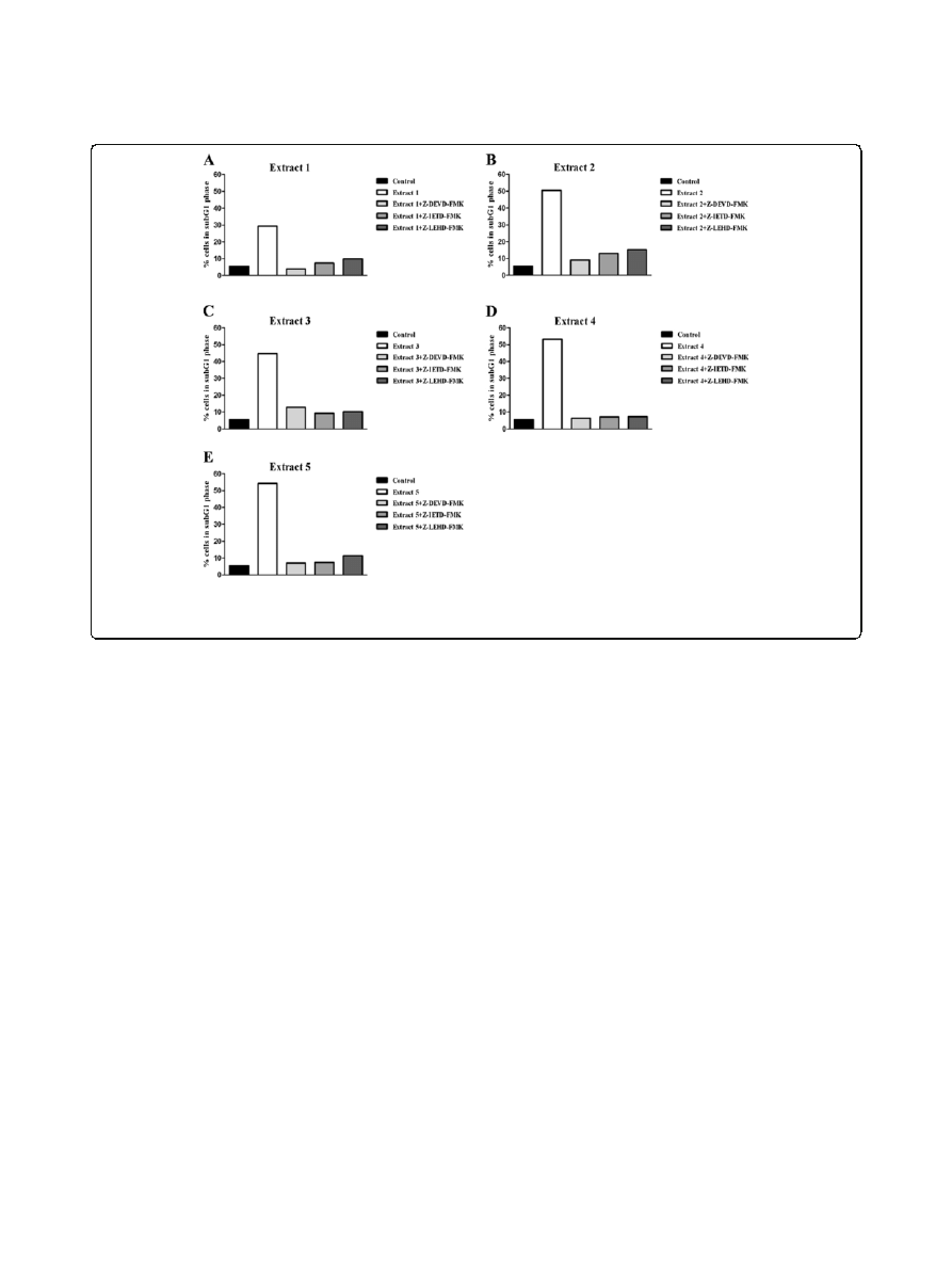
Determination of target caspases
To identify the caspases involved in the apoptotic cell
death pathway induced by the investigated extracts,
the percentages of HeLa cells pretreated with caspase
inhibitors in the subG1 phase were determined. HeLa
cells were preincubated for 2 h with specific caspase
inhibitors (at a final concentration of 40
μM). These were:
Z-DEVD-FMK, a caspase-3 inhibitor, Z-IETD-FMK, a
caspase-8 inhibitor and Z-LEHD-FMK, a caspase-9 inhibi-
tor. Caspase inhibitors were purchased from R&D Systems
(Minneapolis, USA). Five tested extracts were applied to
the HeLa cells at concentrations that corresponded to the
IC
90
values obtained after 72 h. For each extract, one sam-
ple of HeLa cells was not treated with an inhibitor and
served as a reference sample. After 24 h of incubation,
cells were harvested and fixed in 70% ethanol on ice.
Samples were stored at
−20°C for one week before PI
staining. Changes in the percentages of cells in the subG1
phase were determined using a FACSCalibur Flow
Cytometer and analyzed using CELLQuest software.
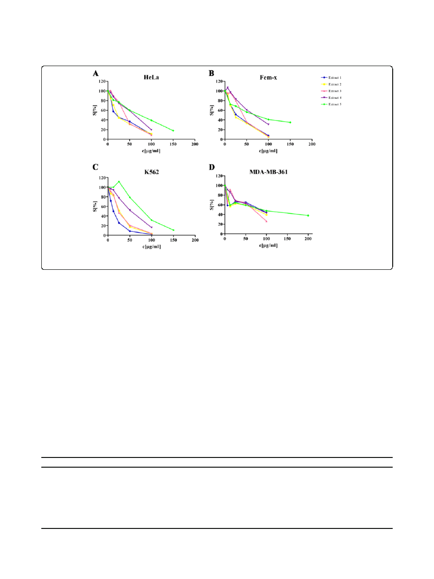
Figure 1 Survival of HeLa (A), Fem-x (B), K562 (C) and MDA-MB-361 cells (D) grown for 72 h in the presence of increasing
concentrations of
Helichrysum zivojinii extracts, determined by MTT test. Representative graphs are shown.
Table 2 Concentrations of five
Helichrysum zivojinii extracts, which induced 50% decrease in the target cancer cell
survival, determined by MTT test
HeLa
Fem-x
K562
MDA-MB-361
Extract 1 IC
50
[
μg/ml]
24.63 ± 4.12
28.85 ± 5.49
11.78 ± 0.94
81.74 ± 6.27
Extract 2 IC
50
[
μg/ml]
20.11 ± 4.49
23.64 ± 1.41
23.82 ± 6.54
81.74 ± 13.31
Extract 3 IC
50
[
μg/ml]
37.98 ± 2.33
47.04 ± 4.79
27.52 ± 4.96
79.93 ± 13.49
Extract 4 IC
50
[
μg/ml]
56.70 ± 6.05
74.84 ± 7.55
50.37 ± 3.28
69.96 ± 11.70
Extract 5 IC
50
[
μg/ml]
84.68 ± 10.39
77.29 ± 6.55
74.88 ± 7.57
94.92 ± 6.85
Cisplatin IC
50
[
μM]
5.60 ± 1.41
5.02 ± 0.59
5.35 ± 0.70
28.23 ± 5.04
Time of continuous agent
’s action was 72 h.
Mati
ć et al. BMC Complementary and Alternative Medicine 2013, 13:36
Page 4 of 12
http://www.biomedcentral.com/1472-6882/13/36
Results
Chemical analysis of plant extracts
The hexane (1) and dichloromethane extracts (2)
presented similar
1
H NMR spectra (recorded in CDCl
3
).
The signals in the
1
H NMR spectra of both extracts
pointed to a complex mixture with prevailing relatively
non-polar substances (region
δ 0.8–2.2). LC/DAD ana-
lysis revealed the presence of a flavonoid with the mo-
lecular formula C
18
H
16
O
7
(344) in the hexane extract,
while the flavanone naringenin and flavone apigenin
were confirmed in the dichloromethane extract, also by
LC/DAD analysis. Three compounds found in both
extracts are presented in Table 1, with their presumed mo-
lecular formulae and measured m/z values corresponding
to identified ions obtained by ESI ToF mass spectrometry.
According to LC/DAD analysis, these compounds unfortu-
nately did not absorb in the UV spectrum, suggesting the
presence of structures without a chromophore. The ethyl-
acetate extract (3) and n-butanol extracts (4) presented
1
H NMR spectra (recorded in DMSO-d
6
) with approxi-
mately identical groups of signals in the regions
δ 12.5–
13.9 (hydroxy protons),
δ 5.9-8.1 (protons on the aromatic
ring that is substituted with one or more hydroxy groups),
and
δ 4.7–5.5 (group of protons corresponding to a sugar
unit). The
1
H NMR spectrum of the most abundant metha-
nol extract (5), also recorded in DMSO-d
6
, resembles the
spectra of the ethyl-acetate and n-butanol extracts by the
presence of signals in the region
δ 5.9–8.1 (aromatic
protons), as well as by the presence of a signal at
δ 13.9
(hydroxy proton); it differs from the last two spectra by
possessing a more pronounced region at
δ 4.7–5.5, which is
responsible for the protons of sugar units. LC/DAD
analysis and ESI ToF mass spectrometry of these three
more polar extracts (3, 4 and 5) revealed quite similar
constituents in each of them. Apart from chlorogenic acid,
two groups of flavonoid compounds were detected: flavon-
oid O-glycosides, and flavonoid aglycons. Among the O-
glycosides, the glucoside (or possibly galactoside) of quer-
cetin, the glucoside of apigenin, the glucoside of kaempferol
(or possibly luteolin), and another glucoside of kaempferol
(possibly luteolin or 6-hydroxyapigenin), were present. Fla-
vonoid aglycones, such as apigenin and naringenin, were
detected. Phthalic acid was found in extracts 2
–5.
In vitro cytotoxic activity
The cytotoxicity of the five isolated extracts was tested
against selected cancer cell lines: human cervix ade-
nocarcinoma HeLa, human melanoma Fem-x, human
myelogenous leukemia K562 and human breast adeno-
carcinoma MDA-MB-361 cells. All investigated extracts
exerted selective dose-dependent cytotoxic actions on
malignant cells. The decrease in survival of target cancer
cells induced by the five Helichrysum zivojinii extracts is
shown in Figure 1 and Table 2.
In general, extracts 1 and 2 (as well as cisplatin, which
served as a positive control) exhibited the highest cyto-
toxic actions against target malignant cell lines; extracts
3 and 4 displayed less pronounced cytotoxicity; extract 5
had the lowest cytotoxic action.
With regard to the specific sensitivities of the different
cells to the cytotoxic activities of the extracts, it is im-
portant to note that K562 cells were the most sensitive
to the cytotoxic actions of extracts 1 and 3. HeLa and
Fem-x cells exhibited a lower sensitivity, while the sensi-
tivity of breast cancer MDA-MB-361 cells to the toxic
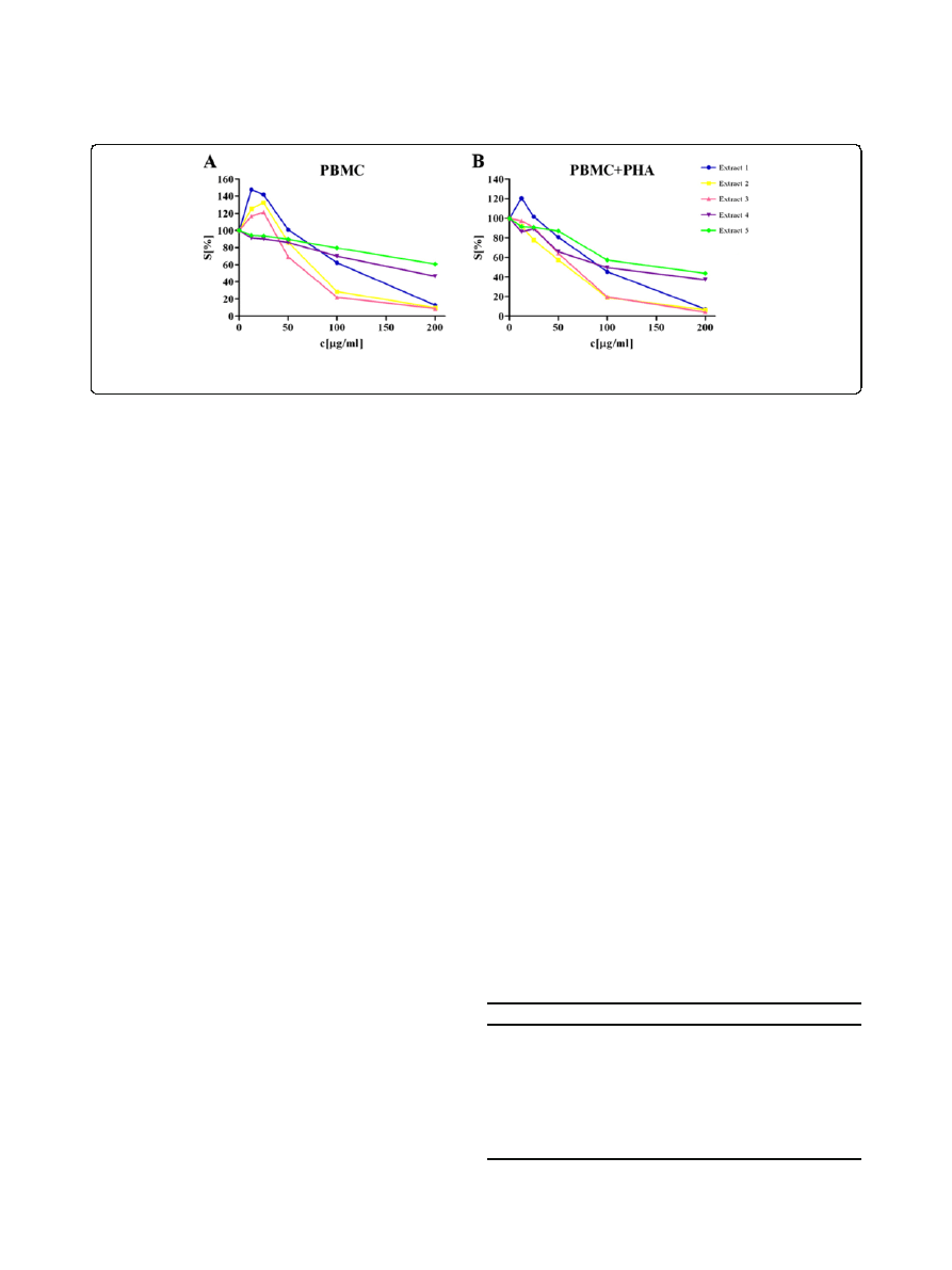
Figure 2 Survival of resting PBMC (A) and PHA-stimulated PBMC (B) grown for 72 h in the presence of increasing concentrations of
Helichrysum zivojinii extracts, determined by MTT test. Representative graphs are shown.
Table 3 Concentrations of five
Helichrysum zivojinii
extracts, which induced 50% decrease in target PBMC
survival, determined by MTT test
PBMC
PBMC + PHA
Extract 1 IC
50
[
μg/ml]
131.59 ± 9.93
99.07 ± 8.02
Extract 2 IC
50
[
μg/ml]
81.74 ± 0.50
67.65 ± 11.37
Extract 3 IC
50
[
μg/ml]
74.82 ± 6.51
61.86 ± 5.53
Extract 4 IC
50
[
μg/ml]
185.17
92.20 ± 9.01
Extract 5 IC
50
[
μg/ml]
> 200
128.12 ± 35.46
Cisplatin IC
50
[
μM]
> 33.34
> 33.34
Time of continuous agent
’s action was 72 h.
Mati
ć et al. BMC Complementary and Alternative Medicine 2013, 13:36
Page 5 of 12
http://www.biomedcentral.com/1472-6882/13/36
actions of the tested extracts was the lowest (being
several times lower than that of the other cell lines to
extracts 1, 2 and 3 especially).
Considering the possible effects of applied antitumor
drugs on normal healthy immunocompetent cells, com-
ponents of the antitumor immune response, their viabil-
ity is significant for tumor control. For that reason, the
activities of the investigated Helichrysum zivojinii ex-
tracts were evaluated against healthy unstimulated and
PHA-stimulated PBMC (Figure 2 and Table 3). It should
be noted that these extracts overall exhibited weaker cyto-
toxic effects against unstimulated PBMC than against
stimulated PBMC. Moreover, extracts 2 and 3 exerted a
more pronounced cytotoxicity against unstimulated and
PHA-stimulated PBMC than extracts 1, 4 and 5.
Table 4 Selectivity in the antitumor action of five
Helichrysum zivojinii extracts
Selectivity
coefficient in the
antitumor action
IC
50
PBMC/
IC
50
HeLa
IC
50
PBMC
+ PHA/
IC
50
HeLa
IC
50
PBMC/
IC
50
K562
IC
50
PBMC
+ PHA/
IC
50
K562
Extract 1
5.34
4.02
11.17
8.41
Extract 2
4.06
3.36
3.43
2.84
Extract 3
1.97
1.63
2.78
2.25
Extract 4
3.27
1.63
3.68
1.83
Extract 5
> 2.36
1.51
2.67
1.71
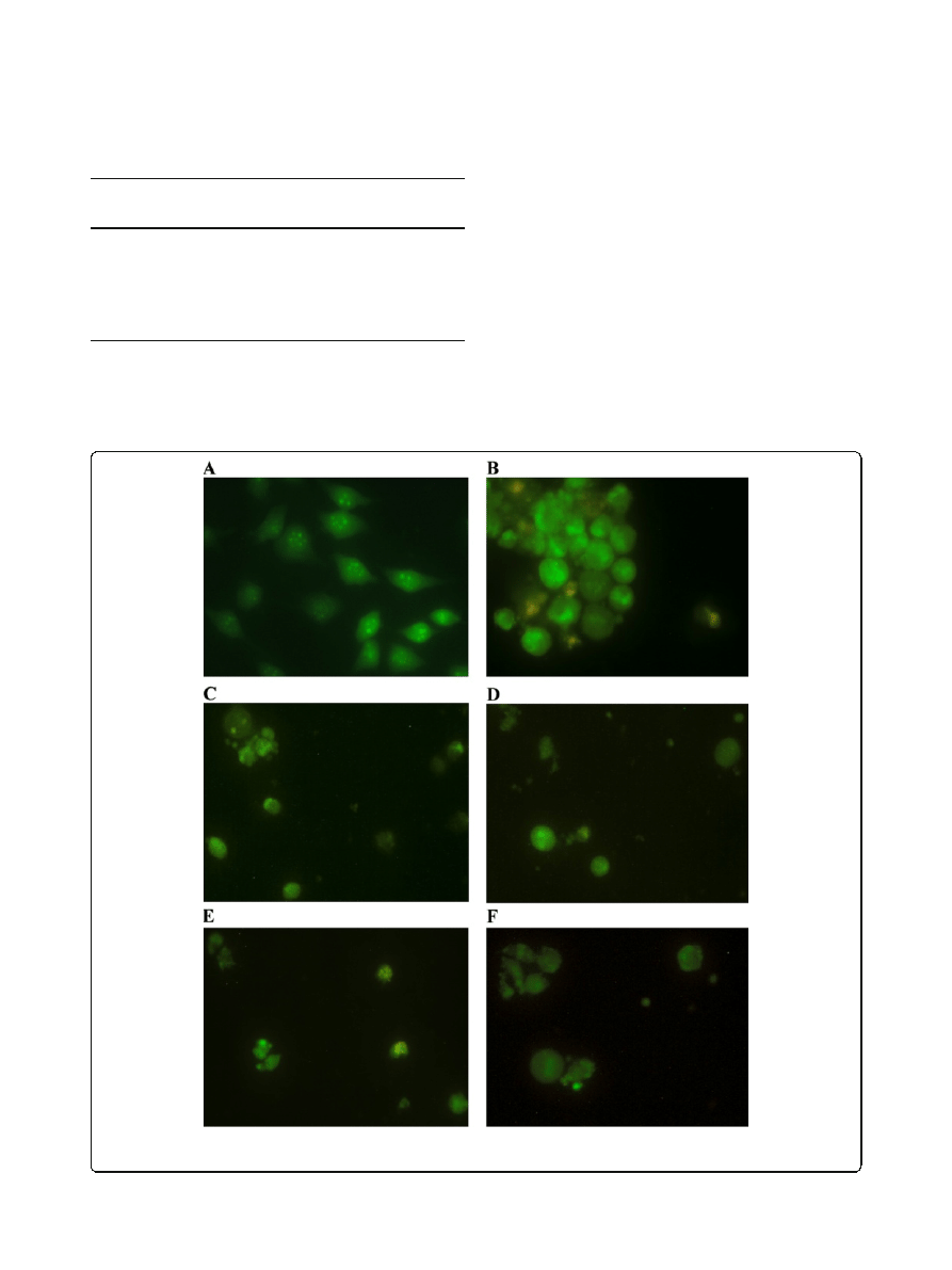
Figure 3 Photomicrographs of acridine orange/ethidium bromide-stained control HeLa cells (A), and HeLa cells treated with different
Helichrysum zivojinii extracts for 24 h (extracts 1–5, photomicrographs B-F, consecutively).
Mati
ć et al. BMC Complementary and Alternative Medicine 2013, 13:36
Page 6 of 12
http://www.biomedcentral.com/1472-6882/13/36
In order to further evaluate the anticancer potential of
the extracts, the selectivity in the antitumor action
against specific malignant cell line in comparison to
healthy PBMC was determined as well. These data are
presented in Table 4 from which it can be observed that
extract 1 exhibited highly selective antitumor action, es-
pecially against K562 cells. Extract 2 also displayed good
selectivity in its antitumor action.
Morphological analysis of HeLa cell death mode
In order to determine whether the investigated plant
extracts have pro-apoptotic activities, we performed mor-
phological analysis by fluorescent microscopy of acridine
orange/ethidium bromide-stained HeLa cells, exposed to
the extracts. Microscopic examination revealed that all
five extracts applied at IC
90
concentrations induced
apoptosis in target HeLa cells after 24 h treatment
(Figure 3). The morphological characteristics of apoptotic
cell death, such as cell shrinkage, condensation and even
fragmentation of nucleus, as well as the presence of
orange-red stained cells at late stages of apoptosis or sec-
ondary necrosis (the latter was observed in extract 1-
treated HeLa cells) and apoptotic bodies. These analyses
confirmed that the cytotoxicity of the Helichrysum
zivojinii
extracts is based on their prominent pro-
apoptotic effects.
Analysis of changes in cell cycle phase distribution
Examination of changes in the cell cycle phase distribu-
tion of HeLa cells treated with these extracts for 24, 48
and 72 h was done to elucidate the mechanisms of the
observed cytotoxic actions (Figure 4). Results from this
Figure 4 Changes in the cell cycle phase distribution of HeLa cells induced by the
Helichrysum zivojinii extracts after 24 (A,B), 48 (C,D)
and 72 h (E) treatment (applied concentrations of tested extracts corresponded to IC
50
and IC
90
values determined for 72 h). (C- control
HeLa cells, 1
– 5 – corresponding extracts are numbered consecutively). Representative graphs are shown.
Mati
ć et al. BMC Complementary and Alternative Medicine 2013, 13:36
Page 7 of 12
http://www.biomedcentral.com/1472-6882/13/36
analysis showed a time - dependent increase in the per-
centages of HeLa cells in the subG1 phase after exposure
to an IC
50
concentration of all of the tested extracts. Add-
itionally, exposure to extracts at IC
90
concentrations
induced significant increases in the percentages of cells
in the subG1 phase 24 h after exposure. It should be
mentioned that the investigated extracts induced a slight
accumulation of HeLa cells in the S phase after 72 h.
Examination of the cell cycle changes that were induced
after exposure for 72 h to IC
90
for each extract was not
performed because at this time point and at this extract
concentration low numbers of mostly dead or dying cells
were present in the sample.
Since the Helichrysum zivojinii extracts exhibited the
pro-apoptotic activities against cervix adenocarcinoma
HeLa cells, the identification of target caspases involved
in the apoptotic pathway was performed. The presence
of the specific caspase inhibitors (caspase-3 inhibitor,
caspase-8 inhibitor or caspase-9 inhibitor) significantly
reduced the percentages of apoptotic subG1 HeLa cells
treated with each of the five plant extracts, as shown in
Figure 5. The effect of the caspase-3 inhibitor on HeLa
cells treated with extracts is shown in Figure 6. It can be
seen that there is an increase in rounded, but attached
and live HeLa cells treated with the caspase-3 inhibitor
before the addition of the extracts in relation to target
HeLa cells only exposed to the tested extracts.
Discussion
The plant kingdom provides a rich source of compounds
with promising cancer chemopreventive and cancer thera-
peutic potential. The main drugs currently used in clinical
practice in the treatment of malignant diseases originate
from plants: vinca alkaloids, taxanes, camptothecins and
epipodophyllotoxins [23]. Over the past years, the focus of
modern anticancer drug discovery has been on a wide
variety of natural compounds, especially on phenolic
compounds. Phytochemicals have been reported to affect
different intracellular signaling pathways implicated in
the initiation, promotion and progression of cancer.
The antitumor effects of plant constituents have been
associated with the induction of carcinogen detoxifying
enzymes, the scavenging of free radicals, anti-inflammatory
activity, cell cycle arrest, the triggering of apoptosis, inhib-
ition of tumor angiogenesis and invasiveness [1-4].
The antioxidant and anti-inflammatory activities of
extracts and isolated compounds from plants belonging to
the large genus Helichrysum have been well documented
[9,11,15,18]. Arzanol, a phloroglucinyl
α-pyrone, is a con-
stituent of Helichrysum italicum that has been reported to
inhibit the NF-
κB transcription factor, cyclooxygenase and
lipooxygenase, as well as the release of proinflammatory
cytokines [11,18]. Regarding the link between inflam-
mation and cancer, chemicals with anti-inflammatory
properties targeting the molecules of signaling cascades
Figure 5 Effects of specific caspase inhibitors on the percentages of apoptotic subG1 HeLa cells treated with
Helichrysum zivojinii
extracts for 24 h (A - extract 1, B - extract 2, C - extract 3, D - extract 4, E - extract 5). (Z-DEVD-FMK - caspase-3 inhibitor; Z-IETD-FMK -
caspase-8 inhibitor; Z-LEHD-FMK - caspase-9 inhibitor).
Mati
ć et al. BMC Complementary and Alternative Medicine 2013, 13:36
Page 8 of 12
http://www.biomedcentral.com/1472-6882/13/36
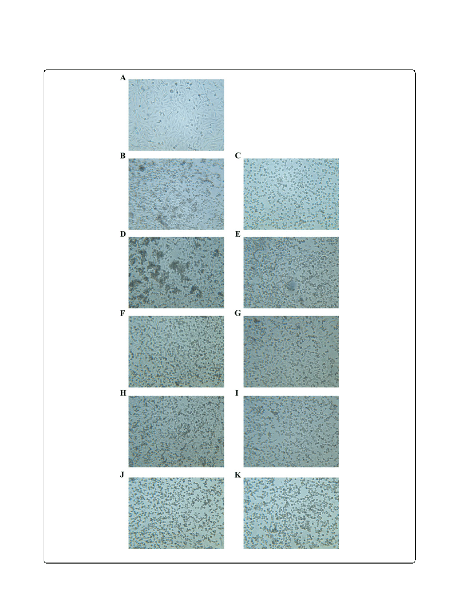
Figure 6 (See legend on next page.)
Mati
ć et al. BMC Complementary and Alternative Medicine 2013, 13:36
Page 9 of 12
http://www.biomedcentral.com/1472-6882/13/36

implicated in inflammation and carcinogenesis may be
useful as cancer chemopreventive drugs.
On the other hand, data about the potential anticancer
activity of extracts and phytochemicals of plants from the
genus Helichrysum are scarce. The antiproliferative effect
of the ethanol extract from Helichrysum maracandicum
towards SENCAR mouse skin transformed cells has been
demonstrated [20]. This extract suppressed the expres-
sion of p38 MAP kinase. An examination of the arzanol
properties showed that this compound, which was isolated
from Helichrysum italicum, did not exert cytotoxic action
against monkey VERO cells at concentrations up to
40
μM [24]. In contrast, another study showed that
arzanol at a concentration of 50
μM significantly
suppressed the survival of human lung carcinoma A549
cells [18]. It should be mentioned that methanolic extracts
prepared from different Helichrysum species were found
to inhibit DNA topoisomerase I [19]. Moreover, the cyto-
toxicity of Helichrysum gymnocephalum essential oil to-
wards human breast adenocarcinoma MCF-7 cells has
been documented [17].
The results presented herein demonstrate the selective
dose-dependent cytotoxic actions of the five extracts
isolated from the endemic plant species Helichrysum
zivojinii
against target cancer cell lines and against healthy
immunocompetent PBMC that have been stimulated to
proliferate, while their cytotoxic actions were not as
pronounced against unstimulated PBMC. The observed
selectivity in the antitumor effects of the extracts against
specific malignant cell types could be attributed to the
actions of different Helichrysum zivojinii constituents on
target molecules of the signal transduction pathways that
regulate cell proliferation and apoptosis. Furthermore,
each of the investigated extracts exhibited considerably
stronger cytotoxicity to HeLa, Fem-x and K562 cells when
compared to PBMC, both resting and PHA-stimulated,
which points to the cancer specificity of their actions. It is
noteworthy that when these extracts were applied at
concentrations that were highly cytotoxic to malignant
cells, they demonstrated very low toxicity towards healthy
immunocompetent PBMC, the key players in immune
defenses against tumors. The good selectivity of their
antitumor actions highlights the significant anticancer
potential of Helichrysum zivojinii extracts. The promi-
nent antitumor properties of these extracts need to be
examined further in in vivo studies.
It should be stressed that all of these extracts exhibited
weaker cytotoxic effects against unstimulated PBMC in
comparison to stimulated PBMC. This finding indicates
that the extracts possess the ability to inhibit the proli-
feration of PHA-stimulated PBMC. Thus, these agents
may even suppress certain immune functions, particu-
larly non-specific antigen stimulation. Additionally, the
observed lower activities against resting PBMC than
against mitogen-stimulated PBMC point to components
in pathways regulating cell proliferation as the possible
molecular targets of the Helichrysum zivojinii extracts.
However, it is very important to note that when extracts 1,
2 and 3 were applied at lower concentrations, they
stimulated the proliferation of resting PBMC. This growth
stimulation effect of lower concentrations of extracts is in
accordance with the well-known effect of very small doses
of X rays on enhanced proliferation of irradiated cells [25].
The observed effects of low concentrations of these
extracts on one of the main components of the immune
response point to the possibility of their use to enhance
immunity. It would be interesting to investigate their ac-
tion towards different PBMC subpopulations and eluci-
date the potential mechanisms through which they
stimulate proliferation. The possible immunostimulatory
effects of extracts at lower concentrations might be
explained by a modulation in lymphocyte cytokine pro-
duction, including IL-2, IFN-
γ, as well as IL-4 and IL-6.
The immunoregulatory actions of phenolic compounds,
such as quercetin, kaempferol and apigenin, have been
reported [26-28]. Considering the presented results, the
effects of the extracts on PBMC might be mediated
through NF-
κB.
Examination of in vitro cytotoxicity revealed that extracts
1 and 2 might be a significant source of novel promising
anticancer compounds in view of their pronounced cyto-
toxic activities against HeLa, Fem-x and especially against
K562 cells, as well as their high selectivity in the antitumor
actions against cancer cells in comparison to healthy
PBMC. Chemical analyses of the Helichrysum zivojinii
extracts showed the presence of phenolic compounds
whose antitumor potential has already been documented.
The bioactive flavone apigenin that was found in extracts
2
–5 has been reported to exhibit anticancer activities
against different types of malignant cells including breast,
cervical, ovarian, prostate, colon, gastric, liver and lung
cancers, as well as skin and thyroid cancer, diverse
hematological malignancies and neuroblastoma [28]
and references cited therein]. The cytotoxic activities of
extracts 2
–5 may be at least in part due to the flavonoid
naringenin. This flavonoid has been shown to exert
(See figure on previous page.)
Figure 6 Effects of pretreatment of HeLa cells with caspase-3 inhibitor (Z-DEVD-FMK), exposed to
Helichrysum zivojinii extracts
(applied concentrations of tested extracts corresponded to IC
90
values determined for 72 h). A
– control; B – Extract 1, C – Extract 1 + Z-
DEVD-FMK; D
– Extract 2, E – Extract 2 + Z-DEVD-FMK; F – Extract 3, G – Extract 3 + Z-DEVD-FMK; H – Extract 4, I – Extract 4 + Z-DEVD-FMK;
J
– Extract 5, K – Extract 5 + Z-DEVD-FMK.
Mati
ć et al. BMC Complementary and Alternative Medicine 2013, 13:36
Page 10 of 12
http://www.biomedcentral.com/1472-6882/13/36

cytotoxicity towards various malignant cell lines, such as
breast cancer cell lines (MCF-7, MDA-MB-231), cervix
adenocarcinoma (HeLa), liver cancer (HepG2, Hep3B,
Huh7), pancreas cancer (PK-1), colon cancer (Caco-2),
stomach cancer (KATOIII, MKN-7) and leukemia cells
(Jurkat, HL-60, U937, NALM-6, THP-1) [29-31]. Add-
itionally, the cancer-preventive and cancer-suppressive
properties of quercetin, whose O-glycosides were identi-
fied in extracts 3, 4 and 5, have been documented as well
[32]. The antiproliferative and pro-apoptotic effects of
quercetin were shown against the HeLa cell line [33].
Morphological analysis of the mode of HeLa cell death,
together with the cell cycle analysis, showed that the treat-
ment of HeLa cells with higher concentrations of the
examined extracts induced apoptotic cell death. To con-
firm the pro-apoptotic action of the tested Helichrysum
zivojinii
extracts and to identify the caspases implicated in
the employed apoptotic pathways, specific caspase inhibi-
tors were used (Z-DEVD-FMK, Z-IETD-FMK, Z-LEHD-
FMK). A prominent decrease in the percentages of subG1
apoptotic HeLa cells after treatments with each of the
tested extracts in combination with specific caspase inhi-
bitors compared to the percentages of subG1 cells after
treatments with only the corresponding extracts, indicates
that each of the five extracts induced apoptosis through
the activation of caspase-3, the main effector caspase, as
well as through the activation of caspase-8 and caspase-9.
We conclude that the constituents of the Helichrysum
zivojinii
extracts triggered apoptosis in HeLa cells through
the intrinsic pathway mediated by caspase-9, and the ex-
trinsic pathway mediated by caspase-8. In addition, the
crosstalk between these two apoptotic pathways should
also be considered. Due to their ability to promote apop-
totic cell death in cancer cells, the investigated extracts and
their constituents may have significant anticancer potential.
It is worth noting that antitumor drugs that induce
apoptosis and thereby suppress the further growth of
tumors play important roles in the clinical treatment of
malignancies. In addition to the pronounced inhibition
of proliferation and survival of target malignant cells,
the lower cytotoxicity of Helichrysum zivojinii extracts
against healthy PBMC is a promising lead for future
studies.
Conclusions
Data from this in vitro study clearly demonstrate the
prominent antitumor potential of five extracts prepared
from the endemic plant species Helichrysum zivojinii,
which can be attributed to their selective and pronounced
antiproliferative and pro-apoptotic actions towards spe-
cific malignant cells in comparison to healthy PBMC. Pro-
spective cancer-suppressive effects of the tested extracts
should be further evaluated in in vivo experiments.
Competing interests
The authors declare that they have no competing interests.
Authors
’ contributions
IM performed all analyses of the anticancer properties of investigated
extracts, interpreted obtained data and wrote the first and last version of the
manuscript. IA participated in design of the study, prepared extracts,
performed chemical characterization, interpreted data and wrote the part of
the manuscript.
ŽŽ participated in acquisition and analysis of data. MJ carried
out chemical analyses of the extracts. VV and SM participated in design of
the study and interpreted obtained data. ZJ designed the research on
anticancer properties of tested extracts, interpreted obtained data,
participated in writing the manuscript and critically revised the manuscript.
All authors have read and approved the final version of the manuscript.
Acknowledgments
The authors are grateful to the Ministry of Education, Science and
Technological Development of the Republic of Serbia for the financial
support (Projects 175011 and 172053). Also the authors would like to thank
Tatjana Petrovi
ć for her excellent technical assistance. For the supply of plant
material, we thank National park Gali
čica, Ohrid, Macedonia. We thank
Andon Bojad
ži and Oliver Avramoski, National park Galičica.
Author details
1
Institute of Oncology and Radiology of Serbia, Pasterova 14, 11000,
Belgrade, Serbia.
2
Institute for Chemistry, Technology and Metallurgy,
University of Belgrade, Njego
ševa 12, 11000, Belgrade, Serbia.
3
Faculty of
Chemistry, University of Belgrade, Studentski trg 16, 11000, Belgrade, Serbia.
Received: 13 September 2012 Accepted: 12 February 2013
Published: 18 February 2013
References
1.
Amin ARMR, Kucuk O, Khuri FR, Shin DM: Perspectives for cancer
prevention with natural compounds. J Clin Oncol 2009, 27:2712
–2725.
2.
Mehta RG, Murillo G, Naithani R, Peng X: Cancer chemoprevention by
natural products: how far have we come? Pharm Res 2010, 27:950
–961.
3.
Neergheen VS, Bahorun T, Taylor EW, Jen LS, Aruoma OI: Targeting specific
cell signaling transduction pathways by dietary and medicinal
phytochemicals in cancer chemoprevention. Toxicology 2010, 278:229
–241.
4.
Surh YJ: Cancer chemoprevention with dietary phytochemicals. Nat Rev
Cancer 2003, 3:768
–780.
5.
Lourens ACU, Viljoen AM, van Heerden FR: South African Helichrysum
species: a review of the traditional uses, biological activity and
phytochemistry. J Ethnopharmacol 2008, 119:630
–652.
6.
Passalacqua NG, Guarrera PM, De Fine G: Contribution to the knowledge
of the folk plant medicine in Calabria region (Southern Italy). Fitoterapia
2007, 78:52
–68.
7.
Red
žić SS: The ecological aspect of ethnobotany and
ethnopharmacology of population in Bosnia and Herzegovina. Coll
Antropol 2007, 31:869
–890.
8.
Sezik E, Yesilada E, Honda G, Takaishi Y, Takeda Y, Tanaka T: Traditional
medicine in Turkey X. Folk medicine in Central Anatolia. J Ethnopharmacol
2001, 75:95
–115.
9.
Aiyegoro OA, Okoh AI: Preliminary phytochemical screening and in vitro
antioxidant activities of the aqueous extract of Helichrysum longifolium
DC. BMC Complement Altern Med 2010, 10:21.
10.
Angioni A, Barra A, Arlorio M, Coisson JD, Russo MT, Pirisi FM, Satta M,
Cabras P: Chemical composition, plant genetic differences, and
antifungal activity of the essential oil of Helichrysum italicum G. Don ssp.
microphyllum (Willd) Nym. J Agric Food Chem 2003, 51:1030
–1034.
11.
Appendino G, Ottino M, Marquez N, Bianchi F, Giana A, Ballero M, Sterner O,
Fiebich BL, Munoz E: Arzanol, an anti-inflammatory and anti-HIV-1
phloroglucinol
α-pyrone from Helichrysum italicum ssp. microphyllum.
J Nat Prod 2007, 70:608
–612.
12.
Aslan M, Deliorman Orhan D, Orhan N, Sezik E, Yesilada E: In vivo
antidiabetic and antioxidant potential of Helichrysum plicatum ssp.
plicatum capitulums in streptozotocin-induced-diabetic rats.
J Ethnopharmacol 2007, 109:54
–59.
Mati
ć et al. BMC Complementary and Alternative Medicine 2013, 13:36
Page 11 of 12
http://www.biomedcentral.com/1472-6882/13/36

13.
Meyer JJ, Afolayan AJ, Taylor MB, Engelbrecht L: Inhibition of herpes
simplex virus type 1 by aqueous extracts from shoots of Helichrysum
aureonitens (Asteraceae). J Ethnopharmacol 1996, 52:41
–43.
14.
Nostro A, Bisignano G, Cannatelli MA, Crisafi G, Germanò MP, Alonzo V:
Effects of Helichrysum italicum extract on growth and enzymatic activity
of Staphylococcus aureus. Int J Antimicrob Agents 2001, 17:517
–520.
15.
Sala A, Recio MC, Giner RM, Máñez S, Ríos JL: New acetophenone
glucosides isolated from extracts of Helichrysum italicum with
antiinflammatory activity. J Nat Prod 2001, 64:1360
–1362.
16.
Süzgeç S, Meriçli AH, Houghton PJ, Çubukçu B: Flavonoids of Helichrysum
compactum and their antioxidant and antibacterial activity. Fitoterapia
2005, 76:269
–272.
17.
Afoulous S, Ferhout H, Raoelison EG, Valentin A, Moukarzel B, Couderc F,
Bouajila J: Helichrysum gymnocephalum essential oil: chemical composition
and cytotoxic, antimalarial and antioxidant activities, attribution of the
activity origin by correlations. Molecules 2011, 16:8273
–8291.
18.
Bauer J, Koeberle A, Dehm F, Pollastro F, Appendino G, Northoff H, Rossi A,
Sautebin L, Werz O: Arzanol, a prenylated heterodimeric phloroglucinyl
pyrone, inhibits eicosanoid biosynthesis and exhibits anti-inflammatory
efficacy in vivo. Biochem Pharmacol 2011, 81:259
–268.
19.
Kucukoglu O, Ozturk B, Kamataki T, Topcu Z: Inhibitory activities of Helichrysum
taxa on mammalian type I DNA topoisomerase. Pharm Biol 2006, 44:189
–193.
20.
Yagura T, Motomiya T, Ito M, Honda G, Iida A, Kiuchi F, Tokuda H, Nishino H:
Anticarcinogenic compounds in the Uzbek medicinal plant, Helichrysum
maracandicum. J Nat Med 2008, 62:174
–178.
21.
Mosmann T: Rapid colorimetric assay for cellular growth and survival:
application to proliferation and cytotoxicity assays. J Immunol Methods
1983, 65:55
–63.
22.
Ohno M, Abe T: Rapid colorimetric assay for the quantification of
leukemia inhibitory factor (LIF) and interleukin-6 (IL-6). J Immunol
Methods 1991, 145:199
–203.
23.
Nobilli S, Lippi D, Witort E, Donnini M, Bausi L, Mini E, Capaccioli S: Natural
compounds for cancer treatment and prevention. Pharmacol Res 2009,
59:365
–378.
24.
Rosa A, Deiana M, Atzeri A, Corona G, Incani A, Melis MP, Appendino G,
Dessí MA: Evaluation of the antioxidant and cytotoxic activity of arzanol,
a prenylated
α-pyrone-phloroglucinol etherodimer from Helichrysum
italicum subsp. microphyllum. Chem Biol Interact 2007, 165:117
–126.
25.
Wu B, Wei Y, Liu FQ, Zhang Q, Wang CB, Bai H: Biological effects of low
dose X-irradiation on human bone marrow mesenchymal stem cells.
J Exp Hematol/Chinese Assoc Pathophysiol 2011, 19:1214
–1217.
26.
Nair MP, Kandaswami C, Mahajan S, Chadha KC, Chawda R, Nair H, Kumar N,
Nair RE, Schwartz SA: The flavonoid, quercetin, differentially regulates Th-1
(IFN
γ) and Th-2 (IL-4) cytokine gene expression by normal peripheral blood
mononuclear cells. Biochim Biophys Acta 2002, 1593:29
–36.
27.
Okamoto I, Iwaki K, Koya-Miyata S, Tanimoto T, Kohno K, Ikeda M, Kurimoto
M: The flavonoid Kaempferol suppresses the graft-versus-host reaction
by inhibiting type 1 cytokine production and CD8+ T cell engraftment.
Clin Immunol 2002, 103:132
–144.
28.
Shukla S, Gupta S: Apigenin: a promising molecule for cancer prevention.
Pharm Res 2010, 27:962
–978.
29.
Kanno S, Tomizawa A, Hiura T, Osanai Y, Shouji A, Ujibe M, Ohtake T, Kimura K,
Ishikawa M: Inhibitory effects of naringenin on tumor growth in human cancer
cell lines and sarcoma S-180-implanted mice. Biol Pharm Bull 2005, 28:527
–530.
30.
Kanno S, Tomizawa A, Ohtake T, Koiwai K, Ujibe M, Ishikawa M: Naringenin-
induced apoptosis via activation of NF-
κB and necrosis involving the loss of
ATP in human promyeloleukemia HL-60 cells. Toxicol Lett 2006, 166:131
–139.
31.
Park JH, Jin CY, Lee BK, Kim GY, Choi YH, Yeong YK: Naringenin induces
apoptosis through downregulation of Akt and caspase-3 activation in
human leukemia THP-1 cells. Food Chem Toxicol
2008, 46:3684
–3690.
32.
Murakami A, Ashida H, Terao J: Multitargeted cancer prevention by
quercetin. Cancer Lett 2008, 269:315
–325.
33.
Vidya Priyadarsini R, Senthil Murugan R, Maitreyi S, Ramalingam K, Karunagaran
D, Nagini S: The flavonoid quercetin induces cell cycle arrest and
mitochondria-mediated apoptosis in human cervical cancer (HeLa) cells
through p53 induction and NF-
κB inhibition. Eur J Pharmacol 2010, 649:84–91.
doi:10.1186/1472-6882-13-36
Cite this article as: Mati
ć et al.: In vitro antitumor actions of extracts
from endemic plant Helichrysum zivojinii. BMC Complementary and
Alternative Medicine 2013 13:36.
Submit your next manuscript to BioMed Central
and take full advantage of:
•
Convenient online submission
•
Thorough peer review
•
No space constraints or color figure charges
•
Immediate publication on acceptance
•
Inclusion in PubMed, CAS, Scopus and Google Scholar
•
Research which is freely available for redistribution
Submit your manuscript at
www.biomedcentral.com/submit
Mati
ć et al. BMC Complementary and Alternative Medicine 2013, 13:36
Page 12 of 12
http://www.biomedcentral.com/1472-6882/13/36
Document Outline
- Abstract
- Background
- Methods
- Plant extracts
- Instrumentation and chromatographic conditions
- Cell culture
- Preparation of peripheral blood mononuclear cells
- Treatment of cancer cell lines
- Treatment of PBMC
- Determination of target cell survival
- Morphological evaluation of HeLa cell death
- Cell cycle analysis
- Determination of target caspases
- Results
- Discussion
- Conclusions
- Competing interests
- Authors’ contributions
- Acknowledgments
- Author details
- References
Wyszukiwarka
Podobne podstrony:
In Vitro Anticancer Activity of Ethanolic Extract
In vitro cytotoxicity screening of wild plant extracts
In vitro corrosion resistance of titanium made using differe
In vitro biological effects of titanium rough surface obtain
2001 In vitro fermentation characteristics of native and processed cereal grains and potato
Gender and Racial Ethnic Differences in the Affirmative Action Attitudes of U S College(1)
Evaluation of in vitro anticancer activities
Cytotoxicity and Modes of Action of the Methanol Extracts
Composition and Distribution of Extracellular Polymeric Substances in Aerobic Flocs and Granular Slu
05 Defined Networks of Neuronal Cells in Vitro
In vitro studies of plasma
Modanese Paradox of Virtual Dipoles in the Einstein Action (2000)
więcej podobnych podstron