
Go to:
Go to:
Curr Mol Med. Jan 2014; 14(1): 69
–95.
Published online Jan 2014. doi:
10.2174/15665240113136660079
PMCID: PMC3905716
Molecular Diagnosis in Autoimmune Skin Blistering Conditions
J.V. Otten
,
T. Hashimoto
,
M. Hertl
,
A.S. Payne
, and
C. Sitaru
Department of Dermatology, University of Freiburg, Hauptstrasse 7, D-79104 Freiburg, Germany
Department of Dermatology, Kurume University, 67 Asahimachi, Kurume, Fukuoka 830-0011, Japan
Department of Dermatology and Allergology, University of Marburg, Baldingerstraße, D-33043 Marburg, Germany
Department of Dermatology, University of Pennsylvania, Philadelphia, Pennsylvania, PA 19104, USA
BIOSS Centre for Biological Signalling Studies, D-79108 Freiburg, Germany
Address correspondence to this author at the Department of Dermatology, University of Freiburg, Hauptstrasse 7, D-79104 Freiburg, Germany; Tel:
+49-(0)761-27067690; Fax: +49-(0)761-27068290; E-mail:
cassian@mail.sitaru.eu
, Email:
cassian.sitaru@uniklinik-freiburg.de
Received January 7, 2013; Revised March 12, 2013; Accepted June 4, 2013.
Copyright
© 2013 Bentham Science Publishers
This is an open access article distributed under the terms of the Creative Commons Attribution License (
http://creativecommons.org/licenses/by/2.5/
), which
permits unrestrictive use, distribution, and reproduction in any medium, provided the original work is properly cited.
Abstract
Blister formation in skin and mucous membranes results from a loss of cell-cell or cell-matrix adhesion and is a common
outcome of pathological events in a variety of conditions, including autoimmune and genetic diseases, viral and bacterial
infections, or injury by physical and chemical factors. Autoantibodies against structural components maintaining cell-cell
and cell-matrix adhesion induce tissue damage in autoimmune blistering diseases. Detection of these autoantibodies either
tissue-bound or circulating in serum is essential to diagnose the autoimmune nature of disease. Various
immunofluorescence methods as well as molecular immunoassays, including enzyme-linked immunosorbent assay and
immunoblotting, belong to the modern diagnostic algorithms for these disorders. There is still a considerable need to
increase awareness of the rare autoimmune blistering diseases, which often show a severe, chronic-relapsing course,
among physicians and the public. This review article describes the immunopathological features of autoimmune bullous
diseases and the molecular immunoassays currently available for their diagnosis and monitoring.
Keywords: Autoantibodies, autoantigens, basement membrane, desmosome, ELISA, extracellular matrix,
hemidesmosome, immunoassay, immunoblotting, immunofluorescence microscopy.
INTRODUCTION
Autoimmune blistering diseases are classified into four major groups, including pemphigus, the pemphigoids,
epidermolysis bullosa acquisita, and dermatitis herpetiformis (Table
1
) [
1
,
2
]. Autoimmune bullous diseases are organ-
specific autoimmune diseases associated with pathogenic autoantibodies against structural proteins that maintain cell-cell
and cell-matrix adhesions in the skin and mucous membranes [
1
,
3
]. Cell-cell adhesion in the epidermis is mainly
maintained by desmosomes and adherens junctions. The extracellular portions of desmosomal cadherins link neighboring
keratinocytes, whereas their intracellular regions bind to desmosomal plaque proteins, which mediate the interaction of
desmosomes with the keratin intermediate filament cytoskeleton. Major autoantigens in the pemphigus group of diseases
include desmogleins, desmocollins and the plaque proteins desmoplakin, periplakin and envoplakin (Fig.
1
) [
1
,
4
].
Structural components of the basement membrane that maintain cell-matrix adhesion and may function as autoantigens in
subepidermal blistering diseases include the intracellular plectin and BP230, which interacts with the transmembrane
hemidesmosomal
Fig. (1)
Schematic representation of major skin autoantigens. The autoantigens shown here
are molecules involved in maintaining the cell-cell and cell-matrix adhesion. The
transmembrane desmosomal cadherins of neighboring cells, desmogleins and
desmocollins, confer ...
Table 1.
Major Autoantigens in Bullous Diseases
C
URRENT
M
OLECULAR
M
EDICINE
Bentham Science Publishers
1
2
3
4
*,1,5
1
2
3
4
5
*
Molecular Diagnosis in Autoimmune Skin Blistering Conditions
http://www.ncbi.nlm.nih.gov/pmc/articles/PMC3905716/
1 von 26
25.08.2014 13:05

Go to:
components collagen XVII/BP180 and α β integrin. Laminin 332 in the lower lamina lucida and lamina densa is a known
ligand for α β integrin. Beside other ubiquitous proteins like perlecan and nidogen, laminin 332 (previously known as
epiligrin or laminin 5) and collagen IV form a network in the lamina densa [
5
]. Laminin γ1 chain, present in laminins 511
and 311 was identified as target of autoantibodies in anti-p200 pemphigoid. In the epidermal basement membrane, laminin
γ1 interacts with integrins α β and α β . Both laminin 332 and its ligand collagen VII of the anchoring fibrils, may be
targeted by autoantibodies in subepidermal blistering diseases [
1
,
3
,
5
].
Due to major advances over the last few decades in identifying the autoantigens in autoimmune blistering disease, rapid
and specific laboratory diagnostic tests have become reality and are currently widely available.
Astute clinical observation and skillful histopathological examination are essential for suspecting an autoimmune
blistering disease [
1
]. The clinical examination should include careful evaluation of skin and mucosal surfaces. The
Nikolsky sign should be tested by applying pressure to the perilesional or normal skin to determine if blisters can be
extended or induced in normal-appearing skin, characteristic of the pemphigus group of diseases [
6
,
7
]. The Tzanck smear
is a simple and inexpensive ancillary diagnostic tool that can provide rapid cytologic information. Optimally, the test is
performed on a fresh blister (< 24 hrs-old). Material is gently scraped from the base of a vesicle, blister, or pustule, onto a
slide and is allowed to air dry and then stain with different dyes, including Giemsa, toluidine blue, and methylene blue [
8
,
9
]. The routine histological examination is performed on a biopsy of a fresh vesicle or blister (< 1 day-old), and helps to
reveal the level of blister formation as well as the presence and features of the inflammatory infiltrate [
10
].
However, diagnosis of an autoimmune blistering disease requires detection of tissue-bound and/or circulating
autoantibodies to confirm the autoimmune nature of disease. Deposits of immunoreactants (typically immunoglobulins
and complement components) in the perilesional skin are detected by direct immunofluorescence (IF) microscopy, which
remains the gold standard for the diagnosis of autoimmune bullous diseases [
10
,
11
]. An accurate diagnosis further relies
on the characterization of autoantibody specificity using different immunoassays, including indirect IF, immunoblotting,
enzyme-linked immunosorbent assay (ELISA), and immunoprecipitation [
1
,
10
].
By indirect IF microscopy circulating antibodies are detected in the patients' sera by incubating with epithelial substrates
such as human skin, esophagus and bladder. The indirect IF on salt-split skin, obtained by incubation of human skin in a
1M NaCl solution, showing a cleavage within the lamina lucida delivers “semi-molecular” information on the identity of
the autoantigens at the dermal-epidermal junction based on their localization on the epidermal or dermal side of the
artificial cleavage [
10
,
12
].
The enzyme-linked immunosorbent assay (ELISA) is a sensitive and easy-to-perform test allowing for the characterization
of the autoantibody specificity. Several immunoassays using purified native and recombinant proteins have been
developed for detection of autoantibodies specific for the main autoantigens in the autoimmune blistering diseases (Table
2
). Autoantibodies against specific antigens may be detected also by immunoblotting, which may be performed using both
recombinant antigens and extracts of skin or cultured skin cells and is most relevant especially when no ELISA systems
are available [
1
].
Table 2.
Quantitative Immunoassays for the Detection of Autoantibodies in Autoimmune
Blistering Skin Diseases
Immunoprecipitation is the technique of precipitating an autoantigen out of solution using patient serum. Serum is mixed
with an extract of radioactively-labeled keratinocytes or their medium and the formed immune complexes are recovered
by adding protein A/G-beads [
1
]. This method was used to identify several autoantigens and was historically the gold
standard diagnostic test in paraneoplastic pemphigus and anti-epiligrin (laminin 332) mucous membrane pemphigoid [
1
,
13
,
14
].
Using these methods autoimmune blistering may be easily differentiated from blistering due to other causes, including
infections, genodermatoses, metabolic and other inflammatory diseases. Incomplete phenotypes, disease associations and
particular pathological constellations may occasionally however make diagnosis a challenging task. By considering for
instance that vesiculo-bullous eruption due to acute herpes or zoster infection may occasionally occur especially in elderly
and severely immunosuppressed patients with autoimmune blistering disease, the informed practitioner may avoid the
“disease flare” trap [
15
,
16
]. In the following sections, we review the clinical and histologic presentation of the
autoimmune blistering diseases, followed by the molecular studies available for their diagnosis.
PEMPHIGUS DISEASES
The term pemphigus is the latinized form of the Greek πέμ ιξ (pemphix) meaning bubble or blister and was first used by
6 4
6 4
3 1
6 4
Molecular Diagnosis in Autoimmune Skin Blistering Conditions
http://www.ncbi.nlm.nih.gov/pmc/articles/PMC3905716/
2 von 26
25.08.2014 13:05

Go to:
Wichmann at the end of the 18 century to describe bullous diseases [
17
,
18
]. Pemphigus comprises a group of
life-threatening autoimmune blistering conditions characterized by acantholytic intraepithelial blister and caused by
autoantibodies against intercellular adhesion molecules [
1
,
19
]. Different clinical forms of pemphigus are characterized by
their distinctive autoantigens, histo- and immunopathological findings. A characteristic histolopathological feature of
pemphigus is acantholysis, which results from the loss of cell-cell adhesion and is defined as detachment of individual or
grouped keratinocytes [
1
,
19
].
PEMPHIGUS VULGARIS
Pemphigus vulgaris (PV) is characterized by suprabasal acantholytic blister formation and autoantibodies against the
keratinocyte surface proteins [
1
]. Several autoantigens have been described in PV, including desmoglein 3, desmoglein 1,
and less frequently, desmocollin 3, acetycholine α9 receptor and pemphaxin [
20
-
26
]. The profile of autoantibodies against
desmogleins 1 and 3 correlates well with the clinical form of PV [
27
]. Autoantibodies against desmoglein 3 are present in
patients with mucosal-dominant PV, while reactivity against desmogleins 1 and 3 is characteristic of muco-cutaneous PV
[
21
,
27
]. The levels of IgG, but also IgE autoantibodies against desmoglein 3 correlate with disease activity in PV patients
[
26
,
28
]. While their detection may be helpful for diagnosing pemphigus, the pathogenic relevance of IgA and IgE
autoantibodies against desmogleins has not been experimentally demonstrated. IgG4 autoantibodies in pemphigus vulgaris
are indicative of active disease, whereas IgG1 autoantibodies are mainly found in remission [
29
].
The production of pathogenic autoantibodies in pemphigus is T cell-dependent. Autoreactive Th2 cell responses directed
against the extracellular domain of desmoglein 1 and 3 have been conclusively documented in pemphigus patients [
30
,
31
]. PV can be also considered as an autoimmune disorder associated with a Treg dysfunction since PV patients have less
desmoglein 3-reactive type 1 regulatory T cells than healthy controls [
32
].
Pemphigus vulgaris may be precipitated by drugs and UV exposure (Table
3
) [
33
-
36
]. Thiol drugs, such as penicillamine
and captopril, are the most common inciting agents, but further drugs, including penicillins, cephalosporins, enalapril,
rifampin, and nonsteroidal antiinflammatory agents were reported to be associated with pemphigus [
33
-
35
].
Table 3.
Drugs Reported as Putative Triggers of Autoimmune Blistering Diseases
Clinically, PV usually involves initially the oral cavity and is characterized by flaccid blisters and erosions causing pain
that may result in weight loss and malnutrition. Nasal, vaginal and anal mucosa may be also affected in PV. Therefore, the
diagnosis of PV should be considered in all patients with mucosal erosive lesions. In patients with mucocutaneos disease
blisters also affect the skin. Skin blisters are typically flaccid and easily eroded and can arise on healthy-appearing or on
erythematous skin (Fig.
2a
). Blisters may be painful or, less frequently, itchy. Erosions in the skin folds may develop into
vegetative lesions, typical for the clinical form of PV known as pemphigus vegetans. In a very small number of patients
with PV, classified as cutaneous-type PV, no mucous membrane involvement is observed, despite autoantibodies against
both desmogleins 1 and 3 [
21
,
27
,
37
]. Pemphigus herpetiformis is another rare clinical variant of PV, which manifests as
cutaneous vesicles in a herpetiform pattern with rare mucosal involvement and autoantibodies against desmoglein 3 [
1
,
38
].
Fig. (2)
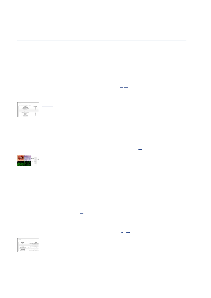
Major clinical and laboratory findings in pemphigus vulgaris. (a) Extensive
erosions with crusts and hyperpigmentation on the back of a 34-year old male
patient with muco-cutaneous pemphigus vulgaris. (b) Histopathological
examination reveals suprabasal ...
Diagnosis of PV should be considered in patients with persistent blisters and erosions of mucous membranes and skin.
This suspicion is further strengthened by a positive Nikolsky sign. The diagnosis is secured by histology demonstrating
suprabasal acantholysis and detection of tissue-bound IgG with an intercellular (“cobblestone”, “fishnet”) pattern by direct
IF microscopy and of circulating IgG autoantitbodies binding to the intercellular junctions of epithelial cells by indirect IF
microscopy on monkey esophagus and recognizing desmoglein 3 by ELISA (Table
4
).
Table 4.
Diagnostic Criteria for Pemphigus Vulgaris
th
Molecular Diagnosis in Autoimmune Skin Blistering Conditions
http://www.ncbi.nlm.nih.gov/pmc/articles/PMC3905716/
3 von 26
25.08.2014 13:05

Go to:
Histopathological examination typically reveals intraepithelial cleavage with acantholysis, occasionally associated with a
sparse inflammatory infiltrate (Fig.
2b
). The split formation occurs mainly in the suprabasal layer and basal keratinocytes
remain attached to the basement membrane suggesting a “row of tombstones”. By direct immunofluorescence microscopy
of patient perilesional skin intercellular deposits of IgG and occasionally C3 are found (Fig.
2c
) [
1
].
Serum IgG autoantibodies binding with an intercellular pattern to epithelium are revealed by indirect IF microscopy,
which can yield a semi-quantitative autoantibody titer (Fig.
2d
). While different substrates were used for the detection of
pemphigus autoantibodies, monkey esophagus has gained a wide acceptance as a sensitive substrate. The molecular
specificity of pemphigus autoantibodies may be analyzed using sensitive and specific immunoassays, which are
commercially available (Table
2
) [
26
]. ELISA systems using recombinant desmogleins for detecting circulating
autoantibodies are essential for the initial diagnosis and allow monitoring the pathogenic autoantibody levels during
clinical follow-up.
PEMPHIGUS FOLIACEUS
Pemphigus foliaceus (PF) is a superficial variant of pemphigus showing cutaneous lesions and virtually no involvement of
mucous membranes associated with subcorneal cleavage and autoantibodies against desmo-glein 1 [
1
]. Additional clinical
forms of superficial pem-phigus, including pemphigus erythematosus, endemic pemphigus foliaceus (fogo selvagem), and
drug-induced pemphigus foliaceus, share clinical, histo- and immuno-pathological features and may be classified as
subtypes of PF. The main autoantigen of PF is desmoglein 1 [
39
]. Desmoglein 1-specific pathogenic autoantibodies in
patients with PF mainly belong to the IgG4 isotype [
40
].
While the cause of most sporadic PF is still elusive, the induction of fogo selvagem appears to be related to environmental
factors (e.g., molecular mimicry due to infections transmitted by insects) [
41
-
43
]. In certain patients, PF may have been
precipitated by extensive UV exposure, burns or by various drugs, including penicillamine, inhibitors of angiotensin
convertase, cephalosporins, and non-steroidal anti-inflammatory agents (Table
3
).
The blistering lesions, which show preference for seborrhoeic areas, usually start on the trunk, face, and scalp. The onset
of PF is usually characterized by scattered, small superficial blisters, which rapidly transform into scaly, crusted erosions
with a puff pastry-like or cornflake appearance (Fig.
3a
). The Nikolsky sign is positive. Untreated, the lesions confluate
and may progress to an exfoliative generalized erythroderma. PF, like PV, is a chronic disease, but associated with less
mortality. While spontaneous remissions are possible, without adequate treatment, the lesions may persist for several
years.
Fig. (3)
Major clinical and laboratory findings in pemphigus foliaceus. (a) Erythema,
blisters and erosions in a 61-year old male patient with pemphigus foliaceus. (b)
Histopathological examination reveals sub-corneal acantholysis and an
inflammatory infiltrate ...
The diagnosis of PF is suggested by superficial skin blisters and erosions without mucosal involvement, and subcorneal
cleavage by histopathology. The diagnosis is confirmed by demonstrating intercellular deposition of tissue-bound and
circulating autoantibodies by direct and indirect IF microscopy, respectively. The molecular specificity of circulating
autoantibodies is further assessed by ELISA using recombinant desmoglein 1 (Table
5
).
Table 5.
Diagnostic Criteria for Pemphigus Foliaceus
Histopathologically PF is characterized by subcorneal acantholysis, with or without an eosinophilic or neutrophilic
inflammatory infiltrate (Fig.
3b
). Intercellular deposits of IgG and C3 are revealed by direct IF microscopy (Fig.
3c
),
while serum IgG autoantibodies are shown to bind to substrates such as esophagus and human skin with an intercellular
pattern by indirect IF microscopy (Fig.
3d
).
IgG autoantibodies recognizing desmoglein 1, but not desmoglein 3, are measured in serum of PF patients by ELISA
(Table
2
; Fig.
4a
). In PF, the levels of desmoglein 1-specific autoantibodies correlate well with disease activity and are
thus helpful for monitoring serologic and clinical activity during the course of the disease of individual patients (Fig.
4b
)
[
26
].
Fig. (4)
Molecular Diagnosis in Autoimmune Skin Blistering Conditions
http://www.ncbi.nlm.nih.gov/pmc/articles/PMC3905716/
4 von 26
25.08.2014 13:05
Go to:
Detection of desmoglein-specific autoantibodies by ELISA. (a) Pemphigus
vulgaris (PV) may present with only mucosal (m) lesions and autoantibodies
against desmoglein (Dsg) 3 or with mucocutaneous (m/c) involvement and
autoantibodies to both Dsg 1 and ...
PARANEOPLASTIC PEMPHIGUS
Paraneoplastic pemphigus (PNP), first described by Anhalt et al. in 1990, is an autoimmune multi-organ syndrome
associated with neoplasia and autoantibodies against desmosomes [
13
]. In addition to the mucocutaneous disease, which
shares important features with pemphigus vulgaris, pulmonary involvement presenting as bronchiolitis obliterans is a rare,
but potentially fatal manifestation of PNP. Based on the multi-organ involvement, the alternative name of paraneoplastic
autoimmune multi-organ syndrome (PAMS) has been suggested for this paraneoplastic condition [
44
,
45
].
While PNP is usually associated with malignant tumours, especially lymphoproliferative diseases, it may also occur in
association with benign neoplasms (Table
6
). There is extensive evidence clearly showing that PNP is an obligate
paraneoplastic syndrome, strongly suggesting that neoplasms are directly linked to autoimmunity. While the mechanisms
of epithelial autoimmunity induction by tumours are not well understood [
46
,
47
], the development of autoantibodies to
multiple epithelial proteins could be explained by epitope spreading [
48
,
49
]. The influence of tumour progression and
treatment on the autoimmune disease course is variable [
46
,
47
,
50
].
Table 6.
Tumors Associated with Paraneoplastic Pemphigus
Typically, PNP patients suffer from severe mucosal involvement with often extensive, intractable stomatitis. The earliest
and most constant clinical findings in PNP are painful erosions of the oropharynx. Crusted erosions on the vermilion of
the lips are typical and similar to that seen in persons with Stevens-Johnson syndrome. Occasionally, genital, nasal and
ocular mucosal surfaces are also affected [
51
-
54
]. The cutaneous eruption of PNP includes the typical pemphigus
presentation with erythema, blistering, and erosions with positive Nikolski sign, as well as lichenoid lesions resembling
erythema multiforme, graft versus host disease, and lichen planus, may be present (Fig.
5a
).
Fig. (5)
Characteristic findings in paraneoplastic pemphigus. (a) Hemorrhagic erosions
with crusts on the lips and oral cavity of a patient with non-Hodgkin lymphoma.
(b) Interface dermatitis by histopathology (H&E staining). (c)
Immunofluorescence (IF) ...
In a minority of patients with PNP pulmonary involvement manifests itself as obstructive lung disease and progresses to
bronchiolitis obliterans, which responds poorly to immunosuppressive therapy and is a major cause of death, although
chest radiograph findings are often normal [
55
].
Painful, progressive stomatitis with skin blistering and/or lichenoid lesions and constrictive bronchiolitis are suggestive of
PNP. Importantly, both the mucocutaneous disease and the constrictive bronchiolitis in PNP patients may be present prior
to the discovery of the underlying neoplasm [
45
]. In addition, while the immunopathological diagnosis of an autoimmune
blistering disease is straightforward, it may be difficult to distinguish PNP from “ordinary” pemphigus vulgaris.
Therefore, a high degree of suspicion of PNP should trigger specialized diagnostic procedures, including, but not limited,
to the demonstration of periplakin- and envoplakin-specific antibodies by ELISA, immunoprecipitation and/or
immunoblotting as well as studies to identify the underlying tumour (Table
7
) [
52
].
Table 7.
Diagnostic Criteria for Paraneoplastic Pemphigus
Histopathological examination can reveal intraepithelial separation with acantholysis and/or an interface dermatitis (Fig.
5b
). Direct IF microscopy shows deposits of IgG and C3 with an intercellular pattern within the epidermis and also with a
linear pattern along the basement membrane. By indirect IF microscopy serum IgG autoantibodies bind with an
intercellular pattern on esophagus and may also stain the basement membrane zone. A more specific immunopathologic
finding in PNP is the strong binding of IgG autoantibodies to the transitional epithelium of the bladder, which is rich in
Molecular Diagnosis in Autoimmune Skin Blistering Conditions
http://www.ncbi.nlm.nih.gov/pmc/articles/PMC3905716/
5 von 26
25.08.2014 13:05

Go to:
plakins [
56
].
The characterization of the molecular specificity of autoantibodies in PNP patients is is difficult because of the multiple
targets of the paraneoplastic autoimmune responses. Autoantibodies recognize several keratinocyte proteins, including
desmoglein 3 (130 kDa), desmoglein 1 (160 kDa), desmoplakin I (250 kDa), envoplakin (210 kDa), periplakin (190 kDa),
bullous pemphigoid antigen 1 (BP230) (230 kDa), the protease inhibitor alpha-2-macroglobulin-like-1 (170 kD), and
plectin (500 kDa) [
13
,
57
-
60
]. Historically the gold standard for autoantibody detection in PNP was immunoprecipitation
using radioactively-labeled keratinocytes, immunoblotting and ELISA using envo- and periplakin are also increasingly
employed for diagnostic purposes (Table
2
) [
61
,
62
]. However, the fact that ELISA and immunoblotting necessitate
different recombinant proteins as well as epidermal and keratinocyte extracts as substrates limits their use as time- and
cost-effective tools for the PNP diagnosis.
IgA PEMPHIGUS
IgA pemphigus is a clinical pemphigus variant associated with IgA autoantibodies to the surface of keratinocytes [
1
]. The
incidence and prevalence of IgA pemphigus are not known, but are certainly very low [
63
]. Patients with IgA pemphigus
may present with different manifestations, usually assigned to a subcorneal pustular dermatosis or intraepidermal
neutrophilic IgA dermatosis type of disease [
63
].
Circulating IgA autoantibodies in subcorneal pustular dermatosis type of IgA pemphigus target desmocollins [
64
,
65
]. In
contrast, the IgA autoimmune response in patients with intraepidermal neutrophilic dermatosis variant appears to be more
heterogeneous. While desmogleins 1 and 3 may represent minor antigens [
66
-
68
], immunoelectron microscopy studies
suggest that IgA autoantibodies in these patients recognize a not yet identified non-desmosomal transmembranous protein
[
69
]. The pathogenic potential of the IgA autoantibodies have not yet been clearly demonstrated and, in the absence of
animal models of the disease, the pathomechanisms of blister formation in IgA pemphigus are not fully understood [
3
,
70
].
IgA pemphigus is clinically characterized by vesicles and pustules with a subacute clinical onset (Fig.
6a
). The primary
lesion is usually a vesicle or blister, which transforms into a pustule. The trunk and extremities are commonly involved,
but lesions may also occur on the scalp, retroauricular and intertriginous areas [
63
].
Fig. (6)
IgA pemphigus. (a) Clinical picture of a 56-year old woman with IgA pemphigus
showing pustules on the abdomen. Inset: close-up view showing pustules, blisters,
erosions, and crusts on an erythematous background. (b) Histopathological
examination reveals ...
IgA pemphigus should be suspected in patients with vesiculopustular lesions and neutrophil-rich intraepidermal cleavage
by histopathology. Further work-up by direct IF reveals intercellular IgA deposition within the epidermis. Indirect IF
microscopy on monkey esophagus allows the detection of circulating IgA autoantibodies. IgA autoantibodies in
subcorneal pustular dermatosis-type disease may be detected by indirect IF microscopy on desmocollin 1-transfected
COS-7 cells. The autoantigen(s) of intraepidermal neutrophilic dermatosis is still elusive, which has prevented the
development of diagnostic molecular immunoassay for this subtype of IgA pemphigus (Table
8
).
Table 8.
Diagnostic Criteria for IgA Pemphigus
Typical histopathological findings of IgA pemphigus are intraepidermal pustules with a subcorneal localization or within
the entire or mid epidermis associated with rare acantholysis and an infiltrate of neutrophil granulocytes in the upper
dermis and epidermis (Fig.
6b
).
Direct IF microscopy shows IgA deposition with an intercellular pattern in the epidermis, which is occasionally more
pronounced in the upper layers. Weaker deposits for IgG and/or C3 with the same staining pattern may be also present.
Indirect IF microscopy on monkey esophagus shows binding of IgA autoantibodies with an intercellular pattern (Fig.
6c
).
Since indirect IF microscopy has a sensitivity of about only 50% in IgA pemphigus, a more sensitive IF molecular assay
has been developed using desmocollin-transfected COS-7 cells [
65
]. An ELISA using recombinant desmocollin has been
developed, but its diagnostic sensitivity appears to be lower when compared with the indirect IF using desmocollin-
transfected COS-7 cells [
71
]. Immunoblotting using a desmosome-enriched fraction of a bovine snout epidermal extract
can be helpful to detect IgA autoantibodies against desmocollin, altough the sensitivity is similarly low [
64
]. Therefore,
Molecular Diagnosis in Autoimmune Skin Blistering Conditions
http://www.ncbi.nlm.nih.gov/pmc/articles/PMC3905716/
6 von 26
25.08.2014 13:05

Go to:
Go to:
the overall importance of the immunoblotting for the IgA pemphigus diagnosis is limited and reserved to specialized
laboratories.
PEMPHIGOID DISEASES
The pemphigoids are a heterogeneous group of subepidermal autoimmune blistering diseases associated with
autoantibodies targeting components of the anchoring filaments [
1
]. The major clinical variants of the pemphigoid group
include bullous pemphigoid (BP), pemphigoid gestationis (PG), linear IgA disease (LAD), mucous membrane pemphigoid
(MMP) and anti-p200 pemphigoid with an approximate annual incidence of 7, 0.5, 0.5, 1 and undefined cases in one
million, respectively [
72
-
74
].
BULLOUS PEMPHIGOID
Bullous pemphigoid (BP) is a subepidermal blistering disease characterized by autoimmunity against hemidesmosmes
[
75
]. BP is the most common autoimmune blistering disease in North America and Western Europe [
72
-
74
]. A more
recent population-based cohort study found the incidence of bullous pemphigoid to be 4.3 cases per 100,000 person-years
in the United Kingdom [
73
]. BP was first described as a separate entity from pemphigus in 1953 by Lever [
76
]. While
most cases are idiopathic, BP has been reported to be precipitated by ultraviolet irradiation, x-ray therapy, drugs (Table
3
),
and, particularly in children, following vaccination.
Autoantibodies in BP are mainly directed against the transmembrane hemidesmosomal antigens BP180/collagen XVII
(bullous pemphigoid antigen of 180 kDa) and the intracellular plakin BP230 (bullous pemphigoid antigen of 230 kDa)
[
77
,
78
]. In a minority of BP patients, in addition to the reactivity to BP180 or BP230, further antigens, including plectin
and α6 integrin, may be targets of autoantibodies [
79
,
80
]. The transmembrane collagen XVII/BP180 shows a type II
orientation with its non-collagenous N-terminus intracellularly located and a long extracellular domain consisting of 15
interrupted collagenous regions. The 16 non-collagenous (NC16A) domain is the immunodominant region of BP180 in
BP and PG [
81
,
82
]. Therefore, recombinant forms of this region are mainly used for detecting specific autoantibodies in
approximately 85 % of the patients (Table
2
). Extensive clinical and experimental evidence suggests that autoantibodies
against BP180, rather than those directed against the intracellularly located BP230, induce skin blistering by inflammatory
mechanisms involving activation of complement and granulocytes [
3
,
83
].
The onset of BP may be either subacute or acute and is associated with intense pruritus. In some patients, BP shows a
prodromal stage with persistent urticarial or eczematous lesions [
84
]. Clinically, patients with full-blown BP present with
generalized inflammatory skin blistering. Typically, tense blisters, which heal without scarring or milia formation, arise
on an erythematous or urticarial background, on the distal extremities, the trunk and intertriginous areas (Fig.
7a
). A
localized variant of BP, often triggered by local trauma [
85
-
90
] or radiotherapy [
91
], may be seen in a subset of patients.
Involvement of the oral and ocular mucosa is uncommon and, when present, of minor clinical significance. Different rare
clinical variants of BP are summarized in Table
9
.
Fig. (7)
Bullous pemphigoid. (a) Blisters, erosions with crusts on an erythematous
background in a 72-years old male patient with bullous pemphigoid. Inset:
close-up view of blistering skin. (b) The histopathological examination reveals
subepidermal cleavage with ...
Table 9.
Clinical Variants of Bullous Pemphigoid (BP)
BP should be suspected in elderly patients presenting with generalized, itchy erythematous papules urticaria and/or skin
blisters, which are subepidermal and associated with inflammatory cell infiltrates dominated by eosinophil or neutrophil
granulocytes. Demonstration of linear deposits of IgG and C3 at the dermal-epidermal junction of patients' perilesional
skin and circulating IgG autoantibodies binding to the epidermal side of 1 M NaCl-split skin by indirect IF microscopy
confirms the diagnosis of a pemphigoid disease. Measurement of circulating autoantibodies against BP180 and BP230 by
ELISA is helpful for diagnosis and may be used for disease monitoring and guiding management decisions (Table
10
).
Table 10.
Diagnostic Criteria for Bullous Pemphigoid
th
Molecular Diagnosis in Autoimmune Skin Blistering Conditions
http://www.ncbi.nlm.nih.gov/pmc/articles/PMC3905716/
7 von 26
25.08.2014 13:05

Go to:
Histopathology analysis of patients’ lesional skin reveals a subepidermal cleavage typically associated with a dense
inflammatory infiltrate dominated by neutrophils and eosinophils (Fig.
7b
). In some BP patients, dermal-epidermal
separation is associated with only sparse infiltrates of inflammatory cells. The mechanisms of blister formation in this
“paucicellular form” of BP have not yet been investigated [
83
].
By direct IF microscopy of patients’ perilesional skin, linear deposits of C3 and IgG are detected at the dermal-epidermal
junction (Fig.
7c
). Indirect IF microscopy on salt-split skin reveals circulating autoantibodies binding to the epidermal side
of the artificially cleaved substrate (Fig.
7d
). This technique allows to efficiently differentiate BP from several
subepidermal autoimmune blistering diseases with autoantibodies binding to the dermal side of salt-split skin [
10
,
11
].
Currently, ELISA systems using recombinant BP180 and BP230 are widely employed to characterize the molecular
specificity of IgG autoantibodies in BP patients (Table
2
; Fig.
8a
) [
62
,
92
,
93
]. IgE autoantibodies against BP180 appear
to correlate with disease activity and may be useful for diagnosis and monitoring [
94
-
96
]. Alternatively, BP180- and
BP230-specific IgG autoantibodies may be detected by immunoblotting using epidermal or keratinocyte extracts (Fig.
8b
)
[
1
].
Fig. (8)
Molecular specificity of autoantibodies in bullous pemphigoid (BP). (a) Sera from
patients with BP were tested by ELISA using a recombinant form of the 16th
noncollagenous (NC16) A domain of the bullous pemphigoid (BP) antigen 180
and with recombinant ...
PEMPHIGOID GESTATIONIS
PG is a rare blistering disease occurring during pregnancy or gestational trophoblastic diseases and is characterized by
autoimmunity against hemides-mosomal proteins [
97
]. Its incidence is approximately 1 in 20,000 to 50,000 pregnancies.
PG is associated with HLA-DR3 (61-80%) and HLA-DR4 (52%), or both (43-50%), and virtually all patients with a
history of PG have demonstrable anti-HLA antibodies. PG patients show autoantibodies against BP180 and, less
frequently, against BP230 [
78
,
98
,
99
]. These serum autoantibodies, initially designated as the herpes gestationis factor,
mainly belong to the IgG1 subclass and activate the complement system by the classical activation pathway ex vivo
[
100
-
102
]. Interestingly, the autoantibody response in PG patients is more restricted to epitopes within the BP180-NC16A
compared with BP [
103
]. Existing clinical and experimental evidence suggests that binding of BP180-specific
autoantibodies to the basement membrane triggers the activation of Fcγ-dependent inflammatory pathways resulting in
tissue damage and subepidermal blister formation [
3
].
Clinically, PG is characterized by an acute onset of itchy urticarial papules, vesicles and blisters on the abdomen and
trunk, which typically occur during late pregnancy or the immediate post-partum period and worsen with subsequent
pregnancies [
104
]. Usually, patients experience intense, relentless pruritus, which often interferes with daily activities.
Symptoms can fade near the end of pregnancy, but extensive flares at or immediately after delivery are not uncommon.
PG usually resolves spontaneously within weeks to months after delivery, but persistence of disease activity for years
post-partum has also been reported [
105
,
106
]. In up to 10% of the newborn babies of PG patients a mild rash may
develop, which resolves spontaneously in several weeks [
107
,
108
].
PG should be suspected in all pregnant women with pruritic dermatoses. The immunopathological findings, which are
similar to those of BP, allow PG to be distinguished from other pregnancy dermatoses, such as from pruritic urticarial
papules and plaques of pregnancy, prurigo of pregnancy, allergic contact dermatitis, and drug eruptions [
109
-
111
]. The
diagnosis of PG is basically made by demonstrating a pemphigoid in a pregnant patient. Thus, BP and PG share
essentially the same diagnostic criteria and monitoring tools (Table
9
).
In patients with bullous PG, subepidermal cleavage and a rich inflammatory infiltrate dominated by eosinophils are found
by routine histopathological analysis. Direct IF microscopy typically reveals strong linear C3 deposits at the basement
membrane zone in perilesional skin biopsy. IgG deposits are less intense and may not be detected in over 50% of the
patients. The binding of IgG autoantibodies to the epidermal side of the salt-split is demonstrated by indirect IF
microscopy. Indirect IF microscopy may be also used to assess the ability of circulating autoantibodies to fix complement
ex vivo. The test is performed by incubating of cryosections of normal human skin with patient serum, followed by
addition of fresh human normal serum as a source of complement [
100
,
101
,
112
]. Although detection of complement-
fixing autoantibodies in PG patients by the complement-binding test is highly sensitive, the method is not widely used in
the routine diagnosis [
10
].
Autoantibodies against BP180 in PG may be detected by immunoblotting using epidermal and keratinocyte extracts and
ELISA using recombinant forms of the NC16A domain of BP180 [
112
].
Molecular Diagnosis in Autoimmune Skin Blistering Conditions
http://www.ncbi.nlm.nih.gov/pmc/articles/PMC3905716/
8 von 26
25.08.2014 13:05

Go to:
MUCOUS MEMBRANE PEMPHIGOID
Mucous membrane pemphigoid (MMP) is an autoimmune blistering disease involving the mucosae and potentially also
the non-mucosal skin [
1
,
113
]. Scarring of the mucous membranes in MMP is common, hence the previous designation
cicatricial pemphigoid, and may lead to severe life-threatening sequelae. Oral, nasal, ocular, laryngeal, esophageal and
anogenital mucosal membranes may be involved. In a subset of patients showing IgG reactivity to laminin 332 a
significant association with neoplasia has been reported [
114
]. Typically, a more aggressive immunosuppressive regimen
is required to halt disease progression.
Autoantibodies of different isotypes, including different IgG subclasses and IgA target several autoantigens of the dermal-
epidermal junction, including BP180 [
115
,
116
], BP230 [
117
], laminin 332 [
14
], α6β4 integrin [
118
] and collagen VII
[
119
,
120
]. Circulating autoantibodies in individual patients are usually directed to a single target antigen. Approximately
2/3 of the MMP patients demonstrate autoantibodeis against BP180 and up to 1/3 against laminin 332 [
117
,
121
]. The
occurrence of autoantibodies against α β integrin in MMP has been repeatedly reported, but their prevalence is unknown
[
122
,
123
].
MMP is characterized by bullous lesions of the mucous membranes and, less commonly the skin, associated with
moderate pruritus or burning sensation. The ensuing erosions are often painful, heal poorly with scarring. The clinical
manifestations of MMP are heterogenous and dependent on the mucosal site involved [
124
]. Oral and conjunctival
membranes are most commonly affected. Involvement of the oropharynx may result in hoarseness or dysphagia.
Esophageal lesions with progressive scarring disease may lead to stenosis. Patients with ocular involvement may present
with pain or with the sensation of grittiness in the eye, conjunctivitis and/or erosions. Patients often present after ocular
surgery, especially for cataracts. Early changes include keratinization of the conjunctiva and shortening of the fornices.
Later, patients develop entropion with subsequent trichiasis. With progressive scarring, patients may develop
symblepharon, synechiae, and ankyloblepharon. Lacrimal gland and duct involvement leads to decreased tear and mucous
production leading to ocular dryness and further trauma. The ultimate sequelae of ocular involvement are opacification
and blindness. Nasal involvement may manifest as epistaxis, nasal crusting, and discomfort. Other mucosal sites, such as
the perianal area or the genitalia, may be involved causing strictures and urogenital dysfunction.
Skin lesions develop in approximately one third of patients with MMP, manifesting as tense vesicles or bullae that may be
hemorrhagic or pruritic. Blisters may heal with scarring or milia. Scalp involvement may lead to alopecia.
A chronic recurrent vesiculobullous eruption that heals with scarring and occurs predominantly on the head and neck
without significant mucosal involvement was initially designated as Brunsting-Perry pemphigoid [
125
]. However, the
histologic, immunofluorescence and immunoelectron microscopic features in patients with this clinical variant do not
differ compared with other MMP variants [
125
].
The diagnosis of MMP should be considered in all cases of chronic erosions or blistering of mucosal surfaces, especially
when associated with scarring and progressive function loss. The diagnosis is confirmed by demonstration of IgG and C3
deposits at the basement membrane by direct IF microscopy. A negative indirect IF microscopy does not exclude the
diagnosis of MMP because often the autoantibody titers are too low to be detected by this method. Characterization of the
molecular specificity of autoantibodies has important clinical consequences and may be performed by ELISA or
immunoblotting (Table
11
).
Table 11.
Diagnostic Criteria for Mucous Membrane Pemphigoid
Histopathological examination of patients’ lesional skin reveals subepidermal blisters and a mixed inflammatory infiltrate.
Commonly, monocytes, histiocytes, plasma cells as well as eosinophils and neutrophils are seen in mucosal biopsies (Fig.
9b
) [
126
].
Fig. (9)
Mucous membrane pemphigoid. (a) Buccal erosions in a 77-year old female with
mucous membrane pemphigoid. (b) Histopathological examination of mucosa
reveals a sub-epidermal blister and a mixed leukocytic infiltrate. (c) Direct
immunofluorescence microscopy ...
Direct IF microscopy of perilesional skin reveals continuous IgG, C3 or IgA deposition along the epidermal basement
membrane (Fig.
9c
). Indirect IF microscopy on 1 M NaCl-split human skin may show binding of IgG and/or IgA on the
epidermal or dermal side of the cleavage, but is often negative due to low serum reactivity in MMP (Fig.
9d
) [
1
,
10
].
6 4
Molecular Diagnosis in Autoimmune Skin Blistering Conditions
http://www.ncbi.nlm.nih.gov/pmc/articles/PMC3905716/
9 von 26
25.08.2014 13:05

Go to:
Immunoprecipitation, immunoblotting and ELISA are important assays in the diagnosis of MMP because about 50% of
the patients’ sera show negative results in indirect IF microscopy on 1 M NaCl separated human skin.
Immunoprecipitation of cultured keratinocytes for detection of serum antibodies to laminin 332 as well as immunoblotting
with extracellular matrix of cultured human kerytinocytes or purified laminin 332 are highly sensitive and may be used for
antibody detection [
127
]. Autoantibodies are mainly directed against the a3 chain of laminin 332 [
128
]. In two case series
of MMP with a b integrin reactivity, patients with mainly oral involvement show autoantibodies to a6 integrin, whereas
ocular pemphigoid is associated with reactivity to b4 integrin [
129
,
130
].
LINEAR IgA DISEASE
Linear IgA disease (LAD) is a subepidermal blistering disease characterized by linear IgA deposits along the epidermal
basement membrane zone. It was first described in 1901 by Bowen, but not recognized as separate entity until 1979, when
it was separated from dermatitis herpetiformis (DH) [
131
]. LAD has two peaks of onset; it is the most frequent
autoimmune blistering disease in children, but also occurs in adults. Occasionally, LAD appears to be triggered by
administration of drugs, most commonly vancomycin (Table
3
) [
132
].
LAD is clinically and immunopathologically a heterogenous disease and may actually represent a group of IgA-mediated
subepidermal autoimmune blistering disorders rather than a single nosologic entity. While in most patients, IgA
autoantibodies bind to the epidermal side of the salt-split skin by IF microscopy, staining of the dermal side of the
artificial split may be also detected. Different target antigens of the lamina lucida-type of LAD have been reported,
including a 97 kDa protein (LABD97) extracted from epidermis [
133
] and a 120 kDa polypeptide (LAD-1) secreted into
the medium of cultured human keratinocytes [
134
,
135
]. Based on biochemical studies and peptide sequence analyses, it
was subsequently shown that LABD97 and LAD-1 are proteolytic cleavage products of the BP180 ectodomain [
136
,
137
].
Based on cumulative findings of the last decades, the name IgA pemphigoid was suggested to be a more adequate
designation for the lamina lucida-type of LAD [
138
]. The lamina densa-type of LAD is characterized by IgA
autoantibodies recognizing dermal proteins of 180 and 285 kDa [
86
]. Since in some patients, IgA antibodies were shown
to bind to the anchoring fibrils and to specifically recognize collagen VII, a new term of IgA-mediated epidermolysis
bullosa acquisita (EBA) was proposed for this subtype of linear IgA disease [
139
].
The pathomechanisms of subepidermal blister formation by IgA autoantibodies are poorly understood [
3
]. Very recently, it
was shown that IgA autoantibodies from patients with LAD induce granulocyte-dependent dermal-epidermal separation in
cryosections of human skin [
140
].
The clinical presentation of LAD is heterogeneous and may mimic other autoimmune blistering diseases such as bullous
pemphigoid and dermatitis herpetiformis. Cutaneous manifestations in patients with LAD include erythematous papules,
urticarial plaques or vesicobullous eruptions. Lesions may appear as tense arciform bullae in a ‘cluster of jewels’
configuration or as grouped papulovesicles. Frequently, LAD patients develop mucosal involvement with oral and/or
ocular erosions (Fig.
10a
) [
1
].
Fig. (10)
Linear IgA disease. (a) Erythema, blisters, erosions and crusts in a 4-year old child
with linear IgA disease. (b) Histopathological examination reveals subepidermal
cleavage and a rich inflammatory infiltrate dominated by neutrophils. (c) Direct
immunofluorescence ...
LAD should be suspected in all children with blistering skin diseases and in adults with grouped tense blisters or erosions.
The diagnosis is made by demonstrating linear IgA deposits at the dermal-epidermal junction by direct IF microscopy
(Table
12
). Further characterization of the molecular target of the IgA autoantibodies by indirect IF microscopy on
salt-split skin, immunoblotting and ELISA is essential for an exact diagnosis and may have prognostic and therapeutic
implications (Table
2
).
Table 12.
Diagnostic Criteria for Linear IgA Disease
Histopathological examination reveals subepidermal cleavage and a dense inflammatory infiltrate mainly consisting of
neutrophils (Fig.
10b
). Direct IF microscopy reveals linear IgA deposition along the epidermal basement membrane (Fig.
10c
). By indirect IF microscopy on 1 M NaCl-split skin, IgA autoantibodies from LAD patients bind to the epidermal or
dermal side of the split (Fig.
10d
).
6 4
Molecular Diagnosis in Autoimmune Skin Blistering Conditions
http://www.ncbi.nlm.nih.gov/pmc/articles/PMC3905716/
10 von 26
25.08.2014 13:05
Go to:
Go to:
The molecular specificity of IgA autoantibodies may be further characterized by ELISA and immunoblotting.
Immunoblotting using concentrated supernatant of cultured keratinocytes or recombinant BP180 ectodomain is a highly
sensitive method to detect IgA autoantibodies against the shed ectodomain of BP180 (Fig.
11a
) [
134
,
138
,
141
]. Detection
of collagen VII-specific IgA autoantibodies requires the use of dermal extracts or recombinant collagen VII as substrate
for immunoblotting [
139
,
142
]. ELISA systems using recombinant BP180 and collagen VII have been developed for
measuring IgG and IgA autoantibodies (Fig.
11b
) [
138
].
Fig. (11)
Molecular specificity of IgA autoantibodies in pemphigoid diseases. (a) Spent
medium of cultured keratinocytes was concentrated by ammonium sulfate
precipitation, electrophoretically separated by 8% SDSPAGE, transferred on
nitrocellulose and immunoblotted ...
ANTI-p200 PEMPHIGOID
Anti-p200 pemphigoid is an autoimmune subepidermal blistering disease, characterized by autoantibodies against a
200-kDa protein (p200) of the epidermal basement membrane, recently identified as the laminin γ1 chain [
143
-
145
].
Clinically, most reported cases present with tense blisters and urticarial eruptions, which resemble BP or the inflammatory
form of EBA. These patients show IgG and C3 deposits at the dermal-epidermal junction by direct IF microscopy and
circulating IgG autoantibodies staining the dermal side of salt-split skin by indirect IF microscopy [
143
,
144
]. By
immunobloting, these autoantibodies recognize a protein of 200 kDa in dermal extract [
143
,
144
].
Research of the last two decades provided extensive evidence that p200 is distinct from all other known autoantigens
within the dermal–epidermal anchoring complex, including collagen XVII/BP180, BP230, α6β4 integrin, laminin 332, and
collagen VII [
146
,
147
]. Recent studies using dermal extracts separated by 2D electrophoresis followed by mass
spectrometry analysis of proteins spots recognized by the patients' sera identified laminin γ1 chain as the target
autoantigen [
144
]. Interestingly, patients with anti-p200 pemphigoid show skin blisters, but show no pathology in other
organs, although laminin γ1 is widely expressed in different basement membrane zones. A likely explanation of this
finding is that laminin γ1 in the epidermal basement membrane zone may have different posttranslational modifications,
such as glycosylation, compared with laminin γ1 expressed in blood vessels. Differences in posttranslational modification
may allow further possible explanations for the organ specificity of the disease [
144
,
145
]. The pathogenicity of laminin
γ1-specific autoantibodies has not yet been demonstrated in ex vivo or animal models [
148
].
Anti-p200 pemphigoid should be suspected in patients with inflammatory autoimmune subepidermal blistering diseases,
especially in cases with BP-like appearance and circulating IgG autoantibodies staining the dermal side of salt-split skin
by indirect IF microscopy. The diagnosis is confirmed by detecting autoantibodies against a 200 kDa protein or
recognizing laminin γ1 by immunoblotting with dermal extracts or ELISA with recombinant protein, respectively (Table
13
).
Table 13.
Diagnostic Criteria for Anti-p200 Pemphigoid
The histopathology of anti-p200 pemphigoid reveals subepidermal cleavage usually associated with a dense inflammatory
infiltrate dominated by neutrophils. In a few patients, eosinophilic granulocytes may be present within the inflammatory
infiltrate, resulting in a microscopic appearance suggestive of BP [
149
].
Direct IF microscopy of perilesional skin biopsies from patients with anti-p200 pemphigoid shows linear deposits of IgG
and C3 along the epidermal basement membrane (Fig.
12b
). Serum IgG autoantibodies binding to the dermal side of the
salt-split are demonstrated by indirect IF microscopy (Fig.
12c
). By immunoblotting, sera from all patients with anti-p200
pemphigoid recognize a 200-kDa protein (p200) in dermal extracts (Fig.
12d
). Autoantibodies against laminin γ1 may be
measured by ELISA using a recombinant form of the C-terminus of the antigen (Table
2
) [
150
].
Fig. (12)
Anti-p200 pemphigoid (a) Erythema, blisters, erosions and crusts in a 53-year old
patient with anti-p200 pemphigoid. (b) Histopathological examination reveals
subepidermal cleavage and a neutrophil-rich inflammatory infiltrate. (c) Direct
immunofluorescence ...
EPIDERMOLYSIS BULLOSA ACQUISITA
Molecular Diagnosis in Autoimmune Skin Blistering Conditions
http://www.ncbi.nlm.nih.gov/pmc/articles/PMC3905716/
11 von 26
25.08.2014 13:05

Go to:
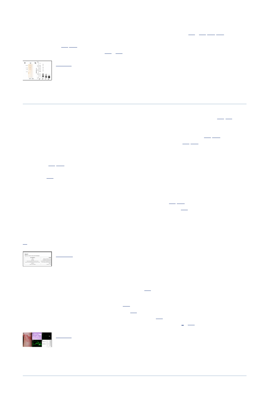
Epidermolysis bullosa acquisita (EBA) is a severe chronic blistering disease of skin and mucous membranes characterized
by subepidermal blisters and tissue-bound and circulating autoantibodies against collagen VII, the main constituent of
anchoring fibrils [
1
,
151
]. The yearly incidence of EBA is at least 0.2%/year/million and is present in about 5% of
unselected patients with subepidermal blistering diseases and autoantibodies against the epidermal basement membrane
zone. EBA is a clinically heterogeneous disease which may present with an inflammatory or non-inflammatory phenotype
[
1
,
152
]. The blister-inducing potential of autoantibodies against collagen VII has been clearly demonstrated and mutually
complementary ex vivo and animal models have been established for this disease [
153
].
Mechanobullous form of EBA may show features highly reminiscent of hereditary dystrophic epidermolysis bullosa, a
disease caused by genetic defects in collagen VII. This form is characterized by extreme skin fragility, trauma-induced
blisters and erosions localized to the extensor skin surface, healing with scars and milia. The inflammatory subtype of
EBA was described, clinically mimicking BP or LAD (Fig.
13a
). EBA patients presenting with an inflammatory
phenotype at the onset can later manifest with non -inflammatory features [
154
,
155
].
Fig. (13)
Diagnostic features of epidermolysis bullosa acquisita (EBA). (a) Clinical picture
of a 61-year old female patient with the inflammatory form of EBA showing
erythema, tense blisters, erosions and crusts on the lateral abdomen. (b)
Histopathology analysis ...
The involvement of mucosal surfaces, especially in the oral cavity, but also of the nasal, conjunctival, pharyngeal, and
laryngeal mucosae, is present in the majority of EBA patients [
156
]. Although often subclinical, the spectrum of mucosal
involvement in EBA resembles MMP and can lead to similar complications, including ankyloglossia, periodontal disease,
scarring and crusting of nasal mucosa, symblepharon, obstruction of nasolacrimal ducts, deformation of the epiglottis,
impaired phonation, dysphagia, esophageal strictures, and supraglottic stenosis requiring emergency tracheostomy [
156
].
EBA is often associated with inflammatory bowel disease and a significant percentage of patients with Crohn disease or
ulcerative colitis show collagen VII-specific autoantibodies [
157
].
The diagnosis of EBA should be suspected in adult patients with skin fragility, trauma-induced blisters, scarring, milia and
nail dystrophy. The inflammatory form of EBA may be clinically and histopathologically indistinguishable from other
pemphigoid diseases. The diagnosis of EBA in all patients is made by demonstrating IgG or IgA autoantibodies against
the dermal side of salt-split skin by indirect IF microscopy, which recognize collagen VII as detected by ELISA or
immunoblotting (Table
14
).
Table 14.
Diagnostic Criteria for Epidermolysis Bullosa Acquisita
Histopathological examination of patients’ lesional skin reveals subepidermal cleavage associated with neutrophilic
infiltrates of variable densities in the upper dermis (Fig.
13b
). By direct IF microscopy, linear IgG and C3 deposits are
found along the epidermal basement membrane (Fig.
13c
). Indirect IF microscopy on human salt-split skin reveals
circulating IgG and/or IgA autoantibodies binding to the blisters floor (Fig.
13d
).
Autoantibodies from EBA patients recognize collagen VII of 290 kDa or its immunodominant region, the non-collagenous
domanin 1, by immunoblotting with dermal extracts or recombinant protein, respectively. ELISA systems using different
recombinant form of collagen VII for detection of specific autoantibodies have been developed and are commercially
available (Table
2
) [
158
-
161
].
DERMATITIS HERPETIFORMIS
Dermatitis herpetiformis (DH) is a chronic subepidermal blistering skin disease characterized by pruritic papulo-vesicular
lesions, typical immunopathological findings, and clinically a good response to sulfone therapy [
162
,
163
]. DH is
associated with distinct HLA haplotypes (DR3 and Dqw2) and is currently regarded as a specific skin manifestation of
celiac disease [
162
-
164
].
DH patients present with diffuse, symmetrical, grouped polymorphic lesions consisting of erythema, urticarial plaques,
papules, clustered herpetiform vesicles and erosions. The most commonly involved sites are the extensor surfaces of the
elbows (90%), knees (30%), shoulders, buttocks, sacral region, and face (Fig.
14a
). The skin lesions commonly associate
with and may be preceded by intense itching and/or burning sensation causing excoriations [
163
,
165
]. The associated
gluten-sensitive enteropathy in DH is often asymptomatic or may manifest with abdominal pain, diarrhoea, iron deficiency
Molecular Diagnosis in Autoimmune Skin Blistering Conditions
http://www.ncbi.nlm.nih.gov/pmc/articles/PMC3905716/
12 von 26
25.08.2014 13:05

Go to:
and reduced growth rates in children [
163
,
165
].
Fig. (14)
Major clinical, histo- and immunopathological features of dermatitis
herpetiformis. (a) Multiple excoriated papules, erosions and crusts on the elbow of
a 44-year old patient with dermatitis herpetiformis. (b) Histopathological
examination shows infiltration ...
DH is a subepidermal autoimmune blistering disease associated with a gluten-sensitive enteropathy and with characteristic
granular IgA deposits in the upper dermis. In its prebullous stage, DH may present with only pruritus and excoriations and
should be distinguished from other pruritic dermatoses, including atopic dermatitis, scabies, papular urticaria, impetigo,
acute dermatitis, nodular prurigo, urticaria and polymorphic erythema by performing a direct IF microscopy analysis.
Serological tests confirm the diagnosis and are very useful to clearly differentiate DH from other autoimmune blistering
diseases such as LAD and BP. In addition, the remission of disease under gluten-free diet and disease relapses or flares
induced by gluten ingestion represent a true “ex juvantibus” diagnostic criterion of DH (Table
15
).
Table 15.
Diagnostic Criteria for Dermatitis Herpetiformis
Histopathological examination in patients' lesional skin reveals an inflammatory infiltrate in the upper dermis and at the
dermo-epidermal junction dominated by neutrophils and eosinophils. These granulocytes form typical papillary
microabscesses which then lead to blister formation in these areas (Fig.
14b
).
Direct IF microscopy from biopsies of uninvolved skin provide optimal results in DH and reveal granular deposits of IgA
along the basement membrane, usually with accentuation in the dermal papillae (Fig.
14c
). Serum IgA autoantibodies
reacting with endomysium may be detected in indirect IF microscopy on monkey esophagus (Fig.
14d
). IgA
autoantibodies specifically recognize the epidermal transglutaminase (TG3) and cross-react with tissue transglutaminase
(TG2) and are a useful marker of bowel damage and diet adherence in DH/celiac disease patients (Table
2
) [
166
,
167
].
These diagnostic measures, especially when the immunopathologic tests are partly negative, but strong suspicion of DH
remains, may be complemented by an aggressive gluten challenge after a gluten-free diet for at least 1 month. Triggering a
flare of the skin eruption in 1-2 days by this “ex juvantibus” test provides a strong further support for a diagnosis of DH
[
165
].
Although not essential for diagnosis, a series of ancillary investigations may be performed for a more accurate global
assessment of the patient with DH, including a small bowel biopsy, HLA testing, screening for autoimmune diseases (e.g.,
thyroid, antinuclear, and citrullinated peptide-specific autoantibodies) and evaluation of malabsorption [
165
,
168
,
169
].
Due to its reliability and efficiency, detection of tissue-bound and serum autoantibodies plays an essential diagnostic role
in autoimmune blistering diseases. Subsequent characterization of the molecular specificity of autoantibodies allows for
developing robust diagnostic algorithms (Fig.
15
), which can help streamlining the laboratory diagnosis of this group of
diseases.
Fig. (15)
Diagnostic algorithm for autoimmune bullous diseases.
PERSPECTIVES FOR THE MOLECULAR DIAG-NOSIS OF AUTOIMMUNE BLISTERING
DIS-EASES
The future development of state-of-the-art diagnostics for autoimmune diseases are heavily dependent on continuous
in-depth fundamental and translational research. The main autoantigens have been already identified and cloned offering
an excellent basis for the development of commercial test kits for the detection of autoantibodies with different specificity,
including against laminin 332 and desmocollins. While most of target antigens are characterized, antigen(s) of the
intraepidermal neutrophilic dermatosis type of IgA pemphigus and the lamina densa type of LAD as well as minor
antigens of other autoimmune blistering diseases still need to be identified. An important, only partly characterized, aspect
is the pathogenic potential of autoantibodies, which may be dependent on their different intrinsic features. A detailed
definition of pathogenic human autoantibodies would allow the development of quantitative tests, which would ideally
Molecular Diagnosis in Autoimmune Skin Blistering Conditions
http://www.ncbi.nlm.nih.gov/pmc/articles/PMC3905716/
13 von 26
25.08.2014 13:05

Go to:
Go to:
Go to:
reflect disease activity in patients.
A further important avenue for improvement is related to automated pre-analytic and analytic steps and data collection for
IF microscopy, ELISA and immunoblotting. While based on the simple concept of detecting structures using probes
conjugated with fluorescent dyes, in its present form, IF is time-consuming, and requires manual handling as well as
interpretation of the data by trained personnel. Relatively extensive experience is needed to properly record and interpret
findings obtained by IF. In addition, IF-based tests are encumbered by a relatively low test-to-test fidelity owing to factors
relating to the method itself (e.g. origin and preparation of the substrate) or to the interpreter. A promising alternative to
the use of standard fluorescence microscopes for direct IF microscopy is the microarray scanner, which allows multiple
antibodies to be visualized simultaneously, obtaining a larger field of view, and facilitating digital recording of images
[
170
]. The need for standardization may be addressed by robotic preparation of slides and development of a
computer-aided IF pattern analysis system [
171
-
174
]. Advanced systems are currently being developed that provide fully
automated readings of IF images and software algorithms for the mathematical description of the IF patterns. Although
full automated IF tests are difficult to imagine at present, the emerging automatic systems may be used for screening and
classification of autoantibodies in routine diagnosis of systemic and organ-specific autoimmune diseases. In this respect,
computer-aided diagnosis systems could support the diagnosis made by specialists, and improve the reproducibility of IF
by overcoming its limitations, especially the inter-observer variability. In addition, these systems should pave the way for
economic data-processing of IF assays. In the long-term, more sophisticated pattern-recognition algorithms and novel
calibration systems should improve standardized quantification of IF image interpretation [
171
-
174
]. Further
miniaturization and automation of the IF diagnostic tests may be achieved by devising complexes or microarrays of
substrates containing the antigens [
175
,
176
]. Thus, small sections of different organ substrates, including salt-split skin,
esophagus, and bladder may be placed on the same slide and incubated with one diluted sample of the patient’s serum. IF
assays using cells expressing recombinant antigens may partly replace the use of ELISAs, immunoblotting, and
immunoprecipitation in the diagnosis of autoimmune diseases. Such cell chips containing spotted cell microarrays may be
constructed by growing and treating cells under normal tissue culture conditions, formaldehyde fixing, and printing
microsamples of each culture onto replicate glass slides [
176
]. These tests using collections of up to hundreds of antigens
are amenable to full automation and high-throughput screening for autoantibodies, and should greatly reduce the costs
involved.
CONCLUDING REMARKS
Autoantibodies against structural components maintaining cell-cell and cell matrix adhesion induce tissue damage and are
exquisite diagnostic markers of autoimmune blistering diseases. The target antigens of blister-inducing autoantibodies
have been extensively characterized over the past decades. Detection of tissue-bound and serum autoantibodies and
characteri-zation of their molecular specificity allows for a rapid and accurate diagnosis of autoimmune blistering
diseases. Various IF methods as well as molecular immunoassays, including ELISA and immunoblotting, became an
essential part of the modern diagnostic algorithms for these disorders. Fundamental and translational research combining
pathogenetic studies, bioinformatic analyses, automation and high throughput approaches will greatly facilitate the
development of a next generation of improved diagnostic tools.
ACKNOWLEDGEMENTS
The work of the authors is supported by grants from the Deutsche Forschungsgemeinschaft SI-1281/4-1 and SI-1281/5-1
(CS), from the European Community's Seventh Framework Programme [FP7-2007-2013] under grant agreement No.
HEALTH-F4-2011-282095 (CS), and from the Medical Faculty of the University of Freiburg (CS). The materials used for
figures were not previously published and were generated at the authors' affiliations.
ABBREVIATIONS
BP = Bullous pemphigoid
DH = Dermatitis herpetiformis
EBA = Epidermolysis bullosa acquisita
ELISA = Enzyme-linked immunosorbent assay
IF = Immunofluorescence
LAD = Linear IgA disease
MMP = Mucous membrane pemphigoid
PF = Pemphigus foliaceus
PNP = Paraneoplastic pemphigus
PV = Pemphigus vulgaris
Molecular Diagnosis in Autoimmune Skin Blistering Conditions
http://www.ncbi.nlm.nih.gov/pmc/articles/PMC3905716/
14 von 26
25.08.2014 13:05

Go to:
Go to:
CONFLICT OF INTEREST
The authors confirm that this article content has no conflicts of interest.
REFERENCES
1. Mihai S, Sitaru C. Immunopathology and molecular diagnosis of autoimmune bullous diseases. J Cell Mol Med.
2007;11:462–481. [
PMC free article
] [
PubMed
]
2. Kneisel A, Hertl M. Autoimmune bullous skin diseases.Part 1 Clinical manifestations. J Dtsch Dermatol Ges.
2011;9:844–56. [
PubMed
]
3. Sitaru C, Zillikens D. Mechanisms of blister induction by autoantibodies. Exp Dermatol. 2005;14:861–875. [
PubMed
]
4. Waschke J. The desmosome and pemphigus. Histochem Cell Biol. 2008;130:21–54. [
PMC free article
] [
PubMed
]
5. VanAgtmael T, Bruckner-Tuderman L. Basement membranes and human disease. Cell Tissue Res. 2010;339:167–188.
[
PubMed
]
6. Arndt KA, Feingold DS. The sign of Pyotr Vasilyewich Nikolsky. N Engl J Med. 1970;282:1154–1155. [
PubMed
]
7. Doubleday CW. Who is Nikolsky and what does his sign mean. J Am Acad Dermatol. 1987;16:1054–1055. [
PubMed
]
8. Kelly B, Shimoni T. Reintroducing the Tzanck smear. Am J Clin Dermatol. 2009;10:141–152. [
PubMed
]
9. Durdu M, Baba M, Seçkin D. The value of Tzanck smear test in diagnosis of erosive vesicular bullous and pustular skin
lesions. J Am Acad Dermatol. 2008;59:958–964. [
PubMed
]
10. Florea F, Sitaru C. Relevance of immunofluorescence methods in clinical dermatology. CML Dermatology.
2011;15:29–45.
11. Gammon WR, Briggaman RA, Inman AO, Queen LL, Wheeler CE. Differentiating anti-lamina lucida and
anti-sublamina densa anti-BMZ antibodies by indirect immunofluorescence on 1. M sodium chloride-separated skin. J
Invest Dermatol. 1984;82:139–144. [
PubMed
]
12. Gammon WR, Briggaman RA, et al. Inman AO3 Differentiating anti-lamina lucida and anti-sublamina densa anti-bmz
antibodies by indirect immunofluorescence on 1. m sodium chloride-separated skin. J. Invest Dermatol. 1984;82:139–144.
[
PubMed
]
13. Anhalt GJ, Kim SC, Stanley JR, et al. Paraneoplastic pemphigus.An autoimmune mucocutaneous disease associated
with neoplasia. N Engl J Med. 1990;323:1729–1735. [
PubMed
]
14. Domloge-Hultsch N, Gammon WR, Briggaman RA, Gil SG, Carter WG, Yancey KB. Epiligrin the major human
keratinocyte integrin ligand is a target in both an acquired autoimmune and an inherited subepidermal blistering skin
disease. J Clin Invest. 1992;90:1628–1633. [
PMC free article
] [
PubMed
]
15. Caldarola G, Kneisel A, Hertl M, Feliciani C. Herpes simplex virus infection in pemphigus vulgaris clinical and
immunological considerations. Eur J Dermatol. 2008;18:440–443. [
PubMed
]
16. Stevenson ML, Levitt JO. Acute zoster in known pemphigus vulgaris and bullous pemphigoid avoiding the "disease
flare" trap. J Am Acad Dermatol. 2011;64:e125–6. [
PubMed
]
17. Raith L. [Johann Ernst Wichmann an unjustly little-known dermatologist of the 18th century. Dermatol Monatsschr.
1983;169:725–727. [
PubMed
]
18. Holubar K. Pemphigus a disease of man and animal.Historical and other perspectives. Int J Dermatol.
1988;27:516–520. [
PubMed
]
19. Stanley JR, Amagai M. Pemphigus bullous impetigo and the staphylococcal scalded-skin syndrome. N Engl J Med.
2006;355:1800–1810. [
PubMed
]
20. Amagai M, Klaus-Kovtun V, Stanley JR. Autoantibodies against a novel epithelial cadherin in pemphigus vulgaris a
disease of cell adhesion. Cell. 1991;67:869–877. [
PubMed
]
21. Ding X, Aoki V, Mascaro JMJ, Lopez-Swiderski A, Diaz LA, Fairley JA. Mucosal and mucocutaneous (generalized)
pemphigus vulgaris show distinct autoantibody profiles. J Invest Dermatol. 1997;109:592–596. [
PubMed
]
22. Nguyen VT, Ndoye A, Grando SA. Pemphigus vulgaris antibody identifies pemphaxin.A novel keratinocyte
annexin-like molecule binding acetylcholine. J Biol Chem. 2000;275:29466–29476. [
PubMed
]
Molecular Diagnosis in Autoimmune Skin Blistering Conditions
http://www.ncbi.nlm.nih.gov/pmc/articles/PMC3905716/
15 von 26
25.08.2014 13:05

23. Nguyen VT, Ndoye A, Grando SA. Novel human alpha9 acetylcholine receptor regulating keratinocyte adhesion is
targeted by Pemphigus vulgaris autoimmunity. Am J Pathol. 2000;157:1377–1391. [
PMC free article
] [
PubMed
]
24. Rafei D, Müller R, Ishii N, Llamazares M, Hashimoto T, Hertl M, Eming R. IgG autoantibodies against desmocollin 3
in pemphigus sera induce loss of keratinocyte adhesion. Am J Pathol. 2011;178:718–723. [
PMC free article
] [
PubMed
]
25. Mao X, Nagler AR, Farber SA, Choi EJ, Jackson LH, Leiferman KM, Ishii N, Hashimoto T, Amagai M, Zone JJ,
Payne AS. Autoimmunity to desmocollin 3 in pemphigus vulgaris. Am J Pathol. 2010;177:2724–2730. [
PMC free article
]
[
PubMed
]
26. Ishii K, Amagai M, Hall RP, et al. Characterization of autoantibodies in pemphigus using antigen-specific enzyme-
linked immunosorbent assays with baculovirus-expressed recombinant desmogleins. J Immunol. 1997;159:2010–2017.
[
PubMed
]
27. Amagai M, Tsunoda K, Zillikens D, Nagai T, Nishikawa T. The clinical phenotype of pemphigus is defined by the
anti-desmoglein autoantibody profile. J Am Acad Dermatol. 1999;40:167–170. [
PubMed
]
28. Nagel A, Lang A, Engel D, et al. Clinical activity of pemphigus vulgaris relates to IgE autoantibodies against
desmoglein 3. Clin Immunol. 2010;134:320–330. [
PubMed
]
29. Dhandha MM, Seiffert-Sinha K, Sinha AA. Specific immunoglobulin isotypes correlate with disease activity
morphology duration and hla association in pemphigus vulgaris. Autoimmunity. 2012;45:516–526. [
PubMed
]
30. Hertl M, Eming R, Veldman C. T cell control in autoimmune bullous skin disorders. J. Clin. Invest.
2006;116:1159–1166. [
PMC free article
] [
PubMed
]
31. Sinha AA. Constructing immunoprofiles to deconstruct disease complexity in pemphigus. Autoimmunity.
2012;45:36–43. [
PubMed
]
32. Veldman C, Höhne A, Dieckmann D, et al. Type i regulatory t cells specific for desmoglein 3 are more frequently
detected in healthy individuals than in patients with pemphigus vulgaris. J. Immunol. 2004;172: 6468–6475. [
PubMed
]
33. Brenner S, Bialy-Golan A, Ruocco V. Drug-induced pemphigus. Clin. Dermatol. 1998;16:393–397. [
PubMed
]
34. Brenner S, Goldberg I. Drug-induced pemphigus. Clin Dermatol. 2011;29:455–457. [
PubMed
]
35. Feng S, Zhou W, Zhang J, et al. Analysis of 6 cases of drug-induced pemphigus. Eur J Dermatol. 2011;21:696–699.
[
PubMed
]
36. Fryer EJ, Lebwohl M. Pemphigus vulgaris after initiation of psoralen and uva therapy for psoriasis. J. Am Acad
Dermatol. 1994;30:651–653. [
PubMed
]
37. Jamora MJJ, Jiao D, Bystryn J. Antibodies to desmoglein 1 and 3 and the clinical phenotype of pemphigus vulgaris. J
Am Acad Dermatol. 2003;48:976–977. [
PubMed
]
38. Lebeau S, Müller R, Masouyé I, Hertl M, Borradori L. Pemphigus herpetiformis analysis of the autoantibody profile
during the disease course with changes in the clinical phenotype. Clin Exp Dermatol. 2010;35:366–372. [
PubMed
]
39. Eyre RW, Stanley JR. Identification of pemphigus vulgaris antigen extracted from normal human epidermis and
comparison with pemphigus foliaceus antigen. J Clin Invest. 1988;81:807–812. [
PMC free article
] [
PubMed
]
40. Rock B, Martins CR, Theofilopoulos AN, et al. The pathogenic effect of IgG4 autoantibodies in endemic pemphigus
foliaceus (fogo selvagem). N Engl J Med. 1989;320:1463–1469. [
PubMed
]
41. Aoki V, Sousa JXJ, Diaz LA. Pathogenesis of endemic pemphigus foliaceus. Dermatol Clin. 2011;29:413–8.
[
PubMed
]
42. Aoki V, Millikan RC, Rivitti EA, Hans-Filho G, Eaton DP, Warren SJP, Li N, Hilario-Vargas J, Hoffmann RG, Diaz
LA. Environmental risk factors in endemic pemphigus foliaceus (fogo selvagem). J Investig Dermatol Symp Proc.
2004;9:34–40. [
PubMed
]
43. Qian Y, Jeong JS, Maldonado M, et al. Cutting edge brazilian pemphigus foliaceus anti-desmoglein 1 autoantibodies
cross-react with sand fly salivary ljm11 antigen. J. Immunol. 2012;189:1535–1539. [
PMC free article
] [
PubMed
]
44. Nguyen VT, Ndoye A, Bassler KD, et al. Classification clinical manifestations and immunopathological mechanisms
of the epithelial variant of paraneoplastic autoimmune multiorgan syndrome a reappraisal of paraneoplastic pemphigus.
Arch Dermatol. 2001;137:193–206. [
PubMed
]
Molecular Diagnosis in Autoimmune Skin Blistering Conditions
http://www.ncbi.nlm.nih.gov/pmc/articles/PMC3905716/
16 von 26
25.08.2014 13:05

45. Maldonado F, Pittelkow MR, Ryu JH. Constrictive bronchiolitis associated with paraneoplastic autoimmune
multi-organ syndrome. Respirology. 2009;14:129–133. [
PubMed
]
46. Anhalt GJ. Paraneoplastic pemphigus the role of tumours and drugs. Br J Dermatol. 2001;144:1102–1104. [
PubMed
]
47. Maverakis E, Goodarzi H, Wehrli LN, Ono Y, Garcia MS. The Etiology of Paraneoplastic Autoimmunity. Clin Rev
Allergy Immunol. 2011;42:135–144. [
PubMed
]
48. Vanderlugt CJ, Miller SD. Epitope spreading. Curr Opin Immunol. 1996;8:831–836. [
PubMed
]
49. Chan LS, Vanderlugt CJ, Hashimoto T, et al. Epitope spreading: lessons from autoimmune skin diseases. J Invest
Dermatol. 1998;110:103–109. [
PubMed
]
50. Zhang J, Qiao Q, Chen X, et al. Improved outcomes after complete resection of underlying tumors for patients with
paraneoplastic pemphigus a single-center experience of 22 cases. J Cancer Res Clin Oncol. 2011;137:229–234. [
PubMed
]
51. Meyers SJ, Varley GA, Meisler DM, Camisa C, Wander AH. Conjunctival involvement in paraneoplastic pemphigus.
Am J Ophthalmol. 1992;114:621–624. [
PubMed
]
52. Anhalt GJ. Paraneoplastic pemphigus. J Investig Dermatol Symp Proc. 2004;9:29–33. [
PubMed
]
53. Frew JW, Murrell DF. Paraneoplastic pemphigus (paraneoplastic autoimmune multiorgan syndrome) clinical
presentations and pathogenesis. Dermatol Clin. 2011;29:419–25. [
PubMed
]
54. Zhu X, Zhang B. Paraneoplastic pemphigus. J Dermatol. 2007;34:503–511. [
PubMed
]
55. Nousari HC, Deterding R, Wojtczack H, et al. The mechanism of respiratory failure in paraneoplastic pemphigus. N
Engl J Med. 1999;340:1406–1410. [
PubMed
]
56. Helou J, Allbritton J, Anhalt GJ. Accuracy of indirect immunofluorescence testing in the diagnosis of paraneoplastic
pemphigus. J Am Acad Dermatol. 1995;32:441–447. [
PubMed
]
57. Kim SC, Kwon YD, Lee IJ, Chang SN, Lee TG. cDNA cloning of the 210-kDa paraneoplastic pemphigus antigen
reveals that envoplakin is a component of the antigen complex. J Invest Dermatol. 1997;109:365–369. [
PubMed
]
58. Kiyokawa C, Ruhrberg C, Nie Z, et al. Envoplakin and periplakin are components of the paraneoplastic pemphigus
antigen complex. J Invest Dermatol. 1998;111:1236–1238. [
PubMed
]
59. Mahoney MG, Aho S, Uitto J, Stanley JR. The members of the plakin family of proteins recognized by paraneoplastic
pemphigus antibodies include periplakin. J Invest Dermatol. 1998;111:308–313. [
PubMed
]
60. Schepens I, Jaunin F, Begre N, et al. The protease inhibitor alpha-2-macroglobulin-like-1 is the p170 antigen
recognized by paraneoplastic pemphigus autoantibodies in human. PLoS ONE. 2010;5:e12250. [
PMC free article
]
[
PubMed
]
61. Hashimoto T, Amagai M, Watanabe K, et al. Characterization of paraneoplastic pemphigus autoantigens by
immunoblot analysis. J Invest Dermatol. 1995;104:829–834. [
PubMed
]
62. Sitaru C, Dähnrich C, Probst C, et al. Enzyme-linked immunosorbent assay using multimers of the 16th
non-collagenous domain of the BP180 antigen for sensitive and specific detection of pemphigoid autoantibodies. Exp
Dermatol. 2007;16:770–777. [
PubMed
]
63. Tsuruta D, Ishii N, Hamada T, et al. IgA pemphigus. Clin Dermatol. 2011;29:437–442. [
PubMed
]
64. Hashimoto T, Ebihara T, Nishikawa T. Studies of autoantigens recognized by IgA anti-keratinocyte cell surface
antibodies. J Dermatol Sci. 1996;12:10–17. [
PubMed
]
65. Hashimoto T, Kiyokawa C, Mori O, et al. Human desmocollin 1 (Dsc1) is an autoantigen for the subcorneal pustular
dermatosis type of IgA pemphigus. J Invest Dermatol. 1997;109:127–131. [
PubMed
]
66. Kárpáti S, Amagai M, Liu WL, Dmochowski M, Hashimoto T, Horváth A. Identification of desmoglein 1 as
autoantigen in a patient with intraepidermal neutrophilic IgA dermatosis type of IgA pemphigus. Exp Dermatol.
2000;9:224–228. [
PubMed
]
67. Hashimoto T, Komai A, Futei Y, Nishikawa T, Amagai M. Detection of IgA autoantibodies to desmogleins by an
enzyme-linked immunosorbent assay the presence of new minor subtypes of IgA pemphigus. Arch Dermatol.
2001;137:735–738. [
PubMed
]
68. Wang J, Kwon J, Ding X, Fairley JA, Woodley DT, Chan LS. Nonsecretory IgA1 autoantibodies targeting desmosomal
Molecular Diagnosis in Autoimmune Skin Blistering Conditions
http://www.ncbi.nlm.nih.gov/pmc/articles/PMC3905716/
17 von 26
25.08.2014 13:05

component desmoglein 3 in intraepidermal neutrophilic IgA dermatosis. Am J Pathol. 1997;150:1901–1907.
[
PMC free article
] [
PubMed
]
69. Ishii N, Ishida-Yamamoto A, Hashimoto T. Immunolocalization of target autoantigens in IgA pemphigus. Clin Exp
Dermatol. 2004;29:62–66. [
PubMed
]
70. Supapannachart N, Mutasim DF. The distribution of IgA pemphigus antigen in human skin and the role of IgA
anti-cell surface antibodies in the induction of intraepidermal acantholysis. Arch Dermatol. 1993;129:605–608. [
PubMed
]
71. Hashimoto T, Yasumoto S, Nagata Y, Okamoto T, Fujita S. Clinical histopathological and immunological distinction in
two cases of IgA pemphigus. Clin Exp Dermatol. 2002;27:636–640. [
PubMed
]
72. Bernard P, Vaillant L, Labeille B, et al. Incidence and distribution of subepidermal autoimmune bullous skin diseases
in three French regions.Bullous Diseases French Study Group. Arch Dermatol. 1995;131:48–52. [
PubMed
]
73. Langan SM, Smeeth L, Hubbard R, Fleming KM, Smith CJP, West J. Bullous pemphigoid and pemphigus vulgaris
incidence and mortality in the UK population based cohort study. BMJ. 2008;337:a180. [
PMC free article
] [
PubMed
]
74. Baican A, Baican C, Chiriac G, et al. Pemphigus vulgaris is the most common autoimmune bullous disease in
Northwestern Romania. Int J Dermatol. 2010;49:768–774. [
PubMed
]
75. Olasz EB, Yancey KB. Bullous pemphigoid and related subepidermal autoimmune blistering diseases. Curr Dir
Autoimmun. 2008;10:141–166. [
PubMed
]
76. Lever W. Pemphigus. Medicine. 1953;32:2–132.
77. Stanley JR, Hawley-Nelson P, Yuspa SH, Shevach EM, Katz SI. Characterization of bullous pemphigoid antigen a
unique basement membrane protein of stratified squamous epithelia. Cell. 1981;24:897–903. [
PubMed
]
78. Diaz LA, Ratrie H, Saunders WS, et al. Isolation of a human epidermal cDNA corresponding to the 180-kDa
autoantigen recognized by bullous pemphigoid and herpes gestationis sera.Immunolocalization of this protein to the
hemidesmosome. . J Clin Invest. 1990;86:1088–1094. [
PMC free article
] [
PubMed
]
79. Laffitte E, Favre B, Fontao L, et al. Plectin an unusual target antigen in bullous pemphigoid. Br J Dermatol.
2001;144:136–138. [
PubMed
]
80. Kiss M, Perényi A, Marczinovits I, et al. Autoantibodies to human alpha6 integrin in patients with bullous
pemphigoid. Ann N Y Acad Sci. 2005;1051:104–110. [
PubMed
]
81. Giudice GJ, Emery DJ, Zelickson BD, Anhalt GJ, Liu Z, Diaz LA. Bullous pemphigoid and herpes gestationis
autoantibodies recognize a common non-collagenous site on the {BP}180 ectodomain. J Immunol. 1993;151:5742–5750.
[
PubMed
]
82. Murakami H, Amagai M, Higashiyama M, et al. Analysis of antigens recognized by autoantibodies in herpes
gestationis.Usefulness of immunoblotting using a fusion protein representing an extracellular domain of the 180 kD
bullous pemphigoid antigen. J Dermatol Sci. 1996;13:112–117. [
PubMed
]
83. Sitaru C. Bullous pemphigoid a prototypical antibody-mediated organ-specific autoimmune disease. J Invest
Dermatol. 2009;129:822–824. [
PubMed
]
84. Feliciani C, Caldarola G, Kneisel A, et al. IgG autoantibody reactivity against bullous pemphigoid (BP) 180 and
BP230 in elderly patients with pruritic dermatoses. Br J Dermatol. 2009;161:306–312. [
PubMed
]
85. Quartey-Papafio CM, Hudson PM. Bullous pemphigoid initially localized to sites of burns (scalds) in a patient on
sulphasalazine for ulcerative colitis. Clin Exp Dermatol. 1994;19:281. [
PubMed
]
86. VandeMaele DM, Reilly JC. Bullous pemphigoid at colostomy site report of a case. Dis Colon Rectum.
1997;40:370–371. [
PubMed
]
87. Torchia D, Caproni M, Ketabchi S, Antiga E, Fabbri P. Bullous pemphigoid initially localized around a urostomy. Int J
Dermatol. 2006;45:1387–1389. [
PubMed
]
88. Batalla A, Peón G, DelaTorre C. Localized bullous pemphigoid at urostomy site. Indian J Dermatol Venereol Leprol.
2011;77:625. [
PubMed
]
89. Pardo J, Rodrguez-Serna M, Mercader P, Fortea JM. Localized bullous pemphigoid overlying a fistula for
hemodialysis. J Am Acad Dermatol. 2004;51:S131–2. [
PubMed
]
Molecular Diagnosis in Autoimmune Skin Blistering Conditions
http://www.ncbi.nlm.nih.gov/pmc/articles/PMC3905716/
18 von 26
25.08.2014 13:05

90. Brodell RT, Korman NJ. Stump pemphigoid. Cutis. 1996;57:245–246. [
PubMed
]
91. Melani L, Giomi B, Antiga E, Torchia D, Caproni M, Fabbri P. Radiation therapy as a trigger factor for initially
localized bullous pemphigoid. Breast J. 2005;11:485–486. [
PubMed
]
92. Kobayashi M, Amagai M, Kuroda-Kinoshita K, et al. BP180 ELISA using bacterial recombinant NC16a protein as a
diagnostic and monitoring tool for bullous pemphigoid. J Dermatol Sci. 2002;30:224–232. [
PubMed
]
93. Yoshida M, Hamada T, Amagai M, et al. Enzyme-linked immunosorbent assay using bacterial recombinant proteins of
human BP230 as a diagnostic tool for bullous pemphigoid. J Dermatol Sci. 2006;41:21–30. [
PubMed
]
94. Iwata Y, Komura K, Kodera M, et al. Correlation of IgE autoantibody to BP180 with a severe form of bullous
pemphigoid. Arch Dermatol. 2008;144:41–48. [
PubMed
]
95. Messingham KAN, Noe MH, Chapman MA, Giudice GJ, Fairley JA. A novel ELISA reveals high frequencies of
BP180-specific IgE production in bullous pemphigoid. J Immunol Methods. 2009;346:18–25. [
PMC free article
]
[
PubMed
]
96. Dresow SK, Sitaru C, Recke A, Oostingh GJ, Zillikens D, Gibbs BF. IgE autoantibodies against the intracellular
domain of BP180. Br J Dermatol. 2009;160:429–432. [
PubMed
]
97. Intong LRA, Murrell DF. Pemphigoid gestationis: pathogenesis and clinical features. Dermatol Clin. 2011;29:447–52.
[
PubMed
]
98. Morrison LH, Labib RS, Zone JJ, Diaz LA, Anhalt GJ. Herpes gestationis autoantibodies recognize a 180-kD human
epidermal antigen. J Clin Invest. 1988;81:2023–2026. [
PMC free article
] [
PubMed
]
99. Kelly SE, Bhogal BS, Wojnarowska F, Whitehead P, Leigh IM, Black MM. Western blot analysis of the antigen in
pemphigoid gestationis. Br J Dermatol. 1990;122:445–449. [
PubMed
]
100. Katz SI, Hertz KC, Yaoita H. Herpes gestationis.Immunopathology and characterization of the HG factor. J Clin
Invest. 1976;57:1434–1441. [
PMC free article
] [
PubMed
]
101. Jordon RE, Heine KG, Tappeiner G, Bushkell LL, Provost TT. The immunopathology of herpes
gestationis.Immunofluorescence studies and characterization of "HG factor". J Clin Invest. 1976;57:1426–1431.
[
PMC free article
] [
PubMed
]
102. Kelly SE, Cerio R, Bhogal BS, Black MM. The distribution of IgG subclasses in pemphigoid gestationis.PG factor is
an IgG1 autoantibody. J Invest Dermatol. 1989;92:695–698. [
PubMed
]
103. Sitaru C, Powell J, Shimanovich I, et al. Pemphigoid gestationis maternal sera recognize epitopes restricted to the
N-terminal portion of the extracellular domain of BP180 not present on its shed ectodomain. Br J Dermatol.
2003;149:420–422. [
PubMed
]
104. Carruthers JA. Herpes gestationis clinical features of immunologically proved cases. Am J Obstet Gynecol.
1978;131:865–867. [
PubMed
]
105. Amato L, Mei S, Gallerani I, Moretti S, Fabbri P. A case of chronic herpes gestationis persistent disease or
conversion to bullous pemphigoid. J Am Acad Dermatol. 2003;49:302–307. [
PubMed
]
106. Boulinguez S, Bédane C, Prost C, Bernard P, Labbé L, Bonnetblanc JM. Chronic pemphigoid gestationis comparative
clinical and immunopathological study of 10 patients. Dermatology. 2003;206:113–119. [
PubMed
]
107. Semkova K, Black M. Pemphigoid gestationis: current insights into pathogenesis and treatment. Eur J Obstet
Gynecol Reprod Biol. 2009;145:138–144. [
PubMed
]
108. Jenkins RE, Hern S, Black MM. Clinical features and management of 87 patients with pemphigoid gestationis. Clin
Exp Dermatol. 1999;24:255–259. [
PubMed
]
109. Ambros-Rudolph CM, Müllegger RR, Vaughan-Jones SA, Kerl H, Black MM. The specific dermatoses of pregnancy
revisited and reclassified results of a retrospective two-center study on 505 pregnant patients. J Am Acad Dermatol.
2006;54:395–404. [
PubMed
]
110. Roth M. Pregnancy dermatoses diagnosis management and controversies. Am J Clin Dermatol. 2011;12:25–41.
[
PubMed
]
111. Beard MP, Millington GWM. Recent developments in the specific dermatoses of pregnancy. Clin Exp Dermatol.
2011;37:1–4. [
PubMed
]
Molecular Diagnosis in Autoimmune Skin Blistering Conditions
http://www.ncbi.nlm.nih.gov/pmc/articles/PMC3905716/
19 von 26
25.08.2014 13:05

112. Sitaru C, Powell J, Messer G, Bröcker E, Wojnarowska F, Zillikens D. Immunoblotting and enzyme-linked
immunosorbent assay for the diagnosis of pemphigoid gestationis. Obstet Gynecol. 2004;103:757–763. [
PubMed
]
113. Chan LS, Ahmed AR, Anhalt GJ, et al. The first international consensus on mucous membrane pemphigoid definition
diagnostic criteria pathogenic factors medical treatment and prognostic indicators. Arch Dermatol. 2002;138:370–379.
[
PubMed
]
114. Egan CA, Lazarova Z, Darling TN, Yee C, Coté T, Yancey KB. Anti-epiligrin cicatricial pemphigoid and relative risk
for cancer. Lancet. 2001;357:1850–1851. [
PubMed
]
115. Bernard P, Prost C, Durepaire N, Basset-Seguin N, Didierjean L, Saurat JH. The major cicatricial pemphigoid antigen
is a 180-kD protein that shows immunologic cross-reactivities with the bullous pemphigoid antigen. J Invest Dermatol.
1992;99:174–179. [
PubMed
]
116. Balding SD, Prost C, Diaz LA, et al. Cicatricial pemphigoid autoantibodies react with multiple sites on the BP180
extracellular domain. J Invest Dermatol. 1996;106:141–146. [
PubMed
]
117. Murakami H, Nishioka S, Setterfield J, et al. Analysis of antigens targeted by circulating IgG and IgA autoantibodies
in 50 patients with cicatricial pemphigoid. J Dermatol Sci. 1998;17:39–44. [
PubMed
]
118. Tyagi S, Bhol K, Natarajan K, Livir-Rallatos C, Foster CS, Ahmed AR. Ocular cicatricial pemphigoid antigen partial
sequence and biochemical characterization. Proc Natl Acad Sci USA. 1996;93:14714–14719. [
PMC free article
]
[
PubMed
]
119. Caux F, Kirtschig G, Lemarchand-Venencie F, et al. IgA-epidermolysis bullosa acquisita in a child resulting in
blindness. Br J Dermatol. 1997;137:270–275. [
PubMed
]
120. Wieme N, Lambert J, Moerman M, Geerts ML, Temmerman L, Naeyaert JM. Epidermolysis bullosa acquisita with
combined features of bullous pemphigoid and cicatricial pemphigoid. Dermatology. 1999;198:310–313. [
PubMed
]
121. Schmidt E, Skrobek C, Kromminga A, et al. Cicatricial pemphigoid: IgA and IgG autoantibodies target epitopes on
both intra- and extracellular domains of bullous pemphigoid antigen 180. Br J Dermatol. 2001;145:778–783. [
PubMed
]
122. Leverkus M, Bhol K, Hirako Y, et al. Cicatricial pemphigoid with circulating autoantibodies to beta4 integrin bullous
pemphigoid 180 and bullous pemphigoid 230. Br J Dermatol. 2001;145:998–1004. [
PubMed
]
123. Rashid KA, Gürcan HM, Ahmed AR. Antigen specificity in subsets of mucous membrane pemphigoid. J Invest
Dermatol. 2006;126:2631–2636. [
PubMed
]
124. Egan CA, Lazarova Z, Darling TN, Yee C, Yancey KB. Anti-epiligrin cicatricial pemphigoid clinical findings
immunopathogenesis and significant associations. Medicine (Baltimore). 2003;82:177–186. [
PubMed
]
125. Hanno R, Foster DR, Bean SF. Brunsting-Perry cicatricial pemphigoid associated with bullous pemphigoid. J Am
Acad Dermatol. 1980;3:470–473. [
PubMed
]
126. Rose C, Schmidt E, Kerstan A, et al. Histopathology of anti-laminin 5 mucous membrane pemphigoid. J Am Acad
Dermatol. 2009;61:433–440. [
PubMed
]
127. Lazarova Z, Sitaru C, Zillikens D, Yancey KB. Comparative analysis of methods for detection of anti-laminin 5
autoantibodies in patients with anti-epiligrin cicatricial pemphigoid. J Am Acad Dermatol. 2004;51:886–892. [
PubMed
]
128. Lazarova Z, Yee C, Lazar J, Yancey KB. IgG autoantibodies in patients with anti-epiligrin cicatricial pemphigoid
recognize the G domain of the laminin 5 alpha-subunit. Clin Immunol. 2001;101:100–105. [
PubMed
]
129. Bhol KC, Dans MJ, Simmons RK, Foster CS, Giancotti FG, Ahmed AR. The autoantibodies to alpha 6 beta 4
integrin of patients affected by ocular cicatricial pemphigoid recognize predominantly epitopes within the large
cytoplasmic domain of human beta 4. J Immunol. 2000;165:2824–2829. [
PubMed
]
130. Bhol KC, Goss L, Kumari S, Colon JE, Ahmed AR. Autoantibodies to human alpha6 integrin in patients with oral
pemphigoid. J Dent Res. 2001;80:1711–1715. [
PubMed
]
131. Chorzelski TP, Jablonska S. IgA linear dermatosis of childhood (chronic bullous disease of childhood). Br J
Dermatol. 1979;101:535–542. [
PubMed
]
132. Fortuna G, Salas-Alanis JC, Guidetti E, Marinkovich MP. A critical reappraisal of the current data on drug-induced
linear immuglobulin A bullous dermatosis.A real and separate nosological entity. J Am Acad Dermatol.
2012;66(6):988–94. [
PubMed
]
Molecular Diagnosis in Autoimmune Skin Blistering Conditions
http://www.ncbi.nlm.nih.gov/pmc/articles/PMC3905716/
20 von 26
25.08.2014 13:05

133. Zone JJ, Taylor TB, Kadunce DP, Meyer LJ. Identification of the cutaneous basement membrane zone antigen and
isolation of antibody in linear immunoglobulin A bullous dermatosis. J Clin Invest. 1990;85:812–820. [
PMC free article
]
[
PubMed
]
134. Marinkovich MP, Taylor TB, Keene DR, Burgeson RE, Zone JJ. LAD-1 the linear IgA bullous dermatosis
autoantigen is a novel 120-kDa anchoring filament protein synthesized by epidermal cells. J Invest Dermatol.
1996;106:734–738. [
PubMed
]
135. Pas HH Kloosterhuis GJ, Heeres K, vanderMeer JB, Jonkman MF. Bullous pemphigoid and linear IgA dermatosis
sera recognize a similar 120-kDa keratinocyte collagenous glycoprotein with antigenic cross-reactivity to BP180. J Invest
Dermatol. 1997;108:423–429. [
PubMed
]
136. Hirako Y, Usukura J, Uematsu J, Hashimoto T, Kitajima Y, Owaribe K. Cleavage of BP180 a 180-kDa bullous
pemphigoid antigen yields a 120-kDa collagenous extracellular polypeptide. J Biol Chem. 1998;273:9711–9717.
[
PubMed
]
137. Schäcke H, Schumann H, Hammami-Hauasli N, Raghunath M, Bruckner-Tuderman L. Two forms of collagen XVII
in keratinocytes.A full-length transmembrane protein and a soluble ectodomain. . J Biol Chem. 1998;273:25937–25943.
[
PubMed
]
138. Csorba K, Schmidt S, Florea F, et al. Development of an ELISA for sensitive and specific detection of IgA
autoantibodies against BP180 in pemphigoid diseases. Orphanet J Rare Dis. 2011;6:31. [
PMC free article
] [
PubMed
]
139. Vodegel RM, deJong MCJM, Pas HH, Jonkman MF. IgA-mediated epidermolysis bullosa acquisita two cases and
review of the literature. J Am Acad Dermatol. 2002;47:919–925. [
PubMed
]
140. van der, Steen LP, Bakema JE, Sesarman A, et al. Blocking fca receptor i on granulocytes prevents tissue damage
induced by iga autoantibodies. J. Immunol. 2012;189:1594–1601. [
PubMed
]
141. Schumann H, Baetge J, Tasanen K, et al. The shed ectodomain of collagen XVII/BP180 is targeted by autoantibodies
in different blistering skin diseases. Am J Pathol. 2000;156:685–695. [
PMC free article
] [
PubMed
]
142. Florea F, Torio-Padron N, Hashimoto T, Sitaru C. Non-scarring skin blistering disease and mucosal lesions with IgA
autoantibodies reactive with collagen VII and IgG reactivity with laminin ?2. Br J Dermatol. 2012;167(4):938–41.
[
PubMed
]
143. Zillikens D, Kawahara Y, Ishiko A, et al. A novel subepidermal blistering disease with autoantibodies to a 200-kDa
antigen of the basement membrane zone. J Invest Dermatol. 1996;106:1333–1338. [
PubMed
]
144. Dainichi T, Kurono S, Ohyama B, et al. Anti-laminin gamma-1 pemphigoid. Proc Natl Acad Sci USA.
2009;106:2800–2805. [
PMC free article
] [
PubMed
]
145. Dainichi T, Koga H, Tsuji T, et al. From anti-p200 pemphigoid to anti-laminin gamma1 pemphigoid. J Dermatol.
2010;37:231–238. [
PubMed
]
146. Zillikens D, Ishiko A, Jonkman MF, et al. Autoantibodies in anti-p200 pemphigoid stain skin lacking laminin 5 and
type VII collagen. Br J Dermatol. 2000;143:1043–1049. [
PubMed
]
147. Liu Y, Shimizu H, Hashimoto T. Immunofluorescence studies using skin sections of recessive dystrophic
epidermolysis bullosa patients indicated that the antigen of anti-p200 pemphigoid is not a fragment of type VII collagen. J
Dermatol Sci. 2003;32:125–129. [
PubMed
]
148. Koga H, Ishii N, Dainichi T, et al. An attempt to develop mouse model for anti-laminin ?1 pemphigoid. J Dermatol
Sci. 2013;70:108–15. [
PubMed
]
149. Dilling A, Rose C, Hashimoto T, Zillikens D, Shimanovich I. Anti-p200 pemphigoid a novel autoimmune
subepidermal blistering disease. J Dermatol. 2007;34:1–8. [
PubMed
]
150. Groth S, Recke A, Vafia K, et al. Development of a simple enzyme-linked immunosorbent assay for the detection of
autoantibodies in anti-p200 pemphigoid. Br J Dermatol. 2011;164:76–82. [
PubMed
]
151. Woodley DT, Briggaman RA, O'Keefe EJ, Inman AO, Queen LL, Gammon WR. Identification of the skin basement-
membrane autoantigen in epidermolysis bullosa acquisita. N Engl J Med. 1984;310:1007–1013. [
PubMed
]
152. Ishii N, Hamada T, Dainichi T, et al. Epidermolysis bullosa acquisita: what's new. J Dermatol. 2010;37:220–230.
[
PubMed
]
Molecular Diagnosis in Autoimmune Skin Blistering Conditions
http://www.ncbi.nlm.nih.gov/pmc/articles/PMC3905716/
21 von 26
25.08.2014 13:05

153. Sitaru C. Experimental models of epidermolysis bullosa acquisita. Exp Dermatol. 2007;16:520–531. [
PubMed
]
154. Remington J, Chen M, Burnett J, Woodley DT. Autoimmunity to type VII collagen epidermolysis bullosa acquisita.
Curr Dir Autoimmun. 2008;10:195–205. [
PubMed
]
155. Caux F. Diagnosis and clinical features of epidermolysis bullosa acquisita. Dermatol Clin. 2011;29:485–91.
[
PubMed
]
156. Luke MC, Darling TN, Hsu R, et al. Mucosal morbidity in patients with epidermolysis bullosa acquisita. Arch
Dermatol. 1999;135:954–959. [
PubMed
]
157. Hundorfean G, Neurath MF, Sitaru C. Autoimmunity against type VII collagen in inflammatory bowel disease. J Cell
Mol Med. 2010;14:2393–2403. [
PMC free article
] [
PubMed
]
158. Chen M, Chan LS, Cai X, O'Toole EA, Sample JC, Woodley DT. Development of an ELISA for rapid detection of
anti-type VII collagen autoantibodies in epidermolysis bullosa acquisita. J Invest Dermatol. 1997;108:68–72. [
PubMed
]
159. Saleh MA, Ishii K, Kim Y, et al. Development of NC1 and NC2 domains of type VII collagen ELISA for the
diagnosis and analysis of the time course of epidermolysis bullosa acquisita patients. J Dermatol Sci. 2011;62:169–175.
[
PubMed
]
160. Pendaries V, Gasc G, Titeux M, et al. Immune reactivity to type VII collagen implications for gene therapy of
recessive dystrophic epidermolysis bullosa. Gene Ther. 2010;17:930–937. [
PubMed
]
161. Licarete E, Ganz S, Recknagel M, et al. Prevalence of collagen VII-specific autoantibodies in patients with
autoimmune and inflammatory diseases. BMC Immunol. 2012;13:16. [
PMC free article
] [
PubMed
]
162. Reunala TL. Dermatitis herpetiformis. Clin Dermatol. 2001;19:728–736. [
PubMed
]
163. Kárpáti S. An exception within the group of autoimmune blistering diseases dermatitis herpetiformis the gluten-
sensitive dermopathy. Dermatol Clin. 2011;29:463–8. [
PubMed
]
164. Reunala T. Dermatitis herpetiformis coeliac disease of the skin. Ann Med. 1998;30:416–418. [
PubMed
]
165. Caproni M, Antiga E, Melani L, Fabbri P. Guidelines for the diagnosis and treatment of dermatitis herpetiformis. J
Eur Acad Dermatol Venereol. 2009;23:633–638. [
PubMed
]
166. Dieterich W, Laag E, Bruckner-Tuderman L, et al. Antibodies to tissue transglutaminase as serologic markers in
patients with dermatitis herpetiformis. J Invest Dermatol. 1999;113:133–136. [
PubMed
]
167. Sárdy M, Kárpáti S, Merkl B, Paulsson M, Smyth N. Epidermal transglutaminase (TGase 3) is the autoantigen of
dermatitis herpetiformis. J Exp Med. 2002;195:747–757. [
PMC free article
] [
PubMed
]
168. Zettinig G, Weissel M, Flores J, et al. Dermatitis herpetiformis is associated with atrophic but not with goitrous
variant of hashimoto's thyroiditis. Eur. J Clin Invest. 2000;30:53–57. [
PubMed
]
169. Nakajima K. Recent advances in dermatitis herpetiformis. Clin Dev Immunol. 2012;2012:914162. [
PMC free article
]
[
PubMed
]
170. Iwamoto S, Iwamoto AT, Cha J, et al. The utility of the DNA microarray scanner to simplify the immunofluorescence
evaluation of autoimmune bullous diseases. Am J Dermatopathol. 2009;31:218–222. [
PMC free article
] [
PubMed
]
171. Hiemann R, Büttner T, Krieger T, Roggenbuck D, Sack U, Conrad K. Challenges of automated screening and
differentiation of non-organ specific autoantibodies on HEp-2 cells. Autoimmun Rev. 2009;9:17–22. [
PubMed
]
172. Sack U, Knoechner S, Warschkau H, Pigla U, Emmrich F, Kamprad M. Computer-assisted classification of HEp-2
immunofluorescence patterns in autoimmune diagnostics. Autoimmun Rev. 2003;2:298–304. [
PubMed
]
173. Rigon A, Soda P, Zennaro D, Iannello G, Afeltra A. Indirect immunofluorescence in autoimmune diseases
assessment of digital images for diagnostic purpose. Cytometry B Clin Cytom. 2007;72:472–477. [
PubMed
]
174. Soda P, Iannello G. Aggregation of classifiers for staining pattern recognition in antinuclear autoantibodies analysis.
IEEE Trans Inf Technol Biomed. 2009;13:322–329. [
PubMed
]
175. Rimm DL, Camp RL, Charette LA, Costa J, Olsen DA, Reiss M. Tissue microarray a new technology for
amplification of tissue resources. Cancer J. 2001;7:24–31. [
PubMed
]
176. Hart T, Zhao A, Garg A, Bolusani S, Marcotte EM. Human cell chips adapting DNA microarray spotting technology
to cell-based imaging assays. PLoS ONE. 2009;4:e7088. [
PMC free article
] [
PubMed
]
Molecular Diagnosis in Autoimmune Skin Blistering Conditions
http://www.ncbi.nlm.nih.gov/pmc/articles/PMC3905716/
22 von 26
25.08.2014 13:05

177. Rosenbach M, Murrell DF, Bystryn J, et al. Reliability and convergent validity of two outcome instruments for
pemphigus. J Invest Dermatol. 2009;129:2404–2410. [
PMC free article
] [
PubMed
]
178. Schmidt E, Dähnrich C, Rosemann A, et al. Novel ELISA systems for antibodies to desmoglein 1 and 3 correlation
of disease activity with serum autoantibody levels in individual pemphigus patients. Exp Dermatol. 2010;19:458–463.
[
PubMed
]
179. Huang Y, Bu D, Zhu Y. Sensitive enzyme-linked immunosorbent assay for detection of circulating anti-peri and envo
autoantibodies in patients with paraneoplastic pemphigus. J Invest Dermatol. 2008;128:S63.
180. Probst C, Schlumberger W, Stöcker W, et al. Development of ELISA for the specific determination of autoantibodies
against envoplakin and periplakin in paraneoplastic pemphigus. Clin Chim Acta. 2009;410:13–18. [
PubMed
]
181. Mouquet H, Drenovska K, Lartigue A, et al. Detection and characterization of anti-envoplakin linker autoantibodies
in paraneoplastic pemphigus using specific bead-based assay. Clin Immunol. 2008;129:304–312. [
PubMed
]
182. Giudice GJ, Wilske KC, Anhalt GJ, et al. Development of an ELISA to detect anti-BP180 autoantibodies in bullous
pemphigoid and herpes gestationis. J Invest Dermatol. 1994;102:878–881. [
PubMed
]
183. Zillikens D, Mascaro JM, Rose PA, et al. A highly sensitive enzyme-linked immunosorbent assay for the detection of
circulating anti-BP180 autoantibodies in patients with bullous pemphigoid. J Invest Dermatol. 1997;109:679–683.
[
PubMed
]
184. Ishiura N, Fujimoto M, Watanabe R, et al. Serum levels of IgE anti-BP180 and anti-BP230 autoantibodies in patients
with bullous pemphigoid. J Dermatol Sci. 2008;49:153–161. [
PubMed
]
185. Chen M, Chan LS, Cai X, O'Toole EA, Sample JC, Woodley DT. Development of an ELISA for rapid detection of
anti-type VII collagen autoantibodies in epidermolysis bullosa acquisita. J Invest Dermatol. 1997;108:68–72. [
PubMed
]
186. Komorowski L, Müller R, Vorobyev A, et al. Sensitive and specific assays for routine serological diagnosis of
epidermolysis bullosa acquisita. J Am Acad Dermatol. 2013;68:e89–95. [
PubMed
]
187. Adornetto G, Volpe G, DeStefano A, et al. An ELIME assay for the rapid diagnosis of coeliac disease. Anal Bioanal
Chem. 2012;403:1191–4. [
PubMed
]
188. Kasperkiewicz M, Dähnrich C, Probst C, et al. Novel assay for detecting celiac disease-associated autoantibodies in
dermatitis herpetiformis using deamidated gliadin-analogous fusion peptides. J Am Acad Dermatol. 2012;66:583–588.
[
PubMed
]
189. Acostamadiedo JM, Perniciaro C. Rogers RS3.Phenytoin-induced linear IgA bullous disease. J Am Acad Dermatol.
1998;38:352–356. [
PubMed
]
190. Armstrong AW, Fazeli A, Yeh SW, Mackool BT, Liu V. Vancomycin-induced linear IgA disease manifesting as
bullous erythema multiforme. J Cutan Pathol. 2004;31:393–397. [
PubMed
]
191. Bitman LM, Grossman ME, Ross H. Bullous drug eruption treated with amputation.A challenging case of
vancomycin-induced linear IgA disease. . Arch Dermatol. 1996;132:1289–1290. [
PubMed
]
192. Brinkmeier T, Angelkort B, Frosch PJ, Herbst RA. [Vancomycin-induced linear IgA dermatosis]. J Dtsch Dermatol
Ges. 2003;1:212–214. [
PubMed
]
193. Khan I, Hughes R, Curran S, Marren P. Drug-associated linear IgA disease mimicking toxic epidermal necrolysis.
Clin Exp Dermatol. 2009;34:715–717. [
PubMed
]
194. Montagnac R, Reguiaï Z, Méhaut S, Bressieux JM, Schillinger F. [Drug induced linear IgA bullous dermatosis].
Nephrologie. 2003;24:287–292. [
PubMed
]
195. Palmer RA, Ogg G, Allen J, et al. Vancomycin-induced linear IgA disease with autoantibodies to BP180 and
LAD285. Br J Dermatol. 2001;145:816–820. [
PubMed
]
196. Whitworth JM, Thomas I, Peltz SA, Sullivan BC, Wolf AH, Cytryn AS. Vancomycin-induced linear IgA bullous
dermatosis (LABD). J Am Acad Dermatol. 1996;34:890–891. [
PubMed
]
197. Baden LA, Apovian C, Imber MJ, Dover JS. Vancomycin-induced linear IgA bullous dermatosis. Arch Dermatol.
1988;124:1186–1188. [
PubMed
]
198. Carpenter S, Berg D, Sidhu-Malik N. Hall RP 3rd, Rico MJ.Vancomycin-associated linear IgA dermatosis. A report
of three cases. J Am Acad Dermatol. 1992;26:45–48. [
PubMed
]
Molecular Diagnosis in Autoimmune Skin Blistering Conditions
http://www.ncbi.nlm.nih.gov/pmc/articles/PMC3905716/
23 von 26
25.08.2014 13:05

199. Piketty C, Meeus F, Nochy D, Poux JM, Jacquot C, Bariety J. Linear IgA dermatosis related to vancomycin. Br J
Dermatol. 1994;130:130–131. [
PubMed
]
200. Geissmann C, Beylot-Barry M, Doutre MS, Beylot C. Drug-induced linear IgA bullous dermatosis. J Am Acad
Dermatol. 1995;32:296. [
PubMed
]
201. Richards SS, Hall S, Yokel B, Whitmore SE. A bullous eruption in an elderly woman.Vancomycin-associated linear
IgA dermatosis (LAD). . Arch Dermatol. 1995;131:1447–8. [
PubMed
]
202. Espagne E, Prost C, Chosidow O, Saïag P, Revuz J, Roujeau JC. [Linear IgA drug-induced dermatosis.Report of 3
cases]. . Ann Dermatol Venereol. 1990;117:898–899. [
PubMed
]
203. Jawitz RS, Krach K, Schapiro B. A vesiculobullous eruption in a hospitalized patient.Diagnosis. Drug-induced linear
IgA bullous dermatosis (LAD) with eosinophils. Arch Dermatol. 2011;147:1443–1448. [
PubMed
]
204. Kuechle MK, Stegemeir E, Maynard B, Gibson LE, Leiferman KM, Peters MS. Drug-induced linear IgA bullous
dermatosis: report of six cases and review of the literature. J Am Acad Dermatol. 1994;30:187–192. [
PubMed
]
205. Combemale P, Gavaud C, Cozzani E, Nicolas JF, Guennoc B, Dusseau JY. [Linear IgA dermatosis induced by
penicillin G]. Ann Dermatol Venereol. 1993;120:847–848. [
PubMed
]
206. Wozniak K, Kowalewski C, Hashimoto T, Ishii N, Glinska-Wielochowska M, Schwartz RA. Penicillin-induced
anti-p200 pemphigoid an unusual morphology. Acta Derm Venereol. 2006;86:443–446. [
PubMed
]
207. Shimanovich I, Rose C, Sitaru C, Bröcker E, Zillikens D. Localized linear IgA disease induced by ampicillin
sulbactam. J Am Acad Dermatol. 2004;51:95–98. [
PubMed
]
208. McWhirter JD, Hashimoto K, Fayne S, Ito K. Linear IgA bullous dermatosis related to lithium carbonate. Arch
Dermatol. 1987;123:1120–1122. [
PubMed
]
209. Primka EJ3rd, Liranzo MO, Bergfeld WF, Dijkstra JW. Amiodarone-induced linear IgA disease. J Am Acad
Dermatol. 1994;31:809–811. [
PubMed
]
210. König C, Eickert A, Scharfetter-Kochanek K, Krieg T, Hunzelmann N. Linear IgA bullous dermatosis induced by
atorvastatin. J Am Acad Dermatol. 2001;44:689–692. [
PubMed
]
211. Durdu M, Baba M, Seçkin D. A case of bullous pemphigoid induced by aspirin. J Am Acad Dermatol.
2011;65:443–444. [
PubMed
]
212. Gabrielsen TO, Staerfelt F, Thune PO. Drug-induced bullous dermatosis with linear IgA deposits along the basement
membrane. Acta Derm Venereol. 1981;61:439–441. [
PubMed
]
213. Valsecchi R, Serra M, Tornaghi A, Cainelli T. [Drug-induced bullous dermatosis with linear IgA]. G Ital Dermatol
Venereol. 1982;117:221–223. [
PubMed
]
214. Bialy-Golan A, Brenner S. Penicillamine-induced bullous dermatoses. J Am Acad Dermatol. 1996;35:732–742.
[
PubMed
]
215. Brenner S, Wolf R, Ruocco V. Drug-induced pemphigus. I. A survey. Clin Dermatol. 1993;11:501–505. [
PubMed
]
216. Aghassi D, Dover JS. Pemphigus foliaceus induced by psoralen-UV-A. Arch Dermatol. 1998;134:1300–1301.
[
PubMed
]
217. Fryer EJ, Lebwohl M. Pemphigus vulgaris after initiation of psoralen and UVA therapy for psoriasis. J Am Acad
Dermatol. 1994;30:651–653. [
PubMed
]
218. Perl S, Rappersberger K, Födinger D, Anegg B, Hönigsmann H, Ortel B. Bullous pemphigoid induced by PUVA
therapy. Dermatology. 1996;193:245–247. [
PubMed
]
219. Abel EA, Bennett A. Bullous pemphigoid.Occurrence in psoriasis treated with psoralens plus long-wave ultraviolet
radiation. Arch Dermatol. 1979;115:988–989. [
PubMed
]
220. Robinson JK, Baughman RD, Provost TT. Bullous pemphigoid induced by PUVA therapy.Is this the aetiology of the
acral bullae produced during PUVA treatment. Br J Dermatol. 1978;99:709–713. [
PubMed
]
221. Deschamps P, Pedailles S, Michel M, Leroy D. Photo-induction of lesions in a patient with pemphigus
erythematosus. Photodermatol. 1984;1:38–41. [
PubMed
]
222. Sanchez-Palacios C, Chan LS. Development of pemphigus herpetiformis in a patient with psoriasis receiving
Molecular Diagnosis in Autoimmune Skin Blistering Conditions
http://www.ncbi.nlm.nih.gov/pmc/articles/PMC3905716/
24 von 26
25.08.2014 13:05

UV-light treatment. J Cutan Pathol. 2004;31:346–349. [
PubMed
]
223. Jappe U, Zillikens D, Bonnekoh B, Gollnick H. Epidermolysis bullosa acquisita with ultraviolet radiationsensitivity.
Br J Dermatol. 2000;142:517–520. [
PubMed
]
224. Kaplan I, Hodak E, Ackerman L, Mimouni D, Anhalt GJ, Calderon S. Neoplasms associated with paraneoplastic
pemphigus a review with emphasis on non-hematologic malignancy and oral mucosal manifestations. Oral Oncol.
2004;40:553–562. [
PubMed
]
225. Wang J, Zhu X, Li R, et al. Paraneoplastic pemphigus associated with Castleman tumor a commonly reported
subtype of paraneoplastic pemphigus in China. Arch Dermatol. 2005;141:1285–1293. [
PubMed
]
226. Dega H, Laporte JL, Joly P, et al. Paraneoplastic pemphigus associated with Hodgkin's disease. Br J Dermatol.
1998;138:196–198. [
PubMed
]
227. Tilakaratne W, Dissanayake M. Paraneoplastic pemphigus a case report and review of literature. Oral Dis.
2005;11:326–329. [
PubMed
]
228. Wang J, Bu DF, Li T, et al. Autoantibody production from a thymoma and a follicular dendritic cell sarcoma
associated with paraneoplastic pemphigus. Br J Dermatol. 2005;153:558–564. [
PubMed
]
229. McKee PH, McClelland M, Sandford JC. Co-existence of pemphigus anti-skeletal muscle antibody and a
retroperitoneal paraganglioma. Br J Dermatol. 1978;99:441–445. [
PubMed
]
230. Eccersley LRJ, Hoffbrand AV, Rustin MHA, McNamara CJ. Paraneoplastic pemphigus associated with systemic
mastocytosis. Am J Hematol. 2009;84:847–848. [
PubMed
]
231. Trüeb RM, Didierjean L, Fellas A, Elias A, Borradori L. Childhood bullous pemphigoid report of a case with
characterization of the targeted antigens. J Am Acad Dermatol. 1999;40:338–344. [
PubMed
]
232. Chimanovitch I, Hamm H, Georgi M, et al. Bullous pemphigoid of childhood autoantibodies target the same epitopes
within the NC16A domain of BP180 as autoantibodies in bullous pemphigoid of adulthood. Arch Dermatol.
2000;136:527–532. [
PubMed
]
233. Cliff S, Holden CA. Pemphigoid nodularis a report of three cases and review of the literature. Br J Dermatol.
1997;136:398–401. [
PubMed
]
234. Powell AM, Albert S, Gratian MJ, Bittencourt R, Bhogal BS, Black MM. Pemphigoid nodularis (non-bullous) a
clinicopathological study of five cases. Br J Dermatol. 2002;147:343–349. [
PubMed
]
235. Schmidt E, Sitaru C, Schubert B, et al. Subacute prurigo variant of bullous pemphigoid autoantibodies show the same
specificity compared with classic bullous pemphigoid. J Am Acad Dermatol. 2002;47:133–136. [
PubMed
]
236. Korman NJ, Woods SG. Erythrodermic bullous pemphigoid is a clinical variant of bullous pemphigoid. Br J
Dermatol. 1995;133:967–971. [
PubMed
]
237. Alonso-Llamazares J, Dietrich SM, Gibson LE. Bullous pemphigoid presenting as exfoliative erythroderma. J Am
Acad Dermatol. 1998;39:827–830. [
PubMed
]
238. Levine N, Freilich A, Barland P. Localized pemphigoid simulating dyshidrosiform dermatitis. Arch Dermatol.
1979;115:320–321. [
PubMed
]
239. Caldarola G, Fania L, Cozzani E, Feliciani C, DeSimone C. Dyshidrosiform pemphigoid a well-defined clinical
entity. Eur J Dermatol. 2011;21:112–113. [
PubMed
]
240. Chan LS, Dorman MA, Agha A, Suzuki T, Cooper KD, Hashimoto K. Pemphigoid vegetans represents a bullous
pemphigoid variant.Patient's IgG autoantibodies identify the major bullous pemphigoid antigen. J Am Acad Dermatol.
1993;28:331–335. [
PubMed
]
241. Mora RG, Nesbitt LTJ, Brantley JB. Lichen planus pemphigoides clinical and immunofluorescent findings in four
cases. J Am Acad Dermatol. 1983;8:331–336. [
PubMed
]
242. Lang PGJ, Maize JC. Coexisting lichen planus and bullous pemphigoid or lichen planus pemphigoides. J Am Acad
Dermatol. 1983;9:133–140. [
PubMed
]
243. Maceyko RF, Camisa C, Bergfeld WF, Valenzuela R. Oral and cutaneous lichen planus pemphigoides. J Am Acad
Dermatol. 1992;27:889–892. [
PubMed
]
Molecular Diagnosis in Autoimmune Skin Blistering Conditions
http://www.ncbi.nlm.nih.gov/pmc/articles/PMC3905716/
25 von 26
25.08.2014 13:05

244. Cohen DM, Ben-Amitai D, Feinmesser M, Zvulunov A. Childhood lichen planus pemphigoides a case report and
review of the literature. Pediatr Dermatol. 2009;26:569–574. [
PubMed
]
Articles from Bentham Open Access are provided here courtesy of Bentham Science Publishers
Molecular Diagnosis in Autoimmune Skin Blistering Conditions
http://www.ncbi.nlm.nih.gov/pmc/articles/PMC3905716/
26 von 26
25.08.2014 13:05
Wyszukiwarka
Podobne podstrony:
BOIE Cewka pdf id 91559 Nieznany
PRZ OPI wyklad 3 v2 pdf id 4033 Nieznany
odpowiedzi pdf id 332621 Nieznany
BATczesc od Trawy pdf id 80765 Nieznany
Optymalizacja w2 pdf id 338946 Nieznany
cukrzyca miazdzyca pdf id 12087 Nieznany
fizyka cz 2 pdf id 176637 Nieznany
Prezentacja pdf id 391045 Nieznany
I KOLO INSTALACJE pdf id 208281 Nieznany
farm 3 PDF id 168032 Nieznany
Optymalizacja w1 pdf id 338945 Nieznany
PDF id 352778 Nieznany
Kieliszek tresc w pdf id 234535 Nieznany
PRZ OPI wyklad 2 v4 pdf id 4033 Nieznany
begg makro PDF id 82389 Nieznany (2)
GEOMETRIA PDF id 189573 Nieznany
ochrona pdf id 791052 Nieznany
pdf 2 id 352781 Nieznany
więcej podobnych podstron