ABC of arterial and venous disease
Non-invasive methods of arterial and venous assessment
Richard Donnelly, David Hinwood, Nick J M London
Although diagnostic and therapeutic decisions in patients with
vascular disease are guided primarily by the history and
physical examination, the use of non-invasive investigations has
increased significantly in recent years, mainly as a result of
technological advances in ultrasonography. This article
describes the main investigative techniques.
Principles of vascular
ultrasonography
In the simplest form of ultrasonography, ultrasound is
transmitted as a continuous beam from a probe that contains
two piezoelectric crystals. The transmitting crystal produces
ultrasound at a fixed frequency (set by the operator according
to the depth of the vessel being examined), and the receiving
crystal vibrates in response to reflected waves and produces an
output voltage. Conventional B mode (brightness mode)
ultrasonography records the ultrasound waves reflected from
tissue interfaces, and a two dimensional picture is built up
according to the reflective properties of the tissues.
Doppler ultrasonography
Ultrasound signals reflected off stationary surfaces retain the
same frequency with which they were transmitted, but the
principle underlying Doppler ultrasonography is that the
frequency of signals reflected from moving objects such as red
blood cells shifts in proportion to the velocity of the target. The
output from a continuous wave Doppler ultrasonograph is
usually presented as an audible signal, so that a sound is heard
whenever there is movement of blood in the vessel being
examined.
Pulsed ultrasonography
Continuous wave ultrasonography provides little scope for
restricting the area of tissue that is being examined because any
sound waves that are intercepted by the receiving crystal will
produce an output signal. The solution is to use pulsed
ultrasonography. The investigator can focus on a specific tissue
plane by transmitting a pulse of ultrasound and closing the
receiver except when signals from a predetermined depth are
returning. This allows, for example, the centre of an artery and
the areas close to the vessel wall to be examined in turn.
Duplex scanners
An important advance in vascular ultrasonography has been
the development of spectral analysis, which delineates the
complete spectrum of frequencies (that is, blood flow velocities)
found in the arterial waveform during a single cardiac cycle.
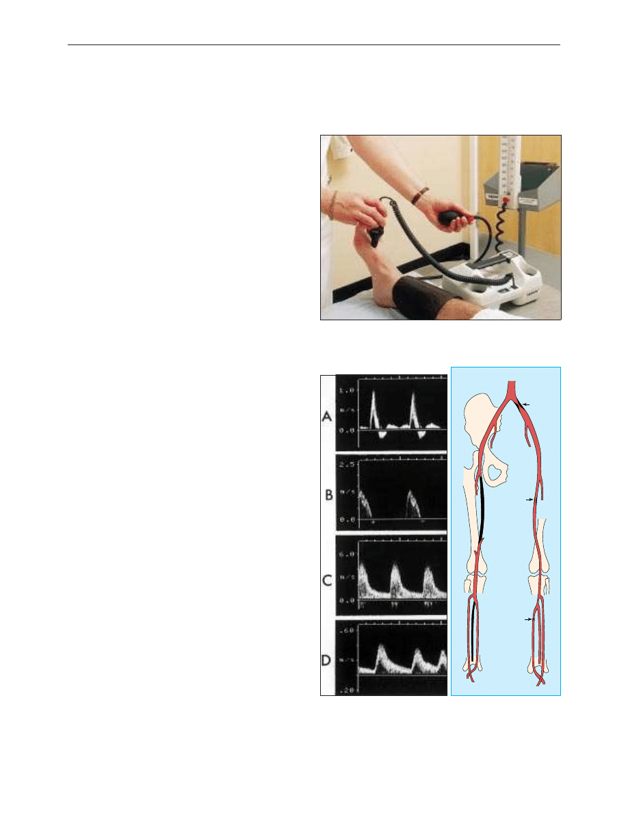
The normal (“triphasic”) Doppler velocity waveform is made up
of three components which correspond to different phases of
arterial flow: rapid antegrade flow reaching a peak during
systole, transient reversal of flow during early diastole, and slow
antegrade flow during late diastole.
Doppler examination of an artery distal to a stenosis will
show characteristic changes in the velocity profile: the rate of
rise is delayed, the amplitude decreased, and the transient flow
reversal in early diastole is lost. In severe disease, the Doppler
Handheld pencil Doppler being used to measure ankle brachial pressure
index
Left: Doppler velocity waveforms: (a) triphasic waveform in normal artery;
(b) biphasic waveform, with increased velocity, through a mild stenosis; (c)
monophasic waveform, with greatly increased velocity, through tight stenosis;
and (d) dampened monophasic waveform, with reduced velocity, recorded
distal to tight stenosis. Right: Anatomical chart used to record position of
stenoses, showing three stenoses with velocity increases of 7
×
, 4
×
, and 3
×
compared with adjacent unaffected arteries
Tight 7x
Significant 3x
4x
Clinical review
698
BMJ
VOLUME 320 11 MARCH 2000 www.bmj.com
on 1 October 2006
bmj.com
Downloaded from
waveform flattens; in critical limb ischaemia it may be
undetectable.
Examination of an arterial stenosis shows an increase in
blood velocity through the area of narrowing. The site(s) of any
stenotic lesions can be identified by serial placement of the
Doppler probe along the extremities. The criteria used to define
a stenosis vary between laboratories, but a twofold increase in
peak systolic velocity compared with the velocity in an adjacent
segment of the artery usually signifies a stenosis of 50% or
more.
By combining the pulsed Doppler system with real time B
mode ultrasound imaging of vessels, it is possible to examine
Doppler flow patterns in a precisely defined area within the
vessel lumen. This combination of real time B mode sound
imaging with pulsed Doppler ultrasonography is called duplex
scanning. The addition of colour frequency mapping (so called
colour duplex or triplex scanners) makes the identification of
arterial stenoses even easier and reduces the scanning time.
Investigations of arterial disease
Ankle brachial pressure index
Under normal conditions, systolic blood pressure in the legs is
equal to or slightly greater than the systolic pressure in the
upper limbs. In the presence of an arterial stenosis, a reduction
in pressure occurs distal to the lesion. The ankle brachial
pressure index, which is calculated from the ratio of ankle to
brachial systolic pressure, is a sensitive marker of arterial
insufficiency.
The highest pressure measured in any ankle artery is used
as the numerator in the calculation of the index; a value >1.0 is
normal and a value < 0.9 is abnormal. Patients with
claudication tend to have ankle brachial pressure indexes in the
range 0.5-0.9, whereas those with critical ischaemia usually have
an index of < 0.5. The index also has prognostic significance
because of the association with arterial disease elsewhere,
especially coronary heart disease.
Diabetic limbs
Systolic blood pressure in the lower limbs cannot be measured
reliably when the vessels are calcified and incompressible—for
example, in patients with diabetes—as this can result in falsely
high ankle pressures. An alternative approach is to use either
the pole test or measurement of toe pressures. Normal toe
systolic pressure ranges from 90-100 mm Hg and is 80-90% of
brachial systolic pressure. A toe systolic pressure < 30 mm Hg
indicates critical ischaemia.
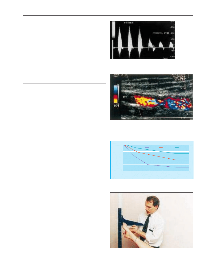
Spectral analysis of blood velocity in a stenosis, and
unaffected area of proximal superficial femoral artery. The
velocity increases from 150 to 300 m/s across the stenosis
Colour duplex scanning of blood flow through stenosis of superficial
femoral artery. Colour assignment (red or blue) depends on direction of
blood flow and colour saturation reflects velocity of blood flow. Less
saturation indicates regions of higher blood flow and deeper colours
indicate slower flow; the absence of flow is coded as black
Years
Patient survival (%)
10
8
6
4
2
0
20
60
800
100
ABPI > 0.85
ABPI 0.4-0.85
ABPI < 0.4
40
Patient survival according to measurements of ankle brachial pressure index
(adapted from McKenna et al, Atherosclerosis 1991;87:119-28)
Pole test for measurement of ankle pressures in patients with calcified
vessels: the Doppler probe is placed over a patent pedal artery and the foot
raised against a pole that is calibrated in mm Hg. The point at which the
pedal signal disappears is taken as the ankle pressure
Relation between increased blood velocity and degree of
stenosis
Diameter of
stenosis (%)
Peak sytolic
velocity* (m/s)
Peak diastolic
velocity* (m/s)
Internal: common
carotid artery
velocity ratio†
0-39
< 1.1
< 0.45
< 1.8
4-59
1.1-1.49
< 0.45
< 1.8
60-79
1.5-2.49
0.45-1.4
1.8-3.7
80-99
2.5-6.1
> 1.4
> 3.7
> 99 (critical) Extremely low
NA
NA
*Measured in lower part of internal carotid artery
†Ratio of peak systolic velocity in internal carotid artery stenosis
relative to proximal measurement in common carotid artery
Clinical review
699
BMJ
VOLUME 320 11 MARCH 2000 www.bmj.com
on 1 October 2006
bmj.com
Downloaded from

Walk test
Exercise testing will assess the functional limitations of arterial
stenoses and differentiate occlusive arterial disease from other
causes of exercise induced lower limb symptoms—for example,
neurogenic claudication secondary to spinal stenosis. A limited
inflow of blood in a limb with occlusive arterial disease results
in a fall in ankle systolic blood pressure during exercise induced
peripheral vasodilatation.
The walk test is performed by exercising the patient for 5
minutes, ideally on a treadmill, but walking the patient in the
surgery or marking time on the spot are adequate. The ankle
brachial pressure index is measured before and after exercise. A
pressure drop of 20% or more indicates significant arterial
disease. If there is no drop in ankle systolic pressure after a 5
minute brisk walk, the patient does not have occlusive arterial
disease proximal to the ankle in that limb.
Duplex scanning
Duplex ultrasonography has a sensitivity of 80% and a
specificity of 90-100% for detecting femoral and popliteal
disease compared with angiography, but it is less reliable for
assessing the severity of stenoses in the tibial and peroneal
arteries. Duplex scanning is especially useful for assessing the
carotid arteries and for surveillance of infrainguinal bypass
grafts where sites of stenosis can be identified before complete
graft occlusion occurs and before there is a fall in ankle brachial
pressure index. The normal velocity within a graft conduit is
50-120 cm/s. As with native arteries, a twofold increase in peak
systolic velocity indicates a stenosis of 50% or more. A peak
velocity < 45 cm/s occurs in grafts at high risk of failure.
Identification of distal vessels for arterial bypass grafting
In critically ischaemic limbs, where occlusive disease tends to be
present at multiple levels, arteriography often fails to show
patent calf or pedal vessels as potential outflows for
femorodistal bypass grafting. Alternative non-invasive
approaches have been developed for preoperative assessment,
including pulse generated run off and dependent Doppler
assessment.
Transcranial Doppler ultrasonography
Lower frequency Doppler probes (1-2 MHz) can be used to
obtain information about blood flow in arteries comprising the
circle of Willis and its principal branches. Mean flow velocities
> 80 cm/s in the middle cerebral artery, or > 70 cm/s in the
posterior and basilar arteries, indicate a serious stenosis.
Transcranial Doppler scanning has several applications but is
especially useful for intraoperative and postoperative
monitoring of patients having carotid endarterectomy.
Helical or spiral computed tomography
Spiral computed tomography is a new, minimally invasive
technique for vascular imaging that is made possible by
combining two recent advances: slip ring computed
tomography (which allows the x ray tube detector apparatus to
rotate continuously) and computerised three dimensional
reconstruction. A helical scan can cover the entire region of
interest (for example, the abdominal aorta from the diaphragm
to the iliac bifurcation) in one 30-40 second exposure, usually in
a single breath hold. This minimises motion artefact and allows
all the scan data to be collected during the first pass of an
intravenous bolus of contrast through the arterial tree—that is
during the time of maximal arterial opacification. A large
number of finely spaced slices from one scan can then be
reconstructed to produce high quality two or three dimensional
images of the contrast enhanced vessels.
Uses of colour duplex scanning
Arterial
x Identify obstructive
atherosclerotic disease:
Carotid
Renal
x Surveillance of
infrainguinal bypass grafts
x Surveillance of lower limb
arteries after angioplasty
Venous
x Diagnosis of deep vein thrombosis
above the knee
x Assessing competence of valves in
deep veins
x Superficial venous reflux:
Assessing patient with recurrent
varicose veins
Identify and locate reflux at
saphenopopliteal junction
x Preoperative mapping of saphenous
vein
Clinical use of transcranial Doppler scanning in adults
x Intraoperative monitoring during carotid endarterctomy:
Shunt function
Cerebral perfusion
x Postoperative montoring after carotid endarterectomy:
Detection of emboli
Formation of carotid thrombus
x Detection of intracranial vasospasm after subarachnoid
haemorrhage
x Detection of middle cerebral artery disease
x Evaluation of collateral circulation in patients with carotid disease
x Evaluation of arteriovenous malformations of the brain
Min after exercise
Ankle brachial pressure index
10
1
20
5
Rest
0
0
0.2
0.3
0.4
0.5
0.6
0.7
0.8
0.9
1
Normal
0.1
Intermittent claudication
Fall in ankle brachial pressure index with exercise in patient with
intermittent claudication and normal subject (adapted from Creager, Vasc
Med
1997;2:231-7)
Spiral computed tomogram of both carotid systems showing a tight stenosis
in the proximal segment of left internal carotid artery
Clinical review
700
BMJ
VOLUME 320 11 MARCH 2000 www.bmj.com
on 1 October 2006
bmj.com
Downloaded from
Magnetic resonance angiography
Magnetic resonance angiography has developed rapidly over
the past five years. It has the advantage of imaging a moving
column of blood and does not require ionising radiation or
iodinated contrast, but the technique has obvious drawbacks in
terms of cost efficiency and accessibility to scanners. A variety of
imaging sequences are used depending on the vessels being
studied and the field strength of the machine. The most
commonly used techniques include time of flight, two and three
dimensional angiography and phase contrast.
Use of a magnetic resonance imaging scanner with a high
field strength (which allows rapid acquisition of data) and a
carefully timed bolus of gadolinium contrast enables high
quality angiographic images to be obtained in a single breath
hold. Magnetic resonance angiography is well established for
examining the cerebral vessels and the carotid arteries, and its
role in other territories is being extended.
Investigations of venous disease
Venous thrombosis
Colour Duplex scanning is both sensitive and specific (90-100%
in most series) for detecting proximal deep vein thrombosis.
Deep veins and arteries lie together in the leg, and the normal
vein appears as an echo-free channel and is usually larger than
the accompanying artery.
Venous ultrasonography is a very accurate method of
identifying deep vein thrombi from the level of the common
femoral vein at the groin crease to the popliteal vein but is less
reliable for diagnosing calf vein thrombosis.
Venous reflux
Colour duplex scanning has revolutionised the investigation of
the lower limb venous system because it allows instant
visualisation of blood flow and its direction. Thus, reflux at the
saphenofemoral junction, saphenopopliteal junction, and
within the deep venous system, including the popliteal vein
beneath the knee and the gastrocnemius veins, can be detected
without invasive techniques. Although venous reflux can be
assessed with a pencil Doppler, this technique misses 12% of
saphenofemoral and 20% of saphenopopliteal junction reflux
compared with colour duplex scanning.
We thank Jean Clarke for expert secretarial assistance; Frances Ryan and
Tim Hartshorne (vascular technicians) and colleagues in the vascular labo-
ratories at Derbyshire Royal Infirmary and Leicester Royal Infirmary; Ken
Callum and Roddy Nash (vascular surgeons) for helpful input to the
manuscript and illustrations; and Jane Wain and staff of the audiovisual
department at Derbyshire Royal Infirmary.
David Hinwood is consultant vascular radiologist, Derbyshire Royal
Infirmary, Derby.
The ABC of arterial and venous disease is edited by Richard
Donnelly, professor of vascular medicine, University of Nottingham
and Southern Derbyshire Acute Hospitals NHS Trust
(richard.donnelly@ nottingham.ac.uk) and Nick J M London,
professor of surgery, University of Leicester, Leicester
(sms16@leicester.ac.uk). It will be published as a book later
this year.
BMJ
2000;320:698-701
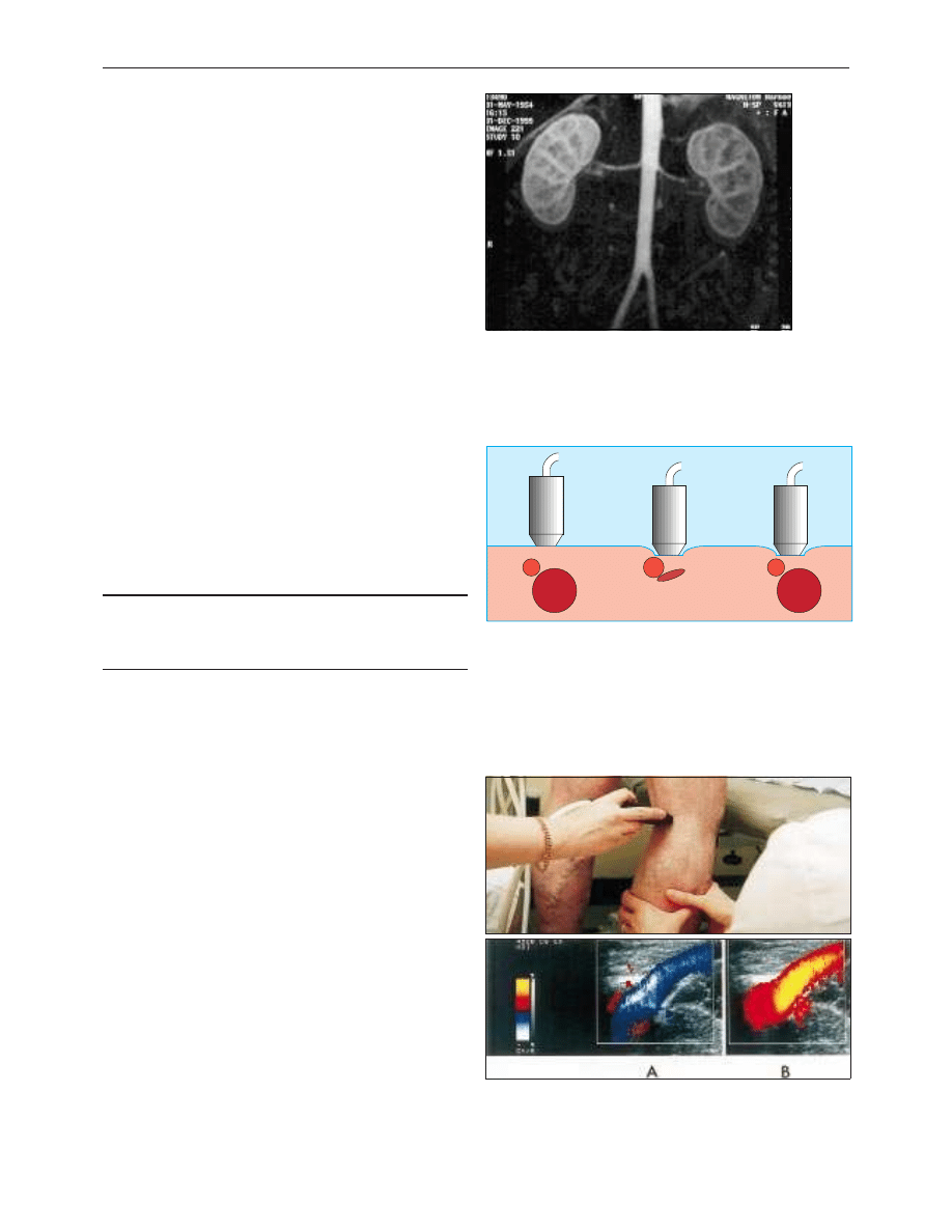
Magnetic resonance angiogram using an intravenous bolus of
gadolinium contrast showing normal renal arteries
Artery
Artery
Artery
Vein
Vein
Vein
Ultrasound detection of deep vein thrombosis. The probe is held lightly on
the skin and advanced along the course of the vein (left). Pressure is applied
every few centimetres by compressing the transducer head against the skin.
The vein collapses during compression if no thrombus is present (middle)
but not if a deep vein thrombus is present (right)
Colour duplex scanning of saphenopopliteal junction. The calf muscles are
manually compressed producing upward flow in the vein (top), which
appears as a blue colour for flow towards the heart (panel A). Sudden
release of the distal compression causes reflux, seen as a red colour
indicating flow away from the heart (panel B)
Criteria for diagnosis of deep vein thrombosis
x Failure of vein to collapse on direct compression
x Visualisation of thrombus within lumen
x Absent or abnormal venous pulsation on Doppler scanning
Clinical review
701
BMJ
VOLUME 320 11 MARCH 2000 www.bmj.com
on 1 October 2006
bmj.com
Downloaded from

ABC of arterial and venous disease
Acute limb ischaemia
Ken Callum, Andrew Bradbury
Limb ischaemia is classified on the basis of onset and severity.
Complete acute ischaemia will lead to extensive tissue necrosis
within six hours unless the limb is surgically revascularised.
Incomplete acute ischaemia can usually be treated medically in
the first instance. Patients with irreversible ischaemia require
urgent amputation unless it is too extensive or the patient too ill
to survive.
Clinical features
Apart from paralysis (inability to wiggle toes or fingers) and
anaesthesia (loss of light touch over the dorsum of the foot or
hand), the symptoms and signs of acute ischaemia are
non-specific or inconsistently related to its completeness. Pain
on squeezing the calf indicates muscle infarction and
impending irreversible ischaemia.
Acute arterial occlusion is associated with intense spasm in
the distal arterial tree, and initially the limb will appear “marble”
white. Over the next few hours, the spasm relaxes and the skin
fills with deoxygenated blood leading to mottling that is light
blue or purple, has a fine reticular pattern, and blanches on
pressure. At this stage the limb is still salvageable. However, as
ischaemia progresses, stagnant blood coagulates leading to
mottling that is darker in colour, coarser in pattern, and does
not blanch. Finally, large patches of fixed staining progress to
blistering and liquefaction. Attempts to revascularise such a
limb are futile and will lead to life threatening reperfusion
injury. In cases of real doubt the muscle can be examined at
surgery through a small fasciotomy incision. It is usually
obvious when the muscle is dead.
Aetiology
Acute limb ischaemia is most commonly caused by acute
thrombotic occlusion of a pre-existing stenotic arterial segment
(60% of cases) or by embolus (30%). Distinguishing these two
conditions is important because treatment and prognosis are
different. Other causes are trauma, iatrogenic injury, popliteal
aneurysm, and aortic dissection.
More than 80% of peripheral emboli arise from the left
atrial appendage in association with atrial fibrillation. They may
also arise from the left ventricle, heart valves, prosthetic bypass
grafts, aneurysmal disease, paradoxical embolism, and atrial
myxoma (rare). In 15% of cases the source of embolus is
obscure. Thrombosis in situ may arise from acute plaque
rupture, hypovolaemia, or pump failure (see below).
Management
General measures
When a patient is suspected to have an acutely ischaemic limb
the case must be discussed immediately with a vascular surgeon.
A few hours can make the difference between death or
amputation and complete recovery of limb function. If there are
no contraindications (acute aortic dissection or multiple trauma,
particularly serious head injury) give an intravenous bolus of
heparin to limit propagation of thrombus and protect the
collateral circulation.
Classification of limb ischaemia
Terminology
Onset:
Acute
Acute on chronic
Chronic
Severity (acute, acute on chronic):
Incomplete
Complete
Irreversible
Definition or comment
Ischaemia < 14 days
Worsening symptoms and
signs ( < 14 days)
Ischaemia stable for > 14 days
Limb not threatened
Limb threatened
Limb non-viable
Symptoms and signs of acute limb ischaemia
Symptoms or signs
Pain
Pallor
Pulseless
Perishing cold
Paraesthesia*
Paralysis*
Comment
Occasionally absent in complete ischaemia
Also present in chronic ischaemia
Also present in chronic ischaemia
Unreliable as ischaemic limb takes on ambient
temperature
Leading to anaesthesia (unable to feel touch
on foot or hand)
Unable to wiggle toes or fingers
*Anaesthesia and paralysis are the key to diagnosing complete ischaemia
that requires emergency surgical treatment
Differentiation of embolus and acute arterial thrombosis
(thrombosis in situ)
Clinical features
Severity
Onset
Limb affected
Multiple sites
Embolic source
Previous claudication
Palpation of artery
Bruits
Contralateral leg
pulses
Diagnosis
Treatment
Embolus
Complete (no
collaterals)
Seconds or minutes
Leg 3:1 arm
Up to 15%
Present (usually atrial
fibrillation)
Absent
Soft, tender
Absent
Present
Clinical
Embolectomy,
warfarin
Thrombosis
Incomplete
(collaterals)
Hours or days
Leg 10:1 arm
Rare
Absent
Present
Hard, calcified
Present
Absent
Angiography
Medical, bypass,
thrombolysis
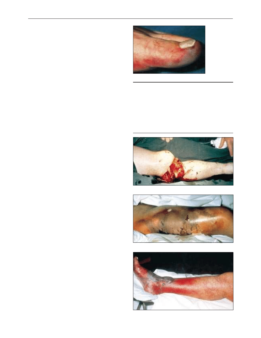
Marble white foot (left of picture) in
patient with acute ischaemia
Clinical review
764
BMJ
VOLUME 320 18 MARCH 2000 www.bmj.com
on 1 October 2006
bmj.com
Downloaded from
Is angiography required?
If ischaemia is complete, the patient must be taken directly to
the operating theatre because angiography will introduce delay,
thrombolysis is not an option, and lack of collateral flow will
prevent visualisation of the distal vasculature. If ischaemia is
incomplete the patient should have preoperative angiography
since simple embolectomy or thrombectomy is unlikely to be
successful, thrombolysis may be an option, and the surgeon
requires a “road map” for distal bypass.
Acute embolus
Embolic occlusion of the brachial artery is not usually limb
threatening, and in elderly people non-operative treatment is
reasonable. Younger patients should have embolectomy to
prevent subsequent claudication, especially if the dominant arm
is affected.
A leg affected by embolus is nearly always threatened and
requires immediate surgical revascularisation. Emboli usually
lodge at the common femoral bifurcation or, less commonly,
the popliteal trifurcation. Femoral embolus is associated with
profound ischaemia to the level of the upper thigh because the
deep femoral artery is also affected. A femoral pulse does not
exclude the diagnosis. Embolectomy can be done under local,
regional, or general anaesthetic.
The adequacy of embolectomy should be confirmed by
angiography while the patient is on the operating table.
On-table thrombolysis should be considered if mechanical
clearance has been unsuccessful. If the embolus has occurred in
an area of longstanding atherosclerotic disease, surgical bypass
may be necessary.
Postoperatively the patient should continue to receive
heparin to prevent formation of further emboli. Many surgeons
postpone heparin for six hours after surgery to reduce the risk
of a haematoma forming. Warfarin reduces the risk of recurrent
embolism, and unless contraindicated, should be prescribed to
all patients long term. Patients should not be given warfarin
without first being on heparin for 48 hours since warfarin can
produce a transient procoagulant state due to inhibition of the
vitamin K dependent anticoagulant proteins C and S.
Opinions differ about how thorough you should be in
establishing the source of emboli. Transthoracic
echocardiography is poor at detecting a thrombus in patients
with atrial fibrillation, and a negative result does not exclude the
diagnosis. Transoesophageal echocardiography provides
excellent views of the left atrium but is moderately invasive and
not universally available. In patients with suspected paroxysmal
tachyarrhythmias, 24 hour electrocardiographic monitoring
should be considered. Even if no source of embolism is found,
anticoagulation should continue long term.
Although immediate loss of a limb after correctly managed
acute embolus is unusual, many series report a 10-20%
in-hospital mortality from heart failure or recurrent embolism,
particularly stroke.
Saddle embolus
Patients with acute embolic occlusion of the aortic bifurcation
have femoral pulses and appear marble white or mottled to the
waist. They may also present with paraplegia due to ischaemia
of the cauda equina, which can be irreversible. Immediate
bilateral embolectomy restores lower limb perfusion, but many
patients subsequently die from reperfusion injury.
Popliteal aneurysm
A popliteal aneurysm can initiate acute ischaemia by forming a
thrombus or acting as a source of emboli. Thrombolysis is often
the best treatment as simple embolectomy or thrombectomy
Factors predisposing to acute thrombosis
Cause
Dehydration
Hypotension
Unusual posture or
activity
Malignancy
Hyperviscosity
Thrombophilia
Comment
Hot weather, diabetes, infection, gastroenteritis
Myocardial infarction, arrhythmia, heart failure,
gastrointestinal haemorrhage, septic shock,
multiple organ failure
Prolonged sitting, kneeling
Solid and haematological
Polycythaemia, thrombocytosis
Protein C or S and antithrombin III
deficiencies; activated protein C resistance;
factor V Leiden; antiphospholipid syndrome
Embolus at popliteal trifurcation
On-table angiograms showing
incomplete clearance of embolus
Aortic occlusion
Clinical review
765
BMJ
VOLUME 320 18 MARCH 2000 www.bmj.com
on 1 October 2006
bmj.com
Downloaded from
usually leads to early rethrombosis and surgical bypass is often
precluded by obliteration of the distal run-off. Once the
circulation is restored, a bypass should be performed to exclude
the aneurysm.
Atheroembolism
Cholesterol emboli are shed from a complex, often acutely
ruptured, atherosclerotic plaque. Distal pulses are usually
present. The patient characteristically presents with the blue toe
(finger) syndrome, which may mimic Raynaud’s phenomenon. If
the blue toe syndrome is not recognised patients may
deteriorate rapidly and require amputation.
Thrombosis in situ
Limbs affected by stable chronic ischaemia do not usually
suddenly deteriorate without a reason—for example, silent
myocardial infarction or underlying, hitherto asymptomatic,
malignancy. Septicaemia, particularly pneumococcal and
meningococcal, may be associated with widespread thrombosis.
Trauma
The commonest causes of non-iatrogenic injury are limb
fractures and dislocations (supracondylar fractures of the
humerus in children, tibial fractures in adults), blunt injuries
occurring in road traffic accidents, and stab wounds. In the
United Kingdom, acute traumatic limb ischaemia is often
iatrogenic, being caused by arterial cannulation (coronary
angioplasty, aortic balloon pump), vascular and orthopaedic
procedures on the limb (especially if exsanguinating
tourniquets are used), or pelvic surgery (cystectomy, anterior
resection) in patients with subclinical aortoiliac disease in whom
the ligated pelvic collaterals form the main blood supply to the
legs. Postoperative assessment of lower limb ischaemia may be
confused by the presence of epidural or spinal anaesthesia.
The presence of distal pulses does not exclude serious
arterial injury. Pulse oximetry, Doppler signals, and
measurement of the ankle brachial pressure index may be
helpful, but in cases of doubt, proceed to angiography.
Intra-arterial drug administration
Intra-arterial drug administration leads to intense spasm and
microvascular thrombosis. The leg is mottled and digital
gangrene is common, but pedal pulses are usually palpable. The
mainstay of treatment is supportive care, hydration to minimise
renal failure secondary to rhabdomyolysis, and full
heparinisation. Vascular reconstruction is almost never
indicated, but fasciotomy may be required to prevent a
compartment syndrome.
Venous gangrene
Venous gangrene can be mistaken for acute limb ischaemia.
However, the leg is invariably swollen and the superficial veins
full. Oedema may make it impossible to palpate pedal pulses,
but Doppler examination will show normal distal waveforms
and pressures. Management includes elevation, heparinisation,
thrombolysis, and treatment of the underlying cause (usually
pelvic or abdominal malignancy).
Aortic dissection
This may cause upper and lower limb ischaemia due to
pinching of the ostia of the relevant arteries by the false lumen.
Thoracic outlet syndrome
Pressure on the subclavian artery from a cervical rib or
abnormal soft tissue band may lead to a post-stenotic dilatation
lined with thrombus, which predisposes to occlusion or
Initial management of acute limb ischaemia
Sensation and movement absent
x Intravenous heparin
x Rapid resuscitation to best medical condition
x Intravenous fluids, catheter, and good urine output
x Urgent surgery—embolectomy or bypass
Sensation and movement present
x Optimise patient to best medical condition
x Admit to hospital
x Intravenous heparin
x Observe limb for signs of deterioration (and act if it occurs)
x Arteriogram when convenient
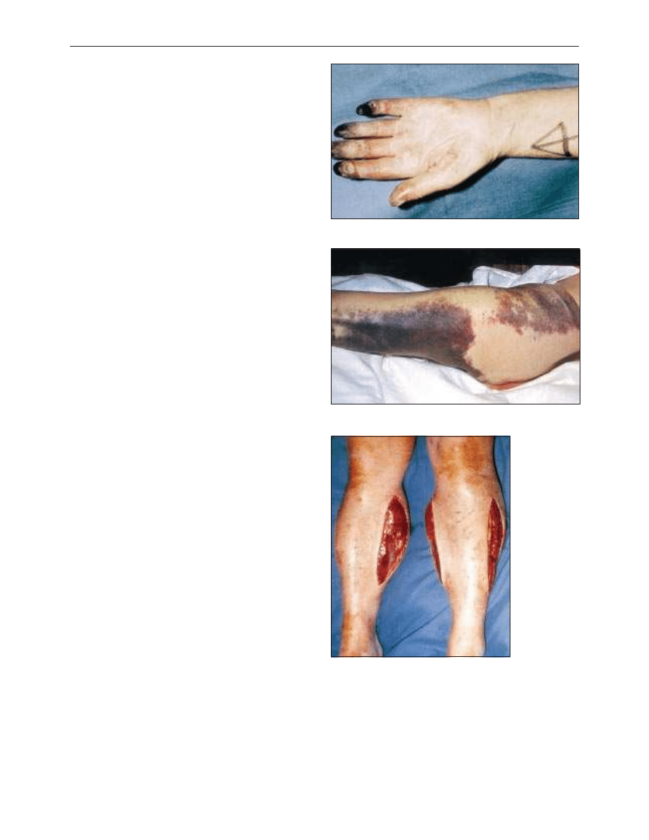
Blue toe syndrome must be promptly identified
Compound fracture of tibia with ischaemia
Ischaemia after intra-arterial drug administration
Venous gangrene
Clinical review
766
BMJ
VOLUME 320 18 MARCH 2000 www.bmj.com
on 1 October 2006
bmj.com
Downloaded from
embolisation. The distal circulation may be chronically
obliterated and digital ischaemia advanced before the thoracic
outlet syndrome is diagnosed. The diagnosis is based on the
results of duplex ultrasonography or angiography, or both.
Treatment options include thrombolysis, thrombectomy or
embolectomy, excision of the cervical rib, and repair of the
aneurysmal segment.
Thrombolysis
In thrombolysis a cannula is embedded into the distal extent of
the thrombus and streptokinase or, preferably, recombinant
tissue plasminogen activator is infused. The technique cannot
be used in patients with complete ischaemia because thrombus
dissolution takes several hours. It is also relatively ineffective
against the organised thrombus present in most peripheral
emboli and is associated with an appreciable minor (20%,
mainly groin haematoma) and major (5%, serious haemorrhage
and stroke) complication rate. Thrombolysis should be
undertaken only in an environment where experienced nursing
and medical staff can closely monitor the patient.
Post-ischaemic syndromes
Reperfusion injury
The reintroduction of oxygenated blood after a period of
ischaemia causes more damage than the ischaemia alone.
Generation of highly reactive, oxygen free radicals is greatly
increased, and these activate neutrophils which migrate into the
reperfused tissue causing injury. For vascular injury to occur
neutrophils must be present and must adhere to the
endothelium. The damaged endothelial cells become more
permeable.
Effects of reperfusion syndrome
Local—
Limb swelling due to increased capillary permeability
causes a compartment syndrome, impaired muscle function due
to ischaemia, and subsequent muscle contracture if the muscle
infarcts.
General—
Acidosis and hyperkalaemia occur due to leakage
from the damaged cells, causing cardiac arrhythmias and
myoglobinaemia, which can result in acute tubular necrosis.
Acute respiratory distress syndrome may also develop, and
gastrointestinal endothelial oedema may lead to increased
gastrointestinal vascular permeability and endotoxic shock.
Compartment syndrome
Increased capillary permeability and oedema on reperfusion in
the calf, where muscles are confined within tight fascial
boundaries, causes an increase in interstitial pressure leading to
muscle necrosis despite apparently adequate inflow—
compartment syndrome. There is swelling and pain on
squeezing the calf muscle or moving the ankle. Palpable pedal
pulses do not exclude the syndrome. The key to management is
prevention through expeditious revascularisation and a low
threshold for fasciotomy. (If in doubt—do it.)
Chronic pain syndromes
Acute complete ischaemia can lead to peripheral nerve injury
that manifests as the chronic pain syndrome, also referred to as
causalgia, reflex sympathetic dystrophy, and many other terms.
If the syndrome is recognised and treated early then many
patients gain prolonged relief from drugs or chemical or
surgical sympathectomy.
We thank Professor C V Ruckley, Mr A Jenkins, and Mr J A Murie for
help with the illustrations.
Digital gangrene due to pressure on subclavian artery from cervical rib
Haematoma due to thrombolysis
Fasciotomy
Ken Callum is consultant surgeon, Derbyshire Royal Infirmary, Derby,
and Andrew Bradbury is professor of surgery, University of
Birmingham, Birmingham Heartlands Hospital, Birmingham.
The ABC of arterial and venous disease is edited by Richard Donnelly,
professor of vascular medicine, University of Nottingham and
Southern Derbyshire Acute Hospitals NHS Trust
(richard.donnelly@nottingham.ac.uk) and Nick J M London, professor
of surgery, University of Leicester, Leicester (sms16@leicester.ac.uk). It
will be published as a book later this year.
BMJ
2000;320:764-7
Clinical review
767
BMJ
VOLUME 320 18 MARCH 2000 www.bmj.com
on 1 October 2006
bmj.com
Downloaded from
ABC of arterial and venous disease
Chronic lower limb ischaemia
Jonathan D Beard
Peripheral vascular disease commonly affects the arteries
supplying the leg and is mostly caused by atherosclerosis.
Restriction of blood flow, due to arterial stenosis or occlusion,
often leads patients to complain of muscle pain on walking
(intermittent claudication). Any further reduction in blood flow
causes ischaemic pain at rest, which affects the foot. Ulceration
and gangrene may then supervene and can result in loss of the
limb if not treated. The Fontaine score is useful when classifying
the severity of ischaemia.
Although many patients with claudication remain stable,
about 150-200 per million of the population progress to critical
limb ischaemia (Fontaine III or IV) each year. Many patients
with critical limb ischaemia can undergo revascularisation,
which has a reasonable chance of saving the limb. A recent
audit by the Vascular Surgical Society found a success rate of
over 70% for these patients. However, many patients still require
major amputation. Rehabilitation of elderly patients after
amputation can prove difficult, with high community costs.
Critical limb ischaemia has been estimated to cost over £200m
a year in the United Kingdom.
Intermittent claudication
History and examination
A history of muscular, cramp-like pain on walking that is
rapidly relieved by resting, together with absent pulses, strongly
supports the diagnosis of intermittent claudication. Disease of
the superficial femoral artery in the thigh results in absent
popliteal and foot pulses and often causes calf claudication.
Disease of the aorta or iliac artery results in a weak or absent
femoral pulse, often associated with a femoral bruit. Disease at
this level may cause calf, thigh, or buttock claudication.
The dorsalis pedis artery lies superficially on the dorsum of
the foot, although its position varies considerably. The posterior
tibial artery lies deeper behind the medial malleolus. Many
healthy people have only one foot pulse. The popliteal pulse
can be difficult to palpate in muscular patients. A prominent
popliteal pulse suggests the possibility of a popliteal aneurysm.
Fontaine classification of chronic leg ischaemia
Stage I Asymptomatic
Stage II Intermittent claudication
Stage III Ischaemic rest pain
Stage IV Ulceration or gangrene, or both
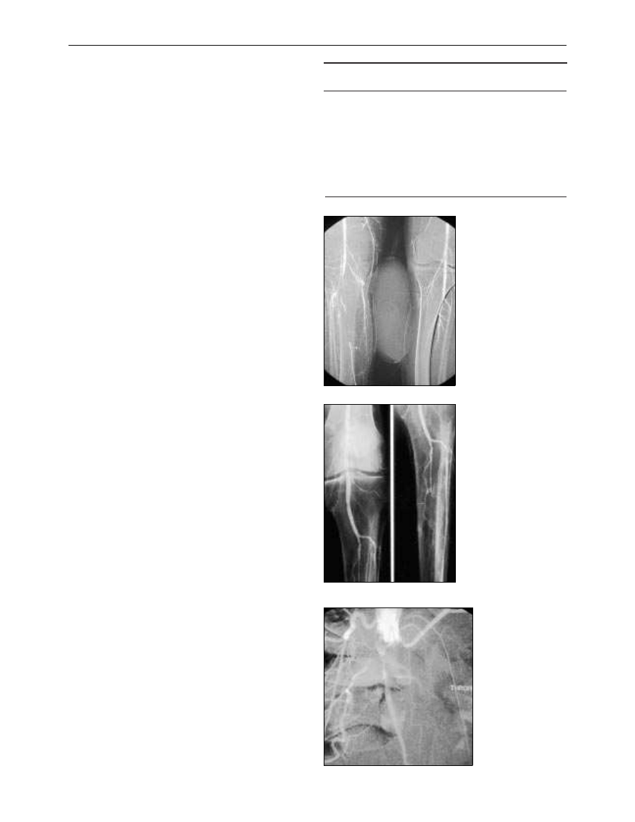
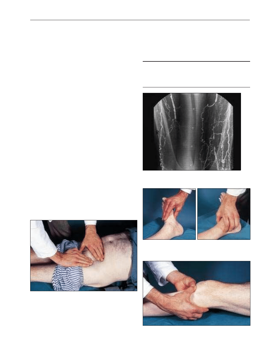
Angiogram showing bilateral occlusions of superficial femoral
arteries in thighs. Collaterals arising from the profunda femoris
artery can functionally bypass this occlusion
Method of palpating dorsalis pedis (left) and posterior tibial (right) pulses.
Examine pulses from the foot of the bed, keeping the fingers flat for the
dorsalis pedis and using the fingertips for the posterior tibial, while applying
counterpressure with the thumb
Method of palpating popliteal artery with patient’s knee slightly flexed. Use
thumbs to apply counterpressure while palpating the artery, which lies deep
in popliteal fossa, with fingers
Method of palpating femoral pulse in skin crease of groin. Counterpressure
on the lower abdomen pushes the skin crease towards the inguinal ligament
and reduces the risk of missing the pulse
Clinical review
854
BMJ
VOLUME 320 25 MARCH 2000 www.bmj.com
on 1 October 2006
bmj.com
Downloaded from

Differential diagnosis
The pain of nerve root compression can be mistaken for
vascular claudication. A careful history can usually distinguish
nerve root compression, especially sciatica due to compression
of the lumbosacral root. However, compression of the cauda
equina due to spinal stenosis can be more difficult to diagnose.
This condition usually causes pain that radiates down both legs.
Although the pain is made worse by walking, it also comes on
after prolonged standing and is not rapidly relieved by rest,
unlike vascular claudication.
Investigation
There are many causes of leg pain that can occur in the
presence of asymptomatic peripheral vascular disease.
Therefore, the absence of pulses does not necessarily imply a
causal link. Furthermore, the presence of pulses at rest does not
exclude symptomatic peripheral vascular disease. A good
history together with an ankle brachial systolic pressure index
of less than 0.9 confirms the diagnosis.
Exercise testing provides an objective measurement of
walking distance, and highlights other exercise limiting
conditions such as arthritis and breathlessness. However,
exercise testing takes time, and many patients find it difficult or
impossible to walk on a treadmill. Only those with a good
history of claudication and normal resting ankle brachial
systolic pressure indexes require an exercise test.
Duplex ultrasound scanning is useful for delineating the
anatomical site of disease in the lower limb. Many hospitals still
use arteriography for this purpose or when the results of
duplex scanning are equivocal. This invasive and expensive
investigation should not be requested unless there is a plan to
proceed with revascularisation, if possible.
Principles of treatment
Intermittent claudication seems a relatively benign condition,
although severe claudication may preclude patients from manual
work. The risk of generalised vascular disease is probably more
important. Patients with claudication have a three times higher
risk of death compared with age matched controls. Modification
of risk factors is therefore vital to reduce death from myocardial
infarction and stroke. All patients should be advised to stop
smoking and take regular exercise. They should also be screened
for hyperlipidaemia and diabetes. Patients with peripheral
vascular disease benefit from regular chiropody, and those with
diabetes should attend a foot clinic. Obesity reduces exercise
capacity, and losing weight will improve the walking distance.
Drug treatment
All patients with peripheral vascular disease benefit from
aspirin (75-300 mg/day) because this reduces the risk of
cardiovascular events. Patients who are intolerant of aspirin
should take dipyridamole (200 mg, twice daily) or clopidogrel
(75 mg/day). Naftidrofuryl may improve the walking distance of
patients with moderate claudication (less than 500 m), but it is
not known if it affects the outcome of the disease. The evidence
to support naftidrofuryl is controversial, and patients prescribed
it should be reassessed after three to six months.
Exercise programmes
A recent meta-analysis of 21 supervised exercise programmes
showed that training for at least six months, by walking to near
maximum pain tolerance, significantly improved pain free and
maximum walking distances. The only controlled trial
comparing an exercise programme with percutaneous
transluminal angioplasty found that exercise was better.
Exercise programmes are cheaper than percutaneous
Factors which may influence the decision to treat
claudication
For
Against
Severe symptoms
Short history
Job affected
Continued smoking
No better after exercise training
Severe angina or chronic
obstructive airways disease
Stenosis or short occlusion
Long occlusion
Proximal disease
Distal disease
Unilateral disease
Multilevel disease
Leg pain
? claudication
Resting ankle brachial
pressure index
Normal
Poor history
Good history
Not vascular
Exercise test
No fall in
pressures
Fall in
pressures
Vascular
claudication
Poor history
Good history
Reduced
Algorithm for investigation of suspected intermittent claudication
Non-invasive
assessment
Modification of risk
factors
Warrants
intervention
Bypass or
endarterectomy
Angioplasty
Intervention
not justified
Arteriography
or duplex scanning
Claudication
Worse
Stable or
improved
Exercise
programme
Algorithm for treatment of intermittent claudication
Treadmills can be used for
objective measurement of walking
distance and for exercise training
Clinical review
855
BMJ
VOLUME 320 25 MARCH 2000 www.bmj.com
on 1 October 2006
bmj.com
Downloaded from
transluminal angioplasty or surgery, although long term
compliance seems poor.
Endovascular techniques
The number of percutaneous transluminal angioplasties
performed for claudication has risen steeply in recent years. In
some situations endovascular techniques have virtually replaced
conventional surgery. Percutaneous transluminal angioplasty
seems best suited for stenoses or short occlusions of the iliac
and superficial femoral vessels, with one year patency rates of
90% and 80% respectively. Angioplasty carries a small but
definite risk of losing the limb because of thrombosis or
embolisation, and patients should be informed of this risk.
Metallic stents push back the atheroma and improve on the
initial lumen gain after angioplasty alone. The indications for
iliac stents include a residual stenosis or dissection after
angioplasty and long occlusions, but there seems little evidence
to justify their routine use. Deployment of stents more distally
has produced disappointing results due to high restenosis rates.
Surgery
The role of bypass for longer arterial occlusions remains poorly
defined because of a lack of proper trials comparing it with
percutaneous transluminal angioplasty and conservative
treatment. Polyester (Dacron) aortobifemoral bypass grafts have
five year patency rates of over 90% but are associated with a
mortality of up to 5%. Complications include graft infection
and postoperative impotence. Femoropopliteal bypass grafting,
using autologous long saphenous vein, polyester, or
polytetrafluoroethylene (Goretex) yields patency rates of less
than 70% at five years. The early patency of prosthetic grafts
seems similar to that of vein grafts, although the long term
results seem less good. Femoropopliteal bypass grafts should
rarely be used for patients with claudication.
Critical limb ischaemia
History and examination
Patients with critical limb ischaemia often describe a history of
deteriorating claudication, progressing to nocturnal rest pain.
Ulceration or gangrene commonly results from minor trauma.
Nocturnal rest pain often occurs just after the patient has fallen
asleep when the systemic blood pressure falls, further reducing
perfusion to the foot. Hanging the foot out of bed increases
perfusion and produces the typical dusky red hue due to loss of
capillary tone. Elevation causes pallor and venous guttering.
Inspect the foot carefully for ulceration under the heel and
between the toes. Swelling suggests deep infection. If you can
palpate foot pulses consider an alternative cause of pain, such
as gout. Patients with critical limb ischaemia require urgent
referral to a vascular surgeon.
Investigation
The ankle brachial systolic pressure index is usually less than
0.5. Arterial calcification may result in falsely increased
pressures, and caution is needed when relying on Doppler
pressures alone, especially in diabetic patients. All patients with
critical limb ischaemia should ideally have arteriography with a
view to endovascular treatment, if feasible. Duplex scanning
may be used instead of angiography and for mapping of the
long saphenous vein before distal bypass surgery. Dependent
Doppler or pulse generated run-off can help to determine the
most suitable artery to receive a distal bypass graft if these
cannot be identified by angiography.
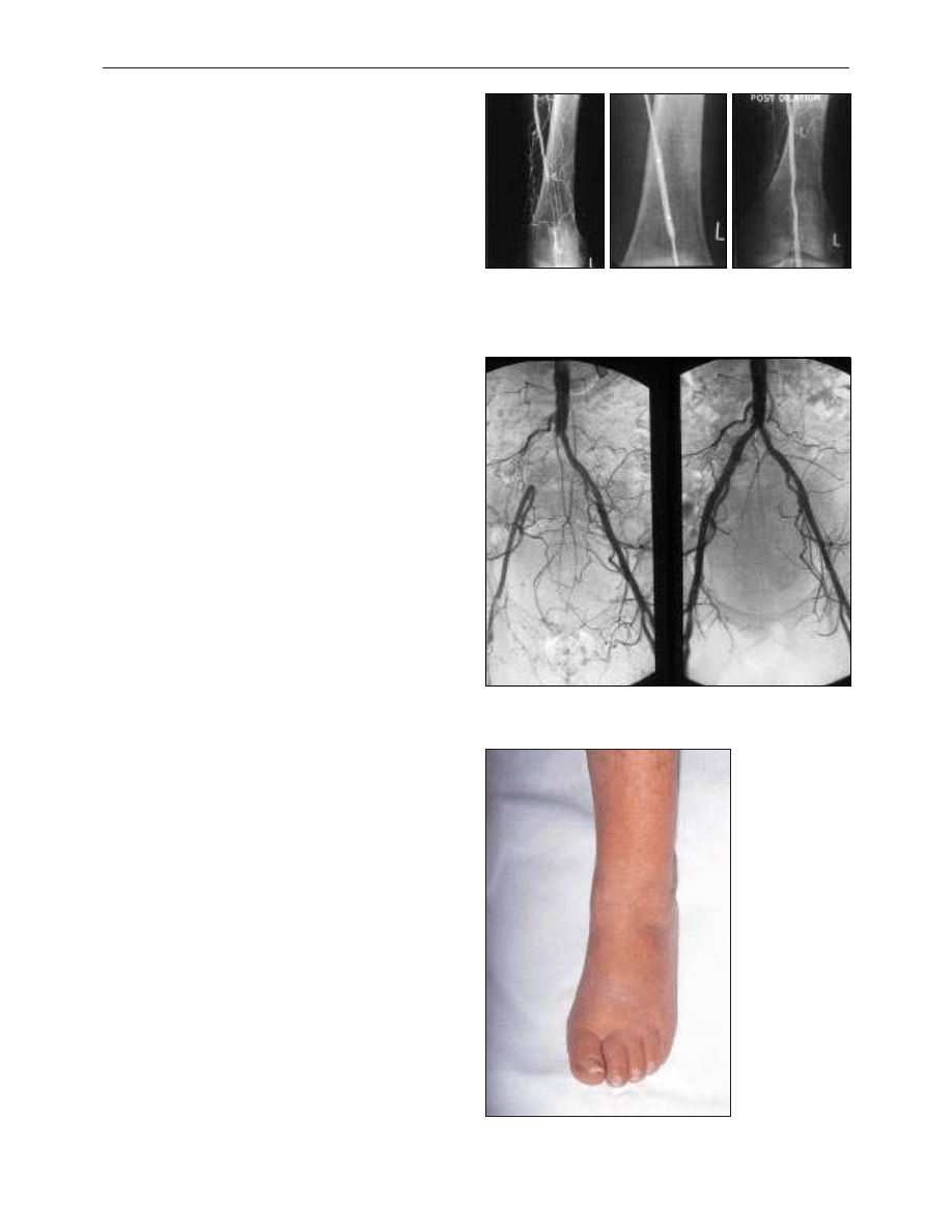
Short occlusion of left popliteal artery (left) treated by percutaneous
transluminal angioplasty. The balloon catheter is passed through the
occlusion over a guide wire and inflated (middle). Appearance after
angioplasty is shown on right
Occlusion of the right common iliac artery before (left) and after (right)
insertion of stent
Critically ischaemic foot displaying typical dusky
red hue on dependency (ischaemic rubor)
Clinical review
856
BMJ
VOLUME 320 25 MARCH 2000 www.bmj.com
on 1 October 2006
bmj.com
Downloaded from

Principles of treatment
The same principles and techniques used to treat claudication
also apply to critical limb ischaemia. However, critical limb
ischaemia is usually caused by multilevel disease, which means
that success rates are lower. Treatment focuses on saving the
limb, although modification of risk factors remains important.
Endovascular treatment
Percutaneous transluminal angioplasty or stenting of proximal
disease may relieve ischaemic rest pain, but healing of
ulceration or gangrene usually requires restoration of foot
pulses. This may necessitate extensive angioplasty of the
superficial femoral, popliteal, and tibial arteries. Good results
have been reported with subintimal angioplasty. Endovascular
treatment can also reduce the magnitude of subsequent
surgery.
Surgery
Patients with a pattern of arterial disease considered unsuitable
for endovascular treatment will usually require surgery. Fit
patients with proximal disease benefit greatly from
aortobifemoral bypass grafting. In unfit patients the options
include crossfemoral bypass for unilateral disease or
axillobifemoral bypass for bilateral disease. These
extra-anatomic procedures have lower patency rates. Many
patients with distal disease will require bypass grafting to the
popliteal or crural arteries below the knee. Autologous vein
grafts give the best patency rates (70% at one year).
Postoperative duplex surveillance may improve patency by
permitting the detection and treatment of vein graft stenoses
before occlusion occurs.
Amputation
Patients with unreconstructable peripheral vascular disease,
fixed flexion deformities, or extensive tissue loss usually require
a major amputation. Preservation of the knee joint has
enormous advantages for wearing artificial limbs and
subsequent mobility. However, there is little point in risking a
non-healing, below knee amputation if the patient seems
unlikely to walk again. Similarly, a patient with good prospects
of wearing an artificial limb will fare better with an above knee
amputation, if below knee amputation seems unachievable.
Local amputation of ulcerated or gangrenous toes will not heal
without revascularisation.
Pain relief
Critical limb ischaemia causes severe pain that requires narcotic
analgesia to provide relief. A slow release opiate such as
morphine seems a good option. Opiates can be supplemented
by non-steroidal anti-inflammatory drugs if these are not
contraindicated. Apart from rehydration, adequate analgesia
alone may be the best treatment for patients with dementia or
other severe comorbidity. If opiate analgesia remains
inadequate, then lumbar sympathectomy (surgical or chemical)
or spinal cord stimulation may help.
About 20-30% of patients with critical limb ischaemia have
unreconstructable disease. A meta-analysis of six randomised
trials of Iloprost, a stable prostacyclin analogue, found that
infusion of this drug reduced the death and amputation rate.
Phantom limb pain may complicate major amputation.
Amitryptyline, carbamazepine, transcutaneous nerve
stimulation, and acupuncture can help in this situation.
Methods of pain relief for critical limb ischaemia
x Slow release opiate analgesia—for example, morphine sulphate
x Prostacyclin analogues*—for example, Iloprost or prostaglandin E
1
(alprostadil)
x Chemical or surgical lumbar sympathectomy
x Dorsal column spinal stimulation
*Not licensed in United Kingdom
Further reading
x Scottish Intercollegiate Guidelines Network. Drug therapy for
peripheral vascular disease. SIGN 1998;27.
x Joint British recommendations on the prevention of coronary heart
disease in clinical practice. Heart 1998;80(suppl):S1-29.
x Leng GC, Fowler B, Ernst F. Exercise for intermittent claudication.
In: Cochrane Library. Issue 4. Oxford: Update Software, 1999.
x Davies AH, Beard JD, Wyatt MG. Essential vascular surgery. London;
W B Saunders, 1999.
Critical limb ischaemia
Limb not
salvageable
Patient moribund
Patient fit
Arteriography or
duplex scanning
Analgesia
Amputation
Non-healing
Healing
Angioplasty
Bypass or
endarterectomy
Limb
salvageable
Algorithm for treatment of critical limb ischaemia
Iliac occlusion
Iliac stenosis
Iliac angioplasty
and
crossover graft
Aortobifemoral
bypass graft
Axillobifemoral
bypass graft
Surgical treatment options for aortoiliac disease
Jonathan D Beard is consultant vascular surgeon at Sheffield Vascular
Institute, Northern General Hospital, Sheffield.
BMJ
2000;320:854-7
The ABC of arterial and venous disease is edited by Richard Donnelly,
professor of vascular medicine, University of Nottingham and
Southern Derbyshire Acute Hospitals NHS Trust (richard.donnelly@
nottingham.ac.uk) and Nick J M London, professor of surgery,
University of Leicester, Leicester (sms16@leicester.ac.uk). It will be
published as a book later this year.
Clinical review
857
BMJ
VOLUME 320 25 MARCH 2000 www.bmj.com
on 1 October 2006
bmj.com
Downloaded from

ABC of arterial and venous disease
Acute stroke
Philip M W Bath, Kennedy R Lees
Acute stroke is now a treatable condition that deserves urgent
specialist attention. Drug treatment and specialist care both
influence survival and recovery. This article considers the
optimal approaches to diagnosis and early management.
Stroke, a sudden neurological deficit of presumed vascular
origin, is a clinical syndrome rather than a single disease. It is a
common and devastating condition that causes death in one
third of patients at six months and leaves another third
permanently dependent on the help of others. Each year in the
United Kingdom there are 110 000 first strokes and 30 000
recurrent strokes; 10 000 strokes occur in people younger than
65 and 60 000 people die of stroke. It is the largest cause of
disability, and more than five per cent of NHS and social
services resources are consumed by stroke patients. Correct
management relies on rapid diagnosis and treatment, thorough
investigation, and rehabilitation.
Assessing the patient
Patients should be assessed at hospital immediately after a stroke.
They may need to go straight to hospital rather than wait to see
their general practitioner since hyperacute treatments such as
thrombolysis must be administered within as little as three hours
after stroke. Ambulance crews can be trained to apply simple
screening questions to identify likely stroke patients.
Stroke is a clinical diagnosis, but brain imaging is required
to distinguish ischaemia from primary intracerebral
haemorrhage. The pattern of neurological signs, including
evidence of motor, sensory, or cortical dysfunction and
hemianopia, can be used to diagnose certain clinical subtypes
and thus to predict prognosis. Other signs also relate to
outcome and may help identify the cause. If neurological
symptoms resolve in less than 24 hours, the traditional
diagnostic label is “transient ischaemic attack” rather than
stroke. However, not all transient ischaemic attacks are
genuinely ischaemic, and many are associated with permanent
cerebral damage: a better term therefore is “mini-stroke.”
Pathophysiology
For practical purposes, there are two types of stroke after
subarachnoid haemorrhage has been excluded. Ischaemia
accounts for 85% of presentations and primary haemorrhage
for 15%. Haemorrhage causes direct neuronal injury, and the
pressure effect causes adjacent ischaemia. Primary ischaemia
results from atherothrombotic occlusion or an embolism. The
usual sources of embolism are the left atrium in patients with
atrial fibrillation or the left ventricle in patients with myocardial
infarction or heart failure.
Vessel occlusion arises from atherosclerosis, typically in the
internal carotid artery just above the carotid bifurcation or from
small vessel disease deep within the brain. Ischaemia causes
direct injury from lack of oxygenation and nutritional support
and sets up a cascade of neurochemical events that lead to
spreading damage. The ischaemia may be reversible if
reperfusion is obtained quickly (now proved in clinical trials),
and the chemical injury may be interrupted by various
neuroprotective drugs (unproved in humans).
Conditions requiring referral to hospital
Admit to hospital
x Neurological deficit lasting 1 hour or more
x Dependent patients—that is, moderate to severe stroke
x Transient ischaemic attack lasting 1 hour or more
x More than one transient ischaemic attack within a week
x Transient ischaemic attack on anticoagulation
x Patient presenting to hospital
x At request of general practitioner
Refer to cerebrovascular clinic
x Independent patient more than 48 hours after stroke (withhold
aspirin)
x Transient ischaemic attack lasting less than 1 hour (give aspirin)
Symptoms and signs of stroke
Anterior circulation strokes
x Unilateral weakness
x Unilateral sensory loss or
inattention
x Isolated dysarthria
x Dysphasia
x Vision:
Homonymous hemianopia
Monocular blindness
Visual inattention
Posterior circulation strokes
x Isolated homonymous
hemianopia
x Diplopia and disconjugate eyes
x Nausea and vomiting
x Incoordination and unsteadiness
x Unilateral or bilateral weakness
and/or sensory loss
Non-specific signs
x Dysphagia
x Incontinence
x Loss of consciousness
Characteristics of subtypes of stroke
Lacunar
Partial
anterior
circulation
Total
anterior
circulation
Posterior
circulation
Signs
Motor or
sensory
only
2 of following:
motor or
sensory;
cortical;
hemianopia
All of:
motor or
sensory;
cortical;
hemianopia
Hemianopia;
brain stem;
cerebellar
% dead at
1 year
10
20
60
20
% dependent
at 1 year
25
30
35
20
Signs of stroke at clinical examination
x Conscious level
x Neurological signs
x Blood pressure
x Heart rate and rhythm
x Heart murmurs
x Peripheral pulses
x Systemic signs of infection or neoplasm
Clinical review
920
BMJ
VOLUME 320 1 APRIL 2000 bmj.com
on 1 October 2006
bmj.com
Downloaded from
Emergency management
Within the first hours after onset of cerebral ischaemia part of
the brain is under threat of death. The infarct core may be
densely ischaemic and will inevitably die, but there is also tissue
with a compromised blood supply balanced on a knife edge
between death and recovery. At this stage, oxygenation and
haemodynamic and metabolic factors are crucial. The
emergency management of stroke requires medical stabilisation
and assessment of factors that may lead to complications (such
as swallowing and hydration); thrombolysis may be considered
(see below). An acute stroke unit concentrates patients,
healthcare staff, resources, and expertise into one area, and such
units may be associated with a better outcome.
Investigations
Patients with acute stroke should have computed tomography
of the brain to distinguish ischaemic and haemorrhagic stroke.
This separation is vital since subsequent investigations and
treatment differ for the two types. Neuroimaging will also
identify conditions mimicking stroke and can help predict
outcome. Ideally, imaging will be performed soon after
admission. Magnetic resonance imaging of the brain may
eventually replace computed tomography since it not only
identifies stroke anatomy but can also assess blood flow and
perfusion in the brain, detect whether lesions are new or old,
and identify carotid artery stenosis.
The extent to which the cause of the stroke should be
investigated depends on several factors, including the likely
degree of recovery, the presence of obvious risk factors, and the
age of the patient; younger patients are more likely to have an
identifiable cause such as an inflammatory or clotting disorder
which may require specific treatment. Although investigations
should be restricted to tests that will inform clinical
management, guidelines can be used to determine which
investigations are needed after stroke.
Swallowing and feeding
Dysphagia affects 35% of stroke patients. It is often
unrecognised after mild stroke and is associated with a poor
outcome, partly because it predisposes to aspiration and
pneumonia and partly because of the nutritional deficit.
Presence of a gag reflex is a poor guide to safe swallowing, and
a formal assessment by trained staff is essential. Fluids are more
difficult to swallow than semisolids. Dysphagic patients should
be fed through a nasogastric tube or percutaneous endoscopic
Death rate (percentage) 30 days, one year, and five years after
different types of stroke
30 days
1 year
5 years
Ischaemic stroke
10
23
52
Intracerebral haemorrhage
52
62
70
Subarachnoid haemorrhage
45
48
52
Conditions that mimic stroke
Diagnosis
Diagnostic features
Decompensation of
previous stroke
Evidence of infection such as urinary or
respiratory tract; metabolic
disturbance
Cerebral neoplasm
(primary or secondary)
Less abrupt onset; primary tumour or
secondary to, for example, lung or
breast cancer
Subdural haematoma
Recent head injury
Epileptic seizure
Possible previous episodes
Traumatic brain injury
History of trauma
Migraine
Less abrupt onset; followed by
headache; younger patients
Multiple sclerosis
Less abrupt onset; possible previous
episodes
Cerebral abscess
Infection
Investigation of stroke
All patients
x Computed tomography (or magnetic resonance imaging)
x Electrocardiography
x Chest radiography
x Full blood count
x Clotting screen
x Electrolyte and creatinine concentrations
Subgroups
x Carotid duplex scanning
x Echocardiography
x Thrombophilia screen
x Immunology screen
x Syphilis serology
x Cerebral angiography (rarely)
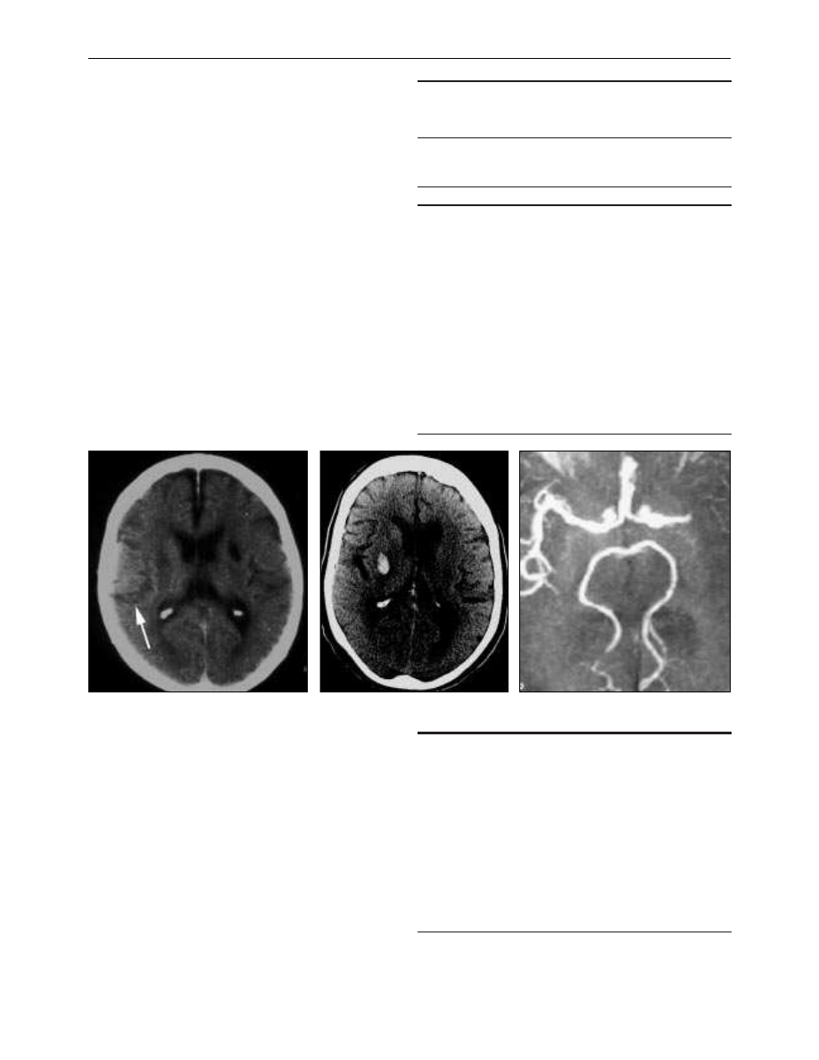
Computed tomogram showing ischaemic stroke
Computed tomogram showing haemorrhagic
stroke
Magnetic resonance angiogram showing middle
cerebral artery occlusion
Clinical review
921
BMJ
VOLUME 320 1 APRIL 2000 bmj.com
on 1 October 2006
bmj.com
Downloaded from

feeding tube until it is safe to resume oral food and fluids. Most
dysphagic patients will not need enteral feeding beyond a few
weeks. However, when and how optimally to feed dysphagic
patients remains to be determined.
Acute intervention
Firm evidence from two large trials has shown that aspirin
(160-300 mg daily by mouth, nasogastric tube, or rectum) started
within 48 hours of onset of acute ischaemic stroke reduces the
risk of subsequent death and disability. However, the effect of
aspirin is small (number needed to treat (NNT) = 77) and is
principally mediated through reducing the risk of early
reinfarction. Neuroimaging is strongly recommended before
starting aspirin. A large trial of unfractionated heparin in stroke
patients found that heparin did not improve outcome, even in
patients with presumed embolic stroke. Heparin may still be
useful in certain groups of patients.
Thrombolysis with alteplase within three hours of onset of
stroke significantly increases the chance of a near complete
recovery (NNT = 7) when administered by specialists. Treatment
up to six hours after stroke has been found less effective in
meta-analysis of randomised controlled trials (estimated
NNT = 12). Thrombolysis is currently licensed for stroke only in
North America and New Zealand, and concerns remain about
its safety.
Neuroprotectant drugs (which may protect neurones from
ischaemia) have, to date, shown no benefit in ischaemic or
haemorrhagic stroke, although several trials are still in progress.
Patients with a large cerebellar infarct or bleed should be
referred for immediate neurosurgical evaluation to facilitate
evacuation of the clot or infarct, or shunting for acute
hydrocephalus, if required. Anticoagulants should be reversed
in patients with primary intracerebral haemorrhage.
Complications of stroke
Stroke may be complicated by several conditions that can alter
outcome adversely. Hyperglycaemia, fever, and hypertension are
each associated with a poor prognosis. In the absence of trial
evidence, raised glucose concentrations should be normalised
and paracetamol given for fever. In contrast, hypertension
should not be treated for the first week since some
antihypertensive drugs (notably calcium channel blockers) seem
to worsen outcome, possibly by reducing regional cerebral
blood flow. Large ischaemic strokes are often complicated by
oedema, swelling, and herniation leading to death; no proved
treatment is available for these complications.
Venous thromboembolic disease (deep vein thrombosis,
pulmonary embolism) develops in half of immobile patients
unless preventive measures are taken. Although compression
stockings reduce the risk of deep vein thrombosis in other
groups of high risk patients, this has not been confirmed in
stroke. A combination of stockings, early mobilisation, adequate
hydration, and aspirin is considered good practice in patients
with ischaemic stroke. Early mobilisation may also reduce the
risk of pressure sores, respiratory tract infections, and urinary
tract infections. When possible, urinary catheters should be
avoided to minimise the risk of infection.
Rehabilitation
The principal aims of rehabilitation are to restore function and
reduce the effect of the stroke on patients and their carers.
Rehabilitation should start early during recovery with
assessment and mobilisation while the patient is in the acute
Acute drug treatments for ischaemic stroke
Aspirin
x Most patients
Heparin (unfractionated or low molecular
weight):
Prophylactic
x Previous venous thromboembolism
x Morbid obesity
Therapeutic
x Carotid artery dissection
x Embolic, recurrent transient ischaemic attacks
Complications of stroke
x Hyperglycaemia
x Hypertension
x Fever
x Infarct extension or rebleeding
x Cerebral oedema, herniation, coning
x Aspiration
x Pneumonia
x Urinary tract infection
x Cardiac dysrhythmia
x Recurrence
x Deep vein thrombosis, pulmonary embolism
Study
Experimental
n/N
Control
n/N
0.5
0.7
1
1.5
Favours treatment
Favours control
2
Peto odds ratio
(95% CI fixed)
Peto odds ratio
(95% CI fixed)
ATLANTIS A 1999
ATLANTIS B 1999
ECASS I 1995
ECASS II 1998
Mori 1992
NINDS 1995
Subtotal (95% CI)
χ
2
=13.23 (df=5), Z=2.97
Total (95% CI)
χ
2
=13.23 (df=5), Z=2.97
64/71
141/307
185/313
187/409
11/19
155/312
729/1431
729/1431
56/71
135/306
185/307
211/391
10/12
192/312
789/1399
789/1399
2.35 (0.95 to 5.82)
1.08 (0.78 to 1.48)
0.79 (0.58 to 1.09)
0.72 (0.55 to 0.95)
0.32 (0.07 to 1.48)
0.62 (0.45 to 0.85)
0.80 (0.69 to 0.93)
0.80 (0.69 to 0.93)
Meta-analysis shows that thrombolysis reduces combined death and
disability from stroke
Stroke patient receiving rehabilitation therapy
Clinical review
922
BMJ
VOLUME 320 1 APRIL 2000 bmj.com
on 1 October 2006
bmj.com
Downloaded from

stroke unit. Once patients are medically stable, they should be
transferred to a stroke rehabilitation unit if further
rehabilitation is required. Formal rehabilitation in a stroke unit
is associated with reduced death and disability (NNT = 12) and a
shorter stay in hospital. Optimal care is multidisciplinary:
doctors, nurses, physiotherapists, occupational therapists,
speech and language therapists, dieticians, psychologists, and
social workers all have a role.
Secondary prevention
Secondary prevention (apart from blood pressure control)
should start shortly after admission. All patients should be
offered lifestyle guidance, including advice to stop smoking,
reduce saturated fat and salt consumption and alcohol intake,
lose weight, and increase exercise. Aspirin started for the
treatment of acute ischaemic stroke should be continued
indefinitely for secondary prevention. The use of alternative or
additional antithrombotic drugs (dipyridamole, clopidogrel, and
warfarin), carotid endarterectomy, and management of
hypertension and hyperlipidaemia after stroke are discussed in
the next article in the series.
The future
Stroke management is now supported by good quality
evidence, but many questions remain unanswered. Whenever
possible, patients should be given the opportunity to enrol in
randomised trials of acute interventions, rehabilitation, or
secondary prevention.
The magnetic resonance image was provided by Professor Alan Moody,
University of Nottingham. The data on thrombolysis were provided by
Dr Joanna Wardlaw, University of Edinburgh.
Healthcare professionals, patients, and carers can obtain
further information about strokes from the Stroke
Association (020 7566 0300), Chest, Heart and Stroke
Association Scotland (0131 225 6963), Chest, Heart, and
Stroke Association Northern Ireland (01232 320184), or
Different Strokes (01908 236033)
Further reading
x Bath PMW. The medical management of stroke. Int J Clin Pract
1997;51:504-10.
x Lees KR. If I had a stroke. . . . Lancet 1998;352 (suppl III):28-30.
x Royal College of Physicians. Stroke audit package. London: RCP,
1994.
x Stroke Units Trialists’ Collaboration. Collaborative systematic review
of the randomised trials of organised inpatient (stroke unit) care
after stroke. BMJ 1997;314:1151-9.
Philip M W Bath is professor of stroke medicine, University of
Nottingham, and Kennedy R Lees is professor of cerebrovascular
medicine, university department of medicine and therapeutics,
Western Infirmary, Glasgow.
The ABC of arterial and venous disease is edited by Richard Donnelly,
professor of vascular medicine, University of Nottingham and
Southern Derbyshire Acute Hospitals NHS Trust (richard.donnelly@
nottingham.ac.uk) and Nick J M London, professor of surgery,
University of Leicester, Leicester (sms16@leicester.ac.uk). It will be
published as a book later this year.
BMJ
2000;320:920-3
My most valuable lesson
A full examination is always useful
It happened two weeks ago. One of the patients attending the
unit that day was a man who had received an allogeneic stem cell
transplant nine months earlier. He presented with a 24 hour
history of right sided chest pain which seemed to be temporally
related to a recent bout of coughing.
The patient duly undressed to the waist and I performed a
respiratory examination, starting with assessment of chest
expansion. My gaze fixed in the direction of my thumbs, I
detected no abnormality. Tracheal position central. Tactile vocal
fremitus and percussion were normal over the anterior and
lateral chest. Auscultation was unremarkable in these areas.
Moving to the patient’s back there were no new chest signs to be
elicited, although my attention was briefly drawn to a small group
of reddish papules in the skin overlying the vertebral process of
T8. Finally I discovered some local tenderness in the area of the
patient’s discomfort, blindly palpating his right lateral chest wall,
and diagnosed an intercostal muscular strain secondary to
coughing. I told the consultant of my findings, and he came to see
the patient himself.
Courtesy dictated that I should remain in attendance while the
repeat assessment took place. The patient’s shirt was once more
removed whereupon my boss took a discernible step backwards
(advice given to me repeatedly when I was preparing for my
membership examination) and then inspected the chest wall with
the patient’s right arm raised.
My pen almost fell out of my hand, poised as it was over the
case notes. There, in full view, was a narrow, intermittent row of
papules, conforming to an arc extending between the patient’s
back and right anterior axillary line.
My consultant turned back to me, directing a quizzical look in
my direction; I suspect it was met by my own open mouthed,
disbelieving one. His words were spoken quietly and distinctly, but
they shook me like thunder:
“It’s zoster, isn’t it?”
Alexander Pope wrote, “A man should never be ashamed to
own he has been in the wrong, which is but saying, in other
words, that he is wiser today than he was yesterday.”
I now have proof, if I needed it, that clinical examination
technique has enormous relevance to our everyday practice.
Furthermore, after the intense embarrassment I suffered on that
fateful day, I now find myself in the new, liberated position that, if
I am ever inclined or encouraged to criticise a colleague’s clinical
oversight in the future, I need only to bring to mind my patient
with his dermatomal eruption.
Richard J A Murrin specialist registrar in haematology, Birmingham
We welcome articles of up to 600 words on topics such as
A memorable patient, A paper that changed my practice, My most
unfortunate mistake
, or any other piece conveying instruction,
pathos, or humour. If possible the article should be supplied on a
disk. Permission is needed from the patient or a relative if an
identifiable patient is referred to. We also welcome contributions
for “Endpieces,” consisting of quotations of up to 80 words (but
most are considerably shorter) from any source, ancient or
modern, which have appealed to the reader.
Clinical review
923
BMJ
VOLUME 320 1 APRIL 2000 bmj.com
on 1 October 2006
bmj.com
Downloaded from
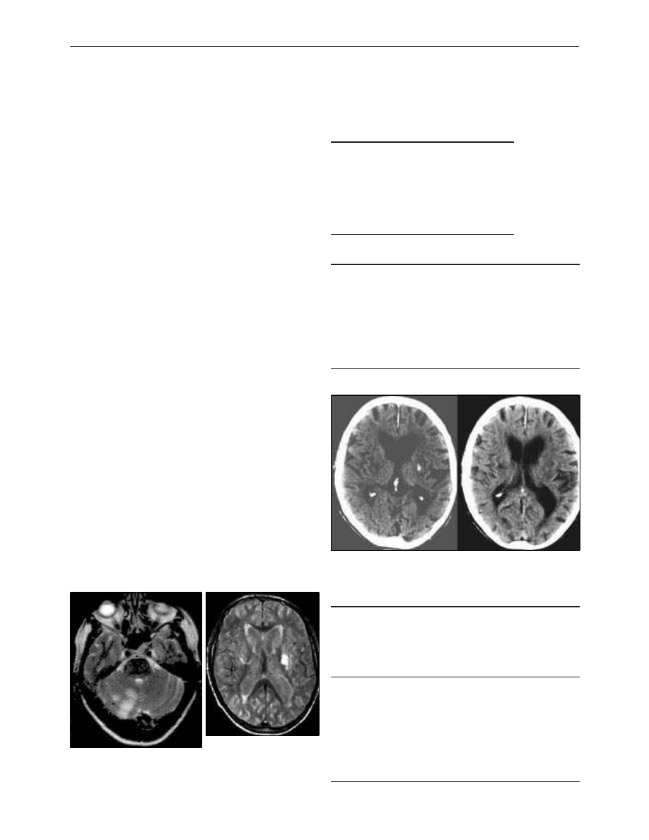
ABC of arterial and venous disease
Secondary prevention of transient ischaemic attack and stroke
Kennedy R Lees, Philip M W Bath, A Ross Naylor
Stroke or transient ischaemic attack is common and likely to be
fatal or cause serious disability. A second stroke will not
necessarily be of the same type as the initial event, although
haemorrhages tend to recur. Patients with previous stroke
commonly succumb to other vascular events, in particular
myocardial infarction. Effective secondary prevention depends
on giving attention to all modifiable risk factors for stroke as
well as treating the causes of the initial stroke. Four questions
should be answered:
Is it acute cerebrovascular disease?
The key features of acute cerebrovascular disease are focal
neurological deficit, sudden onset, and absence of an alternative
explanation. Abrupt onset of a dense hemiparesis before
gradual improvement in a conscious patient rarely causes
doubt, but conditions which commonly mimic stroke must be
considered (see previous article BMJ 2000;320:920-3).
Is it ischaemic or haemorrhagic
stroke?
Neither clinical history nor examination can reliably distinguish
infarction from primary intracerebral haemorrhage. A small
bleed can produce transient symptoms, although these rarely
resolve within an hour.
Cerebral imaging is essential, and the choice and timing of
the scan is important. Haemorrhage is immediately apparent
on computed tomography, but its distinctive appearance
becomes indistinguishable from infarction over a few weeks; for
major symptoms, a computed tomogram taken within two
weeks should still be diagnostic, but a small bleed may be
missed after one week.
Magnetic resonance imaging has a greater sensitivity for
brain stem, cerebellar, and small ischaemic strokes of the brain
than computed tomography. It can also identify haemorrhagic
stroke and remains diagnostic long after signs have become
undetectable on computed tomography.
Risk of recurrence after stroke or transient
ischaemic attack
Stroke
x 8% a year
Transient ischaemic attack
x 8% risk of stroke in first month
x 5% risk of stroke a year thereafter
x 5% risk of myocardial infarction a year
Modifiable risk factors for stroke
x Hypertension
x Smoking
x Diabetes mellitus
x Diet: high salt and fats, low potassium and vitamins
x Excess alcohol intake
x Morbid obesity
x Low physical exercise
x Low temperature
x Cholesterol concentration—at least in patients with coronary disease
Correct imaging techniques for patients with symptoms of
stroke
Symptoms for
< 1 hour
Symptoms
for > 1 hour;
onset < 2
weeks
Symptoms
for > 1 hour;
onset > 2
weeks
Abrupt onset,
typical
cerebrovascular
symptoms
Image only if
anticoagulation
proposed
Computed
tomography
Magnetic
resonance
imaging
Insidious onset
suspicious of
tumour
Not applicable
Computed
tomography
with contrast
Computed
tomography
with contrast
Insidious onset
suggestive of
multiple sclerosis
Not applicable
Magnetic
resonance
imaging
Magnetic
resonance
imaging
Computed tomograms on days 0 (left) and 8 (right) after left subcortical
haemorrhage presenting as a transient ischaemic attack with symptoms
lasting 50 minutes. Note the resolution of diagnostic appearances at day 8
Magnetic resonance image of posterior
fossa of brain in patient with right
cerebellar infarction
T1 weighted magnetic resonance
image of left subcortical
haemorrhage (day 9 in same
patient as computed tomograms
above)
Clinical review
991
BMJ
VOLUME 320 8 APRIL 2000 bmj.com
on 1 October 2006
bmj.com
Downloaded from
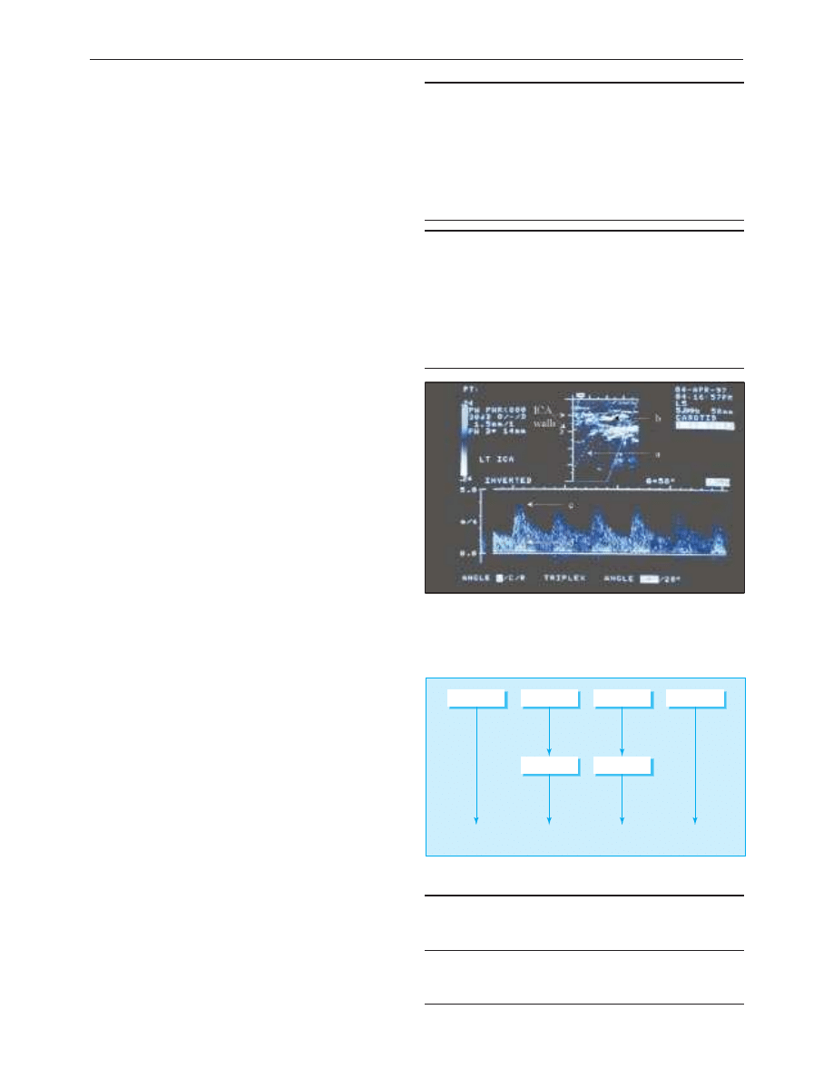
Cardioembolic or vascular aetiology?
Up to a quarter of ischaemic strokes are due to embolism from
the heart or major vessels. In these patients, full anticoagulation
should be considered. Embolic stroke can affect any vascular
territory but can rarely be diagnosed conclusively. Certain
features should prompt a search for an embolic source.
Transthoracic echocardiography is usually adequate, but
transoesophageal echocardiography is justified if the results are
equivocal or the index of suspicion is high.
Anterior or posterior circulation?
The vertebrobasilar arteries supply the brain stem, cerebellum,
and occipital lobes; the cerebral hemispheres are supplied
through the carotid arteries. This distinction is important since
carotid Doppler ultrasonography with a view to endarterectomy
is justified in patients with severe carotid disease only if
symptoms have arisen from the anterior circulation.
Hospital referral
Although the approach to investigation of stroke is simple, few
general practitioners will have open access to the necessary
facilities or see sufficient cases to develop expertise in
interpretation of the results. Patients with suspected stroke need
urgent telephone or fax referral to a “fast track” specialist
cerebrovascular clinic or stroke unit because of the time
limitations on the diagnostic capability of computed tomography
and the limited availability of magnetic resonance imaging.
Management of risk factors
Smoking is an important correctable risk factor and should be
strongly discouraged. The risk of stroke of a smoker returns to
that of a non-smoker within three to five years of stopping
smoking.
Immediate reduction of blood pressure may be deleterious,
but long term risk is inversely related to the blood pressure
achieved. Treatment may therefore be justified even in patients
with “normal” blood pressures. Hypertension should be treated
one to two weeks after a stroke on the basis of British
Hypertension Society guidelines. Patients at high risk of a
further stroke (such as elderly people) derive the greatest
benefit from treatment.
The role of serum cholesterol concentration in the
pathogenesis of stroke remains debatable. Nevertheless, statins
have been shown to reduce the risk of stroke in clinical trials of
patients with coronary heart disease. Lowering cholesterol
concentrations with a statin after atherosclerotic stroke or
transient ischaemic attack probably reduces recurrent events
and the risk of developing ischaemic heart disease. Since stroke
patients represent such a high risk population, the cost of
treatment may be justified.
Diabetes confers a substantial disadvantage for survival and
functional outcome on patients with acute stroke. The
mechanism for this is unknown, but since it is a long term effect,
attempts should be made to normalise blood glucose
concentrations. Blood pressure targets are lower for diabetic
than non-diabetic patients.
Raised plasma homocysteine concentration is increasingly
linked to premature vascular disease and can be easily lowered
through vitamin supplements (folate and pyridoxine). Although
the value of lowering homocysteine concentrations has not
been proved, younger patients with raised plasma homocysteine
concentrations may benefit.
Embolic causes of stroke found on echocardiography
x Mitral stenosis
x Left atrial enlargement ( > 4 cm)
x Dyskinetic or akinetic left ventricle
x Severe global left ventricular dysfunction
x Valvular vegetation
x Left atrial or ventricular thrombus
x Mitral valve calcification
x Calcific aortic valves or stenosis predispose to embolism but may
not justify anticoagulation
Justifications for echocardiography
x Atrial fibrillation
x Heart failure
x Myocardial infarction within
3 months
x Electrocardiographic
abnormalities:
Myocardial infarction or
ischaemia
Bundle branch block
x Cardiac murmur
x Peripheral embolism
x Clinical events in >2 territories:
Right and left hemisphere
Anterior and posterior
circulation
x >2 cortical events (even in
same territory) unless severe
ipsilateral carotid disease
Blood pressure targets (mm Hg) in non-diabetic and diabetic
stroke patients
No diabetes
Diabetes
Titrate to diastolic blood pressure
<85
<80
Optimal blood pressure
< 140/85
< 130/80
Suboptimal blood pressure
>150/90
>140/85
Carotid Doppler ultrasonogram in patient with severe internal carotid artery
stenosis. Upper panel shows angle of insonation (a) of internal carotid artery
and sampling window (b); the velocity of systolic blood flow at the point of
maximal narrowing (c), is nearly 4 m/s (normal <1 m/s). Stenosis causes flow
velocity to increase and produces turbulence, which is seen as shading
within the Doppler spectrum (d)
≥
200/110
Days or
weeks
Months
Months
140-199/90-109
130-139/80-89
≥
140/90
130-139/80-89
< 130/80
Treat
Treat
Reassess
annually
Reassess
in 5 years
Blood pressure thresholds (mm Hg) for treatment after stroke or transient
ischaemic attack (based on British Hypertension Society Guidelines 1999)
Clinical review
992
BMJ
VOLUME 320 8 APRIL 2000 bmj.com
on 1 October 2006
bmj.com
Downloaded from
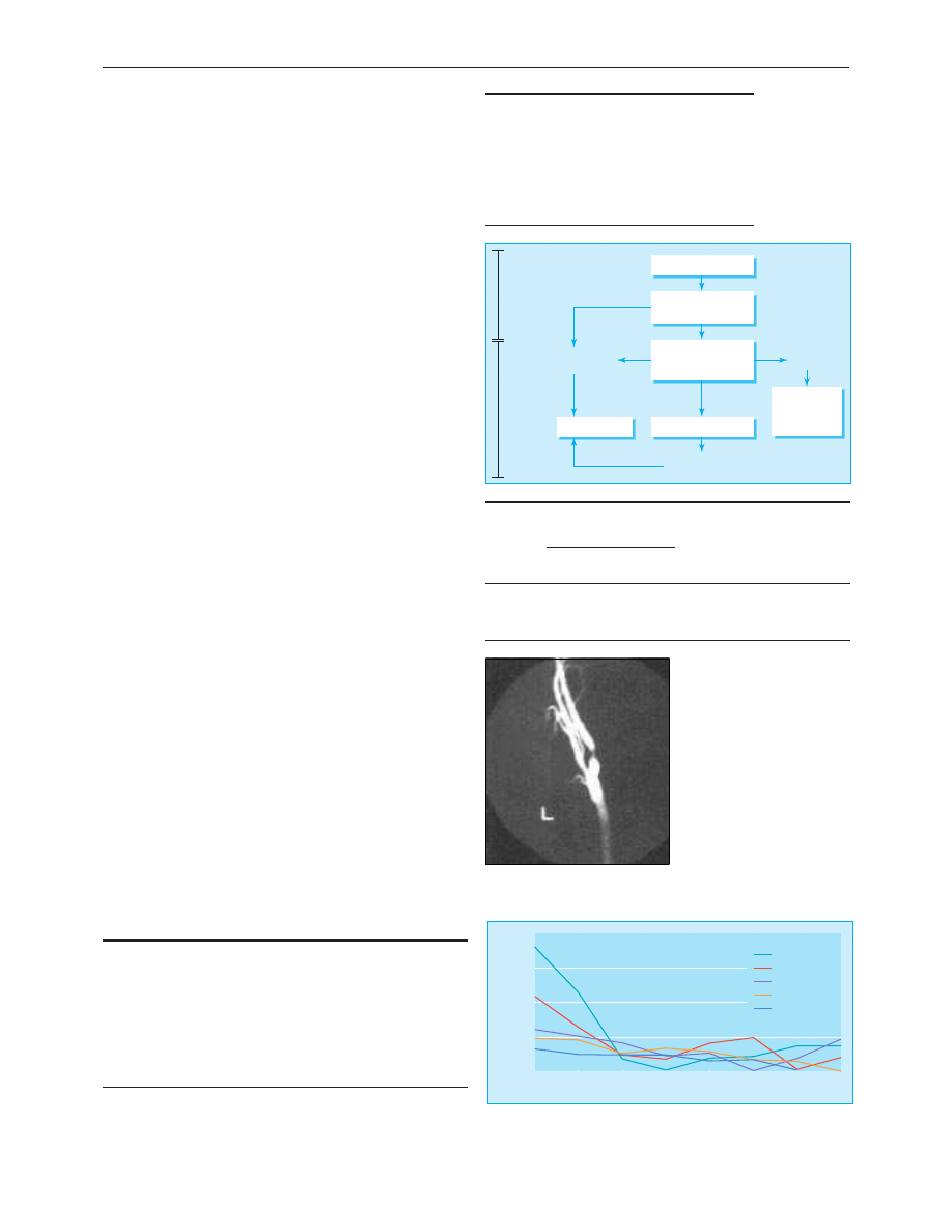
Antiplatelet therapy and
anticoagulation
Patients with atrial fibrillation should receive warfarin if they
have no contraindications, aiming at an international
normalised ratio of 2.0-3.0. Patients with other important
sources of cardiac embolism also benefit from warfarin. Only
patients with mechanical prosthetic heart valves require a
higher international normalised ratio target of 2.5-4.5, although
the exact value depends on the type of valve.
For all other patients with ischaemic stroke, antiplatelet
therapy would be first line treatment. Aspirin is inexpensive and
simple to administer, and its benefits are conclusively proved.
An initial dose of 300 mg followed by 75 mg daily is advised
(higher doses have little advantage but increase gastrointestinal
side effects and bleeding).
Modified release dipyridamole (200 mg twice daily) has an
independent and additive effect to low dose aspirin in
preventing further strokes but not coronary events or overall
mortality. The routine addition of dipyridamole to aspirin for
secondary prevention of strokes may be cost effective.
Clopidogrel (75 mg daily), a new antiplatelet drug, is well
tolerated and was slightly more effective than aspirin in a large
trial. However, it is not cost effective for initial treatment.
Clopidogrel should be used in patients with true intolerance to
aspirin (allergy or intractable side effects on low dose enteric
coated aspirin with or without antiulcer drugs); dipyridamole
alone does not prevent cardiac events.
Carotid surgery and angioplasty
Firm evidence from two large trials has clarified the role of
carotid endarterectomy in patients with ipsilateral severe carotid
stenosis. Patients with severe disease benefit from surgery for up
to 12 months after the most recent cerebral event. The benefit
derived is inextricably linked to the operative risk (stroke or
death within 30 days). In the randomised trials, the operative
mortality in patients with severe disease was 1.0%, the risk of
death or disabling stroke < 4%, and the risk of death or any
stroke < 7.5%. Surgical risk is inversely proportional to surgical
volume, implying that patients should be referred to busy
carotid endarterectomy centres. Surgeons must quote their own
risks rather than results obtained in trials.
Any patient presenting with carotid territory symptoms
should be considered a potential candidate for carotid
endarterectomy, and carotid Doppler ultrasonography should
be done if the patient is fit for surgery. The presence or absence
of a carotid bruit is irrelevant. An ongoing meta-analysis may
further refine the indications, particularly regarding the
management of women and patients with isolated retinal
symptoms, who seem to have a lower overall risk of stroke.
A successful carotid endarterectomy is not a major
procedure, and most patients can be discharged home the day
after surgery. Neither clinical nor ultrasonographic surveillance
Main contraindications to long term
warfarin treatment
x Gastrointestinal bleeding
x Active peptic ulceration
x Frequent falls
x Alcohol misuse
x History of intracranial haemorrhage
Age, by itself, is not a contraindication
Summary of results from European carotid surgery trial
Stenosis
(%)
Incidence of stroke (%)
Absolute risk
reduction (%)
Relative risk
reduction
(%)
Surgery
arm
Medical
arm
0-30
11.8
6.2
−
5.6 at 3 years
None
31-69
16.0
15.0
−
1.0 at 5 years
None
70-99
12.3
21.9
9.6 at 3 years
44 at 3 years
Computed tomography
Aspirin 75-300 mg
Clopidogrel 75 mg
Aspirin intolerance
or contraindication
Haemorrhage excluded
Start aspirin 150-300 mg
Aspirin
tolerant
1-2 weeks
Acute
prophylaxis
of further
vascular
events
Aspirin intolerance
or contraindication
Dipyridamole intolerance
or contraindication
Continue long
term unless
contraindication
develops
Aspirin 75-300 mg and
Dipyridamole 200 mg
twice daily
Secondary
prevention
of stroke
and other
vascular
events
Angiogram showing tight stenosis of
internal carotid artery just distal to
bifurcation
6
Years of follow up
Risk of stroke (%)
5
8
4
3
2
1
7
0
10
15
20
90-99% (n=60)
Stenosis
5
80-89% (n=160)
70-79% (n=169)
50-69% (n=376)
0-49% (n=441)
Risk of stroke in patients not having surgery according to degree of stenosis.
Data from European Carotid Surgery Trialists’ Collaborative Group, Lancet
1998;351:1379-87
Indications for carotid endarterectomy
Surgery not indicated
x Carotid territory symptoms and an ipsilateral 0-69% stenosis
x Complete occlusion of the carotid artery
Surgery indicated
x Carotid territory symptoms within 6 months and an ipsilateral
70-99% stenosis
x Carotid territory symptoms within 12 months and an ipsilateral
80-99% stenosis
Clinical review
993
BMJ
VOLUME 320 8 APRIL 2000 bmj.com
on 1 October 2006
bmj.com
Downloaded from
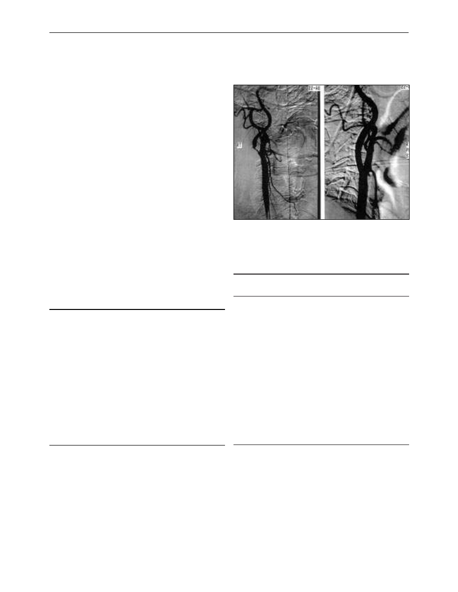
prevents late stroke, and so most patients are discharged from
follow up at six weeks with the proviso that they should be
referred immediately should further cerebral ischaemic
events occur.
In carotid angioplasty the stenosis is dilated by using a
balloon catheter introduced percutaneously through the
femoral artery. The potential advantages of carotid angioplasty
include reduced hospital stay, cranial nerve injury, wound
complications, and other cardiovascular morbidity. The main
concern about carotid angioplasty is the risk of embolic stroke
at the time of the procedure and recurrent stenosis. Carotid
angioplasty aids the management of fibromuscular dysplasia,
radiation injury, and symptomatic restenosis after carotid
endarterectomy. Otherwise, carotid angioplasty should not
be performed outside randomised trials and, as with carotid
endarterectomy, outcomes in individual centres should
be audited.
Complex cases
Secondary prevention of stroke is rightly the province of
general practitioners and the preceding suggestions will cover
most patients with recent stroke. However, patients with
complex conditions will need access to specialist services,
although definitive trial evidence justifying therapeutic
decisions in such cases is often absent. Patients should be
monitored for compliance with treatment and the development
of complications such as renovascular disease, ischaemic heart
disease, and further cerebrovascular problems. Optimal dietary,
smoking, lipid, and blood pressure management is always
required in addition to antithrombotic treatment.
G T McInnes and L Ramsay contributed towards the blood pressure guidelines.
M R Walters supplied some of the pictures and J Overell supplied the antiplatelet
flow chart.
Kennedy R Lees is professor of cerebrovascular medicine, university
department of medicine and therapeutics, Western Infirmary,
Glasgow (k.r.lees@clinmed.gla.ac.uk); Philip M W Bath is professor of
stroke medicine, University Hospital, Nottingham; and A Ross Naylor
is senior lecturer, department of surgery, Leicester Royal Infirmary,
Leicester.
Complex cases that may require hospital referral
Case
Possible treatment
Recurrent stroke or transient
ischaemic attack despite antiplatelet
treatment (treatment failure)
Consider higher doses of
aspirin, addition of
dipyridamole (if not already
prescribed), substitution or
addition of clopidogrel, or
substitution or addition of
warfarin
Recurrent embolic events despite
adequate anticoagulation with
warfarin
Consider adding low dose
aspirin
Recurrent non-haemodynamic
symptoms from inoperable severe
carotid stenosis or serious
intracranial stenosis despite
antiplatelet treatment
Consider warfarin
Hypertension and inoperable severe
carotid stenosis
Consider cerebral blood flow
monitoring (with
ultrasonography or
radionucleotide perfusion
scanning) before
antihypertensive treatment
The ABC of arterial and venous disease is edited by Richard Donnelly,
professor of vascular medicine, University of Nottingham and
Southern Derbyshire Acute Hospitals NHS Trust (richard.donnelly@
nottingham.ac.uk) and Nick J M London, professor of surgery,
University of Leicester, Leicester (sms16@leicester.ac.uk). It will be
published as a book later this year.
BMJ
2000;320:991-4
Severe carotid stenosis before (left) and after carotid angioplasty and
stenting (right)
Further reading
x Antiplatelet Trialists’ Collaboration. Collaborative overview of
randomised trials of antiplatelet therapy. 1. Prevention of death,
myocardial infarction, and stroke by prolonged antiplatelet therapy
in various categories of patients. BMJ 1994;308:81-106.
x EAFT (European Atrial Fibrillation Trial) Study Group. Secondary
prevention in non-rheumatic atrial fibrillation after TIA or minor
stroke. Lancet 1993;342:1255-62.
x Diener HC, Cunha L, Forbes C, Sivenius J, Smets P, Lowenthal A,
for the ESPS-2 Working Group. European Stroke Prevention Study
2. Dipyridamole and acetylsalicylic acid in the secondary prevention
of stroke. J Neurol Sci 1996;143:1-13.
x CAPRIE Steering Committee. A randomised, blinded, trial of
clopidogrel versus aspirin in patients at risk of ischaemic events
(CAPRIE). Lancet 1996;348:1329-39.
x European Carotid Surgery Trialists’ Collaborative Group.
Randomised trial of endarterectomy for recently symptomatic
carotid stenosis. Final results of the MRC European carotid surgery
trial (ECST). Lancet 1998;351:1379-87.
Clinical review
994
BMJ
VOLUME 320 8 APRIL 2000 bmj.com
on 1 October 2006
bmj.com
Downloaded from
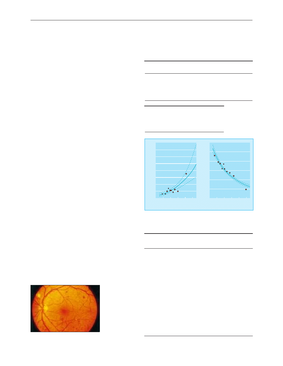
ABC of arterial and venous disease
Vascular complications of diabetes
Richard Donnelly, Alistair M Emslie-Smith, Iain D Gardner, Andrew D Morris
Adults with diabetes have an annual mortality of about 5.4%
(double the rate for non-diabetic adults), and their life expectancy
is decreased on average by 5-10 years. Although the increased
death rate is mainly due to cardiovascular disease, deaths from
non-cardiovascular causes are also increased. A diagnosis of
diabetes immediately increases the risk of developing various
clinical complications that are largely irreversible and due to
microvascular or macrovascular disease. Duration of diabetes is
an important factor in the pathogenesis of complications, but
other risk factors—for example, hypertension, cigarette smoking,
and hypercholesterolaemia—interact with diabetes to affect the
clinical course of microangiopathy and macroangiopathy.
Microvascular complications
A continuous relation exists between glycaemic control and the
incidence and progression of microvascular complications.
Hypertension and smoking also have an adverse effect on
microvascular outcomes. In the diabetes control and
complications trial—a landmark study in type 1 diabetes—the
number of clinically important microvascular endpoints was
reduced by 34-76% in patients allocated to intensive insulin
(that is, a 10% mean reduction in glycated haemoglobin (Hb
A1c
)
concentration from 8.0% to 7.2%). However, these patients also
had more hypoglycaemic episodes. Similarly, in the UK
prospective diabetes study of patients with type 2 diabetes, an
intensive glucose control policy that lowered glycated
haemoglobin concentrations by an average of 0.9% compared
with conventional treatment (median Hb
A1c
7.0% v 7.9%)
resulted in a 25% reduction in the overall microvascular
complication rate. It was estimated that for every 1% reduction
in Hb
A1c
concentration there is a 35% reduction in
microvascular disease.
Retinopathy
Diabetic retinopathy is a progressive disorder classified
according to the presence of various clinical abnormalities. It is
the commonest cause of blindness in people aged 30-69 years.
Damage to the retina arises from a combination of
microvascular leakage and microvascular occlusion; these
changes can be visualised in detail by fluorescein angiography.
A fifth of patients with newly discovered type 2 diabetes have
retinopathy at the time of diagnosis. In type 1 diabetes, vision
threatening retinopathy almost never occurs in the first five
years after diagnosis or before puberty. After 15 years, however,
Risk of morbidity associated with all types of diabetes mellitus
Complication
Blindness
End stage renal disease
Amputation
Myocardial infarction
Stroke
*Compared with non-diabetic patients
Relative risk*
20
25
40
2-5
2-3
Vascular complications of diabetes
Microvascular
Retinopathy
Nephropathy
Neuropathy
Macrovascular
Ischaemic heart disease
Stroke
Peripheral vascular disease
Classification and features of diabetic retinopathy
Grade
Examination features
Symptoms
I Background
retinopathy
Microaneurysms
Small blot haemorrhages
Hard exudates
Not affecting macula
None
II Background with
maculopathy
Leakage in macular region
Capillary occlusion
Hard exudates
Central visual
loss (such as
reading difficulty)
III Preproliferative
retinopathy
Cotton wool spots
Venous abnormalities
Large blot haemorrhages
Intraretinal microvascular
abnormalities
None
IV Proliferative
retinopathy
New vessels on disc or
elsewhere on retina
None, but
complications
cause visual loss
V Advanced
diabetic eye disease
Extensive fibrovascular
proliferation
Retinal detachment
Vitreous haemorrhage
Thrombotic glaucoma
Severe visual loss
Glycated haemoglobin (%)
Rate of progression of retinopathy (per 100 patient-years)
10.0
9.0
8.0
7.0
6.0
5.0
0
4
6
8
10
12
14
16
2
Glycated haemoglobin (%)
Rate of severe hypoglycaemia (per 100 patient-years)
10.0
9.0
8.0
7.0
6.0
5.0
0
40
60
80
100
120
20
Relation between glycaemic control (Hb
A1c
) and risk of progression of
microvascular complications (retinopathy) and severe hypoglycaemia in
patients with type 1 diabetes. Data from the diabetes control and
complications trial. Dotted lines represent 95% confidence intervals
Background retinopathy showing
microaneurysms and small blot haemorrhages
Clinical review
1062
BMJ
VOLUME 320 15 APRIL 2000 bmj.com
on 1 October 2006
bmj.com
Downloaded from
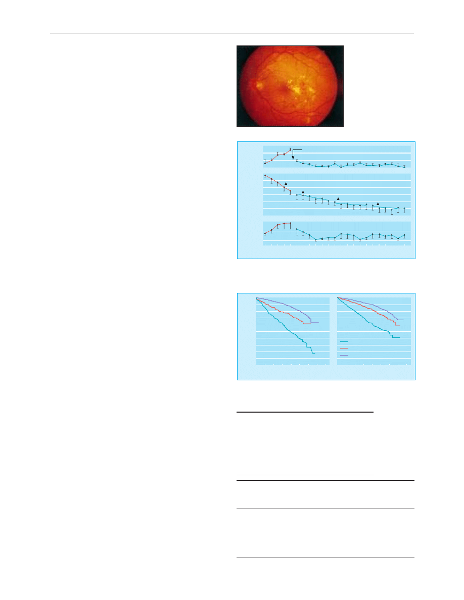
almost all patients with type 1 diabetes and two thirds of those
with type 2 diabetes have background retinopathy.
Vision threatening retinopathy is usually due to
neovascularisation in type 1 diabetes and maculopathy in type 2
diabetes. Depending on the relative contribution of leakage or
capillary occlusion, maculopathy is divided into three types:
exudative maculopathy (when hard exudates appear in the
region of the macula), ischaemic maculopathy (characterised by
a predominance of capillary occlusion which results in clusters
of haemorrhages), and oedematous maculopathy (extensive
leakage gives rise to macular oedema). Treatment of
maculopathy and proliferative retinopathy with laser
photocoagulation prevents further loss of vision rather than
restores diminished visual acuity.
Nephropathy
Diabetic nephropathy is characterised by proteinuria
> 300 mg/24 h, increased blood pressure, and a progressive
decline in renal function. At its most severe, diabetic
nephropathy results in end stage renal disease requiring dialysis
or transplantation, but in the early stages overt disease is
preceded by a phase known as incipient nephropathy (or
microalbuminuria), in which the urine contains trace quantities
of protein (not detectable by traditional dipstick testing).
Microalbuminuria is defined as an albumin excretion rate of
20-300 mg/24 h or 20-200 ìg/min in a timed collection and is
highly predictive of overt diabetic nephropathy, especially in
type 1 diabetes.
The rate of decline in glomerular filtration rate varies widely
between individuals, but antihypertensive treatment greatly
slows the decline in renal function and improves survival in
patients with diabetic nephropathy.
In patients with type 1 diabetes complicated by diabetic
nephropathy, angiotensin converting enzyme inhibitors have
renoprotective effects above those that can be attributed to
reduced blood pressure; they are beneficial even in normotensive
patients and ameliorate other associated microvascular
complications such as retinopathy. In patients with type 2
diabetes, achieving good blood pressure control (which often
requires combination therapy) is more important than the choice
of antihypertensive drug, although angiotensin converting
enzyme inhibitors are the preferred first line treatment
The development of proteinuria is a marker of widespread
vascular damage and signifies an increased risk of subsequent
end stage renal disease and macrovascular complications,
especially coronary heart disease. Microproteinuria and
proteinuria are strongly associated with decreased survival in
both type 1 and type 2 diabetes.
Neuropathy
The diabetic neuropathies present in several ways. The
commonest form is a diffuse progressive polyneuropathy
affecting mainly the feet. It is predominantly sensory, often
asymptomatic, and affects 40-50% of all patients with diabetes.
Reduced sensation can be detected with a monofilament, and
patients with sensory neuropathy as well as other high risk
features need advice on foot care to minimise the risk of
ulceration. Neuropathic foot ulcers can be distinguished from
vascular ulcers, although a mixed aetiology is common.
Macrovascular complications
Atherosclerotic disease accounts for most of the excess
mortality in patients with diabetes. In the UK prospective
diabetes study, fatal cardiovascular events were 70 times more
common than deaths from microvascular complications. The
Clinical features of “high risk” diabetic foot
x Impaired sensation (monofilament)
x Past or current ulcer
x Maceration
x Fungal or gryphotic (thickened or horny) toenails
x Biomechanical problems (corns or callus)
x Fissures
x Clawed toes
Clinical features that distinguish neuropathic and vascular
foot ulcers
Neuropathic
Vascular
Painless
Painful
Located at points of high pressure
Often located at the
extremities
“Punched out” appearance surrounded by callus
Warm foot
Cool ischaemic foot
Bounding foot pulses
Absent foot pulses
Diabetic maculopathy
Years
Albuminuria
(
µ
g/min)
-2
-1
0
1
2
3
4
5
6
7
8
9
750
1250
250
Mean arterial
pressure
(mmHg)
95
105
115
125
Start of antihypertensive treatment
Glomerular
filtration rate
(ml/min/1.73m
2
)
45
65
75
85
95
105
55
Change in glomerular filtration rate
11.3 ml/min/year
3.5 ml/min/year
1.2 ml/min/year
1.3 ml/min/year
Effect of antihypertensive treatment (â blockers and diuretics) on mean
arterial pressure, glomerular filtration rate, and albuminuria in patients with
type 1 diabetes and nephropathy. Reproduced with permission from
Mogensen, Diabetic Med 1995;12:756-9.
Years of follow up from baseline
Survival probability
16
12
8
4
14
10
6
2
0
0
0.2
0.3
0.4
0.5
0.6
0.7
0.8
0.9
1.0
0.1
Years of follow up from baseline
16
12
8
4
14
10
6
2
0
Light
None
Heavy
Type 1
Type 2
Data from WHO multinational study of vascular disease in diabetes showing
survival in patients with type 1 and type 2 diabetes according to degree of
proteinuria (none, slight, or heavy) at baseline. Reproduced with permission
from Stephenson et al, Diabetic Med 1995;12:149-55
Clinical review
1063
BMJ
VOLUME 320 15 APRIL 2000 bmj.com
on 1 October 2006
bmj.com
Downloaded from

relation between glucose concentrations and macrovascular
events is less powerful than for microvascular disease; smoking,
blood pressure, proteinuria, and cholesterol concentration are
more important risk factors for atheromatous large vessel
disease in patients with diabetes.
Hyperlipidaemia is no more common in patients with well
controlled type 1 diabetes than it is in the general population.
In patients with type 2 diabetes, total and low density
lipoprotein cholesterol concentrations are also similar to those
found in non-diabetic people, but type 2 diabetes is associated
with a more atherogenic lipid profile, in particular low
concentrations of high density lipoprotein cholesterol and high
concentrations of small, dense, low density lipoprotein particles.
Hypertension affects at least half of patients with diabetes. In
the UK prospective diabetes study tight blood pressure control
(mean 144/82 mm Hg) achieved significant reductions in the
risk of stroke (44%), heart failure (56%), and diabetes related
deaths (32%), as well as reductions in microvascular
complications (for example, 34% reduction in progression of
retinopathy). One third of patients required three or more
antihypertensive drugs to maintain a target blood pressure
< 150/85 mm Hg. In another recent study (hypertension
optimal treatment study) rates of cardiovascular events in
patients with type 2 diabetes were reduced even further when
combination treatment was used to aim for target diastolic
blood pressures < 80 mm Hg.
Coronary heart disease
The incidence and severity of coronary heart disease events are
higher in patients with diabetes, and several clinical features are
worth noting. The diabetes subgroups in the major secondary
prevention studies of cholesterol reduction (Scandinavian
simvastatin survival study (4S) and cholesterol and recurrent
events (CARE) trial) show a beneficial effect of statins.
Peripheral vascular disease
Atheromatous disease in the legs, as in the heart, tends to affect
more distal vessels—for example, the tibial arteries—producing
multiple, diffuse lesions that are less straightforward to bypass
or dilate by angioplasty. Medial calcification of vessels
(Mönckeberg’s sclerosis) is common and can result in falsely
raised measurements of the ankle brachial pressure index. This
index is therefore less reliable as a screening test in patients
with diabetes and intermittent claudication.
Stroke
Roughly 85% of acute strokes are atherothrombotic, and the rest
are haemorrhagic (10% primary intracerebral haemorrhage and
5% subarachnoid haemorrhage). The risk of atherothrombotic
stroke is two to three times higher in patients with diabetes, but
the rates of haemorrhagic stroke and transient ischaemic attacks
are similar to those of the non-diabetic population. Patients with
Predictors of cardiovascular mortality
Type 1 diabetes
Type 2 diabetes
x Overt nephropathy
x Presence of coronary heart disease
x Hypertension
x Overt proteinuria
x Smoking
x Glycated haemoglobin
x Microalbuminuria
x Hypertension
x Age
Taken from BMJ 1996:313:779-84, Diabetes1995;44:1303-9
70
Age (years)
Estimated hazard ratios
(95% CI)
65
60
55
50
45
40
0.4
1
3
Low density lipoprotein
cholesterol (mmol/l)
5.0
4.5
4.0
3.5
3.0
2.5
High density lipoprotein
cholesterol (mmol/l)
1.3
1.2
1.1
1.0
0.9
Glycated haemoglobin (%)
Estimated hazard ratios
(95% CI)
9
8
7
6
5
0.4
1
3
Systolic blood pressure
(mm Hg)
160
150
140
130
120
110
Current
smokers
Former
smokers
Never
smokers
Estimated hazard ratios for significant risk factors for coronary heart
disease occurring in 335 out of 3055 patients with type 2 diabetes.
Reproduced from Turner et al, BMJ 1998;316:823-8.
Target diastolic blood pressure (mm Hg)
Events per 1000 patient-years
0
<80
(n=499)
<85
(n=501)
<90
(n=501)
10
15
20
25
P value for trend 0.005
30
5
Rates of serious cardiovascular events according to target diastolic blood
pressure in 1500 patients with hypertension and type 2 diabetes. Drawn
from Hansson et al, Lancet 1998;351:1755-62.
Features of coronary heart disease in diabetic patients
Atherosclerosis
Acute myocardial infarction
Revascularisation
x Prevalence of fatal and non-fatal coronary
heart disease events 2-20
×
higher than for
non-diabetics of similar age
x Protective effect of female sex is lost
x Higher incidence of diffuse, multivessel
disease
x Plaque rupture leading to unstable angina
and myocardial infarction is more common
x Superimposed thrombosis more likely
x In-hospital and 6 month mortality double
that in non-diabetics
x Complications (eg, arrhythmias, heart failure,
death) more common
x Reperfusion rates after thrombolysis are
similar to those of non-diabetics, but
reocclusion and reinfarction rates are higher
x Mortality reduced by insulin glucose infusion
immediately after myocardial infarction
x 5 year survival rates after coronary
artery bypass graft or percutaneous
coronary angioplasty lower than for
non-diabetics
x 5 year survival better after coronary
artery bypass graft than percutaneous
coronary angioplasty because of higher
restenosis rates with angioplasty (81% v
66%)
Clinical review
1064
BMJ
VOLUME 320 15 APRIL 2000 bmj.com
on 1 October 2006
bmj.com
Downloaded from

diabetes are probably more prone to irreversible rather than
reversible ischaemic brain damage, and small lacunar infarcts are
common. Stroke patients with diabetes have a higher death rate
and a poorer neurological outcome with more severe disability.
Maintaining good glycaemic control immediately after a stroke is
likely to improve outcome, but the long term survival is reduced
because of a high rate of recurrence. Antihypertensive treatment
is effective in preventing stroke.
Erectile dysfunction
Erectile dysfunction is a common complication of diabetes,
occurring in up to half of men aged over 50 years (compared
with 15-20% in age matched non-diabetic men), although the
exact prevalance is unknown because of likely underreporting.
The underlying pathogenesis is multifactorial, with autonomic
neuropathy, vascular insufficiency, and psychological factors
contributing to the clinical picture. The condition causes
appreciable social and psychological problems for many patients,
and its importance should not be underestimated. The recent
introduction of sildenafil, which is reported to have a 50-70%
success rate in patients with diabetes, is an important advance.
Surveillance and management in
general practice
Screening for diabetes
Up to half of people with type 2 diabetes have vascular
complications at the time of diagnosis. Early detection of
diabetes is therefore essential. Screening (by measuring fasting
blood glucose concentration) should be considered for high risk
patients, especially those who are middle aged and obese, are of
Asian or Afro-Caribbean origin, have a history of gestational
diabetes, or have a family history of diabetes.
Eye screening
The small number of patients with retinopathy in any one
practice (about 50 patients per 10 000 practice list) does not
allow most general practitioners to develop and maintain their
funduscopic skills. Innovative approaches, including the use of
trained community optometrists and mobile retinal
photography units that visit practices annually, can provide a
high standard of retinal screening in the community.
Cardiovascular risk prediction
Identification of patients at highest risk of developing
cardiovascular events allows efforts and resources to be
channelled most effectively. Coronary risk prediction charts
and computer programs such as that recently produced as
Recommendations on prevention of coronary heart disease in clinical practice
Glycaemic control
Blood pressure control
Lipid control
Target
Glycated haemoglobin <7.0% Fasting
blood glucose 4-7 mmol/l
<140/80 mm Hg without macrovascular
disease
<130/80 mm Hg with macrovascular
disease
Serum cholesterol < 5.0 mmol/l in patients
with established coronary heart disease
Primary prevention in those with > 30%
risk of coronary heart disease over 10 years
Treatment
(in addition
to lifestyle
and dietary
advice)
Metformin (first line if body mass
index > 25)
Sulphonylurea
Acarbose
Glitazones (not yet available in United
Kingdom)
Insulin
Combination therapy
Angiotensin converting enzyme inhibitors:
renoprotective but caution in renal artery
stenosis (17% of hypertensive diabetics)
Diuretics
â
blockers
á
blockers
Long acting calcium antagonists
50% of patients will require >3 drugs for
optimum control
Statins (regular monitoring of liver
function)
Years from randomisation
Patients with events (%)
5
4
3
2
1
0
6
8
7
9
0
10
20
Tight control <150/85 mm Hg
Less tight control <180/85 mm Hg
Stroke
Reduction in risk with tight control 44%
(95% confidence interval 11% to 65%, P=0.013)
Kaplan-Meier plot of proportions of patients who developed fatal or
non-fatal stroke, according to blood pressure control. Reproduced from
Turner et al, BMJ 1998;317:703-13.
Normal renal function
• Glomerular hyperfiltration and hyperperfusion
• Glomerular hypertrophy
• Mesangial expansion
• Modest increase in blood pressure
Microalbuminuria
("incipient nephropathy")
Albumin excretion rate
20-300 mg/24 h
Macroproteinuria
("overt nephropathy")
Albumin excretion rate
>300 mg/24 h
Dipstick
negative
Dipstick
positive
End stage renal disease
(dialysis or transplantation)
• Increased albumin excretion rate
• Decreasing glomerular filtration rate
Risk of macrovascular events, especially coronary heart disease
• Uraemia
• Fluid retention
• Increased blood pressure
Transition from normal renal function through to end stage renal disease
requiring dialysis or transplantation. Increasing proteinuria and renal
impairment is associated with an increasing risk of fatal or non-fatal
coronary heart disease, especially in older patients with type 2 diabetes and
other risk factors
Clinical review
1065
BMJ
VOLUME 320 15 APRIL 2000 bmj.com
on 1 October 2006
bmj.com
Downloaded from
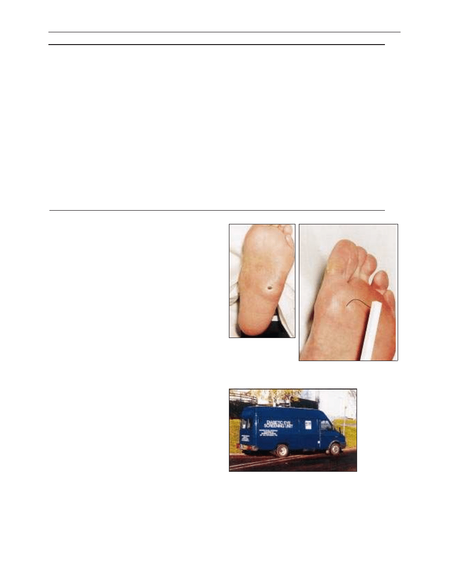
part of the joint British recommendations on prevention of
coronary heart disease in clinical practice will help general
practitioners to implement the findings of recent major
clinical trials.
Annual complications assessment
All patients with diabetes should be offered an annual clinical
assessment concentrating on the prevention, detection, and
management of macrovascular and microvascular complications.
Areas of debate in surveillance of diabetes complications
The value of routine measurements of microalbuminuria in
patients with type 2 diabetes is less clear than in type 1 diabetes.
Arrangements to allow the testing of microalbuminuria in
general practice are not universally available.
The presence of left ventricular hypertrophy is a powerful
predictor of the risk of a cardiovascular event, but screening by
echocardiography or electrocardiography is often not included
as part of the routine annual assessment.
Unlike total cholesterol concentrations and the total
cholesterol to high density lipoprotein cholesterol ratio, the
importance of raised triglyceride concentrations in the risk
profile of patients with type 2 diabetes is unclear.
Team approach to integrated diabetic care
The ongoing care of patients with diabetes, in particular once
they have developed vascular complications, includes a wide
spectrum of healthcare professionals. A systematic, integrated,
and collaborative approach must be developed at a regional
level, with clear lines of communication and the adoption of
locally agreed guidelines for treatment and referral based on
national guidelines—for example, those from the Scottish
Intercollegiate Guideline Network (www.show.scot.nhs.UK/
sign/home.htm).
Alistair M Emslie-Smith is general practitioner, Tayside Centre for
General Practice, Dundee; Iain D Gardner is consultant
ophthalmologist, Derbyshire Royal Infirmary, Derby; and Andrew D
Morris is senior lecturer in medicine and diabetes, Ninewells Hospital
and Medical School, Dundee.
BMJ
2000;320:1062-6
Annual complications assessment
Physical examination
x Body mass index calculation (weight (kg)/(height (m))
2
)
x Blood pressure measurement with patient sitting and
appropriate sized cuff
x Palpation of foot pulses
x Measurement of foot sensation by one or more of following:
10 g monofilament weight (not detected = impaired)
Vibration of 128 Hz tuning fork over medial malleolus
(perception for < 5 secs = impaired)
Assessment of ankle jerk with tendon hammer (less reliable
in elderly people)
x Inspection of feet for nail care, callosities, fissures, fungal
infection, blisters, ulcers, claw toes, prominent metatarsal
heads, and Charcot arthropathy
x Visual acuity in corrected state, using standard 6 m (or 3 m)
Snellen chart. Use pin hole if corrected acuity is >6/9
x Retinal examination by one of:
Direct ophthalmoscopy through pupils dilated with 1%
tropicamide
Combination of direct ophthalmoscopy and slit lamp
biomicroscopy
Retinal photography through fixed site or mobile
non-mydriatic fundus camera
Biochemical analysis
x Dipstick urine analysis for proteinuria
x Urine testing for microalbuminuria in type 1 diabetes
x Blood testing of:
Glycated haemoglobin
Serum creatinine
Serum total cholesterol and high density lipoprotein
cholesterol
History, advice, and education
x Smoking history
x Education and reinforcement of advice on diet, aerobic
exercise, and lifestyle
x Review treatment, including side effects and compliance
x Assess knowledge of diabetes and self management skills,
including warning signs for complications (intermittent
claudication, angina pectoris, foot problems)
x Review footwear provision
x Review need for contact with dietetics, chiropody, orthotics,
and diabetes specialist nurse support
x Advice on erectile dysfunction in men
x Prepregnancy counselling, where appropriate
x Calculate and discuss risk of coronary heart disease and
modification of risk factors
Neuropathic ulcer is a
common complication in
patients with diabetes
Use of monofilament to detect impaired
sensation during annual assessment
Mobile eye screening unit
The ABC of arterial and venous disease is edited by Richard Donnelly,
professor of vascular medicine, University of Nottingham and
Southern Derbyshire Acute Hospitals NHS Trust (richard.donnelly@
nottingham.ac.uk) and Nick J M London, professor of surgery,
University of Leicester, Leicester (sms16@leicester.ac.uk). It will be
published as a book later this year.
Clinical review
1066
BMJ
VOLUME 320 15 APRIL 2000 bmj.com
on 1 October 2006
bmj.com
Downloaded from

organism). Nine samples (8%) from five children (four
boys) grew > 10
5
coliforms/ml, suggesting infection.
However, this was excluded by sterile samples collected
on the same day or on immediate repeat in hospital.
Parents found the pads and bags easy to use and
preferred them to clean catch collections (table) for
both sexes. The pad was considered comfortable,
whereas the bag was distressing, particularly on
removal, often leaking and leaving red marks. Some
found extracting the urine from the pad or emptying it
from the bag to be awkward. Most parents complained
that clean catch collections were time consuming and
often messy; nine gave up after prolonged attempts.
Five parents whose infants voided immediately ranked
it best. The median collection time was 25 minutes for
each method, but parents resented constraining their
children this long for clean catch collections.
Comment
This is the first study of parents’ views of infant urine
collection methods. Pad, bag, and clean catch samples
were equally effective at excluding an infection;
variations in contamination rates balanced collection
failures. Most parents disliked clean catch collections;
their views should be heeded. Most preferred pads to
bags, and they are cheaper. They also found inoculating
dipslides with swabsticks easy; this technique may have
contributed to their relatively low contamination rates.
3 4
Since Kass suggested a diagnostic cut off of a single
bacterial species cultured at > 10
5
/ml, it has been widely
taken as proof of a urine infection and assumed not to
occur from skin contamination, even though his study
5
and others
3
recorded similar false positive rates to ours.
False positive results potentially lead to inappropriate
treatment and imaging. Suprapubic puncture is an
unrealistic alternative in primary care. Although collect-
ing multiple samples would reduce the false positive
rate, it might delay antibiotic treatment.
We thank the parents for volunteering and their thoughtful
comments and Dr Mohammad Raza for microbiological help.
Contributors: MGC had the original idea for the study and is
the guarantor. The study was designed, the data analysed, and
the paper written jointly by all the authors. LDTL carried out
the clinical aspects and DMN and SJP the laboratory aspects of
the study.
Funding: None.
Competing interests: The Children’s Kidney Fund of the
Newcastle University Hospitals special trustees receives royalties
from Ontex UK from sales of Newcastle sterile urine collection
packs.
1
Jadresic L, Cartwright K, Cowie N, Witcombe B, Stevens D. Investigation
of urinary tract infection in childhood. BMJ 1993;307:761-4.
2
Vernon S, Foo CK, Coulthard MG. General practice management of chil-
dren with urinary tract infection: an audit in the former Northern region.
Br J Gen Pract
1997;47:297-300.
3
Hardy JD, Furnell PM, Brumfitt W. Comparison of sterile bag, clean catch
and suprapubic aspiration in the diagnosis of urinary infection in early
childhood. Br J Urol 1976;48:279-83.
4
Macfarlane PI, Houghton C, Hughes C. Pad urine collection for early
childhood urinary-tract infection. Lancet 1999;354:571.
5
Kass EH. Asymptomatic infections of the urinary tract. Trans Assoc Am
Phys
1956;69:56-63.
(Accepted 22 February 2000)
Corrections and clarifications
ABC of vascular disease: Vascular complications of
diabetes
An error crept into a figure in this article by
Richard Donnelly and colleagues (15 April,
pp 1062-6). In the Kaplan-Meier plot at the top of
p 1065, the definition of less tight control should
be < 180/105 (not < 180/85) mm Hg.
Minerva
Minerva slipped up on two names, one personal
and one taxonomic, in the picture article in the
issue of 18 March (p 814). The third author’s name
is R Whitfield; the culture grown was Mycobacterium
marinum
.
Assessments by 44 parents of three methods of home urine
collection
Parents’ assessments
No of parents:
Pad
Bag
Clean
catch
Preference order
First
21
18
5
Second
19
12
13
Third
4
14
26
Open comments
Positive
Easy, hygienic, or quick
25
24
8
Comfortable for child
10
3
0
Negative
Uncomfortable or distressing
1
26
3
Fiddly or messy
9
10
20
Impractical or time consuming
10
0
36
Difficult to get urine out of pad
8
—
—
Red marks left on skin
—
11
—
Too much trouble—gave up (boys)
1 (0)
4 (3)
9 (5)
Email submissions from outside the United Kingdom
We are now offering an email submission service for authors
from outside the UK. The address is papers@bmj.com
Ideally our email server would link seamlessly with our
manuscript tracking system, but for now it does not, which is
why we are offering the service only to authors outside the UK.
Most post in the UK arrives the next day, so UK authors have the
least to gain in speed of delivery from email delivery. As soon as
our systems improve we will invite email submissions from
everyone.
If you choose to send your submission by email please would you
send the text and any tables and figures as attached files, together
with a covering letter giving all your contact details (postal
address, phone, fax, and email address). We can read files created
with most word processing, graphics, and spreadsheet programs.
When your submission is received in our email box you will
receive an automatic acknowledgment to show that it has arrived.
If the submission is incomplete we will contact you and ask you to
resend the missing information.
Once the submission is complete we will register it on our
manuscript tracking system and you will receive a standard
acknowledgment in the post.
Letters to the editor should continue to be sent direct to bmj.com as rapid
responses or to letters@bmj.com
Papers
1313
BMJ
VOLUME 320 13 MAY 2000 bmj.com
on 1 October 2006
bmj.com
Downloaded from
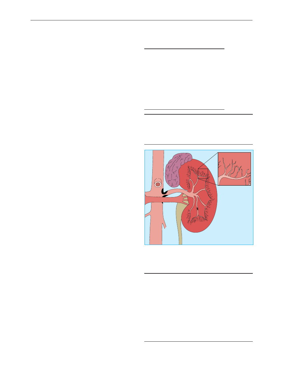
ABC of arterial and venous disease
Renal artery stenosis
Kevin McLaughlin, Alan G Jardine, Jon G Moss
Renal artery stenosis is becoming increasingly common
because of atherosclerosis in an ageing population. Patients
usually present with hypertension and varying degrees of renal
impairment, although silent renal artery stenosis may be
present in many patients with vascular disease. Despite
improvements in diagnostic and interventional techniques,
controversy remains over whether, when, and how to
revascularise the kidneys of patients with renal artery stenosis.
Pathophysiology
The pathophysiology of unilateral renal artery stenosis provides
a clear example of how hypertension develops. Narrowing of
the renal artery, due to atherosclerosis or, rarely, fibromuscular
dysplasia, leads to reduced renal perfusion. The consequent
activation of the renin-angiotensin system causes hypertension
(mediated by angiotensin II), hypokalaemia, and hyponatraemia
(which are features of secondary hyperaldosteronism).
Although these features may be reversed by correcting the
stenosis, a classic presentation is uncommon, and hypertension
is rarely cured in patients with atheromatous renal artery
stenosis. In addition, it is now known that renal artery stenosis is
underdiagnosed and may present as a spectrum of disease from
secondary hypertension to end stage renal failure, reflecting
variation in the underlying disease process. Thus, the presence
of overt, or coincidental, renal artery stenosis usually reflects
widespread vascular disease, with the associated implications for
cardiovascular risk and patient survival.
Clinical features
Atheromatous renal artery stenosis typically occurs in male
smokers aged over 50 years with coexistent vascular disease
elsewhere. It is underdiagnosed and may present with a
spectrum of clinical manifestations. Although conventionally
thought of as a cause of hypertension, atheromatous renal
artery stenosis is not commonly associated with mild to
moderate hypertension. However, it is present in up to a third
of patients with malignant or drug resistant hypertension. Renal
artery stenosis is a cause of end stage renal failure, and patients
commonly present with chronic renal failure (with or without
hypertension). Typical patients have a bland urine sediment and
non-nephrotic range proteinuria, although occasional patients
may have heavy proteinuria with focal glomerulosclerosis on
renal biopsy. Patients may also present with acute renal failure,
particularly those with bilateral renal artery stenosis (or stenosis
of a single functioning kidney) who are taking drugs that block
the renin-angiotensin system.
Less common presentations include recurrent, rapid onset
(“flash”) pulmonary oedema, which is probably a consequence
of fluid retention, and diastolic ventricular dysfunction, which
often accompanies (bilateral) atheromatous renal artery
stenosis. Biochemical abnormalities may also be present in
patients with modest or no serious renal impairment. Patients
with unilateral renal artery stenosis have raised circulating
concentrations of renin and aldosterone and associated
hypokalaemia; in contrast to patients with primary
hyperaldosteronism, their plasma sodium concentration is
Characteristics of renal artery stenosis
Fibromuscular dysplasia
x Young age group
x Predominantly affects women
x Presents as hypertension
x Rarely causes renal impairment
Atherosclerosis
x Older age group
x More common in men
x Affects smokers
x Evidence of atherosclerosis elsewhere
x Causes hypertension—often treatment resistant
x Often associated with renal impairment
Prevalence of atheromatous renal artery stenosis
x 27% of necropsies
x 25% of patients having routine coronary angiography
x 50% of patients having peripheral angiography
x 16-20% of all patients starting renal dialysis
x 25-30% of patients aged over 60 years on dialysis programmes
Clinical features and pointers to diagnosis of renal artery
disease
x Young hypertensive patients with no family history (fibromuscular
dysplasia)
x Peripheral vascular disease
x Resistant hypertension
x Deteriorating blood pressure control in compliant, long standing
hypertensive patients
x Deterioration in renal function with angiotensin converting enzyme
inhibition
x Renal impairment with minimal proteinuria
x “Flash” pulmonary oedema
x > 1.5 cm difference in kidney size on ultrasonography
x Secondary hyperaldosteronism (low plasma sodium and potassium
concentrations)
Atheromatous lesions may affect different sized vessels within the kidney,
and multiple lesions may exist. The site limits the potential for
revascularisation; only lesions within the large vessels are amenable. The
commonest site, at the ostium of the renal artery, is more effectively treated
by stenting. Ulcerated atheromatous plaques may also generate cholesterol
microemboli (particularly after vascular intervention)
Clinical review
1124
BMJ
VOLUME 320 22 APRIL 2000 bmj.com
on 1 October 2006
bmj.com
Downloaded from
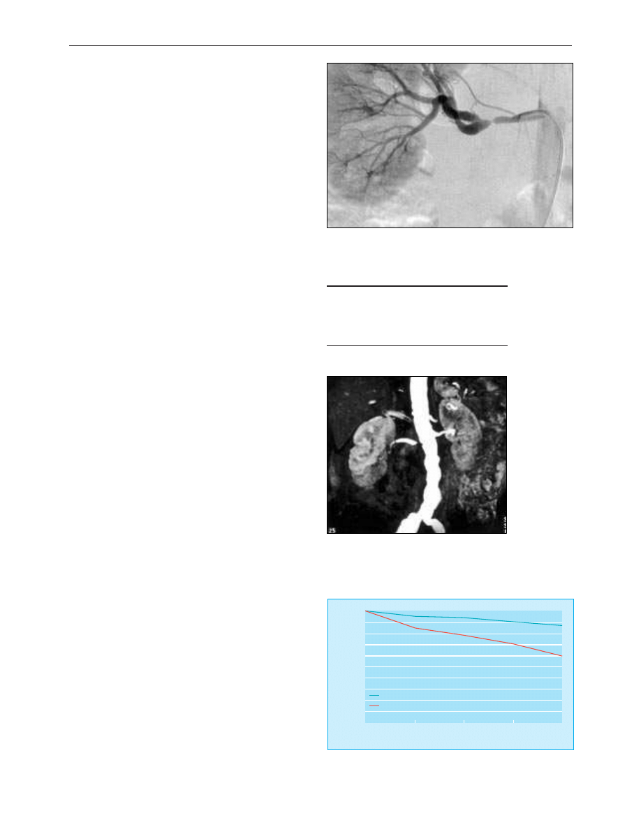
normal or reduced. Patients with bilateral renal artery stenosis
commonly have impaired renal function.
Clinical examination often shows bruits over major vessels,
including the abdominal aorta (a feature of widespread
atherosclerosis), although the classic finding of lateralising
bruits over the renal arteries is uncommon.
Diagnosis
The main differential diagnoses of atheromatous renal artery
stenosis in patients with hypertension and renal impairment are
benign hypertensive nephrosclerosis and cholesterol
microembolic disease. Differentiating between these conditions
may be difficult, particularly as all three can occur
simultaneously.
Angiography remains the standard test for diagnosing
atheromatous renal artery stenosis and is widely available.
However, it is not without risk and may worsen renal function.
Non-invasive imaging techniques are beginning to replace
conventional angiography. Although acceptable results have
been reported by single enthusiastic centres, it remains to be
seen whether these can be reproduced. Each has its own
limitation. Doppler ultrasonography is very operator sensitive
and is often impossible in obese patients. Spiral computed
tomography requires the use of iodinated contrast media and
radiation. Isotope renography (with or without captopril) has
the advantage of providing information on renal function but is
of little value in bilateral disease or when renal function is
seriously impaired. Magnetic resonance angiography is the
most promising imaging technique. It requires no contrast and
permits reconstruction of the image in different planes.
However, use of this technique is limited by lack of access to
magnetic resonance imaging machines in the United Kingdom.
All the above non-invasive tests require variable degrees of
patient cooperation, particularly the ability to hold a breath for
up to 30 seconds for image acquisition. Moreover, none of the
available imaging techniques identify the patients who will
respond to revascularisation. The aim of investigation is to
establish the diagnosis and whether revascularisation is possible
or appropriate. In practice we use ultrasonography to define
renal size and symmetry before proceeding to angiography to
delineate the lesion. Magnetic resonance angiography is
reserved for patients at high risk of angiographic complications.
Prognosis
The rate of progression of atheromatous renal artery stenosis is
difficult to evaluate accurately, and reports from large tertiary
referral centres are likely to be biased by case mix as the studies
have followed selected cohorts. Moreover, follow up may be
limited by the influence of concomitant vascular disease on
survival. Most studies (over a variable follow up period) estimate
the risk of radiological progression of atheromatous renal
artery stenosis to be about 50%, and risk is dependent on initial
severity of the lesion. The rate of occlusion of renal arteries with
greater than 60% stenosis is about 5% a year.
Patients who have bilateral atheromatous renal artery
stenosis with an occluded renal artery are three times more
likely to reach end stage renal failure within two years than
patients who have bilateral disease without occlusion (50% v
18%). The rate of loss of functional renal tissue (implied by loss
of renal size of >1 cm at one year after diagnosis) is about three
times higher for patients with bilateral disease than for those
with unilateral disease (43% v 13%).
Non-invasive imaging techniques
x Doppler ultrasonography
x Captopril renography
x Spiral computed tomography
x Magnetic resonance angiography
Angiogram of renal artery stenosis due to fibromuscular dysplasia of renal
artery. Lesions typically affect the main renal artery, are beaded in
appearance, and may have multiple stenoses
Gadolinium enhanced magnetic resonance angiogram
showing a tight stenosis at the origin of the right renal
artery. The left renal artery is normal
Years
% surviving
4
2
3
1
0
0
20
30
40
50
60
70
80
90
100
No renal artery stenosis
10
Renal artery stenosis
Effect of coexistent renal artery stenosis on survival among patients having
coronary angiography
Clinical review
1125
BMJ
VOLUME 320 22 APRIL 2000 bmj.com
on 1 October 2006
bmj.com
Downloaded from
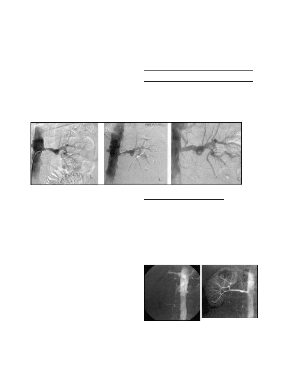
Treatment
Treatment of atheromatous renal artery stenosis must be tailored
to the individual and should be undertaken in the expectation
that revascularisation will prolong life. Hypertension in patients
with renal artery stenosis can be controlled by drugs alone in
almost 90% of cases. Angiotensin converting enzyme inhibitors
reduce the glomerular filtration rate in about one third of patients
with high grade bilateral atheromatous renal artery stenosis.
Although this reduction is reversible in most cases, these drugs
should be used with utmost caution.
Angioplasty is the traditional revascularisation procedure.
Technical advances have revolutionised angioplasty, but few
trials have examined its effects in renal artery stenosis. The
available randomised controlled trials comparing drug
treatment with angioplasty have shown only a modest reduction
in blood pressure or antihypertensive drug requirements after
angioplasty. Unlike treatment of fibromuscular dysplasia, cure of
atheromatous renal artery stenosis by angioplasty alone is rare.
Most atheromatous renal artery stenosis is due to aortic
plaques encroaching on the ostium of the renal artery.
Angioplasty is less than ideal in this situation because of the
elastic recoil of the aortic plaques. The introduction of stents
has helped overcome this problem. A recent randomised
controlled trial comparing stent insertion to angioplasty alone
in patients with ostial stenosis found a higher initial success rate
(88% v 57%) and lower restenosis rate at six months (14% v
48%) in patients who had a stent inserted. Apart from expense
there is little to argue against a policy of primary stenting for
ostial renal artery stenosis. Although the patency rates from
surgical bypass are excellent, surgery should probably be
reserved for patients in whom stenting fails or who develop
complications.
Benefits of revascularisation
Renal function may improve greatly after vascular intervention,
although it is difficult to identify which patients will benefit and
the potential of the kidney distal to the stenosis to recover
function. The available studies in patients with mild to moderate
renal impairment show that renal function improves in 25% of
patients, remains stable in 50%, and deteriorates in 25% after
surgical revascularisation or stenting. Renal length >8 cm on
ultrasonography and the presence of intact glomeruli on renal
biopsy have been suggested as good prognostic markers but
have not been formally studied. Patients in whom renal function
deteriorates after vascular intervention have an extremely poor
prognosis, with most requiring dialysis or dying within one year.
Treatment options for renal artery stenosis
x Drug treatment to reduce blood pressure (avoiding angiotensin
converting enzyme inhibitors if possible)
x Stop smoking
x Lipid lowering treatment
x Preventive treatment for coexistent vascular disease, eg aspirin
x Angioplasty for non-ostial disease and fibromuscular dysplasia
x Stent insertion for ostial atheromatous renal artery stenosis
x Surgical bypass for failed endovascular procedures
Indications for revascularisation
x Resistant hypertension
x Deteriorating renal function
x Critical stenosis, or stenosis with deteriorating function, in single
functioning kidney
x Fibromuscular dysplasia
x Associated heart failure (“flash” pulmonary oedema)
Complications of intervention
x Groin haematoma
x Femoral artery false aneurysm
x Ischaemia distal to puncture site
x Contrast nephrotoxicity
x Renal artery occlusion, dissection, or perforation
x Cholesterol embolism syndrome
Occluded renal artery before (left) and after (right) successful stenting. It is
possible to revascularise an occluded renal artery provided there is distal
reconstitution of the renal artery with collateral vessels maintaining viability.
In this relatively unusual case, the patient required dialysis at presentation
but became dialysis independent after stenting
Endovascular treatment. In this ostial lesion (left) there is no improvement after angioplasty because of elastic recoil (middle). An excellent
morphological result is then achieved by stent placement (right)
Clinical review
1126
BMJ
VOLUME 320 22 APRIL 2000 bmj.com
on 1 October 2006
bmj.com
Downloaded from

Although a modest improvement in blood pressure or a
reduction in antihypertensive drug requirement may be the
goal of revascularisation, renal protection may emerge as a
more important factor. Animal studies have shown that renal
tissue distal to renal artery stenosis undergoes irreversible
ischaemic change, tubular atrophy, interstitial fibrosis, and
glomerulosclerosis despite antihypertensive treatment.
Although similar evidence is not available in humans, insertion
of a stent in patients with renal artery stenosis slowed the rate of
loss of renal function despite modest blood pressure benefits. A
final reason to pursue revascularisation is the fact that patients
with atheromatous renal artery stenosis have a worse prognosis
than any other group on dialysis, with a median survival of 27
months, and prevention of progression to end stage renal
failure may have greatest benefits in this group. The benefits of
revascularisation, and the potential benefits of early intervention
(for example, in patients with coincidental findings of renal
artery stenosis) will need to be established by randomised
controlled trials before the burden of additional procedures is
placed on both radiologists and patients.
Special cases
Fibromuscular dysplasia is a much less common cause of renal
artery stenosis. It principally occurs in young women, who
present with unilateral disease, hypertension, and biochemical
abnormalities. The lesions are graded by their distribution. In
contrast to atheromatous renal artery stenosis, fibromuscular
dysplasia is often cured by intervention, and often by
angioplasty alone.
Patients with renal transplants have accelerated
cardiovascular disease, one of the manifestations of which is the
development of stenosis in the transplanted renal artery. This
produces hypertension, fluid retention, and renal impairment
similar to that found in patients with bilateral renal artery
stenosis and is an indication for revascularisation.
Treatment recommendations
Recommending treatment for renal artery stenosis is difficult
with the quality of the evidence available and the paucity of
controlled trials. Surgical revascularisation is rarely indicated
but may have a role in patients for whom angioplasty or
stenting is not technically feasible or in patients with complex
disease having abdominal vascular surgery. Nephrectomy (by
minimally invasive surgery) may be considered in patients with
unilateral disease whose blood pressure cannot be controlled
because of a small or poorly functioning kidney.
Whatever recommendations we suggest, there will be
disagreement. Firmly held, preconceived beliefs exist on the
treatment of renal artery stenosis despite the fact that there
have been few large outcome studies comparing treatment
strategies. The ASTRAL (angioplasty and stent for renal artery
lesions) study is a British randomised controlled trial that will
compare angioplasty with and without stenting against drug
treatment in 1000 patients with renal artery stenosis. Our future
approach to the management of renal artery stenosis will
depend on the conduct of relevant controlled trials.
The picture of gadolinium enhanced magnetic resonance angiography was
provided by D Wilcock, Leicester Royal Infirmary.
Kevin McLaughlin is assistant professor of nephrology, University of
Calgary, Canada; Alan G Jardine is senior lecturer and consultant
nephrologist, department of medicine and therapeutics, Western
Infirmary, Glasgow (a.g.jardine@clinmed.gla.ac.uk); and Jon G Moss is
consultant interventional radiologist, Gartnavel General Hospital,
Glasgow.
Further reading
x Mailloux LU, Napolitano B, Bellucci AG, Vernance M, Wilkes BM,
Mossey RT. Renal vascular disease causing end-stage renal disease:
incidence, clinical correlates and outcomes: a 20-year clinical
experience. Am J Kidney Dis 1994;24:622-9.
x Plouin PF, Chatellier G, Darne B, Raynaud A. Blood pressure
outcome of angioplasty in atherosclerotic renal artery stenosis: a
randomized trial. Essai Multicentrique Medicaments vs Angioplastie
(EMMA) Study Group. Hypertension 1998;31:823-9.
x Van de Ven PJG, Kaatee R, Beutler JJ, Beek FJ, Woittiez AJ, Buskens
E et al. Arterial stenting and balloon angioplasty in ostial
atherosclerotic renovascular disease: a randomised trial. Lancet
1999;353:282-6.
x Textor SC. Revascularisation in atherosclerotic renal artery disease.
Kidney Int
1998;53:799-811.
x Conlon PJ, Athirakul K, Kovalik E, Schwab SJ, Crowley J, Stack R, et
al. Survival in renovascular disease. J Am Soc Nephrol 1998;9:252-6.
Recommendations for treatment
Patient characteristics
Treatment
Controlled blood pressure
Minimal renal impairment (creatinine
< 150 mmol/l)
Medical
Renal artery stenosis < 50% (unilateral
or bilateral)
Advanced renal failure and
Renal size < 8 cm or
Renal artery occlusion (without distal
recanalisation) or
No evidence of renal function in
affected kidney or
No prospect of recovery
with intervention
Biopsy showing severe irreversible
changes
Renal size > 8 cm and renal artery
stenosis > 50% and
Poorly controlled blood pressure or
Deterioration in renal function with
angiotensin converting enzyme
inhibitors or
Progressive renal impairment
Angioplasty for non-ostial
lesions
Angioplasty plus stent for
ostial disease
The ABC of arterial and venous disease is edited by Richard Donnelly,
professor of vascular medicine, University of Nottingham and
Southern Derbyshire Acute Hospitals NHS Trust (richard.donnelly@
nottingham.ac.uk) and Nick J M London, professor of surgery,
University of Leicester, Leicester (sms16@leicester.ac.uk). It will be
published as a book later this year.
BMJ
2000;320:1124-7
Slope of reciprocal serum creatinine plot
(l/mmol/dayx10
-6
)
After stent
Before stent
-20
-12
-8
-4
0
4
8
12
-16
Effect of stent placement on rate of loss of renal function (expressed as slope
of serum creatinine concentration with time). Overall, a significant reduction
occurred in the rate of loss of renal function, although the individual
response was variable
Clinical review
1127
BMJ
VOLUME 320 22 APRIL 2000 bmj.com
on 1 October 2006
bmj.com
Downloaded from
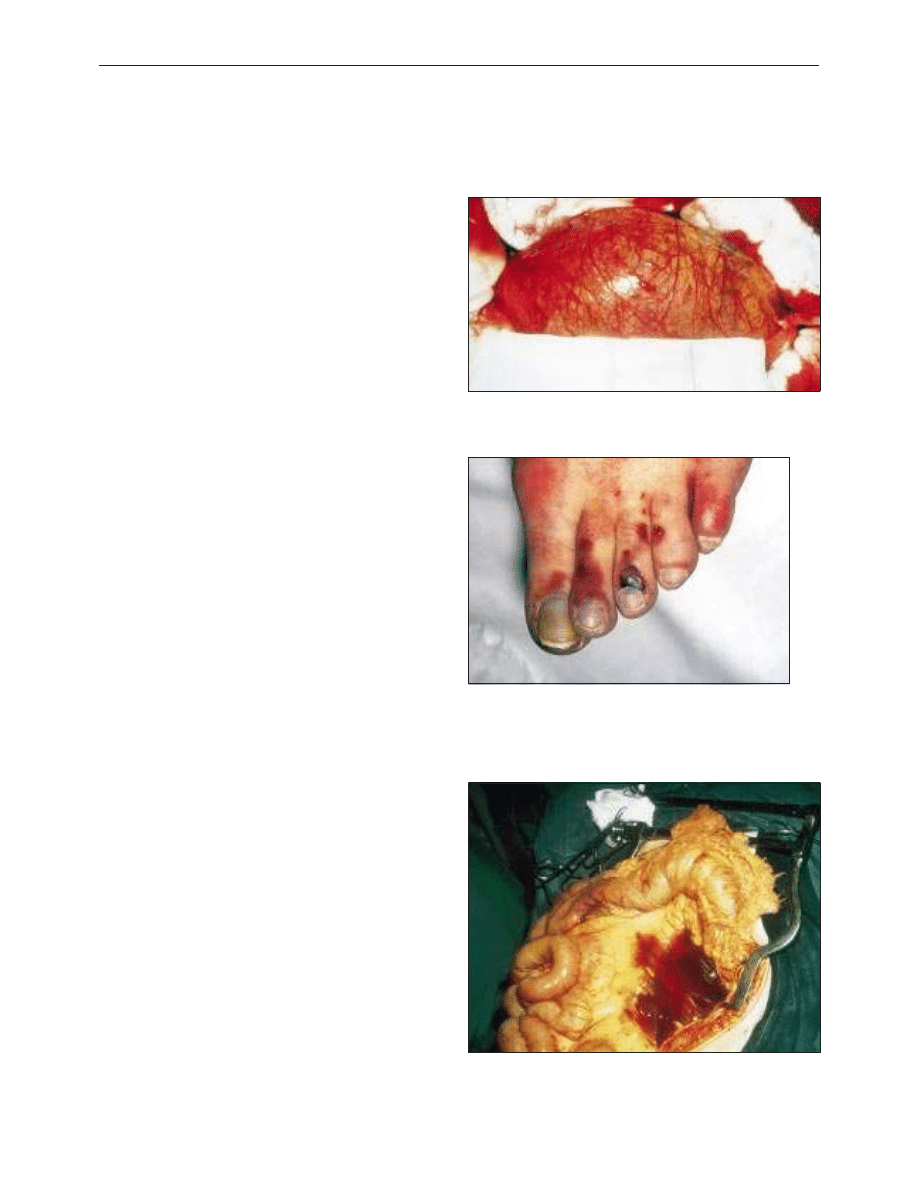
ABC of arterial and venous disease
Arterial aneurysms
M M Thompson, P R F Bell
True arterial aneurysms are defined as a 50% increase in the
normal diameter of the vessel. Clinical symptoms usually arise
from the common complications that affect arterial
aneurysms—namely, rupture, thrombosis, or distal embolisation.
Although the aneurysmal process may affect any large or
medium sized artery, the most commonly affected vessels are
the aorta and iliac arteries, followed by the popliteal, femoral,
and carotid vessels.
Abdominal aortic aneurysms
Aneurysms of the infrarenal abdominal aorta and iliac arteries
coexist to such a degree that they may be considered a single
clinical entity. Abdominal aneurysms usually affect elderly men
( > 65 years), with a prevalence of 5%. In England, abdominal
aneurysm is responsible for over 11 000 hospital admissions
and 10 000 deaths a year. Interestingly, unlike other
atherosclerotic vascular disorders, the prevalence of abdominal
aortic aneurysms is increasing rapidly, and aneurysmal rupture
is now the 13th commonest cause of death in the Western
world.
Clinical presentation
Although abdominal aneurysms may cause symptoms because
of pressure on surrounding structures, about three quarters
remain asymptomatic at initial diagnosis. With the exception of
vague abdominal pain, clinical symptoms usually result from
embolisation or rupture of the aneurysm.
The appearance of microembolic lower limb infarcts in a
patient with easily palpable pedal pulses may suggest the
presence of either popliteal or abdominal aneurysms.
Additionally, patients with embolisation of mural thrombus
from an abdominal aneurysm may present with acute limb
ischaemia due to femoral or popliteal occlusion.
The diagnostic triad of hypovolaemic shock, pulsatile
abdominal mass, and abdominal or back pain is encountered in
only a minority of patients with ruptured abdominal aneurysms.
In general, ruptured abdominal aortic aneurysm should be
considered in any patient with hypotension and atypical
abdominal symptoms. Similarly, the presence of abdominal pain
in a patient with a known aneurysm or pulsatile mass must be
considered to represent a rapidly expanding or ruptured
aneurysm and be treated accordingly. In the community setting,
the death rate from ruptured abdominal aortic aneurysms is
almost 90%, as 80% of patients will die before reaching hospital
and about 50% die during surgery to repair the rupture.
Methods of diagnosis
The sensitivity of abdominal palpation to detect aortic
aneurysms increases with the diameter of the aneurysm, but
palpation is not sufficiently reliable for routine diagnosis.
Similarly, plain abdominal radiography shows a calcified
aneurysmal aortic wall in only half of cases.
The simplest diagnostic test is B mode ultrasonography,
which gives an accurate assessment of both the diameter and
the site of the aneurysm. If more accurate morphological data
are required to determine the exact relation of the aneurysm to
the visceral or renal arteries, detailed cross sectional imaging
Large infrarenal abdominal aortic aneurysm before surgical repair
Clinical picture of “trash foot.” The appearance is caused by multiple
microscopic atheromatous emboli from a large infrarenal aortic
aneurysm. The presence of digital infarcts in a patient with easily
palpable pulses may point to an aneurysmal source of emboli
Ruptured abdominal aneurysm. Rupture usually occurs posteriorly into the
retroperitoneum, which produces a contained leak and allows the possibility
of surgical repair. Free anterior intraperitoneal rupture usually results in
exsanguination
Clinical review
1193
BMJ
VOLUME 320 29 APRIL 2000 bmj.com
on 1 October 2006
bmj.com
Downloaded from
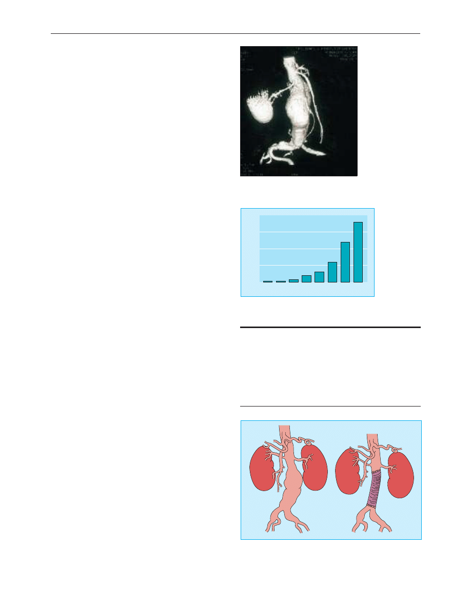
may be obtained by computed tomography or magnetic
resonance angiography.
The diagnosis of ruptured abdominal aortic aneurysms relies
on clinical symptoms. Ultrasonography is used to confirm an
aneurysm if it is difficult to palpate. Computed tomography has a
low specificity (about 75%) for determining the presence of a
rupture and adds little information to routine clinical assessment.
Indications for surgery
Elective surgery
The decision to operate on a patient with an asymptomatic
abdominal aneurysm is based on an analysis of the risk of
aneurysmal rupture compared with the mortality of elective
surgical repair. The risk of rupture is related to many factors,
but the diameter of the aortic aneurysm has historically been
used as the principal determinant.
Unfortunately, little information is available on the rupture
rates of large abdominal aneurysms, but pooled analysis of
existing data suggests that the risk of rupture increases
exponentially in aneurysms above 55-60 mm. This has led to a
broad surgical consensus that aneurysms exceeding 55 mm in
diameter should be surgically repaired if there are no
confounding factors that would substantially increase the risk of
elective surgery.
The treatment for smaller aneurysms has recently been
clarified by the UK small aneurysm trial, which studied 1090
patients with aneurysms of 40-55 mm. The study found a 30 day
operative mortality of 5.8%, mean risk of rupture for small
aneurysms of 1% a year, and no difference in survival between
treatment groups at two, four, or six years. The cost for early
surgery was higher than for surveillance, but early surgery was
associated with improvement in some measures of quality of life.
Emergency treatment
Patients with suspected ruptured aneurysms should be
considered for emergency surgical repair. Several studies have
looked at preoperative risk factors and survival after emergency
repair of an aneurysm. Although there is no precise scoring
system that will allow accurate prediction of survival, the presence
of several predictive factors (age > 80 years, unconsciousness, low
haemoglobin concentration, cardiac arrest, severe
cardiorespiratory disease) can be used to determine patients in
whom the risk of dying during surgery approaches 100%.
Patients with symptomatic aneurysms should be treated as
urgent cases and have the aneurysm repaired. The aetiology of
pain from abdominal aneurysms is not well understood,
although it has been attributed to stretching of the aneurysm
sac or severe inflammation within the aneurysm wall
(inflammatory aneurysms, see below).
Conventional surgical repair
Traditional surgical repair for asymptomatic abdominal aortic
aneurysms involves exposure of the abdominal aorta, aortic and
iliac clamping, and replacement of the aneurysmal segment with
a prosthetic graft. Graft replacement is an effective, durable
procedure, and most centres report 30 day mortality of about 5%,
although this varies with the volume of work and type of hospital.
The mortality associated with surgical repair of aneurysms is
closely related to the “fitness” of the patient for surgery; patients
with severe cardiorespiratory disease have a perioperative
mortality approaching 40%, with most deaths caused by cardiac
events.
Endovascular repair
One of the major developments in vascular surgery over the
past five years has been the introduction of endovascular repair
Factors predisposing to rupture of abdominal aortic
aneurysms
x Diameter of aneurysm
x Diastolic blood pressure
x Chronic obstructive pulmonary disease
x Smoking
x Family history of ruptured aneurysm
x Expansion rate
x Intrinsic biology—inflammation within the aortic wall
x Thrombus-free surface area of aneurysm sac
Computed tomogram showing large infrarenal
abdominal aortic aneurysm
90
100
Aneurysm size (mm)
1 year rupture rate (%)
80
70
60
50
40
30
0
50
75
100
25
Annual rupture rates of abdominal aortic aneurysms
according to size (based on pooled available data)
Conventional repair of abdominal aortic aneurysm. The aneurysmal
segment of the aortoiliac segment is replaced with a prosthetic vascular graft
(usually Dacron or ePTFE), which is sutured to the normal arterial “cuffs”
above and below the aneurysm
Clinical review
1194
BMJ
VOLUME 320 29 APRIL 2000 bmj.com
on 1 October 2006
bmj.com
Downloaded from

of aneurysms. This technique uses an endoprosthesis, which is
delivered through the femoral arteries, to exclude an aneurysm
from the circulation. The endograft is secured to the normal
calibre aorta and iliac arteries using metallic expandable stents
and relies on subsequent thrombosis of the aneurysm to abolish
the risk of rupture.
Endovascular repair has several theoretical advantages over
conventional surgery, and early evidence suggests that
endovascular surgery is better for patients with coexistent
disease, who would be high risk for conventional surgery.
However, the long term durability of endovascular techniques is
unknown, although experience so far shows that up to a quarter
of patients undergoing endovascular aneurysm repair will
require subsequent endovascular interventions to ensure
regression of the aneurysm sac. A multicentre trial of
endovascular repair and conventional surgery has started in the
United Kingdom.
Screening and medical treatment
Ultrasonography-based screening programmes to detect and
treat asymptomatic aneurysms have been proposed as a
mechanism to reduce the mortality from ruptured abdominal
aortic aneurysms. Screening studies report a 2.5% prevalence of
abdominal aortic aneurysms larger than 40 mm in men aged
over 60 years.
Advocates of community screening have suggested that a
single abdominal scan in men aged 65 would exclude 90% of
the population from future aneurysm rupture, and long term
follow up of screened and control populations in Chichester
showed an 85% reduction in rupture in the screened group. A
multicentre randomised trial is currently investigating the
cost-effectiveness of community based aneurysm screening.
One of the problems with screening programmes is that it
identifies many people with small aneurysms. These patients are
not offered any form of treatment other than ultrasonographic
surveillance, which may have implications for quality of life.
However, recent studies have begun to elucidate the molecular
and biochemical mechanisms of aneurysm formation, and
clinical trials of the effectiveness of several groups of drugs to
reduce expansion of small aneurysms are likely. The most
promising of these drugs in experimental studies have been
inhibitors of matrix metalloproteinases.
Variants of abdominal aneurysms
Inflammatory aneurysms
Inflammatory aneurysms are characterised by a dense
inflammatory infiltrate within the aneurysm wall. They are
typically white in appearance and may be densely adherent to
surrounding structures, which could account for the increased
operative mortality in affected patients Patients typically present
with fever, malaise, and abdominal pain.
Thoracoabdominal aneurysms
Thoracoabdominal aneurysms extend to a variable degree from
the thoracic aorta into the abdominal aorta. They typically
Endovascular aneurysm repair. The aneurysm sac is excluded by an
endograft, which is introduced through a remote arteriotomy and anchored
above and below the aneurysm sac
Aortoiliac aneurysm before and after exclusion by a bifurcated endovascular
graft
Inflammatory abdominal aneurysm. The aneurysm is typically white in
appearance and densely adherent to surrounding structures. The duodenum
is being mobilised from the aneurysm sac by sharp dissection
Computed tomogram of thoracoabdominal aneurysm
Potential advantages of endovascular repair over
conventional surgery
x No need for abdominal incision
x Avoidance of aortic cross clamping
x No retroperitoneal dissection
x Improved perioperative cardiorespiratory function
x Reduction in metabolic stress response to aortic aneurysm repair
x Improved renal and gastrointestinal function
x Reduced hospital stay
Clinical review
1195
BMJ
VOLUME 320 29 APRIL 2000 bmj.com
on 1 October 2006
bmj.com
Downloaded from
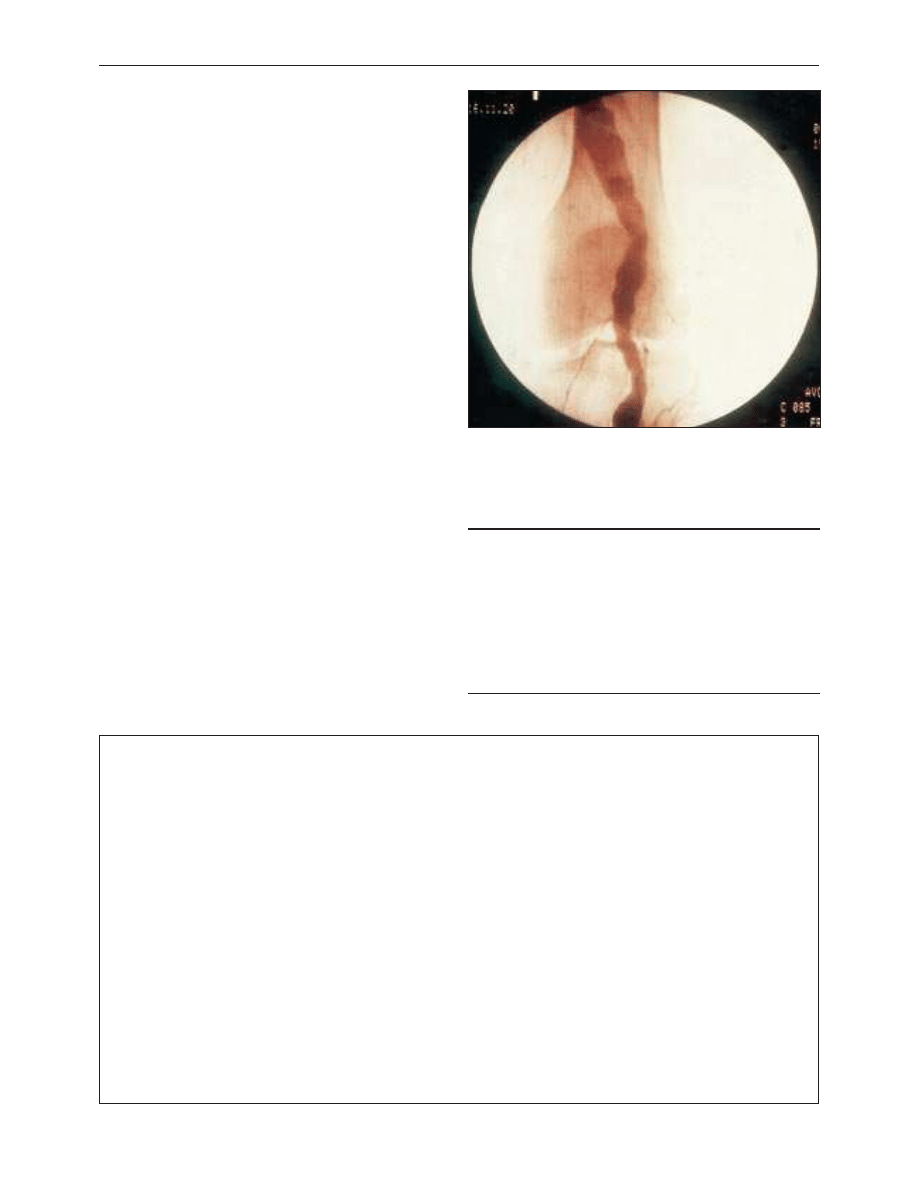
affect the origins of the visceral and renal arteries, which must
be reimplanted into the graft during repair of the aneurysm.
Mortality from repair of thoracoabdominal aneurysms is
significantly higher than that for infrarenal surgery.
Peripheral aneurysms
Popliteal aneurysms comprise 80% of all peripheral aneurysms
and usually exceed 20 mm in diameter. They are associated with
aortic aneurysms (40% of cases), and are frequently bilateral
(50%). In contrast to abdominal aortic aneurysms, patients with
popliteal aneurysms usually present with acute limb ischaemia
secondary to aneurysm thrombosis or distal embolisation. A
diagnosis of popliteal aneurysms is suggested by easily palpable
popliteal pulses and confirmed by duplex ultrasonography.
Patients who develop acute limb ischaemia due to popliteal
thrombosis or embolism have a relatively poor prognosis (15%
amputation rate) because of occlusion of the run off vessels. In
these cases patients should have saphenous vein bypass and
ligation of the popliteal aneurysm with clearance of the crural
vessels by balloon thrombectomy or thrombolysis. The
indications for elective surgery for asymptomatic popliteal
aneurysms are based on suggestive evidence only, with most
clinicians opting for surgical treatment when the aneurysm
exceeds 25 mm in diameter. Results of bypass of asymptomatic
popliteal aneurysms are excellent, with five year graft patency of
80% and limb salvage of 98%.
Femoral artery aneurysms are the second commonest
peripheral aneurysm. Patients present with local pressure
symptoms, thrombosis, or distal embolisation. Surgical treatment
of true femoral aneurysms relies on the principles of excluding
the aneurysm and restoration of blood flow in the limb.
M M Thompson is consultant vascular and endovascular surgeon,
Leicester Royal Infirmary (MattT11@aol.com), and P R F Bell is
professor of surgery, University of Leicester.
The ABC of arterial and venous disease is edited by Richard
Donnelly, professor of vascular medicine, University of Nottingham
and Southern Derbyshire Acute Hospitals NHS Trust
(richard.donnelly@ nottingham.ac.uk) and Nick J M London,
professor of surgery, University of Leicester, Leicester
(sms16@leicester.ac.uk). It will be published as a book later this year.
Key references
x Thompson MM, Sayers RD. Arterial aneurysms. In: Beard JD,
Gaines P, eds. Companion to specialist surgical practice. Vol VII. Arterial
surgery
. London: W B Saunders, 1998.
x UK Small Aneurysm Trial Participants. Mortality results for
randomised controlled trial of early elective surgery or
ultrasonographic surveillance for small abdominal aortic
aneurysms. Lancet 1998;352:1649-55.
x Scott RAP, Wilson NM, Ashton HA, Kay DM. Influence of screening
on the incidence of ruptured abdominal aortic aneurysm: 5-year
results of a randomised controlled study. Br J Surg 1995;82:1066-70.
BMJ
2000;320:1193-6
Arteriogram of patient with popliteal artery aneurysm showing typical
spiralling appearance of the superficial femoral and popliteal arteries
A memorable anaesthetic
“Shouldn’t we do something about the fire?”
Can this have happened only about 40 years ago? It would be
unthinkable now, for many reasons. What makes it memorable is
the aspect of saving life; we all, in our disillusioned moments,
make cynical jokes about saving life, but on this occasion I may
actually have saved several lives all in one go. I was a second year
clinical student, doing part of my obstetric attachment in January
in one of the colder parts of Britain.
Late one evening, the obstetric registrar asked me if I would
like to accompany him on a flying squad call; a parturient in a
remote farmhouse some 18 miles away had a retained placenta.
We set off in a taxi. The driver had only a vague idea of where we
were going, and needed to stop frequently at pubs to ask
directions. After every stop his driving seemed to get more erratic.
We eventually arrived at the house, where the farmer’s wife lay
unconcerned in bed, while the infant slept peacefully in a cot. An
open fire burned merrily in the huge fireplace.
The GP, smart in tweed jacket and plus fours, radiated an air of
confidence and competence, though he must have known there
was little he could do. I don’t remember seeing the farmer, but the
presence of the husband in the delivery room was not then
usually accepted. It was decided that the placenta should be
manually removed in the bed, under open ether anaesthesia.
While preparations were being made for this, I had uneasy
memories of accidents involving ether and flames in the
chemistry laboratory at school.
Eventually, I plucked up courage to say, “Shouldn’t we do
something about the fire?” This was agreed to be sensible, and we
carried out the blazing logs in buckets to a safe part of the frost
bound farmyard. The ether was poured by the GP and the
placenta was delivered without loss of life and farmhouse.
The journey back to the hospital seemed shorter than the
outward journey; the pubs were closed by then. All had gone well,
but I wonder what the Confidential Inquiry into Maternal
Mortality would have said if we had all gone up in flames.
Peter Young consultant anaesthetist, Gloucestershire
We welcome articles of up to 600 words on topics such as
A memorable patient, A paper that changed my practice, My most
unfortunate mistake
, or any other piece conveying instruction,
pathos, or humour. If possible the article should be supplied on a
disk. Permission is needed from the patient or a relative if an
identifiable patient is referred to.
Clinical review
1196
BMJ
VOLUME 320 29 APRIL 2000 bmj.com
on 1 October 2006
bmj.com
Downloaded from

ABC of arterial and vascular disease
Secondary prevention of peripheral vascular disease
Sean Tierney, Fiona Fennessy, David Bouchier Hayes
Most patients with peripheral vascular disease may be reassured
that, with respect to their legs, the condition usually runs a
benign course. Less than one third of patients will require any
surgical or radiological intervention and only 5% will have
amputation. However, peripheral vascular disease is an
independent predictor of increased risk of cardiovascular death.
Half of patients presenting with peripheral vascular disease
have symptoms of coronary artery disease or
electrocardiographic abnormality, 90% have abnormalities on
coronary angiography, and 40% have duplex evidence of
carotid artery disease.
Symptomatic peripheral vascular disease carries at least a
30% risk of death within five years and almost 50% within 10
years, primarily due to myocardial infarction (60%) or stroke
(12%). The risks are more than doubled in patients with severe
disease (requiring surgery), but even asymptomatic patients
(ankle brachial pressure index < 0.9) have a twofold to fivefold
increased risk of fatal or non-fatal cardiovascular events.
Although modification of risk factors has not been shown to
prevent progression of peripheral vascular disease or loss of
limbs, detection of disease mandates an aggressive approach to
modifying risk factors in order to reduce the risk of fatal and
non-fatal myocardial infarction and stroke. The approach to
risk reduction in patients with peripheral vascular disease is
based on extrapolation from results of large studies of patients
with coronary artery disease.
Modification of risk factors
Effective reduction of the risk of cardiovascular disease depends
on coordinated and stringent modification of identifiable risk
factors to prevent progression or new disease and the use of
drugs to correct existing abnormalities. Stopping smoking,
correction of hyperlipidaemia and hypertension, and
optimisation of diabetic control are the cornerstones of
secondary prevention of cardiovascular disease. Lesser benefits
are also likely to accrue through weight reduction in obese
patients, the institution of regular exercise, and dietary
modification. Additional risk factors have been identified but
are uncommon and their treatment is of unproved value.
Cigarette smoking
Cigarette smoking contributes to a third of all deaths from
coronary artery disease, doubles the risk of stroke, and is almost
ubiquitous among patients with peripheral vascular disease.
Synergy between smoking and other risk factors substantially
increases the risks of cardiovascular death associated with these
factors. After a myocardial infarction or stroke, the risk of
recurrence is reduced by 50% in those who stop smoking (even
among long term heavy smokers). Firm evidence also exists that
stopping smoking increases walking distance by twofold to
threefold in 85% of patients with intermittent claudication.
Furthermore, in patients requiring surgical bypass, patency
rates are better among those who successfully stop smoking.
Because as few as 4% of established smokers in the general
population successfully stop smoking without assistance,
measures to improve on this are essential in the secondary
All patients with peripheral vascular disease should have
their risk factors for coronary artery disease assessed and,
if appropriate, modified according to current guidelines
Risk factors for cardiovascular disease
x Cigarette smoking
x Hyperlipidaemia
x Hypertension
x Diabetes mellitus
x Obesity
x Physical inactivity
x Diet high in saturated fats
x Hyperhomocysteinaemia
x Raised Lp(a) lipoprotein concentrations
x Hypercoagulable states
Excised atherosclerotic plaque
Gum (46 studies)
χ
2
= 54.5
Patch (30 studies)
χ
2
= 34.33
Intranasal spray (46 studies)
χ
2
= 1.22
Inhaler (4 studies)
χ
2
= 1.34
Sublingual (2 studies)
χ
2
= 0.1
1405/7235
1225/8929
107/448
84/490
49/243
1065/9247
458/7903
52/439
44/486
31/245
1.63 (1.48 to 1.78)
1.77 (1.58 to 1.97)
2.27 (1.61 to 3.20)
2.08 (1.43 to 3.04)
1.73 (1.07 to 2.80)
Study
Experimental Control
Peto odds ratio
(95% CI)
Peto odds ratio
Favours treatment
Favours control
0.1 0.2
1
5 10
Summary of results of meta-analysis of nicotine gum, patch, intranasal spray,
and sublingual tablet versus control for stopping smoking. Maximum follow
up 6-12 months
Clinical review
1262
BMJ
VOLUME 320 6 MAY 2000 bmj.com
on 1 October 2006
bmj.com
Downloaded from
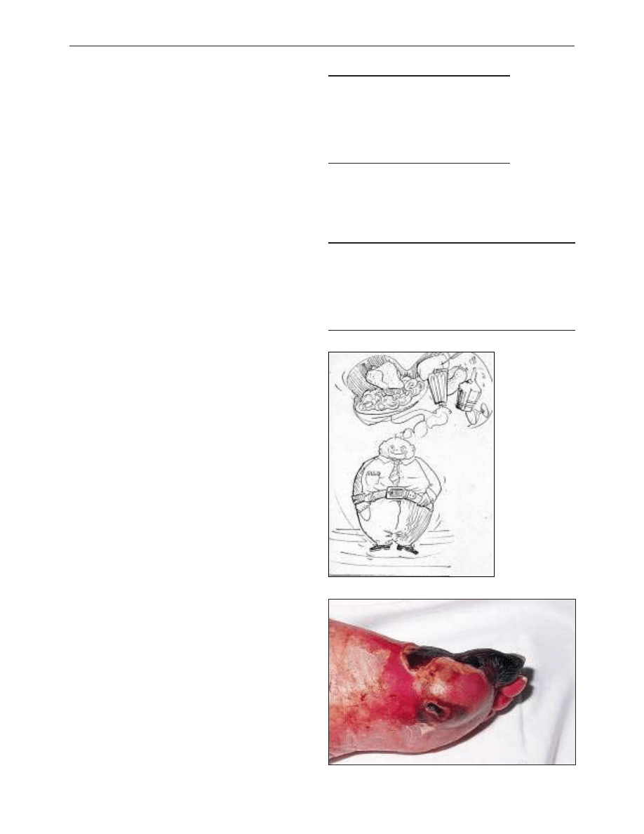
prevention of cardiovascular disease. Modern smokers are
clearly able to ignore punitive taxes and health warnings on
packaging. They respond better to short (5-10 minutes)
counselling from doctors, particularly if they are recovering
from myocardial infarction (50% success rates). Rates of
stopping smoking have been increased to 70% by the addition
of telephone based counselling.
Surprisingly, only half of current smokers in one study had
been encouraged to stop smoking, and fewer had been
specifically counselled. Hospitals caring for patients with
cardiovascular disease can help by offering support
programmes. The use of nicotine replacement (chewing gum or
patches), which is safe for patients with stable cardiovascular
disease, is effective when combined with counselling.
Hyperlipidaemia
Epidemiological data clearly indicate an association between
total cholesterol concentration and the risk of cardiovascular
death. Dietary measures may reduce serum cholesterol and low
density lipoprotein cholesterol concentrations by about 10%,
but long term compliance is poor.
The use of statins (hydroxymethylglutaryl coenzyme A
reductase inhibitors) to lower total and low density lipoprotein
cholesterol concentrations by 30-40% reduces the risk of
cardiovascular death by up to 42% among patients younger
than 70 years with established disease. The only statins licensed
for secondary prevention are simvastatin and pravastatin.
Statins also reduce triglyceride concentrations. Patients with
severe dyslipidaemia require specialised treatment.
Although there are no published studies examining the
effect of lipid reduction in patients with isolated peripheral
vascular disease, patients with peripheral disease are included as
a subgroup in current large trials such as the heart protection
study and the Medical Research Council bezafibrate in patients
with lower extremity arterial disease (LEADER) trial study.
Because of their high risk of ischaemic heart disease patients
with peripheral vascular disease who have a serum cholesterol
concentration over 5.5 mmol/l should be treated. All patients
should therefore have their lipid concentrations measured even
if they do not require specific treatment for their peripheral
vascular disease.
Diabetes mellitus
The adverse effect of diabetes on serum lipid concentrations
and the accelerated atheromatous process in diabetic patients
act synergistically with the result that 80% die of cardiovascular
disease. The atheromatous process particularly affects smaller
more distal vessels, which makes surgical reconstruction more
difficult or impossible. Diabetic patients are therefore more
likely to require amputation than other patients with peripheral
vascular disease. Modification of other risk factors is particularly
important in this population, and the absolute benefits of
reducing cholesterol concentration are likely to be greater in
diabetic patients.
Poor glycaemic control in patients with type 2 diabetes is
associated with an increased risk of cardiovascular
complications, but the value of tighter control in preventing
major cardiac events remains unclear. However, tight glycaemic
control has beneficial effects on serum lipid concentrations, and
the DIGAMI (diabetes insulin glucose after myocardial
infarction) study showed that improved glycaemic control
benefits patients after myocardial infarction. Careful attention to
Measures to encourage stopping smoking
x Public health education
x Taxes
x Smoke free hospitals and workplace
x Advice from doctor
x Nurse case managers
x Support groups and counselling
x Nicotine replacement therapy
Dietary modifications in hyperlipidaemia
x Reduce total fat intake ( < 30% of total energy)
x Reduce saturated fat intake ( < 7% of energy) and substitute with
unsaturates
x Decrease dietary cholesterol intake ( < 200 mg/day)
x Increase fibre intake
x Moderate alcohol intake
x Aim for ideal body mass index (21-25)
Foot of diabetic patient with peripheral vascular disease
Clinical review
1263
BMJ
VOLUME 320 6 MAY 2000 bmj.com
on 1 October 2006
bmj.com
Downloaded from

foot care is particularly important in diabetic patients with
peripheral vascular disease.
Hypertension
The rationale for treating hypertension is largely based on its
adverse effects on the heart and cerebrovascular system.
Conventional guidelines such as those produced by the second
working party of the British Hypertension Society are
applicable to patients with peripheral vascular disease.
Although it might seem preferable to use vasodilators in
patients with peripheral vascular disease, â blockers are safe if
they are required to obtain adequate blood pressure control.
Specific therapeutic measures
Diet
Observational studies suggest that diets rich in fish, fruit,
vegetables, and fibre but low in saturated fat may protect against
cardiovascular disease. Dietary supplementation with fish oil
(which is rich in n-3 polyunsaturated fatty acids) has recently
been shown to protect against cardiovascular death after
myocardial infarction in a large trial, but other trials have failed
to find such a benefit.
Vitamins and trace elements may also alter cardiovascular
risk through other mechanisms. Antioxidants (vitamins E and
C) help prevent the oxidation of low density lipoprotein
cholesterol, which is a key step in the atherogenic process.
Studies in patients with peripheral vascular disease have shown
no specific benefits of antioxidants, but no large scale trials have
been done.
Cardiovascular risk is independently related to plasma
homocysteine concentrations, and it has been proposed that
dietary supplementation with folic acid (which reduces
homocysteine concentrations) may aid in the primary
prevention of cardiovascular disease. However, no large
prospective trials have been done. Severe
hyperhomocysteinaemia ( > 10 ìmol/l), which may respond to
vitamin supplementation, should be considered in patients with
premature atherosclerotic disease without other risk factors. In
the absence of strong evidence for routine vitamin
supplementation, the most prudent approach is to recommend
a balanced diet rich in fruit, vegetables, and whole grains.
Exercise
Physical rehabilitation programmes form an important part of
reducing the risk of cardiovascular disease. If no supervised
exercise programme is available, patients with intermittent
claudication should be advised to walk for an hour a day,
pausing to rest whenever claudication pain develops. This
results in a 20-200% improvement in walking distance.
Although it has been suggested that repeated
ischaemia-reperfusion injury provoked by walking might have
deleterious systemic effects, regular exercise actually reduces
concentrations of serum inflammatory markers.
Antiplatelet therapy
Aspirin has been proved effective in the secondary prevention
of cardiovascular events and death in patients with established
atherosclerosis. Patients with peripheral vascular disease who do
not have specific contraindications should receive 75 mg aspirin
daily. The cost effectiveness of newer antiplatelet drugs remains
to be determined.
Further reading
x Bainton D, Sweetnam P, Baker I, Elwood P. Peripheral vascular
disease: consequence for survival and association with risk factors in
the Speedwell prospective heart disease study. Br Heart J
1994;72:128-32.
x Leng GC, Price JF, Jepson RG. Lipid-lowering for lower limb
atherosclerosis. In: Cochrane Library. Issue 4. Oxford: Update
Software, 1999.
x Scottish Intercollegiate Guidelines Network. Lipids and the primary
prevention of coronary heart disease. SIGN 1999;40.
x Department of Health. Standing medical advisory statement on the use
of statins.
Leeds: DoH, 1997. (Circular EL (97) 41.)
x O’Brien E, Bouchier Hayes D, Fitzgerald D, Atkins N. The arterial
organ in cardiovascular disease: ADAPT (arterial disease
assessment, prevention, and treatment) clinic. Lancet
1998;352:1700-1.
x American Heart Association Writing Group. Optimal risk factor
management in the patient after coronary revascularization.
americanheart.org/Scientific/statements/1994/129401.html
x American Heart Association. Antioxidant consumption and risk of
coronary heart disease: emphasis on vitamin C, vitamin E, and beta
carotene. Circulation 1999; 99:591-5.
A balanced diet rich in fruit, vegetables, and fish may protect against
cardiovascular disease
Clinical review
1264
BMJ
VOLUME 320 6 MAY 2000 bmj.com
on 1 October 2006
bmj.com
Downloaded from

Conclusion
Although individual specialties tend to treat arterial lesions in
isolation, this approach ignores the real risk to patients from the
effects of the same disease on other vascular beds. Although
simple measures (diet, exercise, stopping smoking) may be
adequate for some, the management of many patients is
complex. Patients at high risk of cardiovascular disease are best
managed in an integrated fashion, and the concept of an
arterial disease assessment, prevention, and treatment clinic to
overcome the often haphazard management of risk factors
seems the best way forward. Precise risk can be determined by
using a computer program based on history, examination, and
laboratory results and a specific programme of modification
instituted and monitored. This holistic approach to the
management of cardiovascular disease is the best way to
minimise the risk of disease progression and premature death
in patients with peripheral vascular disease.
The meta-analysis data on nicotine replacement therapy were taken
from Silagy et al. Cochrane library. Issue 4. Oxford: Update Software,
1999.
Sean Tierney is lecturer in surgery (stierney@rcsi.ie), Fiona Fennessy
is a research fellow, and David Bouchier Hayes is professor of surgery,
Beaumont Hospital, Dublin, Ireland.
The ABC of arterial and venous disease is edited by Richard Donnelly,
professor of vascular medicine, University of Nottingham and
Southern Derbyshire Acute Hospitals NHS Trust (richard.donnelly@
nottingham.ac.uk) and Nick J M London, professor of surgery,
University of Leicester, Leicester (sms16@leicester.ac.uk). It will be
published as a book later this year.
BMJ
2000;320:1262-5
Prevention and treatment
Risk factor modification
Vascular surgeon
Neurologist
Ophthalmologist
Cardiologist
Nephrologist
Diabetologist
Family doctor
Hypertension clinic
Lipid clinic
ADAPT clinic
Risk factor assessment
Lifestyle
Family history
Smoking
Alcohol
Salt intake
Obesity
Sedentary or active
Blood pressure
Urine analysis, fasting glucose and insulin
Fasting cholesterol, triglycerides, and high
and low density lipoprotein cholesterol
Thyroid function tests
Homocysteine*
Thrombophilia screen*
* Measure if peripheral vascular disease is
premature (patient under 50 years)
Target organ assessment
Kidney
• Urine analysis (including microalbumin)
• Biochemistry (urea, creatinine,
electrolytes, urate)
• Haematology (haemoglobin, film, white
cell count)
Heart
• Electrocardiography (echocardiography,
stress test, and angiography selectively)
Brain
• Carotid duplex scan in symptomatic
patients
Arterial organ
• Funduscopy; when indicated, arterial
tonometry and biochemical analysis of
endothelial function
Arterial disease assessment, prevention, and treatment (ADAPT) clinics provide a common strategy for all patients with cardiovascular
disease regardless of target organ affected
The health benefits from collecting for Christian Aid
The World Health Organisation has defined health as a state of
physical, mental, and social wellbeing, not merely the absence of
disease. House to house collection of charity envelopes is a
exercise that can be done in three health promoting rounds. The
first round is the mental health promotion exercise. Find and
locate the right roads and work out the shortest way to go round
them. Should I do one side at a time or cross the road at strategic
junctions? Get the rules clear. Who does the corner house? Is it
me or the unknown person who may be collecting from the other
road? Should gates be left open or shut and what do I do about
those dogs? Then how do I respond to the little notice as I walk to
a front door that says, “You are being photographed”? Should I
brush my remaining hair and try to look suitably smart or cover
my face and run past? The next brain teaser is searching for letter
boxes, the variation in letter box locations is truly amazing. I
discover that a doctor who I know professionally has no letter
box; it is a good way of avoiding unwanted junk mail or bills.
Next on the list is physical exercise. This combines the walk
round with door bell hunting, door hammering, and being able
to beat a smart retreat when appropriate. It is also good exercise
in being rejected, lectured at—“charity begins at home” or “if
those lazy so and soes would do some work they would not need
any charity”—or being welcomed by someone with a promptly
opened door, a smile, and the envelope ready. With a bit of luck
there is some weight lifting before the end as the collected
envelopes begin to weigh you down. A five pound note does not
weigh too much but 2p coins and 20 large washers do.
Social exercise comes with the last round. It is part of the rules
of the game that envelopes should be opened and counted by at
least two people. A good neighbour comes across the road. Tea
and talk as the piles of coins grow. Foreign coins and other
extraneous objects go to one side. Counting pennies into the little
plastic bags is a skill that should be easy, but even easier to get
wrong. Counting done and the tea drunk. Was it worth it? Am I
any healthier as a result? I hope so, but it has informed me of the
great generosity of many people who live in this area; and what
other form of exercise can be so beneficial for those nameless
ones far away whose health may depend on the money raised?
Martin Schweiger consultant in communicable disease control, Leeds
We welcome articles of up to 600 words on topics such as
A memorable patient, A paper that changed my practice, My most
unfortunate mistake
, or any other piece conveying instruction,
pathos, or humour. If possible the article should be supplied on a
disk. Permission is needed from the patient or a relative if an
identifiable patient is referred to. We also welcome contributions
for “Endpieces,” consisting of quotations of up to 80 words (but
most are considerably shorter) from any source, ancient or
modern, which have appealed to the reader.
Clinical review
1265
BMJ
VOLUME 320 6 MAY 2000 bmj.com
on 1 October 2006
bmj.com
Downloaded from
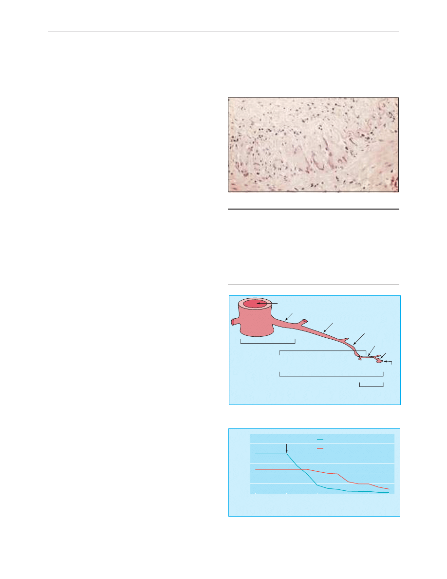
ABC of arterial and vascular disease
Vasculitis
C O S Savage, L Harper, P Cockwell, D Adu, A J Howie
Vasculitis is inflammation of blood vessel walls. The clinical and
pathological features are variable and depend on the site and
type of blood vessels that are affected. Diseases in which
vasculitis is a primary process are called primary systemic
vasculitides.
The main types of vasculitides can be described using
clinical features and pathological findings according to the
Chapel Hill Consensus Conference. These names and
definitions will be followed in this article. Definitive classification
of systemic vasculitis is unsatisfactory since aetiology and
pathogenesis are rarely known, and clinical and histological
features overlap. Vasculitis may also occur as a secondary
feature in other diseases, such as systemic lupus erythematosus
and rheumatoid arthritis.
Fever, night sweats, malaise, myalgia, and arthralgia are
common in all types of vasculitis. Active vasculitis is usually
associated with an acute phase response with an increase in C
reactive protein concentration, erythrocyte sedimentation rate,
or plasma viscosity.
Large vessel vasculitis
Giant cell arteritis (temporal arteritis)
Clinical features include unilateral throbbing headache, facial
pain, and claudication of the jaw when eating. Visual loss is a
feared symptom and may be sudden and painless, affecting part
or all of the visual field. Diplopia may also occur. Giant cell
arteritis is the most common type of primary systemic vasculitis
with an incidence of 200/million population/year.
Treatment is with high dose corticosteroids (40-60 mg/day),
which should be started as soon as the diagnosis is suspected to
avoid visual loss. The diagnosis is confirmed by biopsy of the
affected artery, done within 24 hours of starting corticosteroids.
The corticosteroid dose may be reduced to 10 mg/day over six
months and then more slowly to a maintenance of
5-10 mg/day. Maintenance treatment may be required for two
years. The disease is monitored by measuring C reactive protein
concentrations, erythrocyte sedimentation rate, or plasma
viscosity.
Takayasu’s arteritis
Takayasu’s arteritis is most common in Asia and the Far East
and affects women more than men. Disease of the arteries
supplying the arms, head, neck, and heart leads to the aortic
arch syndrome with claudication of the arm, loss of arm pulses,
variation in blood pressure of more than 10 mm Hg between
the arms, arterial bruits, angina, aortic regurgitation, syncope,
stroke, and visual disturbance. The descending aortic syndrome
may cause bowel ischaemia or infarction, renovascular
hypertension, and renal impairment.
Diagnosis is by angiography or magnetic resonance
angiography. Treatment of acute disease in patients with high C
reactive protein concentration or erythrocyte sedimentation
rate is with corticosteroids. Cytotoxic drugs such as
cyclophosphamide can be added if steroids alone do not
control the disease. Surgery or angioplasty may be required for
stenoses once active inflammation has been controlled.
Definitions of large vessel vasculitis
Giant cell arteritis (temporal arteritis)
x Granulomatous arteritis of aorta and its major branches, especially
extracranial branches of carotid artery
x Often affects temporal artery
x Usually occurs in patients older than 50 years
x Often associated with polymyalgia rheumatica
Takayasu’s arteritis
x Granulomatous inflammation of aorta and its major branches
x Usually occurs in patients younger than 50 years
Temporal artery biopsy specimen with giant cell inflammation
Aorta (large artery)
Renal artery (medium sized artery)
Giant cell (temporal) arteritis
and Takayasu's arteritis
Polyarteritis nodosa and
Kawasaki disease
Microscopic polyangiitis, Wegener's granulomatosis and
Churg-Strauss syndrome
Henoch Schönlein purpura, essential
cryoglobulinaemic vasculitis and antiglomerular
basement membrane antibody mediated disease
Lobar artery (medium sized artery)
Arcuate artery (small artery)
Interlobular artery
(small artery)
Arteriole
Glomerulus
Spectrum of systemic vasculitides organised according to predominant size of
vessels affected (adapted from Jennette et al, Arthritis Rheum 1994;37:187-92)
0
9 days before
treatment
Diagnosis and
treatment
12
d
ay
s
21
d
ay
s
27
d
ay
s
40
d
ay
s
100
150
200
250
300
C reactive protein (mg/l)
Treatment with
immunosuppression
50
Erythrocyte sedimentation rate (mm/h)
C reactive protein concentration (>10 mg/l) and erythrocyte sedimentation
rate (>18 mm/h) are raised at time of diagnosis of giant cell arteritis but fall
to normal levels after starting immunosuppressant therapy
Clinical review
1325
BMJ
VOLUME 320 13 MAY 2000 bmj.com
on 1 October 2006
bmj.com
Downloaded from

Medium vessel vasculitis
Polyarteritis nodosa
Polyarteritis nodosa is uncommon in the United Kingdom. It is
associated with hepatitis B virus in some patients. Arterial disease
leads to ischaemia or infarction within affected organs. The
condition can affect the gut causing bleeding or perforation, the
heart causing angina or myocardial infarction, the kidneys
causing cortical infarcts leading to hypertension and renal failure,
and the peripheral nerves causing mononeuritis multiplex.
Hepatitis may reflect the presence of hepatitis B virus.
Diagnosis is based on the presence of arterial aneurysms on
angiography of the renal, hepatic, splanchnic, or splenic
circulations. Biopsy of affected muscle or nerve may confirm the
presence of vasculitis. Treatment of polyarteritis associated with
hepatitis B virus requires an antiviral drug such as interferon
alfa combined with short course, high dose corticosteroids and
plasma exchange. Non-hepatitis B virus polyarteritis usually
responds to corticosteroids alone, although cyclophosphamide
may be required for patients with more severe disease.
Kawasaki disease
Kawasaki disease affects children, usually under the age of 12
years. In Japan the incidence exceeds 100/100 000 children
younger than 5 years but is less than 5/100 000 in this age
group in the United Kingdom.
The most serious feature of Kawasaki disease is coronary
artery disease; aneurysms occur in a fifth of untreated patients
and may lead to myocardial infarction. They can be detected by
echocardiography. High dose intravenous immunoglobulins
reduce the prevalence of coronary artery aneurysms, provided
that treatment is started within 10 days of onset of the disease.
Low dose aspirin is recommended for thrombocythaemia.
Small vessel vasculitis associated with
antineutrophil cytoplasmic antibody
Small vessel vasculitides are being recognised more frequently,
mainly because of increased awareness. Estimates of incidence
have increased from fewer than 5 cases per million population in
the early 1980s to over 20 per million. The early symptoms of
these disorders are non-specific with fever, malaise, arthralgia,
myalgia, and weight loss, and patients in whom such symptoms
are persistent should be screened for antineutrophil cytoplasmic
antibodies (ANCA); have their erythrocyte sedimentation rate
and C reactive protein concentration measured; and have their
urine tested for blood with a dipstick. Early diagnosis is essential
to prevent potentially life threatening renal and pulmonary
injury. Delays in diagnosis are unfortunately common, and this
often leads to serious morbidity. Once respiratory or renal disease
develops, the course is usually rapidly progressive.
Wegener’s granulomatosis
Upper respiratory tract disease occurs in more than 90% of cases
and includes sinusitis; nasal crusting, bleeding, obstruction, and
collapse of the nasal bridge; serous otitis media with conductive
deafness; and tracheal stenosis. Limited Wegener’s refers to
disease that affects only the respiratory tract at the time of
diagnosis; many cases evolve to systemic disease. Lung disease is
common with cough, haemoptysis, and dyspnoea and may
progress to life threatening pulmonary haemorrhage. The
kidneys are affected in up to 80% of cases; blood, protein, and
casts are present in the urine and should be examined by dipstick
testing and microscopy. If untreated, there is loss of renal
function, often within days. Other features include purpuric
rashes, nail fold infarcts, and ocular manifestations including
Definitions of medium sized vessel vasculitis
Polyarteritis nodosa
x Necrotising inflammation of medium and small arteries without
glomerulonephritis, pulmonary capillaritis, or disease of other
arterioles, capillaries, or venules
Kawasaki disease
x Arteritis affecting large, medium, and small arteries and associated
with mucocutaneous lymph node syndrome
x Coronary arteries are usually affected and aorta and veins may be
affected
x Usually occurs in children
Features of mucocutaneous lymph node syndrome in
Kawasaki disease
x Fever for > 5 days
x Conjunctival congestion
x Changes to lips and oral cavity: dry, red, fissured lips; strawberry
tongue; reddening of oral and pharyngeal mucosa
x Changes of peripheral extremities: red palms and soles; indurative
oedema; desquamation of finger tips during convalescence
x Macular polymorphous rash on trunk
x Swollen cervical lymph nodes
At least five features must be present
Definitions for diagnosis of vasculitides often associated with
antineutrophil cytoplasm antibodies
Wegener’s granulomatosis
x Granulomatous inflammation of the respiratory tract
x Necrotising vasculitis affecting small to medium sized vessels
(capillaries, venules, arterioles, and arteries)
x Necrotising glomerulonephritis is common
Microscopic polyangiitis (microscopic polyarteritis)
x Necrotising vasculitis with few or no immune deposits affecting
small vessels (capillaries, venules, arterioles, and arteries)
x Necrotising arteritis of small and medium sized arteries may be
present
x Necrotising glomerulonephritis very common
x Pulmonary capillaritis often occurs
Churg-Strauss syndrome
x Eosinophil rich and granulomatous inflammation of respiratory tract
x Necrotising vasculitis affecting small to medium sized vessels
x Blood eosinophilia ( > 1.5
×
10
9
/l )
x Usually associated with asthma
Renal angiogram showing multiple
arterial aneurysms
Clinical review
1326
BMJ
VOLUME 320 13 MAY 2000 bmj.com
on 1 October 2006
bmj.com
Downloaded from

conjunctival haemorrhages, scleritis, uveitis, keratitis, proptosis, or
ocular muscle paralysis due to retro-orbital inflammation. The
disease can affect the gut causing haemorrhage, the heart causing
coronary artery ischaemia, and the neurological system causing
sensory neuropathy or mononeuritis multiplex.
The two pathological hallmarks of Wegener’s disease are
chronic granulomatous inflammation and vasculitis. Granulomas
(localised microscopic collections of macrophages) are not always
present. Granulomas in the lung may coalesce into large masses
which cavitate. The vasculitis affects capillaries particularly in the
lung, causing lung haemorrhage, and glomeruli, causing
glomerulonephritis that may be segmental, global, focal, or diffuse
with thrombosis, necrosis of capillary loops, and accumulation of
cells in Bowman’s space. Affected arteries or arterioles show an
inflammatory infiltrate and fibrinoid necrosis. There is no
deposition of immunoglobulins within the kidney or vessel walls.
Microscopic polyangiitis (microscopic polyarteritis)
Microscopic polyangiitis has many similarities to Wegener’s
granulomatosis, but disease of the upper respiratory tract is
uncommon in microscopic polyangiitis, although pulmonary
haemorrhage may occur. Patients with microscopic polyangiitis
usually have glomerulonephritis, and, rarely, disease may be
limited to the kidney. No granuloma formation is seen.
Diagnosis
Diagnosis is based on typical clinical features, biopsy (usually of
kidney, occasionally of nasal mucosa or lung) and presence of
antineutrophil cytoplasmic antibodies. These antibodies usually
have specificity for the enzymes proteinase-3 or myeloperoxidase
and can be detected by enzyme linked immunosorbent assay
(ELISA). In indirect immunofluorescence tests, antineutrophil
cytoplasmic antibodies against proteinase-3 produce a
cytoplasmic staining pattern (cANCA) and antibodies against
myeloperoxidase produce perinuclear staining pattern (pANCA).
Combined testing by ELISA and indirect immunofluorescence is
recommended as this increases specificity at the cost of a 10%
reduction of sensitivity. Sometimes antineutrophil cytoplasmic
antibody tests are negative, particularly if disease is limited to the
upper respiratory tract. Antibody titres usually fall and may
disappear completely when the disease is in remission.
Treatment
Treatment of Wegener’s granulomatosis and microscopic
polyangiitis comprises induction of remission and then
maintenance of remission. Multicentre trials are in progress to
assess the place of pulse cyclophosphamide, plasma exchange,
and methylprednisolone in treatment and to assess the
optimum duration of maintenance therapy. Methotrexate is
sometimes used instead of cyclophosphamide for patients with
no renal disease. Relapses occur in 40-50% of patients during
the first five years, so lifelong monitoring for recurring disease
activity is essential. The five year survival rate is over 80%.
Churg-Strauss syndrome
Churg-Strauss syndrome is associated with an atopic tendency,
usually asthma. It may affect coronary, pulmonary, cerebral, and
splanchnic circulations. Rashes with purpura, urticaria, and
subcutaneous nodules are common. Glomerulonephritis may
develop, but renal failure is uncommon.
Diagnosis depends on presence of typical clinical features,
biopsy of skin, lung, and kidney, and blood eosinophilia. About
25% of patients are positive for cANCA, 50% for pANCA, and
25% have no antineutrophil cytoplasmic antibodies.
Many patients respond to high dose corticosteroids alone,
although cyclophosphamide may be required for patients with
Specificity and sensitivity of ANCA serology testing for
Wegener’s granulomatosis and microscopic polyangiitis
(adapted from Hagen et al, Kidney Int 1998;53:743-53)
Specificity/
sensitivity (%)
Specificity of assays (related to disease controls)
Indirect immunofluorescence:
cANCA
95
pANCA
81
ELISAs:
PR3-ANCA
87
MPO-ANCA
91
Combined indirect immunofluorescence and ELISA:
cANCA/PR3-ANCA positive
99
pANCA/MPO-ANCA positive
99
Sensitivity of combined testing
Wegener’s granulomatosis
73
Microscopic polyangiitis
67
Treatment of small vessel vasculitis
Induction therapy (to 3 months after remission, usually 6 months
from diagnosis)
x Cyclophosphamide, 2.0 mg/kg/day (maximum 200 mg/day). Age
> 60 years, reduce dose by 25%, > 75 years by 50%
x Prednisolone, 1 mg/kg/day (maximum 80 mg/day) reduced weekly
to 25mg/day by 8 weeks and then more slowly to 10 mg/day by 6
months
In severe, life threatening disease (eg, pulmonary haemorrhage, severe
crescentic glomerulonephritis with creatinine > 500 ìmol/l), consider
plasma exchange, 7-10 treatments over 14 days, or three pulses of
methylprednisolone, 15 mg/kg/day for 3 days
Maintenance therapy (to 18-24 months, longer if clinically indicated)
x Azathioprine, 2.0 mg/kg/day (maximum 200 mg/day). Age > 60
years, reduce dose by 25%, > 75 years by 50%
x Prednisolone, 5-10 mg/day
Relapse therapy
x Major relapse: return to induction therapy
x Minor relapse: increase dose of corticosteroids
Stop cyclophosphamide or azathioprine if white blood count 4x10
9
/l;
restart with a dose reduced by at least 25 mg when white blood
count > 4x10
9
/l on two consecutive tests
Consider gastric and bone protection, and fungal and Pneumocystis
carinii
prophylaxis
Cavitating granulomatous lesion in right lung of patient with
Wegener’s granulomatosis
Clinical review
1327
BMJ
VOLUME 320 13 MAY 2000 bmj.com
on 1 October 2006
bmj.com
Downloaded from
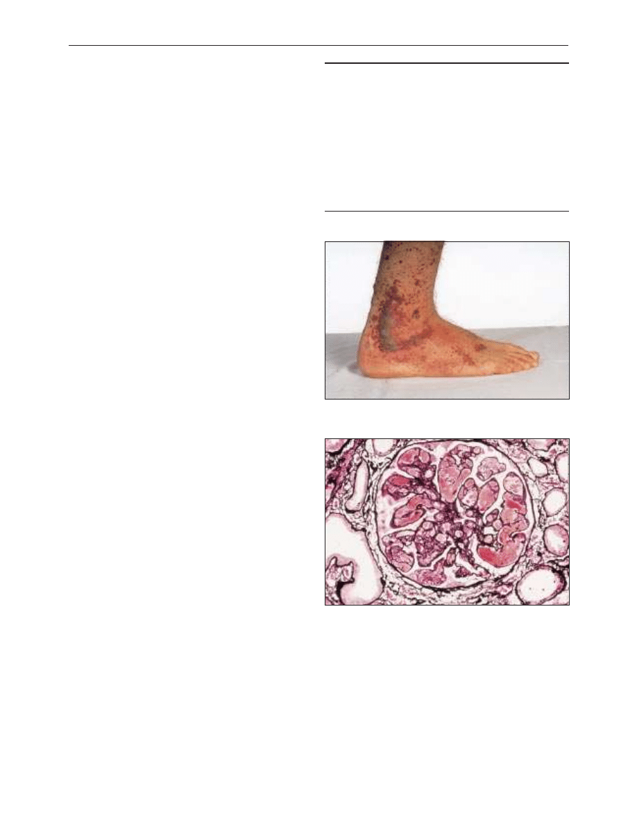
more severe disease. Asthma requires conventional treatment
but the recently introduced leukotriene receptor antagonist
drugs have been causally linked with the Churg-Strauss
syndrome and should be avoided in these patients.
Small vessel vasculitis without
antineutrophil cytoplasmic antibodies
Henoch-Schönlein purpura
Henoch-Schönlein purpura is most common in children but
can occur at any age. Typical clinical features are purpura over
the lower limbs and buttocks, haematuria, abdominal pain,
bloody diarrhoea, and arthralgia.The pathological hallmarks
are deposition of immunoglobulin A at the dermoepidermal
junction and within the glomerular mesangium, with a
mesangial hypercellular glomerulonephritis. Some patients
develop a glomerular lesion resembling that seen in small vessel
vasculitis. Renal disease may occur without the rash or other
typical features.
The disease is usually self limiting and only supportive
treatment is required. Corticosteroids and immunosuppression
may be needed for vasculitic glomerulonephritis or serious gut
haemorrhage and ischaemia.
Cryoglobulinaemic vasculitis (“mixed, essential”)
Cryoglobuins are immunoglobulins that precipitate in the cold.
The mixed cryoglobulin consists of a monoclonal
immunoglobulin M rheumatoid factor complexed to polyclonal
immunoglobulin G. Vasculitis develops when cryoglobulins
deposit in blood vessels. Mixed essential cryoglobulinaemia is
due to hepatitis C virus infection in over 80% of cases. Other
causes of cryoglobulinaemia include dysproteinaemias,
autoimmune diseases, and chronic infections. Serum
complement C4 and C3 concentrations are reduced. Clinical
features include palpable purpura, arthralgia, distal necroses,
peripheral neuropathy, abdominal pain, and
glomerulonephritis. Renal biopsy specimens typically have the
appearance of subendothelial membranoproliferative
glomerulonephritis with intraglomerular deposits.
In cryoglobulinaemia associated with hepatitis C, treatment
is directed at the viral infection. Interferon alfa over six months
is beneficial, but many patients relapse when treatment is
stopped. Prednisolone with or without immunosuppressants
has been used successfully in acute severe disease. The role of
plasma exchange remains unsubstantiated.
Isolated cutaneous leukocytoclastic vasculitis
This is often associated with a drug hypersensitivity response
and improves when the drug is stopped. Occasional patients
may require corticosteroids for severe disease.
Antiglomerular basement membrane antibody mediated
disease (Goodpasture’s disease)
No Chapel Hill definition exists for this rare disease, which has
considerable overlaps with antineutrophil cytoplasmic antibody
associated vasculitis. The hallmarks are a rapidly progressive
global and diffuse glomerulonephritis, as seen in small vessel
vasculitides, or presence of pulmonary haemorrhage, or both.
Diagnosis depends on finding antibodies to glomerular
basement membrane in the serum and linear staining for
immunoglobulin G along the glomerular basement membrane.
The antibodies have been implicated in disease pathogenesis.
About 15-30% of patients have detectable antineutrophil
cytoplasmic antibodies. Treatment is as for small vessel vasculitis
with addition of daily plasma exchange until antiglomerular
basement membrane antibodies are no longer detectable.
Definitions of non-ANCA associated small vessel vasculitis
Henoch-Schönlein purpura
x Vasculitis with IgA dominant immune deposits affecting small
vessels (capillaries, venules, or arterioles)
x Affects skin, gut, and glomeruli
x Associated with arthralgia or arthritis
Cryoglobulinaemic vasculitis
x Vasculitis with cryoglobulin immune deposits affecting small vessels
x Associated with cryoglobulins in serum
x Skin and glomeruli often affected
Isolated cutaneous leukocytoclastic vasculitis
x Isolated cutaneous leukocytoclastic angiitis without systemic
vasculitis or glomerulonephritis
x May evolve into systemic vasculitis
C O S Savage is professor of nephrology, University of Birmingham; L
Harper is specialist registrar, P Cockwell is consultant physician, and
D Adu is consultant physician, Queen Elizabeth Hospital,
Birmingham; and A Howie is reader in renal pathology, University of
Birmingham.
The ABC of arterial and venous disease is edited by Richard Donnelly,
professor of vascular medicine, University of Nottingham and
Southern Derbyshire Acute Hospitals NHS Trust (richard.donnelly@
nottingham.ac.uk) and Nick J M London, professor of surgery,
University of Leicester, Leicester (sms16@leicester.ac.uk). It will be
published as a book later this year.
BMJ
2000;320:1325-8
Purpuric rash on lower limb of patient with Henoch-Schönlein purpura
Renal biopsy specimen showing intraglomerular deposit of cryoglobulins
Clinical review
1328
BMJ
VOLUME 320 13 MAY 2000 bmj.com
on 1 October 2006
bmj.com
Downloaded from
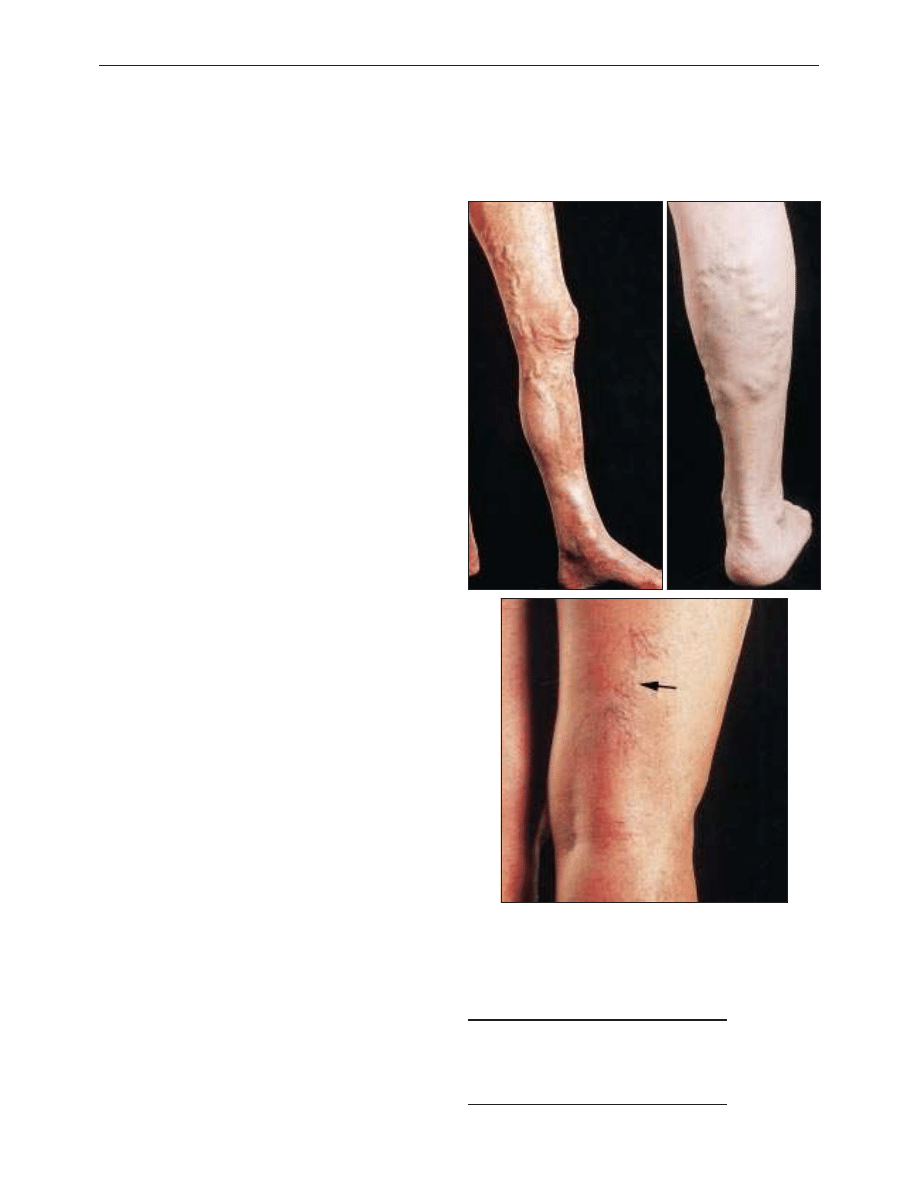
ABC of arterial and venous disease
Varicose veins
Nick J M London, Roddy Nash
Varicose veins are tortuous, twisted, or lengthened veins. Unless
the enlargement is severe, size alone does not indicate
abnormality because size can vary depending on ambient
temperature and, in women, hormonal factors. In addition,
normal superficial veins in a thin person may appear large,
whereas varicose veins in an obese person may be hidden.
Varicose veins can be classified as trunk, reticular, or
telangiectasia. Telangiectasia are also referred to as spider veins,
star bursts, thread veins, or matted veins. Most varicose veins are
primary; only the minority are secondary to conditions such as
deep vein thrombosis and occlusion, pelvic tumours, or
arteriovenous fistulae.
Incidence and prevalence
A study of people aged 35 to 70 years in London in 1992
concluded that the prevalence of varicose veins in men and
women was 17% and 31% respectively. Although varicose veins
have traditionally been considered commoner in women, a
recent study from Edinburgh of people aged 18 to 64 years
found that the prevalence of trunk varices was 40% in men and
32% in women. Over 80% of the total population had reticular
varicosities or telangiectasia. There are few studies on the
incidence of varicose veins; however, the Framingham study
found that the two year incidence of varicose veins was
39.4/1000 for men and 51.9/1000 for women.
Pathophysiology and risk factors
The theory that varicose veins result from failure of valves in
the superficial veins leading to venous reflux and vein dilatation
has been superseded by the hypothesis that valve incompetence
follows rather than precedes a change in the vein wall. Thus, the
vein wall is inherently weak in varicose veins, which leads to
dilatation and separation of valve cusps so that they become
incompetent. This theory is strongly supported by the
observation that the dilatation of varicose veins is initially distal
to the valve; if the primary abnormality was descending valve
incompetence, the initial dilatation should be proximal to the
valve.
Risk factors for varicose veins include increasing age and
parity and occupations that require a lot of standing. There is
no evidence that social class, smoking, or genetic makeup
influence the prevalence of varicose veins. Obesity is associated
with the development of varicose veins in women but not in
men.
Symptoms
The Edinburgh vein study recently compared the prevalence of
symptoms in men and women with and without varicose veins.
In men, the only symptom that was significantly associated with
trunk varices was itching, whereas in women, heaviness or
tension, aching, and itching were significantly associated with
trunk varices. No association was found between reticular
varices and lower limb symptoms in either men or women.
Symptoms associated with varicose veins
x Heaviness
x Tension
x Aching
x Itching
Trunk varices are varicosities in the line of the long (top, left) or short (top,
right) saphenous vein or their major branches. Reticular veins (arrow,
bottom) are dilated tortuous subcutaneous veins not belonging to the main
branches of the long or short saphenous vein, and telangiectasia (bottom)
are intradermal venules <1 mm
Clinical review
1391
BMJ
VOLUME 320 20 MAY 2000 bmj.com
on 1 October 2006
bmj.com
Downloaded from
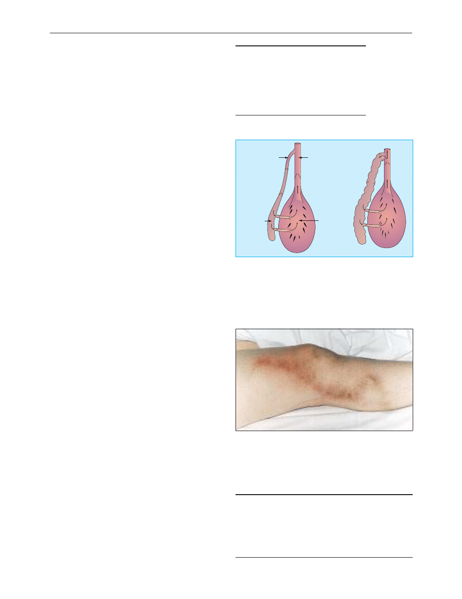
Complications of varicose veins
Some complications of varicose veins, such as haemorrhage
and thrombophlebitis, result from the varicose veins themselves,
whereas others, such as oedema, skin pigmentation, varicose
eczema, atrophie blanche, lipodermatosclerosis, and venous
ulceration result from venous hypertension. The size of varicose
veins does not seem related to the degree of venous
hypertension. Indeed, 40% of limbs with ulceration due to
superficial venous incompetence do not have visible varicose
veins. Venous ulceration is discussed in a subsequent article.
Recurrent varicose veins
In the United Kingdom, about 20% of varicose vein surgery is
for recurrence, and the estimated annual cost of such surgery is
£11m. Recurrent varicose veins can result from inadequate or
defective primary surgery or the development of new sites of
reflux. Improved patient assessment and more rigorous primary
surgery should reduce the socioeconomic impact of recurrent
varicose veins.
Clinical management
History
It is important to determine precisely why the patient has
sought treatment. One third of patients presenting with varicose
veins have symptoms unrelated to their varicose veins or are
worried about deterioration or complications. Such patients
simply need reassurance. It is important to determine whether
the patient has had deep vein thrombosis or thrombophlebitis
and any family history of deep vein thrombosis. History of these
conditions increases the risk of deep vein thrombosis after
varicose vein surgery and may lead to a decision not to operate.
Patients with a history of deep vein thrombosis or
thrombophlebitis who have surgery should receive
perioperative subcutaneous heparin prophylaxis. It is important
to note whether women are taking the contraceptive pill or
hormone replacement therapy. Any history of skin changes is
also important because affected patients are at high risk of
developing ulceration.
Examination and investigation
Examination of the patient should include an abdominal
examination to exclude some of the secondary causes of
varicose veins. With the patient standing, note the distribution
of the varicose veins, in particular whether they are long or
short saphenous, or both. Secondary skin changes should be
noted. Most vascular surgeons would then investigate the
patient in the clinic using handheld Doppler or colour duplex
scanning. Although ideally all patients presenting with varicose
veins would have colour duplex scanning, the NHS does not
have the resources to allow this. Patients with recurrent varicose
veins should be scanned to determine the precise site of
recurrence. Patients with varicose veins in limbs with a history
of deep vein thrombosis or thrombophlebitis should be
scanned to make sure that the superficial veins are not acting as
collaterals in the presence of deep vein obstruction. Scanning is
also essential for patients with venohypertensive skin changes. If
the deep veins are competent in the presence of refluxing
superficial veins, superficial venous surgery is potentially
curative.
Treatment
As discussed above, about a third of patients presenting with
varicose veins will require only reassurance or explanation that
Complications of varicose veins
x Haemorrhage
x Thrombophlebitis
x Oedema
x Skin pigmentation
x Atrophie blanche
x Varicose eczema
x Lipodermatosclerosis
x Venous ulceration
Indications for colour duplex scanning of varicose veins
x Recurrent varicose veins
x History of superficial thrombophlebitis
x History of deep venous thrombosis
x Varicose eczema
x Haemosiderin staining
x Lipodermatosclerosis
x Venous ulceration
Saphenous
vein
Deep outflow
tract
Deep (pump)
compartment
Superficial
compartment
Mechanisms of failure of calf muscle pump and venous hypertenion.
Superficial veins do not normally allow reflux of blood (left). However, if
superficial veins are incompetent (right), some of the blood ejected by the
calf muscle pump during systole refluxes back down the superficial veins
into the calf muscle pump during diastole. This retrograde circuit can
overload the calf muscle pump, leading to dilatation and failure. The
subsequent rise in end diastolic volume leads to venous hypertension.
Adapted from Browse et al
Thrombophlebitis presents as severe pain, erythema, pigmentation over, and
hardening of the vein. Thrombophlebitis in varicose veins results from stasis,
whereas thrombophlebitis occurring in normal veins should alert clinicians
to the possibility of an underlying malignancy or thrombophilia. Recurrent
thrombophlebitis in varicose veins raises the possibility of underlying
thrombophilia
Clinical review
1392
BMJ
VOLUME 320 20 MAY 2000 bmj.com
on 1 October 2006
bmj.com
Downloaded from
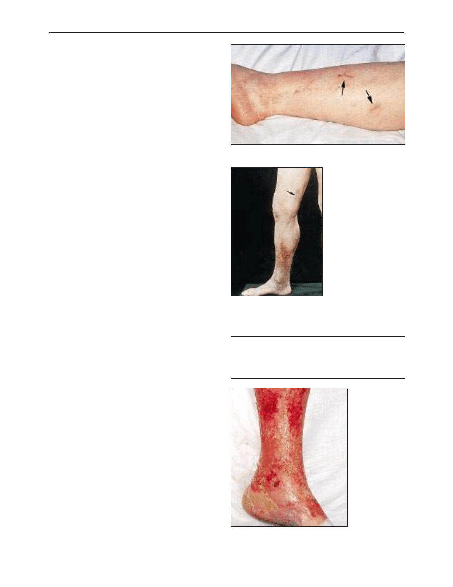
their symptoms are not related to their varicose veins. Patients
whose main symptoms are aching or oedema may benefit from
compression hosiery. Indeed, if it is uncertain whether the
patient’s symptoms are caused by varicose veins, a trial of
compression hosiery may help; a response to compression
indicates that surgery may be beneficial.
The treatment options for trunk varices are injection
sclerotherapy or surgery. The use of injection sclerotherapy for
trunk varices has fallen in recent years, partly because of
concerns about complications such as skin staining and
ulceration and also because up to 65% of patients treated by
sclerotherapy develop recurrent varicose veins within five years.
Currently, sclerotherapy is most commonly used to treat
residual varicosities after surgery. Surgery is generally directed
at the underlying abnormality, in the form of saphenofemoral
or saphenopopliteal disconnection, and in the case of long
saphenous varices, stripping of the long saphenous vein with
multiple avulsions.
Many patients can be treated as day cases, most can return
to driving after one week, and the time off work varies between
one and three weeks depending on the patient’s occupation.
The risk of serious complication (deep venous thrombosis,
pulmonary embolism, or arterial or nerve injury) is less than
1%, but roughly 17% of patients will suffer minor complications,
most commonly temporary saphenous or sural nerve neuralgia.
All patients should be warned of this possibility. After surgery,
20-30% of patients develop recurrent varicose veins within 10
years.
Reticular varices are not connected to major trunk varices
and are treated by sclerotherapy or avulsion through small stab
incisions. Patients who present with capillary telangiectasia
should have colour duplex scanning because roughly 25% will
have clinically unapparent long or short saphenous
incompetence. The telangiectasia are treated by microinjections,
laser, or high intensity light. The last two methods are being
increasingly used.
Management of complications
Thrombophlebitis
There is no indication for antibiotics in patients with
thrombophlebitis. Patients should be referred to a vascular
specialist and surgery considered because thrombophlebitis
tends to recur if the underlying venous abnormality is not
corrected. Colour duplex studies have shown that up to a
quarter of patients with superficial thrombophlebitis have
underlying deep venous thrombosis, and it has therefore been
suggested that all patients with thombophlebitis should have
duplex scanning to exclude deep vein thrombosis. However, a
more realistic suggestion is that patients with phlebitis
extending up the long saphenous vein towards the
saphenofemoral junction should have urgent duplex scanning.
If the thrombus extends into the femoral vein, urgent
saphenofemoral ligation should be considered.
Bleeding varicose veins
Bleeding varicose veins can be stemmed by raising the foot
above the level of the heart and applying compression. The
patients should then be seen by a vascular surgeon with a view
to correcting the underlying abnormality. If the deep veins are
incompetent, the patient should wear compression hosiery.
Varicose eczema, lipodermatosclerosis, and venous ulceration
Patients with varicose eczema or lipodermatosclerosis require
colour duplex scanning to define the underlying venous
Management of thrombophlebitis
x Crepe bandaging to compress vein and minimise propogation of
thrombus
x Analgesia (preferably non-steroidal anti-inflammatory drug)
x Low dose aspirin
One of the complications of injection sclerotherapy is brown skin
pigmentation (arrows)
Skin pigmentation is due to
haemosiderin deposition. This patient
also has thrombophlebitis of the long
saphenous vein with overlying
pigmentation (arrow)
Varicose eczema occurs over prominent varicose
veins and in the lower third of the leg. It may be
dry, scaly, and vesicular or weeping and ulcerated
Clinical review
1393
BMJ
VOLUME 320 20 MAY 2000 bmj.com
on 1 October 2006
bmj.com
Downloaded from

abnormality. Generally, if the only abnormality is superficial
venous incompetence this should be surgically corrected. If,
however, the deep veins are incompetent, then superficial
surgery will not help and the patient should be treated with a
topical steroid and wear compression hosiery.
Areas of controversy
Whose varicose veins should be treated?
In the absence of clear national guidelines the decision about
who should receive varicose vein surgery under the NHS is
being made at a local level. In general terms patients with only
cosmetic problems are not treated whereas patients with skin
changes (eczema, lipodermatosclerosis, and ulceration) are
treated. The most controversial group is patients with
symptomatic trunk varices and no skin changes. Unfortunately,
there is no way of predicting which limbs with varicose veins
will subsequently develop venous ulceration, and it is clearly not
sensible to operate on the 30% of the population with varicose
veins in order to prevent 1% developing an ulcer.
Varicose veins, deep vein thrombosis, contraceptive pill, and
hormone replacement therapy
Although varicose veins increase the risk of deep vein thrombosis
after major abdominal or orthopaedic surgery, there is no
evidence that primary varicose veins are a risk factor for
spontaneous deep vein thrombosis. Similarly, there is no evidence
that women with varicose veins who take the contraceptive pill or
hormone replacement therapy are at increased risk of deep vein
thrombosis compared with women without varicose veins.
Evidence exists, however, that women with varicose veins who
take the pill are more likely to develop thrombophlebitis; a
history of thrombophlebitis is therefore a contraindication to the
pill and a reason for stopping the pill in current takers. Although
not evidence based, the same considerations should probably
apply to hormone replacement therapy.
Varicose vein surgery, contraceptive pill, and hormone
replacement therapy
Although there is no evidence that varicose vein surgery is high
risk for deep vein thrombosis, women taking the combined
contraceptive pill or hormone replacement therapy are at
increased risk of deep vein thrombosis after varicose vein surgery.
Women taking the combined contraceptive pill should either stop
the pill four weeks before surgery and restart two weeks later or
receive subcutaneous heparin prophylaxis. If the pill is stopped
advice must be given about alternative contraception. Women
taking hormone replacement therapy should continue taking it
and receive heparin thromboprophylaxis.
Further reading
x Franks PJ, Wright DD, Moffatt CJ, Stirling J, Fletcher AE, Bulpitt CJ,
et al. Prevalence of venous disease: a community study in West
London. Eur J Surg 1992:158:143-7.
x Brand FN, Dannenberg AL, Abbott RD, Kannell WB. The
epidemiology of varicose veins: the Framingham study. Am J Prev Med
1988;4:96-101.
x Bradbury A, Evans C, Allan P, Lee A, Ruckley CV, Fowkes FGR.
What are the symptoms of varicose veins? Edinburgh vein study cross
sectional population survey. BMJ 1999;318:353-6.
x Campbell B. Thrombosis, phlebitis, and varicose vein surgery. BMJ
1996;312:198-9.
x Royal College of General Practitioners’ oral contraception study:
oral contraceptives, venous thrombosis, and varicose veins. J R Coll
Gen Pract
1978;28:393-9.
x Drugs in the peri-operative period: 3—Hormonal contraceptives
and hormone replacement therapy. Drug Ther Bull 1999;37:78-80.
x Tibbs DJ. Varicose veins and related disorders. Oxford:
Butterworth-Heinemann, 1992.
x Tibbs DJ, Sabiston DC, Davies MG, Mortimer PS, Scurr JH. Varicose
veins, venous disorders, and lymphatic problems in the lower limbs.
Oxford:
Oxford University Press, 1997.
x Ruckley CV, Fowkes FGR, Bradbury AW. Venous disease. London:
Springer-Verlag, 1998.
x Browse NI, Burnand KG, Irvine AT, Wilson NM. Diseases of the veins.
London: Arnold, 1999.
Roddy Nash is consultant surgeon at Derbyshire Royal Infirmary,
Derby. The ABC of arterial and venous disease is edited by Richard
Donnelly, professor of vascular medicine, University of Nottingham
and Southern Derbyshire Acute Hospitals NHS Trust
(richard.donnelly@ nottingham.ac.uk) and Nick J M London,
professor of surgery, University of Leicester, Leicester
(sms16@leicester.ac.uk). It will be published as a book later this year.
BMJ
2000;320:1391-4
Varicose veins with
complications
Uncomplicated trunk varicose veins
Reticular veins and telangiectasia
High priority
Medium priority
Low priority
Demand management. High priority cases should be refered for vascular
assessment. Many regions are not offering NHS treatment for medium and
low priority cases on the basis that resources are needed more for other
conditions
One hundred years ago
Ophthalmology in Spain
Many travellers in Spain have been struck by the number of blind
persons everywhere met with. Dr. Camuset, writing in 1874, said
that the streets of the large towns literally swarm with blind folk.
They go about in bands of five or six, begging, and rolling about
their sightless orbs to excite compassion. For the most part the
blindness is caused by purulent ophthalmia or affections of the
cornea consecutive to granular ophthalmia. A few years later Dr.
Vignes in a trip to San Sebastian saw a number of cases of
granular ophthalmia and many of keratitis, which he attributed to
the bad food and filthy habits of life of the inhabitants of the
whole province of Guypuzcoa. Professor Hirschberg, of Berlin,
who was present at the International Congress of Hygiene held in
Madrid in 1897, bore emphatic witness to the prevalence of eye
diseases in Spain. Nowhere, not even in Egypt, had he met with so
many blind persons as in Spain; among other instances he
mentioned that he had seen a band of musicians consisting of
nine blind men. He attributed this state of things to the neglect of
ophthalmology in Spain, where he said there was not a single
professor of the subject in any of the medical schools. He added
that there was no public institute for the treatment of diseases of
the eye, and that such cases were treated either in the general
hospitals or in small private institutions. (BMJ 1900;ii:947)
Clinical review
1394
BMJ
VOLUME 320 20 MAY 2000 bmj.com
on 1 October 2006
bmj.com
Downloaded from
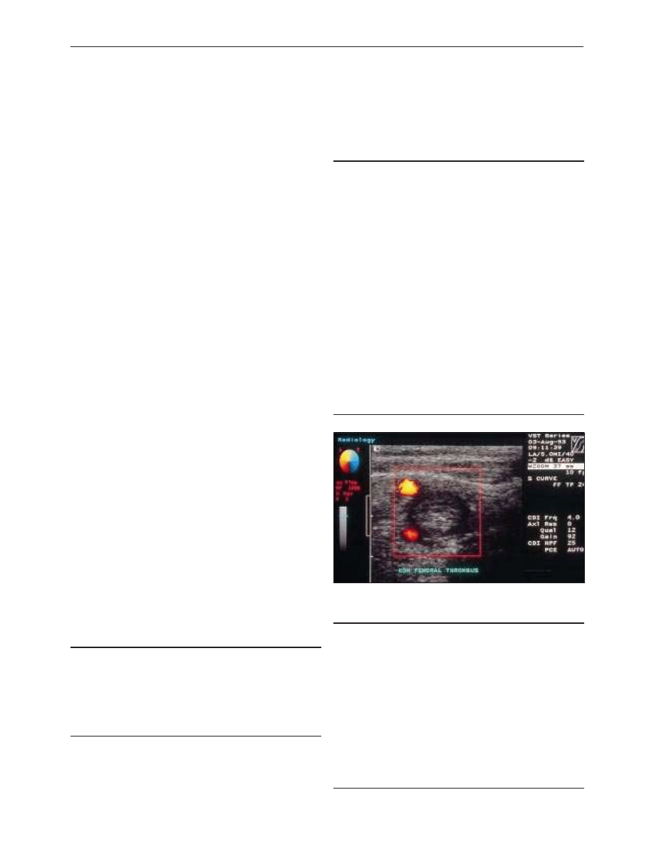
ABC of arterial and venous disease
Swollen lower limb—1: General assessment and deep vein thrombosis
W Peter Gorman, Karl R Davis, Richard Donnelly
The most common cause of leg swelling is oedema, but
expansion of all or part of a limb may be due to an increase in
any tissue component (muscle, fat, blood, etc). A correct
diagnosis requires consideration of whether the swelling is acute
or chronic, symmetrical or asymmetrical, localised or
generalised, and congenital or acquired. Chronic swelling,
particularly if asymmetrical, is usually a sign of chronic oedema
arising from venous or lymphatic disease, whereas symmetrical
lower limb swelling suggests a systemic or more central cause of
oedema, such as heart failure or nephrotic syndrome. Oedema
develops when the rate of capillary filtration (lymph formation)
exceeds lymphatic drainage, either because of increased
capillary filtration, inadequate lymphatic flow, or both.
Extracellular fluid volume is controlled prinicpally by the
lymphatic system, which normally compensates for increases in
capillary filtration. Most oedemas arise because filtration
overwhelms the lymph drainage system. Increased capillary
filtration may occur because of raised venous pressure,
hypoalbuminaemia, or increased capillary permeability due to
local inflammation. The two main causes of a swollen lower
limb are deep vein thrombosis and lymphoedema (a failure of
the lymph drainage system). This article concentrates on deep
vein thrombosis and next week’s article on lymphoedema.
Deep vein thrombosis
Thrombosis usually develops as a result of venous stasis or slow
flowing blood around venous valve sinuses; extension of the
primary thrombus occurs within or between the deep and
superficial veins of the leg and the propagating clot causes venous
obstruction, damage to valves, and possible thromboembolism.
Deep vein thrombosis is often asymptomatic.
Assessment and investigation
Various clinical features suggest deep vein thrombosis, but the
findings of physical examination alone are notoriously
unreliable. Deep vein thrombosis is confirmed in only one out
of every three cases suspected clinically. Confirmation of a
suspected deep vein thrombosis requires use of one or more
investigations, and the confirmation rate rises with the number
of clinical risk factors. Identification of an underlying cause, if
any, will guide both the treatment and the approach to
secondary prevention.
The standard investigation is contrast venography, but this
invasive procedure is painful, often technically difficult and time
consuming, and occasionally complicated by thrombosis and
Causes of swelling of lower limb
Acute
x Deep vein thrombosis
x Cellulitis
x Superficial thrombophlebitis
x Joint effusion or
haemarthrosis
x Haematoma
x Baker’s cyst
x Torn gastrocnemius muscle
x Arthritis
x Fracture
x Acute arterial ischaemia
x Dermatitis
Chronic
Congenital vascular abnormalities
x Haemangioma
x Klippel-Trenaunay syndrome
Venous disease
x Post-thrombotic syndrome
x Lipodermatosclerosis
x Chronic venous insufficiency
x Venous obstruction
Lymphoedema
x Cancer treatment
x Infection
x Tumour
x Trauma
x Pretibial myxoedema
Other
x Heart failure
x Reflex sympathetic dystrophy
x Idiopathic oedema of women
x Hypoproteinaemia, such as
cirrhosis, nephrotic syndrome
x Armchair legs
x Lipoedema
Risk factors for deep vein thrombosis
x Age > 40 years
x Underlying malignancy
x Obesity
x Presence of varicose veins
x Personal or family history of deep vein thrombosis or pulmonary
embolism
x Any surgical procedure lasting more than 30 minutes—especially
orthopaedic, neurosurgical, urological, and gynaecological surgery
x Paralysis or immobility—for example, recent stroke
x Combined contraceptive pill
x Hormone replacement therapy
x Pregnancy and puerperium
x Serious illness—for example, heart failure, myocardial infarction,
sepsis, inflammatory bowel disease
x Presence of hypercoagulable disorders
Colour duplex scan of deep vein thrombosis in common femoral vein
adjacent to artery
Clinical features of acute deep vein thrombosis
x Calf pain or tenderness, or both
x Swelling with pitting oedema
x Swelling below knee in distal deep vein thrombosis and up to groin
in proximal deep vein thrombosis
x Increased skin temperature
x Superficial venous dilatation
x Cyanosis can occur with severe obstruction
Clinical review
1453
BMJ
VOLUME 320 27 MAY 2000 bmj.com
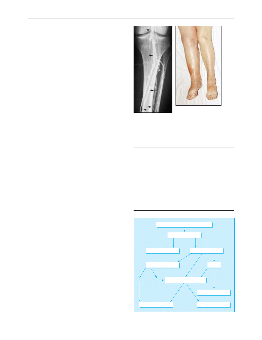
extravasation of contrast. Recent developments in non-invasive
testing mean that venography is now unnecessary in most cases,
particularly in suspected first proximal vein thrombosis.
The accuracy of non-invasive techniques varies with the
clinical circumstances. For example, compression
ultrasonography and impedance plethysmography are accurate
for detecting symptomatic proximal (ileofemoral) deep vein
thrombosis, but both techniques are less satisfactory in
asymptomatic patients and for detecting distal (calf vein)
thrombosis. Compression ultrasonography has become the
preferred first line investigation (see BMJ 2000;320:698-701).
Imaging techniques are generally much less satisfactory in
patients with suspected recurrent deep vein thrombosis, when
confirmation of the diagnosis requires evidence of new
thrombus formation—for example, the appearance of a new
non-compressible venous segment on ultrasonography or a
new intraluminal filling defect on venography.
Measurement of circulating d-dimer concentrations (a
byproduct of fibrin production) is a useful adjunct to
ultrasonography, with 98% sensitivity for deep vein thrombosis
and a high negative predictive value. The sensitivity of the test is
lower for smaller calf vein thrombi. However d-dimer
concentrations rise as a non-specific response to concomitant
illness, not just thrombosis, so d-dimer testing can be
misleading in patients admitted to hospital for other reasons.
A combination of diagnostic approaches—for example,
compression ultrasonography coupled with clinical pretest
probability scoring or d-dimer measurements, or both, gives
better diagnostic accuracy than any single investigation. Lensing
et al have recently shown that the combination of compression
ultrasonography and d-dimer measurement is an efficient
diagnostic approach, with a rate of subsequent
thromboembolism less than 1% in patients with false negative
results who were not treated with heparin. A robust
investigational algorithm has been devised that does not include
routine use of venography.
Complications
The main complications of deep vein thrombosis are
pulmonary embolism, post-thrombotic syndrome, and
recurrence of thrombosis. Proximal thrombi are a major source
of morbidity and mortality. Distal thrombi are generally smaller
and more difficult to detect non-invasively, and their prognosis
and clinical importance are less clear. However, a fifth of
untreated newly developing calf vein thrombi extend
proximally, and a quarter are associated with long term
symptoms of post-thrombotic syndrome; it is therefore
appropriate to treat proved significant calf vein thrombosis.
Post-thrombotic syndrome develops as a result of high
venous pressure due to thrombotic damage to valves. It
complicates 50-75% of deep vein thromboses, and there is a
strong association with ipsilateral recurrence. Clinical features
include pain, swelling, dermatitis, and ulceration. Proximal deep
vein thrombosis is associated with a higher frequency and
greater severity of post-thrombotic syndrome, but the risk is
halved by use of graded compression stockings after deep vein
thrombosis.
Prevention
Patients at significantly increased risk of deep vein
thrombosis—for example, those having major pelvic or
abdominal surgery for cancer or those with a history of
pulmonary embolism or deep vein thrombosis who have
Clinical model to determine pretest probability of deep vein
thombosis (3 points=high risk, 1 or 2=moderate, 0=low)
Score
Active cancer (treatment ongoing, or within 6 months, or
palliative)
1
Paralysis, paresis or recent plaster immobilisation of the legs
1
Recent immobilisation > 3days or major surgery within 12
weeks requiring general or regional anaesthesia
1
Localised tenderness along the distribution of the deep
venous system
1
Entire leg swollen
1
Calf swelling > 3 cm than asymptomatic side (measured
10 cm below tibial tuberosity)
1
Pitting oedema confined to the symptomatic leg
1
Collateral superficial veins (non-varicose)
1
Alternative diagnosis equally or more likely than deep vein
thrombosis
−
2
Venogram showing
thrombus in lower leg
Right ileofemoral deep vein
thrombosis
Patient with suspected deep vein thrombosis
Compression ultrasonography
Positive
Positive
Positive
Low
Moderate High
Negative
Negative
Negative
Repeat ultrasonography at one week
Treat for deep vein thrombosis
Treat for deep vein thrombosis
Deep vein thrombosis excluded
Deep vein thrombosis excluded
Consider clinical probability
D
-dimer
Management options
Algorithm for investigation of deep vein thrombosis
Clinical review
1454
BMJ
VOLUME 320 27 MAY 2000 bmj.com
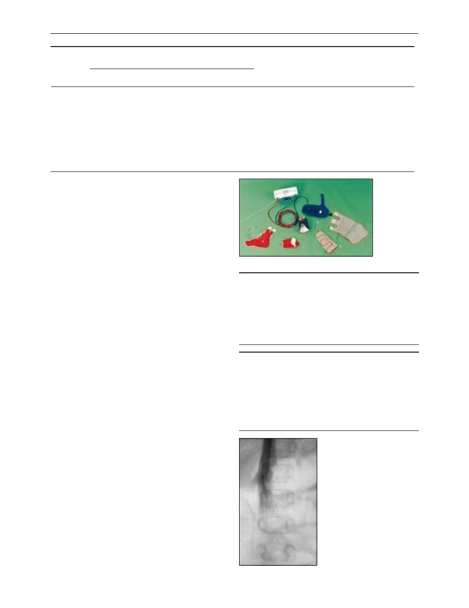
serious trauma or illness or are having major surgery—should
be given prophylaxis. Early mobilisation and graduated
compression stockings are effective, and antiplatelet drugs such
as aspirin provide additional protection.
Pneumatic compression devices have been proved effective
when used perioperatively and in some groups of medical
patients. Low dose unfractionated heparin (5000 units two
hours before surgery and 8-12 hourly postoperatively) given by
subcutaneous injection reduces the rate of postoperative
thromboembolism in general surgical patients by 65%, with
little increase in the risk of serious bleeding. Low molecular
weight heparins are effective and have some advantages over
unfractionated heparin, particularly in high risk patients such as
those having hip replacement.
Treatment
Treatment is aimed at reducing symptoms and preventing
complications. Elevation of the leg with some flexion at the knee
helps reduce swelling, early mobilisation is beneficial, and use of
graded compression stockings achieves a 60% reduction in
post-thrombotic syndrome.
It is important to establish effective anticoagulation rapidly.
Patients are usually given an intial intravenous heparin bolus of
5000 units followed by unfractionated or low molecular weight
heparin for at least five days. With unfractionated heparin an
intravenous constant infusion and subcutaneaous injection twice
daily are equally effective. A heparin algorithm should be used to
adjust the dose. The activated partial thromboplastin time should
be checked six hourly until the target is reached and then daily to
maintain the international normalised ratio at 1.5-2.5. The
platelet count should be checked at the start of treatment and on
day 5 to exclude thrombocytopenia. Warfarin should be started
on day 1, with the dose determined by a warfarin algorithm. The
target ratio is 2-3, and heparin can be stopped when the target
ratio is maintained for more than 24 hours.
Patients with deep vein thrombosis who do not need to be
in hospital (around 35%) can be treated with subcutaneous low
molecular weight heparin in the community. This can be
administered subcutaneously once or twice daily. Low
molecular weight heparin has the advantages of a slightly lower
rate of haemorrhage and thrombocytopenia and more reliable
absorption after injection, and anticoagulation monitoring is
not required routinely. Warfarin should be started on day 1, and
the duration of treatment guided by the risk profile.
Other approaches
Inferior vena cava filters reduce the rate of pulmonary
embolism but have no effect on the other complications of deep
Absolute risks of venous thrombotic complications in procedures or conditions of low, moderate, and high risk
Risk level
Risk (%)
Examples
Deep vein
thrombosis
Proximal deep vein
thrombosis
Fatal pulmonary
embolism
Low
< 10
< 1
0.01
Minor surgery, trauma, or medical illness
Major surgery at age < 40 with no other risk factors
Moderate
10-40
1-10
0.1-1
Major surgery with another risk factor
Major trauma, medical illness, or burns
Emergency caesarean section in labour
Minor surgery with previous deep vein thrombosis,
pulmonary embolism, or thrombophilia
Lower limb paralysis
High
40-80
10-30
1-10
Major pelvic or abdominal surgery for cancer
Major surgery, trauma, or illness with previous pulmonary
embolish, deep vein thrombosis, or thrombophilia
Duration of anticoagulation in patients with deep vein
thrombosis
x Transient cause and no other risk factors: 3 months
x Idiopathic: 3-6 months
x Ongoing risk—for example, malignancy: 6 -12 months
x Recurrent pulmonary embolism or deep vein thrombosis: 6-12
months
x Patients with high risk of recurrent thrombosis exceeding risk of
anticoagulation: indefinite duration (subject to review)
Indications for insertion of an inferior vena cava filter
x Pulmonary embolism with contraindication to anticoagulation
x Recurrent pulmonary embolism despite adequate anticoagulation
Controversial indications:
x Deep vein thrombosis with contraindication to anticoagulation
x Deep vein thrombosis in patients with pre-existing pulmonary
hypertension
x Free floating thrombus in proximal vein
x Failure of existing filter device
x Post pulmonary embolectomy
Pneumatic compression devices
Vena cavagram showing umbrella
delivery device inserted into the inferior
vena cava through the jugular vein. The
filter has been released just below the
renal veins. Inferior vena cava filters help
prevent pulmonary embolism but not
other complications of deep vein
thrombosis, including recurrent
thrombosis
Clinical review
1455
BMJ
VOLUME 320 27 MAY 2000 bmj.com

vein thrombosis. Thrombolysis should be considered in patients
with major proximal vein thrombosis and threatened venous
infarction. Surgical embolectomy is restricted to life threatening
proximal thrombosis where all else has failed.
Pregnancy
Anticoagulating doses of heparin are given for deep vein
thrombosis in pregnancy. It is essential to confirm the presence
of a thrombus objectively. This is usually done by compression
ultrasonography (serially if necessary).
Unfractionated heparin or low molecular weight heparin
(which has a better risk profile but is not licensed in United
Kingdom for this indication) should then be continued
throughout the pregnancy and stopped temporarily before
delivery. Anticoagulation should be restarted in the puerperium
and continued for six weeks to three months. Warfarin is usually
contraindicated during pregnancy because it is teratogenic and
increases risk of maternal and fetal haemorrhage perinatally. It
can be restarted 48 hours after delivery.
Deep vein thrombosis in pregnancy and puerperium and in
women taking contraceptive pill or hormone replacement
therapy
x Normal pregnancy is a hypercoagulable state
x Deep vein thrombosis occurs antepartum in 0.6/1000 women aged
< 35 years and 1.2/1000 women > 35 and postpartum in 0.3/1000
and 0.7/1000 respectively
x Age, operative delivery, personal or family history, and
thrombophilia are particular risks
x Heparin does not cross the placenta and is not secreted in breast
milk
x Prolonged heparin therapy raises concerns about osteoporosis,
heparin-induced thrombocytopenia, and allergy
x The combined contraceptive pill increases the relative risk of deep
vein thrombosis by 3-4 times
x Hormone replacement therapy also increases the relative risk of
deep vein thrombosis by 3-4 times but is associated with a 10 fold
higher absolute risk because of the older age group
Further reading and useful references
x Levick JR. An introduction to cardiovascular physiology. 2nd ed.
Oxford: Butterworth-Heinemann, 1995.
x Lensing WA, Prandoni P, Prins MH, Buller HR. Deep vein
thrombosis. Lancet 1999;353:479-85.
x Second Thromboembolic Risk Factors (THRiFT II) Consensus
Group. Risk of and prophylaxis for venous thromboembolism in
hospital patients. Phlebology 1998;13:87-97.
x Kearon C, Julian JA, Math M, Newman TE, Ginsberg JS.
Non-invasive diagnosis of deep venous thrombosis. Ann Intern Med
1998;128:663-77.
x Anderson DR, Wells PS. Improvements in the diagnostic approach
for patients with suspected deep vein thrombosis or pulmonary
embolism. Thromb Haemost 1999;82:878-86.
x Prins MH, Hutten BA, Koopman MMW, Buller HR. Long-term
treatment of venous thromboembolic disease. Thromb Haemost
1999;82:892-8.
W Peter Gorman is consultant physician and Karl R Davis is clinical
research fellow, Southern Derbyshire Acute Hospitals NHS Trust,
Derby.
The ABC of arterial and venous disease is edited by Richard Donnelly,
professor of vascular medicine, University of Nottingham and
Southern Derbyshire Acute Hospitals NHS Trust (richard.donnelly@
nottingham.ac.uk) and Nick J M London, professor of surgery,
University of Leicester, Leicester (sms16@leicester.ac.uk). It will be
published as a book later this year.
BMJ
2000;320:1453-6
A book that changed me
Understanding today’s Joads
Like all great books The Grapes of Wrath by John Steinbeck absorbs
and entertains but also alters your perspective on life. Its principal
theme is intolerance; its uncomfortable lesson is that prejudice lies
beneath the surface of us all; its optimistic conclusion is the courage
of the human spirit and the kernel of good that remains when all
else is stripped away. The poetic style and vernacular dialogue take
time at first, but once it hooks you it won’t let go.
It is the story of the Joad family. They are Oklahoma farmers
whose lives are ruined by the Dust Bowl of the 1930s; forced from
their land and fuelled by the American Dream of opportunity
they travel west to California. They endure dreadful hardship on
the way, and when they arrive their optimistic hopes and dreams
are shattered by the hostility they encounter.
Steinbeck’s greatest skill is showing us how the Joads engender
this hostility, prickling our own innate prejudices. They are crude,
crassly naive, violent, and hypocritical—they seem unable to make
the link that they are being driven from the land as their
forefathers drove the native Indians from it. Yet once you get to
know them, feel the pathos of their humanity, and share their
experience you become so deeply attached that you are appalled
not only at the treatment they receive but also at your own initial
prejudicial reaction.
I have just reread the book after 20 years, and its relevance
persists. The homeless, the travellers in their caravans next to our
busy dual carriageways, the asylum seekers and refugees in Dover
bedsits or imprisoned in detention centres—these are all today’s
Joads.
Throughout the novel Steinbeck parodies faith that has no
moral depth; he uses many biblical themes and images, and some
passages of the book are written in a quasiscriptural tone. The
Bible developed out of a collective human need to give life
context, meaning, and a moral structure, but Steinbeck suggests
that this message has been lost along with its contemporary
relevance. In a secular, multicultural age The Grapes of Wrath
shows us that storytelling remains (second only to life itself) the
pre-eminent form in teaching us about ourselves and our world.
It is a book that teaches empathy, compassion, and tolerance, but
perhaps its greatest lesson is that these things need to be taught,
and constantly retaught, to us all.
Simon Curtis general practitioner, Oxford
We welcome articles of up to 600 words on topics such as
A memorable patient, A paper that changed my practice, My most
unfortunate mistake
, or any other piece conveying instruction,
pathos, or humour. If possible the article should be supplied on a
disk. Permission is needed from the patient or a relative if an
identifiable patient is referred to. We also welcome contributions
for “Endpieces,” consisting of quotations of up to 80 words (but
most are considerably shorter) from any source, ancient or
modern, which have appealed to the reader.
Clinical review
1456
BMJ
VOLUME 320 27 MAY 2000 bmj.com
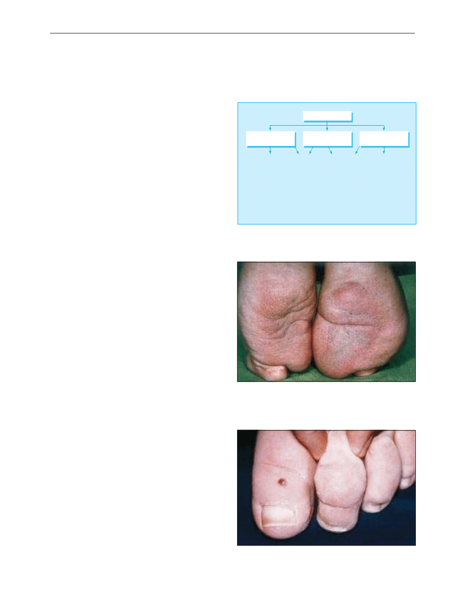
ABC of arterial and venous disease
Swollen lower limb—2: Lymphoedema
Peter S Mortimer
Lymph conducting pathways may become reduced in number,
obliterated, obstructed, or dysfunctional (because of failure of
contractility or valve incompetence). A lack of sensitive methods
for investigation makes it difficult to distinguish between these
mechanisms. A defect in the lymph conducting pathways leads
to primary lymphoedema; in practice this means no identifiable
outside cause can be found. Secondary lymphoedema is due to
factors originating outside the lymphatic system.
Primary lymphoedema
Congenital lymphoedema presenting at or soon after birth is
rare. A family history suggests Milroy’s disease. Swelling
invariably affects both lower limbs, but the upper limbs and face
may also swell.
Limb swelling may be the presenting and major
manifestation of congenital lymphatic malformations either in a
pure form—for example, diffuse lymphangioma—or in
combination with a congenital vascular syndrome—for example,
Klippel-Trenaunay syndrome (varicose veins, excessive long
bone growth, and vascular birthmark).
Most forms of primary lymphoedema present after puberty
with foot and ankle swelling. Women are more often affected,
and the condition may be familial—for example, Meige’s
disease. Lymph reflux due to lymphatic vessel hypertrophy or
megalymphatics is clinically distinguishable.
Secondary lymphoedema
Lymphoedema manifesting with sudden onset of swelling of
one whole leg suggests proximal obstruction. Pelvic causes of
venous or lymphatic obstruction such as tumour or thrombosis
must be excluded. In the Western world cancer treatment—for
example, surgery or radiotherapy) is the commonest cause.
Cancer itself rarely presents with lymphoedema except in
advanced cases presenting late, such as prostate cancer, where
venous obstruction may coexist. Relapsed tumour should always
be considered in someone with limb swelling after apparent
curative cancer treatment.
Filariasis is probably the most common cause of secondary
lymphoedema worldwide and should be considered in any
patient with lymphoedema who has travelled or lived in an
endemic area.
Clinical diagnosis of lymphoedema
The clinical diagnosis of lymphoedema depends on the history
and characteristic skin changes. Although most swelling occurs
in the subcutaneous layer, the skin becomes thicker (as
demonstrated by the inablity to pinch a fold of skin at the base
of the second toe), skin creases become enhanced, and a warty
texture (hyperkeratosis) and papillomatosis develop. Such skin
changes are termed “elephantiasis.”
The differential diagnosis includes venous oedema,
“armchair legs,” and lipodystrophy or lipoedema, which is often
misdiagnosed as lymphoedema.
Primary lymphoedema
Familial
Congenital (onset at or
soon after birth)
1. Congenital vascular
malformations with
lymphangioma
2. Chylous lymphoedema
1. Distal hypoplasia type
(bilateral below knee
swelling)
2. Proximal obstructive
type (unilateral whole
leg swelling)
3. Lymph reflux
(bilateral whole
leg swelling)
1. Meige's disease
2. Distichiasis-
lymphoedema
syndrome
Milroy's
disease
Postpubertal onset
Classification of primary lymphoedema
Primary lymphoedema with bilateral below knee swelling due to hypoplasia
of peripheral lymphatic vessels
Kaposi-Stemmer sign: inability to pinch a fold of skin at base of second toe
because of thickened skin indicates lymphoedema
Clinical review
1527
BMJ
VOLUME 320 3 JUNE 2000 bmj.com
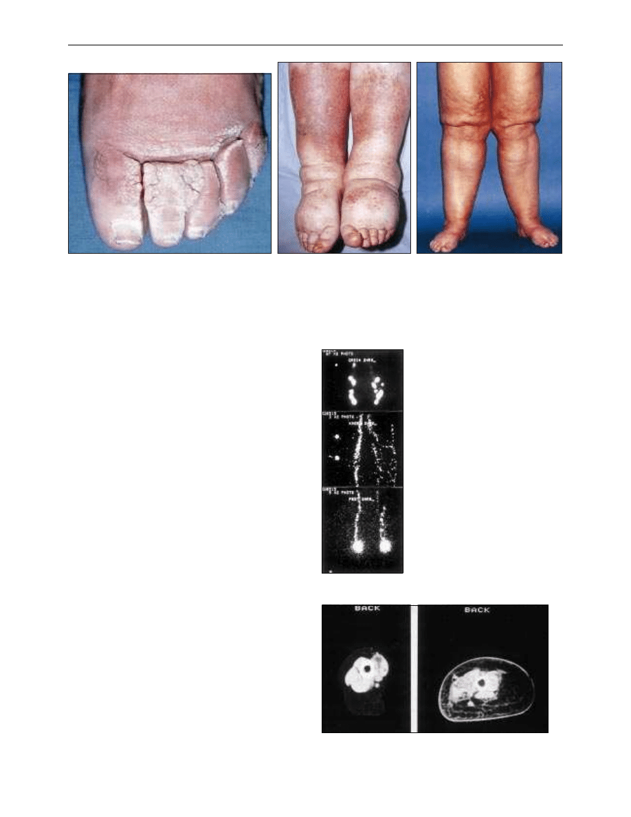
Investigation of lymphoedema
Lymphoscintigraphy (isotope lymphography)
Lymphoscintigraphy is the best investigation for identifying
oedema of lymphatic origin. Radiolabelled colloid or protein is
injected into the first web space of each foot and monitored
using a gamma camera as it moves to the draining lymph
nodes. Measurement of tracer uptake within the lymph nodes
after a defined interval will distinguish lymphoedema from
oedema of non-lymphatic origin. The appearance of tracer
outside the main lymph routes, particularly in the skin (dermal
backflow), indicates lymph reflux and suggests proximal
obstruction. Poor transit of isotope from the injection site
suggests hypoplasia of the peripheral lymphatic system.
Direct contrast x ray lymphography (lymphangiography)
After the lymph vessels have been identified with a vital dye, a
contrast medium such as Lipiodol is administered directly into
a peripheral lymphatic vessel, usually in the dorsum of the foot.
In a normal limb the lymphangiogram will show opacification
of five to 15 main collecting vessels as they converge on the
lowermost inguinal lymph nodes. In patients with lymphatic
obstruction the contrast medium will often reflux into the
dermal network, so called “dermal backflow.”
Computed tomography and magnetic resonance imaging
Both computed tomography and magnetic resonance imaging
detect a characteristic “honeycomb” pattern in the
subcutaneous compartment that is not seen with other causes
of oedema. In post-thrombotic syndrome the muscle
compartment deep to the fascia is enlarged, whereas in
lymphoedema it is unchanged. Thickening of the skin is also
characteristic of lymphoedema, although it is not diagnostic.
Magnetic resonance imaging is more informative than
computed tomography because it can detect water.
Management of lymphoedema
Most patients with lymphoedema are just told to live with it, but
this is neither necessary nor acceptable.
Dilatation of upper dermal lymphatics with consequent fibrosis
gives rise to papillomatosis
Armchair legs (elephantiasis nostras
verrucosis) develop in patients who sit in
a chair day and night with their legs
dependent. Patients with with cardiac or
respiratory disease, stroke, spinal damage,
or arthritis are predisoposed to this
condition
Lipoedema only affects women and causes
swelling between hip and ankle with sparing
of the foot. The condition is symmetrical. The
skin and subcutaneous tissues are soft and
often tender with easy bruising
Lymphoscintigraphy. Radiolabelled colloid or
protein is injected into the first web space of
each foot and followed with a gamma camera
as it moves to the draining lymph nodes. Tracer
can be seen within the main lymphatic channels
and lymph nodes as well as within the infection
site. Collateral drainage is seen within the left
thigh
Computed tomogram showing sections through normal thigh (left) and
thigh with lymphoedema (right). Note thickened skin and honeycomb
pattern
Clinical review
1528
BMJ
VOLUME 320 3 JUNE 2000 bmj.com

Physical treatment to reduce swelling
Treatment is aimed at controlling lymph formation and
improving lymph drainage through existing lymphatic vessels
and collateral routes by applying normal physiological
processes which stimulate lymph flow.
Prevention of infection
Prevention of acute inflammatory episodes (cellulitis or
lymphangitis) is crucial because they can cause severe
constitutional upset and deterioration in swelling. Care of the
skin, good hygiene, control of skin diseases such as tinea pedis,
and careful antiseptic dressings after minor wounds are all
important. Antibiotics must be given promptly when an acute
inflammatory episode occurs. In recurrent cellulitis the only
effective treatment is prophylactic antibiotics—for example,
phenoxymethylpenicillin 500 mg daily, for an indefinite period.
Drug treatment for lymphoedema
Diuretics are of little benefit in lymphoedema because their
main action is to limit capillary filtration. Improvement in
patients who are taking diuretics suggests that the predominant
cause of the oedema is not lymphatic. The benefit of
benzopyrones, such as coumarin or flavonoids, remains
unproved.
Surgery
Surgery is of value in a few patients in whom the size and
weight of a limb inhibit its use and interfere with mobility after
physical treatment. Surgery is aimed at either removing
excessive tissue (reducing or debulking operations) or bypassing
local lymphatic defects.
Peter S Mortimer is consultant skin physician, St George’s Hospital
and Royal Marsden Hospital, London.
The ABC of arterial and venous disease is edited by Richard
Donnelly, professor of vascular medicine, University of Nottingham
and Southern Derbyshire Acute Hospitals NHS Trust
(richard.donnelly@ nottingham.ac.uk) and Nick J M London,
professor of surgery, University of Leicester, Leicester
(sms16@leicester.ac.uk). It will be published as a book later this year.
BMJ
2000;320:1527-9
Physical treatment for lymphoedema
Process
Effect
Exercise
Dynamic muscle contractions encourage both
passive (movement of lymph along tissue planes
and non-contractile lymph vessels) and active
(increased contractility of collecting lymph
vessels) drainage
Compression
(hosiery)
Opposes capillary filtration
Acts as a counterforce to muscle contractions
(so generating greater interstitial pressure
changes)
Manual lymphatic
drainage
Form of massage that stimulates lymph flow in
more proximal, normally draining lymphatics to
“siphon” lymph from congested areas
(particularly trunk)
Multilayer
bandaging
Used as an intensive treatment in combination
with exercise to reduce large, misshapen lower
limbs and permit subsequent maintenance
treatment with hosiery
Pneumatic
compression
Softens and reduces limb volume but can
forcibly displace fluid into trunk and genitalia.
Hosiery must always be worn afterwards
Elevation
Does not stimulate lymph drainage but lowers
venous pressure and therefore filtration,
allowing lymph drainage to catch up
Further reading
x Ko DS, Lerner R, Klose G, Cosimi AB. Effective treatment of
lymphedema of the extremities. Arch Surg 1998;133:452-8.
x Mortimer PS. The swollen limb and lymphatic problems. In: Tibbs
DJ, Sabiston DC, Davies MG, Mortimer PS, Scurr JH. Varicose veins,
venous disorders and lymphatic problems in the lower limb
. Oxford:
Oxford University Press, 1997.
x Levick JR. An introduction to cardiovascular physiology. 2nd ed. Oxford:
Butterworth-Henemann, 1995.
An old friend
It was high time I chucked away my old stethoscope. Much was
wrong with that battered, grubby Littman: the rubber ring
missing from the bell, the warped metal end tube, the left ear
piece fallen off. All of which had little to do with the fact that I
could never hear anything through it.
Junior medical days had been a nightmare, the eminent
physician inviting us all to listen to his classic case of reverse
splitting of the fifth heart sound, and me barely able to hear
lup-dup. Eventually I developed a perverse pride in this disability:
I argued that such sounds were so highly subjective that they
could not support any diagnostic inference, and thus, in an
epistemological sense, they were not there. I became entrenched
in this view after a visit to Sri Lanka, where the chest pathology
could be seen and heard from the end of the bed, just as the
medical textbooks’ Edwardian authors would have encountered it;
and with comparable social conditions at its root. With an attitude
like that, I was obviously destined for provincial obscurity and
public health.
Mid-life approached, and the millennium, and a spirit of chuck
out the old, bring in the new. If I was going to carry a stethoscope
at all let it be a decent one. But I sensed that there were a couple
of last tasks in store for the old one.
The meningitis immunisation clinic was crowded just before
Christmas, so I left my paperwork, pocketed the Littman and
walked up through town to lend a hand. And there it sat on the
desk, like a badge of office, through a busy but rewarding day.
Luckily that was all it was required to do: imagine a public health
doctor using a stethoscope? That’s like seeing a psychiatrist plying
a tendon hammer, or an orthopaedic surgeon with a pleasant
bedside manner—nice in a curiously old fashioned way.
The “holidays” were spent sitting by my beloved, occasionally
fetching her juice or medicines, as machinery hissed and tubing
snaked around our little bedroom. Late afternoon on Boxing Day
the crisis came, her breathing faded, and her pupils widened with
that vision that is beyond life. I disconnected the assorted
gadgetry and noted the time. And then I solemnly placed my
stethoscope on her chest, and heard absolutely nothing at all.
Graham Sutton consultant in public health, Wakefield
We welcome articles of up to 600 words on topics such as
A memorable patient, A paper that changed my practice, My most
unfortunate mistake
, or any other piece conveying instruction,
pathos, or humour. If possible the article should be supplied on a
disk. Permission is needed from the patient or a relative if an
identifiable patient is referred to. We also welcome contributions
for “Endpieces,” consisting of quotations of up to 80 words (but
most are considerably shorter) from any source, ancient or
modern, which have appealed to the reader.
Clinical review
1529
BMJ
VOLUME 320 3 JUNE 2000 bmj.com
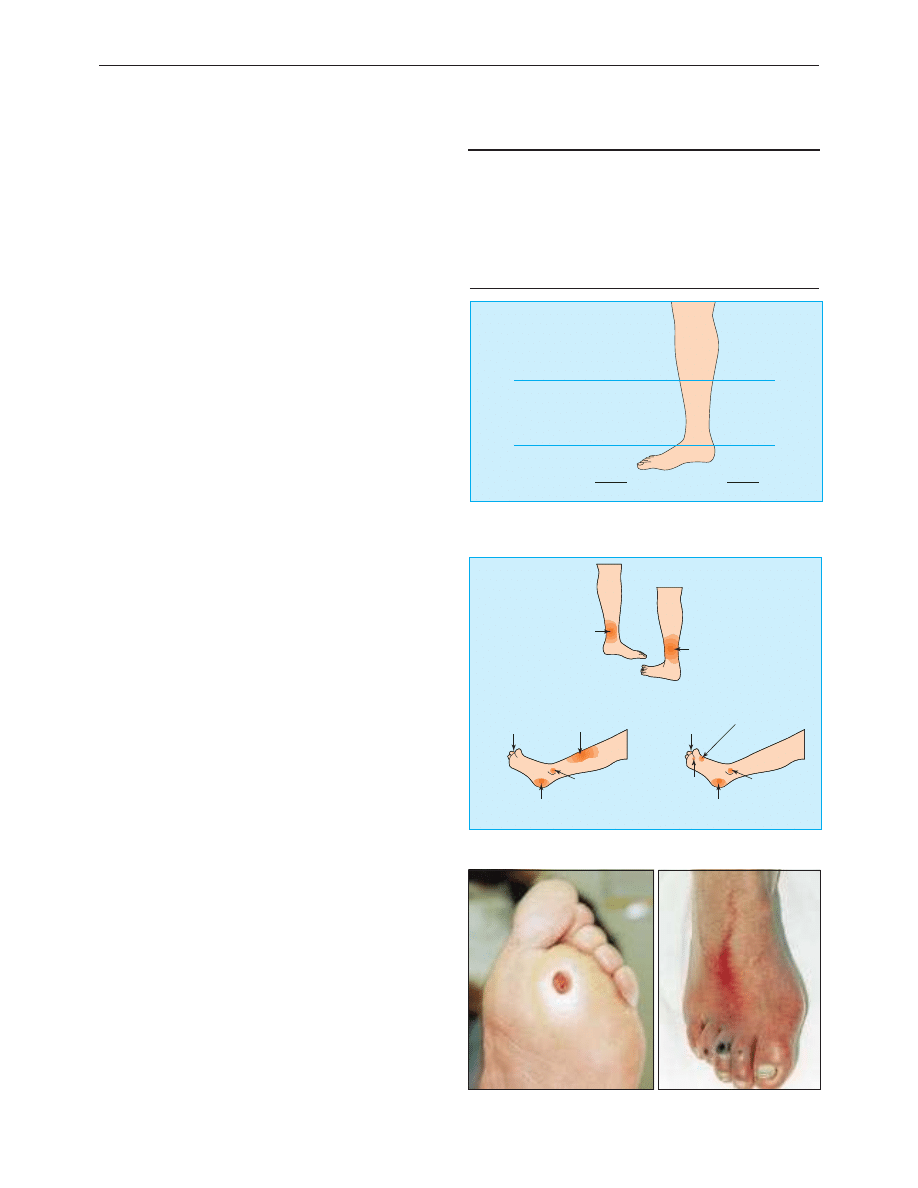
ABC of arterial and venous disease
Ulcerated lower limb
Nick J M London, Richard Donnelly
Ulceration of the lower limb affects 1% of the adult population
and 3.6% of people older than 65 years. Leg ulcers are
debilitating and painful and greatly reduce patients’ quality of life.
Ulcer healing has been shown to restore quality of life. Lower
limb ulceration tends to be recurrent, and the total annual cost of
leg ulceration to the NHS has been estimated at £400m.
Aetiology
Venous disease, arterial disease, and neuropathy cause over 90%
of lower limb ulcers. It is useful to divide leg ulcers into those
occurring in the gaiter area and those occurring in the forefoot
because the aetiologies in these two sites are different. At least
two aetiological factors can be identified in one third of all
lower limb ulcers.
Venous ulcers most commonly occur above the medial or
lateral malleoli. Arterial ulcers often affect the toes or shin or
occur over pressure points. Neuropathic ulcers tend to occur on
the sole of the foot or over pressure points. Apart from
necrobiosis lipoidica, diabetes is not a primary cause of
ulceration but often leads to ulceration through neuropathy or
ischaemia, or both. The possibility of malignancy, particularly in
ulcers that fail to start healing after adequate treatment, should
always be borne in mind. The commonest malignancies are
basal cell carcinoma, squamous cell carcinoma, and melanoma.
Patients with reduced mobility or obesity may develop
ulceration in the gaiter area because of venous hypertension
resulting from inadequate functioning of the calf muscle pump.
The commonest causes of vasculitic ulcers are rheumatoid
arthritis, systemic lupus, and polyarteritis nodosa. The blood
dyscrasias that most commonly lead to leg ulceration are sickle
cell disease, thalassaemia, thrombocythaemia, and
polycythaemia rubra vera.
Clinical assessment
History
It is important to determine the duration of ulceration and
whether it is a first episode or recurrent. Pain is a major
problem for patients with leg ulcers unless there is a
neuropathic component. Lack of pain therefore suggests a
neuropathic aetiology. Systemic diseases that may contribute to
the development of the leg ulceration (such as diabetes or
rheumatoid arthritis) should be noted, as should a history of
trauma, deep vein thrombosis, or varicose vein treatment.
Patients should also be asked about their mobility.
Examination
Examination of the leg should include palpation of pulses and a
search for the signs of venous hypertension, including varicose
veins, haemosiderin pigmentation, varicose eczema, atrophie
blanche, and lipodermatosclerosis. The range of hip, knee, and
ankle movement should be determined, and sensation should
be tested—for example, with a monofilament—to exclude a
peripheral neuropathy.
In patients with ulcers on the sole of the foot, the sole
should be examined for signs of ascending infection, including
proximal tenderness and appearance of pus on proximal
Causes of lower limb ulceration
x Venous disease
x Arterial disease
x Mixed venous-arterial disease
x Neuropathy
x Trauma
x Obesity or immobility
x Vasculitis
x Malignancy
x Underlying osteomyelitis
x Blood dyscrasias
x Lymphoedema
x Necrobiosis lipoidica
diabetecorum
x Pyoderma gangrenosum
x Self inflicted
Non-venous
Venous
8%
5%
43%
87%
49%
Calf
Gaiter
Foot
8%
100%
100%
Distribution of non-venous and venous ulcers of lower limb. The majority of
venous ulcers are in the gaiter area and the majortiy of non-venous ulcers in
foot
Venous
Arterial
Neuropathic
Above medial malleoli
Anterior shin
Over malleoli
Over toe joints
Under heel
Above lateral malleoli
Over malleoli
Inner side of
first metatarsal
head
Under
metatarsal
head
Over toe joints
Under heel
Common sites of venous,
arterial, and neuropathic ulceration. Adapted from
Tibbs et al
Neuropathic ulcers on sole of foot and dorsum of toe joints
Clinical review
1589
BMJ
VOLUME 320 10 JUNE 2000 bmj.com
on 1 October 2006
bmj.com
Downloaded from
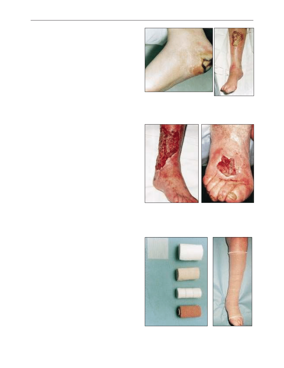
compression of the sole. Note any surrounding callus typical of
neuropathic ulceration and look for tracking to affect the bones
of the foot.
Investigation
Patients with foot ulceration should be referred to hospital for
investigation because many will have underlying arterial
ischaemia that requires prompt intervention. Diabetic patients
with signs of infection should have plain radiography of the
foot to look for osteomyelitis. Patients with venous ulceration
should have their ankle brachial pressure index measured and
can be managed either primarily in the community by trained
nurses or referred to hospital for further investigation into the
underlying venous abnormality.
Management
The management of the more unusual causes of lower leg
ulceration is based on treating the underlying disease. However,
because venous disease affects up to 30% of the population, it is
not uncommon for patients with, for example, rheumatoid
arthritis to have lower limb ulceration caused by venous disease.
Indeed, in up to half of patients with rheumatoid arthritis and
leg ulcers the ulceration is due to venous disease rather than to
the rheumatoid arthritis.
Venous ulceration
Debate continues not only about how venous ulcers should be
treated but also where they should be treated. It has recently
been suggested that patients with leg ulcers should have an
initial assessment in a hospital vascular clinic, with patients who
are unlikely to benefit from surgery then being cared for in the
community. Although this approach has the potential for large
cost savings, clinical trials are required to establish cost
effectiveness. There is no evidence that any form of drug
treatment improves venous ulcer healing, and antibiotics should
be used only if the patient has cellulitis.
Community management
Current evidence suggests that the mainstay of the community
management of venous ulceration should be graduated
compression bandaging. The compression bandaging should be
elastic and have multiple layers with a simple, non-adherent
dressing underneath. For compression bandaging to be safely
applied the ankle brachial pressure index must be at least 0.8.
Nurses caring for patients with venous ulcer need to be trained to
measure the ankle brachial pressure index and apply
compression bandages safely. The bandages should be changed
once or twice a week. The healing rate depends on the initial size
of the ulcer, but 65-70% of venous ulcers heal within six months.
The skin on the lower leg should be kept moist with an
emollient such as simple aqueous cream or 50:50 liquid:white
paraffin, and surrounding eczema should be treated with a
topical steroid. It is important to keep both the primary wound
dressing and any medicaments used as “bland” as possible
because many patients with venous ulcers develop a contact
dermatitis to wound care products.
Hospital treatment
Patients referred to a hospital clinic will have colour duplex
scanning to define the underlying venous abnormality. Recent
studies have shown that about 60% of patients with venous
ulcers have isolated superficial venous incompetence with
normal deep veins. Evidence is mounting that patients with long
saphenous or short saphenous incompetence in the presence of
Arterial ulcer affecting the heel and shin
Venous ulcers usually occur above the malleoli (left) but may affect the
dorsum of the foot (right)
Components of Charing Cross four layer bandaging regimen. The primary
wound dressing (left) is a non-adherent dressing, over which are placed
(middle; top to bottom in order of use) wool, crêpe, Elset, and Coban
bandages. The bandages (right) need replacing once or twice a week
Clinical review
1590
BMJ
VOLUME 320 10 JUNE 2000 bmj.com
on 1 October 2006
bmj.com
Downloaded from
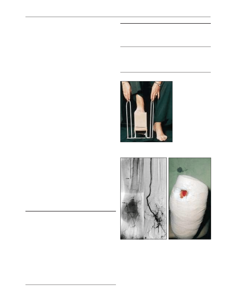
normal deep veins should have surgery to correct the venous
abnormality in the leg and allow ulcer healing. Patients with
refluxing deep veins do not benefit from superficial venous
surgery and are best managed by compression bandaging in the
community.
Prevention of recurrence
The five year recurrence rate of healed venous ulcers can be as
high as 40%, and preventing recurrence is therefore very
important. The rate of recurrence in patients who have had
surgery to correct superficial venous incompetence has not yet
been established, but it is expected to be low. In patients with
healed ulcers who have not had surgery, the mainstay of
preventing recurrence is graduated elastic compression hosiery.
One study found that ulcers recurred in 19% of patients
wearing class 2 compression hosiery and in 69% of
non-compliant patients. However, elderly patients with arthritis
of the knee or hip may struggle to apply class 2 compression
hosiery, and class 1 hosiery is a sensible compromise. Such
patients may find a hosiery applicator useful.
Arterial ulceration
For arterial ulcers to heal, the underlying arterial abnormality
must be corrected. Patients therefore require colour duplex
scanning of their arterial system or diagnostic arteriography to
define the underlying arterial abnormality. Angioplasty is the
treatment of choice because bypass grafting in patients with
ulcers carries an increased risk of wound or graft infection. For
patients in whom angioplasty is not possible, some form of
bypass operation, preferably using the saphenous vein, should
be attempted.
Neuropathic ulceration
The commonest cause of neuropathic ulceration is diabetes,
and many diabetic patients with neuropathic ulceration will also
have an arterial problem that requires correction. In many
hospitals diabetic patients with foot ulcers are managed in
specialist foot clinics run by a combination of diabetes
physicians, vascular surgeons, specialist nurses, and podiatrists.
The principles behind treatment are to optimise blood supply,
debride callus and dead tissue, treat active infection, and protect
the ulcerated area so that healing can occur. This often requires
the use of a protective plaster boot with a window cut out at the
site of the ulcer. Once healing has occurred, the patient is fitted
with footwear designed to minimise trauma and protect bony
prominences.
Classes of compression stocking: most patients can be
managed with below knee class 2 stockings
Class
Pressure at
ankle
(mm Hg)
Uses
1
14-17
Mild varicose veins
2
18-24
Treatment and prevention of venous ulcer
recurrence
3
25-35
Treatment of severe venous hypertension
and ulcer prevention in large diameter
calves
The ABC of arterial and venous disease is edited by Richard Donnelly,
professor of vascular medicine, University of Nottingham and
Southern Derbyshire Acute Hospitals NHS Trust (richard.donnelly@
nottingham.ac.uk) and Nick J M London, professor of surgery,
University of Leicester, Leicester (sms16@leicester.ac.uk). It will be
published as a book later this year.
BMJ
2000;320:1589-91
Key references
x Ruckley CV. Caring for patients with chronic leg ulcer. BMJ
1998;316:407-8.
x Scottish Intercollegiate Guidelines Network. The care of patients
with chronic leg ulcer. SIGN 26 1998;July.
x Fletcher A, Cullum N, Sheldon TA. Systematic review of
compression treatment for venous leg ulcers. BMJ 1997;315:576-80.
x Scriven JM, Hartshorne T, Bell PRF, Naylor AR, London NJM.
Single-visit venous ulcer assessment clinic: the first year. Br J Surg
1997;84:334-6.
x Tibbs DJ, Sabiston DC, Davies MG, Mortimer PS, Scurr JH. Varicose
veins, venous disorders, and lymphatic problems in the lower limbs.
Oxford:
Oxford University Press, 1997.
x Ruckley CV, Fowkes FGR, Bradbury AW. Venous disease. London:
Springer-Verlag, 1998.
x Browse NL, Burnand KG, Irvine AT, Wilson NM. Diseases of the veins.
London: Arnold, 1999.
x Task Force on Chronic Venous Disorders of the Leg. The
management of chronic venous disorders of the leg. Phlebology
1999;14(suppl 1).
Patients may find an applicator helps with
putting on compression hosiery
Occlusion (arrows) of distal posterior
tibial artery before (left) and after
angioplasty (right)
Protective plaster boot with window
cut out
Clinical review
1591
BMJ
VOLUME 320 10 JUNE 2000 bmj.com
on 1 October 2006
bmj.com
Downloaded from
Wyszukiwarka
Podobne podstrony:
ABC Investigation of liver and biliary disease
ABC Of Occupational and Environmental Medicine
ABC Of Conflict and Disaster
ABC Of Occupational and Environmental Medicine
ABC Liver abscesses and hydaid disease
7 questioning the coherence of HPD Westen jrnl of mental and nerv disease 2008
2011 2 MAR Chronic Intestinal Diseases of Dogs and Cats
ABC Of Liver,Pancreas and Gall Bladder
Pharmacokinetics of intraosseous and central venous drug delivery during cardiopulmonary resuscitati
Pharmacokinetics of intraosseous and?ntral venous drug?livery during?rdiopulmonary resuscitation
Application of Magnetic Resonance Spectroscopy in the Mental Diseases of Schizophrenia and Autism
Role of Antibodies in Controlling Viral Disease Lessons from Experiments of Nature and Gene Knockout
więcej podobnych podstron