
Formation of new chromosomes as a virulence
mechanism in yeast
Candida glabrata
Silvia Pola´kova´
a
, Christian Blume
a
, Julia´n ´
Alvarez Za´rate
a
, Marek Mentel
a,b
, Dorte Jørck-Ramberg
b
, Jørgen Stenderup
c
,
and Jure Pisˇkur
a,b,1
a
Department of Cell and Organism Biology, Lund University, SE-22362 Lund, Sweden;
b
Department of Systems Biology, Technical University of Denmark,
DK-2800 Lyngby, Denmark; and
c
Department of Clinical Microbiology, Regionshospitalet Herning, DK-7400 Herning, Denmark
Edited by John A. Carbon, University of California, Santa Barbara, CA, and approved January 9, 2009 (received for review September 30, 2008)
In eukaryotes, the number and rough organization of chromo-
somes is well preserved within isolates of the same species. Novel
chromosomes and loss of chromosomes are infrequent and usually
associated with pathological events. Here, we analyzed 40 patho-
genic isolates of a haploid and asexual yeast, Candida glabrata, for
their genome structure and stability. This organism has recently
become the second most prevalent yeast pathogen in humans.
Although the gene sequences were well conserved among differ-
ent strains, their chromosome structures differed drastically. The
most frequent events reshaping chromosomes were translocations
of chromosomal arms. However, also larger segmental duplica-
tions were frequent and occasionally we observed novel chromo-
somes. Apparently, this yeast can generate a new chromosome by
duplication of chromosome segments carrying a centromere and
subsequently adding novel telomeric ends. We show that the
observed genome plasticity is connected with antifungal drug
resistance and it is likely an advantage in the human body, where
environmental conditions fluctuate a lot.
chromosome rearrangements
兩 evolution 兩 genome stability 兩
pathogenicity
兩 segmental duplications
G
ene content and chromosome organization differ from
species to species. However, in most eukaryotes, including
yeasts, the chromosome structure seems to be well preserved
within the members of the same species, where regular sexual
cycles help to preserve the genome organization. Genomic
instability, including aneuploidy and changes in chromosome
structure, is apparently very low and usually associated with
pathological events, for example, in cancer development in
mammals (1, 2). In sexual species, chromosomal rearrangements
can lead to sexual isolation and subsequent speciation (3).
Candida glabrata, which is the second most prevalent yeast
pathogen in humans, has been traditionally classified as a haploid
and asexual organism. The genome of one strain has been
recently sequenced (4), and the genome structure now provides
a tool for understanding C. glabrata virulence. Rapid changes in
C. glabrata genomic organization have been reported in many
clinical studies (5–7). In addition, isolates from one patient often
exhibit 2 or 3 different karyotypes and during infection the
chromosome pattern can change within a few days (6). So far the
mechanisms behind the genome flexibility, adaptability, and
virulence have been only poorly understood in this yeast.
Results and Discussion
Clinical Isolates of
C. glabrata.
We analyzed 40 clinical isolates of
C. glabrata obtained from Danish patients that were randomly
selected from the Danish Statens Serum Institute collection
(
). All isolates were confirmed to belong to the C.
glabrata species by sequencing of the D1/D2 domain of LSU
rDNA and partial sequencing of mitochondrial SSU rDNA
(
). In addition, a more detailed phylogenetic relation-
ship among isolates was resolved by sequencing and analysis of
the sequences belonging to the fast evolving intergenic spacer
(IGS) region between the nuclear genes CDH1 and ERP6 on
chromosome A (see Fig. 2 and
). The variability among
strains for this sequence was
⬍2%.
Karyotypes Vary A Lot.
The yeast chromosomes were separated by
pulsed-field gel electrophoresis (PFGE). Although the strains,
based on the 3 sequenced loci, show little sequence variability,
their karyotypes were quite variable. The separated chromo-
somes were hybridized with 93 single-gene probes and 1 multi-
gene probe corresponding to LSU rDNA (
). In the
initial mapping we used 2 unique probes per chromosome: 1
from the middle (labeled ‘‘m’’) and 1 from near a chromosome
end (labeled ‘‘er’’ for right or ‘‘el’’ for left) (
and
). The karyotype of C. glabrata CBS 138, the sequenced
strain, was used as a reference, and the expected position of the
probes confirmed by hybridization. The largest chromosome size
polymorphisms were associated with either translocations of
chromosomal arms or interchromosomal duplications involving
12 of 13 chromosomes (
). The two largest chromosomes,
M and L, contain the rDNA locus and showed pronounced
variability in size without any translocations detected. A copy
number variation of the rDNA cluster or intrachromosomal
segmental duplications/deletions could explain the observed
polymorphism. In a few cases, the LSU rDNA probe also
hybridized to smaller chromosomes, probably as a result of
recent translocation events.
Molecular Mechanisms Behind Rearrangements.
Fifteen strains, cov-
ering chromosome changes of all 40 isolates, were selected for
further mapping, using close to 100 gene probes (
), to
understand the organization of centromeres, telomeres, chromo-
some number, some segmental duplications, and translocations
(Fig. 1 and
). Seven reciprocal and 4 nonreciprocal
translocations were observed, identifying all but CBS 138 chromo-
some C to be involved in these rearrangements. Note that if the
translocation involved a chromosome end
⬍20 kb, our approach
could not classify it as reciprocal. Three of these nonreciprocal
translocations were found in several strains, likely representing an
early event during the evolutionary history of C. glabrata (Fig. 2).
However, the origin of these translocation events is much younger
than the chromosomal translocations reported for the Saccharo-
myces sensu stricto sister species (8).
Interchromosomal duplications represent a subclass of trans-
locations. Five events, where a duplicated chromosomal segment
was translocated to another chromosome or fused with another
duplicated segment originating from a different chromosome,
Author contributions: S.P. and J.P. designed research; S.P., C.B., J.A
´ .Z., M.M., and D.J.-R.
performed research; J.S. contributed new reagents/analytic tools; S.P., C.B., D.J.-R., and J.P.
analyzed data; and S.P. and J.P. wrote the paper.
The authors declare no conflict of interest.
This article is a PNAS Direct Submission.
Freely available online through the PNAS open access option.
1
To whom correspondence should be addressed. E-mail: jure.piskur@cob.lu.se.
This article contains supporting information online at
www.pnas.org/cgi/content/full/
2688 –2693
兩 PNAS 兩 February 24, 2009 兩 vol. 106 兩 no. 8
www.pnas.org
兾cgi兾doi兾10.1073兾pnas.0809793106
were observed. The duplicated segments ranged in size from 40
to 700 kb, which is similar to the previously observed Saccha-
romyces cerevisiae duplications generated during growth under
laboratory conditions (9). However, one should note that the
observed C. glabrata duplications were characterized in the
‘‘native’’ isolates.
The comparison of chromosomal rearrangements and phylo-
genetic relationships positioned the origins of the observed
rearrangement events to specific branches of the phylogenetic
tree (Fig. 2). Only in a single case did similar translocations not
cluster together on the phylogenetic tree; the translocations
between chromosome M and F in isolates Y645 and Y650 may
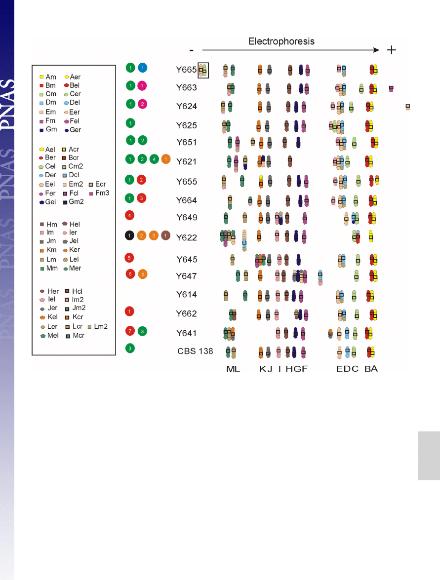
Fig. 1.
Electrophoretic karyotypes and detailed mapping of selected C. glabrata clinical isolates. A minimum of 4 probes labeled with the same color were used
per chromosome: 1 or 2 from the middle of the chromosome marked by squares and 2 from the near of the chromosome ends, left marked by pentagons and
right marked by circles, and 1 from close to the centromere marked with a bold black square (for details see
and
). Using CBS 138 as the
standard, the obtained results can be explained as 7 reciprocal translocations (RT), marked by red circles: event 1, reciprocal translocation between the right arm
of chromosome H (H
R
) and the left arm of chromosome L (L
L
) (RT between H
R
and L
L
); event 2, RT between K
R
and A
R
; event 3, RT between L
L
and M
L
; event 4,
RT between G
R
and D
L
; event 5, RT between M
R
and F
R
; event 6, RT between J
R
and E
R
; event 7, RT between K
L
and J
R
and 4 nonreciprocal translocations (NRT),
marked by green circles: event 1, translocation of the left arm of chromosome I (I
L
) onto chromosome L (NRT of I
L
onto L) (note that NTR of L onto I is equally
probable in the common ancestor of all of the strains from Y650 down to CBS 138, see
and Fig. 2); event 2, NRT of L
L
onto F; event 3, NRT of D onto L (in
all strains but Y641, CBS 138 chromosome D has a different configuration, therefore it is likely that CBS 138 chromosome D originated in this branch by a
translocation event); event 4, NRT of G
R
onto L. Also several segmental duplications can be observed and are divided into different classes (9). Class III duplications
are marked by orange circles, and class II are marked by brown circles. Class III: event 1, interchromosomal duplication (ID) of the left chromosomal arm of
chromosome E (E
L
) and its translocation onto chromosome G (ID of E
L
onto G); event 2, ID of D
R
onto B; event 3, ID of F
L
onto J; event 4, ID of I
L
onto D. Class
II: event 1, ID of E
L
and M
L
(duplication of 2 segments from different chromosomes fused together). The chromosomes E and C in the strain Y665, marked by black
square and blue circle, respectively, did not move into the gel in the electrophoretic field. The black circle stands for fusion of chromosome E and D in the strain
Y622 and is further explained in Fig. 3. Appearance of a novel chromosome is marked by pink circles, encompassing a segment from chromosome F event 1 and
encompassing a segment from the chromosome E event 2.
Pola´kova´ et al.
PNAS
兩 February 24, 2009 兩 vol. 106 兩 no. 8 兩 2689
GENETICS
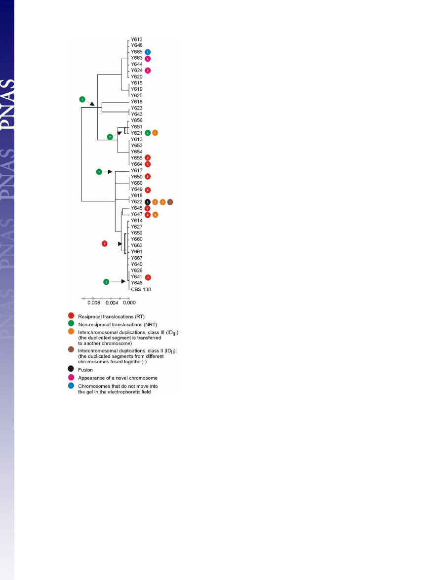
represent independent events. It is noteworthy that even isolates
having the identical intergenic sequences showed chromosomal
reorganizations, suggesting a high frequency of chromosome
remodeling events. For example, Y649, Y650, and Y666 belong
to the same phylogenetic cluster but have undergone different
chromosomal rearrangements (Fig. 2).
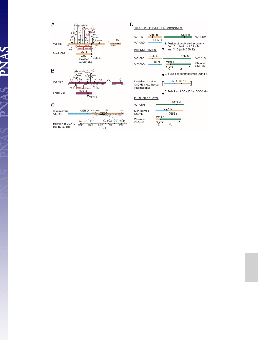
Novel Chromosomes.
Although in a majority of strains we found
13 chromosomes, 2 isolates, Y624 and Y663, exhibited 14 (Figs.
1 and 3). The novel chromosomes are small and seem to be
composed of a large 120- to 200-kb segmental duplication that
carries the centromere region. In Y624, the novel 120-kb chro-
mosome contains a partial duplication of chromosome E with a
subsequent deletion of 40–60 kb (Fig. 3A and
). In Y663,
the novel 200-kb chromosome is a partial duplication of chro-
mosome F (Fig. 3B and
). These new chromosomes appear
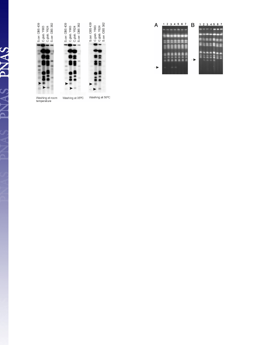
to have acquired telomeres. An oligonucleotide mimicking the
C. glabrata telomeric repeat gave a clear and specific Southern
analysis signal with the 2 small chromosomes, indicating that the
most terminal chromosome parts have a structure similar to
other chromosomes (Fig. 4). As described in S. cerevisiae (10),
when a segment containing an active centromere is duplicated,
DNA ends can acquire telomeres by initiating a recombination-
dependent DNA replication. Apparently, C. glabrata also pos-
sesses an effective mechanism to ‘‘add’’ viable chromosome ends.
In addition to de novo chromosome generation, we observed
chromosome fusions. For example, Y622 carries 2 chimeric
chromosomes, a fusion of chromosomes D and E, and a fusion
of parts of chromosome E and M (Fig. 3 C and D and
The original chromosome E is missing from the isolate. A 50- to
80-kb region carrying the chromosome E centromere is deleted
from the chromosome D
⫹ E chimera. However, these se-
quences are present in the chromosome E
⫹ M chimera. Because
the segment of chromosome E in this chimera is larger than the
segment deleted from the chromosome D
⫹ E chimera, it is
likely that the chromosome E segment was duplicated and
translocated to chromosome M before the chromosome E
centromere was deleted from the chromosome D
⫹ E chimera.
These structures of these novel fusion chromosomes show that it
is important that the chimeric chromosomes contain a single
active centromere so as not to destabilize chromosome parti-
tioning during mitosis (11). Surprisingly, Y665 showed only 11
chromosomal bands (Fig. 1). However, the probes specific for the
chromosome E and C genes hybridized to the loading well,
indicating that these 2 chromosomes adopted a structure that
does not allow them to move into the gel. Chromosome circu-
larization, observed in other yeasts (12), could explain such a
retarded movement in the electrophoretic field.
Another interesting aspect of the novel chromosomes are their
breakage points (
). C. glabrata does not posses the major
repeat sequences, like Candida albicans, and transposons, like S.
cerevisiae (4). However, several classes of mini- and mega-
satellites have been found (13), and two of them coincide with
the internal deletions of the Y624 minichromosome (Fig. 3A and
) and the Y622 fusion chromosome D
⫹E (Fig. 3C and
). In addition, some of the observed breakpoints cor-
relate with the regions, which were involved in the ‘‘historical’’
rearrangements taking place during the evolution of Hemiasco-
mycetes (
Our C. glabrata isolates with segmental aneuploidy and extra
chromosomes did not exhibit any significant delay in cell pro-
liferation when compared with other isolates. However, the
genetic stability of the novel chromosomes was decreased. When
C. glabrata Y624 and Y663, carrying extra chromosomes, were
grown in liquid yeast extract/peptone/dextrose (YPD) medium
for 70 generations, a number of the resulting cells lost the extra
chromosome (Fig. 5). The loss was observed in 40% of the
resulting progeny originating from Y624 and in 70% of the
progeny derived from Y663. Is there any advantage then to
keeping the duplicated regions and novel chromosomes?
Duplicated Genes.
Aneuploidy by gain of small chromosomes or
segmental aneuploidy were the most prevalent events on the left
arm of chromosome E (chrE
L
, 3 events of 8) and the left arm of
chromosome F (chrF
L
, 2 events of 8) (Fig. 1). Several genes on
chrE
L
and chrF
L
potentially play a role in C. glabrata interaction
with the host. Both duplicated segments of ChrF
L
encode a
transporter of the ATP-binding cassette family (CAGL0F01419g)
that is highly similar to S. cerevisiae AUS1. The small chromosome
Fig. 2.
Chromosomal rearrangements and phylogenetic relationship among
41 C. glabrata clinical isolates. The tree is based on the IGS region between
CAGL0A00605g and CAGL0A00627g on chromosome A, and the scale bars
represent the number of base substitutions per site. Specific events are placed
on the phylogenetic tree. The symbols illustrating chromosome changes are as
in Fig. 1.
2690
兩 www.pnas.org兾cgi兾doi兾10.1073兾pnas.0809793106
Pola´kova´ et al.
F encodes an ortholog of S. cerevisiae ABC transporter PDR5
(CAGL0F02717g) known in C. glabrata as PDH1. Its paralog,
known as CDR1 (CAGL0M01760g), is present on chromosome M
and was found duplicated in Y622. ABC transporters are implicated
in pleiotropic drug resistance, and therefore the duplications could
increase the level of drug resistance. Inspection of the chrE
L
arm
showed that the 3 duplicated segments, in Y621, Y622, and Y624,
encode the cluster of the S. cerevisiae YPS orthologs coding for
extracellular glycosyl phosphatidylinositol-linked aspartyl proteases
(CAGL0E01419g,
CAGL0E01727g,
CAGL0E01749g,
CAGL0E01771g,
CAGL0E01793g,
CAGL0E01815g,
CAGL0E01837g, CAGL0E01859g, CAGL0E01881g) and an
ortholog of PLB3 encoding phospholipase B (CAGL0E02321g).
The novel chromosome in Y624 has deleted a large part of this
region but kept YPS2 (CAGL0E01419g) encoding a protease and
PLB3 (CAGL0E02321g) encoding a phospholipase B. Secreted
aspartyl proteases and a phospholipase B have been shown to play
an important role in C. albicans virulence (14–16). Recently, Kaur
et al. (17) have shown that in C. glabrata the aspartyl protease-
encoding genes are required for survival in macrophages.
In C. albicans an aneuploidy and a specific segmental aneuploidy,
consisting of an isochromosome composed of 2 left arms of
chromosome 5, have been found to occur in response to antifungal
drug selection (18). Increases and decreases in drug resistance were
strongly associated with gain and loss of this isochromosome that
bears genes involved in azole drug resistance. In C. glabrata, the
original isolate of Y663 carrying the minichromosome could tol-
erate 129.6 mg/L of fluconazole (
). The Y663 clones, which
lost the minichromosome (Fig. 5) could only tolerate 14.4 mg/L of
fluconazole. In addition, when Y663 was grown in YPD in the
presence of azole (at a concentration of 40 mg/L) for 70 generations,
all resulting progeny still kept the extra minichromosomes, whereas
in the absence of azole, the loss of minichromosome was observed
in 70% of the progeny, indicating that the extra chromosome F
confers a growth advantage in the presence of the drug. Apparently,
the increase and decrease in gene dosage is a strategy used by C.
glabrata to overcome environmental pressure, such as the presence
of antifungal agents. The observed variable genome structure in the
examined pathogenic isolates is therefore an adaptation on the
different environmental conditions provided by each individual
patient and his/her therapeutic regime.
C. glabrata Genome Is Very Dynamic.
It was recently reported by
Torres et al. (19) that aneuploidy in S. cerevisiae is associated with
a proliferative disadvantage. However, in C. glabrata, segmental
duplications, chromosomal rearrangements, and extra chromo-
somes occur and persist at high frequency.
The high occurrence of certain chromosome events in C.
glabrata, i.e., chromosome fusions, possible circularizations, non-
reciprocal translocations, and novel chromosomes, suggests
some sort of telomere dysfunction. So far, 3 telomeric proteins
(Rap1, Sir3, Rif1) involved in transcriptional silencing have been
analyzed in C. glabrata (20, 21). Our inspection of the sequenced
C. glabrata genome shows that homologues of S. cerevisiae TEN1
and RIF2 are missing. The 2 proteins function in telomere end
protection and length regulation (22, 23). Notably, deletion
of RIF2 in S. cerevisiae leads to recombination-dependent,
telomerase-independent telomere elongation (24).
The observed genome dynamics in C. glabrata has in general not
been seen in other yeasts, except in some mutant backgrounds.
However, in certain parasitic protozoa, such as Leishmania, a great
variation in karyotypes has been reported, and in addition, aneu-
ploidy, gene amplification, and deletion have been reported to be
associated with changes in the drug resistance and virulence (25,
26). Elevated chromosome dynamics is not compatible with sexual
lifestyle and meiosis but beneficial for adaptation to changing
Fig. 3.
Segmental duplications involved in the origin of novel chromosomes. (A) Y624 carries a duplication of chromosome E corresponding to a
⬇120-kb
fragment, which has subsequently deleted a 40- to 60-kb segment. (B) Y663 carries a duplication of chromosome F corresponding to
⬇200 kb. Black stripes
symbolize the position of centromeres (CEN). WT stands for CBS 138 chromosome architecture. The last genes identified as present on the novel chromosomes
are marked by red squares. The previously described genes involved in virulence or drug resistance are marked in red. (C) CBS 138 chromosome D and E fusion
found in Y622. Note a large deletion in the CEN E region. (D) A model illustrating the origin of the chromosome D and E fusion found in Y622. The centromere
(CEN) of the original chromosome E (ChE) was removed by a deletion of a
⬇50- to 80-kb region, thereby eliminating a dicentric chromosome structure. The
deleted region also covering centromere E was retained on chimeric chromosome composed of the left arm of chromosome E and the left arm of chromosome
M. The centromere E fragment in the chimeric chromosome is of a larger size as the region deleted around centromere E in the monocentric D plus E chromosome.
Pola´kova´ et al.
PNAS
兩 February 24, 2009 兩 vol. 106 兩 no. 8 兩 2691
GENETICS
environmental conditions. Apparently, C. glabrata has to ‘‘sacrifice’’
its sexual nature to better tolerate the consequences of the en-
hanced genome mutability. The observed elevated adaptability
potential should well be taken into account when developing future
drugs against this increasingly invasive human pathogen.
Materials and Methods
Yeast Strains. The isolates of C. glabrata originate from Danish patients, were
collected at Danish hospitals from 1986 to 1999, and were initially identified
by carbon assimilation tests (27). The clinical isolates used in this study were
randomly selected from a collection of
⬇250 isolates (see also ref. 28 and
). The yeast strains were grown at 25 °C in YPD medium consisting of 1%
yeast extract, 1% Bacto Peptone, and 2% glucose and in the minimal medium
(SD) consisting of 0.17% YNB (yeast nitrogen base without amino acids and
without ammonium sulfate), 0.5% ammonium sulfate, and 2% dextrose.
PCR and Sequencing of PCR Products. Genomic DNA was extracted according
to Philippsen et al. (29). Two regions, nuclear 26S ribosomal RNA coding D1/D2
domain and the mitochondrial small rRNA (SSU), were analyzed to confirm the
species identity. The D1/D2 domain of the nuclear 26rDNA (LSU rDNA) was
amplified with primers NL1 (5
⬘P-GCA TAT CAA TAA GCG GAG GAA AAG-3⬘P)
and NL4 (5
⬘P-GGT CCG TGT TTC AAG ACG G-3⬘P). In the case of the mitochon-
drial small rRNA (SSU), YM5 (5
⬘P-AAG AAT ATG ATG TTG GTT CAG A-3⬘P) and
YM13 (5
⬘P-ATT CTA CGG ATC CTT TAA ACC A-3⬘P) primers were used. Two
primers (NL1, NL4) were used to sequence the D1/D2 domain and 2 primers
(YM5, YM13) were used to sequence the mitochondrial SSU gene.
The IGS region between the nuclear CAGL0A00605g and CAGLA00627g
genes on chromosome A was amplified with primers 00605 (5
⬘P-C TCA CAA
ATG GAT TCC TTA AAG AGT TCG-3
⬘P) and 00627 (5⬘P-GT C ACC AGA GTT GGA
GTA CAT GTA G-3
⬘P), and the following conditions were applied: 94 °C initial
denaturation for 3 min, then 35 cycles of 45 s at 94 °C, 1 min at 52 °C, and 1 min
at 72 °C, followed by 72 °C for 5 min (1 cycle). The IGS region (CAGL0A00605g–
CAGL0A00627g) comprising 690 bases was sequenced with primers 00605 and
00627 by MWG Biotech.
The DNA probes (see
and
) for Southern blot analyses were
obtained by PCR amplification using C. glabrata CBS 138 genomic DNA as a
template. PCR conditions for all probes were 94 °C initial denaturation for 3
min, then 35 cycles of 45 s at 94 °C, 45 s at 56 °C, and 1 min at 72 °C, followed
by 72 °C for 5 min (1 cycle).
Phylogenetic Relationships. The sequences of the IGS region were aligned by
using the ClustalX (1.83) program (30), and the relationship was inferred by
using the neighbor-joining method (31). The phylogenetic tree was linearized
assuming equal evolutionary rates in all lineages (32). The tree is drawn to
scale, with branch lengths in the same units as those of the evolutionary
distances used to infer the phylogenetic tree. The evolutionary distances were
computed by using the Maximum Composite Likelihood method (33) and are
in the units of the number of base substitutions per site. All positions con-
taining gaps and missing data were eliminated from the dataset (Complete
deletion option). There were a total of 671 positions in the final dataset.
Phylogenetic analyses were constructed in MEGA4 (34).
PFGE and Southern Hybridization. Chromosomes of all clinical isolates were
separated by PFGE using a CHEF Mapper XA (Bio-Rad) and a 5-step program,
as follows: step 1, 240-s pulse for 6 h; step 2, 160-s pulse for 13 h; step 3, 120-s
pulse for 10 h; step 4, 90-s pulse for 10 h; and step 5, 60-s pulse for 3 h. The
included angle was 60° and the voltage was 150 V (4.5 V/cm). Chromosomes
and membranes were prepared as described (35). ORF DNA on the membrane
was detected by Southern blot analysis using
32
P-labeled PCR products as
probes (GE Healthcare) (
and
). After prehybridization, the
membrane was hybridized (0.25 M Na
2
HPO
4
, 7% SDS, 1 mM EDTA) at 60 °C for
15 h and washed twice at room temperature for 5 min and once at 60 °C for
30 min with 2% SDS, 100 mM Na
2
HPO
4
. The membrane was stripped by using
the hot SDS procedure protocol (GE Healthcare) and rehybridized more than
once. Signals were detected by using Imaging Screen-K (35
⫻ 43 cm; Bio-Rad)
and Personal Molecular Imager FX (Bio-Rad). We used 93 single-gene probes
and 1 multigene probe to detect and analyze the chromosomal rearrangements.
In the case of the telomeric probe, the 5
⬘ end of the telomeric oligonucle-
otide sequence (32-mer containing 2 16-bp telomere repeats, TCTGGGTGCT-
GTGGGGTCTGGGTGCTGTGGGG) was labeled with [
␥
32
P]-ATP with T4 polynu-
cleotide kinase (Abgene). The membrane was prehybridized and hybridized at
room temperature and washed at different temperature stringencies for 30 min.
Batch Culture Growth and Chromosome Stability. All clinical isolates and C.
glabrata CBS 138 strain were grown in 50 mL of YPD at 25 °C to OD
600
⫽ 10.
Cultures were inoculated with 120
L of overnight culture, and OD
600
was
measured every 3 h to determine the cell doubling time.
To study instability of extra chromosomes the strains Y624 and Y663 were first
plated on YPD medium to isolate single colonies. The single colonies were
analyzed by PFGE to confirm the aneuploidy. Three independent aneuploid
single colonies from each isolate were inoculated in 2 mL of YPD and cultivated
for 70 generations at 25 °C. Every day the cultures were reinoculated, and 2
L of
the old culture was transferred into new liquid YPD. Finally, 14 single colonies
were checked by PFGE for loss of the extra chromosomes. To determine the
doubling time, 3 single colonies with and 3 single colonies without extra chro-
mosome were grown in 50 mL of SD medium at 25 °C to OD
600
⫽ 7. Cultures were
inoculated with 120
L of overnight culture, and OD
600
was measured every 3 h.
Antifungal Drugs. Fluconazole susceptibility was measured on RPMI medium
1640 agar plates (0.84% RPMI medium 1640 with
L
-glutamine and no bicar-
bonate, 3.45% Mops, 2% glucose, 1.5% bacto agar adjusted to pH 7 with 1 M
NaOH) with 6 different concentrations of fluconazole (4.8, 14.4, 43.2, 129.6,
388.8, and 1166.4 mg/L). Appropriate 10 times dilutions of each cell suspension
Fig. 4.
Hybridization of C. glabrata telomeric oligonucleotide (32-mer
containing 2 16-bp telomere repeats) to separated chromosomes of 2 S.
cerevisiae strains (CBS 439 and CBS 382) and 2 C. glabrata strains containing
extra chromosomes (Y663 and Y624; see also Fig. 1). Hybridization was per-
formed at room temperature, and the membranes were washed at indicated
temperatures. Black arrows indicate the positions of the 2 novel chromo-
somes. Note that the 2 chromosomes hybridized to the telomeric probe even
at the most stringent washing conditions. A slightly weaker hybridization
signal from small chromosomes is caused by their instability during the prop-
agation (in other words, the mini-chromosomes are not present at the sto-
chiometrical concentration).
Fig. 5.
Chromosome loss in Y624 and Y663 grown in YPD for 70 generations.
(A) Karyotypes of the parental Y624 strain (line 1) and 6 randomly selected
progeny cell lineages (lines 2–7). The position of the small chromosome is
indicated by an arrow. (B) Karyotypes of the parental strain Y663 (line 2) and
6 randomly selected progeny lineages (lines 1 and 3–7). The position of the
small chromosomes is indicated by an arrow.
2692
兩 www.pnas.org兾cgi兾doi兾10.1073兾pnas.0809793106
Pola´kova´ et al.

(containing 10, 10
2
, and 10
3
cells) were plated on RPMI medium 1640 agar
plates. The plates were incubated for 48 h at 37 °C.
In the case of strain Y663, the aneuploid single colonies were additionally
grown for 70 generations in YPD
⫹ 40 mg/L fluconazole. Every day the cultures
were reinoculated by transfer of 2
L of the old culture into a new liquid YPD ⫹
fluconazole. Appropriate dilutions of each individual suspension were plated
on YPD, and the 14 resulting single colonies were checked by PFGE for chromo-
some loss.
ACKNOWLEDGMENTS. We thank Linda Hellborg, Morten Kielland-Brandt,
Torsten Nilsson-Tillgren, and Ken Wolfe for comments on the early version
of this manuscript and Eimantas Astromskas for technical assistance in
some experiments. This work was supported by the Swedish Research
Council and the Fysiografen, Crafoord, Lindstro¨m, Lawski, and So¨rensen
Foundations.
1. Gauwerky CE, Croce CM (1993) Chromosomal translocations in leukemia. Semin Cancer
Biol 4:333–340.
2. Sharpless NE, et al. (2001) Impaired nonhomologous end-joining provokes soft tissue
sarcomas harboring chromosomal translocations, amplifications, and deletions. Mol
Cell 8:1187–1196.
3. Delneri D, et al. (2003) Engineering evolution to study speciation in yeasts. Nature
422:68 –72.
4. Dujon B, et al. (2004) Genome evolution in yeasts. Nature 430:35– 44.
5. Klempp-Selb B, Rimek D, Kappe R (2000) Karyotyping of Candida albicans and Candida
glabrata from patients with Candida sepsis. Mycoses 43:159 –163.
6. Shin JH, et al. (2007) Changes in karyotype and azole susceptibility of sequential
bloodstream isolates from patients with Candida glabrata candidemia. J Clin Microbiol
45:2385–2391.
7. Lin CY, Chen YC, Lo HJ, Chen KW, Li SY (2007) Assessment of Candida glabrata strain
relatedness by pulsed-field gel electrophoresis and multilocus sequence typing. J Clin
Microbiol 45:2452–2459.
8. Fischer G, James SA, Roberts IN, Oliver SG, Louis EJ (2000) Chromosomal evolution in
Saccharomyces. Nature 405:451– 454.
9. Koszul R, Caburet S, Dujon B, Fischer G (2004) Eukaryotic genome evolution through
the spontaneous duplication of large chromosomal segments. EMBO J 23:234 –243.
10. McEachern MJ, Haber JE (2006) Break-induced replication and recombinational telo-
mere elongation in yeast. Annu Rev Biochem 75:111–135.
11. McClintock B (1984) The significance of responses of the genome to challenge. Science
226:792– 801.
12. Naito T, Matsuura A, Ishikawa F (1998) Circular chromosome formation in a fission
yeast mutant defective in two ATM homologues. Nat Genet 20:203–206.
13. Thierry A, Bouchier C, Dujon B, Richard GF (2008) Megasatellites: A peculiar class of
giant minisatellites in genes involved in cell adhesion and pathogenicity in Candida
glabrata. Nucleic Acids Res 36:5970 –5982.
14. Ghannoum MA (2000) Potential role of phospholipases in virulence and fungal patho-
genesis. Clin Microbiol Rev 13:122–143.
15. Mukherjee PK, et al. (2001) Reintroduction of the PLB1 gene into Candida albicans
restores virulence in vivo. Microbiology 147:2585–2597.
16. Naglik J, Albrecht A, Bader O, Hube B (2004) Candida albicans proteinases and
host/pathogen interactions. Cell Microbiol 6:915–926.
17. Kaur R, Ma B, Cormack BR (2007) A family of glycosylphosphatidylinositol-linked
aspartyl proteases is required for virulence of Candida glabrata. Proc Natl Acad Sci USA
104:7628 –7633.
18. Selmecki A, Forche A, Berman J (2006) Aneuploidy and isochromosome formation in
drug-resistant Candida albicans. Science 313:367–370.
19. Torres EM, et al. (2007) Effects of aneuploidy on cellular physiology and cell division in
haploid yeast. Science 317:916 –924.
20. Haw R, Yarragudi AD, Uemura H (2001) Isolation of a Candida glabrata homologue of
RAP1, a regulator of transcription and telomere function in Saccharomyces cerevisiae.
Yeast 18:1277–1284.
21. Castan˜o I, et al. (2005) Telomere length control and transcriptional regulation of
subtelomeric adhesins in Candida glabrata. Mol Microbiol 55:1246 –1258.
22. Wotton D, Shore D (1997) A novel Rap1p-interacting factor, Rif2p, cooperates with
Rif1p to regulate telomere length in Saccharomyces cerevisiae. Genes Dev 11:748 –760.
23. Grandin N, Damon C, Charbonneau M (2001) Ten1 functions in telomere end
protection and length regulation in association with Stn1 and Cdc13. EMBO J
20:1173–1183.
24. Teng SC, Chang J, McCowan B, Zakian VA (2000) Telomerase-independent lengthening
of yeast telomeres occurs by an abrupt Rad50p-dependent, Rif-inhibited recombina-
tional process. Mol Cell 6:947–952.
25. Beverley SM (1991) Gene amplification in Leishmania. Annu Rev Microbiol 45:417– 444.
26. Ubeda JM, et al. (2008) Modulation of gene expression in drug-resistant Leishmania is
associated with gene amplification, gene deletion, and chromosome aneuploidy.
Genome Biol 9:R115.
27. Wickerham LJ, Burton KA (1948) Carbon assimilation tests for the classification of
yeasts. J Bacteriol 56:363–371.
28. Mentel M, Spírek M, Jørck-Ramberg D, Piskur J (2006) Transfer of genetic material
between pathogenic and food-borne yeasts. Appl Environ Microbiol 72:5122–5125.
29. Philippsen P, Stotz A, Scherf C (1991) DNA of Saccharomyces cerevisiae. Methods
Enzymol 194:169 –182.
30. Thompson JD, Gibson TJ, Plewniak F, Jeanmougin F, Higgins DG (1997) The CLUSTALX
windows interface: Flexible strategies for multiple sequence alignment aided by
quality analysis tools. Nucleic Acids Res 25:4876 – 4882.
31. Saitou N, Nei M (1987) The neighbor-joining method: A new method for reconstructing
phylogenetic trees. Mol Biol Evol 4:406 – 425.
32. Takezaki N, Rzhetsky A, Nei M (1995) Phylogenetic test of the molecular clock and
linearized trees. Mol Biol Evol 12:823– 833.
33. Tamura K, Nei M, Kumar S (2004) Prospects for inferring very large phylogenies by
using the neighbor-joining method. Proc Natl Acad Sci USA 101:11030 –11035.
34. Tamura K, Dudley J, Nei M, Kumar S (2007) Molecular Evolutionary Genetics Analysis
(MEGA) software version 4.0. Mol Biol Evol 24:1596 –1599.
35. Petersen RF, Nilsson-Tillgren T, Piskur J (1999) Karyotypes of Saccharomyces sensu lato
species. Int J Syst Bacteriol 49:1925–1931.
Pola´kova´ et al.
PNAS
兩 February 24, 2009 兩 vol. 106 兩 no. 8 兩 2693
GENETICS
Wyszukiwarka
Podobne podstrony:
Identification of a cannabimimetic indole as a designer drug in a herbal product forensic toxicol (2
Use of hydrogen peroxide as a biocide new consideration of its mechanisms of biocidal action
feminism and formation of ethnic identity in greek culture
Formation of heartwood substances in the stemwood of Robinia
Andrew Garrett Convergence in the formation of Indo European subgroups
A Propagandist of Extermination, Johann von Leers and the Anti Semitic Formation of Children in Nazi
Role of the Structure of Heterogeneous Condensed Mixtures in the Formation of Agglomerates
Formation of active inclusion bodies in E coli
feminism and formation of ethnic identity in greek culture
Askildson, L Effects of Humour in the Language Classroom Humour as a Padagogical Tool in Theory and
Ehrman; The Role Of New Testament Manuscripts In Early Christian Studies Lecture 1 Of 2
Ehrman; The Role of New Testament Manuscripts in Early Christian Studies Lecture 2 of 2
J Leigh Globalization Reflections of Babylon Intercultural Communication and Globalization in the
SzczygielskiStopa Usage of new soil improvement techniques in road embankment constructions
Formation of heartwood substances in the stemwood of Robinia
Domjan M , Cusato B Pavlovian feed forward mechanisms in the control of social behavior
0415444535 Routledge The New Politics of Islam Pan Islamic Foreign Policy in a World of States Jul 2
Fake crop of Baptistic malicious spirits dummy baptism in the Spirit as taught by pastor John Torell
więcej podobnych podstron