
05.06.2012
Diffuse idiopathic skeletal hyperostosis in ancient clergymen
1/5
www.ncbi.nlm.nih.gov/pmc/articles/PMC2200769/?tool=pmcentrez
Go to:
Go to:
Go to:
Eur Spine J. 2007 August; 16(8): 1129–1135.
Published online 2007 March 28. doi:
10.1007/s00586-007-0342-x
PMCID: PMC2200769
Diffuse idiopathic skeletal hyperostosis in ancient clergymen
J. J. Verlaan
,
F. C. Oner
, and
G. J. R. Maat
Author information ►
Article notes ►
Copyright and License information ►
This article has been
cited by
other articles in PMC.
Abstract
Diffuse idiopathic skeletal hyperostosis (DISH) is a common but often unrecognized systemic disorder observed mainly in the elderly. DISH is
diagnosed when the anterior longitudinal ligament of the spine is ossified on at least four contiguous spinal levels or when multiple peripheral
enthesopathies are present. The etiology of DISH is unknown but previous studies have shown a strong association with obesity and insulin-
independent diabetes mellitus. DISH can lead to back pain, dysphagia, myelopathy, musculoskeletal impairment and grossly unstable spine
fractures after minor trauma. In archeological studies a high prevalence of DISH has been demonstrated in ancient clergymen. The present study
describes the pathological changes of human remains excavated from the abbey court (Pandhof) in the city of Maastricht, The Netherlands.
Human remains of 51 individuals buried between 275 and 1795
CE
were excavated and examined. The remains were investigated according to a
standardized physical anthropological report and individuals demonstrating ossification of spinal ligaments and/or multiple peripheral
enthesopathies were included in the study group. The authors reviewed all available material and after reaching consensus, each abnormality
found was given a diagnosis and subsequently recorded. After examination, 28 individuals were considered to be adult males; 11 adult females;
three adults of indeterminate sex and nine individuals were of sub adult age. The mean age at death for adults was 36.8 years. Seventeen adult
individuals (40.4% of all adults), displayed ossifications of at least four contiguous spinal levels and/or multiple enthesopathies of the appendicular
skeleton and were therefore, assigned the diagnosis DISH. The mean age of these individuals was 49.5 ± 13.0 years. In at least three of these
individuals, DISH had led to extensive ossification and subsequent ankylosis of axial and peripheral skeletal structures. In this population of
(presumably) clergymen and high-ranking citizens, DISH was observed in unusual high numbers at a relatively young age. Some of the examined
cases suggest that DISH may be a seriously incapacitating disorder when the more advanced stages of the disease have been reached. It is
hypothesized that “a monastic way of life” can predispose to DISH. Present demographic trends in obesity and diabetes mellitus as potential co-
factors for the development of DISH warrant further study to investigate its future prevalence.
Keywords: Diffuse idiopathic skeletal hyperostosis, Osteoarchaeology, Paleopathology, Spinal ankylosis
Introduction
Diffuse idiopathic skeletal hyperostosis (DISH) is a common but often unrecognized systemic disorder observed mainly in the elderly [
1
]. DISH is
diagnosed when ossification of the anterolateral aspect of the anterior longitudinal ligament is present on at least four contiguous spinal levels
without involvement of the intervertebral disk space and/or apophyseal (facet) joints [
2
]. The disease can manifest itself also at extraspinal
locations leading to peripheral entheseal ossification and bony spurs [
3
,
4
]. Studies investigating the demographic characteristics of DISH have
found a prevalence ranging from 2.9% in the Korean population, to 10.0% in a Caucasian population with a male/female ratio of 2:1 in most
investigations [
5
,
6
]. All papers pertinent to DISH demonstrate a consistent and marked increase of the disease with advancing age [
7
]. Its etiology
is unknown but several publications have found correlations with metabolic disorders, especially high body mass index and insulin-independent
diabetes mellitus [
8
–
10
]. The difference in prevalence between ethnic groups also suggests genetic factors being involved in the etiology of the
disorder [
5
,
6
,
11
].
From paleopathological studies it becomes apparent that DISH is not a disorder from recent history. Its features have for example been
demonstrated in the excavated remains of an Italian female individual who lived six centuries CE (Common Era); in the Egyptian pharaoh
Ramses II (1302–1213 BCE); and in hominid specimens as old as the Neandertal Shanidar I living approximately 50,000 years ago [
12
–
15
]. Signs
of DISH have even been shown in dinosaurs [
16
]. Although the radiological manifestations of DISH can sometimes be quite dramatic, displaying
extensive ossification of ligamentary structures, its clinical symptoms are usually relatively mild [
17
]. Some authors have described severe
dysphagia, progressive airway obstruction, obstructive sleep apnea and difficult endotracheal intubation due to anterior cervical bony
protuberances while others have noted minor stiffness or mild pain when the abnormalities were located in the thoracic or lumbar spine [
18
,
19
].
The presence of DISH has been associated with unstable spinal fractures, often complicated by permanent neurological deficit, following minor
trauma [
20
,
21
]. Still other studies could not confirm these findings and concluded that DISH is a strict radiological observation without clinical
relevance [
22
,
23
]. Presently, medical or surgical intervention is considered necessary for symptomatic treatment in rare cases only.
The “Onze Lieve Vrouwe” Church (OLVK; Church of Our Lady) in Maastricht is one of the oldest churches in The Netherlands with some parts of
its structure dating back to the eleventh century [
24
]. It was built on the foundation of an early medieval Merovingian Church, which in turn was
built on the remains of a Roman fortress. When an architectural rearrangement was planned for its garden, the abbey court (‘Pandhof’) of the
OLVK was temporarily available for archaeological investigation. Following initial fieldwork it was discovered that part of the ‘Pandhof’ had been
used as a burial place (Fig.
1
) [
25
]. After excavation and archaeological investigation was completed, the human remains of 51 individuals were
sent to our department for physical anthropological examination. During this examination, an unusual high number and particularly strong
expression of DISH was noted in the skeletal material with some individuals demonstrating severe ossification of spinal ligaments and multiple
peripheral enthesopathies. In this study individuals with DISH are presented to demonstrate its high prevalence and describe some aspects of the
wide range of abnormalities resulting from the disease. Furthermore, three cases are highlighted to demonstrate some of the specific
musculoskeletal pathology.
Fig. 1
Photograph of the excavation at the Onze Lieve Vrouwe Kerk in the city of Maastricht
Methods
An archaeological investigation of the Pandhof site was performed and the artifacts found were dated by stratigraphy (dating based on geological
1
1
2
05.06.2012
Diffuse idiopathic skeletal hyperostosis in ancient clergymen
2/5
www.ncbi.nlm.nih.gov/pmc/articles/PMC2200769/?tool=pmcentrez
Go to:
superposition of artifacts), and/or seriation (dating using the change of ‘fashion’ of artifacts over time to estimate the year of burial). After
completion of the archaeological examination the excavated human remains were packed individually and sent to our department. After the bones
of each individual were reconstructed, the joints were articulated and the human remains investigated according to a standardized physical
anthropological report [
26
]. For all excavated individuals, attempts were made to assess sex, age at death and stature. Subsequently, they were
examined for pathological conditions and individuals demonstrating spinal ligament ossifications of four or more contiguous vertebral levels
and/or extensive peripheral enthesopathies were included in the study group [
3
]. Two authors reviewed all available material and after reaching
consensus, each abnormality found was given a diagnosis and recorded. Furthermore, all archaeological records were retrieved to obtain
information about estimated year of burial and possible clues to social status or identification.
Results
A total of 56 graves were discovered during excavation, five of which were empty leaving 51 individuals for investigation. After archaeological
examination, the period of burials was determined to be between 275 and 1795 CE [
25
]. The completeness of the skeletons ranged from poor to
good, while the preservation of bone tissue was moderate to good. After examination of all 51 individuals, 28 were considered to be adult males; 11
were adult females; three adults were of indeterminate sex and nine individuals were of sub adult age. The mean age at death of adults was
36.8 ± 15.9 years, with a range of 20–69 years, based on N = 38 comprising of 27 males, eight females and three individuals of indeterminate sex.
Seventeen adult individuals (17/42 = 40.4% of the adult population; ten males, five females and two individuals of indeterminate sex) displayed
key features of DISH (ossification of spinal longitudinal ligaments without involvement of disk space/facet joints and/or the presence of multiple
peripheral enthesopathies) and were included in the study group. The individuals diagnosed with DISH had been buried in the interval 450–1795
CE
. The mean age of these individuals was 49.5 ± 13.0 years with a range of 23–69 years. Table
1
lists the demographic characteristics of
individuals from the study group and their specific pathologic conditions.
Table 1
Characteristics of 17 individuals with diffuse idiopathic skeletal hyperostosis
Three individuals, from grave 25, 43 and 44 are illustrative examples of DISH and are presented in more detail below.
Case grave 25
The individual in grave 25 was a 47 to 63-year-old male with healed rib fractures (left side fourth and fifth ribs) and vertebral osteoarthritis.
Ossification of the anterior longitudinal ligament at multiple thoracic and lumbar levels, except for the part where the aorta is located, confirmed
the diagnosis DISH (Fig.
2
a, b). It is interesting to note that in rare cases of DISH in individuals with situs inversus, ossification of the anterior
ligament is located on the contralateral side supporting the hypothesis that aorta pulsation may prevent ligament ossification [
27
]. In this
individual, ossification of the ligamentum flavum and partial ossification of the interspinal ligament and supraspinal ligament was also seen (Fig.
2
c, d). Although some osteophytosis was present at the anterior margin of the vertebral bodies of the thoracic and lumbar spine, no other signs of
spinal degenerative changes such as intervertebral or apophyseal joint space narrowing were observed. Extraspinal manifestations of DISH were
seen on the right and left humerus where the subscapular muscle insertion had become ossified (Fig.
2
e). The sternocostal and sternoclavicular
ligaments were ossified leading to a sternum with multiple ribs attached, who on their turn, were at the time of excavation still rigidly attached to
the thoracic spine (Fig.
2
f). In this individual, extensive ossification of numerous cranial, axial and peripheral muscle insertions was also observed.
Fig. 2
Photographs of skeletal remains from a 47 to 63-year-old male (grave 25) showing: a frontal view of
the thoracic spine with obvious ossification of the anterior longitudinal ligament except at the
trajectory of the aorta; b a coronal multiplanar reformatted
(more ...)
Case grave 43
In grave 43, a male 48 to 56 years-old-had been buried. DISH was established by the ossification of the anterior longitudinal and supraspinal
ligament (Fig.
3
a), at multiple levels of the thoracic spine. This part of the spine had subsequently become ankylotic without involvement of the
intervertebral and facet joints. Collapse of the tenth and eleventh thoracic vertebral bodies (probably from traumatic origin), had led to a
considerable kyphotic deformity (Fig.
3
b). This individual also had bilateral osteomyelitis of both tibia and fibula.
Fig. 3
Photographs of skeletal remains from a 48 to 56 year-old-male (grave 43) showing: a a right sided
lateral view of the thoracolumbar spine with ossification of the anterior longitudinal ligament,
vertebral osteophytosis and collapse of the tenth
(more ...)
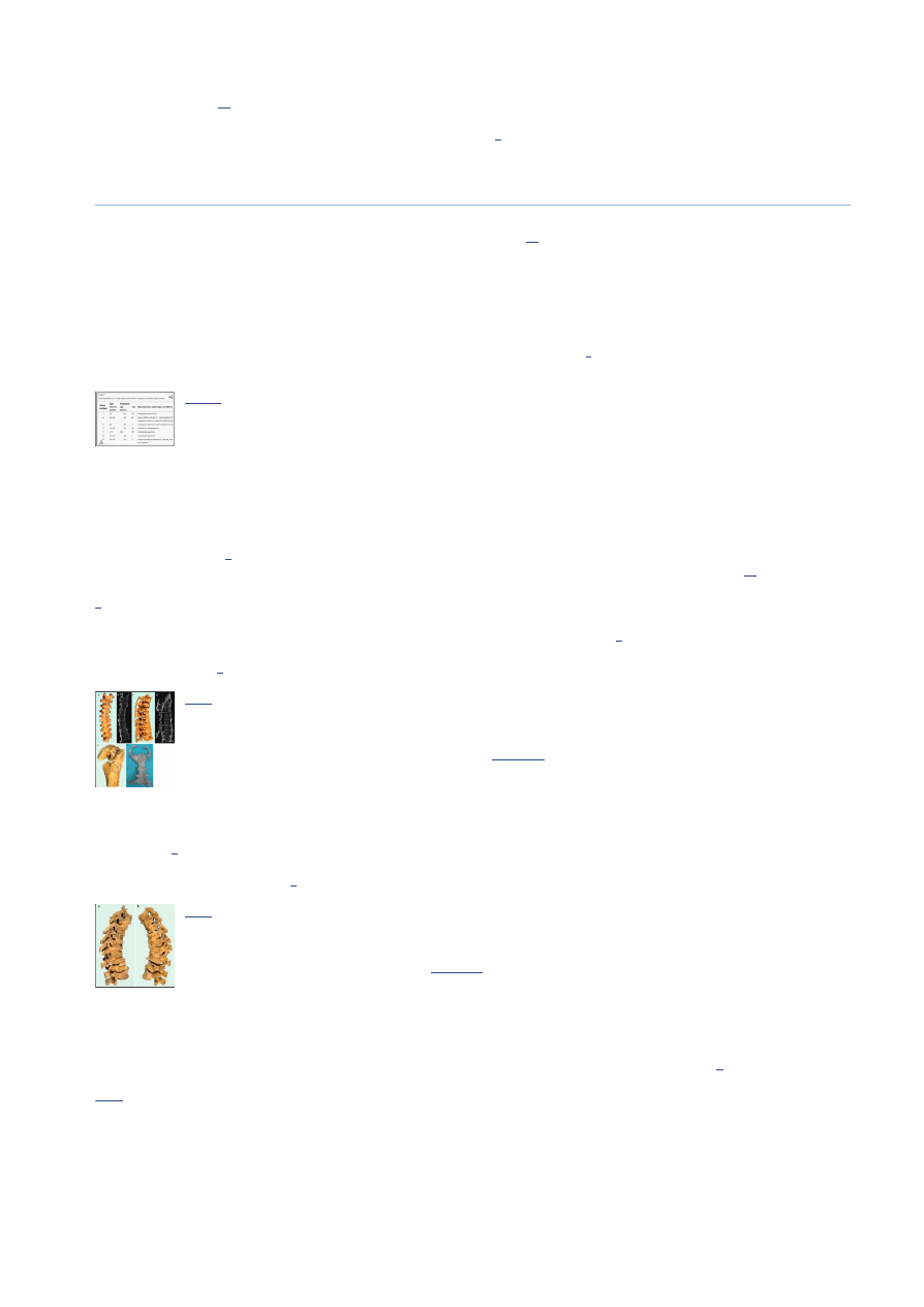
Case grave 44
A 57 year-old-female was buried in grave 44. This individual showed sacralization of the fifth lumbar vertebra and osteochondritis dissecans of the
left glenohumeral joint and the left knee joint. DISH was diagnosed by multiple level ossification of the anterior longitudinal ligament at the
thoracic level, which showed the typical ‘flowing wax’ phenomenon known from clinical radiological examinations (Fig.
4
).
Fig. 4
Photograph of skeletal remains from a 57 year-old-female (grave 44) showing ossification of the anterior longitudinal
ligament exhibiting the typical ‘flowing wax’ phenomenon

05.06.2012
Diffuse idiopathic skeletal hyperostosis in ancient clergymen
3/5
www.ncbi.nlm.nih.gov/pmc/articles/PMC2200769/?tool=pmcentrez
Go to:
Go to:
Go to:
Discussion
Diffuse idiopathic skeletal hyperostosis (DISH) was first described by Forestier [
28
] in the early fifties and subsequently named after him. When it
became clear that peripheral enthesopathies frequently accompanied spinal pathology, suggesting a systemic skeletal condition instead of an
isolated spinal disease, the name was changed into diffuse idiopathic skeletal hyperostosis to better describe the wide range of abnormalities
possible [
29
]. DISH can be distinguished from other ankylosing diseases such as ankylosing spondylitis (Morbus Von Bechterew), because bony
joints, especially the sacroiliac joints are by definition preserved in DISH [
4
].
Ossification of the posterior longitudinal ligament (OPLL) is a disease probably strongly related to DISH and has been studied in depth especially in
Japan where it is a common condition [
30
]. Inamasu et al. [
31
] suggest from their literature review of previously conducted research on OPLL
that it is probably not a single-gene disease; that pathways by which osteogenic induction of spinal ligament cells might occur are as yet poorly
understood and that bone synthesis marker concentrations have not differed significantly from control groups. It is currently unknown whether
the results obtained from research on OPLL may be applicable to DISH as well [
30
].
In the population excavated from the ‘Pandhof’ of the OLVK in the city of Maastricht, DISH was the most frequently encountered pathology.
Seventeen individuals with an average age at death of 49.5 years were diagnosed with this condition on the basis of spinal ligament ossification or
multiple peripheral enthesopathies. The percentage of adult individuals with DISH therefore, was 40.4%. This is an unusual high percentage of
DISH in a relatively young population compared to results from radiological studies in the literature [
7
,
11
]. In the study by Rogers and Waldron
[
32
], DISH was found to be present in 11.5% (6 out of 52 excavated males), of the population buried in the church and chapels from The Royal
Mint Medieval site in London while the prevalence of DISH in a nearby lay cemetery where the general population (likely to be peasants/farmers),
was buried, was 0% (0 out of 99 males). They suggested this difference in prevalence (11.5 vs. 0%) to be caused by a difference in occupation and
access to food [
33
]. In the present study the prevalence of DISH was almost fourfold that reported by Rogers and co-authors. Janssen and Maat
[
34
], found in their paleopathological study DISH to be present in 100% of the inspected clergymen (N = 27, with a mean age of 56 years ranging
from 43 to 75 years and living between 1070 and 1521 CE) excavated from the Saint Servaas Basilica also in the city of Maastricht. Since the
present study group has in common with the clergymen from the Saint Servaas Basilica that the individuals were probably enjoying a high status
(as can be concluded from the location of their burial within the confines of the church normally reserved for priests, monks and high-status
citizens), and likely had plentiful supply of food, it is not unreasonable to suggest that the associated ‘monastic way of life’, a term coined by Rogers
and Waldron [
32
], may have predisposed to DISH [
33
,
35
]. From descriptions of the dietary habits of monks in the Middle Ages in the discussion
section of the Rogers article, it can be argued that saturated (animal-) fats combined with small portions of vegetables and ample alcoholic
beverages may have often been on the menu, not unlike the diet of a considerable part of modern Western society. The 17 individuals with DISH
from the present study were buried in the interval 450–1795 CE. Therefore, it is hypothesised that the interred of the OLVK (and the Merovingian
Church before that), may in general have had an abundant food supply during this time period. Although genetic factors may play an important
role in the etiology of DISH, to our best knowledge, no historical evidence suggests a closer genetical relationship between monks than between the
general population and monks, therefore, a genetical predisposition for monks to develop DISH cannot be demonstrated. From current research it
can be learned that high body mass index and insulin-independent diabetes mellitus are risk factors for developing DISH further supporting,
though certainly not proving, that the lifestyle of ancient clergymen (sedentary work; abundance of food), may have predisposed them to develop
DISH [
36
,
37
].
Examination of the skeletal material with severe abnormalities suggests that DISH may have seriously affected the locomotor and probably also
cardiopulmonary apparatus of individuals with this condition. For example, ankylosis of the thoracic cage forced the individual from grave 25 to
ventilate by diaphragm contraction only and this would have led to restriction of pulmonary function and subsequent physical impairment.
Furthermore, when the physiological range of motion of multiple bony joints from the axial and peripheral skeleton becomes limited due to
surrounding soft tissue ossification, it is not unreasonable to suggest that affected individuals may have had clinical symptoms that are detectable
by physical examination. The individual from grave 25 probably also suffered from restricted abduction and endorotation (estimated to be between
45–60 degrees for both maneuvers before ossified soft tissue impingement), in the left glenohumeral joint. Due to postmortem absence of the
articular surface of the humeral head on the contralateral side, no right shoulder impingement could be estimated.
In conclusion, DISH may actually be a clinically relevant condition although presumably only so when the more advanced stages of the disease
have been reached. Regarding the presently known clinical symptoms, the diagnosis DISH should be considered in patients of middle or advanced
age with unexplainable back pain or peripheral arthralgia and/or restricted motion and should alert the physician for the presence of fracture
dislocations in DISH patients complaining of back pain after relatively minor trauma [
1
].
Acknowledgments
The authors would like to thank Joost Roelofs for his technical assistance during the acquisition of computed tomography data from the skeletal
material.
References
1. Belanger TA, Rowe DE. Diffuse idiopathic skeletal hyperostosis: musculoskeletal manifestations. J Am Acad Orthop Surg. 2001;9:258–267.
[
PubMed
]
2. Resnick D. Diffuse idiopathic skeletal hyperostosis. AJR Am J Roentgenol. 1978;130:588–589. [
PubMed
]
3. Maat GJR, Mastwijk RW, Velde EA. Skeletal distribution of degenerative changes in vertebral osteophytosis, vertebral osteoarthritis and DISH.
Int J Osteoarchaeol. 1995;5:289–298. doi: 10.1002/oa.1390050308. [
Cross Ref
]

05.06.2012
Diffuse idiopathic skeletal hyperostosis in ancient clergymen
4/5
www.ncbi.nlm.nih.gov/pmc/articles/PMC2200769/?tool=pmcentrez
4. Resnick D, Niwayama G. Radiographic and pathologic features of spinal involvement in diffuse idiopathic skeletal hyperostosis (DISH).
Radiology. 1976;119:559–568. [
PubMed
]
5. Julkunen H, Knekt P, Aromaa A. Spondylosis deformans and diffuse idiopathic skeletal hyperostosis (DISH) in Finland. Scand J Rheumatol.
1981;10:193–203. [
PubMed
]
6. Kim SK, Choi BR, Kim CG, et al. The prevalence of diffuse idiopathic skeletal hyperostosis in Korea. J Rheumatol. 2004;31:2032–2035.
[
PubMed
]
7. Weinfeld RM, Olson PN, Maki DD, et al. The prevalence of diffuse idiopathic skeletal hyperostosis (DISH) in two large American Midwest
metropolitan hospital populations. Skeletal Radiol. 1997;26:222–225. doi: 10.1007/s002560050225. [
PubMed
] [
Cross Ref
]
8. Denko CW, Malemud CJ. Body mass index and blood glucose: correlations with serum insulin, growth hormone, and insulin-like growth
factor-1 levels in patients with diffuse idiopathic skeletal hyperostosis (DISH). Rheumatol Int. 2006;26:292–297. doi: 10.1007/s00296-005-0588-
8. [
PubMed
] [
Cross Ref
]
9. Kiss C, Szilagyi M, Paksy A, et al. Risk factors for diffuse idiopathic skeletal hyperostosis: a case-control study. Rheumatology (Oxford)
2002;41:27–30. doi: 10.1093/rheumatology/41.1.27. [
PubMed
] [
Cross Ref
]
10. Sarzi-Puttini P, Atzeni F. New developments in our understanding of DISH (diffuse idiopathic skeletal hyperostosis). Curr Opin Rheumatol.
2004;16:287–292. doi: 10.1097/00002281-200405000-00021. [
PubMed
] [
Cross Ref
]
11. Kiss C, O’Neill TW, Mituszova M, et al. Prevalence of diffuse idiopathic skeletal hyperostosis in Budapest, Hungary. Rheumatology (Oxford)
2002;41:1335–1336. doi: 10.1093/rheumatology/41.11.1335. [
PubMed
] [
Cross Ref
]
12. Canci A, Marchi D, Caramella D, et al. Coexistence of melorheostosis and DISH in a female skeleton from Magna Graecia (sixth century BC).
Am J Phys Anthropol. 2005;126:305–310. doi: 10.1002/ajpa.20077. [
PubMed
] [
Cross Ref
]
13. Chhem RK, Schmit P, Faure C. Did Ramesses II really have ankylosing spondylitis? A reappraisal. Can Assoc Radiol J. 2004;55:211–217.
[
PubMed
]
14. Crubezy E, Trinkaus Shanidar E. A case of hyperostotic disease (DISH) in the middle Paleolithic. Am J Phys Anthropol. 1992;89:411–420. doi:
10.1002/ajpa.1330890402. [
PubMed
] [
Cross Ref
]
15. Russell AS. Ankylosing spondylitis or DISH in ancient mummies. Can Assoc Radiol J. 2004;55:335. [
PubMed
]
16. Rothschild BM. Diffuse idiopathic skeletal hyperostosis as reflected in the paleontologic record: dinosaurs and early mammals. Semin Arthritis
Rheum. 1987;17:119–125. doi: 10.1016/0049-0172(87)90034-5. [
PubMed
] [
Cross Ref
]
17. Beyeler C, Schlapbach P, Gerber NJ, et al. Diffuse idiopathic skeletal hyperostosis (DISH) of the elbow: a cause of elbow pain? A controlled
study. Br J Rheumatol. 1992;31:319–323. doi: 10.1093/rheumatology/31.5.319. [
PubMed
] [
Cross Ref
]
18. Ebo DG, Uytterhaegen PJ, Lagae PL, et al. Choking, sore throat with referred otalgia and dysphagia in a patient with diffuse idiopathic skeletal
hyperostosis (DISH). Acta Clin Belg. 2005;60:98–101. [
PubMed
]
19. Naik B, Lobato EB, Sulek CA. Dysphagia, obstructive sleep apnea, and difficult fiberoptic intubation secondary to diffuse idiopathic skeletal
hyperostosis. Anesthesiology. 2004;100:1311–1312. doi: 10.1097/00000542-200405000-00039. [
PubMed
] [
Cross Ref
]
20. Hendrix RW, Melany M, Miller F, et al. Fracture of the spine in patients with ankylosis due to diffuse skeletal hyperostosis: clinical and
imaging findings. AJR Am J Roentgenol. 1994;162:899–904. [
PubMed
]
21. Houk RW, Hendrix RW, Lee C, et al. Cervical fracture and paraplegia complicating diffuse idiopathic skeletal hyperostosis. Arthritis Rheum.
1984;27:472–475. doi: 10.1002/art.1780270418. [
PubMed
] [
Cross Ref
]
22. Fahrer H, Barandum R, Gerber NJ, et al. Pelvic manifestations of diffuse idiopathic skeletal hyperostosis (DISH): are they clinically relevant?
Rheumatol Int. 1989;8:257–261. doi: 10.1007/BF00270981. [
PubMed
] [
Cross Ref
]
23. Schlapbach P, Beyeler C, Gerber NJ, et al. Diffuse idiopathic skeletal hyperostosis (DISH) of the spine: a cause of back pain? A controlled study.
Br J Rheumatol. 1989;28:299–303. doi: 10.1093/rheumatology/28.4.299. [
PubMed
] [
Cross Ref
]
24. Meertens Institute, Koninklijke Nederlandse Akademie van Wetenschappen 2005.
25. De Koning J (1997) Voorlopig verslag van de stratigrafische opgraving in de Pandhof van de Onze Lieve Vrouwekerk te Maastricht 1997.
Cultural heritage from the City of Maastricht.
26. Maat GJR, Panhuysen RGAM, Mastwijk RW. Manual for the physical anthropological report. Barge’s Anthropologica. 1999;6:1–50.
27. Ciocci A. Diffuse idiopathic skeletal hyperostosis (DISH) and situs viscerum inversus. Report of a single case. Clin Exp Rheumatol. 1987;5:159–
160. [
PubMed
]
28. Forestier J R-QJ. Senile ankylosing hyperostosis of the spine. Ann Rheum Dis. 1950;9:321–330. doi: 10.1136/ard.9.4.321. [
PMC free article
]
[
PubMed
] [
Cross Ref
]
29. Resnick D, Shapiro RF, Wiesner KB, et al. Diffuse idiopathic skeletal hyperostosis (DISH) [ankylosing hyperostosis of Forestier and Rotes-
Querol]. Semin Arthritis Rheum. 1978;7:153–187. doi: 10.1016/0049-0172(78)90036-7. [
PubMed
] [
Cross Ref
]
30. Tsukahara S, Miyazawa N, Akagawa H, et al. COL6A1, the candidate gene for ossification of the posterior longitudinal ligament, is associated
with diffuse idiopathic skeletal hyperostosis in Japanese. Spine. 2005;30:2321–2324. doi: 10.1097/01.brs.0000182318.47343.6d. [
PubMed
]
[
Cross Ref
]
31. Inamasu J, Guiot BH, Sachs DC. Ossification of the posterior longitudinal ligament: an update on its biology, epidemiology, and natural
history. Neurosurgery. 2006;58:1027–1039. doi: 10.1227/01.NEU.0000215867.87770.73. [
PubMed
] [
Cross Ref
]
32. Rogers J, Waldron T. DISH and the monastic way of life. Int J Osteoarchaeol. 2001;11:357–365. doi: 10.1002/oa.574. [
Cross Ref
]
33. Waldron T. DISH at Merton Priory: evidence for a “new” occupational disease? Br Med J (Clin Res Ed) 1985;291:1762–1763. doi:
10.1136/bmj.291.6511.1762. [
Cross Ref
]
34. Janssen HAM, Maat GJR (1999) Canons buried in the stiftskapel of the Saint Servaas Basilica at Maastricht. In: Barge’s Anthropologica, vol 5.
2nd edn. pp 1–43.

05.06.2012
Diffuse idiopathic skeletal hyperostosis in ancient clergymen
5/5
www.ncbi.nlm.nih.gov/pmc/articles/PMC2200769/?tool=pmcentrez
35. Panhuysen RGAM (2005) Demography and health in early medieval Maastricht. Prosopographical observations on two cemetaries. Academic
dissertation.
36. Bray GA, Bellanger T. Epidemiology, trends, and morbidities of obesity and the metabolic syndrome. Endocrine. 2006;29:109–117. doi:
10.1385/ENDO:29:1:109. [
PubMed
] [
Cross Ref
]
37. Daniels Regulation of body mass and management of childhood overweight. Pediatr Blood Cancer. 2005;44:589–594. doi:
10.1002/pbc.20353. [
PubMed
] [
Cross Ref
]
Articles from European Spine Journal are provided here courtesy of Springer-Verlag
Wyszukiwarka
Podobne podstrony:
AT2H Basics Education in Ancient India
AT2H Science War in Ancient India
AT2H History Seafaring in Ancient India
Political and legal thought in ancient China Confucius
Speculations on the Origins and Symbolism of Go in Ancient China
elpenor second lesson in ancient greek (voices tenses verb translating) PART 1
Notario F , Food and Counter cultural Identity in Ancient Cynicism
SERGIS Dog sacrifice in ancient and modern greece from the sacrificial ritual to dog torture
Khipu Accounting in Ancient Peru
PARAMHANS, S A 1989, Astronomy in ancient India its importance, insight and prevalance
Donald H Mills The Hero and the Sea, Patterns of Chaos in Ancient Myth (pdf)(1)
elpenor second lesson in ancient greek (voices tenses verb translating) PART 2
Chiaradonna R , Universals in Ancient Medicine, 2013
Skeletal Rearrangements in natural products synthesis
Ebook History Egypt Sacred Geometry In Ancient Egypt
Self Identification with Diety and Voces in Ancient Egyptian and Greek Magick by Laurel Holmstrom
więcej podobnych podstron