
276
Kardiochirurgia i Torakochirurgia Polska 2010; 7 (3)
WADY WRODZONE
Address for correspondence: dr n. med. Ireneusz Haponiuk, Oddział Kardiochirurgii Dziecięcej Pomorskiego Centrum Traumatologii im. Miko-
łaja Kopernika w Gdańsku, ul. Nowe Ogrody 1-6, 80-803 Gdańsk, Polska, tel./fax +48 58 322 08 51, Email: ireneusz_haponiuk@poczta.onet.pl
Abstract
Asymptomatic pericardial defects are rare and found mostly
incidentally during cardiac surgery. In one third of all cases
absence of pericardium accompanies various congenital heart
defects, and is diagnosed intraoperatively. We present a case
of an 8-month-old male infant with Gerbode-type septal de-
fect (LV-RA communication) with rightward rotation of the
heart and left-sided partial pericardial defect, with pericardial
adhesions and symptoms of left-sided pressure pneumotho-
rax while opening the pericardial sac. The unique co-existence
of Gerbode septal communication and position anomaly of the
heart with congenital defect of the pericardium caused the ef-
fect of valvular air-trapping mechanism in the area of pericar-
dial wall discontinuity that needed a change of the operative
strategy prior to cardiopulmonary bypass. Taking into account
the patient’s past medical history the pericardial defect could
be responsible for pericardial adhesions as a reaction to re-
current pulmonary infections spreading via persistent pleuro-
pericardial communication.
Key words: congenital pericardial defect, Gerbode septal de-
fect, congenital heart defects, paediatric cardiac surgery.
Streszczenie
Bezobjawowe wrodzone ubytki osierdzia rozpoznaje się prze-
ważnie przypadkowo podczas zabiegów kardiochirurgicznych.
W ok. 30% przypadków ubytkom osierdzia towarzyszą inne
wrodzone wady serca. W pracy zaprezentowano przypadek
8-miesięcznego chłopca z ubytkiem w przegrodzie międzyko-
morowej typu Gerbode (komunikacja lewa komora – prawy
przedsionek) w zrotowanym sercu z historią przewlekłych infek-
cji dróg oddechowych. W trakcie zabiegu operacyjnego korekcji
wady serca stwierdzono zrosty osierdziowe oraz lewostronną
odmę opłucnową. Powstała ona po otwarciu worka osierdzio-
wego w mechanizmie pułapki powietrznej przez istniejący uby-
tek osierdzia w okolicy uszka lewego przedsionka. Zrosty osier-
dziowe mogły powstać na skutek komunikacji jamy opłucnowej
lewej z workiem osierdziowym, która stanowiła potencjalną
przyczynę odczynu osierdziowego na przebyte infekcje dróg od-
dechowych.
Słowa kluczowe: wrodzony ubytek osierdzia, ubytek międzyko-
morowy typu Gerbode, wrodzone wady serca, kardiochirurgia
dziecięca.
Background
Congenital pericardial defects are rare malformations
with variable clinical presentations [1, 2]. Pericardial sac de-
fects can be partial or complete, as it is commonly known in
some mammals. The anomalies are still poorly known, the
majority of literature reports are based on incidentally di-
agnosed cases, thus it is impossible to ascertain their total
prevalence. Asymptomatic patients remain undiagnosed,
otherwise the defects are discovered incidentally during
cardiac surgery. The patients with congenital pericardial
defects are referred for surgery for unrelated conditions, or
the diagnosis is given postmortem [1]. To the best of our
knowledge, we present the first case of a patient with con-
genital pericardial sac defect with congenital Gerbode type
ventricular septal defect in an infant described in the lit-
erature. The case presented is twice as interesting because
of preoperative diagnosis of Gerbode defect and necessary
change of the operative strategy due to destabilizing pres-
sure pneumothorax before cardiopulmonary bypass was
commenced. Taking into account the patient’s past medi-
cal history the pericardial defect could be responsible for
pericardial adhesions as a reaction to recurrent pulmonary
infections spreading via persistent pleuro-pericardial com-
munication.
Congenital pericardial defect with Gerbode type
septal defect in rotated heart: report of a case
Ubytek worka osierdziowego oraz ubytek w przegrodzie
międzykomorowej typu Gerbode w zrotowanym sercu – opis przypadku
Ireneusz Haponiuk, Maciej Chojnicki, Radosław Jaworski, Mariusz Sroka, Mariusz Steffek,
Piotr Czauderna
Oddział Kardiochirurgii Dziecięcej Pomorskiego Centrum Traumatologii im. Mikołaja Kopernika w Gdańsku
Kardiochirurgia i Torakochirurgia Polska 2010; 7 (3): 276–279
Kardiochirurgia i Torakochirurgia Polska 2010; 7 (3)
277
WADY WRODZONE
Case report
An eight-month-old male infant; 7600 g birth weight,
was admitted to the Department of Paediatric Cardiac
Surgery, Mikołaj Kopernik Hospital in Gdańsk (Poland)
with the diagnosis of ventricular septal defect (VSD) and
hypothyreosis. The boy had a history of permanent respi-
ratory tract infections prior to the admission with a re-
markably bad clinical course. On admission his cardiac
examination was remarkable for medial displacement of
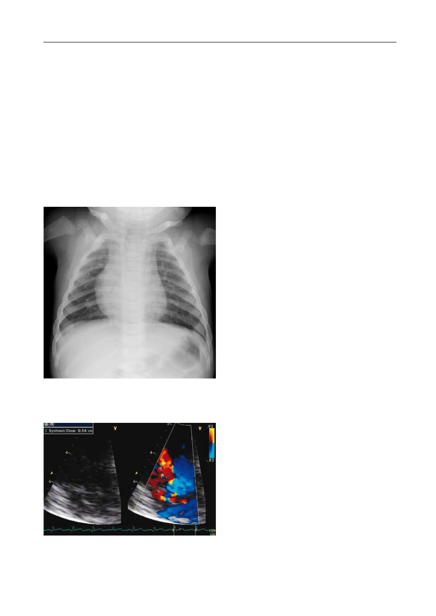
the apex right to the midclavicular line. Preoperative chest
X-ray showed flattening and elongation of the left ventricu-
lar contour and right atrium enlargement (Fig. 1). ECG proved
regular sinus rhythm with the right axis and partial right
bundle branch block (PRBBB). Preoperative Echo examina-
tion showed 5.4 mm Gerbode VSD with left ventricle-right
atrium (LV-RA) communication and mild tricuspid regurgita-
tion, the LV function and diameters of the heart chambers
were normal (Fig. 2).
The patient was referred for scheduled ventricular sep-
tal defect repair. After classic median sternotomy and small
thymus removal during the pericardiotomy unexpected
pericardial adhesions in the area of ventricle body and
right atrium (RA) and its appendage (RAA) were found. At
the time of meticulous dissection there was growing left
pressure pneumothorax with hemodynamic destabiliza-
tion, bradycardia and pressure drop. The pneumothorax
was relieved after insertion of an external suction line into
the left pleura via the pericardial defect, further identified
in the area of the left atrial appendage (LAA). The pericar-
dial sac defect was functionally closed by the LAA causing
air-trapping mechanism of the left pressure pneumothorax.
The heart rotation (40 degrees) in the longitudinal heart
axis was found with the left ventricle (LV) in the front and RA
body situated deep in the pericardium and dorsally. The left
pleura was opened with pleural adhesions as the remnants
of recurrent pleural infections. The cardiac procedure was
performed in cardiopulmonary bypass and moderate hypo-
thermia with antegrade cardioplegic arrest, the heart was
opened through RA incision. The Gerbode defect in the area
of the tricuspid valve annulus with LV-RA communication
was closed with running suture fresh autologous pericardial
patch. The tricuspid valve inspection showed no dysfunc-
tion. The pericardial defect and pericardiotomy were left
open. Postoperative course was uncomplicated. In the first
postoperative echo after surgery there was a small LV-RA
leakage that disappeared in further examinations. The child
was discharged home on the 8
th
postoperative day in a good
general condition. One year follow-up was uneventful.
Discussion
Congenital pericardial defects are rare findings with
variable clinical presentation and can be complete or partial
[1, 2]. These are still poorly known anomalies, sometimes
overlooked, with a total number of less than 150 cases re-
ported. Most frequently pericardial defects are left-sided
(86%) and are related to premature atrophy of the left duct
of Curvier during embryological development. Congeni-
tal complete pericardial absence is thought to be due to
premature atrophy of the left common cardiac vein with
insufficient blood supply to the pleuropericardium, which
leads to its agenesis during pregnancy [1]. Pericardial sac
defects appear three times more frequently in males than in
females in a white-Caucasian population. Thirty percent of
patients demonstrate associated congenital heart defects
as the most frequent ones: atrial septal defect, bicuspid
aortic valve, patent ductus arteriosus, tetralogy of Fallot, as
well as others, like pulmonary sequestration, bronchogenic
cysts, VATER syndrome (vertebral defects, anal atresia, tra-
cheoesophageal fistula, radial and renal dysplasia), Marfan’s
syndrome, Pallister-Killian syndrome [1, 3-5]. Pericardial de-
fects are more frequent in patients with congenital skeletal
malformations (Holt-Oram syndrome) [2]. Complications
Fig. 2. Preoperative Echo examination showed 5.4 mm Gerbode
VSD (marked on the scan) with LV-RA communication and mild
tricuspid regurgitation
Fig. 1. Preoperative chest X-ray showed flattening and elongation
of the left ventricular contour and right atrium enlargement, with
no symptoms that could indicate the presence of left-sided peri-
cardial sac defects

Kardiochirurgia i Torakochirurgia Polska 2010; 7 (3)
278
Congenital pericardial defect with Gerbode type septal defect in rotated heart: a report of a case
associated with pericardial defects depend on their extent.
Complete absence of the entire pericardium or absence of
the whole left or right side is usually associated with an
excellent prognosis [6]. A small increase in preload may
cause ventricular dilatation because of the loss of ventricu-
lar restraint, as it can be demonstrated in volume overload
patients with pericardium left open after regular cardiac
procedures. One case of severe tricuspid regurgitation due
to cardiac hypermobility was reported and it required surgi-
cal intervention [6]. The partial absence is more dangerous.
Entrapment of parts of the heart through defects may lead
to strangulation of the atria, appendages and parts of the
ventricles [7]. Congenital pericardial defect was reported
with the presence of acute myocardial necrosis in an adult
patient free of coronary artery disease, or there was evi-
dence of impingement by a pericardial rim [8].
Clinical presentation of pericardial deficiency is non-spe-
cific. In most cases the defect is discovered incidentally in an
asymptomatic patient [2]. There are reports of non-specific
symptoms like mild cough, upper respiratory infection symp-
toms, brief chest wall throw during exercise usually not as-
sociated with chest pain, shortness of breath, palpitations,
dyspnoea or respiratory distress leading to syncope [1, 8].
Sometimes the dyspnoea causes the “shifting heart” symp-
tom, which is reported by selected patients. Physical exami-
nation may reveal significantly displaced apical pulse area,
which may be palpated in the anterior or midaxillary line,
as well as basal ejection murmurs, apical midsystolic clicks,
or even systolic murmurs of undetermined origin. Patients
may incidentally present some complications clearly related
to the defect, like herniation and incarceration of the myo-
cardium, predominantly the LAA and ventricles. All complica-
tions have been associated with presentations varying from
chest pain to infarction, syncope, tricuspid regurgitation
and sudden death [6-8]. The ECG is not pathognomonic and
typically reveals bradycardia and right bundle branch block
(RBBB), poor R-wave progression secondary to leftward dis-
placement of precordial transitional zone is common, promi-
nent P-waves in the mid-precordial leads denote right atrial
overload. Echocardiographic findings are related to cardiac
levoposition and increased mobility within the chest [8].
These include unusual echocardiographic windows marked
change in cardiac position in the direction of the pericardial
hole with changing patient position on the examination ta-
ble (cardioptosis) and with abnormal cardiac cycle (swing-
ing heart), with abnormal septal motion and false-positive
appearance of the RV cavity dilation. Classic features of the
chest X-ray include levoposition of the heart, confirmed by
absence of the right heart border projecting on the right of
the vertebral column and left cardiac border straightening
and elongation (snoopy sign), as well as deep and very well-
defined aortopulmonary window caused by the absence of
the left pericardium and pleura allowing the left lung to in-
vaginate into pericardial space [8]. Ultimately, the diagnosis
is made or confirmed by CT scan or MRI. These diagnostic
techniques not only prove initial diagnosis, but also deline-
ate an extent of the defect, which provides important infor-
mation for further patient’s management strategy [1, 9].
Therapeutic options are based on small, retrospective
series which recommend surgical interventions only for pa-
tients with complications related to pericardial deficiency
[1, 6]. These include both surgical closure or elongation of
the pericardial defect with regard to initial problem caused
by the pathology [2]. Asymptomatic left total defects usu-
ally do not require surgical treatment because of the small
risk of circulatory complications, while the left-sided partial
pericardial defects are more controversial. Small and mod-
erate in size left-sided pericardial defects are considered for
prophylactic surgery by some, while others suggest treat-
ing only symptomatic patients [2, 6, 8]. Otherwise there are
many reports suggesting that both symptomatic and non-
symptomatic patients should be followed by prophylactic
operation to reduce the risk of death from cardiac structure
herniation and incarceration. On the other hand, patients
with partial absence of the pericardium (isolated); nowa-
days the focus is to manage the symptoms rather than pro-
phylactic management. An interesting and still more and
more available option is thoracoscopy intervention that
provides safe and effective minimally invasive surgical in-
tervention in the area of the pericardium as well as in the
heart structures, possible even in a small patient [10].
In the presented case, we were challenged by the unique
situation of a patient with Gerbode defect in a rotated heart
and partial left pericardial defect, which was difficult to
diagnose before surgery because of atypical position of the
heart. Left-sided pericardial sac defects are usually seen
with left cardiac border invagination to left pleural space
in routine chest X-ray, although the heart borders were
changed because of its irregular position. We also consid-
ered that both facts (partial absence of left pericardium and
rightward heart axis change) might have not been related
in embryonic development, thus we cannot exclude two
independent factors causing right deviation and left-sided
pericardial defect early in fetal life. Unexpected intraopera-
tive complication made us modify the dissection technique
nearby ECC. This complication gives another important
argument in the discussion whether pericardial defect can
be of special importance in selected cases.
References
1. Abbas AE, Appleton CP, Liu PT, Sweeney JP. Congenital absence of the peri-
cardium: case presentation and review of literature. Int J Cardiol 2005;
98: 21-25.
2. Skalski J, Wites M, Haponiuk I, Przybylski R, Grzybowski A, Zembala M,
Religa Z. A congenital defect of the pericardium. Thorac Cardiovasc Surg
1999; 47: 401-404.
3. Boscherini B, Galasso C, Bitti ML. Abnormal face, congenital absence of
the left pericardium, mental retardation, and growth hormone deficiency.
Am J Med Genet 1994; 49: 111-113.
4. Lu C, Ridker PM. Echocardiographic diagnosis of congenital absence of the
pericardium in a patient with VATER association defects. Clin Cardiol 1994;
17: 503-504.
5. Zakowski MF, Wright Y, Ricci A Jr. Pericardial agenesis and focal aplasia cutis
in tetrasomy 12p (Pallister-Killian syndrome). Am J Med Genet 1992; 42:
323-325.

Kardiochirurgia i Torakochirurgia Polska 2010; 7 (3)
279
WADY WRODZONE
6. van Son JA, Danielson GK, Schaff HV, Mullany CJ, Julsrud PR, Breen JF.
Congenital partial and complete absence of the pericardium. Mayo Clin
Proc 1993; 68: 743-747.
7. Jones JW, McManus BM. Fatal cardiac strangulation by congenital partial
pericardial defect. Am Heart J 1984; 107: 183-185.
8. Brulotte S, Roy L, Larose E. Congenital absence of the pericardium presen-
ting as acute myocardial necrosis. Can J Cardiol 2007; 23: 909-912.
9. Murat A, Artas H, Yilmaz E, Ogur E. Isolated congenital absence of the peri-
cardium. Pediatr Cardiol 2008; 29: 862-864.
10. Haponiuk I, Nachulewicz P, Laniewski-Wollk, Burczynski P, Maruszewski B.
Thoracoscopic obliteration of the left atrial appendage in a child with pro-
tein S deficiency – a prophylaxis of thromboembolic complications. Kar-
diochir Torakochir Pol 2008; 5: 287-291.
Wyszukiwarka
Podobne podstrony:
Plebaniak, Robert On best proximity points for set valued contractions of Nadler type with respect
sd74175 Quadruple D Type Flip Flops With Clear
Antczak, Tadeusz Sufficient optimality criteria and duality for multiobjective variational control
Image Processing with Matlab 33
L 5590 Short Sleeved Dress With Zipper Closure
M 5190 Long dress with a contrast finishing work
O'Reilly How To Build A FreeBSD STABLE Firewall With IPFILTER From The O'Reilly Anthology
M 5450 Dress with straps
Dance, Shield Modelling of sound ®elds in enclosed spaces with absorbent room surfaces
kurs excel (ebook) statistical analysis with excel X645FGGBVGDMICSVWEIYZHTBW6XRORTATG3KHTA
03 Teach Yourself Speak Greek With Confidence
Get Started with Dropbox
Osteochondritis dissecans in association with legg calve perthes disease
M 5588 Long Sleeveless Dress With Shaped Trim
Dealing with competency?sed questions
5 2 1 8 Lab Observing ARP with the Windows CLI, IOS CLI, and Wireshark
11 2 4 5 Lab ?cessing Network?vices with SSH
2 1 4 9 Lab Establishing a Console Session with Tera Term
więcej podobnych podstron