®
IRDye Fluorescent AFLP
Protocol for Large Plant
Genome Analysis
Published May, 2010. Updates
to this protocol will be posted at
http://biosupport.licor.com
Doc# 988-11159
Model 4300
DNA Analyzer
®
®

IRDye
®
Fluorescent AFLP
®
Protocol for Large Plant Genome Analysis
Doc# 988-11159
Page 1
www.licor.com
Contents
Page
I.
Required Reagents and Materials ....................................................................1
II.
Introduction .......................................................................................................2
III. AFLP
®
Template Preparation Kit ......................................................................5
IV.
AFLP
®
Selective Amplification Kit ....................................................................7
V. Gel
Electrophoresis ..........................................................................................8
VI.
Image Collection and Analysis..........................................................................9
VII.
AFLP
®
Troubleshooting Guide........................................................................13
I. Required Reagents and Materials
AFLP
®
Components:
Sufficient material is provided for 100 DNA templates and up to 1600 reactions for
selective AFLP
®
reactions. Store all components at -20 °C.
AFLP
®
Template Preparation Kit Components
Volume
•
Maize DNA (75 ng/µl) . . . . . . . . . . . . . . . . . . . . . . . . . . . . . . . . . . . . . . . . . . . . . . . . . . . . . . . . . . 10 µl
•
EcoR
I/
Mse
I enzyme mix [1.25 units/µl each in 10 mM Tris-HCl (pH 7.4), 250 mM NaCl,
0.1 mM EDTA, 1 mM DTT, 200 µg/ml BSA, 50% (v/v) glycerol, 0.15% Triton X-100 . . . . . . . . . . 100 µl
•
5X reaction buffer [50 mM Tris-HCl (pH 7.5, 50 mM Mg-acetate, 250 mM K-acetate] . . . . . . . . . 250 µl
•
T4 DNA ligase [5 units/µl in 10 mM Tris-HCl (pH 7.4), 0.1 mM EDTA, 1 mM DTT,
50 mM KCl, 200 µg/ml BSA, 50% (v/v) glycerol] . . . . . . . . . . . . . . . . . . . . . . . . . . . . . . . . . . . . . . 50 µl
•
Adapter mix [
EcoR
I/
Mse
I adapters, 0.4 mM ATP, 10 mM Tris-HCl (pH 7.5),
10 mM Mg-acetate, 50 mM K-acetate]. . . . . . . . . . . . . . . . . . . . . . . . . . . . . . . . . . . . . . . . . . . . . 1.2 ml
•
TE buffer [10 mM Tris-HCl (pH 8.0), 1.0 mM EDTA] . . . . . . . . . . . . . . . . . . . . . . . . . . . . . . . . . . 9.0 ml
•
Water, deionized . . . . . . . . . . . . . . . . . . . . . . . . . . . . . . . . . . . . . . . . . . . . . . . . . . . . . . . . . . . . . 1.0 ml
•
AFLP
®
pre-amp primer mix . . . . . . . . . . . . . . . . . . . . . . . . . . . . . . . . . . . . . . . . . . . . . . . . . . . . . 2.0 ml
•
AFLP
®
protocol . . . . . . . . . . . . . . . . . . . . . . . . . . . . . . . . . . . . . . . . . . . . . . . . . . . . . . . . . . . . . . . . one
AFLP
®
Selective Amplification Kit Components
Volume
•
Pre-amp Maize DNA . . . . . . . . . . . . . . . . . . . . . . . . . . . . . . . . . . . . . . . . . . . . . . . . . . . . . . . . . . . 16 µl
•
Mse
I primers (containing dNTPs)
Primer M-CAA . . . . . . . . . . . . . . . . . . . . . . . . . . . . . . . . . . . . . . . . . . . . . . . . . . . . . . . .400 µl
Primer M-CAC . . . . . . . . . . . . . . . . . . . . . . . . . . . . . . . . . . . . . . . . . . . . . . . . . . . . . . . .400 µl
Primer M-CAG . . . . . . . . . . . . . . . . . . . . . . . . . . . . . . . . . . . . . . . . . . . . . . . . . . . . . . . .400 µl
Primer M-CAT . . . . . . . . . . . . . . . . . . . . . . . . . . . . . . . . . . . . . . . . . . . . . . . . . . . . . . . . .400 µl
Primer M-CTA . . . . . . . . . . . . . . . . . . . . . . . . . . . . . . . . . . . . . . . . . . . . . . . . . . . . . . . . .400 µl
Primer M-CTC. . . . . . . . . . . . . . . . . . . . . . . . . . . . . . . . . . . . . . . . . . . . . . . . . . . . . . . . .400 µl
Primer M-CTG . . . . . . . . . . . . . . . . . . . . . . . . . . . . . . . . . . . . . . . . . . . . . . . . . . . . . . . .400 µl
Primer M-CTT . . . . . . . . . . . . . . . . . . . . . . . . . . . . . . . . . . . . . . . . . . . . . . . . . . . . . . . . .400 µl

LI-COR Biosciences
Doc# 988-11159
Page 2
www.licor.com
•
IRDye
®
infrared dye-labeled (either IRDye
®
700 or IRDye
®
800)
EcoR
I primers (1 µM)
Primer E-AAC . . . . . . . . . . . . . . . . . . . . . . . . . . . . . . . . . . . . . . . . . . . . . . . . . . . . . . . . .100 µl
Primer E-AAG . . . . . . . . . . . . . . . . . . . . . . . . . . . . . . . . . . . . . . . . . . . . . . . . . . . . . . . . .100 µl
Primer E-ACA . . . . . . . . . . . . . . . . . . . . . . . . . . . . . . . . . . . . . . . . . . . . . . . . . . . . . . . . .100 µl
Primer E-ACT . . . . . . . . . . . . . . . . . . . . . . . . . . . . . . . . . . . . . . . . . . . . . . . . . . . . . . . . .100 µl
Primer E-ACC . . . . . . . . . . . . . . . . . . . . . . . . . . . . . . . . . . . . . . . . . . . . . . . . . . . . . . . . .100 µl
Primer E-ACG . . . . . . . . . . . . . . . . . . . . . . . . . . . . . . . . . . . . . . . . . . . . . . . . . . . . . . . . .100 µl
Primer E-AGC . . . . . . . . . . . . . . . . . . . . . . . . . . . . . . . . . . . . . . . . . . . . . . . . . . . . . . . . .100 µl
Primer E-AGG . . . . . . . . . . . . . . . . . . . . . . . . . . . . . . . . . . . . . . . . . . . . . . . . . . . . . . . . .100 µl
•
10X amplification buffer [100 mM Tris-HCl (pH 8.3), 15 mM MgCl
2
, 500 mM KCl] . . . . . . . . . . 2.2 ml
•
Blue stop solution . . . . . . . . . . . . . . . . . . . . . . . . . . . . . . . . . . . . . . . . . . . . . . . . . . . . . . . . . . . . 8.0 ml
•
AFLP
®
protocol . . . . . . . . . . . . . . . . . . . . . . . . . . . . . . . . . . . . . . . . . . . . . . . . . . . . . . . . . . . . . . . . one
Additional Material Required
In addition to the kit components, the following items are required, but not included:
•
LI-COR
®
DNA Analyzer.
•
Programmable thermal cycler.
•
Mineral oil or liquid wax (if thermal cycler is not equipped with a heated lid).
•
Microcentrifuge capable of generating a relative centrifugal force of 14,000 x
g.
•
1.5 ml microcentrifuge tubes or 0.2 or 0.5 ml thin-walled microcentrifuge tubes (depending on thermal
cycler). For high throughput experiments, 96-well plates are recommended.
•
Pipettes capable of dispensing 0.3 to 2 µl, 1.0 to 20 µl, and 20 to 200 µl.
•
Taq DNA polymerase.
II. Introduction
Amplified Fragment Length Polymorphism (AFLP
®
) is a DNA fingerprinting technique developed by Key-
gene N. V. (1, 2). Since 1995, AFLP
®
has been widely used for genetic diversity assessment (2, 3), linkage
map construction (4, 5, 6), and gene profiling analyses (7, 8) in various genomes. A typical AFLP
®
analysis
consists of five major steps (as illustrated in Figure 1).
The first step is a restriction digest in which genomic DNA is cut by two restriction enzymes (a rare cutter
such as
EcoR
I, and a frequent cutter such as
Mse
I) to generate small DNA fragments.
Step two is a ligation in which double-stranded DNA adapters are ligated to the ends of the restricted DNA
fragments to generate templates for amplification.
Step three is a pre-amplification in which two primers, complementary to the adapter-ligated ends with one
pre-selected nucleotide at the 3’ end, are employed to amplify flanking regions containing the primer bind-
ing site and the restriction site.
Step four is a selective amplification in which selective primers, with an additional 1 to 3 nucleotides at the
3’ end, are employed to amplify subsets of pre-amplified templates. In step five, selective amplification
products are separated by denaturing polyacrylamide gel electrophoresis.
IRDye
®
Fluorescent AFLP
®
Protocol for Large Plant Genome Analysis
Doc# 988-11159
Page 3
www.licor.com
Figur
e 1. Sc
hematic illustr
ation of
AFLP
®
anal
ysis.
A color v
ersion of this figur
e can seen at
http://www
.licor
.com/bio/applications/4300_applications/aflp.jsp.
1.
2.
3.
4.
5.

LI-COR Biosciences
Doc# 988-11159
Page 4
www.licor.com
The popularity of this fingerprinting and mapping technique has created the need for increased throughput
via the automation of AFLP
®
analyses. Toward this end, LI-COR automated DNA analyzers can be used to
efficiently generate true AFLP
®
images (3, 4, 5, 6, 9). When compared to conventional AFLP
®
detection
methods, LI-COR
®
Biosciences’ automated system provides at least four major advantages.
• IRDye
®
labels are much safer than alternative radioactive labels.
• Image data can be obtained from the automated system in several hours rather than the two to four days
required for radioactive or silver staining procedures.
• The sensitivity of the automated system and availability of IRDye labeled AFLP
®
primers reduce overall
cost and eliminate labeling steps.
• AFLP
®
images are scored quickly with software such as SAGA
MX
(LI-COR). Software automation elimi-
nates multiple data entry steps when scoring markers and preparing data for phylogeny programs such as
PAUP, Treecon, or NTSYS and mapping software such as Mapmaker.
The protocols that follow are for plants having genomes ranging from 5 x 10
8
to 6 x 10
9
bp.
References
1.
Zabeau, M. and P. Vos. 1993. European Patent Application, publication number EP 0534858.
2.
Vos, P., R. Hogers, M. Bleeker, M. Reijans, T. van de Lee, M. Hornes, A. Frijters, J. Pot, J. Peleman, M. Kuiper, and M. Zabeau.
1995. AFLP: A new technique for DNA fingerprinting.
Nucleic Acids Research
23:4405-4414.
3.
Qiu, J., E. van Santen, and M. Campos-Andrada. 1999. AFLP analysis of
Lupinus luteus
and
L. cosentinii
using near infrared flu-
orescence labeled primers. P324-327. In: E. van Santen, M. Wink, S. Weissmann, and P. Roemer (eds). Lupin, an ancient crop
for the new millennium. Proc. of the 9th International Lupin Conference, Canterbury, New Zealand.
4.
Remington, D.L., R.W. Whetten, B.-H. Liu, and D.M. O'Malley. 1999. Construction of an AFLP genetic map with nearly com-
plete genome coverage in
Pinus taeda
.
Theoretical and Applied Genetics
98:1279-1292.
5.
Yang, W., D. B. Weaver, B. L. Nielsen, and J. Qiu. 2001. Molecular mapping of a new gene for resistance to frogeye leaf spot of
soybean in ‘Peking’.
Plant Breeding
120: 73-78.
6.
Klein, P.E., R.R. Klein, S.W. Cartinhour, P.E. Ulanch, J. Dong, J.A. Obert, D.T. Morishige, S.S. Schlueter, K.L. Childs, M. Ale, and
J.E. Mullet. 2000. A high throughput AFLP-based method for constructing integrated genetic and physical maps: progress
toward a sorghum genome map.
Genome Res
. 10:789-807.
7.
Bachem, C. W. B., R. S. van der Hoeven, S. M. de Bruijin, D. Vreugdenhil, M. Zabeau, and R. G. F. Visser. 1996. Visualization
of differential gene expression using a novel method of RNA fingerprinting based on AFLP: Analysis of gene expression during
potato tuber development.
Plant J.
9:745-753.
8.
Durranta, W. E., O. Rowlanda, P. Piedras, K.E. Hammond-Kosack, and J. D. G. Jones. 2000. cDNA-AFLP reveals a striking
overlap in race-specific resistance and wound response gene expression profiles.
Plant Cell
. 12: 963-977.
9.
Myburg, A.A., D.L. Remington, D.M. O'Malley, R.R. Sederoff, R.W. Whetten. 2001. High-throughput AFLP analysis using infra-
red dye-labeled primers and an automated DNA sequencer. BioTechniques 30:348-357.

IRDye
®
Fluorescent AFLP
®
Protocol for Large Plant Genome Analysis
Doc# 988-11159
Page 5
www.licor.com
III. AFLP
®
Template Preparation Kit
Preparing Genomic DNA Template
High quality genomic DNA is critical for obtaining reproducible AFLP
®
results. Contaminants in poor qual-
ity DNA may inhibit restriction, resulting in incomplete digestion that will produce variable AFLP
®
banding
patterns following amplification. Although quantity is not as critical as quality, reliable quantification of
DNA templates will ensure uniformity of AFLP
®
data among multiple individuals. Generally, 100 ng of
genomic DNA per template is sufficient.
Note:
Use of A260/A280 ratio to justify DNA quality may not be reliable and quantification based on A260 reading is
often incorrect by several fold. Use of a fluorometer or gel with a set of standards is recommended.
Restriction Digestion of Genomic DNA
Adapter Ligation
1.
Add the following to a 0.2 ml PCR tube (on ice):
5X reaction buffer
2.5 µl
Template DNA (100 ng in
≤
9 µl)
≤
9.0 µl
EcoR
I/
Mse
I enzyme mix
1.0 µl
Deionized water
to 12.5 µl
TOTAL VOLUME
12.5 µl
2.
Mix gently, centrifuge briefly, and incubate the mixture at 37°C for 2 hours.
3.
Incubate the mixture for 15 minutes at 70°C to inactivate the restriction enzymes and place tube
on ice.
Notes:
• A PCR thermal cycler is recommended for incubation in both steps 2 and 3. Program for one cycle of
37°C for 2 hours, one cycle of 70°C for 15 minutes, and 4°C soak.
1.
Add the following to the previous tube (on ice):
Adapter mix
12.0 µl
T4 DNA ligase
0.5 µl
COMBINED VOLUME
25.0 µl
2.
Mix gently by pipetting up and down. Centrifuge briefly and incubate the mixture at 20°C for
2 hours.
3.
After incubation, perform a 1:10 dilution of the ligation mixture by transferring 10 µl of the mixture
to a new 0.5 ml microcentrifuge tube, adding 90 µl of TE buffer, and mixing well.
4.
Store unused portion (15 µl) of the ligation mixture at -20°C for future experiments.
LI-COR Biosciences
Doc# 988-11159
Page 6
www.licor.com
Pre-amplification
1.
On ice, add the following to a PCR tube (size depends on thermal cycler used):
Diluted (1:10) ligation mixture (from steps above)
2.5 µl
AFLP
®
Pre-amp primer mix
20.0 µl
PCR reaction buffer (10X)
2.5 µl
Taq DNA polymerase (5 units/µl)
0.5 µl
TOTAL VOLUME
25.5 µl
Notes:
• Kit was optimized using Taq DNA polymerase (Cat. No. 1146173) and 10X PCR reaction buffer (Cat.
No. 1271318) from Roche Molecular Biochemicals. Choice of DNA polymerase is left to the user;
however, some modification or optimization may be required. Generally, 1 unit of Taq DNA poly-
merase per 10 µl reaction is recommended. For contents of suggested PCR buffer, see 10X amplifica-
tion buffer listed earlier in this protocol under the AFLP Selective Amplification Kit Componants
(page 2).
2.
Mix gently by pipetting up and down. Centrifuge briefly and cap tightly. Add 2 drops of liquid wax
or mineral oil if your thermal cycler is not equipped with a heated lid.
3.
Place PCR tube in the thermal cycler and run the following program.
5.
Perform a 1:40 dilution by pipetting 5 µl of the pre-amplification DNA mixture into a 0.5 ml micro-
centrifuge tube and adding 195 µl of ddH
2
O or low TE buffer (1.0 mM Tris-Cl, pH 8.0, 0.1 mM
EDTA). The diluted pre-amp DNA solution is sufficient for 100 selective AFLP
®
amplifications.
Notes:
• Dilution factors (1:10, 1:20, 1:50, etc.) of the pre-amp DNA may vary, depending on species and tem-
plates.
• Low TE buffer is not included in this kit.
6.
Store the unused portion (~20 µl) of the pre-amp template mixture at -20°C.
Program:
Step
Temperature (°C)
Time
1.
94 30
seconds
2.
56 1
minute
3.
72 1
minute
4.
4 hold
20 cycles total

IRDye
®
Fluorescent AFLP
®
Protocol for Large Plant Genome Analysis
Doc# 988-11159
Page 7
www.licor.com
IV. AFLP
®
Selective Amplification Kit
Selective AFLP
®
Amplification
Selective amplifications are generally performed in 96-well microplates. Before beginning, determine how
samples and primer combinations will be arranged in the plate. If an 8-channel syringe or pipetter will be
used to load the gel, this partially determines how the plate will be set up (see Model 4300 Applications
Manual or Model 4200 Genetic Analysis Manual for details).
The following duplex (one MseI with two IRDye-labeled EcoRI primers) PCR protocol is based on an 11 µl
total reaction volume per template-primer set. It is recommended that two labeled primers with similar T
m
be used. A guide for selecting EcoRI/MseI pairs for several major crop species is provided in Table 1.
Taq DNA polymerase working mix (see recipe below)
6.0 µl
Diluted pre-amp DNA
2.0 µl
MseI primer containing dNTPs
2.0 µl
IRDye 700 labeled EcoRI primer A
0.5 µl
IRDye 800 labeled EcoRI primer B
0.5 µl
TOTAL VOLUME
11.0 µl
Note: If a monoplex (one MseI with either one IRDye 700- or one IRDye 800-labeled EcoRI primer) PCR is to be con-
ducted, eliminate primer A or primer B (total volume equals 10.5 µl).
Reagent preparations
Because of the small amount of several reagents to be pipetted, it is strongly recommended to make a mas-
ter reagent mixture involving multiple reagents whenever possible to reduce systematic and pipetting
errors.
A) Taq DNA Polymerase Working Mix
(recipe for 200 µl, which is sufficient for 33 reactions):
Deionized water
158.0 µl
10X Amplification buffer
40.0 µl
Taq DNA polymerase (5 units/µl)*
2.0 µl
TOTAL VOLUME
200.0 µl
* Choice of DNA polymerase is left to the user; however, some modification to number of units needed may be
required.
B) Taq DNA Polymerase and Primer Mix:
The amount needed depends on how many DNA templates are used per primer pair combination. For
instance, to fingerprint 33 DNA templates using M-CAC coupled with E-AAC (IRDye 700 labeled) and
E-AGG (IRDye 800 labeled) primer combinations, you will need to combine 198 µl (= 6.0 µl x 33) of Taq
DNA polymerase working mix, 66 µl (= 2.0 µl x 33) of M-CAC primer solution, 16.5 µl (= 0.5 µl x 33) of
IRDye 700 E-AAC, and 16.5 µl (= 0.5 µl x 33) of IRDye 800 E-AGG primers into a 1.5 ml tube. (A small
amount of additional master mix is usually needed to compensate the loss due to pipetting errors.) Mix
gently, centrifuge briefly, place on ice, and cover the ice bucket.
Note: IRDye
®
infrared dye-labeled primer is light-sensitive. To minimize exposure to light, wrap all tubes containing
IRDyes (labeled primers and reaction mixes) with aluminum foil.
LI-COR Biosciences
Doc# 988-11159
Page 8
www.licor.com
Thermal Cycling
V. Gel Electrophoresis
For AFLP
®
gel electrophoresis, LI-COR
®
25-cm plates and KB
Plus
(6.5%) gel are recommended. Gels with
higher acrylamide concentration can be prepared using 40-50% polyacrylamide stock solutions from other
manufacturers (see 4300 Applications Manual or 4200 Genetic Analysis Manual). The 0.25 mm thickness
spacers and rectangular combs (either 48- or 64-tooth) are often the best choice. Some additional recom-
mendations follow:
■
Set voltage to 1500V, power to 40W, current to 40 mA, and temperature to 45°C. Set scan speed to 2 for the
Model 4300 or 4 for the Model 4200.
1.
Pipette 9.0 µl of Taq Polymerase and Primer Mix into each tube or well, then add 2.0 µl of each
diluted pre-amp template to bring the volume to 11 µl per tube or well. Add a drop of liquid wax or
mineral oil to each well if the thermal cycler has no heated lid.
2.
Spin briefly. If 96-well plates have been used, centrifuge the microplate(s) at 3000 rpm for a few
seconds to settle all the reagents to the bottom of the well. If such a centrifuge is not available, tap
the plate(s) gently on a table or pipette all the reagents down to the bottom of the well.
3.
Perform PCR using a "touchdown" program:
* Annealing temperatures are 65, 64.3, 63.6, 62.9, etc., for the 12 cycles.
4.
Add 5.0 µl of Blue Stop Solution to each well, mix thoroughly, centrifuge briefly, and denature for
3 minutes at 94°C. After 3 minutes, place on ice and load immediately.
Program:
Step
Temp
Time
1.
94 °C
30 seconds
2.
65 °C
30 seconds
3.
72 °C
1 minute
4.
94 °C
30 seconds
5.
65 °C minus 0.7 °C
per cycle *
30 seconds
6.
72 °C
1 minute
7.
94 °C
30 seconds
8.
56 °C
30 seconds
9.
72 °C
1 minute
10.
soak at 4 °C
12 cycles
23 cycles

IRDye
®
Fluorescent AFLP
®
Protocol for Large Plant Genome Analysis
Doc# 988-11159
Page 9
www.licor.com
■
Pre-run the gel for 30 minutes. Flush the wells completely with a 20 cc syringe to remove urea percipitate or
pieces of gel before loading.
■
Load about 0.8 to 1.0 µl of each denatured sample using either the 8-channel Hamilton syringe or pipette.
Load about 0.8 to 1.0 µl of the molecular size standard (50-700 bp) in the designated lanes. Two or three
marker lanes are usually used for 48- or 64-well gels, respectively. The run will take about 3 hours to collect
fragments up to 700 bp. The first bands will normally show up about 25 minutes after the run is started.
■
After the first run is complete, gels can be reloaded once with a new set of samples, as long as the gel
apparatus has not been moved. To reload a gel, create a new run, load samples and molecular weight
markers, and start the second run (do not pre-run the gel).
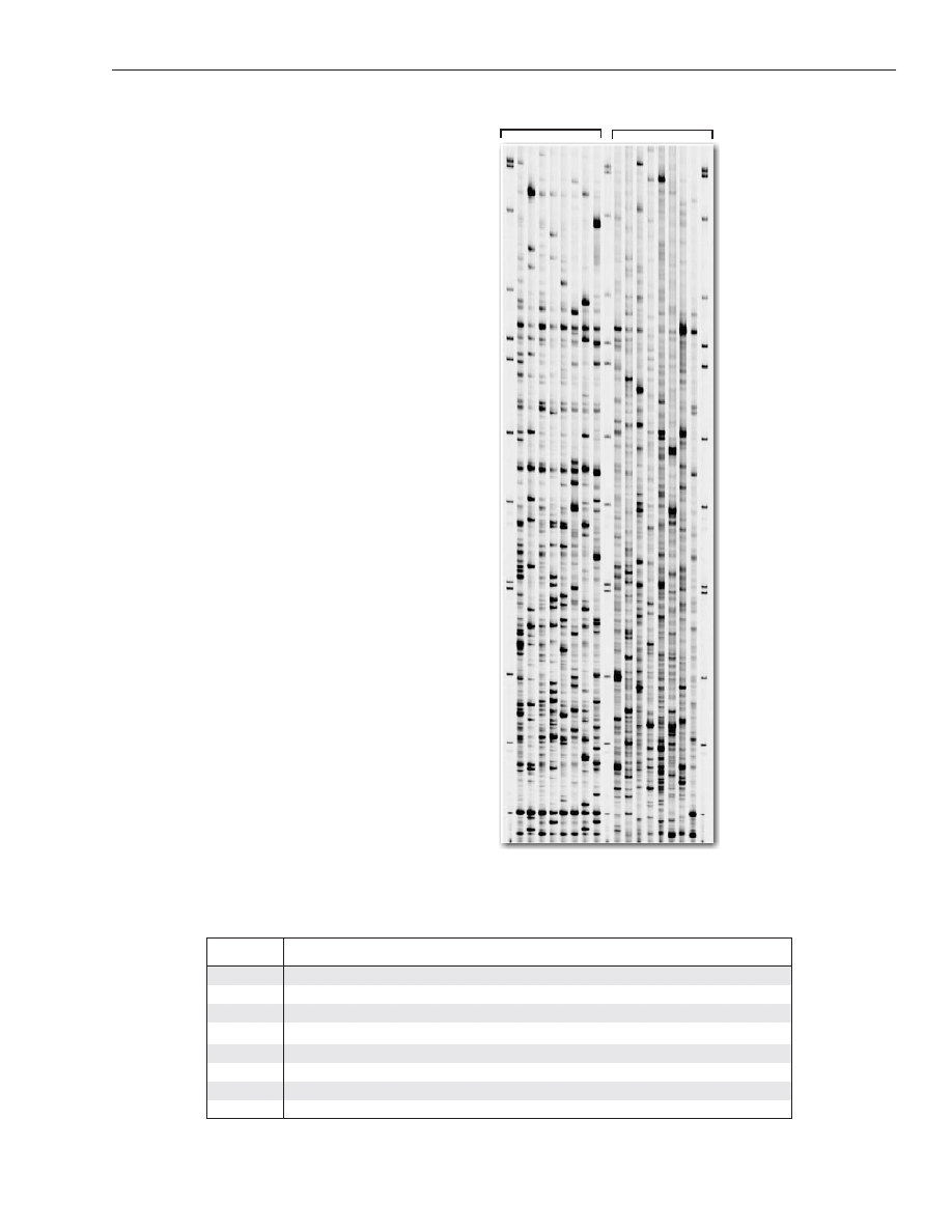
VI. Image Collection and Analysis
AFLP
®
data (TIF images) from IRDye-labeled samples are automatically collected in real time during elec-
trophoresis. Typical AFLP
®
fingerprints of control maize DNA, generated by several different EcoR1/Mse1
primer combinations, are shown in Figure 2.
It is strongly recommended that you perform an AFLP
®
analysis using the control maize DNA samples and
the protocols provided. Compare your images to Figure 2 prior to conducting other AFLP
®
analyses. Similar
AFLP
®
banding patterns should be obtained. However, slight variations in the number of bands (especially
faint bands) and the intensity of individual bands may be observed due to factors such as different Taq DNA
polymerases, gel matrices, and running buffers.
Image data can be quickly viewed, printed, scored, analyzed, and converted into numerical data files using
SAGA
MX
, or other software. Articles cited earlier in this protocol provide additional guidance.
LI-COR Biosciences
Doc# 988-11159
Page 10
www.licor.com
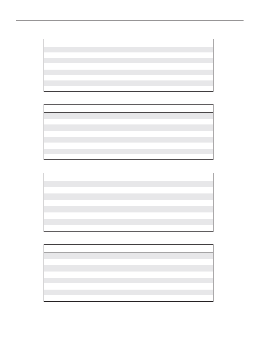
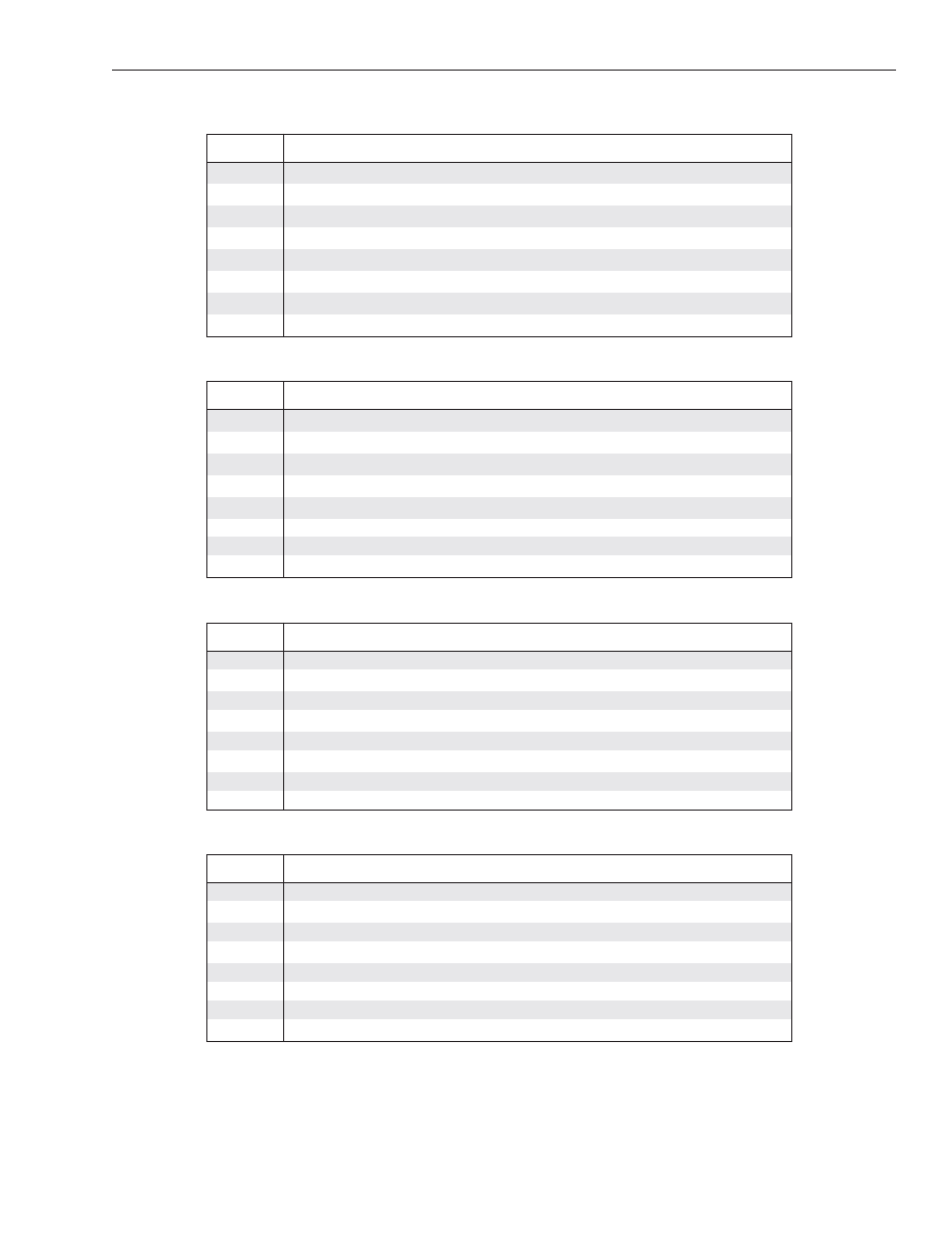
Table 1. Primer pair combinations recommended (
■
) for conducting selective amplifications.
Barley:
M-CAA
M-CAC
M-CAG
M-CAT
M-CTA
M-CTC
M-CTG
M-CTT
E-AAC
■
■
■
■
■
■
■
■
E-AAG
■
■
■
■
■
■
E-ACA
■
■
■
■
■
■
■
■
E-ACC
■
■
■
■
■
■
■
■
E-ACG
■
■
■
■
■
■
■
■
E-ACT
■
■
■
■
■
■
■
E-AGC
■
■
■
■
■
■
■
E-AGG
■
■
■
■
■
■
■
■
Figure 2. AFLP
®
fingerprints of control maize
DNA.
Panel A: AFLP
®
fingerprints of control maize DNA
using IRDye700 labeled EcoRI primer E-AGG
with eight MseI primers: M-CAA (1), M-CAC (2),
M-CAG (3), M-CAT (4), M-CTA (5), M-CTC (6),
M-CTG (7), and M-CTT (8).
Panel B: AFLP
®
fingerprints of control maize DNA
using MseI primer M-CAG with eight IRDye700
labeled EcoRI primers: E-AAC (a), E-AAG (b),
E-ACA (c), E-ACT (d), E-ACC (e), E-ACG (f), E-AGC
(g), and E-AGG (h).
Panel A
Panel B
bp
- 500
- 460
- 400
- 364
- 350
- 300
- 255
- 200
- 145
- 100
- 50
1 2 3 4 5 6 7 8
b c d e f g h
a
IRDye
®
Fluorescent AFLP
®
Protocol for Large Plant Genome Analysis
Doc# 988-11159
Page 11
www.licor.com
Lettuce:
Maize:
Canola:
Pepper:
M-CAA
M-CAC
M-CAG
M-CAT
M-CTA
M-CTC
M-CTG
M-CTT
E-AAC
■
■
■
■
■
■
■
■
E-AAG
■
■
■
■
■
■
E-ACA
■
■
■
■
■
■
■
■
E-ACC
■
■
■
■
■
■
■
■
E-ACG
■
■
■
■
■
■
E-ACT
■
■
■
■
■
■
■
E-AGC
■
■
■
■
■
■
■
■
E-AGG
■
■
■
■
■
■
■
M-CAA
M-CAC
M-CAG
M-CAT
M-CTA
M-CTC
M-CTG
M-CTT
E-AAC
■
■
■
■
■
■
■
E-AAG
■
■
■
■
■
■
■
E-ACA
■
■
■
■
■
■
■
E-ACC
■
■
■
■
■
■
■
■
E-ACG
■
■
■
■
■
■
E-ACT
■
■
■
■
E-AGC
■
■
■
■
■
■
■
E-AGG
■
■
■
■
■
■
■
■
M-CAA
M-CAC
M-CAG
M-CAT
M-CTA
M-CTC
M-CTG
M-CTT
E-AAC
■
■
■
■
■
E-AAG
■
■
■
■
■
E-ACA
■
E-ACC
■
■
■
■
E-ACG
E-ACT
■
■
■
E-AGC
E-AGG
■
■
■
■
■
M-CAA
M-CAC
M-CAG
M-CAT
M-CTA
M-CTC
M-CTG
M-CTT
E-AAC
■
■
■
■
E-AAG
■
■
■
■
■
■
■
E-ACA
■
■
■
■
■
■
■
E-ACC
■
■
■
■
■
■
■
■
E-ACG
■
■
■
■
■
■
■
■
E-ACT
■
■
■
■
■
■
■
E-AGC
■
■
■
■
■
■
■
■
E-AGG
■
■
■
■
■
■
■
LI-COR Biosciences
Doc# 988-11159
Page 12
www.licor.com
Potato:
Sunflower:
Sugar Beet:
Tomato:
M-CAA
M-CAC
M-CAG
M-CAT
M-CTA
M-CTC
M-CTG
M-CTT
E-AAC
■
E-AAG
■
E-ACA
■
■
■
E-ACC
■
■
■
E-ACG
■
■
E-ACT
■
■
■
■
■
E-AGC
■
■
■
■
E-AGG
■
■
■
■
M-CAA
M-CAC
M-CAG
M-CAT
M-CTA
M-CTC
M-CTG
M-CTT
E-AAC
■
■
■
■
■
■
■
E-AAG
■
■
■
■
■
■
E-ACA
■
■
■
■
■
E-ACC
■
■
■
■
■
■
■
■
E-ACG
■
■
■
■
■
■
E-ACT
■
■
■
■
■
■
■
■
E-AGC
■
■
■
■
■
■
■
E-AGG
■
■
■
■
■
■
■
M-CAA
M-CAC
M-CAG
M-CAT
M-CTA
M-CTC
M-CTG
M-CTT
E-AAC
■
■
■
■
■
■
■
■
E-AAG
■
■
■
■
■
■
E-ACA
■
■
■
■
■
■
■
■
E-ACC
■
■
■
■
■
■
■
■
E-ACG
■
■
■
■
■
■
■
■
E-ACT
■
■
■
■
■
■
E-AGC
■
■
■
■
■
■
■
■
E-AGG
■
■
■
■
■
■
■
■
M-CAA
M-CAC
M-CAG
M-CAT
M-CTA
M-CTC
M-CTG
M-CTT
E-AAC
■
■
■
■
■
■
■
■
E-AAG
■
■
■
■
■
■
■
E-ACA
■
■
■
■
■
■
■
■
E-ACC
■
■
■
■
■
■
■
■
E-ACG
■
■
■
■
■
■
■
■
E-ACT
■
■
■
■
■
■
■
■
E-AGC
■
■
■
■
■
■
■
■
E-AGG
■
■
■
■
■
■
■

IRDye
®
Fluorescent AFLP
®
Protocol for Large Plant Genome Analysis
Doc# 988-11159
Page 13
www.licor.com
VII. AFLP
®
Troubleshooting Guide
Troubleshooting AFLP
®
Reactions
Problem
Possible Cause
Solution / Prevention
No bands or weak bands.
IRDye
®
infrared dye-labeled
EcoRI primer(s) not added or
degraded.
Be sure to add EcoRI primer(s) and avoid
exposure of labeled primers to light.
Not enough template DNA.
Be sure to have
≥75 ng of DNA per
restriction digest reaction.
DNA contaminated (e.g., high
salt, EDTA, SDS, or protein).
Extract with phenol/chloroform followed
by ethanol precipitation.
Incorrect PCR conditions.
Verify the cycling program temperature,
cycle number, and time; make sure that
the thermal cycler is operating correctly.
Evaporation during thermal
cycling.
Cover the reactions with mineral oil or
liquid wax; centrifuge briefly before incu-
bation; check that caps fit correctly when
using thermal cyclers with heated lids.
Too few high molecular
weight bands.
Sub-optimal primer pair.
Use suggested primer pair for a given
species (see Table 1).
Sub-optimal quantities of
primer and/or pre-amplified
DNA.
Try a different quantity of IRDye EcoRI
primer (0.3 to 0.8 µl per 11 µl selective
PCR reaction) and a different dilution of
pre-amplified DNA template (1:10 to 1:50).
Many high molecular
weight bands.
Partial digestion.
Purify DNA.
Bands only partway up the
gel.
Poor DNA and/or low poly-
merase activity.
Purify DNA templates and use fresh Taq
DNA polymerase.
Missing lanes (nonspe-
cific, variable).
Evaporation during thermal
cycling.
Cover the reactions with oil or wax; cen-
trifuge briefly before incubation; be sure
that caps fit correctly when using thermal
cyclers with heated lids.
Pipetting error.
Verify addition and mixing of all reaction
components.
Sub-optimal duplexed
AFLP
®
results.
Competition effects of two
labeled EcoRI primers over
one MseI primer.
Choose two labeled EcoRI primers with
similar predicted Tm. If the problem per-
sists, try monoplex PCR first and pool
samples later.

LI-COR Biosciences
Doc# 988-11159
Page 14
www.licor.com
Troubleshooting Gel Images
Problem
Possible Cause
Solution / Prevention
Blurry bands.
Improper gel formation.
Recast gel using fresh solutions and allow
gel to polymerize
≥ 45 minutes.
Smeared bands.
Too much labeled primer(s).
Try less labeled EcoRI primer(s).
Samples not denatured.
Add 5 µl of stop/loading buffer and heat
the sample at 95°C for 3 minutes immedi-
ately before loading gel.
Incorrect electrophoresis
conditions.
For running a 25 cm gel (LI-COR
®
6.5%
KB
Plus
), set temperature to 45°C, voltage
to 1500- 2000 volts, current to 40 mA,
power to 40 W, and scan speed to 2
(Model 4300).
Differences between gel and
running buffer.
Verify that the buffer in the gel and the
running buffer are the same concentration
(1X TBE).
Wavy bands.
The gel surface did not poly-
merize evenly.
Recast a gel and make sure that wells are
free of excessive urea and unpolymerized
gel solution.
Wells are not formed
properly.
Make sure to pull a rectangular-tooth
comb straight out carefully.
Binding silane not used in gel
preparation.
Apply binding silane to front plate before
casting gel.
Air bubbles in gel.
Recast gel, tap gel apparatus to ensure
smooth flow of gel.
Smiling gels.
Uneven gel thickness.
Do not over-tighten the rails or upper
buffer tank to avoid uneven thickness of
the gel.
Outer lane(s) missing.
Comb is not centered.
Be sure to insert the comb in the center of
the gel.
Improper running buffer.
Be sure to use freshly made buffer (1X
TBE) to perform gel electrophoresis.

Notice to purchaser: Limited License
The AFLP
®
technique is covered by U.S. Patent 6,045,994 and other patents or patent
applications owned by Keygene N.V. This product is sold under license from Keygene N.V.,
The Netherlands. This kit may be used for research purposes, excluding medical research.
Medical research means any and all medical diagnostic, pharmaceutical, and forensic
research in connection with humans, animals, and micro-organisms, including but not
limited to:
1. Diagnosis, detection and analysis of disease and disease predisposition in humans and ani-
mals;
2. Diagnosis and detection of human and animal pathogens;
3. Treatment of disease and disease predisposition in humans and animals;
4. Discovery, development and testing of pharmaceutical drugs, medical devices and medical
diagnostics;
5. Paternity, forensic and identity testing in humans.
No right is granted, by implication or estoppel, to use this kit for any activity other than
research activities for the user’s own benefit, such other activities including, but not limited to,
production activities, commercial activities and any activities for the commercial benefit of
third parties, such as contract research and commercial services of any kind, including,
without limitation, reporting results of the user’s activities for a fee or other commercial
consideration.
4647 Superior Street
• P.O. Box 4000 • Lincoln, Nebraska 68504 USA
Technical Support: 800-645-4260
North America: 800-645-4267
International: 402-467-0700 • 402-467-0819
LI-COR GmbH (Germany, Austria, Switzerland, Czech Republic, Hungary, Slovakia): +49 (0) 6172 17 17 771
LI-COR UK Ltd.: +44 (0) 1223 422104
www.licor.com
LI-COR is an ISO 9001 registered company. © 2010 LI-COR Inc. LI-COR and IRDye are trademarks or registered trademarks of LI-COR, Inc. in the
United States and other countries. AFLP is a registered trademark of Keygene N.V., The Netherlands. The LI-COR DNA Analyzer and IRDye reagents
are covered by U.S. patents, foreign equivalents, and patents pending.
®
Document Outline
Wyszukiwarka
Podobne podstrony:
więcej podobnych podstron