
ORIGINAL ARTICLE
Legg-Calvé-Perthes disease: multipositional power Doppler
sonography of the proximal femoral vascularity
Andrea S. Doria
&
Fabiano G. Cunha
&
Marcelo Modena
&
Rui Maciel
&
Laszlo J. Molnar
&
Carlos Luzo
&
Rahim Moineddin
&
Roberto Guarniero
Received: 1 July 2007 / Revised: 19 November 2007 / Accepted: 5 December 2007 / Published online: 7 February 2008
# Springer-Verlag 2008
Abstract
Background Selection of the most appropriate sonographic
scanning approaches for evaluation of hips can improve the
method efficacy and decrease the scanning time.
Objective To determine the sonographic scanning planes
that best assess the proximal femoral vascularity in
asymptomatic and pathologic hips of children with Legg-
Calvé-Perthes disease (LCPD) and evaluate the frequency
(number of hips with evidence of perfusion) and intensity
(number of color pixels per region) of color pixels
representing superficial cartilaginous and deep transphyseal
vascularity in different anatomic regions of pathologic and
asymptomatic hips using multipositional power Doppler
approaches.
Materials and methods Seven scanning approaches (anterior-
sagittal, anterior-transverse, coronal, adduction, perineal, 30°
and 70° of abduction) were applied in 26 pathologic hips of
26 children with LCPD (age range 3
–11 years) and in 25
contralateral asymptomatic hips. The color Doppler signals
seen within the proximal femur were analyzed both
qualitatively (overall/regional frequency) and quantitatively
(intensity).
Results The coronal (P=0.009) and 30° abduction
(P=0.047) approaches demonstrated a higher frequency of
color pixels in pathologic than in asymptomatic hips. The
anterior-sagittal, 30° abduction, adduction and anterior-
transverse planes performed best of all approaches (P=0.02)
to assess deep transphyseal perfusion. The physis demon-
strated a greater intensity of color signals representing
intraosseous vascularity than other regions in pathologic
hips (P=0.027), as noted with the anterior-sagittal approach.
There was a tendency (P=0.06) towards a greater intensity
of pixels representing cartilaginous vascularity in patholog-
ic hips
’ physes with the coronal approach.
Conclusion Specific sonographic scanning planes are rec-
ommended for assessment of the proximal femoral vascu-
larity of LCPD hips. The physis is the anatomic region that
presents with the greatest intensity of color signals in
pathologic hips.
Keywords Legg-Calvé-Perthes disease .
Power Doppler sonography . Hips . Children . Vascularity
Introduction
Although the normal vascular anatomy of the human
growing femoral head has been extensively investigated in
cadaveric studies by Trueta (46 specimens) [
], Lauritzen (6
specimens) [
] and Chung (150
specimens) [
], the literature on the vascularity of normal
and pathologic hips assessed with noninvasive in vivo
imaging techniques such as ultrasonography is scarce.
Pediatr Radiol (2008) 38:392
–402
DOI 10.1007/s00247-007-0726-4
A. S. Doria
:
L. J. Molnar
Heart Institute, Diagnostic, Imaging,
Hospital das Clinicas FMUSP,
Sao Paulo, Brazil
F. G. Cunha
:
M. Modena
:
R. Maciel
:
C. Luzo
:
R. Guarniero
Department of Orthopedic Surgery, Hospital das Clinicas FMUSP,
Sao Paulo, Brazil
R. Moineddin
Department of Public Health, University of Toronto,
Toronto, Canada
Permanent address:
A. S. Doria (
*)
Department of Diagnostic Imaging,
The Hospital for Sick Children,
555 University Ave,
Toronto M5G1X8, Canada
e-mail: andrea.doria@sickkids.ca

Previous studies have demonstrated the presence of
cartilaginous vascular canals that arise in the normal
epiphysis and course towards the metaphysis traversing
the growing physis [
]. These normal vascular canals
enhance after intravenous administration of gadolinium
[
] and are well evaluated with MRI [
]. However, an
MRI examination requires sedation in young children.
Sonography arises as an appealing imaging modality for
assessment of children
’s joints because it does not involve
radiation and does not require sedation. Previous authors
[
] have reported the ability of power Doppler
sonography to detect vascularity within cartilaginous
canals of normal neonatal proximal femoral chondroe-
piphyses. Previous Doppler sonography studies [
have investigated the normal and pathologic proximal
femoral vascularity in growing joints after the neonatal
period. However, no previous investigation has been
conducted to evaluate the effect of different sonographic
scanning approaches on the ability to depict proximal
femoral vascularity.
The major blood supply to the femoral head arises from
the ascending cervical arteries (branches of the lateral
circumflex arteries) that run subsynovially along the
femoral neck. A secondary blood source is the medial
circumflex artery, which partially supplies the femoral head
and neck [
]. Legg-Calvé-Perthes disease (LCPD) is an
avascular necrosis of the proximal growing femoral
epiphysis [
] that is likely caused by an interruption of
the blood supply from the retinacular arteries, branches of
the ascending cervical arteries, to the proximal femoral
epiphysis, with associated decreased flow in the medial
circumflex artery [
]. Recognizing the scanning
approaches that best identify the healing-related neovascu-
larity in pathologic hips that sustained prior local ischemia
and the normal vascularity in asymptomatic hips is crucial
for further sonographic investigation of the vascularity of
the growing proximal femur. Although power Doppler
sonography might not be able to depict the vascularity
arising from the medial circumflex artery (posterior access)
we hypothesized that it could identify the deep transphyseal
flow as a result of healing in pathologic hips of patients
with LCPD.
The purpose of our study was twofold: (1) to determine
the scanning planes that provided best qualitative and
quantitative assessment of the proximal femoral vascularity
(overall and regionally) in asymptomatic and pathologic
hips of children with LCPD using multipositional power
Doppler sonography; and (2) to evaluate the frequency
(number of hips with evidence of perfusion) and intensity
(number of color pixels per region) of cartilaginous and
deep transphyseal vascularity in different anatomic regions
of pathologic and asymptomatic hips using multipositional
approaches.
Materials and methods
Patients
This study was approved by the Research Ethics Com-
mittee of our institution and informed consent was
obtained from the parents of all patients. Children with
LCPD who agreed to participate in the study and who had
not undergone prior surgery of their hips were included in
the study. The diagnosis of LCPD was made based on
clinical (limping hip with pain at mobilization) and radio-
graphic findings. Of 26 patients included, 18 (69.2%) also
underwent scintigraphy within a short time (range 0
–
11 days) from the power Doppler examination, which
ensured that the observations on sonography represented
real changes related to the revascularization process of the
disease as noted by analysis of the images according to
the Conway
’s patterns of neovascularization/revasculariza-
tion of the femoral head in LCPD [
In 26 children with LCPD (18 boys, 8 girls; age 3.1
–
11.5 years, mean 7.1 years), 26 consecutive symptomatic
hips (11 left, 15 right) were evaluated. Of the 26 children,
15 (54%) were younger than 7 years and 12 (46%) were
7 years or older. Twenty five children had unilateral disease
and one had bilateral disease. In the patient with bilateral
disease only the right hip, which had not undergone prior
surgery, was included in the study. The duration of symptom-
atology ranged from 1 month to 5.5 years (mean 12.8 months).
The 25 contralateral asymptomatic hips (14 left, 11 right)
of the children included in the study were also assessed.
Definitions
Superficial cartilaginous vascularity is characterized by
power Doppler signals visualized externally to the cortical
bone of the secondary ossification center of the proximal
femoral epiphysis. These signals represent vascular canals
that course through the epiphyseal and superficial physeal
cartilage of the femoral head (Fig.
Deep transphyseal perfusion is represented by power
Doppler signals visualized along the physis in the vicinity
of the epiphyseal secondary ossification center and meta-
physis (Fig.
Sonographic image acquisition
Whenever possible, unenhanced power Doppler sonograms
of the hips were obtained in the supine position using seven
scanning approaches (Fig.
): (1) anterior-sagittal (25
asymptomatic hips, 26 pathologic hips), with the hips in
neutral position (extension and slight external rotation), and
the transducer placed along the longitudinal axis of the
femoral shaft; (2) anterior-transverse (25 asymptomatic
Pediatr Radiol (2008) 38:392
–402
393
hips, 26 pathologic hips), with the transducer placed
perpendicular to the plane of the anterior-sagittal approach;
(3) coronal (25 asymptomatic hips, 26 pathologic hips),
with the transducer placed on the lateral side of the patient
’s
hip angled at 90° in relation to the plane of the anterior-
sagittal approach; (4) adduction (16 asymptomatic hips, 16
pathologic hips), with the probe placed on the same
position as for the sagittal-anterior approach but with the
hip in slight internal rotation and the knee crossing over the
contralateral joint; (5) perineal (19 asymptomatic hips, 17
pathologic hips), with the probe placed on the internal
aspect of the thigh at the level of the femoral head,
immediately lateral to the perineum area, with the hip
abducted at 45° in relation to a sagittal line crossing the
central aspect of the body; (6) hip at 30° abduction in
relation to the aforementioned imaginary line (22 asymp-
tomatic hips, and 22 pathologic hips), with the probe placed
on the same position as for the sagittal-anterior approach;
(7) hip at 70° abduction (15 asymptomatic hips, 12
pathologic hips) in relation to the imaginary line, with the
probe placed on the same position as for the sagittal-
anterior approach. Standardized foam pads were used to
abduct the patients
’ thighs at preestablished angles for the
sake of reproducibility of measurements. All efforts were
made to avoid causing any discomfort or pain to the
patients during the examinations. Specific scanning
approaches were not performed or immediately discontin-
ued if they caused any discomfort or pain to the patient.
The sonographic examinations were performed with an
HDI 5000 scanner (Advanced Technology Laboratories,
Bothell, WA) by the same operator, who was unaware of
the physiologic status of the hips (pathologic vs. asymp-
tomatic), using a 10-MHz linear-array transducer. Imaging
depth was adjusted to the body habitus of each patient in
order to provide full view of the patient
’s femoral head.
Otherwise identical technical parameters (medium filter,
pulse repetition frequency 700 Hz, and 79% color gain
settings) were used for all examinations. Doppler sono-
graphic examination was limited to a maximum period of
5 min per approach for each hip. Only vessels that could be
identified within the established time frame and for which a
pulsed Doppler waveform could be obtained were consid-
ered for evaluation.
Sonographic imaging analysis
Qualitative assessment Two radiologists (A.S.D. and L.J.
M.) graded the presence or absence of color pixel signals
within the proximal femur (binary response) with regard to:
1. Overall assessment of color Doppler signal in the
proximal femur
2. Region of the proximal femur: epiphysis, physis or
metaphysis (Fig.
)
3. Type of vascularity: superficial cartilaginous or deep
physeal (Fig.
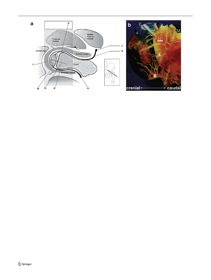
Fig. 1 Anatomy of the arterial blood supply to the proximal femur in
relation to the secondary ossification center of the proximal epiphysis,
physis and metaphysis. a Cross section of the proximal femur at the
level of the joint capsule: a superficial cartilaginous vascularity
(visualized on color Doppler sonography); b anterior ascending cervical
artery; c lateral circumflex artery; d posterior ascending cervical artery;
e medial circumflex artery; f epiphysis; g physis (where deep
transphyseal perfusion can be identified in pathologic hips); h meta-
physis (Drawing modified with permission from reference 15). b
Superficial cartilaginous vascularity extending externally to the cortical
bone at the level of the metaphysis and physis of the proximal femur.
Normal vascularity arising from the intracapsular subsynovial arterial
ring in the femoral head of a cadaveric specimen of a 9-month-old girl.
The anterior half of the specimen, perfused with barium sulfate and
shown in the coronal plan, demonstrates the intracapsular subsynovial
ring (white arrows) with branches of the anterior ascending cervical
arteries (B) crossing through the periphery of the cartilaginous
epiphysis and other sets of blood vessels supplying different areas of
the ossification center (black arrows, A). E epiphysis; P physis; M
metaphysis (reprinted with permission from reference
)
394
Pediatr Radiol (2008) 38:392
–402
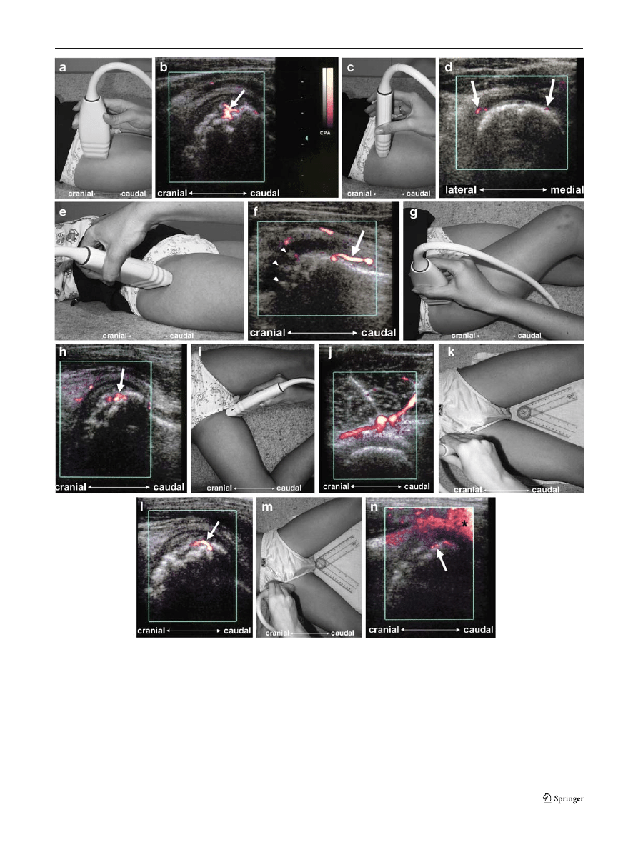
Fig. 2 a, c, e, g, i, k, m Photographs obtained during sonographic
scanning in children in the supine position using the anterior-sagittal
(a), anterior-transverse (c), lateral-sagittal (e), adduction (g), perineal
(i), 30° of abduction (k), and 70° of abduction (m) approaches. b, d, f,
h, j, l, n Power Doppler signals in the pathologic hip of an 8-year-old
boy using the anterior-sagittal (b), anterior-transverse (d), [coronal]
(f), adduction (h), perineal (j), 30° of abduction (l), and 70° of
abduction (n) scanning approaches. h, l, n Color pixels represent
vessels located within the fragmented femoral head (arrows) of
affected hips. d, f Power Doppler signals visualized externally to the
bone contour within the ossifying cartilage of the femoral head and
along the external contour of the proximal femoral metaphysis are
shown using the anterior-transverse (d) and coronal (f) approaches
(arrows). f, n Note cartilaginous vascular canals seen with the coronal
plane (f arrowheads) and a flash artefact from the femoral artery
(asterisk) with the 70° abduction approach (n)
Pediatr Radiol (2008) 38:392
–402
395

Power Doppler signals could be identified either within a
single region or within multiple regions of the proximal
femur. The readers reviewed the images on both digital
format and on videotapes. They were blinded to the clinical
history of the patients; however, the pathologic status of the
hips could be assumed in some patients based on the
characteristic imaging features of LCPD on gray-scale US
imaging. The two readers reached consensus as to the
presence or absence of color pixels in specific anatomic
regions for discordant cases (3/26, 11.5%, overall analysis).
Quantitative assessment The overall and regional quantita-
tive assessment of the superficial cartilaginous and deep
transphyseal proximal femoral vascularity was performed
by counting the number of color pixels spread through the
three regions of the proximal femur. The images obtained
were printed as hard copies and digitalized by an HR5-PRO
color scanner (Kye Systems, Taipei, Taiwan) using 200 dpi,
with 100% image size. The region-of-interest (ROI) of the
sonographic image measured 7.5×9.5 cm (width×height)
and was saved in a JPG image file format. The images were
transferred to a PC and analyzed using Photoshop 5.0
(Adobe Systems, Mountain View, CA). On completion of
the image analysis, the data were matched to each patient
’s
clinical information and the sonographic scanning approach
used.
Statistical analysis
The number of hips with evidence of perfusion (frequency)
presenting with color pixels that represented superficial
cartilaginous or deep transphyseal perfusion was determined
according to different sonographic scanning approaches,
pathologic or asymptomatic status of the hip joint, and
anatomic region of the hip using Fisher
’s exact test. The
numbers of color pixels per region (intensity) identified
in different anatomic regions of pathologic and asymp-
tomatic hips were compared using two-tailed Student
’s
t-tests, 95% confidence intervals or one-factor analysis of
variance, as appropriate. To assess the ability to detect
pixels of the sonographic scanning approaches frequencies
were grouped using the Duncan test. P values less than
0.05 were considered significant.
Results
Superficial cartilaginous vascularity of proximal femur
Overall assessment of frequency of color pixels
Table
shows the results of the overall frequency (quali-
tative assessment) and intensity (quantitative assessment) of
Table 1 Comparison of overall frequency (qualitative assessment) and intensity (quantitative assessment) of color pixels representing superficial
proximal femoral cartilaginous vascularity between pathologic and asymptomatic hips
Scanning approach
Hip
Frequency
Intensity
No. of hips
No. (%) of hips
with signal
P value
a
No. of hips
No. of color
pixels per region
(mean±SD)
P value
b
Anterior-sagittal
Pathologic
26
12 (46.1)
0.07
12
479±401
0.5
Asymptomatic
25
5 (20)
5
461±278
Anterior-transverse
Pathologic
26
7 (26.9)
0.29
7
571±250
0.35
Asymptomatic
25
3 (12)
3
354±113
Coronal
Pathologic
26
11 (42.3)
0.009
11
481±320
0.24
Asymptomatic
25
2 (8)
2
266±50
Adduction
Pathologic
16
2 (12.5)
1.0
2
392±170
0.67
Asymptomatic
16
2 (12.5)
2
321±99
30° abduction
Pathologic
23
10 (43.5)
0.047
10
489±265
0.09
Asymptomatic
22
3 (13.6)
3
332±58
70° abduction
Pathologic
10
0 (0)
1.0
–
c
Asymptomatic
10
0 (0)
Perineal
Pathologic
10
1 (10)
1.0
–
c
Asymptomatic
10
1 (10)
a
Fisher
’s exact test.
b
Two-tailed Student
’s t-test.
c
Quantitative assessment of color pixels identified with the perineal and 70° abduction approaches are not included because insufficient data were
obtained.
396
Pediatr Radiol (2008) 38:392
–402
color pixels representing the superficial proximal femoral
cartilaginous vascularity in pathologic and asymptomatic
hips. Both the coronal and 30° abduction approaches
demonstrated a greater frequency of color pixels in patho-
logic hips as compared with asymptomatic hips (P=0.009
coronal approach; P=0.047 30° abduction approach) with a
tendency towards the anterior-sagittal approach also show-
ing a difference in the frequency of color pixels between
pathologic and asymptomatic hips (P=0.07). The 30°
abduction approach demonstrated superficial proximal
femoral cartilaginous vascularity in 10 of 23 pathologic
hips (43.5%), the coronal approach in 11 of 26 pathologic
hips (42.3%) and the anterior-sagittal approach in 12 of 26
pathologic hips (46.1%). No significant differences in the
intensity of color pixels were found between pathologic and
asymptomatic hips with any of the approaches (Table
).
With regard to the frequency of detection of proximal
femoral vascularity according to patient age group, no
differences in the frequency of color pixels identified in the
proximal femora of children younger than 7 years or 7 years
or older were noted with any of the sonographic scanning
approaches. Although not reaching statistical significance,
superficial cartilaginous vascularity pixels tended to be
depicted more frequently in children less than 7 years of
age than in older children with the coronal approach in
pathologic hips (P=0.08) and with the adduction approach
in asymptomatic hips (P=0.07).
Regional assessment of intensity of color pixels
No definite color signals were identified within the epiphyseal
cartilage of asymptomatic hips. The anterior-sagittal approach
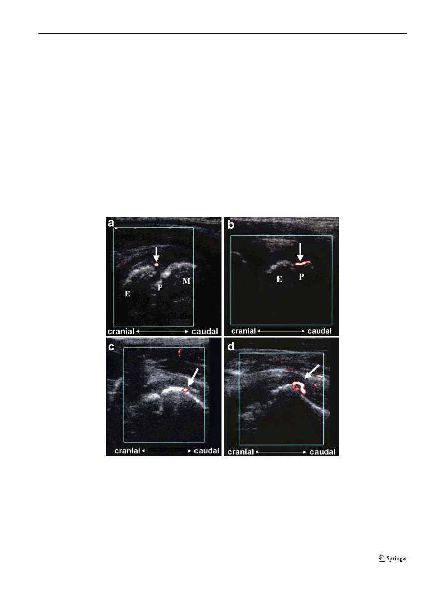
Fig. 3 Superficial cartilaginous vascularity. a Intracapsular vascularity
in the affected hip of a 5-year-old boy demonstrated with power
Doppler US: a vessel runs parallel to the bone contour and passes
through the peripheral perichondrial fibrocartilaginous complex at the
physis (arrow), going from the bone metaphysis into the cartilaginous
epiphysis (E epiphysis, P physis, M metaphysis). Anterior-sagittal
approach. b Intracapsular vascularity in the asymptomatic hip of a 3½-
year-old boy seen with power Doppler US: Doppler signal is identified
within the superficial physeal cartilage externally to the bone surface
(arrow) (E epiphysis, P physis). Anterior-sagittal approach. c
Vascularity of the affected hip of a 10-year-old girl shown with power
Doppler US: a tortuous vessel (arrow) is identified crossing the bone
contour towards the inner aspect of the metaphysis. Note the anatomic
contiguity between the subsynovial intracapsular ring and the intra-
osseous vascularity at the metaphysis level. Anterior-sagittal approach.
d Vascularity of the normal hip in a 5-year-old boy demonstrated by
power Doppler US: single signal (arrow) is visualized on the cortical
bone of the lower region (metaphysis) of the femoral head, likely
representing a metaphyseal branch of an ascending cervical artery.
Anterior-sagittal approach
Pediatr Radiol (2008) 38:392
–402
397

Table 2 Comparison of regional frequency and intensity of color pixels representing superficial cartilaginous vascularity in the epiphysis, physis
and metaphysis of proximal femora affected by LCPD and in asymptomatic hips using various power Doppler sonography scanning approaches
Scanning approach
Hip
Region
Frequency
Intensity
No. of hips
No. of regions
with signal
P value
a
No. of regions
with signal
No. of color
pixels per region
(mean±SD)
P value
b
Anterior-sagittal
Pathologic
Epiphysis
26
1
0.02
1
175
–
Physis
26
8
8
569±459
Metaphysis
26
3
3
343±20
Asymptomatic
Epiphysis
25
0
0
0
Physis
25
4
4
343±101
Metaphysis
25
1
1
933
Anterior-transverse
Pathologic
Epiphysis
26
1
0.19
1
798
–
Physis
26
5
5
566±265
Metaphysis
26
1
1
368
Asymptomatic
Epiphysis
25
0
0
0
Physis
25
2
2
392±129
Metaphysis
25
1
1
279
Coronal
Pathologic
Epiphysis
26
2
0.25
2
217±17
0.06
Physis
26
6
6
568±146
Metaphysis
26
3
3
484±313
Asymptomatic
Epiphysis
25
0
0
0
Physis
25
2
2
266±50
Metaphysis
25
0
0
0
Adduction
Pathologic
Epiphysis
16
0
1.0
0
0
–
Physis
16
1
1
512
Metaphysis
16
1
1
272
Asymptomatic
Epiphysis
16
0
0
0
Physis
16
2
2
321±99
Metaphysis
16
0
0
0
30° abduction
Pathologic
Epiphysis
23
1
0.1
1
413
–
Physis
23
6
6
469±257
Metaphysis
23
3
3
554±370
Asymptomatic
Epiphysis
22
0
0
0
Physis
22
3
3
332±58
Metaphysis
22
0
0
0
70° abduction
Pathologic
Epiphysis
10
0
–
0
0
–
Physis
10
0
0
0
Metaphysis
10
0
0
0
Asymptomatic
Epiphysis
10
0
0
0
Physis
10
0
0
0
Metaphysis
10
0
0
0
Perineal
Pathologic
Epiphysis
10
1
1.0
1
235
–
Physis
10
0
0
0
Metaphysis
10
0
0
0
Asymptomatic
Epiphysis
10
0
0
0
Physis
10
0
0
0
Metaphysis
10
1
1
652
No differences in the frequency or intensity of color pixels representing superficial cartilaginous vascularity were noted between the epiphysis and
metaphysis, or between physis and metaphysis of the proximal femur of pathologic hips.
a
Fisher
’s exact test.
b
Two-tailed Student
’s t-test.
398
Pediatr Radiol (2008) 38:392
–402

was able to demonstrate a difference in the frequency of
visualization of color pixels representing superficial cartilag-
inous vascularity in the physis compared with the epiphysis in
pathologic hips (P=0.02; Fig.
; Table
). Although not
reaching statistical significance, there was a tendency
towards the coronal approach showing a greater intensity
of color pixels representing superficial cartilaginous vascu-
larity at the level of the physis (mean number of pixels 568±
146) compared to the epiphysis (mean number of pixels 217±
17) in pathologic hips (P=0.06; Table
). No differences were
noted in the mean number of pixels identified within the
metaphysis and physis of pathologic hips, or within the meta-
physis and epiphysis of pathologic hips with any of the
sonographic scanning approaches (Fig.
; Table
).
Deep transphyseal vascularity of proximal femur
Overall assessment of frequency of color pixels
The proportion of cases where color signals were identified
with the anterior-sagittal (21/26, 80.8%; Table
), 30°
abduction (17/23, 73.9%; Table
), adduction (9/17, 52.3%;
Table
) and anterior-transverse (13/26, 50%; Table
approaches was significantly different from the proportion
of cases identified with the lateral-sagittal (=6/26, 23%;
Table
), 70° abduction (1/10, 10%; Table
) and perineal
(0/10, 0%; Table
) approaches in pathologic hips
(P=0.018). No definite color pixels representing deep
transphyseal perfusion were identified within the proximal
femur of asymptomatic hips. Differences in the frequency
of cases where deep transphyseal vascularity could be
depicted in pathologic and asymptomatic hips were noted
with most of the approaches (Table
). Conversely, there
were no significant differences in the mean number of
pixels representing deep perfusion between the scanning
approaches investigated (Table
).
Regional assessment of frequency and intensity of color
pixels
The anterior-sagittal approach was able to detect differences
in deep transphyseal vascularity between the physis and
epiphysis of pathologic hips in terms of both the frequency
(P=0.01) and the number of color pixels (physis 890±241
vs. epiphysis 262±64, P=0.03; Table
).
Discussion
Qualitatively, the results of our study showed that the
anterior-sagittal, coronal and 30° abduction sonographic
approaches were good planes for assessment of the overall
cartilaginous vascularity in hips affected by LCPD. How-
ever, the sonographic scanning approaches used in this
Table 3 Comparison of overall frequency (qualitative assessment) and intensity (quantitative assessment) of color pixels representing deep
transphyseal proximal femoral vascularity in pathologic and asymptomatic hips
Scanning approach
Hip
Frequency
Intensity
No. of hips
No. (%) of hips
with signal
P value
a
No. of hips
No. of color
pixels per region
(mean±SD)
P value
b
Anterior-sagittal
Pathologic
26
21 (80.8)
<0.05
17
846±916
<0.05
Asymptomatic
25
0 (0)
0
0
Anterior-transverse
Pathologic
26
13 (50)
<0.05
11
364±165
<0.05
Asymptomatic
25
0 (0)
0
0
Coronal
Pathologic
26
6 (23)
<0.05
6
346±154
<0.05
Asymptomatic
25
0 (0)
0
0
Adduction
Pathologic
17
9 (52.3)
<0.05
8
422±143
<0.05
Asymptomatic
17
0 (0)
0
0
30° abduction
Pathologic
23
17 (73.9)
<0.05
13
831±959
<0.05
Asymptomatic
22
0 (0)
0
0
70° abduction
Pathologic
10
1 (10)
–
2
1130±746
<0.05
Asymptomatic
22
0 (0)
0
0
Perineal
Pathologic
10
0 (0)
–
2
947±1272
<0.05
Asymptomatic
22
0 (0)
0
0
a
Fisher
’s exact test.
b
P values based on 95% confidence intervals for frequency and intensity of color pixels. The 95% confidence intervals of none of the mean
intensity values for pathologic hips included a value of zero, indicating that the mean values of color pixels identified in pathologic and
asymptomatic hips are different. One-factor ANOVA comparing the pixel intensities across the different scanning approaches for pathologic hips
failed to reveal any differences (P=0.93).
Pediatr Radiol (2008) 38:392
–402
399

study were not able to identify vascularity within the
epiphyseal cartilage of asymptomatic hips. We hypothesize
that a number of the vessels present in the normal
epiphyseal cartilage of children with ages ranging from 3
to 11 years may not be prominent enough to be depicted
sonographically. This contrasts with the vascularity present
in the normal physeal cartilage or within the pathologic
epiphyseal cartilage in the same age group of patients with
LCPD, which can be identified with power Doppler
sonography.
From an anatomic point of view, although the articular
cartilage receives its nourishment from synovial fluid and
subchondral vessels, numerous vascular canals course
through the epiphyseal and physeal cartilage to provide
nourishment to the ossifying cartilage [
]. In ischemic
processes such as LCPD we would expect these vascular
canals to enlarge in order to provide better nourishment to
the ossifying cartilage. The observations of this study
support this hypothesis.
Previous authors [
] used the anterior-sagittal plane to
evaluate to the best advantage the vascularity of the anterior
ascending cervical arteries both in pathologic and asymp-
tomatic hips. On the basis of findings of this study, other
planes also enable visualization of cartilaginous flow in the
proximal femur. The lateral (coronal) approach is useful in
the depiction of vascular signals that come through the
notch between the greater trochanter and the lateral aspect
of the femoral neck, where the vascularity is expected to be
prominent in pathologic hips. However, the greater tro-
chanter itself is a barrier to the passage of the ultrasonic
beam, preventing proper evaluation of the branches of the
ascending cervical arteries in some circumstances.
Theoretically, whereas abduction tends to decrease the
area of exposure of the femoral head while it moves toward
the acetabulum, adduction of the hip makes the femoral
head rotate in relation to the acetabulum, offering a better
surface to insonate, which theoretically would help detec-
tion of the proximal femoral vascularity. However, on the
basis of the findings of this study, the adduction approach
did not improve identification of color pixels when
compared to other approaches. The perineal and 70°
abduction approaches were not useful for assessment of
Table 4 Comparison of regional intensity of color pixels representing deep transphyseal vascularity in the epiphysis, physis and metaphysis of
proximal femora affected by LCPD using various power Doppler sonography scanning approaches
Scanning approach
Region
Frequency
Intensity
No. of hips
No. of regions
with signal
P value
a
No. of regions
with signal
No. of color
pixels per region
(mean±SD)
P value
b
Anterior-sagittal
Epiphysis
26
3
0.01
3
262±111
0.03
Physis
26
12
12
890±835
Metaphysis
26
6
6
1051±1247
Anterior-transverse
Epiphysis
26
5
1.0
5
376±208
0.96
Physis
26
6
6
380±143
Metaphysis
26
2
2
286±190
Coronal
Epiphysis
26
1
0.35
1
264
Physis
26
4
4
382±186
Metaphysis
26
1
1
286
Adduction
Epiphysis
17
1
0.09
1
569
Physis
17
6
6
375±155
Metaphysis
17
2
2
492±11
30° abduction
Epiphysis
23
5
0.34
5
317±303
0.09
Physis
23
9
9
1101±1190
Metaphysis
23
3
3
881±716
70° abduction
Epiphysis
10
1
–
–
c
–
c
–
Physis
10
1
Metaphysis
10
1
Perineal
Epiphysis
10
1
–
–
c
–
c
–
Physis
10
1
Metaphysis
10
1
No differences in the frequency or intensity of color pixels representing deep transphyseal vascularity were noted between epiphysis and
metaphysis, or between physis and metaphysis of the proximal femur of pathologic hips.
a
Fisher
’s exact test.
b
Two-tailed Student
’s t-test.
c
Quantitative assessment of color pixels identified with the perineal and 70° abduction approaches are not included because insufficient data were
obtained.
400
Pediatr Radiol (2008) 38:392
–402

deep transphyseal vascularity. Nevertheless, the ischemic
effect of hyperabduction of the femoral chondroepiphysis,
which has been previously reported [
,
] in the
context of hip dysplasia, is not something that clinicians are
concerned about during therapy of LCPD. With the perineal
approach many soft-tissue planes interpose between the
probe and the femoral head, which limits the use of this
approach for Doppler sonographic assessment of the
vascularity of the hip. The 70° abduction approach impedes
visualization of the proximal femoral vascularity and results
in greater exposure of the femoral artery, which can produce
artefacts that hinder proper evaluation of vascularity.
Previous studies [
] have shown that the cartilaginous
canals concentrate in the vicinity of the physeal region. The
anterior-sagittal approach was able to demonstrate a greater
intensity of color pixels representing deep transphyseal
perfusion within the physis of affected hips. There was also a
tendency towards color pixels representing superficial carti-
laginous vessels being more prominent in the physeal region
of affected hips, as noted with the coronal approach. These
results confirm the findings of a previous MRI investigation [
].
The major limitation of this study was its observational
design, which lacked a reference standard measure that
could confirm the accuracy of the data. We used the
presence of Doppler tracing for a given color pixel
representing vascularity as a marker of real blood flow
rather than motion artefacts. Although branches of the
lateral femoral circumflex artery, including the anterior
ascending cervical arteries, comprise the anterior portion of
the intracapsular arterial ring of the hip, branches of the
medial femoral circumflex artery comprise the posterior
portion of the intracapsular arterial ring [
]. The latter
vessels penetrate the proximal femur posteriorly and
therefore are more difficult to visualize using anterior
sonographic approaches [
]. However, given the lack of
direct comparison between color Doppler pixels and a
corresponding reference standard method we were unable
to differentiate color pixels related to branches of the lateral
or medial femoral circumflex arteries. Also, we should
consider that multiple tests of hypotheses were performed
in the statistical analysis of this study, which could have led
to the significant associations and trends seen. A much
larger sample size would have been required to test
hypotheses for seven different scanning approaches if we
had planned to control for the problem of multiple testing
using a post-hoc test such as the Bonferroni correction test.
In this case, a P value of at least 0.007 would have been
required for a test to be considered statistically significant.
Power Doppler hip sonography is an imaging technique
that holds potential value for follow-up of patients with
LCPD and evaluation of the extent of injuries to the
cartilage in musculoskeletal trauma as further information
in the field accumulates. Overall results of our study show
that the superficial cartilaginous vascularity and intraoss-
eous/deep transphyseal vascularity of pathologic hips can
be more effectively evaluated using specific sonographic
scanning approaches, notably the 30° abduction plane. This
sonographic plane is effective for assessment of both
superficial cartilaginous vascularity and intraosseous/deep
transphyseal vascularity. The physis was the anatomic
region of the proximal femur that presented with greater
intensity of color signals representing deep transphyseal
vascularity in pathologic hips, as noted with the anterior-
sagittal approach. The intensity of pixels was greater in
affected hips compared with asymptomatic hips. We
recommend the use of specific sonographic scanning planes
to assess the proximal femoral vascularity of LCPD hips.
Selection of appropriate scanning approaches for evaluation
of hips can improve the efficacy of the method and
decrease the scanning time.
Acknowledgement
We would like to thank Dr. Robert B. Salter for
his valuable suggestions and comments and Luke Itani for the
graphical design for Fig.
References
1. Trueta J (1957) The normal vascular anatomy of the human
femoral head during growth. J Bone Joint Surg Br 39B:358
–
394
2. Lauritzen J (1974) The arterial supply to the femoral head in
children. Acta Orthop Scand 45:724
–736
3. Ogden JA (1974) Changing patterns of proximal femoral
vascularity. J Bone Joint Surg Am 56A:941
–950
4. Chung SMK (1976) The arterial supply of the developing
proximal end of the human femur. J Bone Joint Surg Am
58A:961
–970
5. Wilsman N, Van Sickle D (1970) The relationship of cartilage
canals to the initial osteogenesis of secondary centers of
ossification. Anat Rec 168:381
–392
6. Wilsman N, Van Sickle D (1972) Cartilage canals, their
morphology and distribution. Anat Rec 173:79
–94
7. Barnewolt CE, Shapiro F, Jaramillo D (1997) Normal gadolini-
um-enhanced MR images of the developing appendicular
skeleton: part I. Cartilaginous epiphysis and physis. AJR 169:
183
–189
8. Bearcroft PW, Berman LH, Robinson AHN et al (1996)
Vascularity of the neonatal femoral head: in vivo demonstration
with power Doppler US. Radiology 200:209
–211
9. Keller MS (1996) Sonographic detection of femoral head
vascularity in neonates. Radiology 200:28
–29
10. Amodio J, Rivera R, Prinkney L et al (2006) The relationship
between alpha angle and resistive index of the femoral epiphysis
in the normal and abnormal infant hip. Pediatr Radiol 36:841
–
844
Pediatr Radiol (2008) 38:392
–402
401

11. Schwartz DS, Keller MS, Fields JM et al (1998) Arterial
waveforms in the femoral heads of healthy neonates. AJR
170:465
–466
12. Doria AS, Guarniero R, Molnar LJ et al (2000) Three-dimensional
(3D) contrast-enhanced power Doppler imaging in Legg-Calvé-
Perthes disease. Pediatr Radiol 30:871
–874
13. Doria AS, Guarniero R, Cunha FG et al (2002) Contrast-
enhanced power Doppler sonography: assessment of revascular-
ization flow in Legg-Calvé-Perthes disease. Ultrasound Med Biol
28:171
–182
14. Graif M, Schweitzer ME, Nazarian L et al (1998) Color Doppler
hemodynamic evaluation of flow to normal hip. J Ultrasound Med
17:275
–280
15. Robben SG, Lequin MH, Diepstraten AFM et al (2000) Doppler
sonography of the anterior ascending cervical arteries of the hip.
AJR 174:1629
–1634
16. Jaramillo D, Shapiro F (1998) Growth cartilage
– normal
appearance, variants and abnormalities. MRI Clin North Am
6:455
–471
17. Gruber H, Lachman RS, Rimoin D (1990) Quantitative histology
of cartilage vascular canals in the human rib: findings in normal
neonates and children, and in achondrogenesis II-hypochondro-
genesis. J Anat 173:69
–75
18. Conway JJ (1993) A scintigraphic classification of Legg-Calvé-
Perthes disease. Semin Nucl Med 23:274
–295
19. Salter RB (1999) Textbook of disorders and injuries of the
musculoskeletal system, 3rd edn. Williams & Wilkins, Baltimore,
pp 339
–350
20. Lutfi AM (1970) Mode of growth, fate and functions of cartilage
canals. J Anat 106:135
–145
21. Jaramillo D, Villegas-Medina OL, Doty DK et al (1996)
Gadolinium-enhanced MR imaging demonstrates abduction-
caused hip ischemia and its reversal in piglets. AJR 166:879
–
887
22. Salter RB, Kostuik J, Dallas S (1969) Avascular necrosis of the
femoral head as a complication of treatment for congenital
dislocation of the hip in young children: a clinical and
experimental investigation. Can J Surg 12:44
–61
402
Pediatr Radiol (2008) 38:392
–402
Document Outline
- Legg-Calvé-Perthes disease: multipositional power Doppler sonography of the proximal femoral vascularity
Wyszukiwarka
Podobne podstrony:
Intertrochanteric osteotomy in young adults for sequelae of Legg Calvé Perthes’ disease—a long term
Legg Calvé Perthes Disease in Czech Archaeological Material
Multicenter study for Legg Calvé Perthes disease in Japan
Femoral head vascularisation in Legg Calvé Perthes disease comparison of dynamic gadolinium enhanced
A recurrent mutation in type II collagen gene causes Legg Calvé Perthes disease in a Japanese family
Acute chondrolysis complicating Legg Calvé Perthes disease
Osteochondritis dissecans in association with legg calve perthes disease
Interruption of the blood supply of femoral head an experimental study on the pathogenesis of Legg C
Dynamic gadolinium enhanced subtraction MR imaging – a simple technique for the early diagnosis of L
Modified epiphyseal index for MRI in Legg Calve Perthes disease (LCPD)
Hip Arthroscopy in Legg Calve Perthes Disease
Legg Calve Perthes’ disease
Legg Calve Perthes’ disease
Legg Calve Perthes disease The prognostic significance of the subchondral fracture and a two group c
Computerized gait analysis in Legg Calve´ Perthes disease—Analysis of the frontal plane
Coxa magna quantification using MRI in Legg Calve Perthes disease
Osteochondritis dissecans in association with legg calve perthes disease
Interruption of the blood supply of femoral head an experimental study on the pathogenesis of Legg C
Legg Perthes disease in three siblings, two heterozygous and one homozygous for the factor V Leiden
więcej podobnych podstron