
Impact of opiate addiction on neuroinflammation in HIV
Desiree Byrd
&
Jacinta Murray
&
Gabriella Safdieh
&
Susan Morgello
Received: 13 March 2012 / Revised: 8 June 2012 / Accepted: 8 June 2012 / Published online: 14 July 2012
# Journal of NeuroVirology, Inc. 2012
Abstract To investigate the independent and interactive
effects of opiate addiction and HIV on neuroinflammation,
we measured microglial/macrophage activation and astro-
gliosis in multiple regions of human brain. Samples of
thalamus, frontal gray matter, and frontal white matter were
obtained from 46 individuals categorized as: HIV negatives,
HIV-negative opiate addicts, HIV positives, HIV-positive
opiate addicts, HIV encephalitis (HIVE), and HIVE opiate
addicts. Activated brain microglia/macrophages and astro-
cytosis were quantified by morphometric analysis of immu-
nohistochemical stains for CD68, HLA-D, CD163, and
GFAP. The effects of HIV grouping, opiate addiction, and
their interaction on expression of the markers were exam-
ined in a series of two-way ANOVAs. In opiate addicts,
there was generally higher baseline expression of CD68 and
HLA-D in HIV negatives, and lower expression in HIV and
HIVE, compared to individuals without opiate abuse. Thus,
for these markers, and for GFAP in frontal gray, opiates
were associated with attenuated HIV effect. In contrast, for
CD163, opiates did not significantly alter responses to HIV,
and HIV effects were variably absent in individuals without
opiate abuse. The divergent impact that opiate addiction
displays on these markers may suggest a generally immu-
nosuppressive role in the CNS, with decreased HIV-
associated activation of markers CD68 and HLA-D that
potentially reflect neurotoxic pathways, and preservation
of CD163, thought to be an indicator of neuroprotective
scavenger systems. These results suggest a complex impact
of opiates on neuroinflammation in baseline and virally
stimulated states.
Keywords HIV . Neuroinflammation . Microglia .
Neuropathology . Opiate
Introduction
Significant comorbidity exists between HIV infection and
substance use disorders, such that the two have been con-
sidered
“interlinked epidemics” (Nath et al.
). Abuse of
opiates is a major transmission route, while abuse of other
illicit substances, such as cocaine and methamphetamine,
has become a primary risk factor for HIV infection (CDC
). Literature documenting diverse immunomodulatory
effects of substances of abuse (SOA) has evolved, with
unclear applicability to the progression of HIV or its neuro-
biology. In part, this is because of the complexities of the
D. Byrd
:
J. Murray
:
S. Morgello (
*)
Department of Neurology, The Mount Sinai School of Medicine,
One Gustave L. Levy Place, Box 1137, New York, NY 10029,
USA
e-mail: susan.morgello@mssm.edu
D. Byrd
Department of Psychiatry, The Mount Sinai School of Medicine,
One Gustave L. Levy Place, Box 1137, New York, NY 10029,
USA
G. Safdieh
Department of Pediatrics, NYU Langone Medical Center,
550 First Avenue,
New York, NY 10016, USA
S. Morgello
Department of Neuroscience and Pathology,
The Mount Sinai School of Medicine,
One Gustave L. Levy Place, Box 1137, New York, NY 10029,
USA
J. Neurovirol. (2012) 18:364
–373
DOI 10.1007/s13365-012-0118-x

SOA epidemic, where polysubstance use patterns and med-
ical co-morbidities make it difficult to disentangle indepen-
dent effects in HIV-positive individuals with SOA histories.
Heroin is a common substance of abuse for HIV-positive
persons who reside in NYC, where it is estimated that over
110,000 people are living with HIV (New York City
Department of Health and Mental Hygiene
). One of the
mechanisms postulated for the deleterious interaction of HIV
and SOA in the human brain is immunomodulation (Burdo et
al.
). The immunomodulatory effects of opiates make them
of particular interest to neuroAIDS investigations because of the
brain's inflammatory response to HIV, the fact that these sub-
stances modulate immunity in part via CNS receptor-mediated
pathways, and the significant potential for functional and be-
havioral impact on multiple levels (Friedman et al.
Hauser et al.
). However, it is likely that the interaction
of opiates and local neuroimmune responsiveness to HIV is a
highly complex phenomenon, as these substances also influ-
ence characteristics of the blood
–brain barrier and exert effects
on the peripheral immune cells that traffic into the CNS on a
daily basis (Dhillon et al.
; Friedman et al.
; Seelbach
et al.
). Thus, it is not surprising that there are conflicting
reports of immunomodulatory effects in the brain and its cellu-
lar constituents, with or without the presence of HIV. While
opiates are largely considered immunosuppressive, the few
human brain studies that have been performed on intravenous
heroin users have suggested enhanced microglial activation in
response to HIV (Anthony et al.
; Arango et al.
; Chao
et al.
; Hu et al.
). The immunomodulatory effects of
other substances are even less clear. For example, with regard to
cocaine (generally considered pro-inflammatory), studies of
human brain to date have emphasized its catastrophic alterations
to CNS vasculature that result in intracerebral stroke and hem-
orrhage, but have not elucidated what effect it might have on
intrinsic parenchymal immunomodulation (Aggarwal et al.
; Crawford et al.
; Dhillon et al.
; Hauser et al.
; Tyor and Middaugh
Despite potential CNS vulnerability to SOA-HIV-1 inter-
actions, the present literature describing human brain con-
sequences is limited by very few studies of human brain
tissue with appropriate control groups of HIV negatives.
Additionally, existing data has been compiled on primarily
Caucasian cohorts, limiting generalizability to minorities
who currently comprise the majority of HIV-infected adults
in the USA (CDC 2007). In this regard, tissue available
from the Manhattan HIV Brain Bank presents a unique
opportunity to examine neuroinflammation and SOA in the
context of HIV, as the cohort is predominantly composed of
minority individuals with high rates of substance use disor-
ders. The current study aims to add to the growing body of
literature on SOA
–HIV interactions, by examining microglial
activation in HIV-infected and seronegative substance abusers
and comparing the neuroinflammatory influence of opiate
addiction in a well characterized, primarily American ethnic
minority, urban cohort.
Methods
Patient population
Study subjects were selected from the Manhattan HIV Brain
Bank (MHBB; U01MH083501) in New York City, NY. The
MHBB is a longitudinal, observational study of both HIV-
positive and HIV-negative adults. As part of the subject eval-
uations, psychiatric and substance use histories and basic
laboratory data inclusive of CD4 counts and HIV plasma
loads are recorded. Data obtained from this population were
obtained in compliance with the Mount Sinai School of
Medicine IRB. A total of 46 participants were classified
according to positive or negative status for the following
variables: HIV, HIVencephalitis (HIVE), and history of opiate
(heroin) addiction (administration routes for all but one patient
were intravenous). Histories of opiate addiction were ascer-
tained upon interview or chart review, and in 25 patients,
confirmed with a semi-structured psychiatric interview, the
Psychiatric Research Interview for Substance and Mental
Disorders (Hasin et al.
) and in 21, urine toxicology.
Subjects for this study were a subset of the larger holdings
of the MHBB, and were selected by the following criteria:
appropriate clinical data available for accurate opiate charac-
terization and immunological status (CD4 count) and absence
of opportunistic infections, significant anoxic ischemic dam-
age, and other neuropathologies in the regions of interest that
were known to contribute to changes in the microglial/astro-
glial markers of interest. Once these criteria were met by a
sample, we then attempted to match as closely as possible the
demographic characteristics (age, gender, and race/ethnicity)
of each group, so that there was no significant difference
between the groups in mean age, gender composition, and
race/ethnicity. This resulted in the following distribution:
Group 1 HIV negative; no opiate use syndrome (
n07)
Group 2 HIV negative; opiate addict (
n07)
Group 3 HIV positive; no opiate use syndrome (
n010)
Group 4 HIV positive; opiate addict (
n013)
Group 5 HIV encephalitis; no opiate use syndrome (
n04)
Group 6 HIV encephalitis; opiate addict (
n05)
Males accounted for 67 % of the total sample and average
age was 47 years (8.6). For the participants with HIV, median
CD4 count (cells/mm
3
) was 15.5 (range
01−336) and mean
viral load (log
10
) was 4.39 (1.69). Of note, the groups
were composed almost exclusively of African Americans
(43.5 %) and Hispanics (50 %). Finally, 44 % of the
opiate addicts had documentation of being subsequently
J. Neurovirol. (2012) 18:364
–373
365

maintained on methadone and 36 % were taking or
receiving non-methadone opiates.
Human brain processing
At the time of patient demise, brains were obtained and
routinely processed as has been previously published
(Morgello et al.
). A minimum of 57 routine hematox-
ylin and eosin-stained sections were examined for each
brain. Neuropathologic analysis to diagnose the presence
of HIVE was performed by a board-certified neuropatholo-
gist (SM). With only one exception, the brains used in this
study were free of active opportunistic infection or tumor;
the one exception was an opiate addict with HIVE (group 6),
who had a circumscribed temporal lobe lymphoma which
did not involve the brain regions sampled.
The three regions of interest selected for this study in-
cluded thalamus, frontal gray matter, and frontal white mat-
ter. These regions were selected for their comparability to a
prior study of brain microglial activation in polysubstance-
using Caucasian patients with HIV, and because several of
these regions are sites of predilection for HIV-associated
neuropathologies (Arango et al.
).
Tissue microarray and immunohistochemical staining
Paraffin donor blocks were chosen in the regions of interest
(frontal gray, frontal white, and thalamus), and used to create 12
tissue microarrays (TMAs) (three slides for each region) with
cores of 1.0 mm diameter (Battifora
; Kononen et al.
). This redundancy was to ensure that all 46 patients had
at least two adherent punches in each region. Microarrays were
sectioned at 5
μm, and mounted on coated slides (Fisher super-
frost plus, Fisher Corp) for immunohistochemical staining.
Immunohistochemical staining was performed with four
antibodies. Details of the primary antibodies manufacturer,
clone, and dilutions used are summarized in Table
. After
incubation with the primary antibody, slides were incubated
for 30 min with anti-mouse or anti-rabbit Ig ImmPRESS
reagent (Vector Laboratories, CA) prior to development
with diaminobenzidene chromogen. To minimize run-to-
run variability, all sections were stained with each antibody
in one cycle. Hematoxylin counterstain was applied, to
facilitate identification of the cells of interest.
Morphometric analysis
After immunohistochemical staining, slides were examined
with a Nikon Labphot-2 light microscope. Six successive
fields at ×40 magnification (total area of analysis, 0.03 mm
2
)
were taken from each tissue core with a Coolpix 950 digital
camera attached to the microscope by a Coolpix MDC lens.
The percentage of tissue area occupied by antibody-positive
cells was determined by an automated system developed at the
Mount Sinai School of Medicine (Wu et al.
). Two cores
were analyzed for each antibody in each anatomical region
from each patient, for a total of 12 images. The raw percentage
area measures for positive staining in the 12 images were
recorded in a Microsoft Excel workbook and were averaged
prior to the general analysis.
Validation of TMA as a representation of whole slide IHC
TMAs have the advantage of containing multiple specimens
on a single slide, allowing for more uniformity of immuno-
histochemical staining. Unfortunately, TMAs also reduce the
amount of tissue analyzed. It has been shown for several
common antigen/antibody pairs that two needle cores ade-
quately represent antigen expression on a whole tissue section
with 95 % accuracy; we wished to validate this in our system.
Blocks of deep white matter were chosen from six
MHBB cases. Whole 5-
μM slices were taken from each
block and used in immunohistochemistry using monoclonal
anti-CD68 antibody. The slides were visualized with a light
microscope and 30 random areas of deep white matter were
photographed and used for morphometric analysis. After the
whole sections were cut and stained, a TMA was con-
structed from the residual paraffin blocks. The CD68-
stained slides were used as a guide to select five areas of
deep white matter, which were punched from the blocks
with a 1.0-mm-diameter needle. With six cases represented,
this resulted in a TMA with a total of 30 tissue cores. Five-
micron slices were cut from the TMA block and stained for
CD68 as described. Six images were photographed from
each core and analyzed for area of staining. The data were
organized in Excel and statistical analysis performed.
Pearson's correlation tests were applied to analyze the
number of punches in a TMA that were equivalent to a
standard tissue section in morphometric analysis. Five
Table 1 Antibodies used in staining brain samples
Antigen
Manufacturer
Dilution
CD68
Lysosomal membrane protein, monocyte/ macrophage/microglia
Dako Corp., CA
1:1,000
HLA-D (DP, DQ, DR)
Class 2 major histocompatibility locus
Dako Corp., CA
1:500
CD163
Monocyte/macrophage scavenger receptor, ramified microglia
NovoCastra, UK
1:100
GFAP
Astrocyte intermediate filament
Dako Corp., CA
1:2,500
366
J. Neurovirol. (2012) 18:364
–373

Pearson correlation tests were performed: test 1 used the
average of six area measures from punch 1 against six area
measures generated from the first six images of the slice; test
2 combined 12 area measures generated from punches 1 and
2 against the first 12 measures generated from the slice, and
so on, until the final test was done on 30 images generated
from all five punches against 30 measures generated from
the 30 images of the slice.
Performing the five correlation tests of punch to slide
staining allowed us to determine how many punches were
equivalent to a tissue slice in our quantitative analysis. The
tests revealed that there was an adequate and statistically
significant correlation between two punches and the tissue
slice (
r00.983, p<0.01).
Data analysis
Statistical analyses were performed using SPSS software
(Chicago, IL) version 19.0 for Windows, and repeated using
JMP version 9.0.0 on a Macintosh computer (SAS Institute).
Two-way analysis of variance (ANOVA) was utilized for the
primary analyses of the study. When interaction terms were
significant, follow-up one-way ANOVAs were completed
separately for the opiate and no opiate groups with HIV status
as the independent variable, and post hoc analysis by Tukey's
test. Chi-square analyses were applied to categorical variables.
Independent variables in these analyses included HIV status
(HIV negative, HIV positive, or HIVE) and opiate addiction
history status. The dependent variable was average antibody
staining area for each region of interest. An analysis of the
relationship between microglial/astrocytic cell response and
peripheral immunovirologic status in the HIV and opiate
groups was completed using Spearman's bivariate correla-
tions. Viral loads were log
10
transformed prior to analyses.
Results
Effects of HIV status, opiate addiction, and their interaction,
on neuroimmune and glial markers
To determine the effects of HIV grouping, opiate addiction,
and their interaction on neuroimmune and glial markers, a
series of two-way ANOVAs was performed in each brain
region for each marker. The
p values for the overall models
and interaction terms from these analyses are presented in
Table
. For each opiate group, marker, and region, means
and standard deviations are additionally detailed in Table
along with
p values from simple tests of significance (one-
way ANOVA) for HIV status when overall models indicated
the presence of significant group differences. The effects of
opiate addiction on marker expression in the HIV groups are
depicted in Fig.
.
For CD68, the overall model (two-way ANOVA) dem-
onstrated significant differences in frontal white matter (
F
(5,40)
06.4903, p00.0002) and thalamus (F (5,39)02.6085,
p00.0396), but not frontal gray matter (F (5,40)01.7877,
p00.1375). Significant or trend level interactions between
opiate status (addict, no abuse) and HIV grouping (negative,
positive, and HIVE) were seen for CD68 in frontal white
matter (
p00.0323) and thalamus (p00.0906). In frontal
white matter, for individuals with no opiate abuse, the
HIVE group displayed significantly higher CD68 expression
than HIV-positive and HIV-negative groups (
p<0.0001).
Likewise, in thalamus, there was a stepwise effect of HIV
status in that the HIVE group displayed significantly higher
CD68 expression than HIV positive, who displayed higher
levels than HIV negative (
p00.0645). In contrast to the sig-
nificant or trend level effects of HIV status on CD68 expres-
sion in individuals without history of opiate abuse, in subjects
with opiate addiction, differences in CD68 expression were
not significant in any brain region. For all brain regions, CD68
staining in HIV negatives was greater in individuals with
opiate addiction than in those without; conversely, for all brain
regions, CD68 staining in HIVE positives and HIVE was less
in opiate addicts than in those without. Thus, in the presence
of opiate addiction, baseline levels of CD68 in HIV negatives
were elevated, and the increase of levels in association with
HIV infection was decreased.
For HLA-D, the overall model demonstrated significant
differences in the thalamus (
F (5,39)03.4599, p00.0111)
and trend level differences in frontal gray (
F (5,40)02.2817,
p00.0647), but not frontal white matter (F (5,40)01.3958,
p00.2466). The interaction terms were significant or at
trend level for HLA-D in frontal gray matter (
p00.0716)
and thalamus (
p00.0300), wherein the effect of HIV status
Table 2
p values for two-way
ANOVAs, with mean area
CD68, CD163, HLA-D, and
GFAP staining as outcome, and
HIV group and opiate addiction
status as variables
Significant and trend level
effects are in bold
Region
p value for
CD68
HLA-D
CD163
GFAP
Frontal gray
Overall model
0.1375
0.0647
0.0057
0.0171
HIV × opiate interaction
0.3006
0.0716
0.1326
0.1425
Frontal white
Overall model
0.0002
0.2466
0.0003
0.4187
HIV × opiate interaction
0.0323
0.3853
0.1749
0.6440
Thalamus
Overall model
0.0396
0.0111
0.2058
0.5360
HIV × opiate interaction
0.0906
0.0300
0.8451
0.6852
J. Neurovirol. (2012) 18:364
–373
367
on expression showed a similar attenuation in opiate addicts
as was seen with CD68. Follow-up one-way ANOVAs
revealed that in the absence of opiate abuse, there was a
trend for the HIV-positive group to demonstrate greater
HLA-D staining than the HIV-negative group in frontal
white matter (
F (2,19)03.05, p00.0724). A significant
HIV effect was also observed in the thalamus in the non-
opiate group (
F (2,18)04.30, p00.0308), where post hoc
tests indicated that the HIV positive displayed higher HLA-
D than both the HIV negative and HIVE groups. Differences
in HLA-D expression in opiate addicts were not significant
in any brain region; there was only a trend level effect in
frontal gray matter (
F (2, 23) – 2.87, p00.0783). In this
region, post hoc tests revealed significantly greater HLA-D
staining in the HIVE group than the HIV negatives and HIV
positives.
In contrast to the patterns seen with CD68 and HLA-D,
opiate addiction did not appear to attenuate the response of
CD163 to HIV. For CD163, the overall model demonstrated
significant differences in the frontal gray (
F (5,40)03.8910,
p00.0057) and frontal white matter (F (5,40)06.0320, p0
0.0003), but not thalamus (
F (5,39)01.5207, p00.2058). No
significant or trend level interactions were seen for CD163.
In opiate addicts, the effect of HIV status on CD163 expres-
sion was significant in frontal white matter (
F (2, 23)0
26.85,
p<0.0001) and frontal gray matter (F (2, 23)03.41,
p00.0513). Post hoc tests revealed that the HIVE groups
demonstrated significantly greater CD163 staining than both
the HIV-positive and -negative groups in all regions tested.
In contrast, in the absence of opiate abuse, CD163 did not
show significant HIV effects in frontal white matter, but was
significant in frontal gray matter (
F (2, 19)06.14, p0
0.0093), where both the HIVE and HIV-positive groups
demonstrated significantly greater CD163 staining than the
HIV negatives.
The overall model for GFAP was significant only in
frontal gray matter (
F (5,40)03.1545, p00.0171), where
no interaction effect was observed, but a simple main effect
of HIV status was significant (
F (2, 40)05.660, p00.007).
In the absence of opiate abuse, a significant HIV effect was
observed (
F (2, 19)08.79, p00.0022). Post hoc analyses
revealed that for these subjects without opiate abuse, both
the HIVE and HIV-positive groups demonstrated signifi-
cantly greater GFAP staining than the HIV negatives.
Analyses for subjects with opiate addiction did not reach
statistical significance, but qualitative examination of the
mean staining values demonstrate that the HIVE group
evidenced higher GFAP levels than HIV positive and HIV
negatives (
F (2, 23)02.23, p00.1312).
Thus, in general, individuals with opiate addiction
showed higher mean levels of CD68 and HLA-D staining
in the HIV-negative state, lower CD68 levels in the HIV
positive and HIVE conditions, and lower HLA-D in the
HIV-positive conditions, when contrasted to individuals
without opiate abuse (Table
). This effect was not seen
for CD163, where opiate addicts had generally higher peak
Table 3 Mean area of staining in frontal gray and white matter and thalamus for CD68, CD163, HLA-D, and GFAP
Antigen
Frontal gray matter
Frontal white matter
Thalamus
HIV groups
Opiate addicts
No opiate abuse
Opiate addicts
No opiate abuse
Opiate addicts
No opiate abuse
CD68
HIV neg
0.419 (0.106)
0.314 (0.151)
0.969 (0.273)
0.696 (0.208)
0.835 (0.139)
0.545 (0.263)
HIV pos
0.473 (0.078)
0.659 (0.095)
0.977 (0.200)
1.122 (0.174)
0.842 (0.102)
1.293 (0.204)
HIVE
0.532 (0.126)
0.725 (0.151)
1.564 (0.323)
2.801 (0.276)
0.937 (0.164)
1.437 (0.322)
p value
n.r.
n.r.
0.2843
<0.0001
0.8694
0.0645
HLA-D
HIV neg
0.280 (0.211)
0.167 (0.159)
0.797 (0.287)
0.536 (0.366)
0.839 (0.151)
0.500 (0.364)
HIV pos
0.338 (0.155)
0.676 (0.133)
1.148 (0.210)
1.530 (0.307)
0.940 (0.111)
1.736 (0.282)
HIVE
0.984 (0.250)
0.410 (0.210)
1.341 (0.340)
0.922 (0.485)
0.932 (0.179)
0.684 (0.446)
p value
0.0783
0.0724
n.r.
n.r.
0.8563
0.0308
CD163
HIV neg
0.099 (0.027)
0.045 (0.284)
0.163 (0.047)
0.267 (0.089)
0.182 (0.064)
0.195 (0.098)
HIV pos
0.108 (0.020)
0.155 (0.024)
0.228 (0.034)
0.212 (0.074)
0.215 (0.047)
0.286 (0.076)
HIVE
0.198 (0.032)
0.187 (0.038)
0.655 (0.056)
0.459 (0.118)
0.419 (0.075)
0.406 (0.120)
p value
0.0513
0.0093
<.0001
0.2318
n.r.
n.r.
GFAP
HIV neg
3.679 (1.193)
1.244 (0.750)
10.970 (1.098)
8.492 (1.238)
10.029 (1.530)
7.283 (1.857)
HIV pos
2.958 (0.875)
4.225 (0.627)
10.442 (0.806)
9.656 (1.036)
10.862 (1.122)
10.598 (1.438)
HIVE
6.452 (1.411)
6.167 (0.992)
11.840 (1.300)
11.527 (1.637)
11.757 (1.810)
11.437 (2.273)
p value
0.1312
0.0022
n.r.
n.r.
n.r.
n.r.
Significant and trend level effects are in bold
n.r. when overall models were not significant, follow-up analyses were not run
368
J. Neurovirol. (2012) 18:364
–373
levels in HIVE and variable attenuation in HIV-negative
states. Thus, opiate addiction appeared to attenuate the
significant HIV-associated rise in CD68 and HLA-D, and
enhance CD163 responsiveness.
Effects of CD4 count and plasma viral load
on neuroimmune and glial markers
To determine the relationship between HIV patient (com-
bined HIV and HIVE group) immunovirologic status and
expression of the brain markers of interest, an analysis of the
relationship between CD4 count and plasma viral load to
CD68, HLA-D, CD163, and GFAP was completed within
each opiate grouping (Table
). HIV-positive opiate addicts
did not have a significantly different mean CD4 count than
HIV-positive individuals without opiate abuse (mean (SEM)
CD4 for opiate addicts
076 (21); for no opiate abuse046
(24);
p00.3643); nor did they differ significantly in plasma
HIV load (mean log
10
HIV load for opiate addicts
04.18
(0.42); for no opiate abuse
04.65 (0.45); p00.4450).
We examined the correlation within each opiate group of
immunovirologic indices and expression of brain markers.
In the opiate addicts, the correlation between increasing
CD4 and decreasing expression of CD68 was stronger in
gray matter regions than in the non-abusing population.
Additionally, within the opiate group, there was a negative
correlation with CD163 that was not present in non-abusers.
However, correlation of decreasing GFAP with increasing
CD4 was not present in addicts, and present in gray matter
regions of non abusers. Thus, these correlative analyses
generally showed tighter relationship between attenuation of
microglial markers and increasing CD4 count in the opiate
abusers than non-opiate group, but a reversal of this phenom-
enon with regard to astrocyte marker GFAP. With regard to
viral load, no correlations were seen in the absence of opiate
addiction, whereas addicts had significant correlations of gray
matter CD68 and white matter CD163 with plasma viral load.
Discussion
Documentation and analysis of the immunomodulatory im-
pact of SOA has been undertaken for several decades, with
the fundamental clinical observation that a spectrum of drug
users show increased susceptibility to microbial infections
(Cabral
; Friedman et al.
). With the onset of the
HIV epidemic, concerns arose that SOA would modulate the
natural history of infection, although a fully realized
HIV-
HIV +
HIVE
CD68
Opiate
Addict
No
Opiates
CD163
Opiate
Addict
No
Opiates
a
c
b
e
d
f
i
h
g
l
k
j
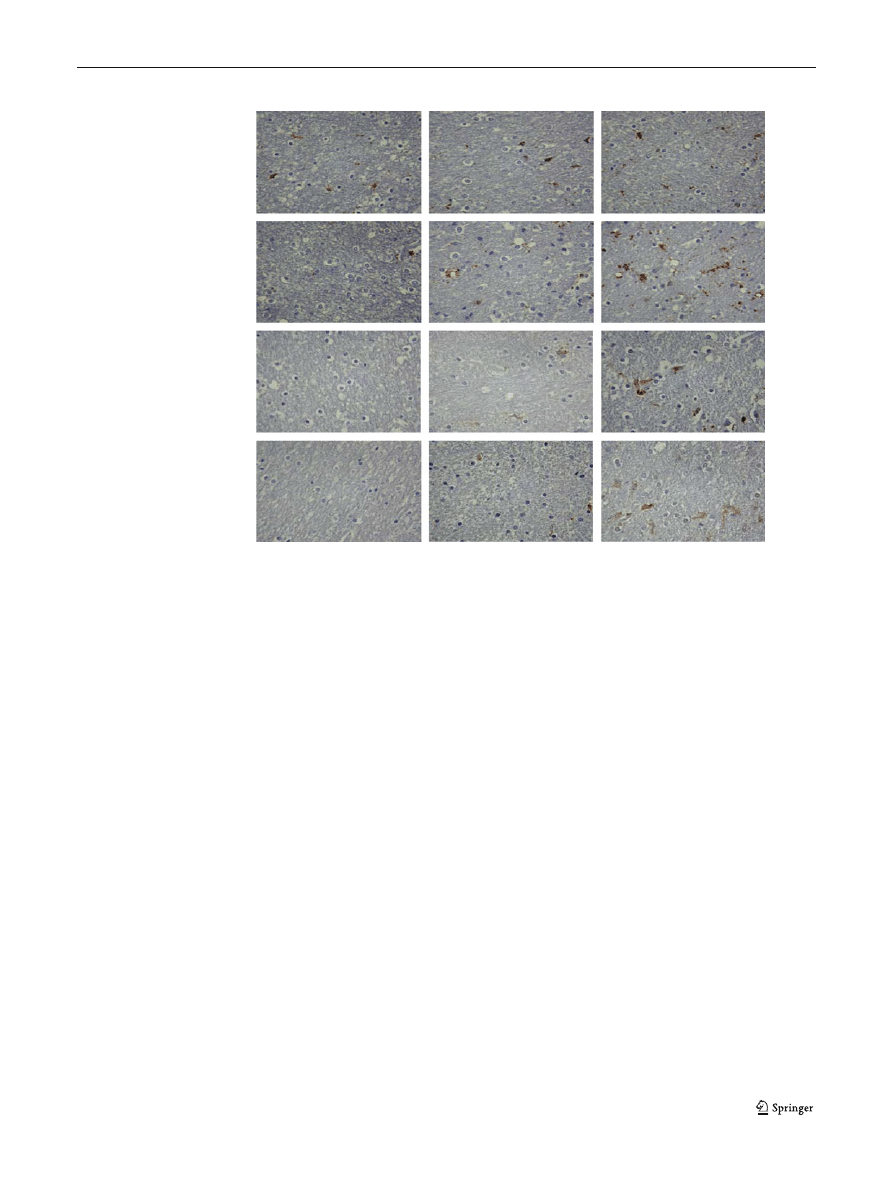
Fig. 1 Immunohistochemical stains for CD68 (a
–f) and CD163 (g–l) in frontal white matter of subjects with and without opiate addiction, with and
without HIV and HIV encephalitis. (Diaminobenzidene chromogen, hematoxylin counterstain, original magnification ×40)
J. Neurovirol. (2012) 18:364
–373
369
observation of this phenomenon and its underlying patho-
genetic mechanism have remained problematic. This may
largely be due to the significant behavioral and medical co-
morbidities in HIV-infected addicts, and in part because of
the polysubstance-using habits of the individuals under
study (Burdo et al.
; Cabral
).
The complex immunomodulatory effects of SOA have
been well documented in animal models and in vitro
systems, and may be mediated at multiple levels, including
receptor-initiated pathways in the CNS and peripheral im-
mune effector cells (Burdo et al.
). Both innate and
adaptive immunity can be significantly altered, and in some
paradigms, SOA bias T helper cell divergence from a pro-
inflammatory, anti-microbial Th1 pathway to the humoral
Th2 (Friedman et al.
). Significant interactions between
these effects and the immunosuppressive impact of HIV
raise concerns of harmful synergies, and recently, focus on
neuroAIDS disorders as an important location for these
synergies has arisen (Berman et al.
). Thus, a nascent
literature examining the impact of SOA on brain inflamma-
tion has evolved, largely in experimental systems, and to a
lesser degree in primary human CNS materials. Access to
and interpretation of primary human materials remain prob-
lematic, as substance users often display multiple con-
founds, have polysubstance habits, and CNS tissues and
fluids are not easily acquired. In this regard, the MHBB
represents a unique resource, as it follows a large number of
HIV-infected individuals with substance use disorders, is
targeted to their nervous system characterization, and the
program banks CSF and brain tissue when available.
In general, exogenous opiates are considered immuno-
suppressive, although it has been noted that most experi-
mental paradigms investigating this phenomenon have
utilized acute administration, and more conflicting results
may be obtained in models with subacute or chronic admin-
istration (Eisenstein et al.
). With regard to studies
examining the impact of opiates on CNS cells, it has been
demonstrated that activation of mu opioid receptors inhibits
microglial cell chemotaxis and promotes their apoptosis,
supporting the notion of an anti-inflammatory role within
the brain (Chao et al.
; Hu et al.
). However, in the
presence of HIV tat, astrocytes treated with morphine
increase their release of MCP-1, RANTES, and IL-6, poten-
tially contributing to a pro-inflammatory environment,
although it should be noted that not all chemokines are pro-
inflammatory and some may contribute to diminished respon-
siveness (El-Hage et al.
; Hauser et al.
). It has also
been demonstrated that morphine enhances HIV replication in
peripheral blood mononuclear cells, and has been shown to
potentiate TNF alpha production from human microglial cell
cultures (Peterson et al.
). The effects of morphine are
mediated through mu opioid receptors and are thought to
result from enhanced reactivity to other stimuli (such as
HIV or its proteins) (Peterson et al.
). In contrast, human
microglia exposed to the kappa opioid peptide dynorphin
demonstrate direct upregulation of TNF alpha and IL-6 in
the absence of other immune stimuli (Peterson et al.
Studies of opioids in animal models of HIV infection
have arrived at contradictory conclusions regarding immu-
nomodulation. Mice given systemic morphine showed in-
creased numbers of macrophages/microglia at intracerebral
Table 4 Correlations between CD4 count and plasma HIV load with
brain staining for CD68, HLA-D, CD163, and GFAP in population
stratified by opiate addiction (Spearman's rho and
p values)
CD4 correlate with
Opiate addicts
No opiate abuse
r
p
r
p
CD68
Frontal gray
−0.7427
0.0004
−0.3590
0.2074
Frontal white
−0.2573
0.3027
−0.5264
0.0531
Thalamus
−0.6110
0.0071
−0.5529
0.0403
HLA-D
Frontal gray
−0.3392
0.1685
0.4648
0.0941
Frontal white
0.0290
0.9089
0.3877
0.1708
Thalamus
0.1058
0.6760
0.1674
0.5673
CD163
Frontal gray
−0.5187
0.0274
−0.0154
0.9583
Frontal white
−0.4793
0.0442
0.1189
0.6855
Thalamus
−0.2448
0.3275
−0.0903
0.7588
GFAP
Frontal gray
−0.4627
0.0532
−0.6718
0.0085
Frontal white
−0.0052
0.9837
−0.4427
0.1129
Thalamus
−0.1732
0.4918
−0.6498
0.0119
HIV load correlate with
Opiate addicts
No opiate abuse
r
p
r
p
CD68
Frontal gray
0.5721
0.0206
0.1342
0.6474
Frontal white
0.4354
0.0919
0.4268
0.1280
Thalamus
0.6746
0.0041
0.4158
0.1392
HLA-D
Frontal gray
0.3685
0.1602
−0.1320
0.6528
Frontal white
−0.1635
0.5453
0.0616
0.8343
Thalamus
0.1159
0.6690
0.1760
0.5472
CD163
Frontal gray
0.3195
0.2277
0.3388
0.2360
Frontal white
0.5305
0.0345
−0.1606
0.5833
Thalamus
0.2065
0.4428
0.0594
0.8401
GFAP
Frontal gray
0.2853
0.2841
0.2156
0.4591
Frontal white
−0.1545
0.5677
0.1584
0.5886
Thalamus
−0.1560
0.5639
0.1408
0.6311
370
J. Neurovirol. (2012) 18:364
–373

sites of tat injection (El-Hage et al.
). However, simian
models have shown conflicting results regarding enhanced
viral virulence, with some studies showing beneficial, and
others, detrimental effects of chronic morphine administra-
tion (Burdo et al.
). For SIV neuropathogenesis, one
study has documented enhanced CNS replication in the
context of chronic morphine exposures (Kumar et al.
).
In the context of the contradictory findings of animal and
in vitro analyses, there have been few human brain studies
published to examine the role of opiates in the neuroimmune
response to HIV. All have been conducted in the Caucasian
Edinburgh cohort, utilizing the same basic technique (im-
munohistochemistry) as the current study (Anthony et al.
; Arango et al.
; Tomlinson et al.
). One of
these studies observed an upregulation of microglia in the
brains of HIV-negative intravenous drug users (IVDU)
when contrasted with HIV-negative individuals without
drug addiction, and failed to find a significant difference in
the density of microglial cells between HIV-negative IVDU
and HIV-positive, pre-symptomatic IVDU (Tomlinson et al.
). Two studies examining the response to HIV enceph-
alitis demonstrated trends for increased staining of microglia
in IVDU when compared to non-IVDU individuals with
HIV and HIVE; however, significance at
p<0.05 was not
achieved in any region (Anthony et al.
; Arango et al.
).
In the context of these prior studies, our analysis of a
predominantly minority cohort grouped into HIV negative,
HIV positive, and HIVE, shows that with opiate addiction,
there is an attenuation of macrophage/microglial activation,
with tendencies for enhanced expression of CD68 and HLA-
D in the absence of HIV, and diminution of an HIV-
associated increase. Our results suggest that there may be
a bi-phasic effect of chronic opiates in the CNS with regard
to these stimulatory molecules: in the absence of other CNS
immune stimuli, chronic opiate exposure may actuate glial-
stimulatory effects, resulting in enhanced baseline immune
responsiveness. When challenged with a pathogen such as
HIV, the chronically opiate exposed brain may conversely
show a decreased ability to respond, with impaired micro-
glial function. Of interest, when plasma samples derived
from members of the MHBB cohort were examined for
levels of lipopolysaccharide (LPS), individuals with intra-
venous heroin abuse were found to have higher levels than
those without, suggesting that decreased immune respon-
siveness to intestinal bacterial pathogens may be operant in
this cohort (Ancuta et al.
). When injected systemically,
LPS is capable of activating microglia and stimulating brain
cytokine response in both the absence and presence of
intrinsic brain pathologies (Combrinck et al.
; Perry
; Teeling et al.
). Our findings, taken together with
the literature on LPS, may lead to the following hypothesis:
with chronic opiates, attenuation of response to gut
pathogens may result in basal CNS stimulation on the basis
of circulating factors such as LPS. When infection enters the
brain, as with HIVE, there may then be continued imped-
ance to normal responsiveness in the context of the nervous
system.
Of interest in the present study was the differential effect
of opiates on CD163 expression, when contrasted with
CD68 and HLA-D. CD163 is a hemoglobin scavenger re-
ceptor, and marks perivascular macrophages in the normal
human CNS (Fabriek et al.
; Kim et al.
). These
cells are critical to antigen presentation and co-stimulation,
and are important targets of HIV infection (Fabriek et al.
; Kim et al.
). They are important in the transmis-
sion of systemic inflammatory stimuli to the CNS and are
constitutively activated (Galea et al.
). There is a sug-
gestion in the literature that these cells are important in the
anti-inflammatory response; in HIV neuropathology, it has
already been noted that the pattern of CD163 expression is
different and distinguishable from the pattern of other
markers of activated microglia, including HLA-DR
(Roberts et al.
). Thus, the divergent effects noted in
the present study build on these initial observations: while
opiates abrogate the effect of HIV on stimulatory molecules
CD68 and HLA-D, their apparent enhancement of HIV
effects on CD163 suggests that this may be a critical com-
ponent of suppressing CNS immune responsiveness. Further
examination of this potential pathway is indicated.
Interesting divergence was also seen with regard to
microglial and astrocytic markers and their correlation with
immunovirologic indices in opiate and non-opiate groups.
While HIV-opiate interactions were present in microglia in
multiple regions of brain in this study, a similar effect was
not seen for astrocytes. Furthermore, non-opiate groups
showed correlations of GFAP with CD4, but not opiate
addicts. This may suggest that mechanisms of chronic opiate
action on the CNS may vary between cell compartments.
Alternatively, it may be that the one marker we chose to
study, GFAP, may not reflect the full extent of opiate inter-
actions in this cell compartment. Further study may be
warranted.
Finally, the apparent contradiction between the present
study and prior studies of the predominantly Caucasian
Edinburgh cohort needs to be addressed. While our study
found evidence of opiate-associated immunosuppression,
the opposite effect was seen in subjects from Edinburgh
(Anthony et al.
; Arango et al.
; Tomlinson et al.
). There are many potential reasons for this discrepan-
cy. First, it has been noted that the Edinburgh group is
largely polysubstance, and it is unclear how the neurobio-
logical impact of their SOA utilization was determined to be
a result of the opiate component. In our cohort, most indi-
viduals were maintained on methadone or other medical
opiates. This raises the possibility, or even probability, that
J. Neurovirol. (2012) 18:364
–373
371

the effects witnessed in brains from the MHBB reflect
chronic utilization of non-heroin opiates, and not other
SOA. Another factor that was not reported in the
Edinburgh studies was the comparative CD4 and HIV load
in analytic groups, which may have biased reactivities. In
MHBB, the significant differences that were observed were
not due to systemic immunovirologic parameters. It is also
possible that because of the dramatically different demo-
graphic compositions of the two samples, differences may
reflect a disparity based on some unidentified racial charac-
teristic. Future studies are indicated, using larger sample
sizes, with more attention to the temporal and quantitative
aspects of the SOA, and any potential demographic variabil-
ity. Additionally, it will be essential to correlate the results of
this study with behavioral and cognitive phenotypes, as
neuroinflammation is critical to the clinical manifestations
of neuroAIDS disorders.
Acknowledgments
The authors thank the participants and staff of
the Manhattan HIV Brain Bank. This work is supported by grants
U01MH083501 from the National Institutes of Mental Health (NIMH)
and Neurological Disorders and Stroke (NINDS) (to SM) and
UL1RR029887 from the National Center for Research Resources
(NCRR) (to the Mount Sinai School of Medicine).
References
Aggarwal SK, Williams V, Levine SR, Cassin BJ, Garcia JH (1996)
Cocaine-associated intracranial hemorrhage: absence of vasculitis
in 14 cases. Neurology 46:1741
–1743
Ancuta P, Kamat A, Kunstman KJ, Kim EY, Autissier P, Wurcel A,
Zaman T, Stone D, Mefford M, Morgello S, Singer EJ, Wolinsky
SM, Gabuzda D (2008) Microbial translocation is associated with
increased monocyte activation and dementia in AIDS patients.
PLoS One 3:e2516
Anthony IC, Ramage SN, Carnie FW, Simmonds P, Bell JE (2005)
Does drug abuse alter the microglial phenotype and cell turnover
in the context of advancing HIV infection? Neuropathol Appl
Neurobiol 31:325
–338
Arango JC, Simmonds P, Brettle RP, Bell JE (2004) Does drug abuse
influence the microglial response in AIDS and HIV encephalitis?
AIDS 18:S69
–S74
Battifora H (1986) The multitumor (sausage) tissue block: novel meth-
od for immunohistochemical antibody testing. Lab Invest 55:244
–
248
Berman JW, Carson MJ, Chang L, Cox BM, Fox HS, Gonzalez RG,
Hanson GR, Hauser KF, Ho WZ, Hong JS, Major EO, Maragos
WF, Masliah E, McArthur JC, Miller DB, Nath A, O'Callaghan
JP, Persidsky Y, Power C, Rogers TJ, Royal W 3rd (2006)
NeuroAIDS, drug abuse, and inflammation: building collabora-
tive research activities. J Neuroimmune Pharmacol 1:351
–399
Burdo T, Katner S, Taffe M, Fox H (2006) Neuroimmunity, drugs of
abuse, and neuroAIDS. J Neuroimmune Pharmacol 1:41
–49
Cabral GA (2006) Drugs of abuse, immune modulation, and AIDS. J
Neuroimmune Pharmacol 1:280
–295
CDC (2007) HIV/AIDS Fact sheet: a glance at the HIV/AIDS epidemic.
Center for Disease Control and Prevention (CDC)
Chao CC, Hu S, Shark KB, Sheng WS, Gekker G, Peterson PK (1997)
Activation of mu opioid receptors inhibits microglial cell chemo-
taxis. J Pharmacol Exp Ther 281:998
–1004
Combrinck MI, Perry VH, Cunningham C (2002) Peripheral infection
evokes exaggerated sickness behaviour in pre-clinical murine
prior disease. Neuroscience 112(1):7
–11
Crawford FC, Wood ML, Wilson SE, Mathura VS, Hollen TR, Geall F,
Kolippakkam DN, Mullan MJ (2006) Cocaine induced inflamma-
tory response in human neuronal progenitor cells. J Neurochem
97:662
–674
Dhillon NK, Peng F, Bokhari S, Callen S, Shin SH, Zhu X, Kim KJ,
Buch SJ (2007) Cocaine-mediated alteration in tight junction
protein expression and modulation of CCL2/CCR2 axis across
the blood-brain barrier: implications for HIV-dementia. J
Neuroimmune Pharmacol 3(1):52
–56
Eisenstein TK, Rahim RT, Feng P, Thingalaya NK, Meissler JJ (2006)
Effects of opioid tolerance and withdrawal on the immune system.
J Neuroimmune Pharmacol 1:237
–249
El-Hage N, Gurwell JA, Singh IN, Knapp PE, Nath A, Hauser KF
(2005) Synergistic increases in intracellular Ca2+, and the release
of MCP-1, RANTES, and IL-6 by astrocytes treated with opiates
and HIV-1 Tat. Glia 50:91
–106
El-Hage N, Wu G, Wang J, Ambati J, Knapp PE, Reed JL, Bruce-
Keller AJ, Hauser KF (2006) HIV-1 tat and opiate-induced
changes in astrocytes promote chemotaxis of microglia through
the expression of MCP-1 and alternative chemokines. Glia 53:132
–
146
Fabriek BO, van Haastert ES, Galea I, Polfliet MMJ, Dopp ED, van
den Heuvel MM, van den Berg TK, de Groot CJA, der Valk PV,
Dijkstra CD (2005) CD163-positive pervascular macrophages in
the human CNS express molecules for antigen recognition and
presentation. Glia 51:297
–305
Friedman H, Pross S, Klein TW (2006) Addictive drugs and their
relationship with infectious diseases. FEMS Immunol Med
Microbiol 47:330
–342
Galea I, Felton LM, Waters S, van Rooijen N, Perry VH, Newman TA
(2008) Immune-to-brain signaling: the role of cerebral CD163-
positive macrophages. Neurosci Lett 448:41
–46
Hasin D, Trautman K, Miele G, Samet S, Smith M, Endicott J (1996)
Psychiatric Research Interview for Substance and Mental Disorders
(PRISM): reliability for substance abusers. Am J Psychiatry
153:1195
–1201
Hauser KF, El-Hage N, Stiene-Martin A, Maragos WF, Nath A,
Persidsky Y, Volsky DJ, Knapp PE (2007) HIV-1 neuropatho-
genesis: glial mechanisms revealed through substance abuse. J
Neurochem 100:567
–586
Hu S, Sheng WS, Lokensgard JR, Peterson PK (2002) Morphine induces
apoptosis of human microglia and neurons. Neuropharmacology
42:829
–836
Kim WK, Alvarez X, Fisher J, Bronfin B, Westmoreland S, McLaurin
J, Williams K (2006) CD163 identifies perivascular macrophages
in normal and viral encephalitic brains and potential precursors to
perivascular macrophages in blood. Am J Pathol 168(3):822
–834
Kononen J, Bubendorf L, Kallioniemi A, Barlund M, Schraml P,
Leighton S, Torhorst J, Mihatsch MJ, Sauter G, Kallioniemi OP
(1998) Tissue microarrays for high-throughput molecular profil-
ing of tumor specimens. Nat Med 4:844
–847
Kumar R, Orsoni S, Norman L, Verma AS, Tirado G, Giavedoni LD,
Staprans S, Miller GM, Buch SJ, Kumar A (2006) Chronic
morphine exposure causes pronounced virus replication in cere-
bral compartment and accelerated onset of AIDS in SIV/
SHIV-infected Indian rhesus macaques. Virology 354:192
–
206
Morgello S, Gelman BB, Kozlowski PB, Vinters HV, Masliah E,
Cornford M, Cavert W, Marra C, Grant I, Singer EJ (2001) The
National NeuroAIDS Tissue Consortium: a new paradigm in brain
372
J. Neurovirol. (2012) 18:364
–373

banking with an emphasis on infectious disease. Neuropathol
Appl Neurobiol 27:326
–335
Nath A, Hauser KF, Wojna V, Booze RM, Maragos W, Prendergast M,
Cass W, Turchan JT (2002) Molecular basis for interactions of
HIV and drugs of abuse. J Acquir Immune Defic Syndr 31(Suppl):
S62
–S69
New York City Department of Health and Mental Hygiene (2011) New
York City HIV/AIDS annual surveillance statistics. New York
City Department of Health and Mental Hygiene, New York
Perry VH (2004) The influence of systemic inflammation on inflam-
mation in the brain: implications for chronic neurodegenerative
disease. Brain Behav Immun 18:407
–413
Peterson PK, Molitor TW, Chao CC (1998) The opioid-cytokine con-
nection. J Neuroimmunol 83:63
–69
Roberts ES, Masliah E, Fox HS (2004) CD163 identifies a unique
population of ramified microglia in HIV encephalitis (HIVE). J
Neuropathol Exp Neurol 63(12):1255
–1264
Seelbach MJ, Brooks TA, Egleton RD, Davis TP (2007) Peripheral
inflammatory hyperalgesia modulates morphine delivery to the
brain: a role for P-glycoprotein. J Neurochem 102:1677
–1690
Teeling JL, Felton LM, Deacon RMJ, Cunningham C, Rawlins JNP,
Perry VH (2007) Sub-pyrogenic systemic inflammation impacts
on brain and behavior, independent of cytokines. Brain Behav
Immun 21:836
–850
Tomlinson GS, Simmonds P, Busuttil A, Chiswick A, Bell JE (1999)
Upregulation of microglia in drug users with and without pre-
symptomatic HIV infection. Neuropathol Appl Neurobiol
25:369
–379
Tyor WR, Middaugh LD (1999) Do alcohol and cocaine abuse alter the
course of HIV-associated dementia complex? J Leukoc Biol
65:475
–481
Wu HS, Murray J, Morgello S (2008) Segmentation of brain immuno-
histochemistry images using clustering of linear centroids and
regional shapes. J Imaging Sci Technol 52:040502
–040502-11
J. Neurovirol. (2012) 18:364
–373
373
Document Outline
- Impact of opiate addiction on neuroinflammation in HIV
Wyszukiwarka
Podobne podstrony:
possible impacts of climatic warming on polar bears
Latour The Impact of Science Studies on Political Philosophy
The Impact of Countermeasure Spreading on the Prevalence of Computer Viruses
The Impact of Countermeasure Propagation on the Prevalence of Computer Viruses
The impact of network structure on knowledge transfer an aplication of
possible impacts of climatic warming on polar bears
Karpińska Krakowiak, Małgorzata The Impact of Consumer Knowledge on Brand Image Transfer in Cultura
Impact of agricultural biotechnology on biodiversity
Impact of Computer Viruses on Society
Do methadone and buprenorphine have the same impact on psychopathological symptoms of heroin addicts
THE IMPACT OF SOCIAL NETWORK SITES ON INTERCULTURAL COMMUNICATION
Impact of resuscitation system errors on survival from in hospital cardiac arrest
Impact of resuscitation system errors on survival from in-hospital cardiac arrest, MEDYCYNA, RATOWNI
The impact of Microsoft Windows infection vectors on IP network traffic patterns
Impact of Artificial Gummy Fingers on Fingerprint Systems
Marina Post The impact of Jose Ortega y Gassets on European integration
The Impact of Mary Stewart s Execution on Anglo Scottish Relations
L R Kominz The Impact of Tourism on Japanese Kyogen (Asian Ethnology Vol 47 2, 1988)
więcej podobnych podstron