
Human skeleton

Human skeleton
The human skeleton consists of both fused and individual
bones supported and supplemented by ligaments,
tendons, muscles and cartilage. It serves as a scaffold
which supports organs, anchors muscles, and protects
organs such as the brain, lungs and heart. The biggest
bone in the body is the femur and the smallest is the
stapes bone in the middle ear. In an adult, the skeleton
comprises around 13% of the total body weight, and half
of this weight is water.
Fused bones include those of the pelvis and the cranium.
Not all bones are interconnected directly: There are three
bones in each middle ear called the ossicles that
articulate only with each other. The hyoid bone, which is
located in the neck and serves as the point of attachment
for the tongue, does not articulate with any other bones
in the body, being supported by muscles and ligaments.

Organization
Much of the human skeleton maintains the ancient
segmental pattern present in all vertebrates
(mammals, birds, fish, reptiles and amphibians)
with basic units being repeated. This segmental
pattern is particularly evident in the vertebral
column and in the ribcage.There are 206 bones in
the adult human skeleton, a number which varies
between individuals and with age - newborn
babies have 270 bones some of which fuse
together. These bones are organized into a
longitudinal axis, the axial skeleton, to which the
appendicular skeleton is attached.

Function
Support
The skeleton provides the framework which supports the
body and maintains its shape. The pelvis and associated
ligaments and muscles provide a floor for the pelvic
structures. Without the ribs, costal cartilages, and the
intercostal muscles the lungs would collapse.
Movement
The joints between bones permit movement, some allowing
a wider range of movement than others, e.g. the ball and
socket joint allows a greater range of movement than the
pivot joint at the neck. Movement is powered by skeletal
muscles, which are attached to the skeleton at various sites
on bones. Muscles, bones, and joints provide the principal
mechanics for movement, all coordinated by the nervous
system.

Function c.d.
Protection
The skeleton protects many vital organs:
The skull protects the brain, the eyes, and the middle
and inner ears.
The spine protects the spinal cord.
The rib cage, spine, and sternum protect the lungs,
heart and major blood vessels.
The clavicle and scapula protect the shoulder.
The ilium and spine protect the digestive and
urogenital systems and the hip.
The patella and the ulna protect the knee and the
elbow respectively.
The carpals and tarsals protect the wrist and ankle
respectively.

Function c.d.
Blood cell production
The skeleton is the site of haematopoiesis, which takes place
in red bone marrow. Marrow is found in the center of long
bones.
Storage
Bone matrix can store calcium and is involved in calcium
metabolism, and bone marrow can store iron in ferritin and
is involved in iron metabolism. However, bones are not
entirely made of calcium, but a mixture of chondroitin
sulfate and hydroxyapatite, the latter making up 70% of a
bone.
Endocrine regulation
Bone cells release a hormone called osteocalcin, which
contributes to the regulation of blood sugar (glucose) and fat
deposition. Osteocalcin increases both the insulin secretion
and sensitivity, in addition to boosting the number of insulin-
producing cells and reducing stores of fat.

Gender-based differences
There are many differences between the male and
female human skeletons. Most prominent is the
difference in the pelvis, owing to characteristics
required for the processes of childbirth. The shape of a
female pelvis is flatter, more rounded and proportionally
larger to allow the head of a fetus to pass. Men tend to
have slightly thicker and longer limbs and digit bones
(phalanges), while women tend to have narrower rib
cages, smaller teeth, less angular mandibles, less
pronounced cranial features such as the brow ridges
and external occipital protuberance (the small bump at
the back of the skull), and the carrying angle of the
forearm is more pronounced in females. Females also
tend to have more rounded shoulder blades.
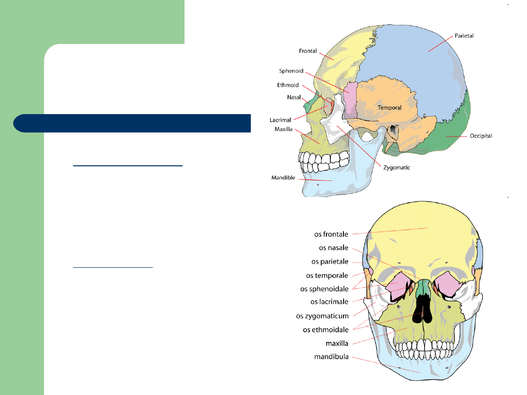
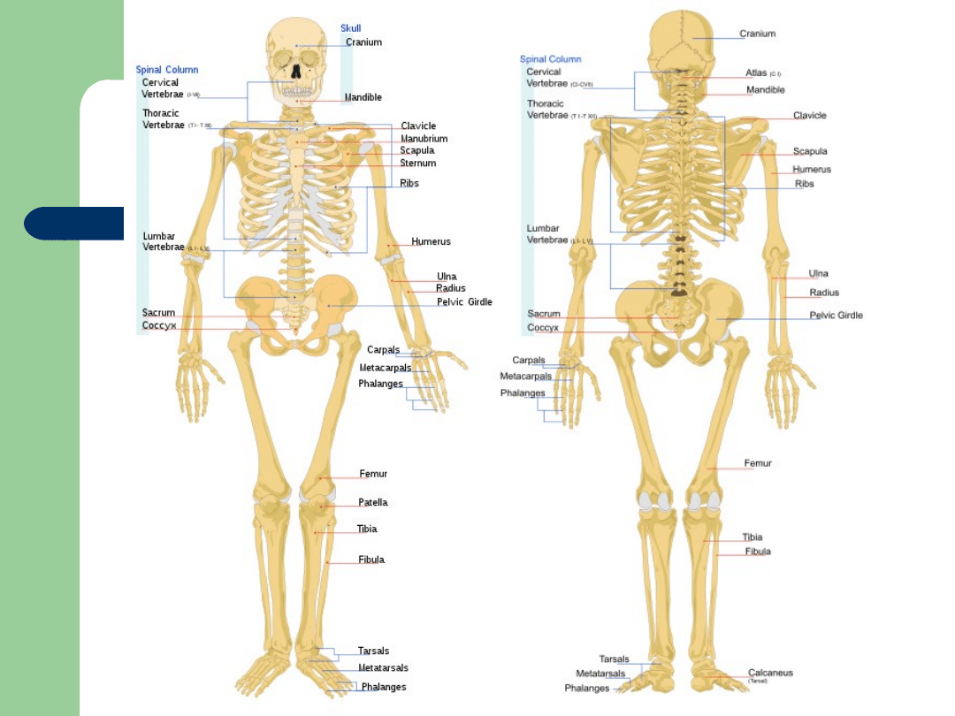
In the skull (22):
The Cranial bones:
–
frontal bone
–
parietal bone (2)
–
temporal bone (2)
–
occipital bone
–
sphenoid bone
–
ethmoid bone
Facial bones:
–
mandible
–
maxilla (2)
–
palatine bone (2)
–
zygomatic bone (2)
–
nasal bone (2)
–
lacrimal bone (2)
–
vomer bone
–
inferior nasal conchae (2)
Bones
Bones
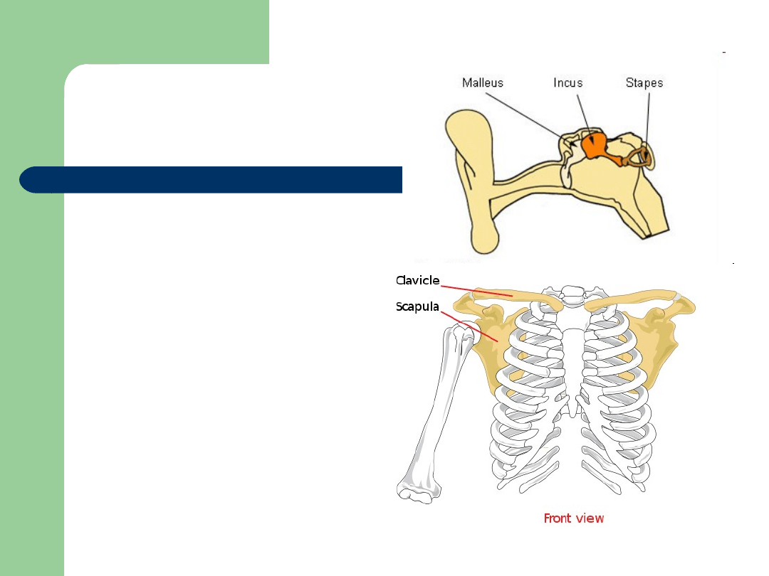
In the middle ears (6):
- malleus (2)
- incus (2)
- stapes (2)
In the throat (1):
- hyoid bone
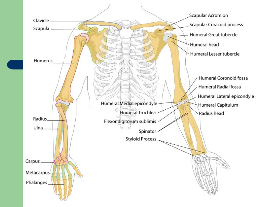
In the shoulder girdle (4):
- scapula or shoulder blade (2)
- clavicle or collarbone (2)
Bones
In the thorax (25 or 27):
- sternum
–
Can be considered as three different bones;
manubrium, body of sternum (gladiolus) and xiphoid
process
- ribs (2 x 12)
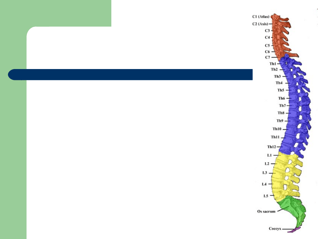
In the vertebral column (24):
- cervical vertebrae (7)
- thoracic vertebrae (12)
- lumbar vertebrae (5)

Bones
In the arms (2):
- humerus (2)
In the forearms (4):
- radius (2)
- ulna (2)
In the hands (54):
Carpal (wrist) bones:
–
scaphoid bone (2)
–
lunate bone (2)
–
triquetral bone (2)
–
pisiform bone (2)
–
trapezium (2)
–
trapezoid bone (2)
–
capitate bone (2)
–
hamate bone (2)
Metacarpus (palm) bones:
–
metacarpal bones (5 × 2)
Digits of the hands (finger bones or
phalanges):
–
proximal phalanges (5 × 2)
–
intermediate phalanges (4 × 2)
–
distal phalanges (5 × 2)
Bones

Bones
In the pelvis (4):
- coccyx (4 or 5 fused)
- sacrum (5 fused)
- hip bone (innominate bone or coxal bone)
(2)
In the thighs (2):
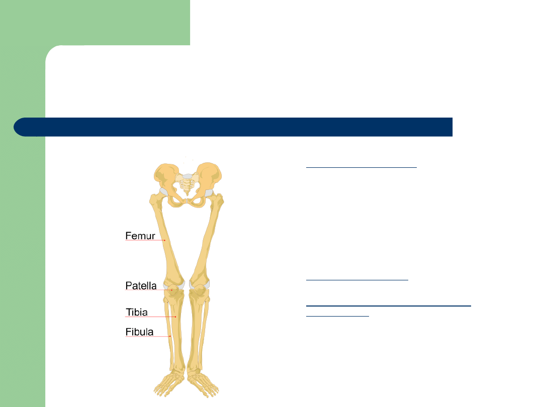
- femur (2)
Bones
In the legs (6):
- patella (2)
- tibia (2)
- fibula (2)
In the feet (52):
Tarsal (ankle) bones:
–
calcaneus (heel bone) (2)
–
talus (2)
–
navicular bone (2)
–
medial cuneiform bone (2)
–
intermediate cuneiform bone
(2)
–
lateral cuneiform bone (2)
–
cuboid bone (2)
Metatarsus bones:
–
metatarsal bone (5 × 2)
Digits of the feet (toe bones or
phalanges):
–
proximal phalanges (5 × 2)
–
intermediate phalanges (4 × 2)
–
distal phalanges (5 × 2)

The End
Thank you for your
attention!
Document Outline
- Slide 1
- Slide 2
- Slide 3
- Slide 4
- Slide 5
- Slide 6
- Slide 7
- Slide 8
- Slide 9
- Slide 10
- Slide 11
- Slide 12
- Slide 13
- Slide 14
- Slide 15
- Slide 16
Wyszukiwarka
Podobne podstrony:
Human skeleton 2
Human Skeleton
AN INSTANCE OF DENTAL MODIFICATION ON A HUMAN SKELETON FROM NIGER, WEST AFRICA
Human Development Index
Human Terrain System
konspekt ?humanizacja sztuki
Maslow (1943) Theory of Human Motivation
nauki human w med id 315728 Nieznany
human development14 technical notes
human mimicry
2002 mol genetics of human cognition MolInterv
skeleton, STOMATOLOGIA, Anatomia
Psychology and Cognitive Science A H Maslow A Theory of Human Motivation
Cosmic and Human Metamorphoses
więcej podobnych podstron