
Glycolysis
Copyright © 1998-2007 by Joyce
J. Diwan.
All rights reserved.
Biochemistry of
Metabolism
Glycolysis
takes place in the
cytosol
of cells.
Glucose enters the Glycolysis pathway by
conversion to
glucose-6-phosphate
.
Initially there is energy input corresponding to
cleavage of two ~P bonds of ATP.
H
O
OH
H
OH
H
OH
CH
2
O
PO
3
2
H
OH
H
1
6
5
4
3
2
glucose-6-phosphate
H
O
O H
H
O H
H
O H
CH
2
O H
H
O H
H
H
O
O H
H
O H
H
O H
CH
2
O
PO
3
2
H
O H
H
2
3
4
5
6
1
1
6
5
4
3
2
A T P A DP
M g
2+
glucose glucose-6-phosphate
Hexokinase
1. Hexokinase
catalyzes:
Glucose + ATP
glucose-6-P + ADP
The reaction involves nucleophilic attack of
the C6 hydroxyl O of glucose on P of the
terminal phosphate of ATP.
ATP binds to the enzyme as a complex with
Mg
++
.
Mg
++
interacts with negatively charged
phosphate oxygen atoms, providing charge
compensation & promoting a favorable
conformation of ATP at the active site of the
Hexokinase enzyme.
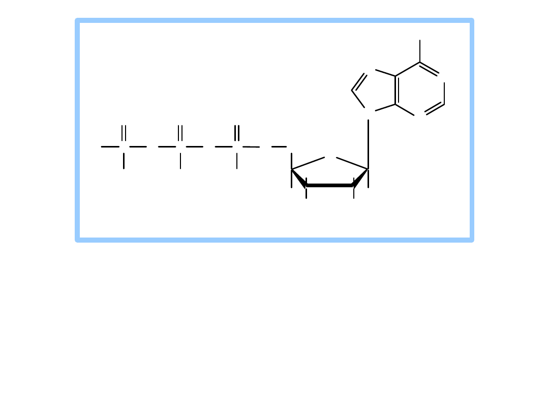
N
N
N
N
NH
2
O
OH
OH
H
H
H
CH
2
H
O
P
O
P
O
P
O
O
O
O
O
O
O
adenine
ribose
ATP
adenosine triphosphate
The reaction catalyzed by Hexokinase is
highly
spontaneous
.
A phosphoanhydride bond of ATP (
~P
) is cleaved.
The phosphate ester formed in glucose-6-
phosphate has a lower G of hydrolysis.
H
O
O H
H
O H
H
O H
CH
2
O H
H
O H
H
H
O
O H
H
O H
H
O H
CH
2
O
PO
3
2
H
O H
H
2
3
4
5
6
1
1
6
5
4
3
2
A T P A DP
M g
2+
glucose glucose-6-phosphate
Hexokinase
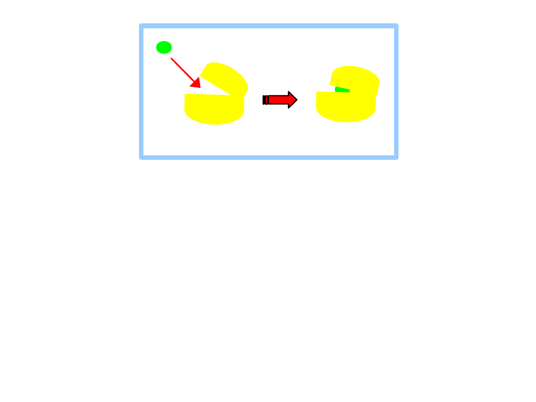
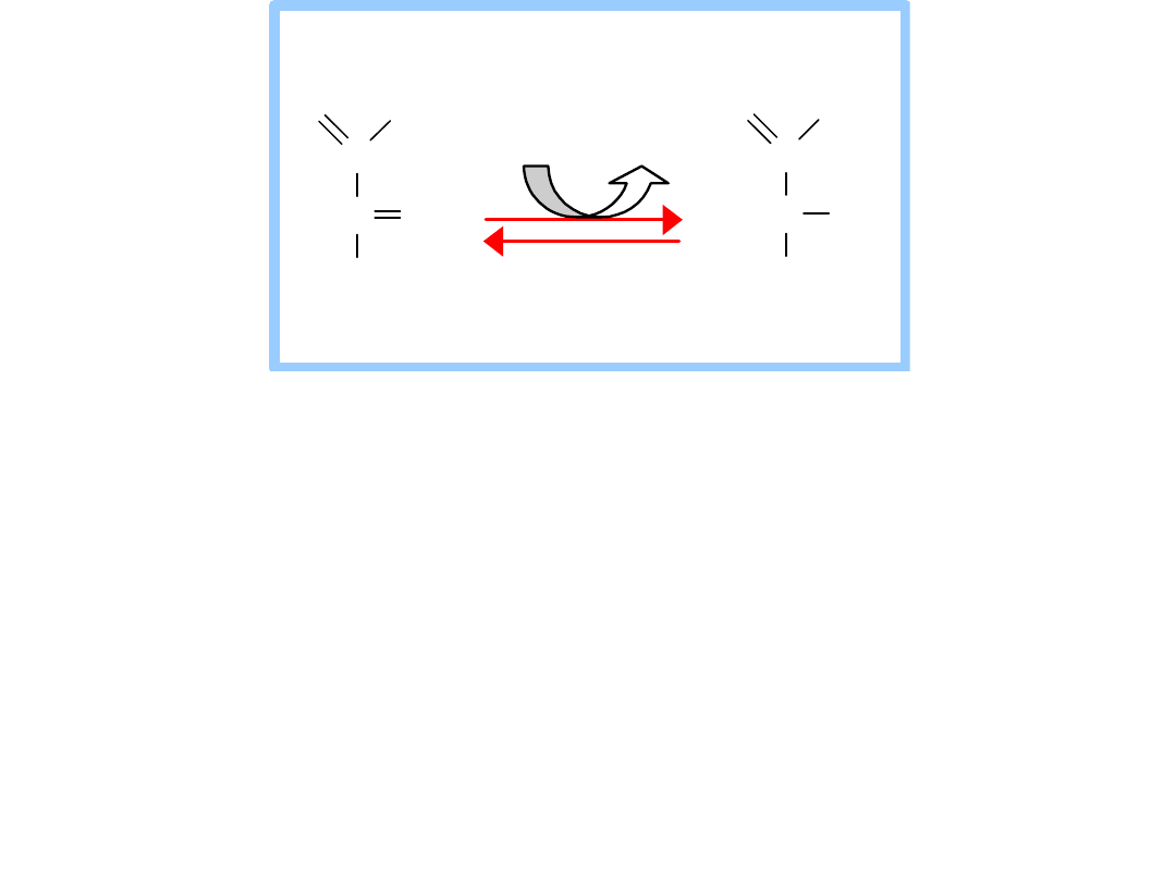
the
C6 hydroxyl
of the
bound glucose is
close to
the terminal
phosphate
of
ATP, promoting catalysis.
water is excluded
from the active site.
This prevents the enzyme from catalyzing ATP
hydrolysis, rather than transfer of phosphate to
glucose.
glucose
Hexokinase
H
O
O H
H
O H
H
O H
CH
2
O H
H
O H
H
H
O
O H
H
O H
H
O H
CH
2
O
PO
3
2
H
O H
H
2
3
4
5
6
1
1
6
5
4
3
2
A T P A DP
M g
2+
glucose glucose-6-phosphate
Hexokinase
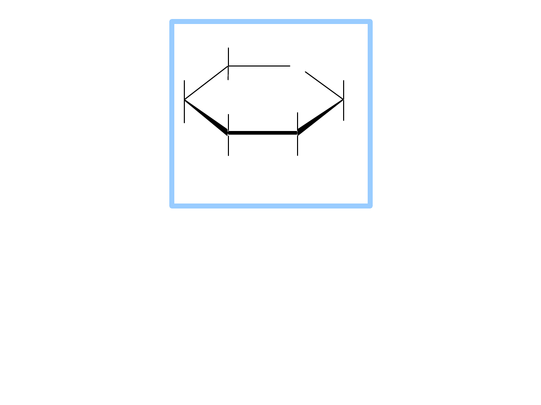
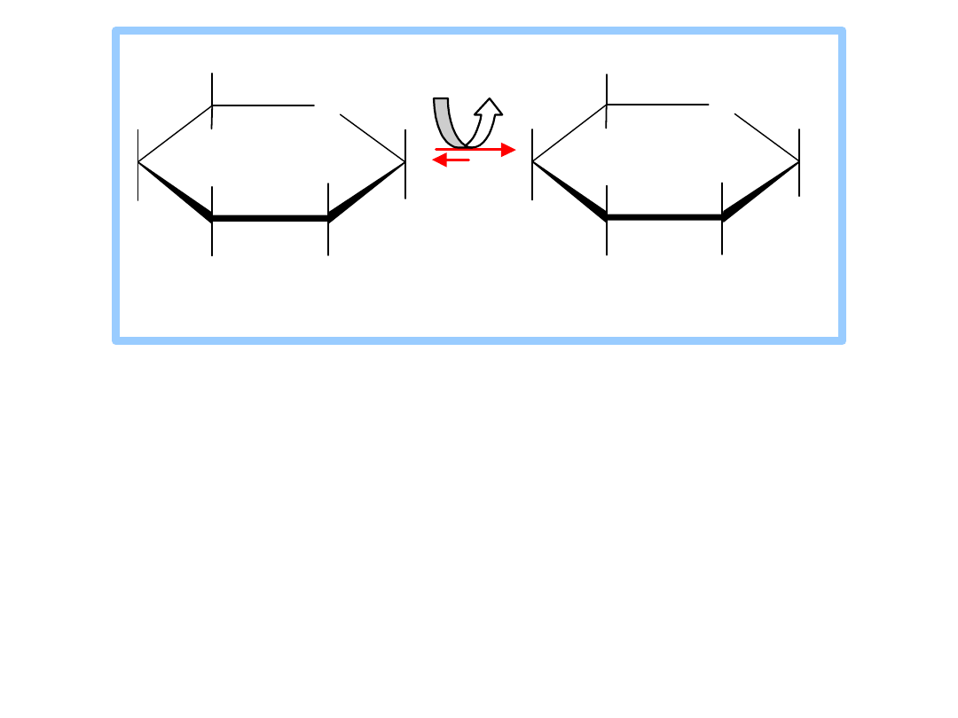
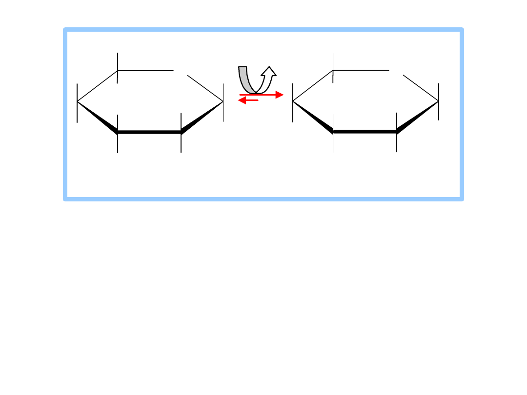
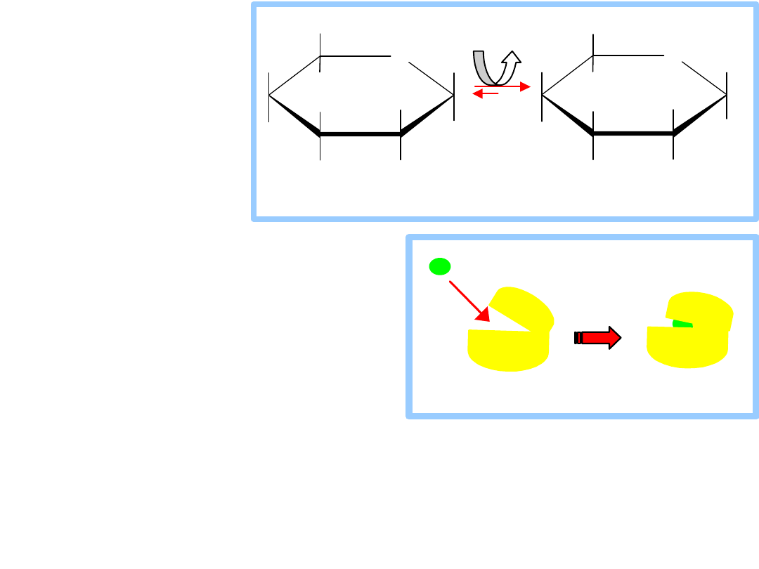
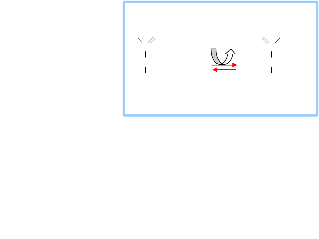
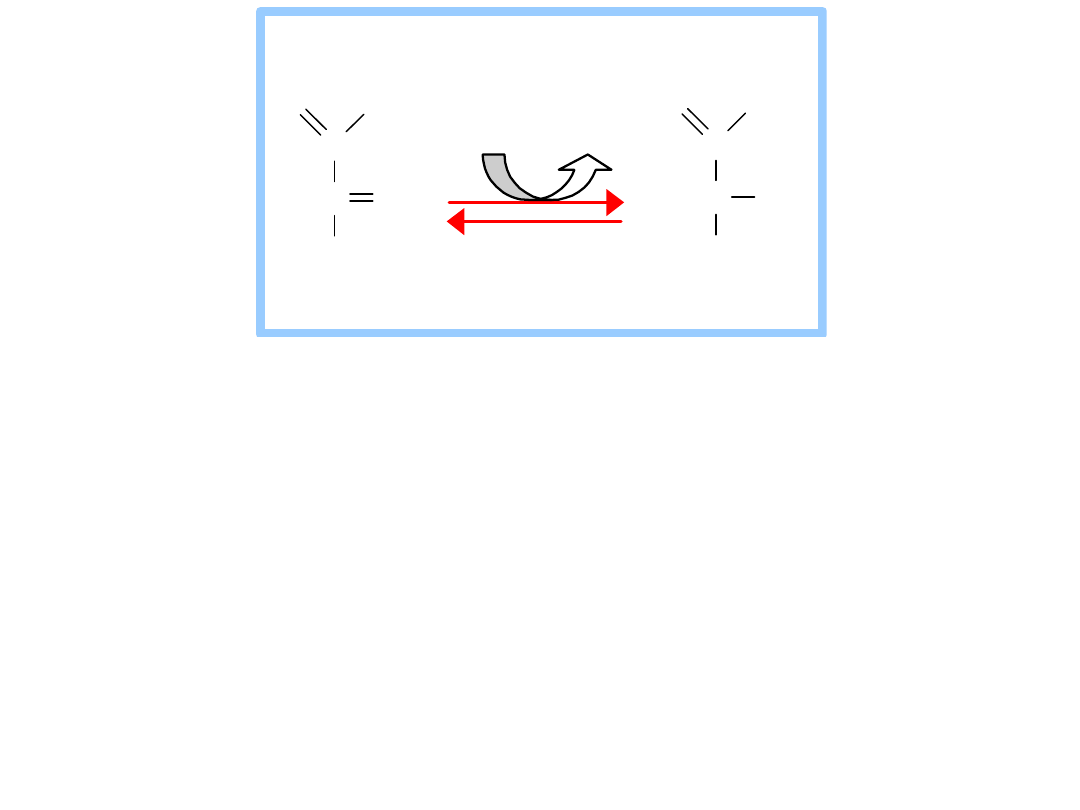
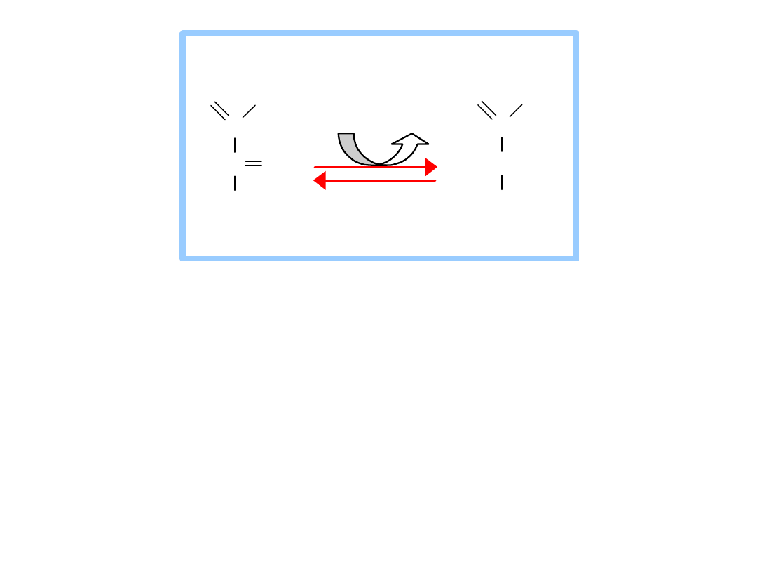
Induced fit:
Glucose
binding
to
Hexokinase
stabilizes a
conformatio
n
in
which:
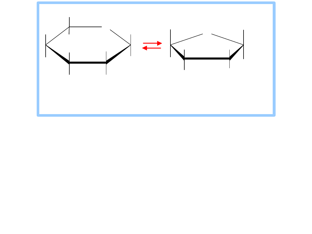
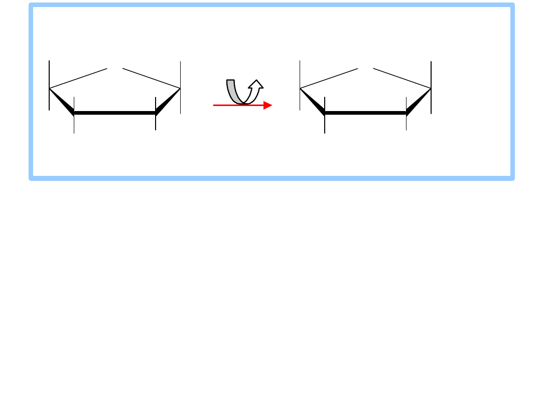
It is a
common motif
for an enzyme active site to
be located at an interface between protein
domains that are connected by a flexible hinge
region.
The
structural flexibility
allows access to the
active site, while permitting precise positioning
of active site residues, and in some cases
exclusion of water, as substrate binding
promotes a particular conformation.
glucose
Hexokinase
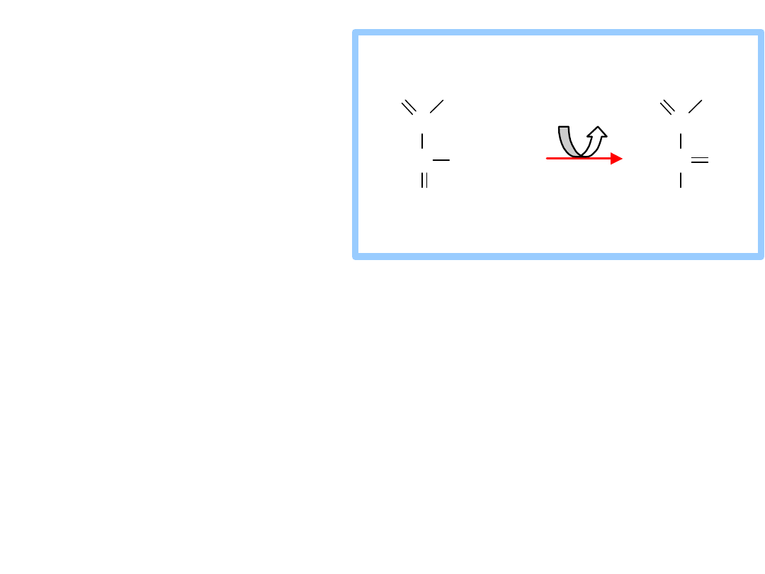
2. Phosphoglucose Isomerase
catalyzes:
glucose-6-P
(aldose)
fructose-6-P
(ketose)
The mechanism involves acid/base catalysis, with
ring opening, isomerization via an
enediolate
intermediate
, and then ring closure. A similar
reaction catalyzed by Triosephosphate Isomerase
will be presented in detail.
H
O
O H
H
O H
H
O H
CH
2
O PO
3
2
H
O H
H
1
6
5
4
3
2
CH
2
O PO
3
2
O H
CH
2
O H
H
O H
H
H
HO
O
6
5
4
3
2
1
glucose-6-phosphate fructose-6-phosphate
Phosphoglucose Isomerase
3. Phosphofructokinase
catalyzes:
fructose-6-P + ATP
fructose-1,6-bisP
+ ADP
This highly
spontaneous
reaction has a
mechanism similar to that of Hexokinase.
The Phosphofructokinase reaction is the
rate-
limiting step
of Glycolysis.
The enzyme is highly
regulated
, as will be
discussed later.
CH
2
O PO
3
2
O H
CH
2
O H
H
O H
H
H
HO
O
6
5
4
3
2
1
CH
2
O PO
3
2
O H
CH
2
O
PO
3
2
H
O H
H
H
HO
O
6
5
4
3
2
1
A T P A D P
M g
2 +
fructo se-6-pho sphate fructo se-1,6-bispho sphate
P ho spho fructo k inase
4.
Aldolase
catalyzes:
fructose-1,6-
bisphosphate
dihydroxyacetone-P +
glyceraldehyde-3-P
The reaction is an
aldol cleavage
, the reverse
of an aldol condensation.
Note that C atoms are renumbered in products
of Aldolase.
6
5
4
3
2
1
CH
2
O PO
3
2
C
C
C
C
CH
2
O PO
3
2
O
HO
H
H
O H
H
O H
3
2
1
CH
2
O PO
3
2
C
CH
2
O H
O
C
C
CH
2
O PO
3
2
H
O
H
O H
+
1
2
3
fructose-1,6-
bisphosphate
A ldolase
dihydroxyacetone glyceraldehyde-3-
phosphate phosphate
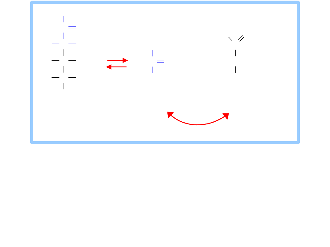
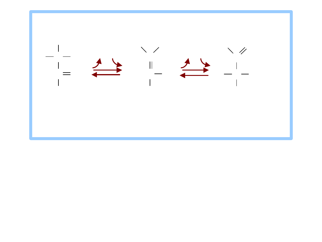
Triosephosphate Isomerase
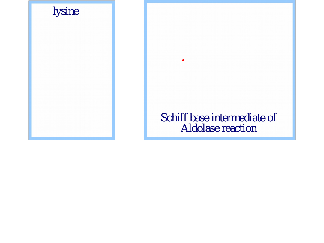
A
lysine
residue at the active site functions
in catalysis.
The
keto
group of fructose-1,6-bisphosphate
reacts with the -amino group of the active
site lysine, to form a protonated
Schiff base
intermediate.
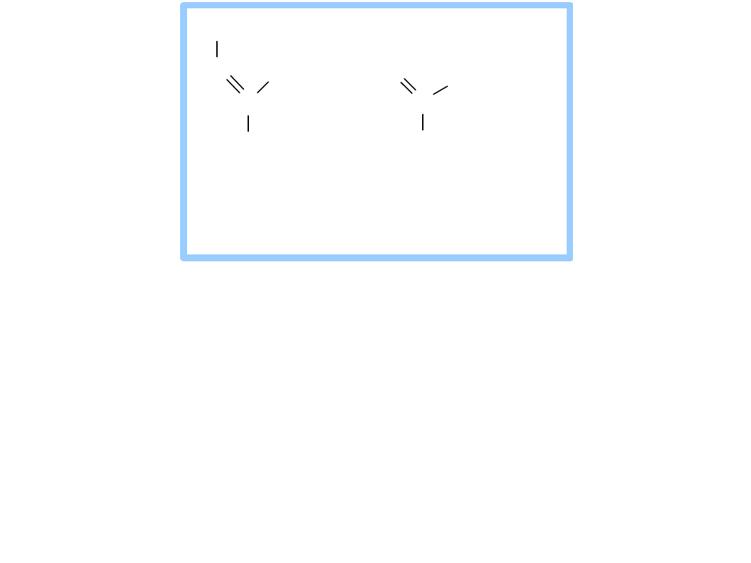
Cleavage of the bond between C3 & C4
follows.

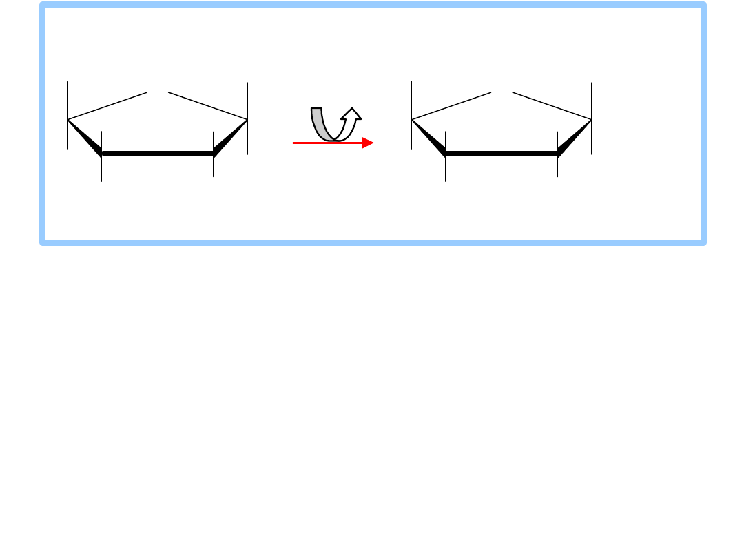
5. Triose Phosphate Isomerase (TIM)
catalyzes:
dihydroxyacetone-P
glyceraldehyde-
3-P
Glycolysis continues from glyceraldehyde-3-P.
TIM's K
eq
favors dihydroxyacetone-P. Removal of
glyceraldehyde-3-P by a subsequent
spontaneous reaction allows throughput.
6
5
4
3
2
1
CH
2
O PO
3
2
C
C
C
C
CH
2
O PO
3
2
O
HO
H
H
O H
H
O H
3
2
1
CH
2
O PO
3
2
C
CH
2
O H
O
C
C
CH
2
O PO
3
2
H
O
H
O H
+
1
2
3
fructose-1,6-
bisphosphate
A ldolase
dihydroxyacetone glyceraldehyde-3-
phosphate phosphate
Triosephosphate Isomerase
The ketose/aldose conversion involves
acid/base catalysis
, and is thought to proceed
via an
enediol
intermediate, as with
Phosphoglucose Isomerase.
Active site Glu and His residues are thought to
extract and donate protons during catalysis.
C
C
CH
2
O PO
3
2
O
C
C
CH
2
O PO
3
2
H
O
H
O
H
C
C
CH
2
O PO
3
2
H
O
H
O
H
H
H
O
H
H
+
H
+
H
+
H
+
d i h y d ro x y a c e to n e e n e d io l g l y c e ra ld e h y d e -
p h o s p h a te i n te rm e d i a te 3 - p h o s p h a te
T rio s e p h o s p h a te Is o m e ra s e
C
CH
2
O PO
3
2
O
O
C
CH
2
O PO
3
2
HC
O
O H
pro po sed
enedio late
interm ediate
pho spho gly co late
transitio n state
analo g
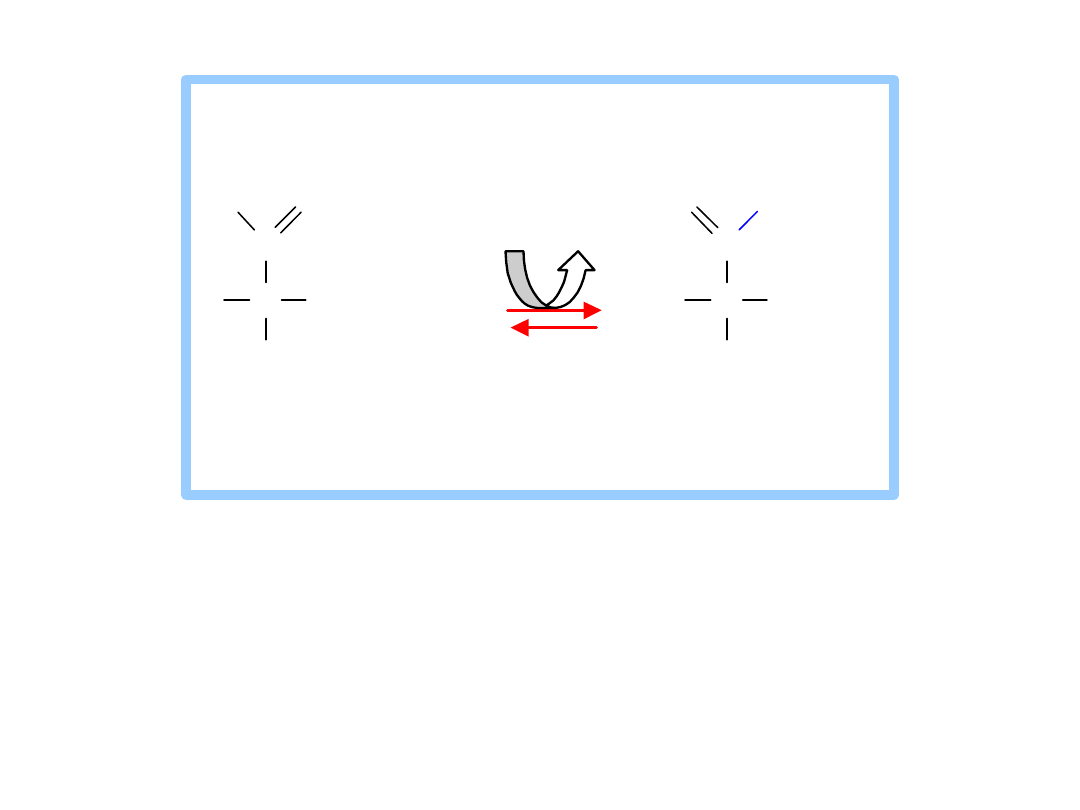
2-Phosphoglycolate
is a
transition state
analog
that binds tightly at the active site of
Triose Phosphate Isomerase (TIM).
This inhibitor of catalysis by TIM is similar in
structure to the proposed enediolate
intermediate.
TIM is judged a "perfect enzyme." Reaction
rate is limited only by the rate that substrate
collides with the enzyme.
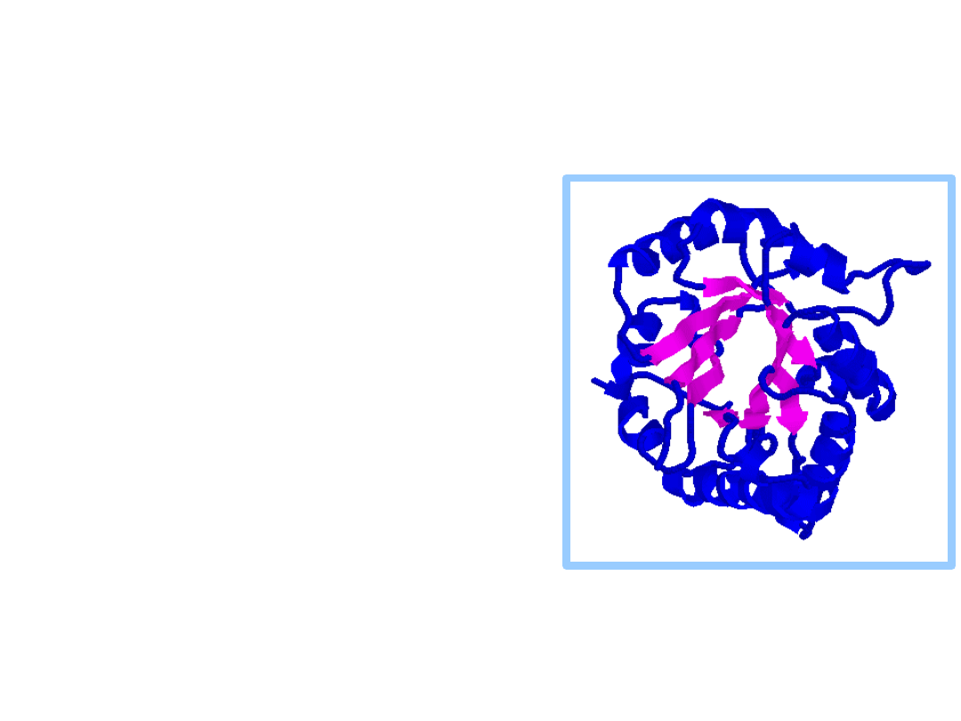
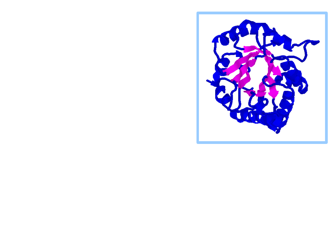
TIM
Triosephosphate
Isomerase structure is
an
barrel
, or TIM
barrel.
In an barrel there are
8 parallel -
strands surrounded by 8
-helices.
Short loops connect
alternating -strands &
-helices.
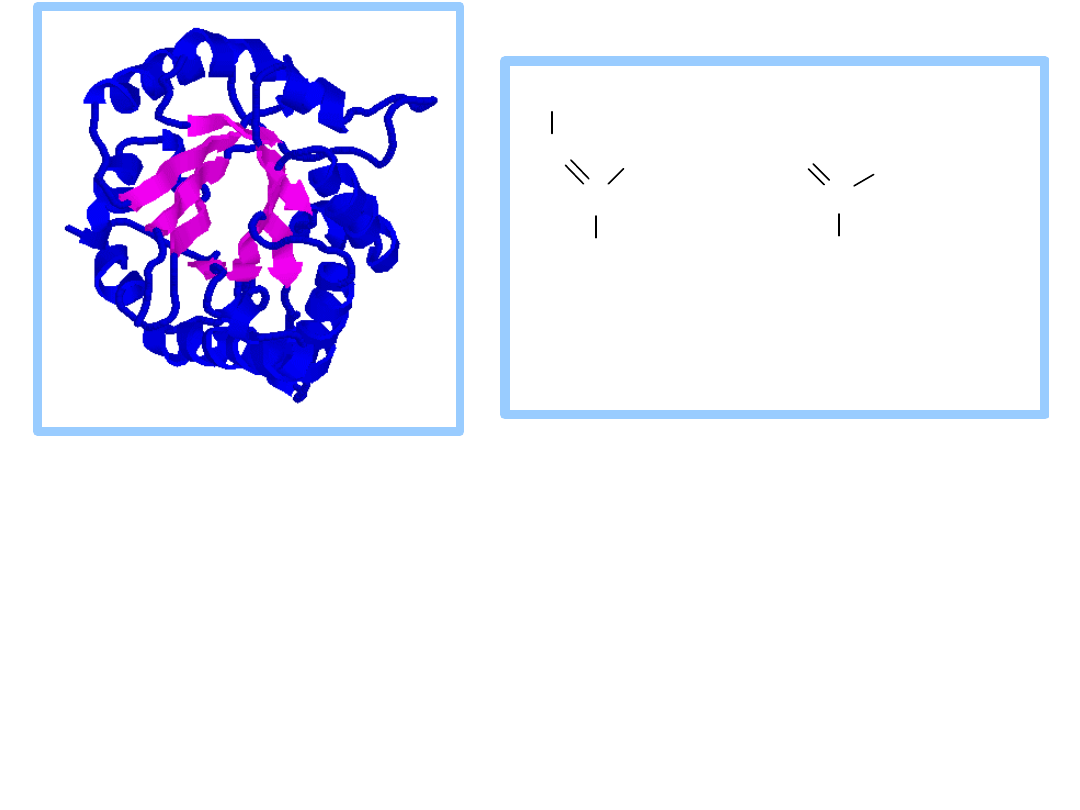
TIM
TIM barrels
serve as
scaffolds for active site
residues in a diverse
array of enzymes.
Residues of the
active
site
are always at the
same end of the barrel,
on C-terminal ends of -
strands & loops
connecting these to -
helices.
There is debate whether the many different
enzymes with TIM barrel structures are
evolutionarily related.
In spite of the structural similarities there is
tremendous
diversity in catalytic functions
of these enzymes and little sequence
homology.
TIM
Explore
the structure of the
Triosephosphate Isomerase (TIM)
homodimer, with the transition state
inhibitor 2-phosphoglycolate
bound to one of the TIM monomers.
Note
the structure of the TIM barrel, and
the loop that forms a lid that closes over the
active site after binding of the substrate.
C
CH
2
O PO
3
2
O
O
C
CH
2
O PO
3
2
HC
O
O H
pro po sed
enedio late
interm ediate
pho spho gly co late
transitio n state
analo g
C
C
CH
2
O PO
3
2
H
O
H
O H
C
C
CH
2
O PO
3
2
O
O PO
3
2
H
O H
+
P
i
+ H
+
N A D
+
N A D H
1
2
3
2
3
1
g l y c e ra l d e h y d e - 1 ,3 - b i s p h o s p h o -
3 - p h o s p h a te g l y c e ra te
G ly c e ra ld e h y d e - 3 - p h o s p h a te
D e h y d ro g e n a s e
6. Glyceraldehyde-3-phosphate
Dehydrogenase
catalyzes:
glyceraldehyde-3-P + NAD
+
+ P
i
1,3-bisphosphoglycerate +
NADH + H
+
C
C
CH
2
O PO
3
2
H
O
H
O H
C
C
CH
2
O PO
3
2
O
O PO
3
2
H
O H
+
P
i
+ H
+
N A D
+
N A D H
1
2
3
2
3
1
g l y c e ra l d e h y d e - 1 ,3 - b i s p h o s p h o -
3 - p h o s p h a te g l y c e ra te
G l y c e ra l d e h y d e - 3 - p h o s p h a te
D e h y d ro g e n a s e
Exergonic oxidation of the aldehyde in
glyceraldehyde- 3-phosphate, to a carboxylic acid,
drives formation of an
acyl phosphate
, a "high
energy" bond (
~P
).
This is the only
step in Glycolysis in which
NAD
+
is
reduced to NADH.
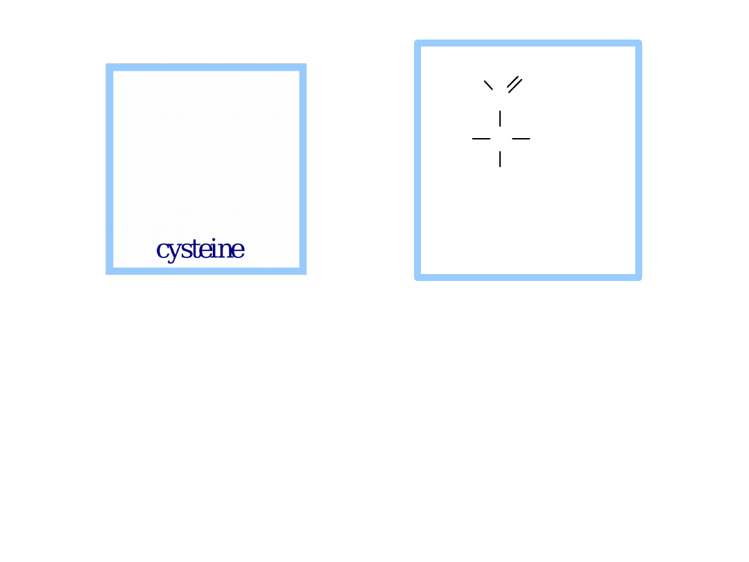
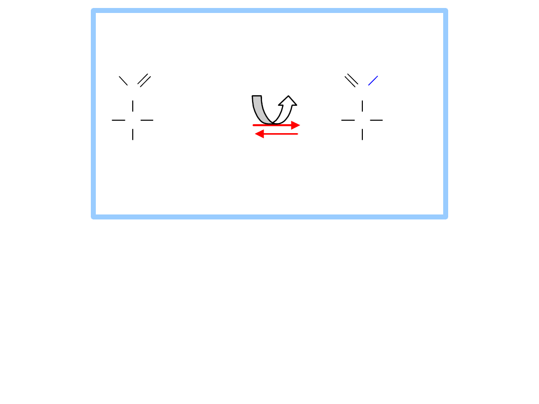
A
cysteine thiol
at the active site of
Glyceraldehyde-3-phosphate Dehydrogenase
has a role in catalysis.
The aldehyde of glyceraldehyde-3-phosphate
reacts with the cysteine thiol to form a
thiohemiacetal
intermediate.
C
C
CH
2
OPO
3
2
H
O
H
OH
1
2
3
glyceraldehyde-3-
phosphate
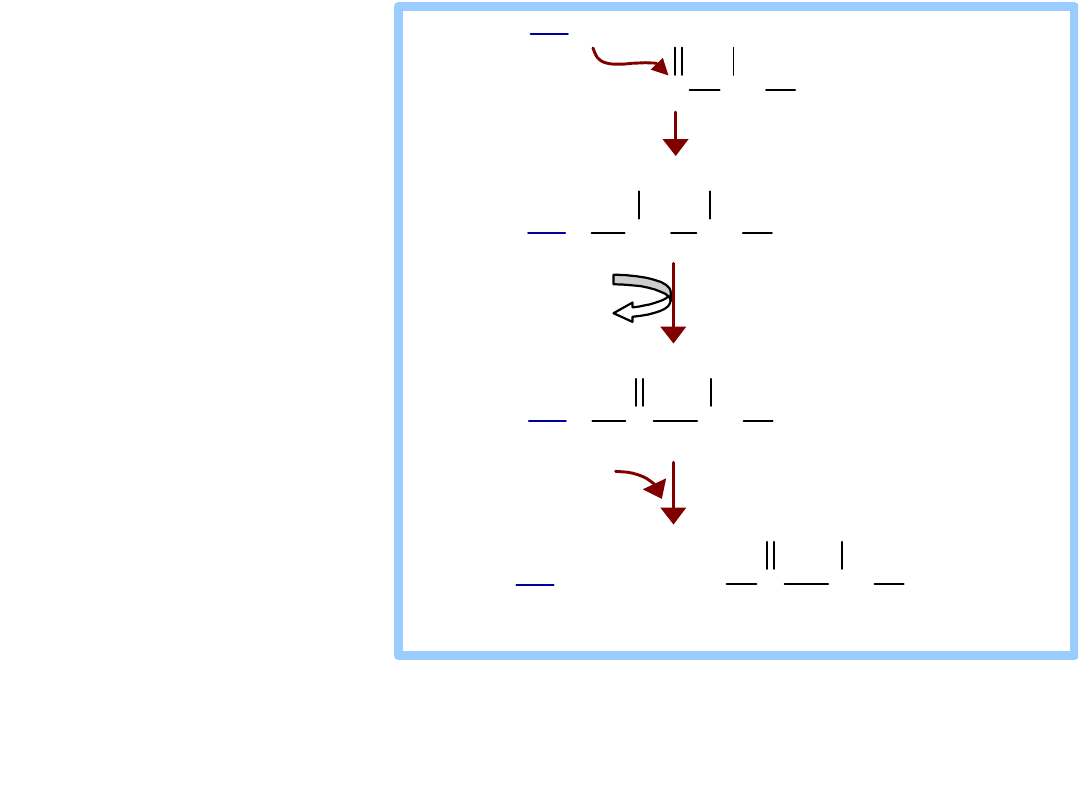
The “high energy” acyl thioester is attacked by
P
i
to yield the acyl phosphate (
~P
) product.
CH
CH
2
OPO
3
2
OH
Enz-Cys
SH
Enz-Cys
S
CH
CH
CH
2
OPO
3
2
OH
OH
Enz-Cys
S
C
CH
CH
2
OPO
3
2
OH
O
HC
NAD
+
NADH
Enz-Cys
SH
P
i
C
CH
CH
2
OPO
3
2
OH
O
O
3
PO
2
O
glyceraldehyde-3-
phosphate
1,3-bisphosphoglycerate
thiohemiacetal
intermediate
acyl-thioester
intermediate
Oxidation to a
carboxylic
acid (in a ~
thioester
)
occurs, as
NAD
+
is
reduced to
NADH
.
Recall that NAD
+
accepts 2 e
plus one H
+
(a hydride) in going to its reduced form.
N
R
H
C
NH
2
O
N
R
C
NH
2
O
H
H
+
2e
+
H
+
NA D
+
NA DH
C
C
CH
2
O PO
3
2
O
O PO
3
2
H
O H
C
C
CH
2
O PO
3
2
O
O
H
O H
A D P A T P
1
2
2
3
3
1
M g
2+
1 ,3 -bispho spho - 3 -p ho spho gly cerate
gly cerate
P ho spho gly cerate K inase
7. Phosphoglycerate Kinase
catalyzes:
1,3-bisphosphoglycerate + ADP
3-
phosphoglycerate +
ATP
This phosphate transfer is reversible (low
G), since one ~P bond is cleaved &
another synthesized.
The enzyme undergoes substrate-induced
conformational change similar to that of
Hexokinase.
C
C
CH
2
O H
O
O
H
O
PO
3
2
2
3
1
C
C
CH
2
O
PO
3
2
O
O
H
O H
2
3
1
3-phosphoglycerate 2-phosphoglycerate
P hosphoglycerate M utase
8. Phosphoglycerate Mutase
catalyzes:
3-phosphoglycerate
2-
phosphoglycerate
Phosphate is shifted from the OH on
C3 to the OH on C2.
C
C
CH
2
O H
O
O
H
O
PO
3
2
2
3
1
C
C
CH
2
O
PO
3
2
O
O
H
O H
2
3
1
3-phosphoglycerate 2-phosphoglycerate
Phosphoglycerate M utase
C
C
CH
2
OPO
3
2
O
O
H
OPO
3
2
2
3
1
2,3-bisphosphoglycerate
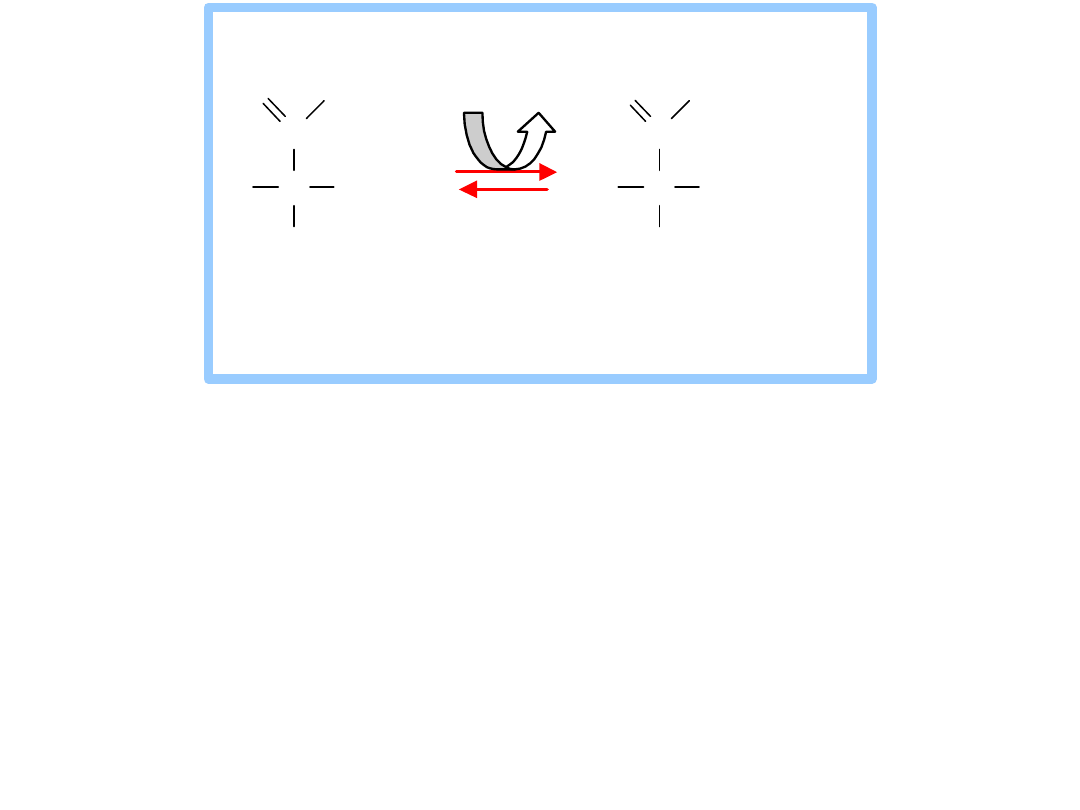
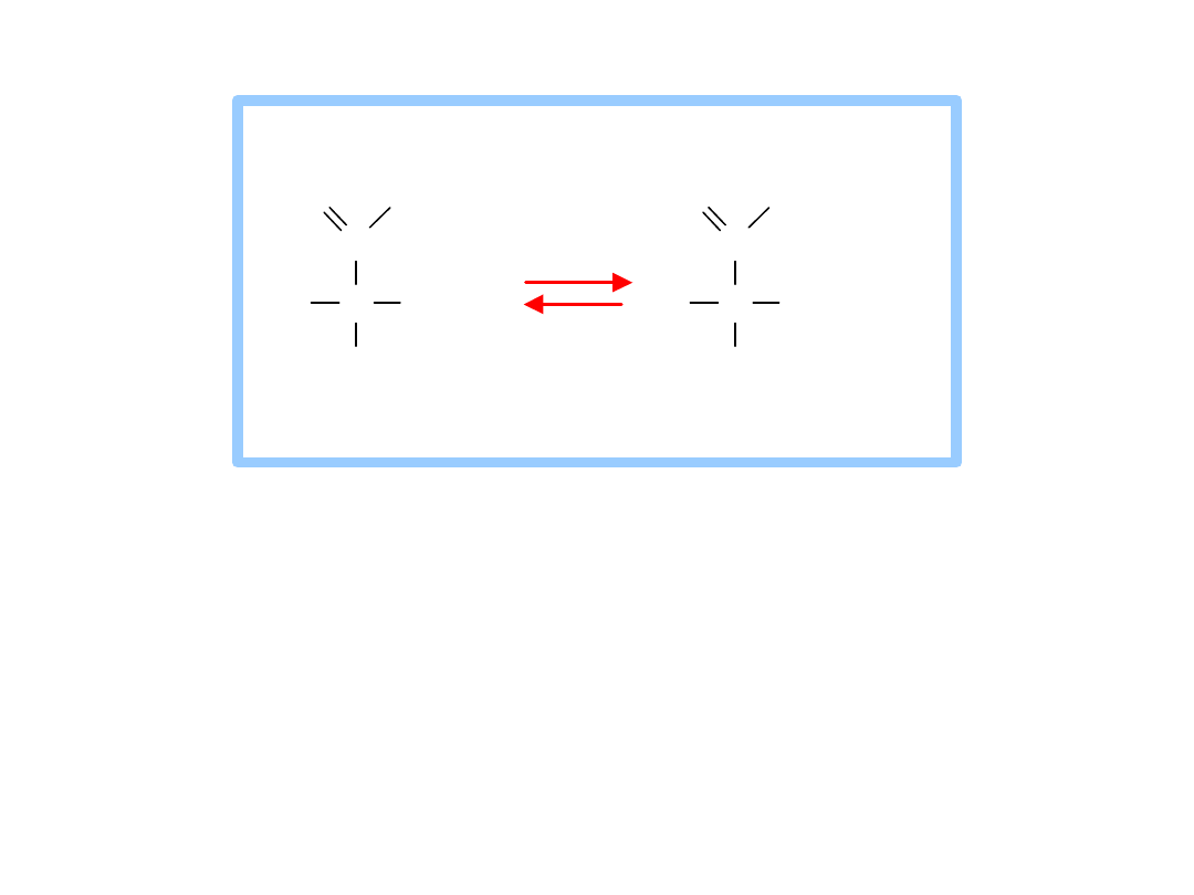
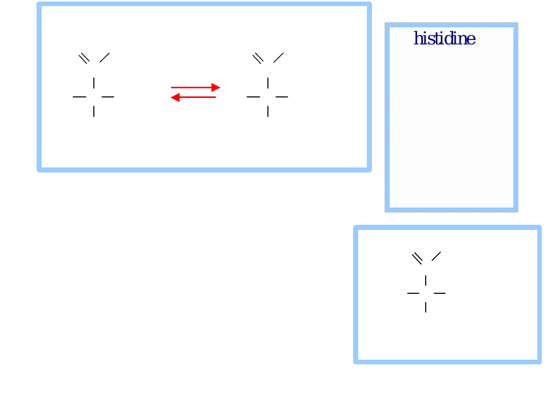
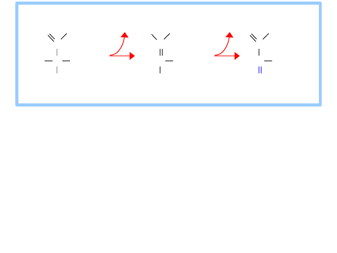
An active site
histidine
side-chain participates in P
i
transfer, by donating &
accepting phosphate.
The process involves a
2,3-bisphosphate
intermediate.
View an
of the
Phosphoglycerate Mutase
reaction.
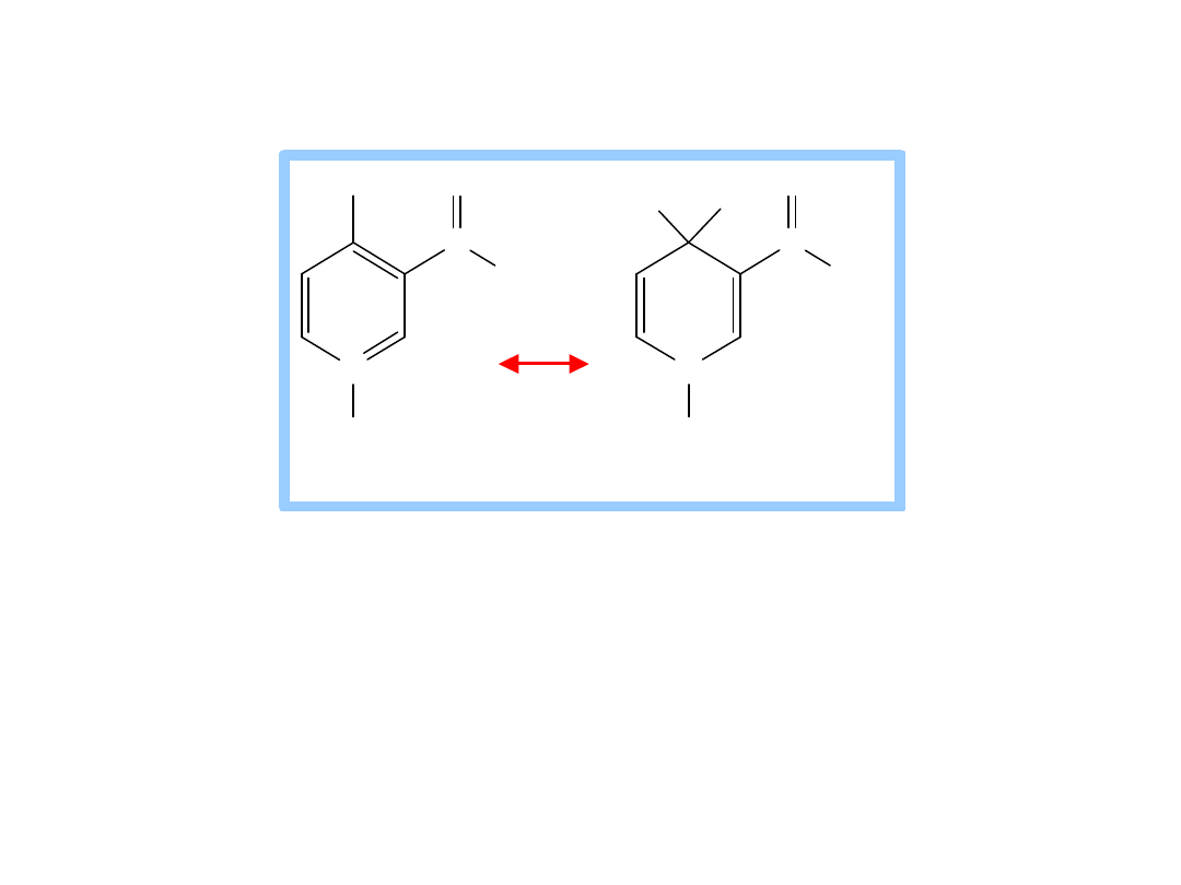
9. Enolase
catalyzes:
2-phosphoglycerate
phosphoenolpyruvate
+
H
2
O
This dehydration reaction is
Mg
++
-
dependent
.
2 Mg
++
ions interact with oxygen atoms of the
substrate
carboxyl
group at the active site.
The Mg
++
ions help to stabilize the enolate
anion intermediate that forms when a Lys
extracts H
+
from C #2.
C
C
C H
2
O H
O
O
H
O P O
3
2
C
C
C H
2
O H
O
O
O P O
3
2
C
C
C H
2
O
O
O P O
3
2
O H
2
3
1
2
3
1
H
2-phosphoglycerate
enolate intermediate
phosphoenolpyruvate
Enolase
10. Pyruvate Kinase
catalyzes:
phosphoenolpyruvate + ADP
pyruvate + ATP
C
C
CH
3
O
O
O
2
3
1
A D P A T P
C
C
CH
2
O
O
O
PO
3
2
2
3
1
phosphoenolpyruvate pyruvate
Pyruvate K inase
This phosphate transfer from PEP to ADP is
spontaneous
.
PEP has a larger G of phosphate
hydrolysis than ATP.
Removal of P
i
from PEP yields an unstable
enol, which spontaneously converts to the
keto form of pyruvate.
Required inorganic
cations
K
+
and Mg
++
bind
to anionic residues at the active site of
Pyruvate Kinase.
C
C
CH
3
O
O
O
2
3
1
A D P A T P
C
C
CH
2
O
O
O
PO
3
2
2
3
1
C
C
CH
2
O
O
O H
2
3
1
phosphoenolpy ruv ate enolpy ruv ate py ruv ate
P yruv ate K inase
Hexokinase
Phosphofructokinase
glucose
Glycolysis
ATP
ADP
glucose-6-phosphate
Phosphoglucose Isomerase
fructose-6-phosphate
ATP
ADP
fructose-1,6-bisphosphate
Aldolase
glyceraldehyde-3-phosphate
+
dihydroxyacetone-phosphate
Triosephosphate
Isomerase
Glycolysis continued
Glyceraldehyde-3-phosphate
Dehydrogenase
Phosphoglycerate Kinase
Enolase
Pyruvate Kinase
glyceraldehyde-3-phosphate
NAD
+
+
P
i
NADH
+
H
+
1,3-bisphosphoglycerate
ADP
ATP
3-phosphoglycerate
Phosphoglycerate Mutase
2-phosphoglycerate
H
2
O
phosphoenolpyruvate
ADP
ATP
pyruvate
Glycolysi
s
continue
d.
Recall
that
there are
2 GAP
per
glucose.

Glycolysis
Balance sheet
for
~P
bonds of ATP:
How many ATP ~P bonds expended? ________
How many ~P bonds of ATP produced?
(Remember there are two 3C fragments from
glucose.) ________
Net production of ~P bonds of ATP per
glucose: ________
2
4
2

Balance sheet
for
~P
bonds of ATP:
2 ATP expended
4 ATP produced (2 from each of two 3C
fragments from glucose)
Net production of
2 ~P
bonds of
ATP
per
glucose
.
Glycolysis - total pathway, omitting H
+
:
glucose + 2 NAD
+
+ 2 ADP + 2 P
i
2 pyruvate + 2 NADH + 2
ATP
In
aerobic organisms
:
pyruvate
produced in Glycolysis is oxidized to
CO
2
via Krebs Cycle
NADH
produced in Glycolysis & Krebs Cycle is
reoxidized via the respiratory chain, with
production of much additional ATP.
They
must reoxidize NADH
produced in
Glycolysis through some other reaction,
because
NAD
+
is needed for the
Glyceraldehyde-3-phosphate Dehydrogenase
reaction.
Usually NADH is reoxidized as
pyruvate
is
converted to a
more reduced
compound.
The complete pathway, including Glycolysis
and the reoxidation of NADH, is called
fermentation
.
C
C
CH
2
O PO
3
2
H
O
H
O H
C
C
CH
2
O PO
3
2
O
O PO
3
2
H
O H
+
P
i
+ H
+
N A D
+
N A D H
1
2
3
2
3
1
g l y c e ra l d e h y d e - 1 ,3 - b i s p h o s p h o -
3 - p h o s p h a te g l y c e ra te
G ly c e ra l d e h y d e - 3 - p h o s p h a te
D e h y d ro g e n a s e
Fermentation
:
Anaerobic
organisms
lack a
respiratory
chain.
C
C
CH
3
O
O
O
C
H
C
CH
3
O
O H
O
N A D H
+
H
+
N A D
+
L actate D ehy dro genase
py ruv ate lactate
E.g.,
Lactate Dehydrogenase
catalyzes
reduction
of the keto in
pyruvate
to a
hydroxyl, yielding
lactate
, as NADH is
oxidized to NAD
+
.
Lactate
, in addition to being an end-product
of fermentation, serves as a
mobile
form of
nutrient energy
, & possibly as a
signal
molecule in mammalian organisms.
Cell membranes contain
carrier
proteins that
facilitate transport of lactate.
C
C
CH
3
O
O
O
C
H
C
CH
3
O
O H
O
N A D H
+
H
+
N A D
+
L actate D ehy dro genase
py ruv ate lactate
Skeletal muscles
ferment glucose to
lactate
during exercise, when the exertion is brief and
intense.
Lactate
released to the
blood
may be taken
up by other tissues, or by skeletal muscle after
exercise, and converted via Lactate
Dehydrogenase back to
pyruvate
, which may
be oxidized in
Krebs Cycle
or (in liver)
converted to back to
glucose
via
gluconeogenesis
C
C
CH
3
O
O
O
C
H
C
CH
3
O
O H
O
N A D H
+
H
+
N A D
+
L actate D ehy dro genase
py ruv ate lactate
Lactate
serves as a
fuel
source for
cardiac
muscle
as well as
brain neurons
.
Astrocytes
, which surround and protect
neurons in the brain,
ferment glucose
to
lactate
and release it.
Lactate
taken up by adjacent neurons is
converted to pyruvate that is oxidized via
Krebs Cycle.
C
C
CH
3
O
O
O
C
CH
3
O
H
C
CH
3
O
H
H
H
N A D H
+
H
+
N A D
+
CO
2
P y ru v ate A lco h o l
D ecarb o x y lase D eh y d ro gen ase
p y ru v ate acetald eh y d e eth an o l
Some anaerobic organisms metabolize
pyruvate to
ethanol
, which is excreted as a
waste product.
NADH
is converted to
NAD
+
in the reaction
catalyzed by Alcohol Dehydrogenase.

Glycolysis
, omitting H
+
:
glucose + 2 NAD
+
+ 2 ADP + 2 P
i
2 pyruvate + 2
NADH + 2 ATP
Fermentation
, from glucose to lactate:
glucose + 2 ADP + 2 P
i
2 lactate +
2 ATP
Anaerobic catabolism
of glucose yields
only 2 “high energy” bonds of ATP.

Glycolysis Enzyme/Reaction
G
o
'
kJ/mo
l
G
kJ/mo
l
Hexokinase
-20.9
-27.2
Phosphoglucose Isomerase
+2.2
-1.4
Phosphofructokinase
-17.2
-25.9
Aldolase
+22.
8
-5.9
Triosephosphate Isomerase
+7.9
negati
ve
Glyceraldehyde-3-P
Dehydrogenase
& Phosphoglycerate Kinase
-16.7
-1.1
Phosphoglycerate Mutase
+4.7
-0.6
Enolase
-3.2
-2.4
Pyruvate Kinase
-23.0
-13.9
*Values in this table from D. Voet & J. G. Voet (2004) Biochemistry, 3
rd
Edition, John Wiley & Sons, New York, p. 613.

Flux
through the Glycolysis pathway is
regulated
by
control of 3 enzymes that catalyze
spontaneous
reactions:
Hexokinase, Phosphofructokinase & Pyruvate
Kinase
.
Local control
of metabolism involves regulatory
effects of varied concentrations of pathway
substrates
or
intermediates
, to benefit the cell.
Global control
is for the benefit of the whole
organism, & often involves
hormone-activated
signal cascades
.
Liver
cells have major roles in metabolism,
including maintaining blood levels various of
nutrients such as glucose. Thus global control
especially involves liver.
Some aspects of global control by hormone-
activated signal cascades will be discussed later.
Hexokinase
is
inhibited
by
product
glucose-6-
phosphate
:
by
competition
at the
active site
by
allosteric
interaction at a
separate
enzyme site.
Cells
trap glucose
by
phosphorylating
it,
preventing exit on glucose carriers.
Product inhibition
of Hexokinase ensures that cells
will not continue to accumulate glucose from the
blood, if [glucose-6-phosphate] within the cell is
ample.
H
O
O H
H
O H
H
O H
CH
2
O H
H
O H
H
H
O
O H
H
O H
H
O H
CH
2
O
PO
3
2
H
O H
H
2
3
4
5
6
1
1
6
5
4
3
2
A T P A DP
M g
2+
glucose glucose-6-phosphate
Hexokinase
Glucokinase
has a
high K
M
for
glucose
.
It is
active
only
at high [glucose]
.
One effect of
insulin
, a hormone produced when blood
glucose is high, is
activation
in liver of
transcription
of the gene that encodes the
Glucokinase
enzyme.
Glucokinase is
not
subject to product inhibition
by
glucose-6-phosphate. Liver will take up &
phosphorylate glucose even when liver [glucose-6-
phosphate] is high.
H
O
O H
H
O H
H
O H
CH
2
O H
H
O H
H
H
O
O H
H
O H
H
O H
CH
2
O
PO
3
2
H
O H
H
2
3
4
5
6
1
1
6
5
4
3
2
A T P A DP
M g
2+
glucose glucose-6-phosphate
Hexokinase
Glucokina
se
is a
variant of
Hexokinase
found in
liver
.

Glucokinase is subject to
inhibition
by
glucokinase regulatory protein
(
GKRP
).
The ratio of Glucokinase to GKRP in liver
changes in different metabolic states,
providing a mechanism for modulating
glucose phosphorylation.
Glucose-6-phosphatase
catalyzes hydrolytic
release of P
i
from glucose-6-P. Thus
glucose
is
released
from the liver to the blood as needed
to maintain blood [glucose].
The enzymes Glucokinase & Glucose-6-phosphatase,
both found in
liver
but not in most other body
cells, allow the liver to control blood [glucose].
Glycogen Glucose
Hexokinase or Glucokinase
Glucose-6-Pase
Glucose-1-P Glucose-6-P Glucose + P
i
Glycolysis
Pathway
Pyruvate
Glucose metabolism in liver.
Glucokinase
,
with high K
M
for glucose,
allows liver
to store
glucose
as glycogen in
the fed
state
when blood
[glucose] is high.
High [glucose]
within liver cells causes a transcription
factor
carbohydrate responsive element binding
protein
(
ChREBP
) to be transferred into the nucleus,
where it activates
transcription
of the gene for Pyruvate
Kinase.
This facilitates converting
excess glucose
to
pyruvate
,
which is metabolized to
acetyl-CoA
, the main precursor
for synthesis of
fatty acids
, for long term energy storage.
C
C
CH
3
O
O
O
2
3
1
A D P A T P
C
C
CH
2
O
O
O
PO
3
2
2
3
1
phosphoenolpyruvate pyruvate
Pyruvate K inase
Pyruvate Kinase
,
the last step
Glycolysis, is
controlled
in
liver
partly by
modulation of the
amount
of
enzyme
.
Phosphofructokinase
is usually the
rate-limiting step
of the Glycolysis pathway.
Phosphofructokinase is
allosterically inhibited by
ATP
.
At
low
concentration, the substrate
ATP
binds
only
at
the
active site
.
At
high
concentration, ATP binds
also
at a low-affinity
regulatory site
, promoting the tense conformation.
CH
2
O PO
3
2
O H
CH
2
O H
H
O H
H
H
HO
O
6
5
4
3
2
1
CH
2
O PO
3
2
O H
CH
2
O
PO
3
2
H
O H
H
H
HO
O
6
5
4
3
2
1
A T P A D P
M g
2 +
fructo se-6-pho sphate fructo se-1,6-bispho sphate
P ho spho fructo k inase
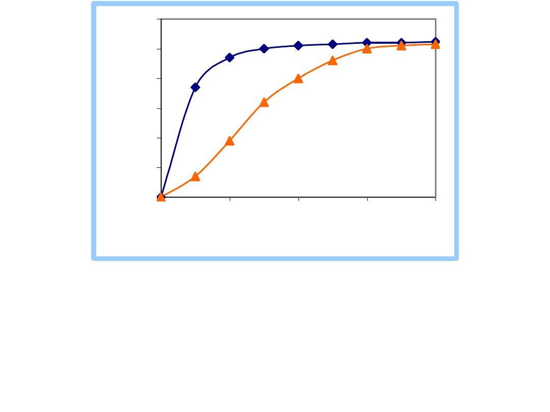
The
tense
conformation of PFK, at
high [ATP]
, has lower
affinity for the other substrate, fructose-6-P.
Sigmoidal
dependence of reaction rate on [fructose-6-P] is seen.
AMP
, present at significant levels only when there is
extensive ATP hydrolysis, antagonizes effects of high
ATP.
0
10
20
30
40
50
60
0
0.5
1
1.5
2
[Fructose-6-phosphate] mM
P
F
K
A
c
ti
v
it
y
high [ATP]
low [ATP]
Inhibition of the Glycolysis enzyme
Phosphofructokinase when [ATP] is high
prevents breakdown of glucose in a pathway
whose main role is to make ATP.
It is more useful to the cell to store glucose as
glycogen when ATP is plentiful.
Glycogen Glucose
Hexokinase or Glucokinase
Glucose-6-Pase
Glucose-1-P Glucose-6-P Glucose + P
i
Glycolysis
Pathway
Pyruvate
Glucose metabolism in liver.
Document Outline
- Slide 1
- Slide 2
- Slide 3
- Slide 4
- Slide 5
- Slide 6
- Slide 7
- Slide 8
- Slide 9
- Slide 10
- Slide 11
- Slide 12
- Slide 13
- Slide 14
- Slide 15
- Slide 16
- Slide 17
- Slide 18
- Slide 19
- Slide 20
- Slide 21
- Slide 22
- Slide 23
- Slide 24
- Slide 25
- Slide 26
- Slide 27
- Slide 28
- Slide 29
- Slide 30
- Slide 31
- Slide 32
- Slide 33
- Slide 34
- Slide 35
- Slide 36
- Slide 37
- Slide 38
- Slide 39
- Slide 40
- Slide 41
- Slide 42
- Slide 43
- Slide 44
- Slide 45
- Slide 46
- Slide 47
- Slide 48
Wyszukiwarka
Podobne podstrony:
prezentacja ang tekst
Prezentacja ang
Wiosna prezentacja ang
prezentacja ang
rozmnarzanie psów starsze, ZOOTECHNIKA, Ang, Prezentacja na ang
Prezentacja1 glikoliza
Ulcerative colitis, Prywatne, Materiały - Rok II, ang, prezentacja
po ang treść prezentacji
ang prezentacja
Ang prezentacja sylwester
prezentacja finanse ludnosci
prezentacja mikro Kubska 2
Religia Mezopotamii prezentacja
Prezentacja konsument ostateczna
Hydrocephalus(ang)
Strategie marketingowe prezentacje wykład
motumbo www prezentacje org
więcej podobnych podstron