
1
Image – the Angel Oak
Lecture #4 – Plant Structure,
Growth And Development

2
Key Concepts:
• What is a kingdom?
• Why study plants?
• What makes a plant a plant?
• The hierarchy of structure – plant cells,
tissues and organs
• Growth
• Primary growth – elongation
• Secondary growth – diameter expansion
• Morphogenesis occurs during growth

3
Image – Linnaeus
Carolus
Linnaeus
(1707-1778)
The founder of
modern
taxonomy
defined
kingdoms by
morphological
similarity

4
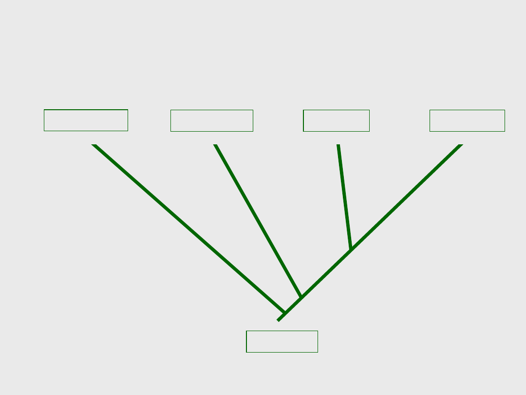
Linnaeus’ Taxonomic
Hierarchy
Taxonomic Category
Example (taxon)
Kingdom
Plantae, also Metaphyta = all plants
Division (phylum)
Magnoliophyta = all angiosperms
Class
Liliopsida = all monocots
Order
Asparagales = related families
(Orchidaceae, Iridaceae, etc)
Family
Orchidaceae = related genera
(Platanthera,
Spiranthes, etc)
Genus
Platanthera = related species
(P. ciliaris, P.
integra, etc)
Specific name/epithet
ciliaris = one species

5
Linnaeus’ Taxonomic
Hierarchy
Taxonomic Category
Example (taxon)
Kingdom
Plantae, also Metaphyta = all plants
Division (phylum)
Magnoliophyta = all angiosperms
Class
Liliopsida = all monocots
Order
Asparagales = related families
(Orchidaceae, Iridaceae, etc)
Family
Orchidaceae = related genera
(Platanthera,
Spiranthes, etc)
Genus
Platanthera = related species
(P. ciliaris, P.
integra, etc)
Specific name/epithet
ciliaris = one species

6
Images – the yellow fringed orchid
Platanthera ciliaris

7
Images – the 3 multicellular kingdoms, animals, fungi and plants
Linnaeus recognized only 2
kingdoms
• If it moved – animal; if it didn’t – plant
• Fungi were lumped with plants
• The microscopic world was largely
unknown

8
Diagram – the 5 kingdom system
The 5 kingdom system – developed in
the 1960’s and used until recently

9
Diagram – 3 domain system of classification
Molecular data supports 3
domain classification scheme
Kingdoms are defined by monophyletic lineage

10
Diagram – transition from 5 kingdom to 3
domain system indicating dynamic nature of
classification
Classification is Dynamic!
Multicellular eukaryotes remain fairly well
defined – the plants, fungi and animals.
Classification of single celled organisms is still
underway.

11
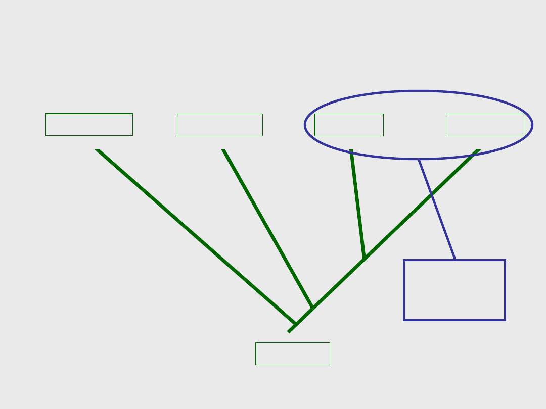
Current Taxonomic
Hierarchy
Taxonomic Category
Example (taxon)
Domain
Eukarya = all eukaryotic organisms
Kingdom
Plantae, also Metaphyta = all plants
Division (phylum)
Magnoliophyta = all angiosperms
Class
Liliopsida = all monocots
Order
Asparagales = related families
(Orchidaceae, Iridaceae, etc)
Family
Orchidaceae = related genera
(Platanthera,
Spiranthes, etc)
Genus
Platanthera = related species
(P. ciliaris, P.
integra, etc)
Specific name/epithet
ciliaris = one species

12
Why Plants?

13
Image – shooting stars
Why Plants?

14
What makes a plant a plant???

15
Images and diagrams – characteristics that
separate plants from other kingdoms

16
What makes a plant a plant???
• Multicellular, eukaryotic organisms with extensive
specialization
• Almost all are photosynthetic, with chloroplasts (=
green)
Some obtain additional nutrition through parasitism or carnivory
Some are saprophytic, entirely without chlorophyll (absorb dead
OM)
• Excess carbohydrates stored as starch (coiled, branched
polymer of glucose)
• Cell walls of cellulose = fibrous (not branched)
polysaccharide = accounts for the relative rigidity of the
cell wall
• Cell division by formation of cell plate
• Most extant plant species are terrestrial (many
characteristics that are adapted for terrestrial life)
• Separated from cyanobacteria by chloroplasts
• Separated from green algae by various adaptations to
terrestrial life
Read this later….

17
Plants were the first organisms
to move onto land
• Occurred about 475mya
• Very different conditions from former
marine habitat
• Many new traits emerged in
adaptation to life on dry land
• Extensive adaptive radiation into
many new ecological niches

18
Diagram – phylogeny of land
plants; same on next slide
Four major
groups of
plants have
emerged since
plants took to
land

19
We will focus
on
angiosperms
Next semester
in 211 you will
learn more
about the
transition from
water to land,
and the
evolution of
reproductive
strategies in all
plants

20
Images – flowering plants
Angiosperms – the flowering
plants:
90% of the Earth’s modern flora

21
Diagram – plant cell; same on next slide
Basic Structure of the Plant Cell –
what’s unique???

22
Basic Structure of the Plant
Cell

23
Critical Thinking
• Do all plant cells have
chloroplasts???
• How can you tell???

24
Critical Thinking
• Do all plant cells have
chloroplasts???
• How can you tell???

25
Image – chloroplast free
white bracts on white-top
sedge
Critical Thinking
• Do all plant cells
have
chloroplasts???
• How can you tell???

26
Diagram – primary and
secondary cell walls; same on
next slide
More on the cell wall:
• All cell walls are
produced by the cell
membrane, outside
• Primary wall is
produced first
Mostly cellulose
• Secondary walls are
produced later
Lignified, so
???
• Secondary walls are
interior to primary
walls

27
More on the cell wall:
• All cell walls are
produced by the
cell membrane
• Primary wall is
produced first
Mostly cellulose
• Secondary walls are
produced later
Lignified, so
• Secondary walls are
interior to primary
walls

28
Micrographs – plant cell types
Five Major
Plant Cell
Types
• Parenchyma
• Collenchyma
• Sclerenchyma
• Xylem elements
• Phloem
elements

29
Micrographs – parenchyma cells
Parenchyma
• Thin primary wall
• No secondary wall
• Many metabolic and storage functions
• Bulk of the plant body

30
Micrograph – collenchyma
cells; same on next slide
Collenchyma
• Thick primary
wall
• No secondary
wall
Implications???
• Support
growing
tissues

31
Collenchyma
• Thick primary
wall
• No secondary
wall
Implications???
• Support
growing tissues

32
Micrograph –
sclerenchma cells;
same on next slide
Sclerenchyma
• Thick secondary wall
• Secondary walls are
lignified
Implications???
• Support mature
plant parts
• Often dead at
maturity

33
Sclerenchyma
• Thick secondary wall
• Secondary walls are
lignified
Implications???
• Support mature
plant parts
• Often dead at
maturity

34
Micrographs – collenchyma and sclerenchyma cell comparison
Collenchyma vs. Sclerenchyma
• Both provide structural support
• Both have thick walls
• Collenchyma = thick primary wall, no lignin
• Sclerenchyma = thick secondary wall, lignified

35
Diagrams and
micrograph –
tracheids and vessel
elements
Xylem
Elements
• Lignified secondary
walls
• Always dead at
maturity (open)
• Function to transport
water and dissolved
nutrients, and to
support the plant
• Tracheids and vessel
elements

36
Micrograph – rings of lignin in
developing vessel element;
same on next slide
Critical
Thinking
• Vessel elements
and the convergent
evolution of rings
• What else looks
like this????
• What is the
function????

37
Critical
Thinking
• Vessel elements
and the
convergent
evolution of
rings
• What else looks
like this????
• What is the
function????

38
Micrograph – phloem elements
Phloem
Elements
• Sieve tube members +
companion cells
• STM lack nucleus,
ribosomes – their
metabolism is controlled
by the companion cells
• Function to transport
the products of
metabolism
• Non-angiosperms have
more primitive phloem
elements

39
Diagram – phloem elements
Critical Thinking
• What might be the functional
advantage of a cell with no
nucleus???

40
Critical Thinking
• What might be the functional
advantage of a cell with no
nucleus???

41
Micrographs – plant cell types
Plants are
Simple
Only Five
Major Cell
Types
• Parenchyma
• Collenchyma
• Sclerenchyma
• Xylem elements
• Phloem elements

42
Diagram – plant tissue types
Tissue
Systems
• Epidermis
• Vascular
• Ground
• Meristem

43
Micrograph
and diagram
– epidermis
Epidermis Tissue:
• Covers the outer surface of
all plant parts
• Shoot surfaces covered
with waxy cuticle
Helps to protect the plant
and prevent desiccation
• Usually a single,
transparent cell layer
• Tight joints; stomata allow
for gas exchange

44
Critical Thinking
• Do roots have a waxy cuticle???
• Why or why not???

45
Critical Thinking
• Do roots have a waxy cuticle???
• Why or why not???
Never forget the importance
of natural selection!!!!!

46
Micrograph –
vascular bundle in
cross section
Vascular Tissue:
• Transports water, solutes,
and metabolic products
throughout the plant
• Confers structural
support
• Includes xylem elements,
phloem elements,
parenchyma and
sclerenchyma fibers

47
Critical Thinking
• Why does vascular tissue give
structural support to a plant???

48
Critical Thinking
• Why does vascular tissue give
structural support to a plant???

49
Micrograph and diagram –
ground tissues in stems and
leaves
Ground Tissue:
• Bulk of the plant
body – pith, cortex
and mesophyll
• Mostly parenchyma
• Most metabolic,
structural and
storage functions

50
Micrograph – herbaceous dicot stem
Critical Thinking
• Is this what the inside of a tree looks
like???

51
Micrograph of herbaceous eudicot stem; image of
woody stem; diagram of woody stem tissue
organization
Critical Thinking
• Is this what the inside of a tree looks
like???

52
Image – new growth at tip of stem
Meristem Tissue:
• How the plant grows
• Cells divide constantly during the
growing season to make new tissues
• More details later

53
Diagram – plant tissue systems
Plants are
Simple
Only Four Major
Tissue Types
• Epidermis
• Vascular
• Ground
• Meristem

54
Tissues Make Organs:
• Roots – anchor the plant, absorb
water and nutrients
• Stems – support the leaves
• Leaves – main site of photosynthesis
• Reproductive organs (flowers, cones,
etc – more later)
All organs have additional functions –
hormone synthesis, transport, etc…

55
Diagram – root and shoot systems
Plant Organ Systems
56
ancestral
paleoherbs
magnoliids
eudicots
monocots
Modern molecular evidence
indicates four classes of
angiosperms

57
Images – water lily and magnolia
Paleoherbs and Magnoliids comprise
about 3% of angiosperms
Paleoherbs
• Aristolochiaceae,
Nymphaeaceae,
etc
Magnoliids
• Magnoliaceae,
Lauraceae,
nutmeg, black
pepper, etc
58
ancestral
paleoherbs
magnoliids
eudicots
monocots
Modern evidence indicates 4 classes
of angiosperms
~ 97% of
angiosperm
s

59
Images – monocots
Monocots include grasses,
sedges, iris, orchids, lilies,
palms, etc…..

60
Images – eudicots
Eudicots include 70+% of all
angiosperms:
• Most broadleaf trees and shrubs
• Most fruit and vegetable crops
• Most herbaceous flowering plants

61
Monocots vs. Eudicots
Monocots
• Flower parts in
multiples of 3
• Parallel leaf venation
• Single cotyledon
• Vascular bundles in
complex
arrangement
• ~90,000 species
Eudicots
• Flower parts in
multiples of 4 or 5
• Netted leaf venation
• Two cotyledons
• Vascular bundles in a
ring around the stem
• Modern classification
indicates 2 small
primitive groups +
eudicots
• 200,000+ species

62
Micrographs – cross sections of eudicot and
moncot roots; same on next 3 slides
Root System Tissue
Organization
Eudicots
Monocots
Epidermis, ground, endodermis, pericycle, vascular
tissues
63
Eudicot root – closeup
Epidermis
Cortex
Endodermis
Pericycle
Vascular
tissues – in
solid core
64
Monocot root – closeup
Epidermis
Cortex
Endodermis
Pericycle
Vascular tissues –
in ring
Pith in the very
center

65
Critical Thinking
• Where do branch roots form???

66
Micrograph – root emerging from pericycle
Critical Thinking
• Where do branch roots form???

67
Micrograph – eudicot and monocot stem
tissue organization; same on next 4
slides
Stem System Tissue
Organization
Eudicots
Monocots
Epidermis, ground, vascular
tissues
68
Eudicot stem – closeup
Epidermis
Cortex
Vascular
tissues –
bundles
in a ring
Pith
69
Monocot stem – closeup
Epidermis
Cortex
Vascular
tissues –
bundles are
scattered

70
Wood forms from a meristem
that links the vascular bundles:

71
Stem System Tissue
Organization
Eudicots
Monocots
Monocots cannot make wood
More on wood formation later

72
Micrograph – cross-section of leaf tissue arrangement
Leaf Tissue Arrangement
Epidermis, ground, vascular
tissues
73
Diagram – leaf tissue arrangement
Leaf closeup
Epidermis
Cortex –
palisade
mesophyll
Cortex –
spongy
mesophyll
Vascular
tissues

74
Micrograph – epidermis
tissue showing stomata
Stomata – pores to allow for gas
exchange and transpiration

75
Diagram – shoot and root systems
See, plants really are simple
• 5 cell types
• 4 tissue types
• 4 organ types

76
Plant Growth
• Remember, most plants are anchored by
roots
• They can’t move to escape or take
advantage of changes in their
environment
• Plants adjust to their environment
• Simple structure + lots of developmental
flexibility allow plants to alter when and
how they grow
Developmental flexibility comes
from meristems

77
Meristem Tissues
• Actively dividing cells that generate
all other cells in the plant body
• Cause indeterminate growth
Stems and roots elongate throughout the
plant’s life (indeterminate primary
growth)
Trees continually expand in diameter
(indeterminate secondary growth)
Branches form in roots and stems

78
Not all plant parts have
indeterminate growth
patterns
Indeterminate:
Roots
and
Stems
These parts grow
throughout the life
of the plant,
exploring new
environments or
responding to
damage
Determinate:
Leaves
Flowers
Fruits
These parts grow
to a genetically +/-
predetermined size
and shape and
then stop – cannot
repair damage

79
Some mature cells can
de-differentiate to
become meristematic
once more!!!
• Primarily occurs in the indeterminate
parts
Stems and roots
• A process that very seldom occurs in
other kingdoms
• Allows stems and roots to repair damage
and form branches and sprouts

80
Critical Thinking
• Can all plant cells de-differentiate???
• What would control this???

81
Critical Thinking
• Can all plant cells de-differentiate???
• What would control this???

82
Critical Thinking
• Can all plant cells de-differentiate???
• What would control this???

83
Growth in Plants:
an irreversible increase in size
due to metabolic processes
(processes that use ATP energy)
• Cell division produces new cells =
function of meristem
• Cell expansion increases the size of the
new cells = up to 80% of size increase
• Cell differentiation occurs during and
after expansion

84
Diagram – planes of cell division and the effect on morphogenesis
The plane of cell division contributes to
morphogenesis
Division in 2 planes forms sheets of cells

85
Critical Thinking
• What tissues are files of cells???
• What tissues are sheets of cells???
• What tissues are 3-D bulky???

86
Critical Thinking
• What tissues are files of cells???
• What tissues are sheets of cells???
• What tissues are 3-D bulky???

87
Growth in Plants:
an irreversible increase in size
due to metabolic processes
(processes that use ATP energy)
• Cell division produces new cells =
function of meristem
• Cell expansion increases the size of the
new cells = up to 80% of size increase
• Cell differentiation occurs during and
after expansion

88
Diagram – how auxin works to promote cell expansion
Auxin-mediated cell
expansion
ATP is used

89
Diagram – cellulose orientation in
primary wall and the effects on
morphogenesis
The direction of cell expansion depends on
cellulose orientation, and contributes to
morphogenesis

90
Growth in Plants:
an irreversible increase in size
due to metabolic processes
(processes that use ATP energy)
• Cell division produces new cells =
function of meristem
• Cell expansion increases the size of the
new cells = up to 80% of size increase
• Cell differentiation occurs during and
after expansion

91
Diagram – patterns
of growth in roots
Expansion
and
differentiation
occur in an
overlapping
zone in all
plant parts

92
REVIEW:
Growth in Plants:
an irreversible increase in size
due to metabolic processes
(processes that use ATP energy)
• Cell division produces new cells =
function of meristem
• Cell expansion increases the size of the
new cells = up to 80% of size increase
• Cell differentiation occurs during and
after expansion

93
Diagram – location
of meristems on the
plant body; next
slide also
Location of
the
meristems
determines
the pattern
of plant
growth
Most
common
meristems:
apical,
axillary and
lateral

94
Apical
meristems
cause
elongation of
roots and
stems

95
Micrograph – longitudinal section showing distribution of tissues in root

96
Images – root cap and mucigel

97
Root Cap
• Protects the meristem
• Secretes mucigel
Eases movement of roots through soil
Secretes chemicals that enhance nutrient
uptake
• Constantly shedding cells
Mechanical abrasion as roots grow through
soil
• Constantly being replenished by
meristem

98
Diagram – longitudinal section of root
showing zones of growth; same on next 2
slides
Primary Growth in Roots

99
Primary Growth in Roots

100
Primary Growth in Roots

101
Micrograph – root hairs
extending from
epidermis; same on
next few slides
Root Hairs
• Form as the epidermis
fully differentiates
• Extensions off epidermal
cells
NOT files of cells
Part of an epidermal cell
• Hugely increase the
surface area of the
epidermis
• 10 cubic cm (double
handful) of soil might
contain 1 m of plant
roots
Mostly root hairs

102
Critical Thinking
• What is the selective advantage of
root hairs???

103
Critical Thinking
• What is the selective advantage of
root hairs???

104
Root Hairs
• By contrast, 10 cc of soil
may contain up to 1000
m of fungal hyphae
(1km!)
These serve a similar
function for the fungus
Ramify throughout the
substrate for maximum
absorption
Some fungi form symbiotic
associations with plant
roots and both organisms
benefit from this huge
absorptive surface area!
More in 211…..

105
Diagram – location of apical meristems
Apical
meristems
cause
elongation of
roots and
stems

106
Micrograph – longitudinal
section of stem showing
apical and axillary
meristems
Apical Meristems in Shoots

107
Critical Thinking
• There is no “shoot cap” – why not???

108
Critical Thinking
• There is no “shoot cap” – why not???

109
Diagram – meristem locations
Axillary
meristems
allow for
branching –
similar in
structure
and function
to apical
meristems
Remember, pericycle
in roots has same
function

110
Micrograph – longitudinal
section of stem showing
apical and axillary
meristems; same on next
two slides
Axillary Meristems in Shoots

111
Primary Growth in Shoots
• Apical
meristem
• Leaf primordia
• Axillary buds

112
As with roots –
cell division
occurs first; zones
of expansion and
differentiation
overlap
Axillary buds may
activate to make
branches, or may
remain dormant

113
Diagram – how stems elongate during primary growth
Primary growth of a shoot – elongation from
the tip

114
Diagram – meristem locations
Lateral
meristems
cause
diameter
expansion
Roots also
expand in
diameter, but it’s
more
complicated –
we’ll save that
for BIOL 300

115
Diagram – lateral meristems
Lateral Meristems = Cambiums

116
Diagram – primary vs. secondary growth
Remember:
Elongation is
primary growth
Diameter
expansion is
secondary
growth

117
Images – cross section of wood and whole tree
Secondary
growth –
diameter
expansion

118
Micrograph – cross section of a
eudicot stem; same on next 2
slides
Eudicot Stem – recall the
arrangement of vascular
bundles
119
Eudicot Stem – recall the
arrangement of vascular
bundles
Vascular
cambium
forms here:

120
Eudicot Stem – recall the
arrangement of vascular
bundles
Vascular
cambium
forms here:
a cylinder of
meristem
tissue
between the
xylem to the
interior and
the phloem
to the
exterior

121
Diagram – location of the
vascular cambium relative to
other tree tissues
Secondary xylem and phloem form
through cell division by the vascular
cambium

122
Diagram – transition from
primary growth to secondary
growth; same on next slide
During primary
growth the vascular
tissues form in
bundles
from the
apical meristem
During secondary
growth the vascular
tissues form in
cylinders
from the
vascular cambium
2
o
xylem to the
inside
2
o
phloem to the
outside

123
Secondary
xylem
accumulate
s

124
Micrograph – cross section of woody
plant showing secondary tissues; same
on next slide
Secondary Xylem = Wood!

125
Annual growth rings are
accumulating rings of secondary
xylem

126
Diagram –
pattern of
accumulation
of secondary
xylem as a tree
grows; same on
next slide
Critical Thinking
• Why do eudicot trees taper???

127
Critical Thinking
• Why do eudicot trees taper???

128
Bark
• All tissues external to the vascular
cambium
• Diameter expansion splits original
epidermis
Bark structurally and functionally
replaces epidermis
• Inner bark
Functional secondary phloem
• Outer bark
Composition varies as tree matures

129
Micrograph – cross section of a tree showing bark formation
Bark Formation

130
Cork Cambium
• Meristematic tissue
• Forms in a cylinder during 2
o
growth
• Divides to produce cork cells
Cells filled with waxy, waterproof suberin
• Eventually cork cambium becomes
cork itself

131
More on cork cambium
• First layer develops from cortex
De-differentiation!!!
• Second layer forms from cortex –
same process
• Third layer forms from cortex…..
• Cortex eventually runs out
• Then what???

132
More on cork cambium
• First layer develops from cortex
De-differentiation!!!
• Second layer forms from cortex –
same process
• Third layer forms from cortex…..
• Cortex eventually runs out
• Then what???

133
More on cork cambium
• First layer develops from cortex
De-differentiation!!!
• Second layer forms from cortex –
same process
• Third layer forms from cortex…..
• Cortex eventually runs out
• Then what???

134
More on cork cambium
• First layer develops from cortex
De-differentiation!!!
• Second layer forms from cortex –
same process
• Third layer forms from cortex…..
• Cortex eventually runs out
• Then what???

135
Diagram – lateral meristems
and the secondary tissues in
a tree; same on next slide
Critical Thinking
• What is the next available layer of
tissue???

136
Critical Thinking
• What is the next available layer of
tissue???

137
More on cork cambium
• First layer develops from cortex
De-differentiation!!!
• Second layer forms from cortex –
same process
• Third layer forms from cortex…..
• Cortex eventually runs out
• Then what???

138
More on cork cambium
• First layer develops from cortex
De-differentiation!!!
• Second layer forms from cortex –
same process
• Third layer forms from cortex…..
• Cortex eventually runs out
• Then what???
139
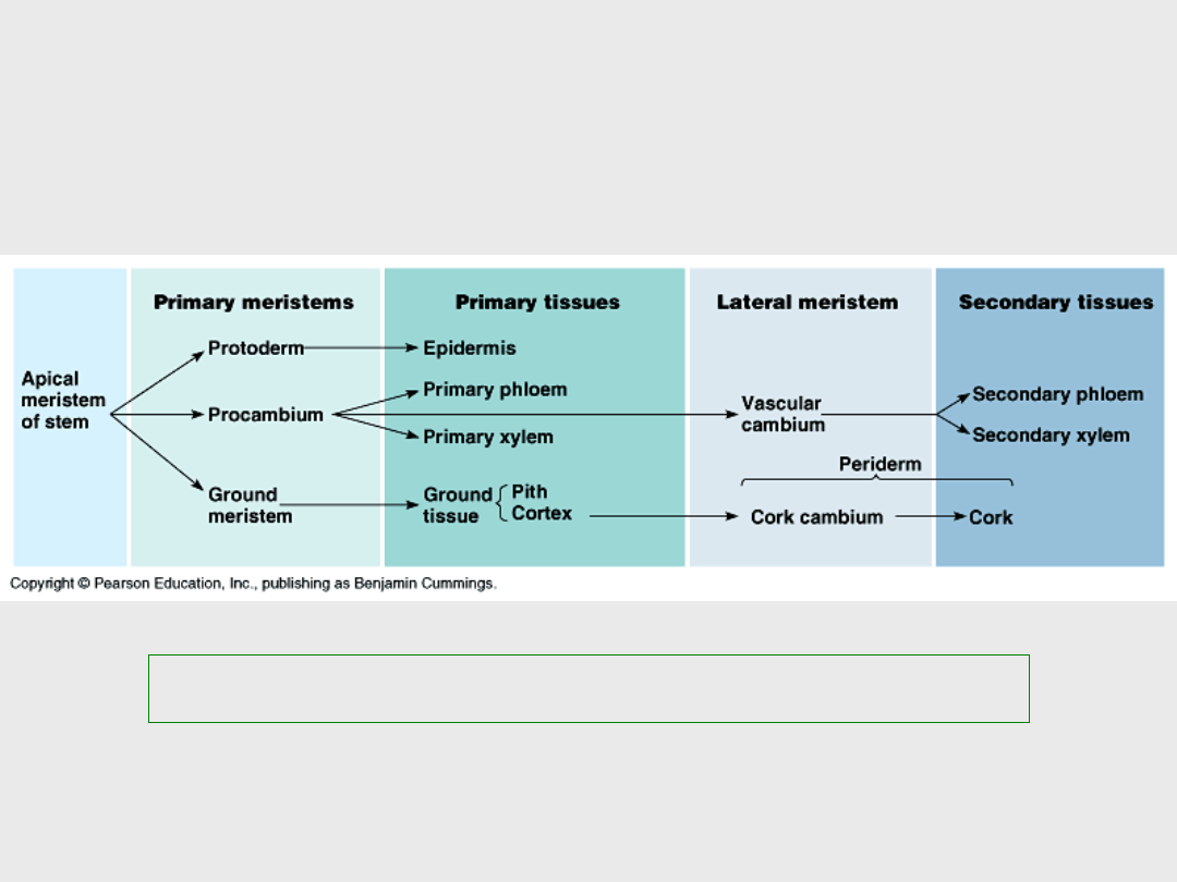
Diagram – how undifferentiated cells develop into the tissues of the plant body
Stem Tissue Derivations and
Fates:
Cells divide, expand and differentiate

140
Review:
Key Concepts:
• What is a kingdom?
• Why study plants?
• What makes a plant a plant?
• The hierarchy of structure – plant cells,
tissues and organs
• Growth
• Primary growth – elongation
• Secondary growth – diameter
expansion
• Morphogenesis occurs during growth

141
Various images and a micrograph of a monocot
stem – an example of one influence of plants on
American history
Monocots, Palmetto Trees,
Ft. Moultrie and the SC State
Flag
Document Outline
- Lecture #4 – Plant Structure, Growth And Development
- Key Concepts:
- Slide 3
- Linnaeus’ Taxonomic Hierarchy
- Slide 5
- Slide 6
- Slide 7
- The 5 kingdom system – developed in the 1960’s and used until recently
- Molecular data supports 3 domain classification scheme
- Classification is Dynamic!
- Current Taxonomic Hierarchy
- Why Plants?
- Slide 13
- What makes a plant a plant???
- Slide 15
- Slide 16
- Plants were the first organisms to move onto land
- Slide 18
- Slide 19
- Angiosperms – the flowering plants: 90% of the Earth’s modern flora
- Basic Structure of the Plant Cell – what’s unique???
- Basic Structure of the Plant Cell
- Critical Thinking
- Slide 24
- Slide 25
- More on the cell wall:
- Slide 27
- Five Major Plant Cell Types
- Parenchyma
- Collenchyma
- Slide 31
- Sclerenchyma
- Slide 33
- Collenchyma vs. Sclerenchyma
- Xylem Elements
- Slide 36
- Slide 37
- Phloem Elements
- Slide 39
- Slide 40
- Plants are Simple Only Five Major Cell Types
- Tissue Systems
- Epidermis Tissue:
- Slide 44
- Slide 45
- Vascular Tissue:
- Slide 47
- Slide 48
- Ground Tissue:
- Slide 50
- Slide 51
- Meristem Tissue:
- Plants are Simple Only Four Major Tissue Types
- Tissues Make Organs:
- Plant Organ Systems
- Modern molecular evidence indicates four classes of angiosperms
- Paleoherbs and Magnoliids comprise about 3% of angiosperms
- Modern evidence indicates 4 classes of angiosperms
- Monocots include grasses, sedges, iris, orchids, lilies, palms, etc…..
- Eudicots include 70+% of all angiosperms:
- Monocots vs. Eudicots
- Root System Tissue Organization
- Eudicot root – closeup
- Monocot root – closeup
- Slide 65
- Slide 66
- Stem System Tissue Organization
- Eudicot stem – closeup
- Monocot stem – closeup
- Wood forms from a meristem that links the vascular bundles:
- Slide 71
- Leaf Tissue Arrangement
- Leaf closeup
- Stomata – pores to allow for gas exchange and transpiration
- See, plants really are simple
- Plant Growth
- Meristem Tissues
- Not all plant parts have indeterminate growth patterns
- Some mature cells can de-differentiate to become meristematic once more!!!
- Slide 80
- Slide 81
- Slide 82
- Growth in Plants: an irreversible increase in size due to metabolic processes (processes that use ATP energy)
- Slide 84
- Slide 85
- Slide 86
- Slide 87
- Auxin-mediated cell expansion
- The direction of cell expansion depends on cellulose orientation, and contributes to morphogenesis
- Slide 90
- Slide 91
- REVIEW: Growth in Plants: an irreversible increase in size due to metabolic processes (processes that use ATP energy)
- Slide 93
- Slide 94
- Slide 95
- Slide 96
- Root Cap
- Primary Growth in Roots
- Slide 99
- Slide 100
- Root Hairs
- Slide 102
- Slide 103
- Slide 104
- Slide 105
- Apical Meristems in Shoots
- Slide 107
- Slide 108
- Slide 109
- Axillary Meristems in Shoots
- Primary Growth in Shoots
- Slide 112
- Slide 113
- Slide 114
- Lateral Meristems = Cambiums
- Slide 116
- Slide 117
- Eudicot Stem – recall the arrangement of vascular bundles
- Slide 119
- Slide 120
- Secondary xylem and phloem form through cell division by the vascular cambium
- Slide 122
- Slide 123
- Secondary Xylem = Wood!
- Annual growth rings are accumulating rings of secondary xylem
- Slide 126
- Slide 127
- Bark
- Bark Formation
- Cork Cambium
- More on cork cambium
- Slide 132
- Slide 133
- Slide 134
- Slide 135
- Slide 136
- Slide 137
- Slide 138
- Stem Tissue Derivations and Fates:
- Review: Key Concepts:
- Monocots, Palmetto Trees, Ft. Moultrie and the SC State Flag
Wyszukiwarka
Podobne podstrony:
Heavy metal toxicity,effect on plant growth and metal uptake
Plant Structure and Function F05
The growth and economic development, Magdalena Cupryjak 91506
Developments in seismic structural analysis and design
The growth and economic development, Magdalena Cupryjak 91506
196 Capital structure Intro lecture 1id 18514 ppt
JOINT CAPABILITIES INTEGRATION AND DEVELOPMENT SYSTEM
Biogas Situation and Developmen Nieznany
fitopatologia, Microarrays are one of the new emerging methods in plant virology currently being dev
1Electronics Progress and development trends
19 Liberalism and Conservatismid 18312 ppt
Ch 23 Plant Structures student
(2)GT 01&02 ideas and examplesid 941 ppt
Collagens structure, function, and biosynthesis
How to create and develop brand value
a relational perspective on turnover examining structural, attitudinal and behavioral predictors
więcej podobnych podstron