
T U T O R I A L
Diagnosis and Management of the
Painful Shoulder. Part 1: Clinical
Anatomy and Pathomechanics
Phillip S. Sizer Jr., MEd, PhD, PT*; Valerie Phelps, PT**; Kerry Gilbert, MPT*
*Texas Tech University Health Sciences Center, Lubbock, Texas
; **International
Academy of Orthopedic Medicine-US, Tucson, Arizona
Abstract: Distinctive anatomical features can be wit-
nessed in the shoulder complex, affording specific patholog-
ical conditions. Disorders of the shoulder complex are
multifactoral and features in both the clinical anatomy and
biomechanics contribute to the development of shoulder
pain. The sternocalvicular, acromioclavicular, glenohumeral,
and scapulothoracic joints must all participate in function of
the shoulder complex, as each biomechanically contributes
to functional movements and clinical disorders witnessed in
the shoulder region. A clinician’s ability to effectively evalu-
ate, diagnose, and treat the shoulder is largely reliant upon
a foundational understanding of the clinical anatomy and
biomechanics of the shoulder complex. Thus, clinicians are
encouraged to consider these distinctions when examining
and diagnosing disorders of the shoulder.
Key Words: acromioclavicular, biomechanics, gleno-
humeral, scapula, shoulder, sternoclavicular
INTRODUCTION
The ability to effectively evaluate, diagnose, and treat
shoulder problems is largely reliant upon a foundational
understanding of the clinical anatomy and biomechan-
ics of the shoulder complex. The shoulder complex is a
significant component of the elevation chain and every
attempt to elevate the upper extremity is dependent
upon the interactions between the glenohumeral,
acromioclavicular, and sternoclavicular joints in concert
with functions at the scapulothoracic junction, cervi-
cothoracic spine, and rib cage (see Figure 1).
1
For
example, upper extremity elevation can be achieved
through glenohumeral flexion or abduction in coopera-
tion with complex movements of the scapula.
2
Kibler
supported this notion when he suggested that the
dynamic function and coordination of scapulothoracic
primary movers, including the serratus anterior, latis-
simus dorsi, and trapezius, are critical to elevation
mobility.
3
PATHOANATOMY
Scapula and Scapulothoracic Junction
The scapula is an irregular flat bone that serves as a
mobile connection with the thorax, as well as an inser-
tion site for numerous muscles. While not a true syn-
ovial articulation, the scapulothoracic junction and
gliding surfaces formed by the subscapularis and serra-
tus anterior fascia allow the junction to serve as a
“joint.”
4
The controlled mobility of this mechanism is
strongly influenced by actions of the rhomboid, trapez-
ius and serratus anterior muscles.
3,4
Bony projections of
the scapula, including the scapular spine, acromion
process, and coracoid process, serve as attachments for
important soft tissue structures. Other clinically relevant
scapular landmarks include the scapular notch, lateral
scapular spine, and the glenoid fossa. The suprascapu-
lar nerve courses through the notch (containing affer-
ent, efferent, and sympathetic fibers) and proceeds to
provide a motor nerve supply to the supraspinatus
Send all Correspondence to: Phillip S. Sizer Jr, MEd, PhD, PT, Texas Tech
University Health Science Center, School of Allied Health, Physical Therapy
Program, 3601 4
th
St., Lubbock, TX 79430, (806) 743-3902.
© 2003 World Institute of Pain, 1530-7085/03/$15.00
Pain Practice, Volume 3, Number 1, 2003 39–57
40 • sizer et al.
muscle and a sensory nerve supply to the acromioclav-
icular joint.
5
Distally, it courses lateral to the spine of
the scapula and, subsequently, innervates the infra-
spinatus muscle.
Moriggl developed a scapular notch classification
system based on architectural shape and relationship of
the notch to the transverse ligament.
6
Reductions in
the ratio of scapular-notch size to nerve-diameter can
lead to suprascapular nerve entrapment. As result, it
appears that a deep-v or pinhole notch configuration
heightens nerve entrapment potential, leaving the
nerve less room for movement and greater incidence of
deformation.
Sternoclavicular Joint (SCJ)
The only direct synovial articular attachment of the
upper extremity to the axial skeleton is observed at the
sternoclavicular joint (SCJ). The SCJ is formed by con-
nection between the sternal manubrium and the clavi-
cle. This joint is anatomically classified as a sellar, or
saddle, mechanism. The joint surfaces are covered with
fibrous cartilage and are completely separated by an
intraarticular fibrocartilage disc, thus creating 2 joint
compartments.
4
The disc serves to increase surface
contact between the joint partners; contributing to the
sellar joint behavior and ensuing motion control. It is
important to note that the disc is attached to the
sternum cranially and caudally but not anteriorly or
posteriorly, allowing relative increased mobility in the
anterior posterior directions.
The ventral and dorsal sternoclavicular ligaments
augment the relatively thin SCJ capsule, thus contribut-
ing to the anterior-posterior stability of the SCJ. While
the dorsal ligament system appears to be the most sig-
nificant stabilizer of the joint,
7
the ventral partner
appears to weaken with increased age, lending to ante-
rior joint subluxation. While nontraumatic anterior
subluxations are benign,
8
they can be cosmetically
undesirable and contribute to SCJ motion deficits in
response to surface incongruity. Conversely, posterior
subluxations are commonly related to trauma and
should be considered a medical emergency, due to
potential compromise to the airway, esophageal, vascu-
lar, and neural structures.
9–12
The interclavicular ligament, which spans between
the cranial-medial ends of the clavicles, contributes to
stability of each SCJ and creates a dynamic influence
between the 2 joints. The costoclavicular ligaments
connect the clavicle to the first rib, thus illustrating a
potential influence that first rib mobility can have
on clavicular function. In addition, a cartilaginous con-
nection anchors interarticular disc to the first rib, thus
reducing disc movement in the superior-inferior direc-
tion. There are no muscles that directly cross the
SCJ and, therefore, the stability and function of the
joint largely depend upon the competency of bony
architecture or inert tissue. However, the sternoclei-
domastoid, pectoralis major, subclavius, and the ster-
nohyoid muscles all attach to the medial end of the
clavicle, indirectly influencing the mobility and stability
of the SCJ.
13
Acromioclavicular Joint
The connection between the scapular acromion and the
clavicle comprises the acromioclavicular joint (ACJ).
Similar to the sternoclavicular joint, the ACJ joint sur-
faces are covered with fibrous cartilage, while being
separated by an intraarticular disc in 20% of the
population. The fibers of the ACJ capsule are confluent
with deltoid and trapezius fascia. In addition, the
capsule is reinforced by the superior and inferior
acromioclavicular ligaments, which provide the primary
ventral and dorsal joint stability. The inferior capsular
ligament is the primary restraint to anterior translation
of the clavicle
14
and compromise to these ligaments will
result in increased anterior-posterior instability. The
extraarticular coracoclavicular ligaments (conoid and
trapezoid) stabilize the ACJ in a cranial-caudal direc-
tion, keeping the scapula from moving downward
in relation to the clavicle and serve as secondary
stabilizers in the anterior-posterior direction.
4,15,16
Lee
et al suggested that the trapezoid component of the
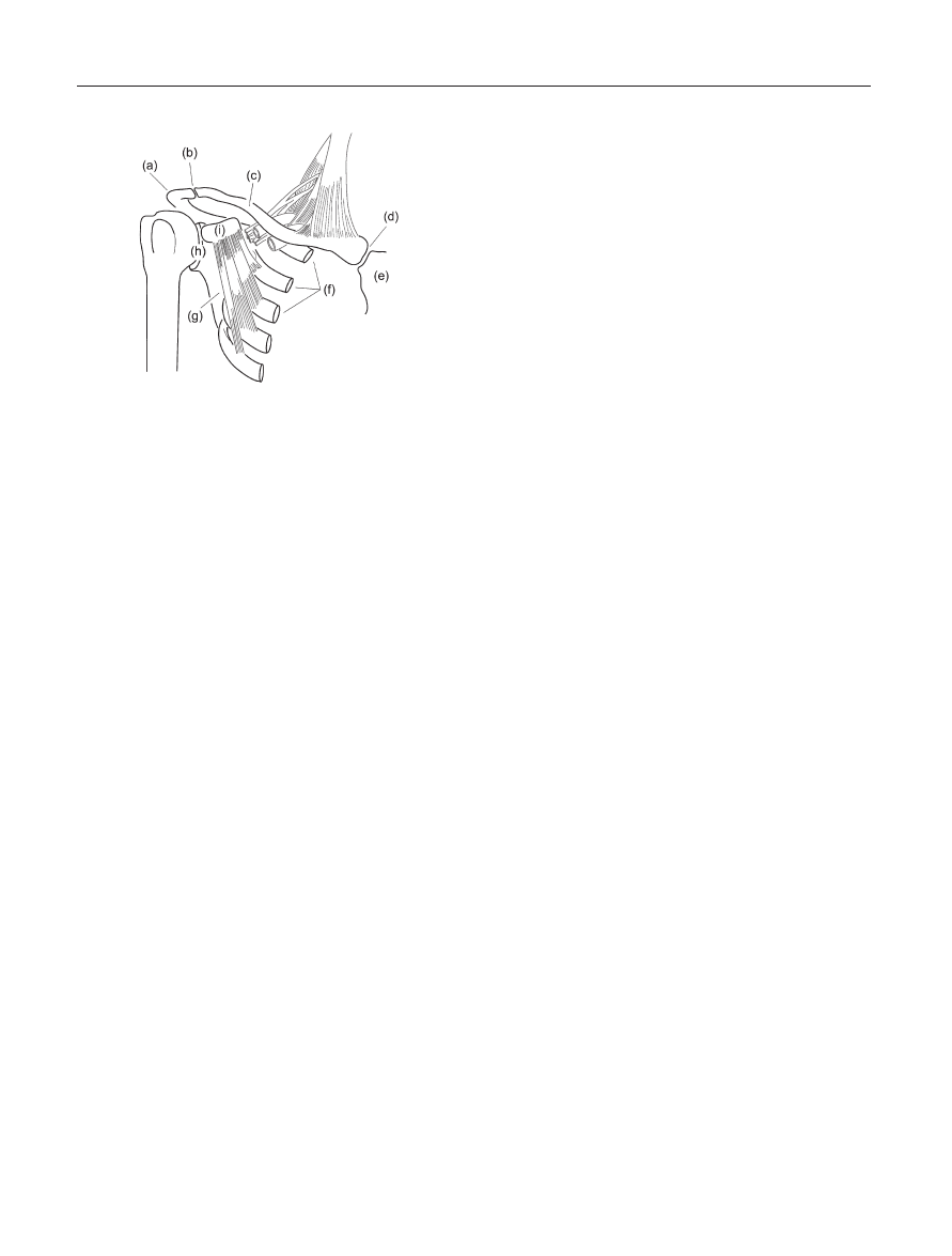
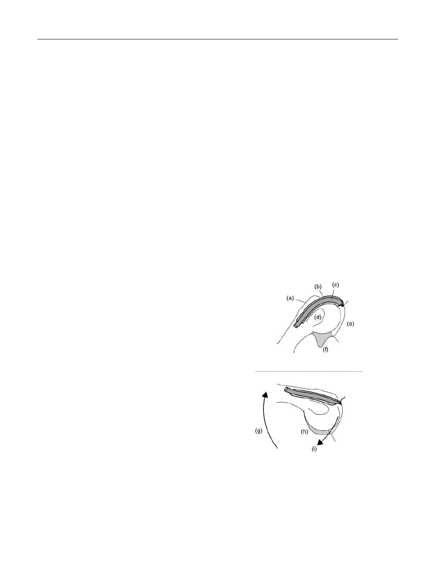
Figure 1. Joint Systems in the Shoulder Complex. (a) Acromion
process; (b) Acromioclavicular joint; (c) Clavicle; (d) Sternoclavic-
ular joint; (e) Sternal manubrium; (f) 1
st
through 3
rd
Ribs; (g)
Scapulothoracic joint; (h) Glenohumeral joint; (i) Coracoid
process.

Diagnosis and Management of the Painful Shoulder • 41
coracoclavicular ligament is the most important of
these secondary stabilizers, especially during posterior
displacement of the clavicle.
14
These ligaments can
be ruptured with a fall on the acromion, leading to a
separated ACJ.
17
Bureau et al suggested that a bursa could form
between the conoid and trapezoid ligaments, creating a
pseudo-articulation. An approximation between the
coracoid process and the clavicle produces a “kissing
coracoid syndrome” that is often accompanied by an
increase in the density of nociceptive fibers under the
clavicle.
18
This affliction can be associated with dys-
plastic changes in the coracoid process or malunion
after distal clavicle fracture. Additionally, it can emerge
after reconstruction of the coracoclavicular ligaments
when the repair is excessively tight and the subclavicu-
lar space is appreciably narrowed.
The coracoacromial ligament attaches to the inferior
AC capsular ligament, as well as the coracohumeral and
coracoclavicular ligaments.
16
This ligament connects
the coracoid to the acromion process and creates a roof
over the supraspinatus and humeral head, producing a
buffer between the rotator cuff and the bony acromion.
However, the coracoacromial ligament is not important
for ACJ stability unless the other ligaments are dis-
rupted. Conversely, this ligament is clinically significant,
in that it serves as a component in an acromiohumeral
impingement syndrome.
The stability of the ACJ is largely dependent upon
the integrity of the previously mentioned capsuloliga-
mentous structures surrounding the joint. The deltoid
and trapezius muscle insertions surround the ACJ, pro-
viding secondary stability. While these muscles do not
provide direct stabilization or create voluntary move-
ment at the ACJ, they should be a focus of a nonoper-
ative stabilization program, should a clinical instability
develop.
Glenohumeral Joint (GHJ)
The glenohumeral joint (GHJ) is composed of the con-
nection between the spheroid humeral head and the
concave glenoid fossa of the scapula. This glenoid
concavity is shallow, creating a relatively small (30%)
contact area with the large humeral head. This rela-
tionship is augmented by the fibrocartilaginous glenoid
labrum, which contributes to 50% of the total socket
depth in the glenohumeral interface.
19
While this rela-
tionship contributes greatly to the overall mobility of
the joint, glenohumeral stability is potentially compro-
mised by this same relationship.
The humeral head is inclined approximately
135–145° upward. This orientation, along with the 11°
upward tilt of the glenoid fossa, lends to the GHJ
maximum loose-packed position of 55° in the scapular
plane. In addition, the humeral head is retroverted
approximately 20° influencing available external and
internal rotation, where an increase in retroversion
lends to increased external rotation.
20
Symeonides et al
correlated decreased retroversion with an increased inci-
dence of anterior instability, suggesting that a combina-
tion of anterior capsular adaptation and decreased
retroversion potentially leads to an increase in anterior
humeral head exposure during the cocking phase of
throwing.
21
In addition, McPherson et al suggested that
the radius of the humeral head does not match that of
the glenoid, lending the GHJ to better restraint of ante-
rior-posterior versus superior-inferior translation.
22
The
restraint to superior translation is largely due to the
acromion and coracoid processes of the scapula, as well
as the coracoacromial ligament. The size of the acromio-
humeral interval (AHI—the space between the
acromion and greater tubercle of the humerus), which
does not appear to be sex or age specific,
23
is typically
1.0–1.5 cm on radiograph.
24
Muscle hypertrophy, dis-
rupted scapular mechanics, GHJ capsular dysfunction,
or architectural changes can compromise this space and
potentially lead to impingement by virtue of increased
pressure within the interval.
25,26
The glenohumeral joint capsule is somewhat loose,
allowing for relative unrestricted motion of the GHJ.
This loose capsule is posteriorly reinforced by the
rotator cuff muscles, while the anterior area is sup-
ported by anterior cuff muscles, as well as the superior,
middle, and inferior glenohumeral ligaments (see Figure
2). The middle glenohumeral ligament is frequently
underdeveloped or absent, while the superior and infe-
rior glenohumeral ligament complexes contribute
largely to the overall stability of the GHJ.
27,28
The supe-
rior glenohumeral ligament originates from the supra-
glenoid tubercle with the long head of the biceps tendon
and inserts at the lesser tubercle, supplying an anterior
sling over the long head biceps tendon.
29
In addition
to the superior, middle, and inferior glenohumeral
ligaments, Kolts et al described a “spiral glenohumeral
ligament” that works in concert with the glenohumeral
ligament complex.
30
This spiral ligament begins at the
infraglenoid tubercle and the axillary portion of the
inferior glenohumeral ligament. It courses upward and
laterally to fuse with the anterior joint capsule, middle
glenohumeral ligament, and superior glenohumeral lig-
42 • sizer et al.
ament, ultimately inserting with the subscapularis
tendon into the lesser tubercle. These ligament fibers
spiral as they course toward their insertions during
external rotation and abduction, thus lending to the
name.
30
The coracohumeral ligament demonstrates 2 divi-
sions that course from the coracoid process to the
greater and lesser tubercles of the humerus, strengthen-
ing the anterior-superior portion of the capsule (see
Figure 2). Because they share attachments at the glenoid
tubercles, the divisions are dynamized by the supra-
spinatus and subscapularis tendons, respectively.
31
The
superficial fibers insert at the greater tuberosity while
the deep fibers connect distal to the supraspinatus inser-
tion, sending slips anteriorly over the biceps tendon.
29
The coracohumeral ligament varies in thickness, but
appears more developed than the fibers of the superior
glenohumeral ligament.
29
Finally, the transverse band
(or “transverse ligament”) connects the 2 divisions
and resists their separation during the pull of their
respective dynamizing muscles.
The tendon of the biceps long head courses between
the greater and lesser tubercles, passing through the
rotator cuff interval that is formed by the 2 divisions of
the coracohumeral ligament (see Figure 2).
9,32
This
tendon dives into the capsule on its way to inserting at
the supraglenoid tubercle, representing 1 opening of the
capsule. This rotator cuff interval is described as a tri-
angular space bordered superiorly by the anterior fibers
of the supraspinatus tendon, inferiorly by the superior
border of the subscapularis tendon, medially by the
corocoid process and laterally by the long head of the
biceps tendon. The floor of the rotator interval is com-
prised of the coracohumeral ligament (CHL), superior
glenohumeral ligament (SGHL), and joint capsule.
29,32
The rotator interval plays a role in resisting inferior
translation of the humeral head, just as Jost et al sug-
gested that the CHL and the SGHL are primarily
responsible for inferior stabilization.
29
Histologically,
the rotator interval contains unorganized collagen and
an occasional congenital defect between the supraspina-
tus and subscapularis tendons, each contributing to
anteroinferior and multidirectional glenohumeral insta-
bility.
32
Conversely, patients with frozen shoulder can
demonstrate a CHL that is comprised of thickened,
dense, highly vascular, pinkish bands of fibrous scar
tissue in the absence of the rotator interval.
33
The
contracted CHL consists of a dense matrix of type
III collagen composed mainly of fibroblasts and myofi-
broblasts, lending to GHJ motion loss in the directions
of flexion and external rotation.
33
The anteroinferior ligament and capsule (see Figure
2) serves as the primary restraint to anteroinferior
glenohumeral dislocation and exhibits higher peak
strains at the glenoid insertion when compared to the
humeral insertion.
34
The synovial membrane lines the
fibrous capsule and secretes synovial fluid into the joint
cavity, creating a normal synovial volume of 20–40 mL
of fluid in the GHJ. The inferior portion of the fibrous
capsule is redundant in order to allow considerable
rotatory and translatory movement associated with
overhead elevation (see Figure 3). A synovial lining
covers this axillary recess, along with the anterior and
posterior bands of the inferior glenohumeral ligament.
A loss in synovial fluid creates the potential for adhe-
sions within this recess, accompanied by a reduced syn-
ovial fluid volume to 2–4 mL and subsequent adhesive
capsulitis. In addition, the size of this recess appears to
influence surgical outcomes, as evidenced by a positive
correlation between decreased capsular capacity and
painful GHJ limitations after rotator cuff repair.
35
The glenoid labrum is a fibrocartilage ring that cir-
cumferentially attaches to the bony rim of the glenoid
fossa (see Figure 4). Nishida et al described 2 fibrocar-
tilage layers within the labrum. The most superficial
layer is found at the articular surface and contains chon-
drocytes that are more suited for compressive load tol-
erance. Conversely, the deep collagen layer provides
cushioning and stabilization. Additionally, the fibrocytic
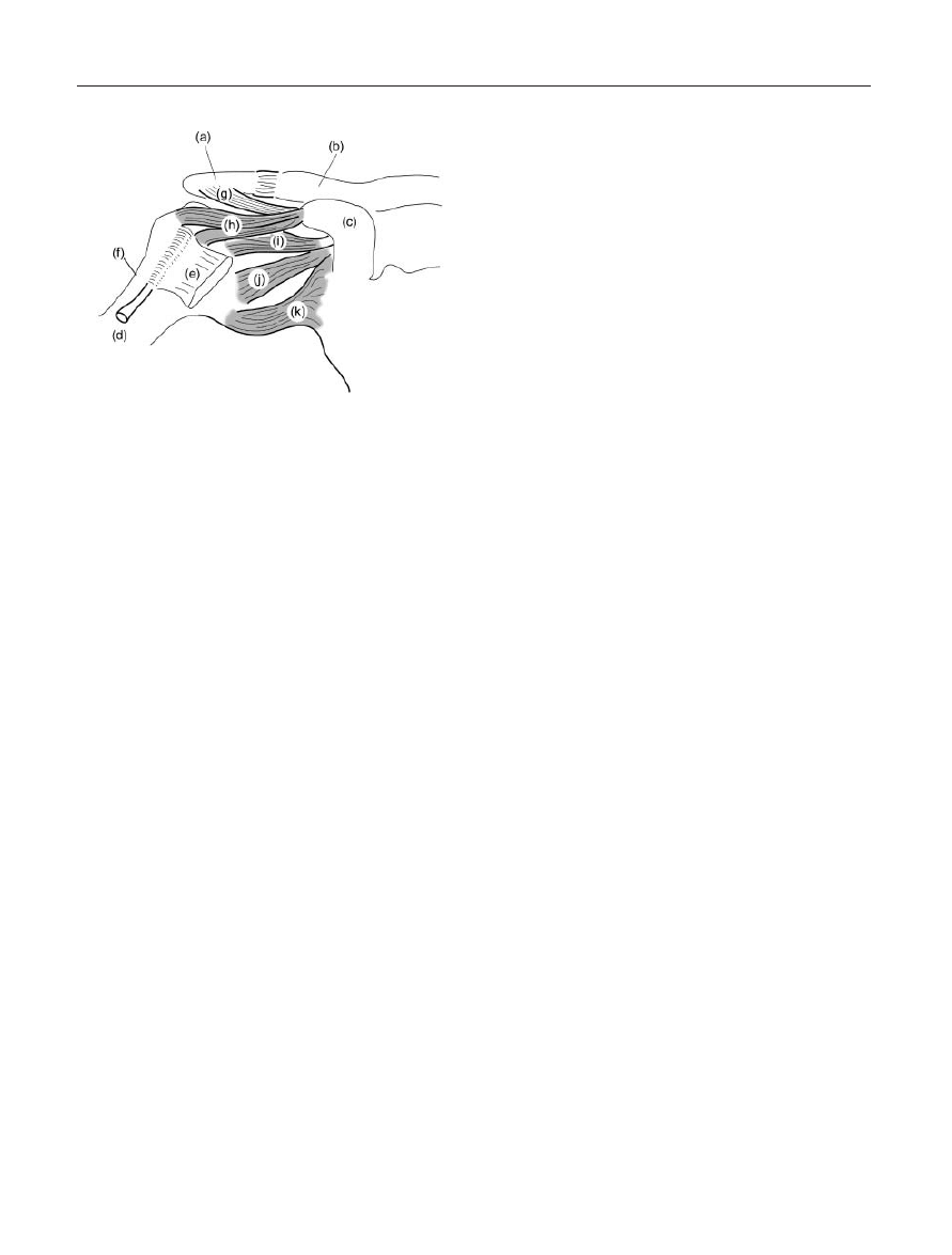
Figure 2. Ligaments in the Glenohumeral Region, Anterior View.
(a) Acromion process; (b) Clavicle; (c) Coracoid process; (d) Biceps
tendon, long head; (e) Subscapularis tendon; (f) Proximal
humerus; (g) Coraco-acromial ligament; (h) Coraco-humeral lig-
ament; (i) Superior anterior glenohumeral ligament; (j) Middle
anterior glenohumeral ligament; (k) Inferior anterior gleno-
humeral ligament.
Diagnosis and Management of the Painful Shoulder • 43
collagenous outer rim resists tensile loading produced
by glenohumeral movement.
36
The labral attachment to
the glenoid bony rim (limbus) varies at different zones
of the labrum. The inferior labrum is firmly attached to
the limbus, while the superior labrum potentially
demonstrates a loose attachment, predisposing it to dis-
ruption.
37
In addition, the attachment of the anterior
labrum appears to be variable, lending it to compromise
during micro- and macrotraumatic events.
38
The glenohumeral capsule, ligaments, and numerous
cuff tendons variably insert into the labrum.
39
For
example, variations in the biceps long head insertion at
the supraglenoid tubercle create differences in subse-
quent labral lesions. The more the biceps tendon inserts
into the labrum versus the limbus or tubercle, the higher
the potential it poses for superior labral lesion. In addi-
tion to increasing GHJ surface area contact, the labrum
promotes stability by creating negative pressure within
the GHJ.
40,41
The introduction of a small opening
(vent) through the labrum appears to increase GHJ
mobility as a consequence of reduced negative pressure
in the joint.
42
Thus, either labral tears or surgically
induced venting could produce increased laxity in the
joint, meriting protective stabilization.
43
The glenohumeral joint capsule is reinforced by the
insertion of the rotator cuff muscles. These muscles
include the supraspinatus, infraspinatus, teres minor,
and the subscapularis.
4,44
These muscles contribute
largely to the overall dynamic stability of the GHJ
through concavity compression,
42
and weakness or
imbalance in any of these muscles may cause changes in
elevation chain function.
43
Therefore, the function of
these muscles must be addressed during any shoulder
stabilization routine.
Musculature
No discussion about the shoulder complex would be
complete without including the clinical implications of
muscles in the shoulder region. The deltoid is consid-
ered the most important mover and dynamic inferior
stabilizer of the GHJ, although pathology of the deltoid
is rare.
38
This reduced injury potential is likely related
to the fibrous bands originating from the anterior corner
of the acromion and proceeding through the middle
portion of the deltoid, apparently providing structural
support for the forces sustained during elevation.
45
While deltoid activity can elevate the greater tubercle
into the acromion, the latissimus dorsi functions to
decrease this compression within the acromiohumeral
interval, meriting latissimus dorsi activation during the
treatment of chronic bursitis.
The scapulothoracic primary movers, including the
rhomboids, serratus anterior, and the trapezius, all con-
tribute to scapular control and stability.
3
Scapular
control is essential to scapulohumeral coordination and
scapulothoracic instability can emerge out of perfor-
Figure 3. Redundant Glenohumeral Capsule. (a) Scapular
glenoid neck and limbus; (b) Glenoid labrum; (c) Inferior
glenohumeral capsular recess; (d) Inferior anterior glenohumeral
ligament; (e) Superior glenohumeral capsule.
Figure 4. Glenoid Labrum. (1) Inferior Cross-Sectional View: (a)
Glenoid fossa; (b) Chondrocytic articular surface; (c) Deep col-
lagenous layer; (2) Anterior Oblique View: (a) Glenoid fossa; (b)
Chondrocytic articular surface; (c) Deep collagenous layer; (d)
Glenoid limbus and neck; (e) Coracoid process; (f) Acromion
process.
44 • sizer et al.
mance deficits demonstrated by any of these muscles.
The ability of the rotator cuff to move and dynamically
stabilize the GHJ is largely dependant upon the ability
of the scapular muscles to provide the GHJ with a stable
base at the scapulothoracic joint. Consequently, clini-
cians should include scapular stabilization in the treat-
ment of numerous shoulder afflictions.
The external insertions of the supraspinatus and
infraspinatus tendons are predisposed to compression
by external structures (such as the acromion) as they
approach the greater tubercle in an inclined direction.
Conversely, the internal insertions demonstrate a verti-
cal course toward the deep aspects of the greater tuber-
cle, lending them to shear, tension, and or compression
loading. Additionally, these fibers appear to be suscep-
tible to bone bruises when compression loaded in
subjects younger than 35 years old. Each of these
impingement behaviors will demonstrate unique clinical
pictures and merit different management strategies (see
Figure 5).
46
Defining the functions of the rotator cuff muscles has
remained controversial. Cooperatively, the entire cuff
system constrains the humeral head during elevation
38
and compresses the humeral head into the glenoid
fossa for stability.
42,43,47
Individually, the infraspinatus
primarily acts as an external rotator, whereas the
supraspinatus is a primary abductor and a secondary
external rotator. Consequently, resisted abduction will
be the most painful clinical test for supraspinatus ten-
dopathies, often followed by resisted external rotation.
Itoi et al divided the supraspinatus into anterior, middle,
and posterior sections. Based on evaluations of the ulti-
mate load/stress and modulus of elasticity, the anterior
strip appeared to be the most biomechanically signifi-
cant for accepting loads.
48
However, select authors
have suggested that the role of the supraspinatus is
overemphasized.
While the popular belief is that this small muscle
keeps the humeral head from migrating cranially during
elevation, Halder et al suggested that the supraspinatus
is less effective in superior stabilization of the GHJ when
compared to the latissimus and teres minor muscles.
38
Thompson et al proposed that the main function of the
supraspinatus is in the early range of abduction, and
reported that increasing activation of the supraspinatus
actually initiates an upward migration of the humeral
head.
49
In addition, Halder et al suggested that the
supraspinatus was ineffective at providing inferior
stabilization when compared to the lateral deltoid
and the coracobrachialis.
38
Wuelker et al found that
supraspinatus activity actually increased GHJ friction
and increased acromiohumeral interval pressure by
8%.
50
Based on these studies, abduction activities
initiated by the supraspinatus should be delayed
with impingement patients in order to reduce the
risk of any symptom provocation associated with
increased interval pressure. Furthermore, lesions must
accompany supraspinatus tears to either the infra-
spinatus or subscapularis for active elevation to be
compromised.
50
This may explain why patients with
isolated supraspinatus tears are still able to elevate their
shoulders.
Historically, the supraspinatus has been evaluated
with isometric abduction testing in the “empty-can”
position, where the shoulder is abducted and internally
rotated. However, Sharkey demonstrated that the infra-
spinatus is more active than the supraspinatus during
the “empty can” test and therefore challenged the test’s
validity for evaluating isolated supraspinatus activity.
51
Additionally, investigators demonstrated an increase in
infraspinatus activity during elevation with internal
rotation, and increased subscapularis activity during ele-
vation with external rotation.
52
Furthermore, Otis sug-
gested that the rotator cuff (especially the infraspinatus)
is most important for elevation during the first 30–60°
of abduction, the subscapularis is most important
during elevation over 60° of abduction. Consequently,
the best method for strengthening the subscapularis may
be resistive training internal rotation prepositioned at
60° abduction.
53
The subscapularis is the most powerful muscle of the
rotator cuff. The subscapularis tendon, which com-
pletely covers the lesser tubercle, primarily acts to inter-
nally rotate the shoulder. Shoulder adduction is
produced by the latissimus dorsi, pectoralis major, teres
Figure 5. Rotator Cuff Insertions. (a) Supraspinatus tendon
approaching the insertion into the greater tubercle of the
humerus; (b) Humeral head; (c) Glenoid limbus, fossa, and accom-
panying labrum; (d) Internal insertion of the supraspinatus; (e)
External insertion of the supraspinatus.
Diagnosis and Management of the Painful Shoulder • 45
major, and teres minor when the shoulder is situated in
the resting position (arm at the side). Conversely, the
subscapularis can serve as an adductor when the shoul-
der is prepositioned in abduction greater than 30°.
Therefore, an isolated subscapularis tendopathy will
produce pain during resisted internal rotation, while
resisted adduction is pain-free when the shoulder is
positioned at the patient’s side. Finally, the superior
fibers of the subscapularis are subject to impingement
within the acromiohumeral interval during flexion
elevation. Equally, the inferior fibers are at risk for
impingement against the coracoid process when the
individual internally rotates the shoulder in a horizon-
tally adducted position (as witnessed during a volleyball
slam or a tennis serve). Therefore, subscapularis must
be considered as a potential cause of a patient’s shoul-
der impingement, meriting specific tests that suggest its
involvement.
The subscapularis serves as an important anterior
stabilizer, especially when eccentrically activated during
functional movements. While this stabilization is pro-
vided through both active and passive constraint, any
failure in the musculotendinous unit could compromise
shoulder stability. Although the insertion of the superior
tendon demonstrates the greatest thickness and highest
tensile load-to-failure, it appears to be the most frequent
failure point, closely followed by the midsubstance of
the inferior tendon. Anteriorinferior dislocation or a
tension load imposed by external rotation and or exten-
sion can injure these regions.
38
The anatomical variation of long head of the biceps
(LHB) proximal insertion has been well described.
54–56
Approximately 50% of the tendons are attached to the
supraglenoid tubercle, whereas 25% are attached only
to the labrum and 25% are attached to both supragle-
noid tubercle and the labrum.
54
This variability lends to
inconsistency in tensile and shear loading properties
within the tendon and labrum, as well as differences in
the lesions produced during macrotraumatic events.
Additionally, the LHB shares fibrous connection to the
superior and inferior glenohumeral ligaments as a com-
ponent in the periarticular fiber system.
57
Furthermore,
Jost et al describes anterior support to the LHB tendon
from the coracohumeral and superior glenohumeral
ligaments.
29
The LHB courses through the intertubercular groove,
acting as a guide for glenohumeral elevation.
58
In
essence, the LHB prevents superior translation of the
humeral head into the acromiohumeral interval, thus
stabilizing the GHJ (see Figure 6).
37,43,59
Levy et al sug-
gested that the LHB functions minimally in isolated
shoulder motion when elbow and forearm motion is
controlled, and therefore the LHB function at the shoul-
der must be based on the passive tensile role of the LHB
tendon or tension associated with elbow and forearm
activity.
60
The long head of the biceps (LHB) is intraarticular
but extra-synovial as it is escorted through the GHJ
joint capsule by its own synovial sheath. This tenosyn-
ovial sheath is often the source of LHB affliction and is
most provocative when the tendon is moved. Biceps
tenosynovitis can be characterized by fibrosis and col-
lagen degeneration, synovial villous or vascular hyper-
plasia, lymphocytic-plasmacytic infiltrates, cartilaginous
metaplasia, and possible ischemic necrosis.
61
This
behavior is related to the structure of the sheath, which
presents with a visceral layer attached to the tendon and
a parietal layer that is harnessed to surrounding struc-
tures by connective tissue. Tendon movement is pro-
duced when the patient’s shoulder is extended in the
Figure 6. Function of the Biceps Long-Head Tendon during
shoulder elevation. (a) Greater tubercle of the humerus; (b)
Tenosynovial sheath surrounding the tendon; (c) Biceps long-
head tendon coursing through the humeral bicipital groove to
its insertion into the glenoid labrum and supra-glenoid tubercle;
(d) Lesser tubercle of the humerus; (e) Glenoid; (f) inferior gleno-
humeral capsular recess; (g) Elevation of the glenohumeral joint
into abduction; (h) Inferior capsular recess stretched under
tension loading; (i) inferior arthrokinematic translation of the
humeral head.

46 • sizer et al.
glenoid plane, thus sliding the visceral layer of the
sheath on the stationary parietal layer. This movement
can produce the patient’s symptoms when the visceral
and parietal layers are inflamed, swollen, and irregu-
lar,
62
potentially producing pain with the movement as
the rough surfaces of the inflamed layers slide across
each other.
Suprahumeral Roof
Components of the suprahumeral roof include the cora-
coacromial ligament, the coracoid and the acromion.
Graichen et al reported a difference in subacromial
space between males (larger) and females (smaller) at
rest and at 30° of abduction, but not at 90° abduction
or during muscle activity. Thus, higher levels of eleva-
tion and muscle activity increases the variability in
subacromial space.
47
Occupying the acromiohumeral
interval are the rotator cuff tendons and the sub-
acromiodeltoid bursa. The bursa plays a large role in
the gliding mechanism,
63
as it is attached to the greater
tubercle of the humerus, supra- and infraspinatus
muscles, acromion, coracoacromial ligament, and the
inferior acromioclavicular ligament.
The bursa may vary in size with a possible segmen-
tal or compartmentalized configuration,
4
thus con-
founding the role of palpation in the diagnosis of
shoulder pain. The acromiohumeral interval can be
compressed as the deltoid and rotator cuff are activated,
resulting in bursal irritation and subsequent symptoms
with any shoulder movement. The bursa is the most
densely innervated structure in the area, whose nerve
supply arises from the articular branches of the supras-
capular and lateral pectoral nerves. The bursa may be
involved in the regulation of shoulder movement as
evidenced by the populations of free nerve endings,
Ruffini endings and Pacinian corpuscles observed in
the surrounding capsuloligamentous structures.
63–66
The bursa is often the primary source of pain with trau-
matic partial rotator cuff tears in patients younger than
40 years old, and as a result of overuse tears in patients
older than 40.
67
In addition, a localized bursal reaction
is described as a degenerative disorder or the result of
acromion distortions.
25,68
Finally, the symptoms asso-
ciated with chronic bursal irritation may stem from
increased substance P and substance P receptor expres-
sion in the synovial region.
69
Vascularization (GHJ)
Branches of the posterior circumflex humeral artery and
the suprascapular artery supply the blood flow to the
posterior rotator cuff. The anterior circumflex humeral
artery and the subscapular and coracoacromial arteries
supply the anterior rotator cuff. Hypovascular zones
known as “zones of lability” exist in the supraspinatus,
infraspinatus, and subscapularis with the supraspinatus
having the highest incidence.
70–72
These hypovascular
zones create early predilection sites for degeneration and
subsequent traumatic tears. The “wringing out” mech-
anism occurs with the individual’s arm at the side, as
the humeral head is pushed cranial against the tendon
of the rotator cuff.
4
Slight abduction may limit the
potential for this “wringing-out,” so many exercises in
the rehabilitation process should be performed in a posi-
tion of slight abduction.
BIOMECHANICS
Upper extremity elevation depends on shoulder-
complex function. Whereas elevation represents every
attempt to lift the upper extremity overhead, an indi-
vidual can elevate the arm forward through flexion in
the parasagittal plane, sideways through abduction in
the frontal plane, or any movement between these
planes in an oblique direction. In addition, an individ-
ual can elevate backward in the parasagittal plane. One
can identify several important structural members
within the elevation chain: the acromioclavicular joint,
or ACJ; the sternoclavicular joint, or SCJ; the gleno-
humeral joint, or GHJ; the scapulothoracic junction, or
STJ; the cervicothoracic junction, or CTJ; and the upper
six ribs in the thoracic spine.
1
Dysfunction at any of
these sites can perpetuate clinical problems, such as
impingement or instability. In addition, movement can
be appreciated in the acromiohumeral interval during
elevation. Within this space one can appreciate move-
ment between the undersurface of the acromion and the
long tendon of the biceps as well as the other soft tissue
structures found in the area, such as the sub-
acromiodeltoid bursa and tendons of the supraspinatus
or infraspinatus. Each of these members demonstrates a
host of unique biomechanical properties that, when
combined, produce complex kinematic and kinetic
behaviors during functional upper extremity movement.
To understand these complex behaviors, one must first
analyze the osteokinematic and arthrokinematic perfor-
mance of each component.
The sternoclavicular joint (SCJ) moves as result of
clavicular movement. Osteokinematic clavicular motion
is a component of shoulder girdle movement in several
planes. The clavicle can swing cranial about a horizon-
tal axis through its proximal end during shoulder girdle
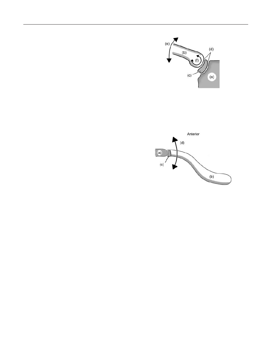
Diagnosis and Management of the Painful Shoulder • 47
elevation, producing arthrokinematic movement at
the sternoclavicular joint in relation to the intraarticu-
lar disc and sternum.
73
Arthrokinematically, the convex
clavicular surface rolls cranial, medial and slightly
dorsal while it slides caudal, lateral, and slightly ventral
(see Figure 7).
4
Conversely, the clavicle swings into
retraction within the transverse plane about a vertical
axis through the sternum, where the concave clavicle
and intraarticular disc will move in relation to the
convex sternum.
73
Here, the concave clavicle and
disc will arthrokinematically rock and glide in a poste-
rior, slightly caudal, and slightly medial direction
(see Figure 8).
4
Although these behaviors are respec-
tively reversed during shoulder girdle depression and
protraction, they are less clinically relevant as patients
rarely present with clinical limitations in either direction
of movement.
The clavicle demonstrates a curved trajectory
upward, backward, and downward during functional
upper extremity elevation (Vanderhelm, 1994).
74
More
specifically, the clavicle elevates and retracts during
functional elevation between 0° and 150°.
1
Conversely,
the clavicle produces a relative depression and protrac-
tion trajectory during elevation greater than 150°, while
never reaching its original starting position (see Figure
9). In addition, functional elevation requires the clavi-
cle to move at the SCJ about a third axis, whereas the
clavicle produces a 50° to 70° accessory backward spin
(see Figure 9).
74
Although a sellar joint should only
allow 2° of freedom (ie, elevation/depression and pro-
traction/retraction), this third degree of freedom is
allowed by deformation within the intraarticular disc.
Thus, SCJ mechanics violate the traditional view of
sellar joint behavior.
In context with the backward spin at the SCJ,
Hollinshead discovered that SCJ movements are greater
during isolated shoulder girdle elevation versus func-
tional elevation of the entire upper extremity.
73
This
author suggested that the difference is associated with
the backward spin, in that the capsuloligamentous
structures about the SCJ are twisted, thus restraining
clavicular elevation or retraction trajectories. While
patients can demonstrate normal accessory movement
in the SCJ at rest or in a shrugged shoulder girdle
position (in absence of the spin), the joint may limit
functional upper extremity elevation in relation to a
limit of the spin movement. This behavior merits the
examination of accessory movement at the SCJ in a
position of upper extremity elevation, in that SCJ cap-
sular adaptations can induce a significant loss of upper
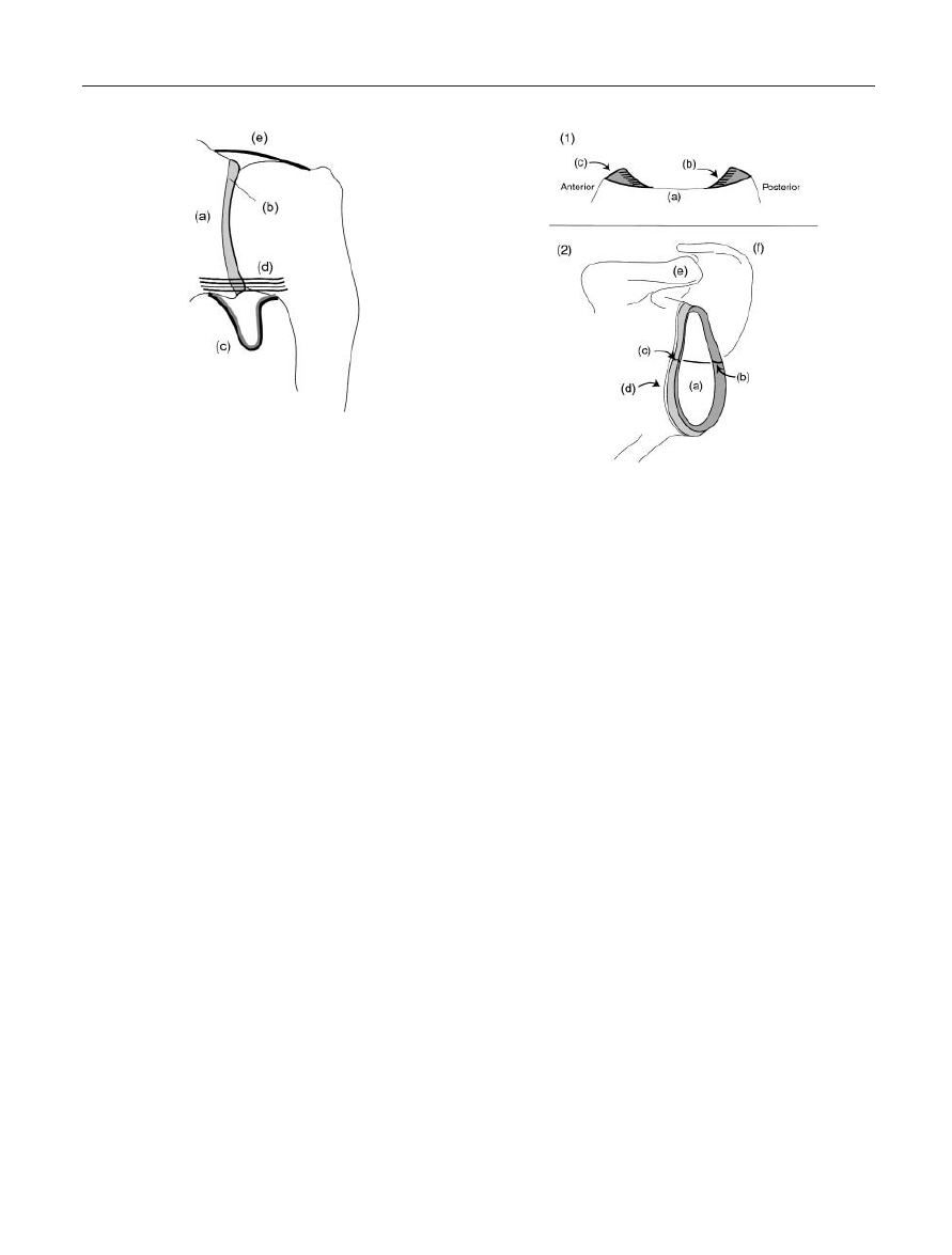
Figure 7. Sternoclavicular Movement in the Frontal Plane during
Shoulder Elevation; (a) Sternal manubrium; (b) Clavicle; (c) Ster-
noclavicular intra-articular disc; (d) Two articular compartments
created by the intra-articular disc within the joint; (e) Elevation
of the clavicle in the frontal plane; (f) Arthrokinematic rolling of
the clavicle on the intraarticular disc during elevation.
Figure 8. Sternoclavicular Movement in the Transverse Plane
during Shoulder Elevation: (a) Sternal manubrium; (b) Clavicle;
(c) Sternoclavicular intra-articular disc; (d) Retraction of the clav-
icle in the transverse plane.
extremity elevation even if isolated shoulder girdle
motion is normal.
The previously defined clavicular behaviors influence
movement at the acromioclavicular joint (ACJ). In addi-
tion, movements of the scapula and acromion influences
movement at this joint during shoulder girdle movement
and upper extremity functional elevation. Moreover, the
majority of movement at the ACJ occurs after 90° of
upper extremity elevation. This is of great interest to
clinicians, as ACJ pain occurs more frequently when the
upper extremity is positioned above 90° elevation. This
is of great interest to clinicians, as ACJ pain occurs more
frequently when the upper extremity is positioned above
90° elevation. Furthermore, the previously mentioned
clavicular spin behaviors translate into a spin at the
ACJ. However, this spin behavior is rarely greater than
10° at the ACJ, versus the 50° to 70° spin witnessed at
the SCJ. This disparity is confusing, because the same
osteokinematic spin of the clavicle produces different

48 • sizer et al.
motion values at the different joints. However, while the
clavicle spins relative to a fixed sternum during func-
tional elevation, the same clavicle is spinning relative to
a moving acromion. Thus, motion at the ACJ is inti-
mately associated with movement of the scapula at the
scapulothoracic junction (STJ).
From an anterior-posterior view, the ACJ is relatively
flat, thus producing a relative rocking of the clavicle on
the acromion during functional upper extremity
elevation. However, the clavicle is convex on a concave
acromion from an aerial view. As previously reported,
the clavicle retracts during functional upper extremity
elevation. As result, the convex clavicle must arthro-
kinematically slide anterior on the concave acromion
during retraction in order to allow functional elevation
above 90°.
4
Any limits in this anterior sliding may
hinder elevation movement, especially above 90°. Con-
versely, capsuloligamentous compromise at the ACJ
may allow excessive translation and the subsequent
sequelae associated with ACJ degeneration, such as
deformation and exostosis.
75,76
Scapular position and movement are essential to total
shoulder complex function. The scapula moves as a
component of the shoulder girdle on the thoracic wall
in a variety of different directions, including upward
or downward rotation, protraction or retraction, and
elevation or depression. Selected motions of the
scapulothoracic junction (STJ) accompany clavicular
movements during functional upper extremity elevation.
McClure et al conducted in-vivo three-dimensional
analyses of scapular movements and found that the
scapula upwardly rotates in the frontal plane, posteri-
orly tilts in the parasagittal plane and externally rotates
in the transverse plane in a nonlinear fashion during
functional elevation. This behavior was repeated at end-
range active external rotation of the glenohumeral joint.
However, internal rotation appeared to have little influ-
ence on STJ behaviors. McClure et al suggested that
the external rotation behavior reduced stress to the ante-
rior glenohumeral joint capsuloligamentous complex
during functional elevation, especially in external rota-
tion/abduction as witnessed with the wind-up phase of
throwing. The investigators suggested that a limit to this
rotation might increase the risk of anterior shoulder
laxity and subsequent instability. Finally, they suggested
that the posterior tilting promoted humeral clearance in
the acromiohumeral interval during elevation, as indi-
viduals who suffer from impingement demonstrate
reduced posterior tilting.
2,77
Other investigators have demonstrated similar find-
ings.
1,74,78,79
In addition, Fung et al found that scapu-
lar upward rotation and retraction (external rotation)
were greatest during abduction elevation (versus flexion
elevation). On the other hand, they found that posterior
tilting was greatest during flexion elevation. Further-
more, Fung et al found that these behaviors occurred
later in the range versus previously documented in-vivo
behaviors.
1
This finding suggested that in-vivo shoulder
girdle behaviors are initiated by dynamic muscular
systems versus the delayed behaviors associated with
the passive, isolated capsuloligamentous influence
witnessed in this study.
Glenohumeral joint (GHJ) movements participate in
the complex angular displacements of the functional
“humerothoracic joint”.
80
Historically, glenohumeral
movements have been labeled as ball-and-socket kine-
matics in concert with the relationship of the convex
humeral head to the concave glenoid fossa and labrum
complex. However, recent investigators have observed
translatory behaviors of the humeral head during ele-
vation activities
47,50,81
that are dynamically constrained
Figure 9. Lateral Cross-Sectional View of the Clavicle. (1) Clavic-
ular Motion during upper elevation 0–150°: (a) Clavicle posi-
tioned at rest with arm at the side; (b) Clavicular position when
upper extremity is elevated; (c) Clavicular elevation vector; (d)
Clavicular retraction vector; (e) Resultant clavicular elevation tra-
jectory; (f) 70° backwards spin of the clavicle. (2) Clavicular
Motion during terminal upper extremity elevation between
150–180°: (a) Relative depression vector; (b) Relative protraction
vector; (c) Resultant clavicular trajectory.
Diagnosis and Management of the Painful Shoulder • 49
by the labrum, capsuloligamentous structures, and
rotator cuff complex.
19,38,43,82
The osteokinematic swing associated with GHJ
abduction is accompanied by superior, inferior and ante-
rior humeral translation. The humeral head translates
superiorly to the superior glenoid fossa within the first
30° of abduction, followed by gradual anterior and infe-
rior translations as the range progresses (see Figure
10).
47,50,81
Inferior translation of the humeral head is
constrained by the superior capsule, superior gleno-
humeral ligament, and coracohumeral ligament when
the humerus is at the subject’s side in the anatomical
resting position.
28
Conversely, the inferior capsuloliga-
mentous complex constrains inferior translation when
the GHJ is abducted. Any affliction that triggers infe-
rior capsular adaptive shortening can reduce inferior
translation of the humeral head and subsequent GHJ
abduction, resulting in a compensatory excess in scapu-
lar tilting and possible impingement behaviors in the
acromiohumeral interval.
35
The osteokinematic swing executed during GHJ
flexion is accompanied by an arthrokinematic spin and
translations of the humeral head. Once again, the
humeral head translates superiorly to the superior
glenoid fossa during the first 30° of flexion, followed by
a gradual posterior and inferior translation as the range
progresses (see Figure 11).
50,81
These behaviors are
potentially constrained by any member of the entire
capsuloligamentous system, as the capsuloligamentous
structures twist during the spin movement of the
humeral head. However, investigators have demon-
strated that the primary physiological constraints to this
behavior are the coracohumeral and superior gleno-
humeral ligaments. Any compromise to these structures,
as witnessed after a rotator cuff interval tear, can
decrease these controls and produce excessive aphys-
iogical translations and subsequent GHJ instability.
28
On the contrary, adaptive shortening of the posterior
capsule can alter the arthrokinematic spin and reduce
the inferior translatory behaviors during GHJ flexion,
resulting in persistent superior positioning and subse-
quent impingement in the acromiohumeral interval (see
Figure 12).
3,83–85
Glenohumeral external rotation is best described as
an osteokinematic rotation of the humerus about the
diaphyseal axis. This movement is accompanied by a
posterior rolling and anterior sliding of the humeral
head on the glenoid complex when the arm is positioned
at the patient’s side. This anterior translatory behavior
is constrained by both the coracohumeral and superior
anterior glenohumeral ligament complex (see Figure
13).
27
Conversely, external rotation performed in a
position of GHJ abduction is best described as an
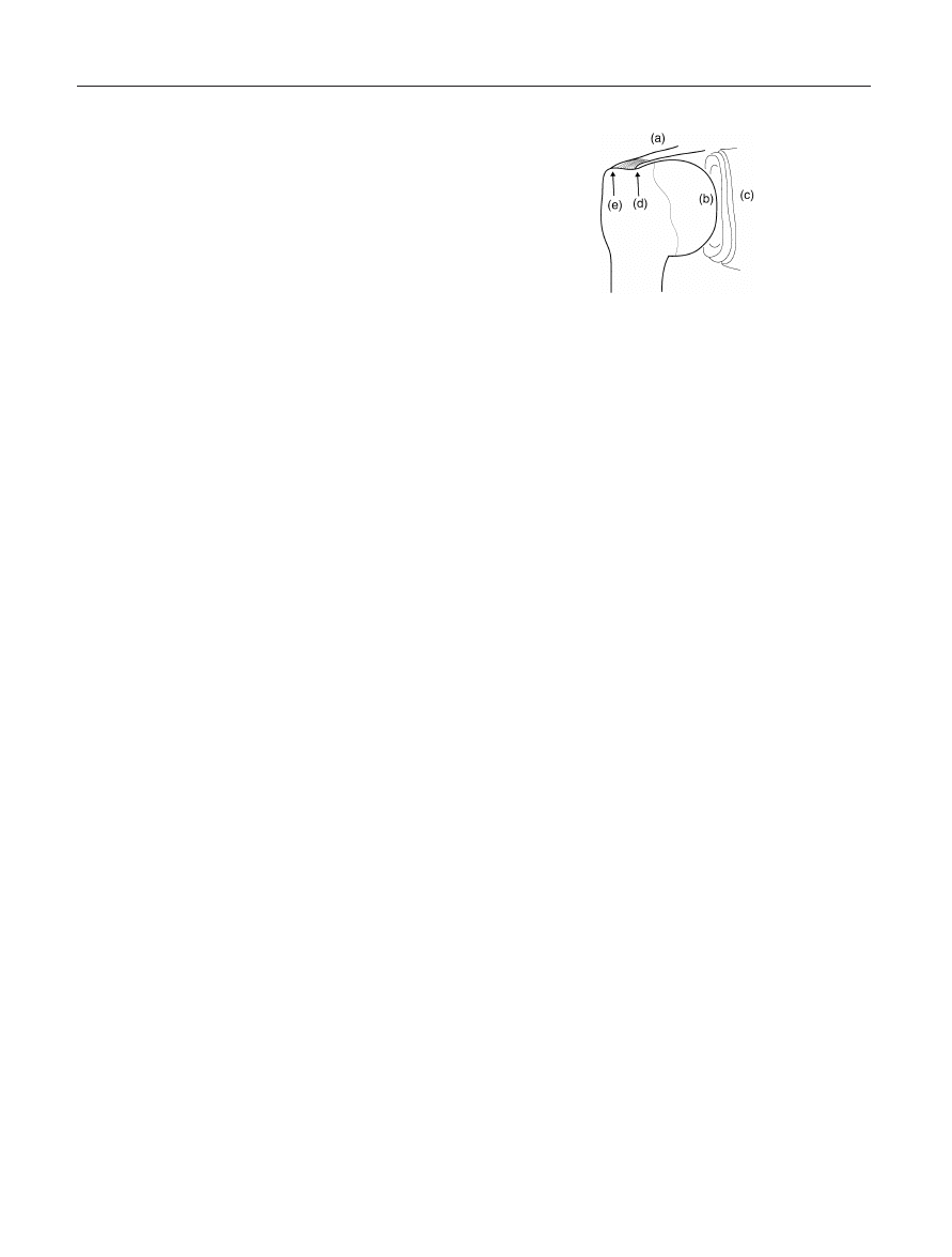
Figure 10. Humeral Head Translation Upon the Glenoid Fossa
During Upper Extremity Abduction Elevation: (1) Humeral head
translation during the first 30° of abduction elevation: (a)
Contact point of the humeral head on the glenoid fossa with the
arm at rest; (b) Course of humeral head translation; (c) Gleno-
humeral contact point with the arm abducted to 30°. (2) Humeral
head translation during 30–180° of abduction elevation: (a)
Course of humeral head translation; (b) Glenohumeral contact
point at end-range abduction elevation.
Figure 11. Humeral Head Translation Upon the Glenoid Fossa
During Upper Extremity Flexion Elevation: (1) Humeral head
translation during the first 30° of flexion elevation: (a) Contact
point of the humeral head on the glenoid fossa with the arm at
rest; (b) Course of humeral head translation; (c) Glenohumeral
contact point with the arm flexed to 30°. (2) Humeral head trans-
lation during 30–180° of flexion elevation: (a) Course of humeral
head translation; (b) Glenohumeral contact point at end-range
flexion elevation.
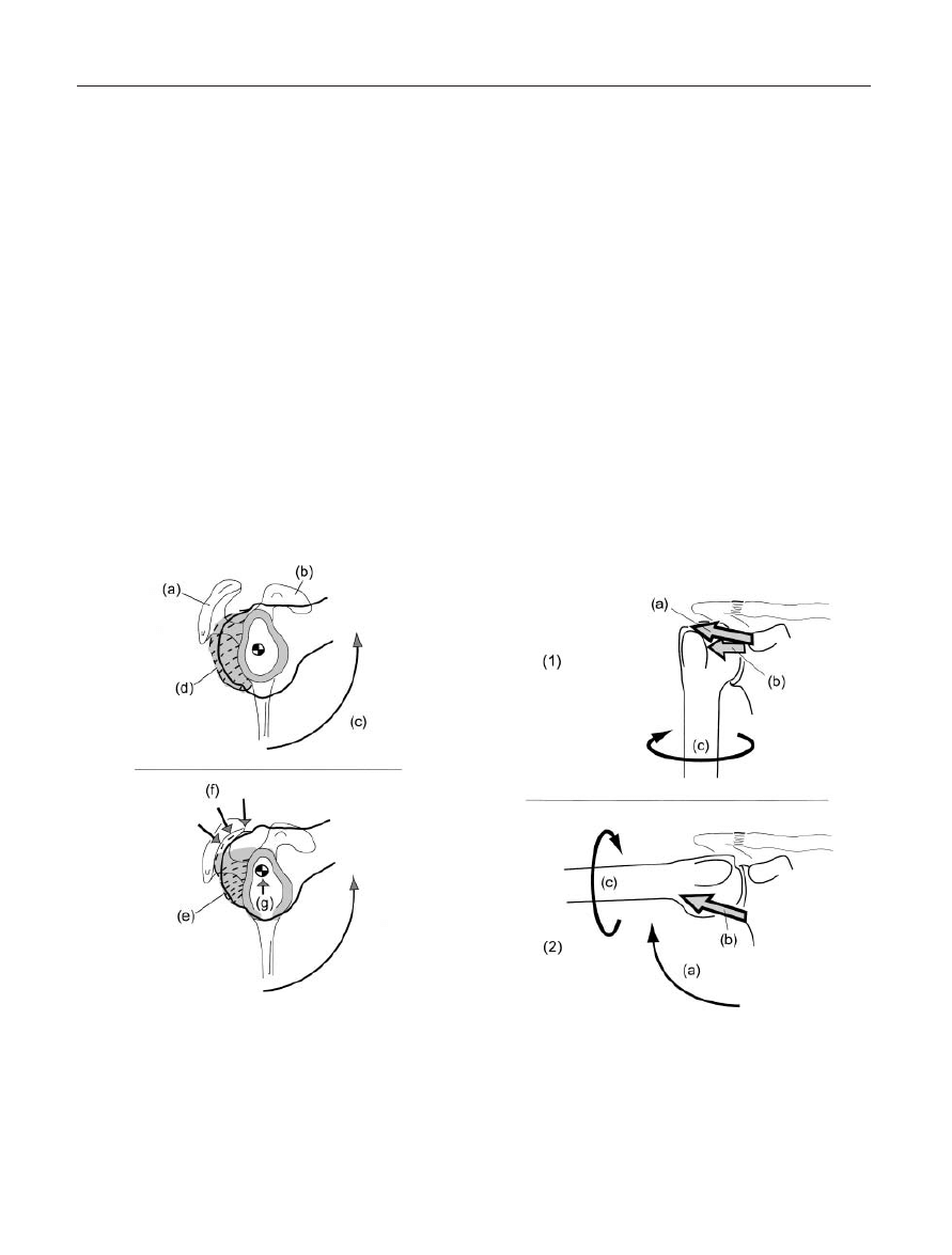
50 • sizer et al.
arthrokinematic spin, due to repositioning of the
humeral head with respect to the glenoid complex. The
inferior anterior glenohumeral ligament serves as the
primary constraint to this particular movement and any
adaptive shortening of this structure could limit exter-
nal rotation in an abducted position (see Figure 13).
Additionally, this limitation may require the scapula and
shoulder girdle to compensate, as witnessed during the
wind-up phase of throwing after Bankhart repair.
27
On
the other hand, elongation of this mechanism could
increase external rotation during cocking, leading to
increased aphysiological motion and risk for rotator
cuff lesions, posterior labral impingement, or SLAP
lesion (Superior Labrum Anterior-Posterior) in the supe-
rior labral quadrant.
86
Finally, the difference in con-
straints based on changes in GHJ position merits clinical
testing for joint limits or laxity in both dependent and
abducted positions.
Similar behaviors can be witnessed during internal
rotation, where the superior posterior capsule con-
strains internal rotation in a dependent GHJ position
and the inferior posterior capsule limits internal rota-
tion while the GHJ is positioned in abduction. Posterior
capsular tightness can limit internal rotation and, as pre-
viously mentioned, can result in sustained superior
humeral head translation during elevation. Limitations
appear to be related to posterior capsular fibrosis and
muscular tightness.
87,88
Additionally, progressive limi-
tation appears to be related to repetitive or sustained
activity in a functionally external rotated position, such
as tennis.
87,89,90
However, as previously discussed
normal posterior structures provide little support to the
stability of the GHJ in the posterior translatory direc-
tion. Rather, posterior stability is afforded by the ante-
rior angulation of the glenoid fossa,
1
the integrity of the
glenoid labrum and the support afforded by the ante-
rior inferior glenohumeral ligament complex.
82
Figure 12. The Diablo Effect; Influence of Posterior Gleno-
humeral Capsular Tightness on Impingement Behaviors Seen
from an Outlet View: (a) Acromion process; (b) Coracoid process;
(c) Elevation of the glenohumeral joint into flexion; (d) Tension
loading of a normal posterior glenohumeral capsule; (e) Tension
loading of an adaptively shortened posterior glenohumeral
capsule; (f) Decreased acromiohumeral interval space, resulting
in elevated interval pressure and subsequent impingement; (g)
Persistent superior positioning of the glenohumeral contact
point during elevation.
Figure 13. Capsulo-Ligamentous Constraints on Glenohumeral
External Rotation: (1) Constraints to External Rotation with the
Humerus Positioned at the Subject’s Side: (a) Tension loading in
the coraco-humeral ligament; (b) Tension loading in the superior
glenohumeral ligament; (c) External rotation of the humerus. (2)
Constraints to External Rotation with the Humerus Positioned at
90° Abduction: (a) Elevation of the glenohumeral joint to 90°
abduction; (b) Tension loading in the inferior glenohumeral lig-
ament complex; (c) External rotation of the humerus.

Diagnosis and Management of the Painful Shoulder • 51
Glenohumeral joint movement is very complex
during functional elevation, due to coupling movements
of the joint and movement of scapula. First, elevation
in abduction requires external rotation of the gleno-
humeral joint to maximize humeral contact with the
glenoid fossa.
91
Second, the glenohumeral joint moves
in concert with the scapulothoracic junction about a
functional axis outside the humeral head during upper
extremity functional elevation.
80
While, rotator cuff
muscle activity appears to persuade humeral head cen-
tralization during elevation,
47
scapular movement helps
to maintain appropriate length tension relationships in
those muscles through full elevation range.
2
This non-
linear behavior is a consequence of concerted functional
coordination between the GHJ and STJ, known as
“scapulohumeral rhythm”.
1,2,92
While the influence
that the direction of elevation has on this rhythm is con-
troversial, investigators have historically suggested that
this behavior can be best described as a ratio of move-
ment between the GHJ and STJ.
92–95
Recently, investigators have identified three distinc-
tive phases of elevation, accompanied by different
scapulohumeral rhythm behaviors occurring in each of
the various phases.
74,92
During the setting phase of ele-
vation (0° to 60°) the scapula seeks a stable position
under the humerus, so to provide a more secure base for
the rolling humeral head. The movement ratio that
is witnessed during this phase is approximately 6 : 1 to
7 : 1 (GHJ : STJ). Throughout this arc, the scapula
“wiggles” as it attempts to establish an optimal posi-
tion. However, because the upper extremity weighs
more than the scapula; the scapula tends to tip or wing
if not sufficiently controlled by activity of the serratus
anterior (especially during eccentric activity associated
with a return from an elevated position).
During the elevation phase (60° to 130°), the scapu-
lohumeral complex produces three-dimensional motion
around the previously mentioned helical oblique axis
outside of the humeral head. The glenoid fossa is appro-
priately positioned under the humeral head during this
phase and the subsequent movement ratio is approxi-
mately 1 : 1. However, this ratio changes again to 5 : 1
during the end-range phase (130° to 180°).
Controversy can be witnessed regarding the impact
that resisted movement has on the scapulohumeral
rhythm. While select investigators have suggested that
an increase in STJ involvement can be observed earlier
in the elevation range of motion when the movement is
resisted,
92,93
others have suggested that resistance has
no impact on the rhythm.
78
Additionally, McQuade et
al found that shoulder muscle fatigue appeared to sig-
nificantly alter scapulohumeral rhythm by decreasing
scapulothoracic movement during elevation.
96
Clini-
cally, disturbances in this rhythm can be linked to
impingement, instability, and elevation limits.
2,97,98
Additionally, attaining full GHJ abduction does not
insure normal functional elevation. Thus, clinicians
should use an elevation preposition when mobilizing the
glenohumeral joint to ensure full restoration of func-
tional elevation.
99
SUMMARY
Distinctive anatomical features prevail in the shoulder
complex, lending to specific pathological conditions.
Clinical conditions in the shoulder complex are multi-
factoral, and both anatomical and biomechanical dis-
turbances participate in the development of affliction.
The sternocalvicular, acromioclavicular, glenohumeral,
and scapulothoracic joints must all participate in func-
tion of the shoulder complex, as each biomechanically
contributes to functional movements and clinical disor-
ders witnessed in the shoulder region. Clinicians are
encouraged to consider the anatomical and biomechan-
ical distinctions in this region when examining and diag-
nosing disorders of the shoulder.
Many painful conditions in the shoulder region share
similar clinical features, creating a diagnostic challenge
and potential confusion for the clinician. A careful
examination that implements specific testing procedures
can lead a clinician to effective diagnosis of the painful
shoulder. Once diagnosed, a clinician should consider
specific management options when attempting to erad-
icate the patient’s symptoms. Clinical examination, dif-
ferential diagnosis, and management options will be
considered in Part II of this series.
REFERENCES
1. Fung M, Kato S, Barrance PJ, Elias JJ, McFarland
EG, Nobuhara K, Chao EY. Scapular and clavicular kinemat-
ics during humoral elevation: a study with cadavers. J Shoul-
der Elbow Surg. 2001;10:278–285.
2. McClure PW, Michener LA, Sennet BJ, Karduna AR.
Direct 3-dimensional measurement of scapular kinematics
during dynamic movements in vivo. J Shoulder Elb Surg.
2001;10:269–277.
3. Kibler WB. Biomechanical analysis of shoulder
during tennis activities. Raquet Sports. 1995;14:79–85.
4. Winkel D, Matthijs O, Phelps V. Diagnosis and
Treatment of the Upper Extremities: Non-operative
Orthopaedic Medicine and Manual Therapy. Gaithersburg,
MD: Aspen Publishers, Inc., 1997.

52 • sizer et al.
5. Shishido H, Kikuchi S. Injury of the suprascapular
nerve in shoulder surgery: An anatomic study. J Shoulder
Elbow Surg. 2001;10:372–376
6. Moriggl B. [Fundamentals, possibilities and limita-
tions of sonography of osteofibrous tunnels in the shoulder
area. 1] Anat Anz. 1997;179:355–373.
7. Spencer EE, Kuhn JE, Huston LJ, Carpenter JE.
Hughes RE. Ligamentous restraints to anterior and posterior
translation of the sternoclavicular joint. J Shoulder Elbow
Surg. 2002;11:43–47.
8. Rockwood CA Jr, Odor JM. Spontaneous atraumatic
anterior subluxation of the sternoclavicular joint. J Bone Joint
Surg. 1989;71:1280–1288.
9. Noda M, Shiraishi J, Mizuno K. Chronic posterior
sternoclavicular dislocation causing compression of a sub-
clavian artery. J Shoulder Elbow Surg. 1997;6:564–569.
10. Rayan GM. Compression brachial plexopathy
cuased by chronic posterior dislocation of the sternoclavicu-
lar joint. J Okla State Med Assoc. 1994;87:7–9.
11. Jougan JB, Lepront DJ, Dromer CE. Posterior dislo-
cation of the sternoclavicular joint leading to mediastinal
compression. Ann Thoracic Surg. 1996;61:711–713.
12. Wasylenko MJ, Busse EF. Posterior dislocation of the
clavicule causing fatal tracheoesophageal fistula. Can J Surg.
1981;24:626–627.
13. Netter FH. Atlas of Human Anatomy. Summit, NJ:
Ciba-Geign Corporation, 1989.
14. Lee KW, Debski RE, Chen CH, Woo SLY, Fu FH.
Functional evaluation of the ligaments of the acromioclavicu-
lar joint during anteroposterior and superoinferior transla-
tion. Am J Sports Med. 1997;25: 858–862.
15. Fukuda K, Craig EV, An KN, Cofield RH, Chao EY.
Biomechanical study of the ligamentous system of the
acromioclavicular joint. J Bone Joint Surg Am. 1986;68:
434–440.
16. Salter EG Jr, Nasca RJ, Shelley BS. Anatomical obser-
vations on the acromioclavicular joint and supporting liga-
ments. Am J Sports Med. 1987;15:199–206.
17. Magee DJ. Orthopedic Physical Assessment.
Philadelphia, Pa: W. B. Saunders Corp, 1997.
18. Bureau NJ, Dussault RG, Keats TE. Imaging of
bursae around the shoulder joint. Skeletal Radiology.
1996;25:513–517.
19. Halder AM, Kuhl SG, Zobitz ME, Larson D, An KN.
Effects of the glenoid labrum and glenohumeral abduction on
stability of the shoulder joint through concavity-compression.
J Bone Joint Surg. 2001;83-A:1062–1069.
20. Kronberg M, Brostrom LA, Soderlund V. Retrover-
sion of the humeral head in the normal shoulder and its rela-
tionship to the normal range of motion. Clin Orthop.
1990;253:113–117.
21. Symeonides PP, Hatzokos I, Christoforides J,
Pournaras J. Humeral head torsion in recurrent anterior
dislocation of the shoulder. J Bone Joint Surg Br. 1995;77:
687–690.
22. Mcpherson EJ, Friedman RJ, An YHH, Chokesi R,
Dooley RL. Anthropometric study of normal glenohumeral
relationships. J Shoulder Elbow Surg. 1997;6:105–112.
23. Anetzberger H, Putz R. [Morphometry of the sub-
acromial space and its clinical relevance]. Unfallchirurg.
1995;98:407–414.
24. Flatow EL, Soslowsky LJ, Ticker JB, Pawluk RJ,
Helper M, Ark J, Mow VC, Bigliani LU. Excursion of the
rotator cuff under the acromion. Patterns of subacromial
contact. Am J Sports Med. 1998;22:779–788.
25. Bigliani LU, Levine WN. Subacromial impingement
syndrome. J Bone Joint Surg. 1997;79A:1854–1867.
26. Riley GP, Harrall RL, Constant CR, Cawston
TE, Hazleman BL. Prevalence and possible pathological
significance of calcium phosphate salt accumulation in
tendon matrix degeneration. Ann Rheum Dis. 1996;55:109–
115.
27. Itoi E, Watanabe W, Yamada S, Shimizu T,
Wakabayashi I. Range of motion after Bankart repair—
Vertical compared with horizontal capsulotomy. Am J Sports
Med. 2001;29:4414–4445.
28. Van der Reis W, Wolf EM. Arthroscopic rotator cuff
interval capsular closure. Orthop. 2001;24:657–661.
29. Jost B, Koch PP, Gerber C. Anatomy and functional
aspects of the rotator interval. J Shoulder Elbow Surg.
2000;9:336–341.
30. Kolts I, Busch LC, Tomusk H, Rajavee E, Eller A,
Russlies M, Kuhnel W. Anatomical compositon of the
anterior shoulder joint capsule: a cadaver study on 12
glenohumeral joints. Ann Anat-anatom Anzeig. 2001;183:53–
59.
31. Gagey O, Bonfait H, Gillot C, Mazas F. [The
mechanics of shoulder elevation. Role of the coracohumeral
ligament]. Rev Chir Orthop Reparatrice Appar Mot. 1985;71
(Suppl 2):S105–S107.
32. Cole BJ, Rodeo SA, O’Brien SJ, Altchek D, Lee D,
DiCarlo EF, Potter H. The anatomy and histology of the
rotator interval capsule of the shoulder. Clin Ortho Rel Rsch.
2001;390:129–137.
33. Omari A, Bunker TD. Open surgical release for
frozen shoulder: Surgical findings and results of the release. J
Shoulder Elbow Surg. 2001;10:353–357.
34. Malicky DM, Soslowsky LJ, Kuhn JE, Bey MJ,
Mouro CM, Raz JA, Liu CA. Total strain fields of the antero-
inferior shoulder capsule under subluxation: a stereoradi-
ogrammetric study. J Biomech Eng—Transactions of the
ASME. 2001;123:425–431.
35. Hato Y, Saitoh S, Murakami N, Seki H, Kobayashi
H, Takaoka K. Shrinkage in the inferior pouch of the scapu-
lohumeral joint is related to posterior pain after rotator cuff
repair: radiographic and arthographic comparison between

Diagnosis and Management of the Painful Shoulder • 53
patients with postoperative pain and those without it. J Shoul-
der Elbow Surg. 2001;10:333–339.
36. Nishida K, Hashizume H, Toda K, Inoue H. Histo-
logic and scanning electron microscopic study of the glenoid
labrum. J Shoulder Elbow Surg. 1996;5:132–138.
37. Huijbregts PA. SLAP lesions: structure, function, and
physical therapy diagnosis and treatment. J Man Manip Ther.
2001;9:71–83.
38. Halder AM, Halder CG, Zhao KD, ODriscoll SW,
Morrey BF, An KN. Dynamic inferior stabilizers of the shoul-
der joint. Clin Biomech. 2001;16:138–143.
39. Mosely HF, Overgaard B. The anterior caps7ular
mechanism in recurrent anterior dislocation of the shoulder.
Morphological and clinical studies with special reference to
the glenoid labrum and gleno-humeral ligaments. J Bone Joint
Surg. 1962;44B:913–927.
40. Habermeyer P, Schuller U. [Significance of the
glenoid labrum for stability of the glenohumeral joint. An
experimental study] Unfallchirurg 1990;93:19–26.
41. Inokuchi W, Sanderhoff OB, Sojbjerg JO, Sneppen
O. The relation between the position of the glenohumeral joint
and the intraarticular pressure: an experimental study. J Shoul-
der Elbow Surg. 1997;6:144–149.
42. Doukas WC, Speer KP. Anatomy, pathophysiology,
and biomechanics of shoulder instability. Orthop Clin N Am.
2001;32:381–391.
43. Levine WN, Flatow EL. The pathophysiology
of shoulder instability. Am J Sports Med. 2000;28:911–
917.
44. Reddy AS, Mohr KJ, Pink MM, Jobe FW. Elec-
tromyographic analysis of the deltoid and rotator cuff muscles
in persons with subacromial impingement. J Shoulder Elbow
Surg. 2000;9:519–523.
45. Lorne E, Gagey O, Quillard J, Hue E, Gagey N.
The fibrous frame of the deltoid muscle. Its functional and
surgical relevance. Clin Orth Rel Res 2001;386:222–
225.
46. Anzilotti KF, Schweitzer ME, Oliveri M, Marone PJ.
Rotator cuff strain: a post-traumatic mimicker of tendonitis
on MRI. Skeletal Radiol. 1996;25:555–558.
47. Graichen H, Bonel H, Stammberger T, Englmeier
KH, Reiser M, Eckstein F. Sex-specific differences of subacro-
mial space width during abduction, with and without muscu-
lar activity, and correlation with anthropometric variables. J
Shoulder Elbow Surg. 2001;10:129–135.
48. Itoi E, Berglund LJ, Grabowski JJ, Schultz FM,
Growney ES, Morrey BF, An KN. Tensile properties
of the supraspinatus tendon. J Ortho Rsch. 1995;13:578–
584.
49. Thompson WO, Debski RE, Boardman ND 3rd,
Taskiran E, Warner JJ, Fu FH, Woo SL. A biomechanical
analysis of rotator cuff deficiency in a cadaveric model. Am J
Sports Med. 1996;24:286–292.
50. Wuelker N, Roetman B, Plitz W, Knop C. [Function
of the supraspinatus muscle in a dynamic shoulder model.]
Unfallchirung. 1994;97:308–313.
51. Sharkey NA, Marder RA, Hanson PB. The entire
rotator cuff contributes to elevation of the arm. J Ortho Rsch.
1994;12:699–708.
52. Rowlands LK, Wertsch JJ, Primack SJ, Spreitzer AM,
Roberts MM. Kinesiology of the empty can test. Am J Phys
Med Rehabil. 1995;74:302–304.
53. Otis JC, Jiang CC, Wickiewicz TL, Peterson MG,
Warren RF, Santner TJ. Changes in the moment arms of the
rotator cuff and deltoid muscles with abduction and rotation.
J Bone Joint Surg Am. 1994;76:667–676.
54. Gigis P, Natsis C, Plyzonis M. New aspects on the
topography of the tendon of the long head of the biceps
brachii muscle: one more stabilizer factor of the shoulder
joint. Bullitin de Association des Anatomistes. 1995;79:9–
11.
55. Pal GP, Bhatt RH, Patel VS. Relationship between
the tendon of the long head of biceps brachii and the glenoid
labrum in humans. Anat Rec. 1991;229:278–280.
56. Vangsness CT, Jorgenson SS, Watson T, Johnson DL.
The origin of the long head of the biceps from the scapula and
glenoid labrum: an anatomical study of 100 shoulders. J Bone
Joint Surg Br. 1994;76:951–954.
57. Huber WP, Putz RV. Periarticular fiber system of the
shoulder joint. Arthroscopy. 1997;13: 680–691.
58. McGough RL, Debski RE, Taskiran E, Fu FH,
Woo SLY. Mechanical properties of long head of biceps
tendon. Knee Surg Sports Traumatol Arthrosc. 1996;3:226–
229.
59. Nidecker A, Guckel C, Hochstetter A. Imaging
the long head of biceps tendon: A pictoral essay emphasizing
magnetic resonance. European J Radiology. 1997;25:177–
187.
60. Levy AS, Kelly BT, Lintner SA, Osbahr DC, Speer
KP. Function of the long head of the biceps at the shoulder:
Electromyographical analysis. J Shoulder Elbow Surg.
2001;10:250–255.
61. Davidson EB, Griffey SM, Vasseur PB, Shields SL.
Histopathological, radiographic, and arthrographic com-
parison of the biceps tendon in normal dogs and dogs with
biceps tenosynovitis. J Am Anim Hosp Assoc. 2000;36:
522–530.
62. Guckel C, Nidecker A. MR arthrographic findings in
tenosynovitis of the long bicipital tendon of the shoulder.
Skeletal Radiol. 1998;27:7–12.
63. Ide K, Shirai Y, Ito H, Ito H. Sensory nerve supply
in the human subacromial bursa.J Shoulder Elbow Surg.
1996;5:371–382
64. Soifer TB, Levy HJ, Soifer FM, Kleinbart F, Vigorita
V, Bryk E. Neurohistology of the subacromial space.
Arthroscopy 1996;12:182–186.

54 • sizer et al.
65. Vangsness CT, Ennis M, Taylor JG, Atkinson R.
Neural anatomy of the glenohumeral ligaments, labrum, and
subacromial bursa. Arthroscopy 1995;11:180–184.
66. Aszmann OC, Dellon AL, Birely BT, McFarland EG.
Innervation of the human shoulder joint and its implications
for surgery. Clin Orthop. 1996;330:202–207.
67. Payne LZ, Altchek DW, Craig EV, Warren RF.
Arthroscopic treatment of partial rotator cuff tears in young
athletes: a preliminary report. Am J Sports Med. 1997;
25:299–305.
68. Ishii H, Brunet JA, Welsh RP, Uhthoff HK. Bursal
reactions in rotator cuf tearing, the impingement syndrome,
and calcifying tendinitis. J Shoulder Elbow Surg. 1997;6:
131–136.
69. Gotoh M, Hamada K, Yamakawa H, Inoue A,
Fukuda H. Increased substance P in subacromial bursa and
shoulder pain in rotator cuff diseases. J Orthop Res.
1998;16:618–621.
70. Determe D, Rongieres M, Kany J, Glasson JM,
Bellumore Y, Mansat M, Becue J. Anatomic study of the
tendinous rotator cuff of the shoulder. Surg Radiol Anat.
1996;18:195–200.
71. Lohr JF, Uhthoff HK. The microvascular pattern
of the supraspinatus tendon. Clin Orthop. 1990;254:35–
38.
72. Ling SC, Chen CF, Wan RX. A study on the vascu-
lar supply of the supraspinatus tendon. Surg Radiol Anat.
1990;12:161–165.
73. Hollinshead WH. Sternoclavicular joint. Pectoral
region, axilla and shoulder. Back and limbs. Anatomy for
Surgeons. 1958;3:265–268.
74. van der Helm FCT. Analysis of kinematic and
dynamic behavior of the shoulder mechanism. J Biomech.
1994;27:527–550.
75. Petersson CJ. Degeneration of the acromioclavicular
joint. A morphological study. Acta Orthop Scand. 1983;54:
434–438.
76. Tiurina TV. [Age-related characteristics of the human
acromioclavicular joint] Arkh Anat Gistol Embriol. 1985;89:
75–81.
77. Werner SL, Gill TJ, Murray TA, Cook TD. Rela-
tionships between throwing mechanics and shoulder distrac-
tion in professional baseball pitchers. Am J Sports Med.
2001;29:354–358.
78. de Groot JH, van Woensel W, van der Helm
FCT. Effect of different arm loads on the position of the
scapula in abduction postures. Clin Biomech. 1999;14:309–
314.
79. Ludewig PM, Cook TM, Nawoczenski DA. Three-
dimensional scapular orientation and muscle activity at
selected positions of humeral elevation. J Orthop Sports Phys
Ther. 1996;24:57–65.
80. Doorenbosch CAM, Mourits AJJM, Veeger DHEJ.
Determination of functional rotation axes during elevation of
the shoulder complex. J Orthop Sports Phys Ther. 2001;31:
133–137.
81. Wuelker N, Wirth CJ, Plitz W, Roetman B. A
dynamic shoulder model: reliability testing and muscle force
study. J Biomech. 1995;28:489–499.
82. Hurschler C, Wulker N, Windhagen H, Plumhoff P,
Hellmers N. Medially based anterior capsular shift of the
glenohumeral joint. Passive range of motion and posterior
capsular strain. Am J Sports Med. 2001;29:346–353.
83. Matzen FA, Arntz CT. Subacromial impingement. In:
Rockwood CA, Matsen FA, eds. The Shoulder. Philadelphia,
Pa: W. B. Saunders Company, 1990.
84. Warner JJ, Micheli LJ, Arslanian LE, Kennedy J,
Kennedy R. Patterns of flexibility, laxity, and strength in
normal shoulders and shoulders with instability and impinge-
ment. Am J Sports Med. 1990;18:366–375.
85. Ticker JB, Beim GM, Warner JJ. Recognition and
treatment of refractory posterior capsular contracture of the
shoulder. Arthroscopy 2000;16:27–34.
86. Pradhan RL, Itoi E, Hatakeyama Y, Urayama
M, Sato K. Superior labral strain during the throwing
motion: A cadevaric study. Am J of Sports Med. 2001;29:488–
92.
87. Chandler TJ, Kibler WB, Uhl TL, Wooten B, Kiser
A, Stone E. Flexibility comparisons of junior elite tennis
players to other athletes. Am J Sports Med. 1990;18:134–
136.
88. Pappas M, Zawacki RM, McCarthy CF. Rehabilita-
tion of the pitching shoulder. Am J Sports Med. 1985;13:223–
235.
89. Ellenbecker TS, Roetert EP, Piorkowski PA, Schulz
DA. Glenohumeral joint internal and external rotation range
of motion in elite junior tennis players. J Orthop Sports Phys
Ther. 1996;24:336–341.
90. Kibler WB, Chander TJ, Livingston BP, Roetert EP.
Shoulder range of motion in elite tennis players: effect of age
and years of tournament play. Am J Sports Med. 1996;24:
279–284.
91. Jobe CM, Iannotti JP. Limits imposed on gleno-
humeral motion by joint geometry. J Shoulder Elbow Surg.
1995;4:281–285.
92. McQuade KJ, Smidt GL. Dynamic scapulohumeral
rhythm: the effects of external resistance during elevation of
the arm in the scapular plane. J Orthop Sports Phys Ther.
1998;27:125–133.
93. Doody SG. Shoulder movements during abduction
in the scapular plane. Arch Phys Med Rehab. 1965;51:711–
720.
94. Inman VT. Saunders JB, Abbott LC. Observations on
the function of the shoulder joint. J Bone Joint Surg.
1944;26:1–30.
95. Walker PS, Poppen NK. Biomechanics of the shoul-
der joint during abduction in the plane of the scapula [pro-
ceedings]. Bull Hosp Joint Dis. 1977;38:107–111.

Diagnosis and Management of the Painful Shoulder • 55
96. McQuade KJ, Wei SH, Smidt GL. Effects of local
muscle fatigue on three-dimensional scapulohumeral rhythm.
Clin Biomech. 1995;10:144–148.
97. Eto M. Analysis of scapulohumeral rhythm for peri-
arthritis scapulohumeralis. J Jap Orthop Assoc. 1991;65:693–
707.
98. Warner JJ, Micheli LJ, Arslanian LE, Kennedy J,
Kennedy R. Scapulothoracic motion in normal shoulders and
shoulders with glenohumeral instability and impingement
syndrome. A study using Moire topographic analysis. Clin
Orthop. 1992;285:191–199.
99. Matthijs, O. Proceedings from the Diagnosis and
Treatment of the Shoulder Complex. March, 1998. Denver,
CO. International Academy of Orthopedic Medicine.

56 • sizer et al.
1.
All of the following clavicular movements are
involved in upper extremity elevation less than
150°, except:
a.
Backward Spin
b.
Elevation
c.
Protraction
d.
Retraction
2.
Which ligament system is most responsible for
stabilizing the acromioclavicular joint in the
frontal plane (ie, . . . in the cranial-caudal
direction)?
a.
Acromioclavicular ligaments
b.
Coracoacromial ligament
c.
Coracoclavicular ligaments
d.
Coracohumeral ligaments
3.
What percentage of the humeral head is in
contact with the glenoid fossa, in absence of the
glenohumeral labrum?
a.
30%
b.
40%
c.
50%
d.
60%
4.
All of the following structures serve as compo-
nents of the rotator cuff interval, except:
a.
Coracohumeral ligament
b.
Infraspinatus tendon
c.
Subscapularis tendon
d.
Superior glenohumeral ligament
5.
Which of the following ligaments associated
with the glenohumeral joint are frequently
underdeveloped?
a.
Coracohumeral ligament
b
Inferior glenohumeral ligament
c.
Middle glenohumeral ligament
d.
Superior glenohumeral ligament
6.
The glenohumeral joint capsule is reinforced by
the tendons of all of the following muscles,
except:
a.
Infraspinatus
b.
Subscapularis
c.
Supraspinatus
d.
Teres Major
7.
Your patient produces severe pain during
resisted shoulder abduction, along with
minimal pain during resisted external rotation.
Which of the following tendopathies would be
most likely responsible for your patient’s pain?
a.
Biceps Tendinitis
b.
Infraspinatus Tendinitis
c.
Subscapularis Tendinitis
d.
Supraspinatus Tendinitis
8.
All of the following muscles are adductors
while the arm is positioned at the patient’s side,
except for:
a.
Latissimus dorsi
b.
Pectoralis major
c.
Subscapularis
d.
Teres Major
9.
Which of the following tendons are considered
intra-articular but extrasynovial as it courses in
proximity of the glenohumeral joint?
a.
Biceps Tendinitis
b.
Infraspinatus Tendinitis
c.
Subscapularis Tendinitis
d.
Supraspinatus Tendinitis
10. All of the following statements are true regard-
ing the subacromiodeltoid bursa, except:
a.
It is often the 1° source of pain with trau-
matic rotator cuff tears in patients younger
than 40 years.
b.
It is the most densely innervated structure
in the glenohumeral region
c.
It may be involved in the neurological reg-
ulation of shoulder movements
d.
It’s size and compartmental configuration
are predictable and consistent across
patients.
11. Which of the following joints includes an
intra-articular disc that creates two joint
compartments?
a.
Acromioclavicular joint
b.
Glenohumeral joint
c.
Scapulothoracic joint
d.
Sternoclavicular joint
12. During which of the following upper extremity
motions is scapular posterior tilting most
prevalent?
a.
Abduction elevation
b.
Extension elevation
c.
Flexion elevation
d.
Internal rotation
13. The coracohumeral ligament serves as a princi-
ple constraint to all of the following move-
ments, except:
Appendix: Continuing Medical Education Questions

Diagnosis and Management of the Painful Shoulder • 57
Answers
1.
c
2.
c
3.
a
4.
b
5.
c
6.
d
7.
d
8.
c
9.
a
10.
d
11.
d
12.
c
13.
a
14.
d
15.
b
a.
Glenohumeral abduction
b.
Glenohumeral external rotation
c.
Glenohumeral flexion
d.
Glenohumeral inferior translation with the
arm at the patient’s side.
14. All of the following structures are essential to
posterior glenohumeral joint stability except:
a.
Anterior angulation of the glenoid fossa
b.
Anterior-inferior glenohumeral ligament
complex
c.
Integrity of the glenoid labrum
d.
Posterior glenohumeral capsule
15
What is the approximate scapulo-humeral
movement ratio produced between the gleno-
humeral joint and scapulothoracic complex at
40° of arm elevation?
a.
A ratio of 1 : 1
b.
A ratio of 7 : 1
c.
A ratio of 3 : 1
d.
A ratio of 5 : 1

Wyszukiwarka
Podobne podstrony:
232 Lasker And The Exchange Variation Of The Ruy Lopez (Part 2) by Steve Wrinn
Learn greek (6 of 7) The nominal system, part I
Learn greek (2 of 7) The greek alphabet, part II
Learn greek (7 of 7) The nominal system, part II
Learn Greek (1 Of 7) The Greek Alphabet, Part I
Conan Creatures of the Hyborian Age Part II
Conan Creatures of the Hyborian Age Part I
Learn greek (6 of 7) The nominal system, part I
v ray basics of the render settings part 4 of 5
Diagnosis and Management of Hemochromatosis
Introduction Blocking stock in warehouse management and the management of ATP
20091002 02?ghans turn over weapons and armament?ches as part of the SRP
Diagnosis and Management of hepatitis
A campaign of the great hetman Jan Zamoyski in Moldavia (1595) Part I Politico diplomatic and milita
więcej podobnych podstron