
MiniReview
Comparison of the epidemiology, drug resistance mechanisms,
and virulence of Candida dubliniensis and Candida albicans
Derek J. Sullivan
, Gary P. Moran, Emmanuelle Pinjon, Asmaa Al-Mosaid,
Cheryl Stokes, Claire Vaughan, David C. Coleman
Microbiology Research Unit, Department of Oral Medicine, Oral Surgery and Oral Pathology, Dublin Dental School and Hospital, University of Dublin,
Trinity College, Dublin 2, Ireland
Received 8 September 2003 ; received in revised form 24 October 2003 ; accepted 27 October 2003
First published online 26 November 2003
Abstract
Candida dubliniensis is a pathogenic yeast species that was first identified as a distinct taxon in 1995. Epidemiological studies have
shown that C. dubliniensis is prevalent throughout the world and that it is primarily associated with oral carriage and oropharyngeal
infections in human immunodeficiency virus (HIV)-infected and acquired immune deficiency syndrome (AIDS) patients. However, unlike
Candida albicans, C. dubliniensis is rarely found in the oral microflora of normal healthy individuals and is responsible for as few as 2% of
cases of candidemia (compared to approximately 65% for C. albicans). The vast majority of C. dubliniensis isolates identified to date are
susceptible to all of the commonly used antifungal agents, however, reduced susceptibility to azole drugs has been observed in clinical
isolates and can be readily induced in vitro. The primary mechanism of fluconazole resistance in C. dubliniensis has been shown to be
overexpression of the major facilitator efflux pump Mdr1p. It has also been observed that a large number of C. dubliniensis strains express
a non-functional truncated form of Cdr1p, and it has been demonstrated that this protein does not play a significant role in fluconazole
resistance in the majority of strains examined to date. Data from a limited number of infection models reflect findings from
epidemiological studies and suggest that C. dubliniensis is less pathogenic than C. albicans. The reasons for the reduced virulence of C.
dubliniensis are not clear as it has been shown that the two species express a similar range of virulence factors. However, although C.
dubliniensis produces hyphae, it appears that the conditions and dynamics of induction may differ from those in C. albicans. In addition,
C. dubliniensis is less tolerant of environmental stresses such as elevated temperature and NaCl and H
2
O
2
concentration, suggesting that
C. albicans may have a competitive advantage when colonising and causing infection in the human body. It is our hypothesis that a
genomic comparison between these two closely-related species will help to identify virulence factors responsible for the far greater
virulence of C. albicans and possibly identify factors that are specifically implicated in either superficial or systemic candidal infections.
= 2003 Federation of European Microbiological Societies. Published by Elsevier B.V. All rights reserved.
Keywords : Candida dubliniensis; Candida albicans ; Epidemiology ; Virulence ; Resistance
1. Introduction
Fungi are important agents of human disease. Amongst
the most important fungal pathogens are yeast species
belonging to the genus Candida. These species can cause
a wide range of human diseases ranging from super¢cial
mucosal infections, such as vulvovaginal (VVC) and oro-
pharyngeal candidosis (OPC), to life-threatening invasive
infections. In the majority of cases OPC and systemic in-
fections occur only in individuals who are severely ill and/
or immunocompromised. In particular, oropharyngeal in-
fections are very commonly diagnosed in human immuno-
de¢ciency virus (HIV)-infected individuals and individuals
with acquired immune de¢ciency syndrome (AIDS), while
deep-seated systemic infections are frequently associated
with patients with neutropenia, for example as a result
of antineoplastic therapy or immunosuppressive therapy
associated with organ transplantation. The most common
cause of candidosis is the polymorphic species Candida
albicans, which can grow as yeast cells, pseudohyphae
and hyphae. It also produces chlamydospores which are
refractile spore-like structures that are mainly produced at
the termini of hyphae under speci¢c environmental condi-
1567-1356 / 03 / $22.00 = 2003 Federation of European Microbiological Societies. Published by Elsevier B.V. All rights reserved.
doi :10.1016/S1567-1356(03)00240-X
* Corresponding author. Tel. : +353 (1) 612 7275 ;
Fax : +353 (1) 6127295.
E-mail address :
(D.J. Sullivan).
FEMSYR 1629 18-12-03
Cyaan
Magenta
Geel
Zwart
FEMS Yeast Research 4 (2004) 369^376

tions in vitro. However, other Candida species, including
Candida glabrata and Candida parapsilosis, are currently
also signi¢cant human pathogens. Indeed, it has been sug-
gested that the incidence of infections caused by non-C.
albicans Candida species is increasing
While performing an in-depth epidemiological analysis
of the Candida species associated with oral candidal infec-
tions in the Irish HIV-infected and AIDS population in
the early 1990s, we identi¢ed an unusual group of isolates
. These were originally identi¢ed as C. albicans because
they were germ tube- and chlamydospore-positive (two
traits previously recognised as being speci¢c for this spe-
cies only), however, when they were ¢ngerprinted using
the C. albicans-speci¢c DNA ¢ngerprinting probe 27A,
the ¢ngerprint patterns obtained were observed to be atyp-
ical and clearly distinct from those of known C. albicans
strains. Following a thorough comparison of the pheno-
types and genotypes of these atypical isolates and repre-
sentative strains of C. albicans (and Candida stellatoidea),
it became increasingly clear that there were many di¡er-
ences between these organisms. This was con¢rmed by
performing a phylogenetic analysis comparing the nucleo-
tide sequences of the V3 variable region of the large sub-
unit ribosomal RNA gene from the atypical isolates and
from C. albicans. The results of this led us to suggest that
the atypical organisms belonged to a novel taxon, which
we called Candida dubliniensis (after the city and Univer-
sity of Dublin)
. The phylogenetic relationship between
C. dubliniensis and the rest of the Candida genus has since
been established on the basis of comparisons between the
nucleotide sequences of a wide range of other genes, in-
cluding the small ribosomal RNA gene
, ACT1
,
MDR1
, CDR1
and ERG3
.
2. Clinical signi¢cance of C. dubliniensis
In order to determine the clinical importance of C. dub-
liniensis and to determine its role in human disease it is
essential to be able to accurately identify the species in
clinical samples. However, due to the phenotypic similar-
ities between C. dubliniensis and C. albicans this can be
problematic. Since its identi¢cation, a large number of
phenotypic and genotypic tests have been developed with
a view to investigating the prevalence of C. dubliniensis in
the human population. Many of these tests have been
described in detail in earlier reviews
and are sum-
marised in
. Very few of these phenotype-based
identi¢cation tests are 100% accurate. However, it has
recently been reported that all 128 C. dubliniensis isolates
tested produced hyphal fringes when incubated at 30‡C for
48^72 h on Pal’s agar (a medium containing bird seed
extract), whereas all of the 124 C. albicans isolates tested
produced smooth colonies when grown under the same
culture conditions (
), thus indicating that this is a
very accurate and reliable di¡erentiation test
. How-
ever, the most de¢nitive methods of identifying C. dublin-
iensis are based on di¡erential ampli¢cation of species-
speci¢c sequences using the polymerase chain reaction
(PCR) and real-time PCR
. Ideally, in the clinical
laboratory more than one method should be used to de-
¢nitively identify C. dubliniensis isolates.
C. dubliniensis was originally identi¢ed because DNA
from this species hybridised very poorly to C. albicans
species-speci¢c DNA ¢ngerprinting probes and had un-
usual karyotype patterns, suggesting that the genomes of
the two species have very distinctive di¡erences
. This
suggestion has since been con¢rmed by a study that iden-
ti¢ed C. dubliniensis-speci¢c semi-repetitive sequences
.
These sequences (i.e. Cd1, Cd24 and Cd25), have been
fully characterised and developed for use as DNA ¢nger-
printing probes in the analysis of C. dubliniensis popula-
tions. The interesting ¢nding from this study and from an
extended follow-up study that compared ¢ngerprints and
sequences derived from the ITS region of the rRNA oper-
on is that C. dubliniensis is comprised of four separate
genotypes
. The signi¢cance of these genotypes has
yet to be established. However, isolates belonging to the
predominant clade (genotype 1) are primarily associated
with carriage and infection in HIV-infected individuals. It
is also apparent that the C. dubliniensis genome undergoes
microevolutionary genomic rearrangements at a far higher
rate than that of C. albicans
. In a thorough investiga-
tion of this phenomenon, Joly et al.
have determined
that the C. dubliniensis genome contains more than twice
the number of RPS sequences than that of C. albicans, and
that these could act as recombination hotspots leading to
non-homologous recombination between chromosomes.
This might explain the unusual karyotypes with small
supernumary chromosomes that are typical of C. dublin-
iensis. What e¡ects this increased level of mitotic recombi-
nation might have on C. dubliniensis is not known. It
could have a bene¢cial e¡ect by facilitating adaptation
to changing environments by modulating phenotypic
traits, such as drug resistance. However, it could also
have a detrimental e¡ect by leading to a loss of hetero-
zygosity.
The development of improved methods for identifying
C. dubliniensis during the past ¢ve years has resulted in a
large volume of published data describing the epidemiol-
ogy of this species. It has been identi¢ed in studies from
every continent and has been found in a wide range of
anatomical sites and clinical samples
. Interestingly,
despite the phenotypic similarities between C. dubliniensis
and C. albicans, the former appears to be only a minor
constituent of the normal oral and vaginal microbial £ora.
In a study on an Irish population of normal healthy in-
dividuals only 3.5% of individuals were found to carry C.
dubliniensis in the oral cavity while the prevalence of this
species in the vagina was found to be even lower
. C.
dubliniensis is most commonly associated with OPC in
HIV-infected and AIDS patients. In a study on oral can-
FEMSYR 1629 18-12-03
Cyaan
Magenta
Geel
Zwart
D.J. Sullivan et al. / FEMS Yeast Research 4 (2004) 369^376
370

didosis in our own laboratory, we have shown that in an
Irish cohort, 26% of HIV-infected and 32% of AIDS pa-
tients with symptoms of OPC harboured C. dubliniensis,
while in patients without symptoms of OPC the levels were
18 and 25%, respectively
. C. dubliniensis was usually
found in combination with other yeast species, especially
C. albicans. However, in up to 10% of cases it was the only
Candida species detectable, indicating that C. dubliniensis
was very likely to be responsible for the symptoms of
disease observed in these cases. C. dubliniensis carriage
and infection were also particularly prevalent in recurrent
cases of infection, following prior treatment with azole
antifungal drugs, such as £uconazole. A high prevalence
of C. dubliniensis in the oral cavities of HIV-infected and
AIDS patients has also been reported in studies from Ger-
many
and the USA
. However, in a number of
other studies, the levels of C. dubliniensis found were sig-
ni¢cantly lower
. The reasons for the disparity in
the prevalence of C. dubliniensis in these studies are not
clear. However, it is possible that the sampling and iden-
ti¢cation methods used, the geographical locale and the
cohort of HIV-infected individuals examined (e.g. intra-
venous drug user, homosexual, etc.) could have contrib-
uted to the di¡erences in the epidemiological data. In ad-
dition, a relatively high prevalence of C. dubliniensis has
also been observed in the oral cavities of patients with
denture stomatitis
, diabetes
and cystic ¢brosis
C. dubliniensis has also been recovered from blood sam-
ples obtained from cases of invasive disease, particularly in
patients who were neutropenic following bone marrow or
solid organ transplantation
. However, despite the
high prevalence of C. dubliniensis in cases of OPC in HIV-
infected individuals, this species is only rarely associated
with systemic infections. In two recent studies, C. dublin-
iensis accounted for approximately 2% of cases of candi-
demia in the UK
and in the USA
; this is in stark
contrast with C. albicans, which accounts for approxi-
mately two thirds of all cases of candidemia. It should
also be noted that in many cases C. dubliniensis isolates
were recovered from blood samples that also yielded bac-
terial species such as Staphylococcus aureus and Escheri-
chia coli, thus in these cases it is impossible to attribute the
symptoms of disease to C. dubliniensis alone. These epide-
miological data suggest that despite the phenotypic simi-
larities between the two species, C. albicans has a compet-
itive advantage in colonising and infecting humans.
In summary, C. dubliniensis is a rare constituent of the
human normal microbial £ora, and this is re£ected in the
low prevalence of C. dubliniensis in invasive infections.
However, C. dubliniensis is a very signi¢cant contributor
to cases of OPC in HIV-infected and AIDS patients and is
clearly pathogenic and present at high levels in these pa-
tients. The reasons for the puzzling disparity between the
levels of C. dubliniensis in HIV- and non-HIV-infected
individuals are not understood. One possible explanation
is that the methods currently used for routinely isolating
oral yeasts (e.g. swabs and rinsing) do not adequately
sample all of the microniches that C. dubliniensis might
inhabit in the oral cavity (e.g. the gingivae). Another pos-
sibility is that C. dubliniensis strains could be acquired
from exogenous sources following su⁄cient depletion of
an individual’s T cell count as HIV infection proceeds.
Another interesting question concerns whether C. dublin-
iensis has only recently emerged as a human pathogen or
whether it has always been associated with human colo-
nisation and infection. From the published epidemiologi-
cal studies it is clear that the vast majority of C. dublin-
iensis strains identi¢ed to date have been found in
collections from HIV-infected patients dating from the
early 1990s onwards, possibly correlating with the intro-
duction of £uconazole for the treatment of oral candidosis
in these patients. However, in several studies of archival
strain collections a small number of C. dubliniensis strains
have been identi¢ed that predate the emergence of HIV
, with one strain dating as far back as the 1950s
.
This suggests that C. dubliniensis has been associated with
human colonisation (and possibly infection), albeit at a
Table 1
Comparison of speci¢c phenotypic traits of C. albicans and C. dubliniensis
Trait
C. albicans
C. dubliniensis
Ref.
Production of germ tubes
++
+
Production of chlamydospores
+
++
Growth at 37‡C
a
++
++
Growth at 42‡C
a
++
3
Growth at 45‡C
a
+
3
Growth in broth containing 6.5% (W/V) NaCl
+
3
Growth on xylose
b
+
3
Growth on lactate
b
+
3
Growth on K-methyl-
D
-glucoside
b
+
3
Growth on trehalose
b
+
T
Colony colour on CHROMagar Candida
light blue/green
dark green
Colony morphology on Pal’s agar
smooth
rough+hyphal fringe
a
Strains grown on potato dextrose agar.
b
Based on data obtained using the ID32C yeast identi¢cation system (bioMe¤rieux, France)
FEMSYR 1629 18-12-03
Cyaan
Magenta
Geel
Zwart
D.J. Sullivan et al. / FEMS Yeast Research 4 (2004) 369^376
371
lower level than C. albicans, for a long time and has very
likely been misidenti¢ed as C. albicans. However, the HIV
pandemic and antifungal therapy appears to have led to its
selection and ultimately to its identi¢cation as a novel
species in 1995.
3. Antifungal drug resistance in C. dubliniensis
As C. dubliniensis was initially isolated from the oral
cavities of HIV-infected patients with recurrent oral can-
didosis, many of whom had previously received azole anti-
fungal drug therapy, it was originally suggested that the
apparent emergence of C. dubliniensis during the early
1990s may have been due to positive selection as a result
of the introduction of novel therapeutic strategies, in a
manner similar to that suggested for the emergence of C.
glabrata. However, this hypothesis may not be correct as
several studies have since shown that the great majority of
C. dubliniensis isolates are inherently susceptible to azole,
polyene and echinocandin antifungals
. Despite
this, it is worth noting that one study has reported that the
geometric mean minimal inhibitory concentration (MIC)
for £uconazole in a group of C. dubliniensis isolates was
signi¢cantly higher than that of a matched cohort of C.
albicans isolates
Resistance to £uconazole in C. dubliniensis was ¢rst de-
scribed by Moran et al. in a group of isolates recovered
from HIV-infected and AIDS patients
. In addition,
this study showed that £uconazole-resistant derivatives
could be generated from susceptible isolates following ex-
posure to £uconazole in vitro, indicating that this species
can rapidly develop resistance. Resistance to £uconazole
in clinical isolates of C. dubliniensis has been reported by
others, and as with C. albicans, resistance is primarily
associated with isolates recovered from HIV-infected pa-
tients
. Exposure of C. dubliniensis isolates to
£uconazole in vitro not only results in the selection of
derivatives with reduced susceptibility, but also increases
the adherence of C. dubliniensis to epithelial cells and re-
sults in increased levels of proteinase secretion
. Thus,
it is possible that £uconazole therapy could provide a se-
lective pressure that favours the growth of C. dubliniensis
over C. albicans under some conditions in the oral cavity.
In one longitudinal study, Martinez et al.
described
the replacement of C. albicans with C. dubliniensis in HIV-
infected patients receiving £uconazole therapy. All 42 pa-
tients included in the study harboured C. albicans at the
outset, 12 of whom went on to develop infections with
£uconazole-resistant C. albicans. However, by the end of
the study, in another eight patients C. albicans was re-
placed by C. dubliniensis in the oral cavity. Surprisingly,
in only two of these eight cases were the C. dubliniensis
isolates resistant to £uconazole in vitro. These results sug-
gest that factors in addition to antifungal drug resistance
might play a role in the positive selection of C. dubliniensis
in the oral cavities of HIV-infected individuals. Clearly,
the e¡ects of £uconazole on oral Candida population dy-
namics are complex and further epidemiological analysis
and in vitro studies on the potential virulence-modulating
e¡ects of £uconazole are required before de¢nitive conclu-
sions can be reached.
The molecular mechanisms of azole resistance in C.
dubliniensis have been investigated in a number of studies
. Homologues of the genes encoding C. albi-
cans drug e¥ux pumps, CDR1 and MDR1, have been
described
in
C.
dubliniensis
(termed
CdCDR1
and
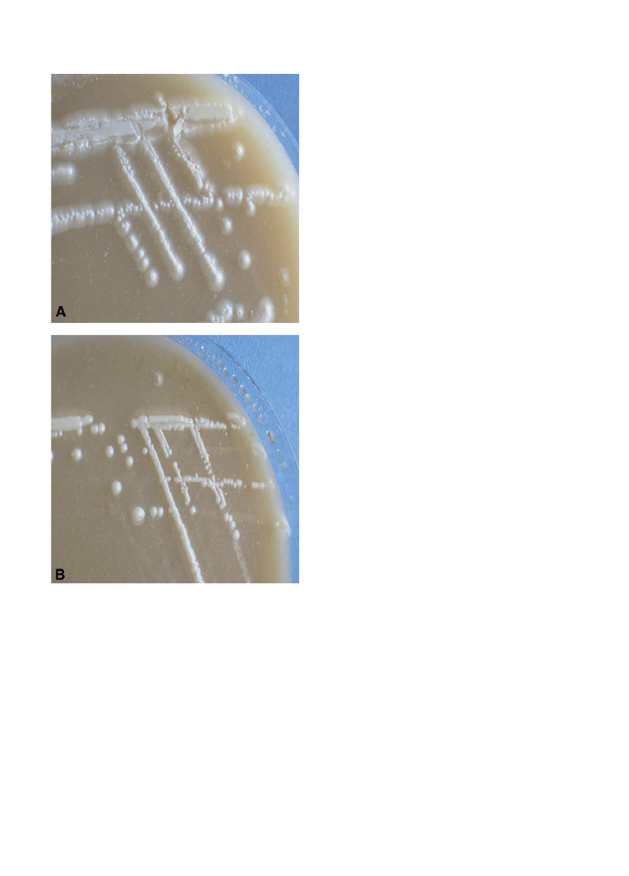
Fig. 1. Photograph of colonies of C. dubliniensis (A) and C. albicans (B)
grown on Pal’s agar at 30‡C for 48^72 h. The C. dubliniensis colonies
are rough and are surrounded by a hyphal fringe, whereas the C. albi-
cans colonies are smooth and have no hyphal fringe.
FEMSYR 1629 18-12-03
Cyaan
Magenta
Geel
Zwart
D.J. Sullivan et al. / FEMS Yeast Research 4 (2004) 369^376
372

CdMDR1, respectively) and have been implicated in the
development of resistance to azole drugs. Moran et al. [5]
analysed the resistance mechanisms in a group of £ucona-
zole-resistant isolates of C. dubliniensis and in vitro gen-
erated £uconazole-resistant derivatives and found that in
each case, £uconazole resistance was associated with up-
regulation of CdMDR1. Similarly, Perea et al.
de-
scribed increased CdMDR1 expression in £uconazole-re-
sistant clinical isolates of C. dubliniensis recovered from
two separate AIDS patients. The importance of CdMDR1
in the development of £uconazole resistance in clinical
isolates of C. dubliniensis was con¢rmed by gene deletion
studies in the £uconazole-resistant strain CM2, which was
rendered £uconazole-susceptible following deletion of
both alleles of CdMDR1
In contrast to C. dubliniensis, where CdMDR1 is the
principle £uconazole e¥ux mechanism identi¢ed so far,
in most isolates of C. albicans increased expression of
CaCDR1 is more commonly identi¢ed as the main medi-
ator of £uconazole resistance. Unlike the £uconazole-spe-
ci¢c Mdr1 pumps, Cdr1 pumps can transport a broad
range of azole drugs, including ketoconazole and itraco-
nazole. Cross-resistance to these azoles is relatively rare in
£uconazole-resistant C. dubliniensis isolates, and this may
re£ect the high prevalence of MDR1-mediated drug resis-
tance in C. dubliniensis. The reason(s) for the apparent
reduced contribution of CdCDR1 overexpression to £uco-
nazole resistance in C. dubliniensis is not clear. However,
Moran et al.
recently provided a likely explanation for
the lack of CdCDR1 expression seen in many £uconazole-
resistant isolates. Approximately 58% of genotype 1 C.
dubliniensis isolates (genotype 1 is the predominant geno-
type recovered from HIV-infected patients) were found to
harbour a nonsense mutation in the CdCDR1 gene, result-
ing in the translation of a truncated, non-functional pro-
tein. But £uconazole-resistant isolates with functional
CdCDR1 alleles may still exhibit increased CdCDR1
mRNA expression in conjunction with CdMDR1. Dele-
tion of both CdCDR1 copies in an isolate coexpressing
both pumps resulted in increased susceptibility to ketoco-
nazole and itraconazole. However, no e¡ect on £ucona-
zole MIC was observed. This was most likely due to the
high levels of CdMDR1 still expressed in this mutant
Point mutations in the ERG11 gene which result in spe-
ci¢c amino acid substitutions in the enzyme 14K-lanosterol
demethylase have also been shown to be important in the
development of £uconazole resistance in C. albicans. To
date, only one study, by Perea et al.
, has described
mutations in the C. dubliniensis CdERG11 gene associated
with £uconazole resistance. Two of the mutations de-
scribed are identical to mutations previously shown to
be involved in £uconazole resistance in C. albicans, namely
G307A and G464S. It has yet to be experimentally veri¢ed
whether the remaining CdERG11 mutations described af-
fect £uconazole susceptibility. These mutations occurred
in isolates which also exhibited increased drug e¥ux
pump expression, indicating that £uconazole resistance
in C. dubliniensis, as in C. albicans, is multifactorial.
Resistance to itraconazole has not yet been described in
clinical isolates of C. dubliniensis, but itraconazole-resis-
tant derivatives (that are also cross-resistant to other
azoles) can be generated in vitro following serial subcul-
ture of susceptible isolates on agar medium containing
increasing concentrations of drug
. Itraconazole-resis-
tant derivatives were found to have altered membrane per-
meabilities compared to susceptible isolates. Analysis of
their membrane sterol contents revealed pro¢les lacking
ergosterol which is consistent with a mutation in the sterol
C5,6-desaturase enzyme encoded by CdERG3. Although
increased expression of CdCDR1 and CdERG11 was also
noted in these derivatives, loss-of-function mutations in
CdERG3 were found to be responsible for the high levels
of azole cross-resistance observed. Mutations in the ERG3
gene have also been associated with azole resistance in C.
albicans
, however, the contribution of this resistance
mechanism to azole drug resistance in Candida species has
not been fully investigated.
In summary, as in C. albicans, the majority of C. dublin-
iensis isolates are susceptible to a wide range of antifungal
agents, thus it seems unlikely that the emergence of C.
dubliniensis in HIV-infected patients has been due to selec-
tion by antifungal therapy. When resistance does emerge
the molecular mechanisms are broadly similar in the two
species. However, there appear to be di¡erences in the
relative roles of the e¥ux proteins Cdr1p and Mdr1p,
probably due to the fact that in a large number of C.
dubliniensis isolates the CdCDR1 gene is defective.
4. Virulence of C. dubliniensis
The epidemiological data on the prevalence of C. dublin-
iensis described above clearly show that this species is only
relatively rarely encountered in the normal oral £ora of
immunocompetent individuals, but is apparently enriched
selectively in the oral cavities of immunocompromised pa-
tients, particularly HIV-infected and AIDS patients who
have received antifungal therapy with £uconazole. It is
also clear that while C. dubliniensis may in some cases
cause systemic infections, it does so far less frequently
than C. albicans. Con¢rmation of this apparent reduced
virulence of C. dubliniensis has been obtained in compar-
ative studies using a mouse model of systemic infection
. Therefore, despite the signi¢cant phenotypic and
genotypic similarities shared between C. albicans and C.
dubliniensis, current evidence from epidemiological and
virulence studies indicates that C. albicans is better
adapted to colonise and cause disease in vivo. This begs
the question, what di¡erences are there between the two
species that render C. albicans more pathogenic ?
Candida infections involve a very complex interaction
between a wide range of host factors and yeast virulence
FEMSYR 1629 18-12-03
Cyaan
Magenta
Geel
Zwart
D.J. Sullivan et al. / FEMS Yeast Research 4 (2004) 369^376
373

determinants that may be di¡erentially expressed depend-
ing on the prevailing environmental conditions. The iden-
ti¢cation and determination of the contribution of speci¢c
virulence factors to various C. albicans infections is cur-
rently a topic of major interest to medical mycologists.
One of the most important virulence factors of C. albicans
is its ability to adhere to certain human tissues. However,
given the range of host tissues which Candida species can
colonise and infect this suggests that these organisms pos-
sess a large number of surface adhesins, very few of which
have actually been characterised. There have been several
studies to date which have compared the adherence of C.
albicans and C. dubliniensis to a range of human cells.
However, it is di⁄cult to meaningfully compare the results
of these studies as di¡erent cells and culture conditions
were used in each case. Depending on the conditions
used, C. dubliniensis was sometimes found to be more
adherent than C. albicans
, while using di¡erent
assay conditions the opposite was also found
. Inter-
estingly, in one study, inclusion of £uconazole in the assay
medium led to an increase in adherence of C. dubliniensis
to Vero cells, suggesting that this might contribute to the
high prevalence of C. dubliniensis in HIV-infected and
AIDS patients receiving £uconazole therapy
. It has
also been shown that C. dubliniensis, unlike C. albicans,
is hydrophobic at 37‡C and that C. dubliniensis expresses
greatly reduced levels of acid-labile mannans in cell sur-
face N-glycans
. Cell surface hydrophobicity has
previously been associated with enhanced adherence and
resistance to phagocytosis
, however, this hydrophobic-
ity does not appear to confer resistance to phagocytosis on
C. dubliniensis
. The only study that investigated the
adherence of C. dubliniensis to a speci¢c host protein com-
pared the adherence of a range of Candida species to mu-
cin, to which C. dubliniensis was found to adhere as
strongly as C. albicans
. A gene family encoding pro-
teins homologous to the ALS family of surface adhesion
glycoproteins in C. albicans has also been identi¢ed in C.
dubliniensis, but it appears that these genes are regulated
di¡erently in the two species
.
Another trait of C. albicans that has been proposed as
an important virulence factor is its ability to grow either in
yeast or in hyphal form (i.e. dimorphism). C. dubliniensis
is the only Candida species other than C. albicans able to
produce germ tubes and true hyphae. However, the dy-
namics of production of hyphae in both species di¡er de-
pending on the culture conditions used ; in most cases in-
duction of hyphae is more e⁄cient in C. albicans (e.g. in
the presence of N-acetyl-
D
-glucosamine
), but con-
versely, under certain circumstances C. dubliniensis produ-
ces hyphae, while C. albicans does not (e.g. on Pal’s agar
) (
). There also appears to be a disparity in the
production of hyphae in vivo, as histopathological analy-
sis of infected kidneys in a systemic mouse infection model
showed that the C. dubliniensis cells were predominantly in
the yeast phase, while the C. albicans strains produced far
higher levels of hyphae and pseudohyphae
. These data
suggest that there are clearly di¡erences in the regulation
and dynamics of induction of hyphae in the two species.
Phenotypic switching is another important C. albicans vir-
ulence factor, however, it has been reported that C. dublin-
iensis can undergo phenotypic switching more frequently
than C. albicans
. The production of a range of extra-
cellular hydrolases, such as the secretory aspartyl protein-
ases (Saps), has been implicated in the pathogenicity of C.
albicans. Southern hybridisation analysis has shown that
C. dubliniensis encodes a similar range of genes as the C.
albicans SAP family. However, phenotypic studies de-
signed to determine the levels of proteinase produced by
C. dubliniensis have yielded contradictory results
. Interestingly, analysis of phospholipase production
in the two species suggests that C. dubliniensis may pro-
duce lower levels than C. albicans
While C. dubliniensis grows well in vitro at 37‡C and
produces bio¢lm under speci¢c conditions
, it has been
reported that its growth rate is less than that of C. albicans
, and that in mixed cultures, C. albicans out-grows
C. dubliniensis
. This suggests that C. albicans has a
competitive growth advantage under the conditions tested.
The di¡erent growth characteristics of the two species are
even more pronounced under conditions of environmental
stress and it has been shown that C. dubliniensis is signi¢-
cantly less tolerant of elevated temperature (e.g. s 42‡C
), osmotic pressure (e.g. 6.5% (w/v) NaCl
) and
oxidative stress (e.g. 10 mM H
2
O
2
).
The comparative virulence of C. dubliniensis and C. al-
bicans is clearly a very complex topic. While some di¡er-
ences in virulence factors have been identi¢ed there are
also numerous contradictory data. A full understanding
of the reasons for the greater capacity of C. albicans to
cause infection will require a concerted e¡ort to compare
the genomes and phenotypes of the two species. In partic-
ular, there is an urgent need to compare the e¡ects of a
greater number of strains belonging to each of the two
species in a wider range of animal models of infection.
The results of these studies should provide valuable infor-
mation concerning the molecular mechanisms of how Can-
dida species cause disease.
5. Conclusions
C. dubliniensis is now ¢rmly recognised as a signi¢cant
human pathogen. Since the introduction of highly active
anti-retroviral therapy (HAART) the incidence of oral
candidosis in HIV-infected and AIDS patients has de-
creased dramatically
. Consequently, the incidence of
C. dubliniensis in OPC has decreased since it ¢rst emerged
in the early 1990s. However, the prevalence of this species
should continue to be monitored in case of changes in the
epidemiology of AIDS (e.g. due to emerging resistance to
HAART or to lack of compliance) and due to the intro-
FEMSYR 1629 18-12-03
Cyaan
Magenta
Geel
Zwart
D.J. Sullivan et al. / FEMS Yeast Research 4 (2004) 369^376
374

duction of novel antifungal agents such as new azole de-
rivatives and the echinocandins. In addition, comparative
genomic analyses using microarrays and genome sequence
data will aid the identi¢cation of genetic di¡erences be-
tween the two species and contribute to the elucidation
of the reasons for the di¡erences in the capacity of these
species to colonise and infect humans, thus improving our
understanding of candidal pathogenicity.
Acknowledgements
Research in the authors laboratory has been supported
by the Irish Health Research Board grants RP04/97,
RP04/99 and RP08/2000 and by the Dublin Dental School
and Hospital.
References
[1] Krcmery, V. and Barnes, A.J. (2002) Non-albicans Candida spp. caus-
ing fungaemia pathogenicity and antifungal resistance. J. Hosp. In-
fect. 50, 243^260.
[2] Sullivan, D.J., Westerneng, T.J., Haynes, K.A., Bennett, D.E. and
Coleman, D.C. (1995) Candida dubliniensis sp. nov.: phenotypic and
molecular characterization of a novel species associated with oral
candidosis in HIV-infected individuals. Microbiology 141, 1507^1521.
[3] Gil¢llan, G.D., Sullivan, D.J., Haynes, K., Parkinson, T., Coleman,
D.C. and Gow, N.A.R. (1998) Candida dubliniensis: phylogeny and
putative virulence factors. Microbiology 144, 829^838.
[4] Donnelly, S.A., Sullivan, D.J., Shanley, D.B. and Coleman, D.C.
(1999) Phylogenetic analysis and rapid identi¢cation of Candida dub-
liniensis based on analysis of ACT1 intron and exon sequences. Mi-
crobiology 145, 1871^1882.
[5] Moran, G.P., Sanglard, D., Donnelly, S.M., Shanley, D.B., Sullivan,
D.J. and Coleman, D.C. (1998) Identi¢cation and expression of mul-
tidrug transporters responsible for £uconazole resistance in Candida
dubliniensis. Antimicrob. Agents Chemother. 42, 1819^1830.
[6] Moran, G., Sullivan, D., Morrschhau«ser, J. and Coleman, D. (2002)
The Candida dubliniensis CdCDR1 gene is not essential for £ucona-
zole resistance. Antimicrob. Agents Chemother. 46, 2829^2941.
[7] Pinjon, E., Moran, G.P., Jackson, C.J., Kelly, S.L., Sanglard, D.,
Coleman, D.C. and Sullivan, D.J. (2003) Molecular mechanisms of
itraconazole resistance in Candida dubliniensis. Antimicrob. Agents
Chemother. 47, 2424^2437.
[8] Sullivan, D. and Coleman, D. (1997) Candida dubliniensis: an emerg-
ing opportunistic pathogen. Curr. Top. Med. Mycol. 8, 15^25.
[9] Sullivan, D. and Coleman, D. (1998) Candida dubliniensis : character-
istics and identi¢cation. J. Clin. Microbiol. 36, 329^334.
[10] Sullivan, D.J., Moran, G., Donnelly, S., Gee, S., Pinjon, E., McCar-
ton, B., Shanley, D.B. and Coleman, D.C. (1999) Candida dublin-
iensis : an update. Rev. Iberoam. Micol. 16, 72^76.
[11] Al-Mosaid, A., Sullivan, D.J. and Coleman, D.C. (2003) Di¡erentia-
tion of Candida dubliniensis from Candida albicans on Pal’s Agar.
J. Clin. Microbiol. 41, 4787^4789.
[12] Park, S., Wong, M., Marras, S.A., Cross, E.W., Kiehn, T.E., Cha-
turvedi, V., Tyagi, S. and Perlin, D.S. (2000) Rapid identi¢cation of
Candida dubliniensis using a species-speci¢c molecular beacon. J. Clin.
Microbiol. 38, 2829^2836.
[13] Selvarangan, R., Limaye, A.P. and Cookson, B.T. (2002) Rapid iden-
ti¢cation and di¡erentiation of Candida albicans and Candida dublin-
iensis by capillary-based ampli¢cation and £uorescent probe hybrid-
ization. J. Clin. Microbiol. 40, 4308^4312.
[14] Tamura, M., Watanabe, K., Imai, T., Mikami, Y. and Nishimura, K.
(2000) New PCR primer pairs speci¢c for Candida dubliniensis and
detection of the fungi from the Candida albicans clinical isolates in
Japan. Clin. Lab. 46, 33^40.
[15] Kurzai, O., Heinz, W.J., Sullivan, D.J., Coleman, D.C., Frosch, M.
and Muhlschlegel, F.A. (1999) Rapid PCR test for discriminating
between Candida albicans and Candida dubliniensis isolates using
primers derived from the pH-regulated PHR1 and PHR2 genes of
C. albicans. J. Clin. Microbiol. 37, 1587^1590.
[16] Elie, C.M., Lott, T.J., Reiss, E. and Morrison, C.J. (1998) Rapid
identi¢cation of Candida species with species-speci¢c DNA probes.
J. Clin. Microbiol. 36, 3260^3265.
[17] Joly, S., Pujol, C., Rysz, M., Vargas, K. and Soll, D.R. (1999) Devel-
opment and characterization of complex DNA ¢ngerprinting probes
for the infectious yeast Candida dubliniensis. J. Clin. Microbiol. 37,
1035^1044.
[18] Gee, S.F., Joly, S., Soll, D.R., Meis, J.F., Verweij, P.E., Polacheck,
I., Sullivan, D.J. and Coleman, D.C. (2002) Identi¢cation of four
distinct genotypes of Candida dubliniensis and detection of microevo-
lution in vitro and in vivo. J. Clin. Microbiol. 40, 556^574.
[19] Joly, S., Pujol, C. and Soll, D.R. (2002) Microevolutionary changes
and chromosomal translocations are more frequent at RPS loci in
Candida dubliniensis than in Candida albicans. Infect. Genet. Evol. 2,
19^37.
[20] Polacheck, I., Strahilevitz, J., Sullivan, D., Donnelly, S., Salkin, I.F.
and Coleman, D.C. (2000) Recovery of Candida dubliniensis from
non-human immunode¢ciency virus-infected patients in Israel.
J. Clin. Microbiol. 38, 170^174.
[21] Sullivan, D., Haynes, K., Bille, J., Boerlin, P., Rodero, L., Lloyd, S.,
Henman, M. and Coleman, D. (1997) Widespread geographic distri-
bution of oral Candida dubliniensis strains in human immunode¢-
ciency virus-infected individuals. J. Clin. Microbiol. 35, 960^964.
[22] Kamei, K., McCullough, M.J. and Stevens, D.A. (2000) Initial case
of Candida dubliniensis infection from Asia : non-mucosal infection.
Med. Mycol. 38, 81^83.
[23] Fisher, J.M., Basson, N.J. and van Zyl, A. (2001) Identi¢cation of
Candida dubliniensis in a HIV-positive South African population. S.
Afr. Dent. J. 56, 599^601.
[24] Ponton, J., Ruchel, R., Clemons, K.V., Coleman, D.C., Grillot, R.,
Guarro, J., Aldebert, D., Ambroise-Thomas, P., Cano, J., Carrillo-
Munoz, A.J., Gene, J., Pinel, C., Stevens, D.A. and Sullivan, D.J.
(2000) Emerging pathogens. Med. Mycol. 38 (Suppl. 1), 225^236.
[25] Tintelnot, K., Haase, G., Seibold, M., Bergmann, F., Staemmler, M.,
Franz, T. and Naumann, D. (2000) Evaluation of phenotypic
markers for selection and identi¢cation of Candida dubliniensis.
J. Clin. Microbiol. 38, 1599^1608.
[26] Lasker, B.A., Elie, C.M., Lott, T.J., Espinel-Ingro¡, A., Gallagher,
L., Kuykendall, R.J., Kellum, M.E., Pruitt, W.R., Warnock, D.W.,
Rimland, D., McNeil, M.M. and Reiss, E. (2001) Molecular epidemi-
ology of Candida albicans strains isolated from the oropharynx of
HIV-positive patients at successive clinic visits. Med. Mycol. 39,
341^352.
[27] Meiller, T.F., Jabra-Rizk, M.A., Baqui, A., Kelley, J.I., Meeks, V.I.,
Merz, W.G. and Falkler, W.A. (1999) Oral Candida dubliniensis as a
clinically important species in HIV-seropositive patients in the United
States. Oral Surg. Oral Med. Oral Pathol. Oral Radiol. Endod. 88,
573^580.
[28] Vargas, K.G. and Joly, S. (2002) Carriage frequency, intensity of
carriage, and strains of oral yeast species vary in the progression to
oral candidiasis in human immunode¢ciency virus-positive individu-
als. J. Clin. Microbiol. 40, 341^350.
[29] Blignaut, E., Pujol, C., Joly, S. and Soll, D.R. (2003) Racial distri-
bution of Candida dubliniensis colonization among South Africans.
J. Clin. Microbiol. 41, 1838^1842.
[30] Willis, A.M., Coulter, W.A., Sullivan, D.J., Coleman, D.C., Hayes,
J.R., Bell, P.M. and Lamey, P.J. (2000) Isolation of C. dubliniensis
from insulin-using diabetes mellitus patients. J. Oral Pathol. Med. 29,
86^90.
FEMSYR 1629 18-12-03
Cyaan
Magenta
Geel
Zwart
D.J. Sullivan et al. / FEMS Yeast Research 4 (2004) 369^376
375

[31] Peltroche-Llacsahuanga, H., Dohmen, H. and Haase, G. (2002) Re-
covery of Candida dubliniensis from sputum of cystic ¢brosis patients.
Mycoses 45, 15^18.
[32] Gottlieb, G.S., Limaye, A.P., Chen, Y.C. and Van Voorhis, W.C.
(2001) Candida dubliniensis fungemia in a solid organ transplant pa-
tient : case report and review of the literature. Med. Mycol. 39, 483^
485.
[33] Meis, J.F., Ruhnke, M., De Pauw, B.E., Odds, F.C., Siegert, W. and
Verweij, P.E. (1999) Candida dubliniensis candidemia in patients with
chemotherapy-induced neutropenia and bone marrow transplanta-
tion. Emerg. Infect. Dis. 5, 150^153.
[34] Sebti, A., Kiehn, T.E., Perlin, D., Chaturvedi, V., Wong, M., Doney,
A., Park, S. and Sepkowitz, K.A. (2001) Candida dubliniensis at a
cancer center. Clin. Infect. Dis. 32, 1034^1038.
[35] Brandt, M.E., Harrison, L.H., Pass, M., Sofair, A.N., Huie, S., Li,
R.K., Morrison, C.J., Warnock, D.W. and Hajjeh, R.A. (2000) Can-
dida dubliniensis fungemia: the ¢rst four cases in North America.
Emerg. Infect. Dis. 6, 46^49.
[36] Kibbler, C.C., Ainscough, S., Barnes, R.A., Gransden, W.R., Holli-
man, R.E., Johnson, E.M., Perry, J.D., Sullivan, D.J. and Wilson,
J.A. (2003) Management and outcome of blood stream infections due
to Candida species in England and Wales. J. Hosp. Infect. 54, 18^24.
[37] Odds, F.C., Van Nu¡el, L. and Dams, G. (1998) Prevalence of Can-
dida dubliniensis isolates in a yeast stock collection. J. Clin. Micro-
biol. 36, 2869^2873.
[38] Martins-Nishikawa, M., Trilles, L., Symoens, F., Swinne, D. and
Nolard, N. (2002) Prevalence of Candida dubliniensis in the BCCM/
IHEM Biomedical Fungi/Yeasts culture collection (isolates before
1990). Med. Mycol. 40, 443^445.
[39] Martinez, M., Lopez-Ribot, J.L., Kirkpatrick, W.R., Coco, B.J.,
Bachmann, S.P. and Patterson, T.F. (2002) Replacement of Candida
albicans with C. dubliniensis in human immunode¢ciency virus-in-
fected patients with oropharyngeal candidiasis treated with £ucona-
zole. J. Clin. Microbiol. 40, 3135^3139.
[40] Moran, G.P., Sullivan, D.J., Henman, M.C., McCreary, C.E., Har-
rington, B.J., Shanley, D.B. and Coleman, D.C. (1997) Antifungal
drug susceptibilities of oral Candida dubliniensis isolates from human
immunode¢ciency virus (HIV)-infected and non-HIV-infected sub-
jects and generation of stable £uconazole-resistant derivatives in vi-
tro. Antimicrob. Agents Chemother. 41, 617^623.
[41] Pfaller, M.A., Messer, S.A., Gee, S., Joly, S., Pujol, C., Sullivan,
D.J., Coleman, D.C. and Soll, D.R. (1999) In vitro susceptibilities
of Candida dubliniensis isolates tested against the new triazole and
echinocandin antifungal agents. J. Clin. Microbiol. 37, 870^872.
[42] Perea, S., Perea, S., Lopez-Ribot, J.L., Wickes, B.L., Kirkpatrick,
W.R., Dib, O.P., Bachmann, S.P., Keller, S.M., Martinez, M. and
Patterson, T.F. (2002) Molecular mechanisms of £uconazole resis-
tance in Candida dubliniensis isolates from Human Immunode¢ciency
Virus-infected patients with oropharyngeal candidiasis. Antimicrob.
Agents Chemother. 46, 1695^1703.
[43] Ruhnke, M., Schmidt-Westhausen, A. and Morschhau«ser, J. (2000)
Development of simultaneous resistance to £uconazole in Candida
albicans and Candida dubliniensis in a patient with AIDS. J. Antimi-
crob. Chemother. 46, 291^295.
[44] Borg-Von Zepelin, M., Niederhaus, T., Gross, U., Seibold, M.,
Monod, M. and Tintelnot, K. (2002) Adherence of di¡erent Candida
dubliniensis isolates in the presence of £uconazole. AIDS 16, 1237^
1244.
[45] Wirsching, S., Moran, G.P., Sullivan, D.J., Coleman, D.C. and
Morschhauser, J. (2001) MDR1-mediated drug resistance in Candida
dubliniensis. Antimicrob. Agents Chemother. 45, 3416^3421.
[46] Miyazaki, Y., Geber, A., Miyazaki, H., Falconer, D., Parkinson, T.,
Hitchock, C., Grimberg, B., Nyswaner, K. and Bennett, J.E. (1999)
Cloning, sequencing, expression and allelic sequence diversity of
ERG3 (C-5 sterol desaturase gene) in Candida albicans. Gene 236,
43^51.
[47] Vilela, M.M., Kamei, K., Sano, A., Tanaka, R., Uno, J., Takahashi,
I., Ito, J., Yarita, K. and Miyaji, M. (2002) Pathogenicity and viru-
lence of Candida dubliniensis : comparison with C. albicans. Med.
Mycol. 40, 249^257.
[48] McCullough, M., Ross, B. and Reade, P. (1995) Characterization of
genetically distinct subgroup of Candida albicans strains from oral
cavities of patients infected with human immunode¢ciency virus.
J. Clin. Microbiol. 33, 696^700.
[49] Jabra-Rizk, M.A., Falkler, W.A., Merz, W.G., Baqui, A.A.M.A.,
Kelley, J.I. and Meiller, T.F. (2001) Cell surface hydrophobicity-as-
sociated adherence of Candida dubliniensis to human buccal epithelial
cells. Rev. Iberoam. Micol. 18, 17^22.
[50] Hazen, K.C., Wu, J.G. and Masuoka, J. (2001) Comparison of the
hydrophobic properties of Candida albicans and Candida dubliniensis.
Infect. Immun. 69, 779^786.
[51] Hazen, K.C. and Glee, P.M. (1995) Cell surface hydrophobicity and
medically important fungi. Curr. Top. Med. Mycol. 6, 1^31.
[52] Peltroche-Llacsahuanga, H., Schnitzler, N., Schmidt, S., Tintelnot,
K., Lutticken, R. and Haase, G. (2000) Phagocytosis, oxidative burst,
and killing of Candida dubliniensis and Candida albicans by human
neutrophils. FEMS Microbiol. Lett. 191, 151^155.
[53] de Repentigny, L., Aumont, F., Bernard, K. and Belhumeur, P.
(2000) Characterization of binding of Candida albicans to small in-
testinal mucin and its role in adherence to mucosal epithelial cells.
Infect. Immun. 68, 3172^3179.
[54] Hoyer, L.L., Fundyga, R., Hecht, J.E., Kapteyn, J.C., Klis, F.M. and
Arnold, J. (2001) Characterization of agglutinin-like sequence genes
from non-albicans Candida and phylogenetic analysis of the ALS
family. Genetics 157, 1555^1567.
[55] Hannula, J., Saarela, M., Dogan, B., Paatsama, J., Koukila-Kahkola,
P., Pirinen, S., Alakomi, H.L., Perheentupa, J. and Asikainen, S.
(2000) Comparison of virulence factors of oral Candida dubliniensis
and Candida albicans isolates in healthy people and patients with
chronic candidosis. Oral Microbiol. Immunol. 15, 238^244.
[56] Slifkin, M. (2000) Tween 80 opacity test responses of various Candida
species. J. Clin. Microbiol. 38, 4626^4628.
[57] Ramage, G., Vande Walle, K., Wickes, B.L. and Lopez-Ribot, J.L.
(2001) Bio¢lm formation by Candida dubliniensis. J. Clin. Microbiol.
39, 3234^3240.
[58] Kirkpatrick, W.R., Lopez-Ribot, J.L., McAtee, R.K. and Patterson,
T.F. (2000) Growth competition between Candida dubliniensis and
Candida albicans under broth and bio¢lm growing conditions.
J. Clin. Microbiol. 38, 902^904.
[59] Pinjon, E., Sullivan, D., Salkin, I., Shanley, D. and Coleman, D.
(1998) Simple, inexpensive, reliable method for di¡erentiation of Can-
dida dubliniensis from Candida albicans. J. Clin. Microbiol. 36, 2093^
2095.
[60] Alves, S.H., Milan, E.P., de Laet Sant’Ana, P., Oliveira, L.O., San-
turio, J.M. and Colombo, A.L. (2002) Hypertonic Sabouraud broth
as a simple and powerful test for Candida dubliniensis screening.
Diagn. Microbiol. Infect. Dis. 43, 85^86.
[61] Martins, M.D., Lozano-Chiu, M. and Rex, J.H. (1998) Declining
rates of oropharyngeal candidiasis and carriage of Candida albicans
associated with trends toward reduced rates of carriage of £ucona-
zole-resistant C. albicans in human immunode¢ciency virus-infected
patients. Clin. Infect. Dis. 27, 1291^1294.
[62] Pincus, D.H., Coleman, D.C., Pruitt, W.R., Padhye, A.A., Salkin,
I.F., Geimer, M., Bassel, A., Sullivan, D.J., Clarke, M. and Hearn,
V. (1999) Rapid identi¢cation of Candida dubliniensis with commer-
cial yeast identi¢cation systems. J. Clin. Microbiol. 37, 3533^3539.
[63] Coleman, D., Sullivan, D., Harrington, B., Haynes, K., Henman, M.,
Shanley, D., Bennett, D., Moran, G., McCreary, C. and O’Neill, L.
(1997) Molecular and phenotypic analysis of Candida dubliniensis : a
recently identi¢ed species linked with oral candidosis in HIV-infected
and AIDS patients. Oral Dis. 3 (Suppl. 1), S96^S101.
FEMSYR 1629 18-12-03
Cyaan
Magenta
Geel
Zwart
D.J. Sullivan et al. / FEMS Yeast Research 4 (2004) 369^376
376
Document Outline
Wyszukiwarka
Podobne podstrony:
Antifungal drug resistance machanism in fungal pathogens
Pathogenesis and antifungal drug resistance of C glabrata
Proteomics of drug resistance in C glabrata
Comparison of Human Language and Animal Communication
Comparison of the Russians and Bosnians
A Comparison of Plato and Aristotle
A Comparison of the Status of Women in Classical Athens and E
Comparison of the U S Japan and German British Trade Rivalr
Comparison of Plato and Aristotle's Political Theories
Comparison Of Judaism And Christianity
Comparison of theoretical and experimental free vibrations of high industrial chimney interacting
Comparison of cartesian vector control and polar
Alta J LaDage Occult Psychology, A Comparison of Jungian Psychology and the Modern Qabalah
12 Angry Men Comparison of the Movie and Play
Proteomics of drug resistance in C glabrata
Comparison of theoretical and experimental free vibrations of high industrial chimney interacting
więcej podobnych podstron