
Published online 19 February 2003
Electrophysiology and brain imaging of biological
motion
Aina Puce
1*
and David Perrett
2
1
Centre for Advanced Imaging, Department of Radiology, West Virginia University, PO Box 9236,
Morgantown, WV 26506-9236, USA
2
School of Psychology, University of St Andrews, St Andrews, Fife KY16 9JU, UK
The movements of the faces and bodies of other conspecifics provide stimuli of considerable interest to
the social primate. Studies of single cells, field potential recordings and functional neuroimaging data
indicate that specialized visual mechanisms exist in the superior temporal sulcus (STS) of both human
and non-human primates that produce selective neural responses to moving natural images of faces and
bodies. STS mechanisms also process simplified displays of biological motion involving point lights mark-
ing the limb articulations of animate bodies and geometrical shapes whose motion simulates purposeful
behaviour. Facial movements such as deviations in eye gaze, important for gauging an individual’s social
attention, and mouth movements, indicative of potential utterances, generate particularly robust neural
responses that differentiate between movement types. Collectively such visual processing can enable the
decoding of complex social signals and through its outputs to limbic, frontal and parietal systems the STS
may play a part in enabling appropriate affective responses and social behaviour.
Keywords: biological motion; event related potentials; functional magnetic resonance imaging; humans;
single-unit electrophysiology; animals
1. INTRODUCTION
Primates, being social animals, continually observe one
another’s behaviour so as to be able to integrate effectively
within their social living structure. At a non-social level,
successful predator evasion also necessitates being able to
‘read’ the actions of other species in one’s vicinity. The
ability to interpret the motion and action of others in
human primates goes beyond basic survival and successful
interactions with important conspecifics. Many of our rec-
reational and cultural pursuits would not be possible with-
out this ability. Excellent symphony orchestras exist not
only owing to the exceptional musicians, but also their
ability to interpret their conductors’ non-verbal instruc-
tions. Conductors convey unambiguously not only the
technical way that the orchestra should execute the piece
of music, but modulate the mood and emotional tone of
the music measure by measure. The motion picture indus-
try owes much of its success today to its silent movie pion-
eers, who could entertain with their non-verbal antics. The
world’s elite athletes rely on the interpretation of other’s
movements to achieve their team’s goals successfully and
foil opponents.
2. HUMAN BEHAVIOURAL STUDIES OF
BIOLOGICAL MOTION PERCEPTION
The perception of moving biological forms can rely on
the ability to integrate form and motion but it can also
*
Author for correspondence (apuce@hsc.wvu.edu).
One contribution of 15 to a Theme Issue ‘Decoding, imitating and
influencing the actions of others: the mechanisms of social interaction’.
Phil. Trans. R. Soc. Lond. B (2003) 358, 435–445
435
Ó 2003 The Royal Society
DOI 10.1098/rstb.2002.1221
rely on the ability to define form from motion (Oram &
Perrett 1994, 1996). The latter is evident in the ingenious
work of Johansson who filmed actors dressed in black with
white dots attached to their joints on a completely black
set ( Johansson 1973). With these moving dots human
observers could reliably identify the walking or running
motions, for example, of another human or an animal
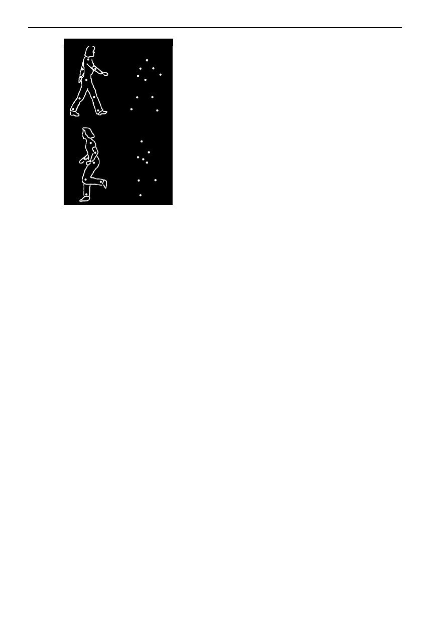
(figure 1). This type of stimulus is known as a Johansson,
point light or biological motion display.
A number of important observations have emerged from
the human behavioural biological motion perception
literature. First, the perceptual effect of observing an indi-
vidual walking or running is severely compromised when
the display is inverted (Dittrich 1993; Pavlova & Sokolov
2000). Second, while biological motion representing loco-
motory movements is recognized the most efficiently,
social and instrumental actions can also be recognized
from these impoverished displays (Dittrich 1993). Third,
biological motion can be perceived even within masks of
dots (Perrett et al. 1990a; Thornton et al. 1998). Fourth,
the gender of the walker (and even the identity of specific
individuals) can be recognized from pattern of gait and
idiosyncratic body movements in these impoverished dis-
plays (Cutting & Kozlowski 1977; Kozlowski & Cutting
1977). Fifth, there is a bias to perceive forward loco-
motion, at the expense of misinterpreting the underlying
form in time-reversed biological motion films (Pavlova et
al. 2002). Finally, observers can discern various emotional
expressions from viewing Johansson faces (Bassili 1978).
In very low light conditions many animals are efficient
at catching prey or evading predators. In such conditions
the patterns of articulation (typical of biological motion)
may be more discernible than the form of stationary ani-
436 A. Puce and D. Perrett
Physiology of biological motion
Figure 1. An example of a biological motion stimulus.
(Adapted from Johansson (1973), with permission from
Percept. Psychophys.)
mals. Indeed, in behavioural experiments it is evident that
point light displays are sufficient for cats to discriminate
the pattern of locomotion of conspecifics (Blake 1993). In
an ingenious behavioural study in cats, a forced choice
task where selection of a biological motion display (of a
cat walking or running) was rewarded with food resulting
in the animals performing significantly above chance. A
series of foil stimuli showing dots changing their spatial
location provided a set of tight controls in this experiment
(Blake 1993).
Evidence for the existence of specialized brain systems
that analyse biological motion (and the motion of humans
and non-humans) comes from neuropsychological lesion
studies. Dissociations between the ability to perceive bio-
logical motion and other types of motion have been dem-
onstrated. Several patients who are to all intents and
purposes ‘motion blind’ can discriminate biological
motion stimuli (Vaina et al. 1990; McLeod et al. 1996).
The opposite pattern, i.e. an inability to perceive biologi-
cal motion yet have relatively normal motion perception
in general, has also been reported (Schenk & Zihl 1997).
3. BIOLOGICAL MOTION PERCEPTION IN
NON-HUMANS
One brain region known as the STP area in the cortex
surrounding the STS has been the subject of considerable
scrutiny ever since cells selective for the sight of faces were
characterized in this region in monkeys (Perrett et al.
1982; Desimone 1991). This STS brain region is known
to be a convergence point for the dorsal and ventral visual
streams. The STP area derives its input from the MST
area in the dorsal pathway and the anterior inferior-
temporal area in the ventral pathway (Boussaoud et al.
1990; Felleman & Van Essen 1991). The cortex of the
STS has connections with the amygdala (Aggleton et al.
1980) and also with the orbitofrontal cortex (Barbas
1988), regions implicated in the processing of stimuli of
social and emotional significance in both human and non-
Phil. Trans. R. Soc. Lond. B (2003)
human primates (reviewed in Baron-Cohen 1995; Bro-
thers 1997; Adolphs 1999).
In addition to having face-specific cells, the cortex of
the STS has other complex response properties. It has
emerged that visual information about the shape and pos-
ture of the fingers, hands, arms, legs and torso all impact
on STS cell tuning in addition to facial details such as the
shape of the mouth and direction of gaze (Desimone et al.
1984; Wachsmuth et al. 1994; Perrett et al. 1984, 1985a;
Jellema et al. 2000). Motion information presumed to
arrive from the dorsal stream projections arrives in the
STS some 20 ms ahead of form information from the ven-
tral stream (figure 2a), but despite this asynchrony, STS
processing overcomes the ‘binding problem’ and only
form and motion arising from the same biological object
are integrated within 100 ms of the moving form becom-
ing visible (Oram & Perrett 1996). Indeed, STS cell inte-
gration of form and motion is widespread and there are
numerous cell types specializing in the processing of dif-
ferent types of face, limb and whole body motion (Perrett
et al. 1985b; Carey et al. 1997; Jellema et al. 2000, 2002;
Jellema & Perrett 2002).
While most STS cells derive sensitivity to body move-
ment by combining signals about the net translation or
rotation of the body with the face and body form visible
at any moment in time, a smaller proportion (20%) of
cells are able to respond selectively to the form of the body
defined through patterns of articulation in point light dis-
plays (Perrett et al. 1990a,b; Oram & Perrett 1994, 1996;
figure 1). These cells tuned to biological motion are selec-
tive for the sight of the same action visible in full light and
when depicted in point light displays.
Cells responding to whole body motion exhibit selec-
tivity for direction of motion and view of the body: most
respond preferentially to compatible motion with the body
moving forward in the direction it faces, though some are
tuned to backward locomotion with the body moving in
the opposite direction to the way it faces (Perrett et al.
1985b, 1989; Oram & Perrett 1996; figure 2b). This cellu-
lar tuning bias for forward locomotion may underlie the
forward bias found in perceptual interpretation of loco-
motion depicted in point light displays (Pavlova et al.
2002).
Responses to purposeful hand object actions such as
reaching for, picking, tearing and manipulating objects
have also been characterized in the STS (Perrett et al.
1989, 1990c; Jellema et al. 2000). These STS cells are
sensitive to the form of the hand performing the action,
and are unresponsive to the sight of tools manipulating
objects in the same manner as hands. Furthermore, the
cells code the spatio-temporal interaction between the
agent performing the action and the object of the action.
For example, cells tuned to hands manipulating an object
cease to respond if the hands and object move appropri-
ately but are spatially separated. This selectivity ensures
that the cells are more responsive in situations where the
agent’s motion is causally related to the object’s motion.
The STS cell populations coding body and hand actions
appear to be exclusively visual, although information from
the motor system does affect other STS cell populations
(Hietanen & Perrett 1996) and modulates STS activity in
humans (Iacoboni et al. 2001; Nishitani & Hari 2001).
Information defined by the visual characterization of

Physiology of biological motion A. Puce and D. Perrett 437
form +
motion
motion
form
130
120
110
100
100
90
80
re
sp
on
se
la
te
nc
y
(m
s)
70
60
50
40
30
20
10
0
re
sp
on
se
(
sp
ik
es
s
_ 1
)
SA
>
>
<
<
<
<
motion
direction
body
view
(a)
(b)
Figure 2. Some response properties of primate STP area neurons elicited by biological motion stimuli. (Adapted from Oram &
Perrett (1994, 1996), with permission.) (a) Average response latencies for neurons with different response properties. (b) An
example of a neuron that does not differentiate between real human motion and biological motion. Also, the strongest
response is in the motion direction compatible with direction of the body.
actions in the STS appears to be relayed via parietal sys-
tems (Gallese et al. 2002) to frontal motor planning sys-
tems. In frontal and parietal areas a neural system has
recently been found to respond selectively both during the
execution of hand actions, and (like STS cells) during the
observation of corresponding actions performed by others.
The frontal region of primate cortex had long been known
to be somatotopically organized for the representation and
control of movements of the mouth and arm (Rizzolatti et
al. 1988). Neurons within area F5 of the monkey premo-
tor cortex have now been labelled ‘mirror’ neurons,
because they discharge when monkeys perform or observe
the same hand actions (di Pellegrino et al. 1992; Rizzolatti
et al. 1996a,b; Gallese et al. 1996). An F5 cell selective
for the action of grasping would respond for example
when the monkey grasps an object in sight or in the dark
(thereby demonstrating motoric properties). The visual
properties of such an F5 cell are strikingly similar to those
described in the STS: both F5 and STS cells will respond
when the monkey observes the experimenter reaching and
grasping an object, but not to the sight of the exper-
imenter’s hand motion alone or the sight of the object
alone. These conjoint properties have led Rizzolatti et al.
(1996a,b) and Gallese et al. (1996) to postulate that the
F5 neurons form a system for matching observation and
executing actions for the grasping, manipulation and
placement of objects. Because the cells additionally
respond selectively to the sound of actions (Kohler et al.
2002), the mirror system may provide a supra-modal con-
ceptual representation of actions and their consequences
in the world. Crucially the properties of the frontal mirror
system indicate that we may understand actions perfor-
med by others because we can match the actions we sense
through vision (and audition) to our ability to produce the
same actions ourselves.
The actions of others are not always fully visible, for
example someone may become hidden from our sight as
they move behind a tree, or their hands may not remain
fully in view as they reach to retrieve an object. The simi-
larity of STS and F5 systems in processing of actions has
become more apparent in experiments investigating the
nature of processing during these moments when actions
Phil. Trans. R. Soc. Lond. B (2003)
are partially or totally occluded from sight. Within the
STS it is now apparent that specific cell populations are
activated when the presence of a hidden person can be
inferred from the preceding visual events (i.e. they were
witnessed passing out of sight behind a screen and have
not yet been witnessed re-emerging into sight, so they are
likely to remain behind the screen; Baker et al. 2001). In
an analogous manner, F5 cells may respond to the sight
of the experimenter reaching to grasp an object. The same
cells are active when the experimenter places an object
behind a screen and then reaches as if to grasp it (even
though the object and hand are hidden from view (Umilta
et al. 2002)). The sight of equivalent reaching when there
is no reason to believe an object is hidden from sight fails
to activate the F5 cells. Thus F5 and STS cells code the
sight of actions on the basis of what is currently visible
and on the basis of the recent perceptual history
(Jellema & Perrett 2002; Jellema et al. 2002).
The manner in which temporal STS and frontal F5 sys-
tems interact is not fully clear, but appears to involve
intermediate processing steps mediated by parietal areas
(Nishitani & Hari 2000, 2001; Gallese et al. 2002). While
STS and F5 cells have similar visual properties they may
subserve distinct functions; the frontal system perhaps
serves to control the behaviour of the self particularly in
dealing with objects (Rizzolatti et al. 1996a,b), whereas the
STS system is specialized for the detection and recog-
nition of the behaviour of others (Perrett et al. 1990c;
Mistlin & Perrett 1990; Hietanen & Perrett 1996).
4. HUMAN NEUROIMAGING AND
ELECTROPHYSIOLOGICAL STUDIES OF
BIOLOGICAL MOTION PERCEPTION
The first suggestion that humans may possess special-
ized biological motion perception mechanisms came from
a point light display depicting a moving body designed to
investigate the response properties of medial temporal/V5,
a region of occipito-temporal cortex known to respond to
motion. In this fMRI study activation was observed in
MT/V5 as well as areas of superior temporal cortex. This
was regarded at that time as surprising, as the activation

438 A. Puce and D. Perrett
Physiology of biological motion
appeared to lie in brain regions traditionally regarded as
participating in auditory speech processing (Howard et al.
1996). Localization of primary auditory cortex was not
performed in this visual stimulation study. In a PET study
published in the same year Johansson displays of body
motion (depicting a person dancing), hand motion
(depicting a hand reaching for a glass and bringing it to
a mouth), object motion (depicting a three-dimensional
structure rotating and pitching) and control conditions,
consisting of either random dot motion or a static display
of randomly placed dots, were shown to a group of healthy
subjects (Bonda et al. 1996). The human motion con-
ditions selectively activated the inferior parietal region and
the STS. Specifically, the body motion condition selec-
tively activated the right posterior STS, whereas the hand
motion condition activated the left intraparietal sulcus and
the posterior STS (Bonda et al. 1996). In a more recent
fMRI study, a Johansson display depicting a walker was
used and the activation contrasted to control conditions
that included a dot display with non-random motion and
a gender discrimination task with real images of faces
(Vaina et al. 2001). Biological motion differentially acti-
vated a large number of dorsal and ventral regions, most
notably the lateral occipital complex, but the STS was not
preferentially activated in this study.
Grossman and colleagues found that biological motion
stimuli depicting jumping, kicking, running and throwing
movements produced more right STS activation than con-
trol motion irrespective of the visual field in which the
biological motion display was presented. Conversely, the
control motion, including scrambled biological motion
displays, activated MT/MST areas and the lateral-occipital
complex (Grossman et al. 2000). Moreover, the STS
region could also be activated by imagining Johansson
stimuli, although the size of the activation was small
(Grossman et al. 2000). While the most robust STS acti-
vation was elicited by viewing upright Johansson displays,
a smaller STS activation signal was also seen to viewing
inverted Johansson displays.
While biological motion clearly activates the STS region
in humans, the function of the region may be more general
in performing a visual analysis of bodies based on either
the characteristic patterns of articulation that comprise
biological motion or information about bodies that can be
derived from static images (Downing et al. 2001); hence
the term ‘extrastriate body area’ has been applied to one
cortical region within the STS complex.
5. BIOLOGICAL MOTION PERCEPTION VERSUS
HUMAN MOTION PERCEPTION
As in non-human primates, responsiveness to Johans-
son-like displays of facial motion is present in STS regions
that also respond to real images of facial motion, e.g. non-
linguistic mouth movements (Puce et al. 2001), although
the per cent magnetic resonance signal change to the
Johansson-like face was smaller than that observed to the
natural facial images. In parallel to the neuroimaging data,
direct measures of neural activity in humans, in the form
of scalp ERPs, are elicited to Johansson-like and real
images of faces (Thompson et al. 2002b), with a promi-
nent negativity occurring at ca. 170 ms post-motion onset
Phil. Trans. R. Soc. Lond. B (2003)
eyes
Puce et al. eye gaze
Wicker et al. eye gaze
Hoffman & Haxby eye gaze
hand
Neville et al. ASL
Bonda et al. hand action
Grezes et al. hand action
Grezes et al. hand movement
Grafton et al. hand grasp
Rizzolatti et al. hand grasp
mouth
Calvert et al. lip reading (STG)
Calvert et al. lip reading (AG)
Puce et al. mouth movement
Puce & Allison mouth movement
body
Howard et al. body movement
Bonda et al. body movement
Senior et al. body movement
Kourtzi & Kanwisher
body movement
Grossman et al. body movement
Figure 3. Centres of activation to viewing the face, hand and
body movements of others obtained from a series of PET
and fMRI studies. (Adapted from Allison et al. (2000), with
permission.)
(N170) over the bilateral temporal scalp. This activity is
significantly greater than that seen to motion controls.
Over the latter half of the 1990s, a series of PET and
fMRI studies examining activation to viewing the motion
and actions of others have pointed to the existence of
cortical networks that preferentially process certain attri-
butes of these high-level visual displays (reviewed by Alli-
son et al. 2000; Blakemore & Decety 2001). Figure 3
displays activation observed in these studies, lying along
the posterior extent of the STS and its ascending limb in
inferior parietal cortex in response to observing move-
ments of the body, hands, eye and mouth. Activation in
these regions can also be elicited to imagining the motion
of others (Grossman et al. 2000), and additionally to
viewing static images of implied motion (Kourtzi &
Kanwisher 2000).
Interestingly, differences in activation patterns can
occur when subjects view compatible versus incompatible
motion of the head or body (Thompson et al. 2002a).
Specifically, the bilateral posterior lateral temporal cortex
is active when viewing compatible motion. By contrast,
viewing incompatible motion activates the right posterior
lateral temporal cortex, left anterior temporal cortex, left
Physiology of biological motion A. Puce and D. Perrett 439
_4
_2
2
4
6
8
_100 0 100 200 300 400 500 600 700 800 900
time (ms)
am
pl
it
ud
e
(m
V)
P400
N170
mV
4
3
2
1
0
_1
_2
_3
_4
group attention
control
mutual gaze exchange
(a)
(b)
0
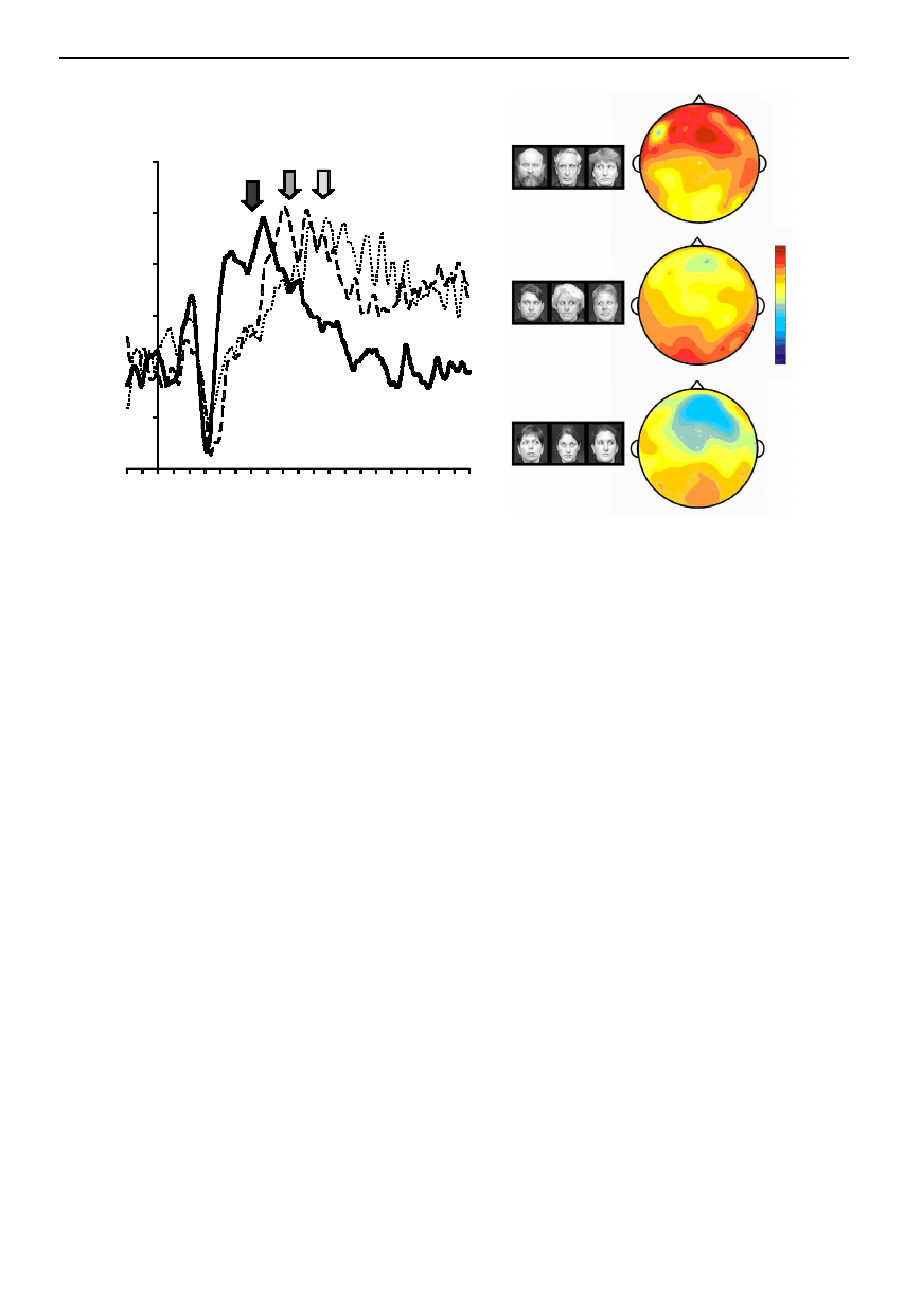
Figure 4. ERPs elicited to a social attention task. (a) ERP waveforms elicited to three conditions: solid line, group attention;
dashed line, mutual gaze exchange; dotted line, control. The arrows indicate a late peak of ERP activity that follows the N170
ERP (P400), which changes its latency as a function of viewing condition. (b) Voltage maps for the three viewing conditions
generated at the peak of P400 activity for the group attention condition (black arrow in (a)). The group attention condition
shows fronto-temporal positivity, whereas the other two conditions show small posterior positivities.
temporoparietal junction and left precentral gyrus. This
extended network of activation might be due to the nov-
elty or salience of the incongruent body and head motion
stimuli (Downar et al. 2002). The differential experience
with compatible and incompatible motion may explain
STS cell sensitivity to the compatibility of motion direc-
tion and body view during the locomotion described
above.
What is unique about the motion of animate beings?
Animals and humans possess articulated joints, enabling
the movement of body parts without having to maintain
a constant spatial relationship in space relative to each
other. This results in the ability to produce a limitless set
of movements. Man-made objects, such as utensils and
tools, in general do not have this capability. Beauchamp
et al. (2002) investigated the differences in brain activation
to these different types of high-level motion stimuli. Inter-
estingly, observing human motion stimuli activated the
STS and observing the motion of tools/utensils activated
cortex ventral to the STS, on the MTG. In another fMRI
experiment in this same study, stimuli depicting articu-
lated and non-articulated human motion were presented.
The STS responded to the articulated human motion and
the MTG to non-articulated motion, indicating that these
high-order processing mechanisms process selectively the
higher-order motion type (Beauchamp et al. 2002).
Grezes et al. (2001) also reported activation differences
between observing rigid and non-rigid motion. Specifi-
cally, they observed an anterior–posterior gradient of acti-
vation in the STS regions, with non-rigid motion
producing the most anterior activation. Additionally, they
observed activation in left intraparietal cortex to non-rigid
Phil. Trans. R. Soc. Lond. B (2003)
biological motion (Grezes et al. 2001). The magnitude of
the activation in the STS to biological motion, and indeed
in other cortical regions, can be coloured by the task
requirements and the attention that the observer places
on the ‘human’ quality of the motion (Vaina et al. 2001).
Additionally, attention to the displayed emotion enhances
fMRI activation in the STS, whereas increased activation
to facial attributes per se, such as identity or isolated fea-
tures, increased activation in all known face-sensitive
cortical regions (Narumoto et al. 2001).
(
a) Social cognition
The limbic system, in conjunction with the orbitofrontal
cortex and the STS, is thought to form a network that is
involved in social cognition (Baron-Cohen 1995; Brothers
1997; Adolphs 1999). One important aspect of social cog-
nition is the identification of the direction of another’s
attention from their direction of gaze or head view (Perrett
et al. 1985a, 1992; Kleinke 1986; Allison et al. 2000;
Emery 2000). Indeed, the existence of an eye direction
detector has been postulated in this hierarchical system of
social cognition, which at its top level allows us to ‘mind-
read’ and infer the intentions of others (Baron-Cohen
1995; Baron-Cohen et al. 1997). While there is evidence
for cell populations coding for eye and attention direction
within STS (Perrett et al. 1985a, 1992), the populations
are not anatomically grouped in such a way that scalp
evoked potentials are necessarily linked to a given eye
direction (Bentin et al. 1996; Eimer 1998; Taylor et al.
2001). Our attention and behaviour can be modified when
confronted with a face with averted gaze. A peripheral
target stimulus is detected by normal subjects more

440 A. Puce and D. Perrett
Physiology of biological motion
efficiently when it lies in the direction of gaze of a central
stimulus face (Friesen & Kingstone 1998; Driver et al.
1999; Hietanen 1999, 2002; Langton & Bruce 2000).
Moreover, patients with unilateral neglect are less likely
to extinguish a contralesional target stimulus when it lies
in the gaze path of a stimulus face (Vuilleumier 2002).
Following the attention direction of someone’s gaze may
be such an over-learned response that it needs little con-
scious awareness.
(
b) Gaze perception
Neuroimaging studies involving gaze perception indi-
cate that there is an active cortical network involving occi-
pito-temporal cortex (fusiform gyrus, inferior temporal
gyrus, parietal lobule and bilateral middle temporal gyri)
when subjects passively view gaze aversion movements
(Wicker et al. 1998). One prominently active region to
viewing eye movements (gaze aversion and also eyes look-
ing at the observer) is the cortex around the STS, parti-
cularly in the right hemisphere, and this same region is
active also to viewing opening and closing movements of
the mouth (Puce et al. 1998). Thus, as is evident from
the single cell responses, the STS region contains neural
populations representing multiple aspects of the appear-
ance of the face (including gaze) and body and their
motion; the STS should not be considered exclusively an
‘eye detector’ or ‘eye processor’. The STS is more acti-
vated during judgements of gaze direction than during
judgements of identity, whereas the fusiform and inferior
occipito-temporal activation is stronger during judgements
of identity than gaze direction (Hoffman & Haxby 2000).
Intracranial ERP recordings from these structures indicate
that the STS responds to facial motion, whereas the ven-
tral-temporal cortex responds more strongly to static facial
images (Puce & Allison 1999). This is not surprising if
one considers that eye gaze direction changes are transient
and their detection might require motion processing sys-
tems, whereas identity judgements can be made indepen-
dently of facial movements. Indeed, the processing of
dynamic information about facial expression and the
processing of static information about facial identity
appear neuropsychologically dissociable (Campbell 1992;
Humphreys et al. 1993).
(
c) Lip reading
Lip reading, an important function for both hearing and
deaf individuals, can be neuropsychologically dissociated
from face recognition (Campbell et al. 1986), in a some-
what similar manner to gaze perception. Normal lip read-
ing uses cortex of the STG in addition to other brain
regions such as the angular gyrus, posterior cingulate,
medial frontal cortex and frontal pole (Calvert et al. 1997).
The STG and surrounding cortex activate bilaterally when
subjects view face actions that could be interpreted as
speech (Puce et al. 1998; Campbell et al. 2001), while
some regions of the posterior right STS activate for the
sight of speech and non-speech mouth movements
(Campbell et al. 2001). Centres of activation to visual
speech appear to overlap those associated with hearing
speech (Calvert et al. 1997), indicating that these regions
receive multimodal inputs during speech analysis
(Kawashima et al. 1999; Calvert et al. 2000). Further
evidence for this multimodal integration is a phenomenon
Phil. Trans. R. Soc. Lond. B (2003)
known as the McGurk effect (McGurk & MacDonald
1976), where what observers hear when listening to speech
sounds is altered by simultaneously viewing mouth move-
ments appropriate to a different speech utterance. Indeed,
magnetoencephalographic recordings of neural activity to
speech stimuli show sensitivity to auditory–visual
mismatch (Sams et al. 1991) with activity 200 ms post-
stimulus augmented when the visual speech does not
correspond to the accompanying auditory speech.
(
d) The mirror neuron system and action
observation/execution
The existence of a mirror neuron system in humans has
been investigated during the manipulation of objects
(Rizzolatti et al. 1996a,b; Binkofski et al. 1999a,b). The acti-
vation in fronto-central regions, seen when subjects observe
and/or execute grasping behaviours, is accompanied by
activity in the parietal cortex and STS (Jeannerod et al.
1995; Iacoboni et al. 1999, 2001; Rizzolatti et al. 2001; Gal-
lese et al. 2002), paralleling the mirror neuron system in
non-human primates.
Additionally, the secondary somatosensory cortex, SII,
located in the temporal operculum is postulated to analyse
the intrinsic properties of the graspable object while acti-
vation observed in the cortex in the intraparietal sulcus
was thought to be related to kineasthetic processes
(Binkofski et al. 1999b), although strictly speaking it is not
part of the mirror neuron system.
The neuroimaging data mesh well with reported dis-
turbances in executing grasping movements in the neuro-
psychological lesion literature. For example, Jeannerod
and colleagues have reported a case with bilateral posterior
parietal lesions of vascular origin where there was no dif-
ficulty in reaching toward the location of the object; how-
ever, a profound deficit in executing the anticipatory
grasping movement with the fingers occurred to nonde-
script objects (cylindrical dowels). Interestingly, there was
no deficit in grasping behaviour when well-known reco-
gnizable objects were used in the same test (Jeannerod et
al. 1994). Mental imagery of hand and finger movements
was found to be impaired in patients with unilateral par-
ietal lesions, who had difficulties in producing movements
with their hands and fingers (Sirigu et al. 1996). It has
been reported that patients with unilateral parietal lesions
have more difficulty in imitating gestures involving their
own bodies relative to movements involving external
objects, particularly if the lesion is in the left hemisphere
(Halsband et al. 2001).
The human STS in its posterior extent has been found
to be active not only to the hand and body movements of
others (see figure 3; Allison et al. 2000), but also to faces
(Puce et al. 1998). Interestingly, ERP recordings indicate
that neural activity can differentiate between types of facial
movements (Puce et al. 2000). Viewing mouth opening
movements produces larger N170 responses relative to
viewing mouth closing movements. A similar N170
response gradient is seen for observing eyes averting their
gaze away from the observer relative to eyes focusing their
gaze on the observer. Augmented neural responses to eye
aversion movements may be a powerful signal that the
observer is no longer the focus of another’s attention.
Similarly, larger N170s to mouth opening movements
might be important for recognizing the beginning of an
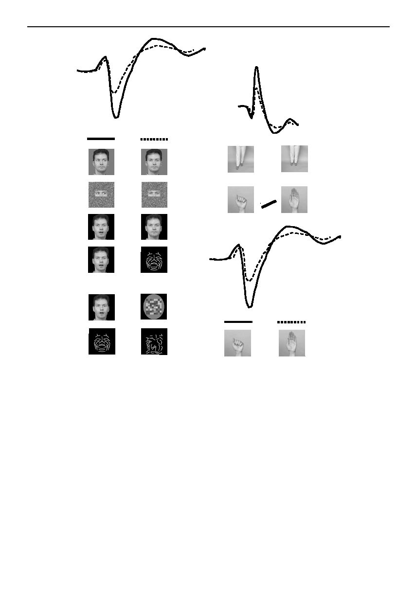
Physiology of biological motion A. Puce and D. Perrett 441
posterior
temporal
N170
>
>
>
>
>
>
>
fronto-central
>
>
P130
P270
posterior
temporal
N170
(a)
(b)
(i)
(ii)
Figure 5. Schematic summary of ERP waveforms elicited in response to observing human motion. (a) Posterior temporal
N170 (solid line) to conditions listed in the left column is larger relative to N170 (dashed line) elicited to conditions listed in
the right column. b(i) Frontocentral ERPs show larger P130 and P270 components across body and hand motion conditions
shown in the left and right columns (solid versus dashed line). b(ii) Posterior temporal N170 (solid line) is larger to hand
closure relative to hand opening (dashed line).
utterance (Puce et al. 2000). With recording electrodes
sited in the STS of epilepsy surgery patients, selective
responses to mouth opening have been elicited (see Alli-
son et al. 2000, box 1). No responses were observed to
mouth closing movements or eye deviations, indicating
that these regions might be responsive during lip reading
(or the sight of gestures and emotional expressions in
which the mouth opens, e.g. during eating and surprise).
The Talairach coordinates of these electrode positions are
comparable to sites of fMRI activation in lip reading
(Calvert et al. 1997).
If eye aversion movements are given a context, late ERPs
that differ as a function of the social significance of the
aversion movement can be elicited (figure 4; A. Cooper and
A. Puce, unpublished data). This was demonstrated in a
visual task where two permanently gaze-averted flanker
faces were presented with a central face that changed its
Phil. Trans. R. Soc. Lond. B (2003)
gaze direction. The central face could look in the same
direction as both flanker faces, setting up an apparently
common focus of attention off to the side (‘group
attention’). Alternatively, if the central face looked away
from the observer in the opposite direction to the other two
faces, a mutual gaze exchange between the central face and
one of the flankers became apparent (‘mutual gaze
exchange’). Finally, the central face could look away from
the observer and the other two flanker faces by looking up
(‘control’). An N170 ERP to the gaze aversion of the cen-
tral face was elicited, and its characteristics did not change
as a function of condition (see also Puce et al. 2000). A
later positive ERP, elicited between 300 and 500 ms post-
motion onset (P400) was seen to differentiate in latency as
a function of viewing condition: group attention produced
the shortest latency response, followed by the mutual gaze
exchange condition and then the control condition.

442 A. Puce and D. Perrett
Physiology of biological motion
Our non-verbal and verbal facial movements usually do
occur in an affective context, and preliminary ERP data
indicate that our brains are very sensitive to these gesture–
affect blends. If facial movements (either non-verbal or
verbal) are combined with different types of affect, tem-
poral scalp N170 peak latency and the amplitude of later
ERP activity can be altered as a function of affect type
(Wheaton et al. 2002b). If gesture–affect combinations are
incongruous, as shown by increased reaction time to class-
ify affect in behavioural data, late ERP activity from 300
to 975 ms post-motion onset is modulated as a function
of not only affect or gesture but also their combination
(Wheaton et al. 2002a). These preliminary data indicate
that the processing of inconsistencies in others’ behaviour
can be detected physiologically.
ERPs, in the form of N170 negativities occurring over
bilateral temporal scalp regions, have been elicited not
only to facial movements but also to hand and body move-
ments (Wheaton et al. 2001). The N170 activity was larger
for observing hand clenching movements relative to hand
opening movements. In addition, ERP activity was also
observed to hand and body motion over the central scalp.
Interestingly, ERP activity was larger to observing a body
stepping forward than to a body stepping back (paralleling
the cellular bias for forward or compatible direction of
locomotion; Perrett et al. 1985b; Oram & Perrett 1994).
Taken together, the ERP differentiation in the hand and
body movements might indicate a stronger neural signal
for potentially threatening movements (Wheaton et al.
2001). When fMRI activation to these movement types is
compared, there is a robust signal within the temporopari-
etal cortex to all of these motion types (Wheaton et al.
2002c). Figure 5 summarizes the main findings from the
ERP studies (Puce et al. 2000; Wheaton et al. 2001;
Thompson et al. 2002b), and indicates that processing
between movement types begins before 200 ms post-
motion onset not only in the posterior temporal cortex but
also in the frontocentral regions, which would be expected
from the distribution of action processing evident in fMRI
and cell recording.
(
e) Gesture and action processing: implications
for disorders of social communication
The processing of non-verbally presented messages, in
the form of face and hand gestures, is crucial for social
primates to be able to interact with one another—and
there are considerable similarities in the high-level biologi-
cal motion processing systems in human and non-human
primates. The importance of comprehending actions of
others may also be evident when such comprehension is
impaired in clinical conditions. Disorders such as autism,
Asperger syndrome, and schizophrenia are characterized
by the inability to form or maintain social relationships.
This can be difficult if the sufferer cannot process
incoming social messages communicated by the bodily
and facial actions of others, or sends inappropriate social
reactions to such signals (e.g. Williams et al. 2001).
Further neuroimaging and neurophysiological studies of
healthy subjects and those with impairments of human
motion processing may shed light on the interactions
between the various components of these high-level bio-
logical motion processing systems.
Phil. Trans. R. Soc. Lond. B (2003)
A.P.’s research has been supported by the National Health and
Medical Research Council (Australia) and the Australia
Research Council.
REFERENCES
Adolphs, R. 1999 Social cognition and the human brain.
Aggleton, J. P., Burton, M. J. & Passingham, R. E. 1980
Cortical and subcortical afferents to the amygdala of the rhe-
sus monkey (Macaca mulatta).
Allison, T., Puce, A. & McCarthy, G. 2000 Social perception
from visual cues: role of the STS region.
Baker, C. I., Keysers, C., Jellema, T., Wicker, B. & Perrett,
D. I. 2001 Neuronal representation of disappearing and hid-
den objects in temporal cortex of the macaque.
Barbas, H. 1988 Anatomic organization of basoventral and
mediodorsal visual recipient prefrontal regions in the rhesus
monkey.
J. Comp. Neurol. 276, 313–342.
Baron-Cohen, S. 1995 Mindblindness: an essay on autism and
theory of mind. Cambridge, MA: MIT Press.
Baron-Cohen, S., Wheelwright, S. & Joliffe, T. 1997 Is there
a ‘language of the eyes’? Evidence from normal adults, and
adults with autism or Asperger syndrome
Bassili, J. N. 1978 Facial motion in the perception of faces and
of emotional expression.
J. Exp. Psychol. Hum. Percept. Perf.
Beauchamp, M. S., Lee, K. E., Haxby, J. V. & Martin, A. 2002
Parallel visual motion processing streams for manipulable
objects and human movements.
Bentin, S., Allison, T., Puce, A., Perez, A. & McCarthy, G.
1996 Electrophysiological studies of face perception in
humans.
J. Cogn. Neurosci. 8, 551–565.
Binkofski, F., Buccino, G., Posse, S., Seitz, R. J., Rizzolatti,
G. & Freund, H. J. 1999a A fronto-parietal circuit for object
manipulation in man: evidence from an fMRI study.
Binkofski, F., Buccino, G., Stephan, K. M., Rizzolatti, G.,
Seitz, R. J. & Freund, H. J. 1999b A parieto-premotor net-
work for object manipulation: evidence from neuroimaging.
Blake, R. 1993 Cats perceive biological motion. Psychol. Sci.
4, 54–57.
Blakemore, S.-J. & Decety, J. 2001 From the perception of
action to the understanding of intention.
Bonda, E., Petrides, M., Ostry, D. & Evans, A. 1996 Specific
involvement of human parietal systems and the amygdala in
the perception of biological motion.
Boussaoud, D., Ungerleider, L. G. & Desimone, R. 1990 Path-
ways for motion analysis: cortical connections of the medial
superior temporal and fundus of the superior temporal visual
areas in the macaque.
J. Comp. Neurol. 296, 462–495.
Brothers, L. 1997 Friday’s footprint: how society shapes the
human mind. New York: Oxford University Press.
Calvert, G. A., Bullmore, E. T., Brammer, M. J., Campbell,
R., Williams, S. C., McGuire, P. K., Woodruff, P. W.,
Iversen, S. D. & David, A. S. 1997 Activation of auditory
cortex during silent lipreading.
Calvert, G. A., Campbell, R. & Brammer, M. J. 2000 Evidence
from functional magnetic resonance imaging of crossmodal
binding in the human heteromodal cortex.
Campbell, R. 1992 The neuropsychology of lipreading.

Physiology of biological motion A. Puce and D. Perrett 443
Campbell, R., Landis, T. & Regard, M. 1986 Face recognition
and lipreading.
Campbell, R., MacSweeney, M., Surguladze, S., Calvert, G.,
McGuire, P., Suckling, J., Brammer, M. J. & David, A. S.
2001 Cortical substrates for the perception of face actions:
an fMRI study of the specificity of activation for seen speech
and for meaningless lower-face acts (gurning).
Carey, D. P., Perrett, D. I. & Oram, M. W. 1997 Recognizing,
understanding and reproducing action. In Handbook of
neuropsychology, vol. 11. Action and cognition (ed. M.
Jeannerod), pp. 111–129. Amsterdam: Elsevier.
Cutting, J. E. & Kozlowski, L. T. 1977 Recognizing friends by
their walk: gait perception without familiarity cues. Bull. Psy-
chonomic. Soc. 9, 353–356.
Desimone, R. 1991 Face-selective cells in the temporal cortex
of monkeys.
Desimone, R., Albright, T. D., Gross, C. G. & Bruce, C. 1984
Stimulus-selective properties of inferior temporal neurons in
the macaque.
di Pellegrino, G., Fadiga, L., Fogassi, V., Gallese, V. & Rizzol-
atti, G. 1992 Understanding motor events: a neurophysiol-
ogical study.
Dittrich, W. H. 1993 Action categories and the perception of
biological motion.
Downar, J., Crawley, A. P., Mikulis, D. J. & Davis, K. D. 2002
A cortical network sensitive to stimulus salience in a neutral
behavioral context across multiple sensory modalities.
Downing, P. E., Jiang, Y. H., Shuman, M. & Kanwisher, N.
2001 A cortical area selective for visual processing of the
human body.
Driver, J., Davis, G., Ricciardelli, P., Kidd, P., Maxwell, E. &
Baron-Cohen, S. 1999 Gaze perception triggers reflexive
visuospatial orienting.
Eimer, M. 1998 Does the face-specific N170 component
reflect the activity of a specialized eye processor?
Emery, N. J. 2000 The eyes have it: the neuroethology, func-
tion and evolution of social gaze.
Felleman, D. J. & Van Essen, D. C. 1991 Distributed hier-
archical processing in the primate cerebral cortex.
Friesen, C. K. & Kingstone, A. 1998 The eyes have it! Reflex-
ive orienting is triggered by nonpredictive gaze. Psychol. Bull.
Rev. 5, 490–495.
Gallese, V., Fadiga, L., Fogassi, L. & Rizzolatti, G. 1996
Action recognition in the premotor cortex.
Gallese, V., Fadiga, L., Fogassi, L. & Rizzolatti, G. 2002
Action representation and the inferior parietal lobule. Atten-
tion Perform. 19, 247–266.
Grezes, J., Fonlupt, P., Bertenthal, B., Delon-Martin, C., Seg-
ebarth, C. & Decety, J. 2001 Does perception of biological
motion rely on specific brain regions?
Grossman, E., Donnelly, M., Price, R., Pickens, D., Morgan,
V., Neighbor, G. & Blake, R. 2000 Brain areas involved in
perception of biological motion.
Halsband, U., Schmitt, J., Weyers, M., Binkofski, F.,
Gru¨tzner, G. & Freund, H. J. 2001 Recognition and imi-
tation of pantomimed motor acts after unilateral parietal and
premotor lesions: a perspective on apraxia.
Hietanen, J. K. 1999 Does your gaze direction and head orien-
tation shift my visual attention?
Phil. Trans. R. Soc. Lond. B (2003)
Hietanen, J. K. 2002 Social attention orienting integrates vis-
ual information from head and body orientation.
Hietanen, J. K. & Perrett, D. I. 1996 Motion sensitive cells in
the macaque superior temporal polysensory area: response
discrimination between self- and externally generated pat-
tern motion.
Behav. Brain Res. 76, 155–167.
Hoffman, E. A. & Haxby, J. V. 2000 Distinct representations
of eye gaze and identity in the distributed human neural sys-
tem for face perception.
Howard, R. J., Brammer, M., Wright, I., Woodruff, P. W.,
Bullmore, E. T. & Zeki, S. 1996 A direct demonstration of
functional specialization within motion-related visual and
auditory cortex of the human brain.
Humphreys, G. W., Donnelly, N. & Riddoch, M. J. 1993
Expression is computed separately from facial identity, and
it is computed separately for moving and static faces: neuro-
psychological evidence.
Iacoboni, M., Woods, R. P., Brass, M., Bekkering, H., Mazzi-
otta, J. C. & Rizzolatti, G. 1999 Cortical mechanisms of
human imitation.
Iacoboni, M., Koski, L. M., Brass, M., Bekkering, H., Woods,
R. P., Dubeau, M. C., Mazziotta, J. C. & Rizzolatti, G. 2001
Reafferent copies of imitated actions in the right superior
temporal cortex. Proc. Natl Acad. Sci. USA 98, 13 995–
13 999.
Jeannerod, M., Decety, J. & Michel, F. 1994 Impairment of
grasping movements following a bilateral posterior parietal
lesion. Neuropsychologia 32, 369–380.
Jeannerod, M., Arbib, M. A., Rizzolatti, G. & Sakata, H. 1995
Grasping objects: the cortical mechanisms of visuomotor
transformation.
Jellema, T. & Perrett, D. I. 2002 Coding of visible and hidden
actions. Attention Perform. 19, 356–380.
Jellema, T., Baker, C. I., Wicker, B. & Perrett, D. I. 2000 Neu-
ral representation for the perception of the intentionality of
hand actions.
Jellema, T., Oram, M. W., Baker, C. I. & Perrett, D. I. 2002
Cell populations in the banks of the superior temporal sulcus
of the macaque and imitation. In The imitative mind: develop-
ment, evolution, and brain bases (ed. A. Meltzoff & W. Prinz),
pp. 267–290. Cambridge University Press.
Johansson, G. 1973 Visual perception of biological motion and
a model of its analysis.
Percept. Psychophys. 14, 202–211.
Kawashima, R., Imaizumi, S., Mori, K., Okada, K., Goto, R.,
Kiritani, S., Ogawa, A. & Fukuda, H. 1999 Selective visual
and auditory attention toward utterances: a PET study.
Kleinke, C. L. 1986 Gaze and eye contact: a research review.
Kohler, E., Keysers, C., Umilta, M. A., Fogassi, L., Gallese,
V. & Rizzolatti, G. 2002 Hearing sounds, understanding
actions: action representation in mirror neurons.
Kourtzi, Z. & Kanwisher, N. 2000 Activation in human
MT/MST by static images with implied motion.
Kozlowski, L. T. & Cutting, J. E. 1977 Recognizing the sex of
a walker from a dynamic point-light display.
Langton, S. R. H. & Bruce, V. 2000 You must see the point:
automatic processing of cues to the direction of social atten-
tion.
J. Exp. Psychol. Hum. Percep. Perf. 26, 747–757.
McGurk, H. & MacDonald, J. 1976 Hearing lips and seeing
voices.
McLeod, P., Dittrich, W., Driver, J., Perrett, D. I. & Zihl, J.
1996 Preserved and impaired detection of structure from
motion in a ‘motion-blind’ patient.

444 A. Puce and D. Perrett
Physiology of biological motion
Mistlin, A. J. & Perrett, D. I. 1990 Visual and somatosensory
processing in the macaque temporal cortex: the role of
‘expectation’.
Narumoto, J., Okada, T., Sadato, N., Fukui, K. & Yonekura,
Y. 2001 Attention to emotion modulates fMRI activity in
human right superior temporal sulcus.
Nishitani, N. & Hari, R. 2000 Temporal dynamics of cortical
representation for action.
Nishitani, N. & Hari, R. 2001 Sign language and mirror neu-
ron system. Neuroimage 12(6), S452.
Oram, M. W. & Perrett, D. I. 1994 Responses of anterior
superior temporal polysensory (STPa) neurons to ‘biological
motion’ stimuli. J. Cogn. Neurosci. 6, 99–116.
Oram, M. W. & Perrett, D. I. 1996 Integration of form and
motion in the anterior superior temporal polysensory area
(STPa) of the macaque monkey.
Pavlova, M. & Sokolov, A. 2000 Orientation specificity in bio-
logical motion perception.
Percept. Psychophys. 62, 889–899.
Pavlova, M., Kra¨geloh-Mann, I., Birbaumer, N. & Sokolov,
A. 2002 Biological motion shown backwards: the apparent-
facing effect.
Perrett, D. I., Rolls, E. T. & Caan, W. 1982 Visual neurons
responsive to faces in the monkey temporal cortex.
Perrett, D. I., Smith, P. A. J., Potter, D. D., Mistlin, A. J.,
Head, A. S., Milner, A. D. & Jeeves, M. A. 1984 Neurones
responsive to faces in the temporal cortex: studies of func-
tional organization, sensitivity to identity and relation to per-
ception
Perrett, D. I., Smith, P. A. J., Potter, D. D., Mistlin, A. J.,
Head, A. S., Milner, A. D. & Jeeves, M. A. 1985a Visual
cells in the temporal cortex sensitive to face view and gaze
direction.
Proc. R. Soc. Lond. B 223, 293–317.
Perrett, D. I., Smith, P. A. J., Mistlin, A. J., Chitty, A. J.,
Head, A. S., Potter, D. D., Broennimann, R., Milner,
A. D. & Jeeves, M. A. 1985b Visual analysis of body move-
ments by neurones in the temporal cortex of the macaque
monkey: a preliminary report.
Perrett, D. I., Harries, M. H., Bevan, R., Thomas, S., Benson,
P. J., Mistlin, A. J., Chitty, A. J., Hietanen, J. K. & Ortega,
J. E. 1989 Frameworks of analysis for the neural represen-
tation of animate objects and actions.
Perrett, D. I., Harries, M. H., Benson, P. J., Chitty, A. J. &
Mistlin, A. J. 1990a Retrieval of structure from rigid and
biological motion; an analysis of the visual response of neu-
rons in the macaque temporal cortex. In AI and the eye (ed.
T. Troscianko & A. Blake), pp. 181–201. Chichester, UK:
Wiley.
Perrett, D. I., Harries, M., Chitty, A. J. & Mistlin, A. J. 1990b
Three stages in the classification of body movements by vis-
ual neurones. In Images and understanding (ed. H. B. Barlow,
C. Blakemore & M. Weston-Smith), pp. 94–108. Cam-
bridge University Press.
Perrett, D. I., Mistlin, A. J., Harries, M. H. & Chitty, A. J.
1990c Understanding the visual appearance and conse-
quence of hand actions. In Vision and action: the control of
grasping (ed. M. A. Goodale), pp. 163–180. Norwood, NJ:
Ablex Publishing.
Perrett, D. I., Hietanen, J. K., Oram, M. W. & Benson, P. J.
1992 Organization and functions of cells responsive to faces
in the temporal cortex.
Phil. Trans. R. Soc. Lond. B 335,
Puce, A. & Allison, T. 1999 Differential processing of mobile
and static faces by temporal cortex. Neuroimage 9(6), S801.
Phil. Trans. R. Soc. Lond. B (2003)
Puce, A., Allison, T., Bentin, S., Gore, J. C. & McCarthy, G.
1998 Temporal cortex activation in humans viewing eye and
mouth movements.
Puce, A., Smith, A. & Allison, T. 2000 ERPs evoked by view-
ing moving eyes and mouths.
Puce, A., Castiello, U., Syngeniotis, A. & Abbott, D. 2001 The
human STS region integrates form and motion. Neuroimage
13(6), S931.
Rizzolatti, G., Camarda, R., Fogassi, L., Gentilucci, M., Lup-
pino, G. & Matelli, M. 1988 Functional organization of
inferior area 6 in the macaque monkey. II. Area F5 and the
control of distal movements.
Rizzolatti, G., Fadiga, L., Gallese, V. & Fogassi, L. 1996a Pre-
motor cortex and the recognition of motor actions.
Res. Cogn. Brain Res. 3, 131–141.
Rizzolatti, G., Fadiga, L., Matelli, M., Bettinardi, V., Paulesu,
E., Perani, D. & Fazio, F. 1996b Localization of grasp rep-
resentations in humans by PET. 1. Observation versus
execution.
Rizzolatti, G., Fogassi, L. & Gallese, V. 2001 Neurophysiolog-
ical mechanisms underlying the understanding and imitation
of action.
Nature Rev. Neurosci. 2, 661–670.
Sams, M., Aulanko, R., Ha¨ma¨la¨inen, M., Hari, R., Lounas-
maa, O. V., Lu, S. T. & Simola, J. 1991 Seeing speech: vis-
ual information from lip movements modifies activity in the
human auditory cortex.
Schenk, T. & Zihl, J. 1997 Visual motion perception after brain
damage: II. Deficits in form-from-motion perception.
Neuropsychologia 35, 1299–1310.
Sirigu, A., Duhamel, J. R., Cohen, L., Pillon, B., Dubois, B. &
Agid, Y. 1996 The mental representation of hand move-
ments after parietal cortex damage.
Taylor, M. J., Edmonds, G. E., McCarthy, G. & Allison, T.
2001 Eyes first! Eye processing develops before face pro-
cessing in children.
Thompson, J. C., Wheaton, K., Berkovic, S. F., Jackson, G. &
Puce, A. 2002a Hemodynamic responses in humans to the
perception of compatible and incompatible body motion. In
The fMRI Experience IV Proc. NIH, Maryland, 2002, 93.
Thompson, J. C., Wheaton, K., Castiello, U. & Puce, A. 2002b
ERPs differentiate between facial motion and motion in gen-
eral. Abstract no. 14221. Academic Press OHBM Annual
Scientific Meeting 2002.
Thornton, I. M., Pinto, J. & Shiffrar, M. 1998 The visual per-
ception of human locomotion.
Umilta, M. A., Kohler, E., Gallese, V., Fogassi, L., Fadiga, L.,
Keysers, C. & Rizzolatti, G. 2001 I know what you are
doing: a neurophysiological study.
Vaina, L. M., LeMay, M., Bienfang, D. C., Choi, A. Y. &
Nakayama, K. 1990 Intact ‘biological motion’ and ‘structure
from motion’ perception in a patient with impaired motion
mechanisms: a case study.
Vaina, L. M., Solomon, J., Chowdhury, S., Sinha, P. & Belli-
veau, J. W. 2001 Functional neuroanatomy of biological
motion perception in humans.
656–11 661.
Vuilleumier, P. 2002 Perceived gaze direction in faces and spa-
tial attention: a study in patients with parietal damage and
unilateral neglect.
Neuropsychologia 40, 1013–1026.
Wachsmuth, E., Oram, M. W. & Perrett, D. I. 1994 Recog-
nition of objects and their component parts: responses of
single units in the temporal cortex of the macaque.
Wheaton, K. J., Pipingas, A., Silberstein, R. & Puce, A. 2001
Neuronal responses elicited to viewing the actions of others.

Physiology of biological motion A. Puce and D. Perrett 445
Wheaton, K. J., Aranda, G. & Puce, A. 2002a ERPs elicited
to combined emotional and gestural movements of the face
as a function of congruency. Abstract no. 14186. Academic
Press OHBM Annual Scientific Meeting 2002.
Wheaton K. J., Aranda, G. & Puce, A. 2002b Affective modu-
lation of gestural and visual speech stimuli: an ERP study.
Abstract no. 14215. Academic Press OHBM Annual Scien-
tific Meeting 2002.
Wheaton, K. J., Thompson, J. C., Berkovic, S. F., Jackson,
G. & Puce, A. 2002c Brain regions responsive to the percep-
tion of human motion. The fMRI Experience IV Proc. NIH,
Maryland 2002, p. 103.
Wicker, B., Michel, F., Henaff, M.-A. & Decety, J. 1998 Brain
regions involved in the perception of gaze: a PET study.
Phil. Trans. R. Soc. Lond. B (2003)
Williams, J. H., Whiten, A., Suddendorf, T. & Perrett, D. I.
2001 Imitation, mirror neurons and autism.
GLOSSARY
ERP: event-related potential
fMRI: functional magnetic resonance imaging
MST: medial superior temporal
MTG: mid-temporal gyrus
PET: positron emission tomography
STG: superior temporal gyrus
STP: superior temporal polysensory
STS: superior temporal sulcus
Wyszukiwarka
Podobne podstrony:
The electrochemical and mechanical behavior of passivated an
Flash on English for Mechanics, Electronics and Technical Assistance
Brain Imaging in Clinical Psychiatry
(autyzm) Autism Mind and Brain
Dan Geometry and the Imagination
Childhood Trauma, the Neurobiology of Adaptation, and Use dependent of the Brain
Duality between Electric and Magnetic Black Holes
Catalogue of the Collection of Greek Coins In Gold, Silber, Electrum and Bronze
Memes and the Exploitation of Imagination
71 1021 1029 Effect of Electron Beam Treatment on the Structure and the Properties of Hard
Relationship?tween mind and Brain
Flash on English for Mechanics, Electronics and Technical Assistance
History of electricity and electronics Pojecia
Flash on English for Mechanics, Electronics and Technical Assistance
David Thoreau Walden (And the Duty of Civil Disobedience) (Ingles)
White Energy from Electrons and Matter from Protons A Preliminary Model Based on Observer Physics
THE GOOGLE STORY, by David A Vise and Mark Malseed
Chomsky N Linguistics and Brain Science Chapter 1
W Roll Poltergeists, Electromagnetism and Consciousness
więcej podobnych podstron