
J
OURNAL OF
C
LINICAL
M
ICROBIOLOGY
, July 2011, p. 2533–2539
Vol. 49, No. 7
0095-1137/11/$12.00
doi:10.1128/JCM.02171-10
Copyright © 2011, American Society for Microbiology. All Rights Reserved.
Mechanical Disruption of Lysis-Resistant Bacterial Cells by Use of a
Miniature, Low-Power, Disposable Device
䌤
†
Peter E. Vandeventer,
1
Kris M. Weigel,
2
Jose Salazar,
1
Barbara Erwin,
1
Bruce Irvine,
3
Robert Doebler,
3
Ali Nadim,
4
Gerard A. Cangelosi,
2
and Angelika Niemz
1
*
Keck Graduate Institute of Applied Life Sciences, 535 Watson Drive, Claremont, California 91711
1
; Seattle Biomedical Research
Institute, 307 Westlake Avenue North, Seattle, Washington 98109
2
; Claremont BioSolutions, 1182 Monte Vista Ave., Suite 11,
Upland, California 91786
3
; and Claremont Graduate University, 150 E. Tenth St., Claremont, California 91711
4
Received 26 October 2010/Returned for modification 3 March 2011/Accepted 25 April 2011
Molecular detection of microorganisms requires microbial cell disruption to release nucleic acids. Sensitive
detection of thick-walled microorganisms such as Bacillus spores and Mycobacterium cells typically necessitates
mechanical disruption through bead beating or sonication, using benchtop instruments that require line
power. Miniaturized, low-power, battery-operated devices are needed to facilitate mechanical pathogen dis-
ruption for nucleic acid testing at the point of care and in field settings. We assessed the lysis efficiency of a
very small disposable bead blender called OmniLyse relative to the industry standard benchtop Biospec
Mini-BeadBeater. The OmniLyse weighs approximately 3 g, at a size of approximately 1.1 cm
3
without the
battery pack. Both instruments were used to mechanically lyse Bacillus subtilis spores and Mycobacterium bovis
BCG cells. The relative lysis efficiency was assessed through real-time PCR. Cycle threshold (C
T
) values
obtained at all microbial cell concentrations were similar between the two devices, indicating that the lysis
efficiencies of the OmniLyse and the BioSpec Mini-BeadBeater were comparable. As an internal control,
genomic DNA from a different organism was spiked at a constant concentration into each sample upstream of
lysis. The C
T
values for PCR amplification of lysed samples using primers specific to this internal control were
comparable between the two devices, indicating negligible PCR inhibition or other secondary effects. Overall,
the OmniLyse device was found to effectively lyse tough-walled organisms in a very small, disposable, battery-
operated format, which is expected to facilitate sensitive point-of-care nucleic acid testing.
Nucleic acid testing has become an important tool in infec-
tious disease diagnosis (4, 25), biothreat detection (14, 30), and
research. Point-of-care or point-of-use applications of nucleic
acid testing, especially in settings with minimal infrastructure,
require novel tools that can perform essential tasks in minia-
turized, inexpensive formats with the same performance char-
acteristics as currently available, expensive, laboratory-based
methods (13).
Lysis of an organism to liberate its genomic material is an
important step in sample preparation for nucleic acid testing.
Many common pathogens can be lysed through chemical
agents, such as detergents and chaotropic salts, or by enzymatic
treatment (8, 31). However, lysis is a significant challenge for
thick-walled microorganisms such as Bacillus anthracis spores
and Mycobacterium tuberculosis cells (13, 18, 22). The multi-
layer structure of Bacillus spores includes an outer cortex and
coat that is resistant to chemical and physical treatments (5,
23). Similarly, mycobacteria have a thick, waxy cell wall that is
difficult to disrupt for the extraction of nucleic acids (9, 17).
High-energy mechanical disruption methods, such as sonica-
tion and bead beating, are commonly used for lysis of thick-
walled organisms, since chemical, heat, freeze-thaw, or enzy-
matic lysis methods alone are less effective (1, 11, 22). Lysis
protocols for mycobacteria have been reported that use low-
energy bead beating (2, 6) in conjunction with heat or chemical
or enzymatic lytic agents, which increase process complexity
and potentially introduce PCR inhibitors. We are not aware of
any published or unpublished methods that can break open
slow-growing mycobacteria by low-energy bead beating alone,
in the absence of other lytic treatments, with the same high
efficiency as the BioSpec Mini-BeadBeater.
Disruption of thick-walled organisms by sonication typically
involves the exposure of a suspension containing the pathogen
and beads to high frequency sound waves that are delivered by
a rapidly oscillating transducer. Lysis by sonication has been
attributed to cavitation, where the rapid formation and shrink-
age of gas bubbles creates high pressures and temperatures (5).
Lysis of thick-walled organisms by bead beating typically in-
volves high-frequency oscillation of a closed tube containing a
suspension of the target organism and beads. The mechanism
of lysis by bead-beating has been attributed to high shear rates
between beads and strong periodic vortical flow fields (13).
The diameter of beads used during mechanical lysis is critical
to lysis efficiency, with 100-
m-diameter beads being more
effective than larger-diameter beads at lysing Gram-positive
bacteria (11, 22).
Bead beating and sonication typically require benchtop de-
vices with significant power demands. The BioSpec Mini-Bead-
Beater (Fig. 1A) and the Sonics VibraCell Ultrasonic system
are among the smallest devices on the market, at respective
sizes of 3,900 cm
3
for the BioSpec Mini-BeadBeater and
⬎7,400 cm
3
for the VibraCell Ultrasonic system, including the
power supply (13). Larger, heavier, and more expensive bead-
* Corresponding author. Mailing address: Keck Graduate Institute
of Applied Life Sciences, 535 Watson Dr., Claremont, CA 91711.
Phone: (909) 607-9854. Fax: (909) 607-9826. E-mail: aniemz@kgi.edu.
† Supplemental material for this article may be found at http://jcm
.asm.org/.
䌤
Published ahead of print on 4 May 2011.
2533
on March 13, 2015 by UCSF Library & CKM
http://jcm.asm.org/
Downloaded from

beating devices exist, which can process multiple samples in
parallel. The BioSpec Mini-BeadBeater device has been used
in previous studies to lyse bacterial spores (13, 20). Mycobac-
terium cells are lysed effectively using the BioSpec Mini-Bead-
Beater (27) and Mini-BeadBeater-8 (12, 15). The BioSpec
Mini-BeadBeater has been used as a standard to benchmark
relative lysis efficiencies of new devices and techniques (13).
Fully integrated sample-to-answer nucleic acid testing sys-
tems are on the market, such as the Cepheid GeneXpert,
which automates sample preparation through sonication, fil-
tration, and solid-phase extraction, followed by amplification
and detection via the real-time PCR (34). Mechanical cell
disruption is facilitated through a miniaturized sonication de-
vice (5). GeneXpert cartridges are available to detect B. an-
thracis (34), M. tuberculosis (7, 16), and Clostridium difficile
(26), three pathogens that are known to be lysis resistant. In
the GeneXpert, sensitive detection of these tough-walled
pathogens is facilitated through mechanical cell disruption via
sonication.
Alternative methods for rapid mechanical lysis of thick-
walled organisms have been developed at Keck Graduate In-
stitute and are now commercialized by Claremont BioSolu-
tions (13). One method uses a disposable, battery-operated/
low-power, light weight, miniaturized, bead blender called the
OmniLyse. This device (Fig. 1B) uses four AAA 1.5-V batter-
ies, weighs approximately 3 g, and is 1.1 cm
3
in volume without
a battery pack or 71 g and 44 cm
3
in volume with a battery
pack. The OmniLyse consists of an injection-molded chamber
with inlet and outlet ports that can be operated in batch or
flowthrough mode and is therefore suitable for processing
large sample volumes. A micro-motor equipped with a custom-
designed precision-cut impellor is installed in such a way as to
dissipate a high amount of mechanical energy within the cham-
ber. The device utilizes a proprietary method for entrapping
and retaining zirconia/silica beads within the lysis chamber
during activation of the motor. High-speed camera video ex-
periments have shown that the motor can drive the impellor at
speeds greater than 30,000 rpm with the chamber filled with
liquid and beads. The kinetic energy imparted onto the beads
in solution generates high shear forces between beads, causing
disruption of cells caught within the shear flow. The system is
a single-use disposable unit, rendered feasible by use of inex-
pensive components.
We herein report a performance evaluation, assessing the
lysis efficiency of the OmniLyse device relative to the industry
standard benchtop Biospec Mini-BeadBeater (Fig. 1), using B.
subtilis spores as a BSL1 surrogate for B. anthracis spores, and
M. bovis BCG, an attenuated vaccine strain, as a BSL2 surro-
gate for M. tuberculosis.
MATERIALS AND METHODS
General reagents.
Standard Taq buffer (B9014S), Taq DNA polymerase
(M0273L), and deoxynucleotide triphosphate (dNTP; N0447S) were purchased
from New England Biolabs. Magnesium chloride (ES25-100ml) was obtained
from Amresco (Solon, OH). Uracil DNA glycosylase (catalog no. 78310) and
PCR nucleotide mix with dUTP (catalog no. 77330) were acquired from USB
(Cleveland, OH). Oligonucleotide primers were ordered from Integrated DNA
Technologies (Coralville, IA) and Sigma-Aldrich (St. Louis, MO). SYBR green
I (S7567) was acquired from Invitrogen (Carlsbad, CA). Power SYBR mix
(catalog no. 4368702) was purchased from Applied Biosystems. Bovine serum
albumin (BSA) was obtained from New England Biolabs (B9001S; Ipswich, MA)
and Thermo Scientific (HyClone SH30574.03; Waltham, MA). TE buffer was
composed of 10 mM Tris and 1 mM EDTA (pH 8.3). B. subtilis spores, strain
GB03, were donated by Gustafson, LLC, now a subsidiary of Bayer Corp.
Aeromonas hydrophila (strain 7966) was obtained from the American Type Cul-
ture Collection (Manassas, VA). The Kinyoun TB Stain Kit K (catalog no.
212315) was ordered from BD (Franklin Lakes, NJ). Gel electrophoresis to size
double-stranded DNA following PCR was performed on the Agilent 2100 Bio-
analyzer (Santa Clara, Ca) by using an Agilent DNA 1000 kit (catalog no.
5067-1504). The Mini-BeadBeater and the Mini-BeadBeater-8 were purchased
from BioSpec (Bartlesville, OK).
OmniLyse device.
Studies performed herein utilized the current version of
Claremont BioSolutions (Upland, CA) OmniLyse device, as shown in Fig. 1B,
with an inlet luer-lock fitting, a stiff outlet tube, and a lead-free micro-motor that
in the current device configuration can operate at full speed for up to 20 min. The
lysis chamber contains 200 mg of 100-
m-diameter acid-washed zirconia/silica
beads. The OmniLyse device was operated using four AAA alkaline batteries at
a nominal voltage of 6 V with a low electrical current between 100 and 200 mA.
Since the typical capacity of an alkaline AAA battery is approximately 1,200
mAh, a single battery pack should operate between 6 and 12 h, which translates
to an expected 250 to 500 lysis preparations of 1.5 min each per battery pack.
This disposable lysis device is fabricated using inexpensive manufacturing meth-
ods. The main body and fittings of the device are produced through inexpensive
injection molding. Also, the motors are mass produced in extremely high quan-
tities and obtained at low cost. Even in low production volumes, Claremont
BioSolutions is currently able to sell the devices within the price range of other
disposables used in bioscience research. As volumes increase, both the cost of
goods and the unit price are anticipated to drop precipitously.
Preparation of B. subtilis spore stocks.
B. subtilis spores were suspended and
washed three times in Nanopure filtered water. After the final wash and resus-
pension, the spores were quantified by using an iNcyto (Chungchongnam, Korea)
C-chip hemocytometer (DHC-N01-2) and through colony counting using LB
agar plates. A Nikon Eclipse TE2000-S microscope was used to view microbial
samples. Both methods resulted in similar spore counts. The Schaeffer-Fulton
staining method (32) was used to confirm that the B. subtilis microorganisms
were present as spores and not as vegetative cells. The spore stocks were stored
at
⫺20°C until further use.
Lysis of B. subtilis spores.
Dilution series of B. subtilis spores ranging from 10
4
to 10
8
spores/ml in the first experiment and from 10
3
to 10
8
spores/ml in the
second experiment were prepared in TE buffer, to which 10
6
copies of purified
Escherichia coli K-12 genomic DNA/ml was added as an internal control. (E. coli
K-12 was grown in nutrient broth at 37°C, and the DNA was purified and
extracted by using a Qiagen [Venlo, Netherlands] DNeasy Blood & Tissue Kit
[catalog no. 69504].) Each sample in this dilution series was processed in tripli-
cate by using the OmniLyse device and the BioSpec Mini-BeadBeater. The same
100-
m-diameter zirconia/silica beads were used in both devices. For lysis using
the OmniLyse device, 500-
l sample aliquots were aspirated from the sample
tube through the stiff tubing into the device and into a syringe attached at the
port with the luer lock and then dispensed back through the device into the sam-
ple tube for a total of three passes, with a total processing time per sample of 1.5
min. Each replicate was performed with a new device. Crude lysates were kept on
ice until samples were tested using PCR. For lysis using the BioSpec Mini-
BeadBeater, 625-
l sample aliquots plus 250 mg of 100-m-diameter zirconia/
silica beads were added to 2-ml conical tubes. The samples were bead beaten at
FIG. 1. Systems for mechanical disruption of tough-walled organ-
isms. (A) Mini-BeadBeater (BioSpec), a typical benchtop instrument.
(B) OmniLyse bead blender (Claremont BioSolutions), a miniaturized,
disposable, battery-operated device.
2534
VANDEVENTER ET AL.
J. C
LIN
. M
ICROBIOL
.
on March 13, 2015 by UCSF Library & CKM
http://jcm.asm.org/
Downloaded from
maximum speed (4,800 rpm) for 1-min pulses, three times, with a 30-s rest on ice
between pulses. When finished, the samples were immediately transferred to
fresh tubes and kept on ice until testing by PCR.
PCR analysis of B. subtilis samples.
After processing, each sample was split
and interrogated with two primer sets, one specific for B. subtilis and the other
specific for E. coli DNA (Table 1). Then, 12.5
l of sample was added to 37.5 l
of master mix. The thermocycling conditions consisted of a 10-min 94°C hot start,
followed by 45 cycles of 94°C for 8 s, 58.7°C for 15 s, and 72°C for 28 s. For the
second experiment, a 37°C 10-min UNG (uracil N-glycosylase) step was added
before the 10-min 94°C hot start step. Each PCR mixture contained 1
⫻ standard
Taq buffer, 0.25 mM concentrations of dNTPs, 0.08 U of Taq Polymerase/
l, 4.5
mM MgCl
2
, 0.3
M concentrations of the forward and reverse (F/R) primers, 0.8
mg of BSA/ml, and 1
⫻ SYBR green I. For the second test dUTPs replaced the
dTTPs (at the same final concentration) and 0.008 U of UNG/
l was added to
the master mix. PCR was performed using the DNA Engine Opticon 2 (Bio-Rad,
Hercules, CA). For the lysis control experiments, each PCR sample contained
0.3
M concentrations of the F/R primers, 0.8 mg of BSA/ml, and 1⫻ Power
SYBR green PCR mix.
Cultivation of M. bovis BCG.
Mycobacterium bovis BCG strain Russia was
grown in Difco Middlebrook 7H9 broth medium supplemented with 10% albu-
min-dextrose-catalase enrichment (BBL), 0.2% glycerol, and 0.05% Tween 80 at
37°C. Cultures were maintained in log-phase growth (optical density at 600 nm
[OD
600
]
⫽ 0.2 to 1.0, where 1 OD
600
⫽ 4.5E8 cells/ml) before use in experiments.
The cells were pelleted by centrifugation at 16,000
⫻ g for 1 min or 3,000 ⫻ g for
30 min and washed once with 1
⫻ phosphate-buffered saline (equivalent culture
volume) and resuspended in TE to the desired concentration. A Kinyoun TB
Stain Kit K was used to confirm the presence of Mycobacterium cells.
Lysis of M. bovis BCG cells.
A dilution series of M. bovis BCG, in TE buffer
containing 10
6
copies of purified Aeromonas hydrophila genomic DNA/ml, was
prepared ranging from 10
4
to 10
8
CFU/ml in the first experiment and from 10
3
to 10
8
CFU/ml in the second experiment. (A. hydrophila was grown on nutrient
agar or in nutrient broth at 28 to 30°C, and the DNA was purified and extracted
by using a MoBio Laboratories [Carlsbad, CA] UltraClean microbial DNA
isolation kit [10].) Then, 4 mg of BSA/ml was added upstream to the TE buffer
in the second experiment. Triplicates of samples in the dilution series were
processed using either the OmniLyse device or the BioSpec Mini-BeadBeater-8.
For lysis using the OmniLyse device, portions (1 ml in the first experiment and
500
l in the second experiment) of the samples were aspirated from the sample
tube through the stiff tubing into the device and into a syringe attached at the
port with the luer lock and then dispensed back through the device into the
sample tube for a total of three passes, with a total processing time per sample
of 1.5 min. The experiment was performed in triplicates, using a new device for
each replicate. For lysis using the BioSpec Mini-BeadBeater-8 device, 1.2 ml or
625
l in the first and second experiments, respectively, plus 250 mg of beads, was
added to 2-ml conical tubes. For the BioSpec Mini-BeadBeater-8, Sigma-Aldrich
106-
m-diameter glass beads (G-4649) were used in the first experiment, and the
same 100-
m-diameter zirconia/silica beads used in the OmniLyse blender were
used in the second experiment. The samples were bead beaten at maximum
speed (2,800 rpm) for 1-min pulses, three times, with a 30-s rest on ice between
pulses (10, 33). Crude lysates were stored at
⫺80°C after processing until tested
using PCR. A control experiment was also performed in which processed sam-
ples were split and either frozen at
⫺80°C or kept on ice until tested using PCR.
PCR analysis of M. bovis BCG samples.
After processing, each sample was
split and interrogated with two primer sets, one specific for M. bovis BCG and the
other specific for A. hydrophila DNA (Table 1). For PCR analyses, 5
l of sample
was added to 20
l of master mix. Each PCR sample contained the F/R primers
0.3
M and 1⫻ Power SYBR green PCR mix. In experiment 1, each PCR sample
further contained 0.8 mg of BSA/ml in the master mix. The thermocycling
conditions consisted of a 10-min 95°C hot start, followed by 45 cycles of 95°C for
15 s, 59°C for 30 s, and 72°C for 30 s. A dissociation curve between 95 and 65°C
was acquired to confirm the identity of the amplicons. PCR was performed using
an Applied Biosystems 7500 Real-Time instrument (Foster City, CA).
RESULTS
We evaluated the capability of the OmniLyse device to me-
chanically disrupt thick-walled organisms and liberate DNA
suitable for PCR amplification, relative to the BioSpec Mini-
BeadBeater and Mini BeadBeater-8, two industry-standard
benchtop mechanical lysis devices. Figure 2 provides an over-
view of the experiments performed. To the extent possible, we
used similar conditions in the OmniLyse and the BioSpec de-
vices. Unless otherwise stated, all data points represent the
averaged cycle threshold (C
T
) values of triplicate runs, with the
error bars representing the standard deviation. In all experi-
ments, we included triplicates of a control sample that only
contained a fixed concentration of the internal control DNA
but none of the targeted microorganism DNA. Since the x axes
in Fig. 3 to 5 represent the concentrations of the targeted
microorganisms, this control sample was added as an addi-
tional point at 0 CFU/ml.
Lysis efficiency.
The targeted microorganisms were B. sub-
tilis spores or M. bovis BCG cells. The C
T
values obtained from
PCRs with primers specific for each organism were used as
indicators of lysis efficiency. If both devices have similar lysis
efficiencies, then similar C
T
values should be obtained at each
concentration of the targeted microorganism. In general, B.
TABLE 1. Sequences of forward and reverse primers used during PCR
Target organism
Primer sequence (5
⬘–3⬘)
Product size (bp)
Reference
Forward
Reverse
B. subtilis
GTTTTGTTCTTTTCCTGTGCC
GCTTCCAGCTTACTGATATCC
128
28
E. coli
ACGCTGCCCGATATAACAAC
GCAATGGCGTAAAAATTGGT
236
21
A. hydrophila
ATTGAGCCGCCTTAACAGG
AACTGTTATCCCCCTCGAC
189
10
M. bovisBCG
CCTGCGAGCGTAGGCGTC
CTCGTCCAGCGCCGCTTC
123
3
a
a
The oligonucleotide primer sequence was modified from reference 3.
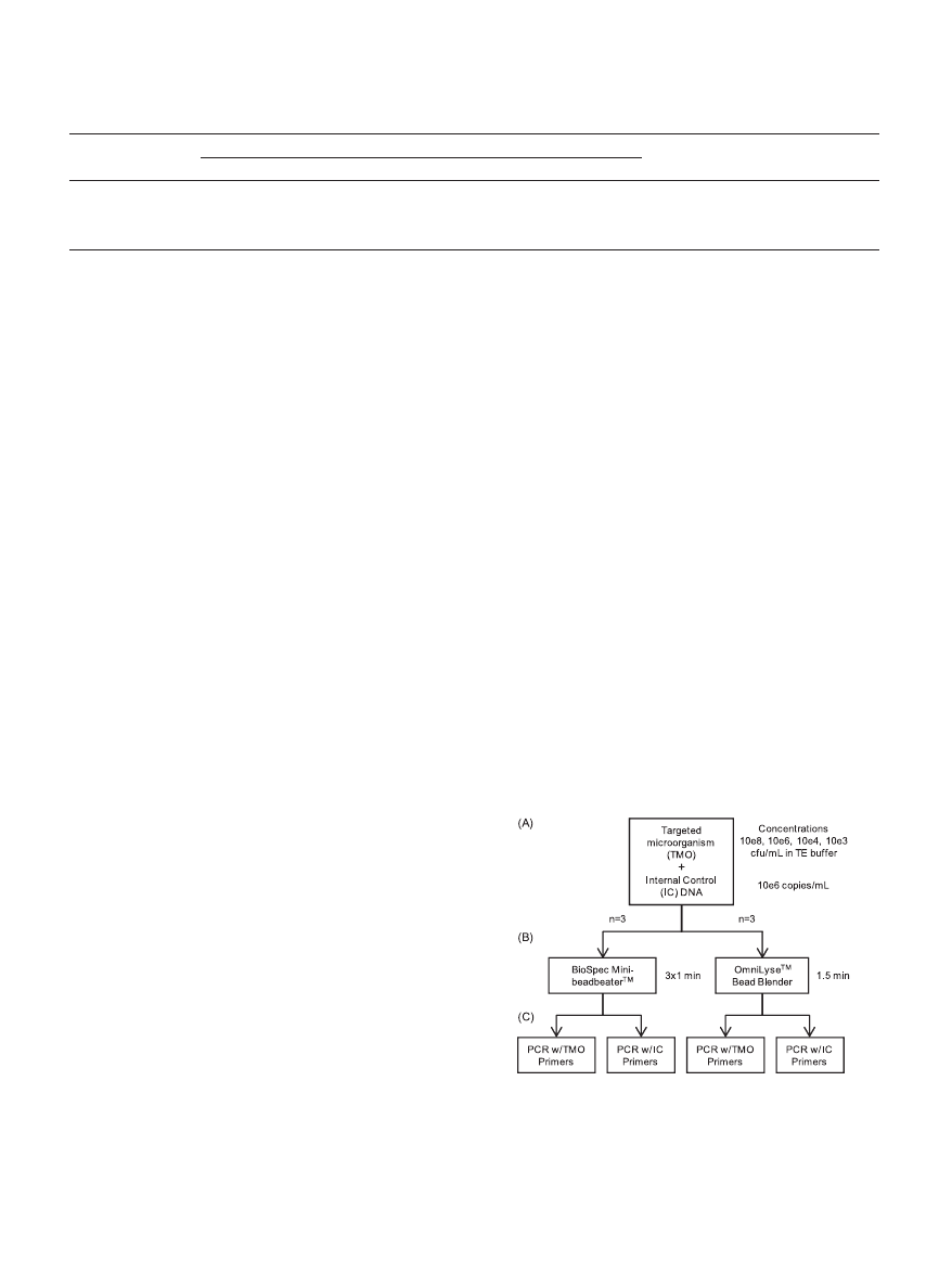
FIG. 2. Procedure overview. (A and B) Serial dilutions of the tar-
geted microorganism (TMO) plus a fixed amount of internal control
(IC) genomic DNA were processed in triplicate using an OmniLyse
device for 1.5 min or a BioSpec Mini-BeadBeater/Mini-BeadBeater-8
in three 1-min pulses, with 30-s rests on ice between pulses. (C) Pro-
cessed samples were split and analyzed by PCR using primers either
specific for the TMO or the IC.
V
OL
. 49, 2011
MECHANICAL PATHOGEN LYSIS USING A MINIATURIZED DEVICE
2535
on March 13, 2015 by UCSF Library & CKM
http://jcm.asm.org/
Downloaded from
subtilis spores processed through either the OmniLyse device
or the BioSpec Mini-BeadBeater, quantified by using B. sub-
tilis-specific primers, yielded comparable C
T
values at all con-
centrations tested in two independent experiments (Fig. 3).
Likewise, M. bovis BCG cells processed through either the
OmniLyse device or the BioSpec Mini-BeadBeater, using M.
bovis BCG-specific primers, resulted in comparable C
T
values
at all concentrations tested in two independent experiments
(Fig. 4). Since the purpose of the present study is to determine
a difference in C
T
value as function of device, independent of
the targeted microorganism’s concentration, the sample size
per device includes all replicates for all concentrations and
both experiments. This experimental design has a statistical
power of
⬎90% for predicting a significant ⌬C
T
of 0.5 between
the two devices, with a standard deviation in C
T
of 0.5 (signal-
to-noise ratio [S/N]
⫽ 1). Based on a two-factor analysis of
variance (ANOVA, see Fig. S1 in the supplemental material),
there is no significant difference between the C
T
values (as
indicators of lysis efficiency) obtained using the BioSpec versus
those obtained using the OmniLyse device, for either B. subtilis
spore lysis (P
⫽ 0.3225) or lysis of M. bovis BCG (P ⫽ 0.7845).
This ANOVA treated the targeted microorganism concentra-
tion as factor 1 and lysis device as factor 2, using a blocked
experimental design, wherein experimental sets (Fig. 3 and 4)
are treated as blocks.
In the first experiment (Fig. 4A), glass beads were used in
the BioSpec device and zirconia/silica beads were used in the
OmniLyse blender. In the second experiment (Fig. 4B), zirco-
nia/silica beads were used in both devices. The C
T
values ob-
tained using the BioSpec device are almost identical in both
experiments, indicating that the bead type used does not ap-
preciably affect the lysis efficiency.
In lysis experiments of B. subtilis spores and M. bovis BCG
cells, averaged C
T
values over the range of concentrations
tested for both devices were within a standard deviation of
each other. Having BSA present in the master mix was found
to be important for obtaining robust and reproducible exper-
imental results when zirconia/silica beads were used in either
device, especially at lower BCG cell concentrations, as dis-
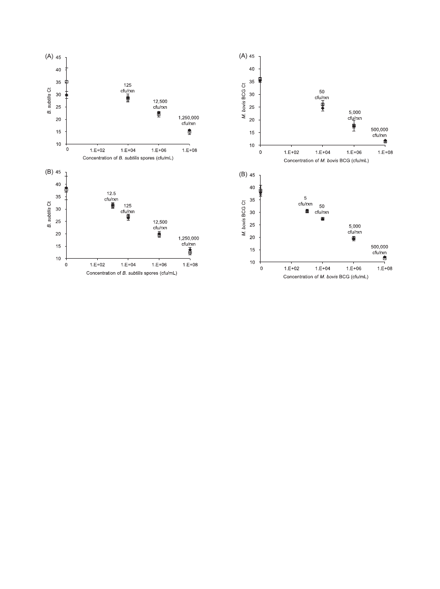
FIG. 3. Lysis of B. subtilis spores. PCR C
T
values are shown for
solutions containing spores at different concentrations, processed us-
ing either the OmniLyse device (}) or the BioSpec Mini-BeadBeater
(
䡺), with primers specific to B. subtilis. The numbers for CFU per
reaction (CFU/rxn) indicated in the graph are theoretical maxima,
based on 12.5
l of lysed sample added to each PCR, and assume no
loss during sample processing. In both experiments A and B, the C
T
values of spores lysed obtained using either device were comparable,
indicating similar lysis efficiency. During experiment A, the no-target
control (0 CFU/ml) samples appeared earlier than expected. Gel elec-
trophoresis results (data not shown) suggested that this was predom-
inantly due to primer dimers.
FIG. 4. Lysis of M. bovis BCG cells. C
T
values are shown for solu-
tions containing M. bovis BCG at different concentrations, processed
using either the OmniLyse device (}) or the BioSpec Mini-Bead-
Beater (
䡺), with primers specific to M. bovis BCG. The numbers for
CFU/rxn indicated in the graph are theoretical maxima, based on 5
l
of lysed sample added to each PCR, and assume no loss during sample
processing. Since mycobacteria are prone to clumping, one CFU may
represent more than one cell or genome. In both experiments A and B,
the C
T
values of BCG cells lysed using either device were comparable,
indicating similar lysis efficiency. For experiment A, only one replicate
was tested for the 10
8
CFU/ml concentration using the OmniLyse
blender. In experiment A, glass beads were used in the BioSpec device,
while zirconia/silica beads were used in all other experiments.
2536
VANDEVENTER ET AL.
J. C
LIN
. M
ICROBIOL
.
on March 13, 2015 by UCSF Library & CKM
http://jcm.asm.org/
Downloaded from
cussed in below. When glass beads were used in the Biospec
device, robust and reproducible results were obtained regard-
less of the cell concentration or presence of BSA.
B. subtilis spores are known to carry extracellular DNA (5,
19). To rule out the possibility that PCR experiments predom-
inantly detected extracellular DNA, we conducted a control
experiment. B. subtilis spores at concentrations of 10
4
, 10
6
, and
10
8
CFU/ml were either lysed using the BioSpec system, fol-
lowed by PCR analysis, or analyzed via PCR without the me-
chanical disruption step. At these concentrations, the unpro-
cessed spores amplified approximately 4 to 6 C
T
values later
than the samples that underwent mechanical lysis (Fig. 6). A
similar experiment was performed for M. bovis BCG cells at
concentrations of 10
8
and 10
6
CFU/ml (see Fig. S2A in the
supplemental material). Again, the unprocessed bacterial cells
amplified approximately 4 to 6 C
T
values later than the samples
that underwent mechanical lysis. Therefore, mechanical lysis
results in 10- to 100-fold more PCR-amplifiable target DNA,
assuming a PCR efficiency of ca. 100%. The true difference is
likely greater since unprocessed control samples probably un-
derwent partial heat lysis during the 10-min, 95°C PCR hot
start step. In another experiment, intact M. bovis BCG cells
were removed from processed and unprocessed samples by
centrifugation prior to PCR. In this case, unprocessed samples
amplified
⬃10 C
T
values later than processed samples (see Fig.
S2B in the supplemental material).
For the M. bovis BCG study, lysed samples were frozen at
⫺80°C prior to PCR processing. To investigate whether this
freeze-thaw step artificially enhanced mycobacterial lysis, we
conducted a separate experiment, wherein M. bovis BCG sam-
ples lysed using the BioSpec device were either kept on ice or
frozen at
⫺80°C prior to PCR processing. The C
T
values ob-
tained using both processing methods were comparable (see
Fig. S2A in the supplemental material), and averages were
within a standard deviation of each other.
Internal control results.
To assess secondary effects such as
DNA degradation and PCR inhibition, we added an internal
control (IC) to each reaction upstream of the lysis step, con-
sisting of a fixed amount of purified genomic DNA from a
different organism. If there are no secondary effects or if un-
accounted secondary effects are similar for both devices and at
each concentration of the targeted microorganism, then the IC
C
T
values should be comparable and relatively constant.
For the B. subtilis lysis experiment, E. coli genomic DNA was
added as an IC and processed by PCR for the first experiment
only, as shown in Fig. 5A. For the M. bovis BCG lysis experi-
ments, A. hydrophila genomic DNA was added as an IC and
processed by PCR in both experiments. The results for IC
amplification were similar in both experiments; data for the
second experiment are shown in Fig. 5B.
Overall, the C
T
values of the internal controls were compa-
rable for the OmniLyse device and the BioSpec Mini-Bead-
Beaters. Cross-amplification, i.e., amplification of the DNA
from the targeted microorganism using IC-specific primers and
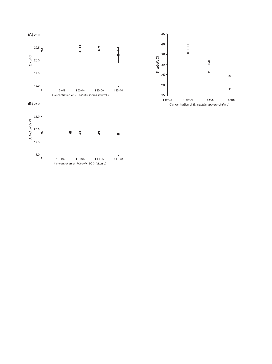
FIG. 5. Assessment of secondary effects on PCR amplification of
internal control DNA. (A) C
T
values for amplification of E. coli
genomic DNA spiked as an internal control at a constant concentra-
tion in B. subtilis suspensions of various concentrations, processed via
the OmniLyse (}) or BioSpec (䡺) device. Based on a 12.5-l sample
added per PCR and assuming no loss during processing, each PCR
should contain 12,500 copies E. coli genomic DNA. (B) C
T
values for
amplification of A. hydrophila genomic DNA spiked as an internal
control at a constant concentration into M. bovis BCG suspensions of
various concentrations, processed via the OmniLyse (}) or BioSpec
(
䡺) device. Based on a 5-l sample added per PCR, and assuming no
loss during processing, each PCR should contain 5,000 copies A. hy-
drophila genomic DNA. Overall, the C
T
values of the internal controls
are reasonably comparable for the OmniLyse device and the BioSpec
Mini-BeadBeater devices, and are independent of the concentration of
the targeted microorganism.
FIG. 6. Control experiment to differentiate detection of B. subtilis
extracellular DNA from detection of DNA as result of B. subtilis spore
lysis. Samples lysed using the BioSpec system followed by PCR analysis
(}) amplify approximately 4 to 6 C
T
values earlier than samples con-
taining the same concentration of B. subtilis spores but analyzed via
PCR without the mechanical disruption step (
䡺). Mechanical lysis
results in 10- to 100-fold more PCR-amplifiable target DNA, assuming
a PCR efficiency of ca. 100%.
V
OL
. 49, 2011
MECHANICAL PATHOGEN LYSIS USING A MINIATURIZED DEVICE
2537
on March 13, 2015 by UCSF Library & CKM
http://jcm.asm.org/
Downloaded from

vice versa, was not observed in either case. In the B. subtilis and
M. bovis BCG experiments, correct amplicon formation was
confirmed via gel electrophoresis and melting-curve analysis,
respectively. Amplification with B. subtilis specific primers for
some of the lower B. subtilis concentrations and the no-target
controls also contained primer dimers.
The presence of BSA in the master mix appeared to be
critical to obtaining reproducible and robust C
T
values follow-
ing mechanical lysis using the zirconia/silica beads in either the
OmniLyse or the BioSpec Mini-BeadBeater-8. When BSA was
omitted in initial experiments with M. bovis BCG, the C
T
val-
ues were inconsistent, with many failed amplifications, espe-
cially at the lower M. bovis BCG concentrations. The C
T
values
of the IC (A. hydrophila genomic DNA) increased with de-
creasing M. bovis BCG concentration. Late amplification was
also observed in the no-target control, containing A. hydrophila
DNA only in buffer that was processed through the respective
bead beating device. Dilution series of the purified A. hydro-
phila DNA not processed through a lysis device did not show
this effect. Adding BSA directly to the bacterial suspension
prior to lysis, or to the master mix following lysis, eliminated
these effects and enabled consistent results.
DISCUSSION
The miniaturized OmniLyse device mechanically lyses B.
subtilis spores and M. bovis BCG cells with efficacy similar to
the industry-standard benchtop BioSpec Mini-BeadBeater,
based on the comparable C
T
values obtained from PCRs with
primers specific for these two microorganisms. Bacillus spores
carry extracellular DNA that may be bound on the bacterial
spore coats. This extracellular DNA is not readily removed by
multiple wash steps and can be amplified during PCR even
without mechanical lysis (5, 19). Although it is possible that
extracellular DNA was coamplified in the experiments de-
scribed here, a control experiment demonstrated that mechan-
ical lysis significantly increased (between 10- and 100-fold) the
amount of PCR-amplifiable target DNA, a finding in line with
the expectation that mechanical lysis is required to achieve a
low limit of detection. In addition, a previous time series lysis
study of DNase-treated B. subtilis spores using the OmniLyse
device clearly demonstrated that this device mechanically dis-
rupts spores and liberates intracellular DNA (13). Likewise,
other studies have confirmed the ability of the BioSpec Mini-
BeadBeater to lyse Bacillus spores (20, 24).
Using the protocol described here, the experiments did not
appear to be confounded by secondary effects such as DNA
degradation or PCR inhibition, based on consistent internal
control C
T
values obtained for both devices and at each con-
centration of the targeted microorganism. For lysis experi-
ments using zirconia/silica beads in either device, the presence
of BSA in the master mix was required for consistent, repro-
ducible results. Although further experiments are required to
fully understand this effect, it is possible that BSA relieves a
PCR-inhibitory effect linked to fragments of zirconia/silica
beads carried into the PCR. This hypothesis is supported by
the observation that when glass beads were used in the Bio-
Spec device in place of zirconia/silica beads, no inhibition was
observed, and reproducible results were obtained at all cell
concentrations with or without BSA. BSA is commonly used as
a PCR facilitator (29, 36), and it may bind to fragments of
zirconia/silica beads carried into the PCR. The inhibitory effect
was unlikely to be caused by cellular debris because the effect
was less prominent at higher concentrations of the targeted
microorganism and was also observed in the no-target controls
containing IC DNA only in buffer processed through a bead-
beating device.
To the extent possible, we used similar conditions in the
OmniLyse and the BioSpec devices, which included the use of
similar or identical beads and bead-to-volume ratios. The pro-
cessing time per sample for the OmniLyse device (1.5 min
without rest), however, was significantly less than for the Bio-
Spec BeadBeater devices (three 1-min pulses with 30-s rests
between pulses). The BioSpec processing time used in the
present study was based on standard published protocols (10,
13, 33, 35), and appears to be the time required using this
system for effective lysis of Bacillus spores and mycobacterial
cells. In most experiments, the same zirconia/silica beads were
used in both devices. BioSpec protocols reported in the liter-
ature use either glass beads or zirconia/silica beads for lysis of
mycobacteria (22, 33, 35). Based on our results (Fig. 4), the use
of glass beads versus zirconia/silica beads in the BioSpec did
not appreciably affect the lysis efficiency using this device and
did not alter the relative lysis efficiency compared to the
OmniLyse.
In summary, for both lysis-resistant bacterial strains studied,
mechanical lysis using the OmniLyse blender and the BioSpec
Mini-BeadBeaters resulted in similar C
T
values for PCR anal-
ysis of processed samples over the range of concentrations
studied. Amplification of IC DNA spiked at a constant con-
centration into the samples upstream of lysis using either de-
vice resulted in consistent PCR C
T
values for all processed
samples. Overall, the OmniLyse device facilitates effective lysis
of tough-walled organisms in a miniaturized, disposable, bat-
tery-operated format, with an efficiency comparable to that of
a widely used benchtop system. Future work will focus on
combining lysis with extraction of nucleic acids within the bead
blender with suitable nucleic acid amplification and detection
methods in a fully integrated cartridge for handheld point-of-
care nucleic acid testing in low-resource settings.
ACKNOWLEDGMENTS
We thank Donald S. Kenney (Gustafson, LLC) for graciously sup-
plying B. subtilis spores for this study.
This study was supported by Public Health Service grant AI090831
from the National Institute of Allergy and Infectious Diseases. P.E.V.
acknowledges support through a Science, Mathematics, and Research
for Transformation (SMART) scholarship from the Department of
Defense.
P.E.V., K.M.W., B.E., G.A.C., and A.N. declare that they have no
conflicts of interest. R.D., A.N., and B.I. each own a minority interest
in Claremont BioSolutions, LLC. J.S. works on a part-time basis for
Claremont BioSolutions, LLC.
REFERENCES
1. Amaro, A., E. Duarte, A. Amado, H. Ferronha, and A. Botelho. 2008. Com-
parison of three DNA extraction methods for Mycobacterium bovis, Myco-
bacterium tuberculosis, and Mycobacterium avium subsp. avium. Lett. Appl.
Microbiol. 47:8–11.
2. Anderberg, R. J., J. A. Strachan, and G. A. Cangelosi. 1995. Purification of
DNA from Mycobacterium species without sonication or phenol. Biotech-
niques 18:217–219.
3. Balamurugan, R., S. Venkataraman, K. R. John, and B. S. Ramakrishna.
2006. PCR amplification of the IS6110 insertion element of Mycobacterium
2538
VANDEVENTER ET AL.
J. C
LIN
. M
ICROBIOL
.
on March 13, 2015 by UCSF Library & CKM
http://jcm.asm.org/
Downloaded from

tuberculosis in fecal samples from patients with intestinal tuberculosis.
J. Clin. Microbiol. 44:1884–1886.
4. Barken, K. B., J. A. J. Haagensen, and T. Tolker-Nielsen. 2007. Advances in
nucleic acid-based diagnostics of bacterial infections. Clin. Chim. Acta 384:
1–11.
5. Belgrader, P., et al. 1999. A minisonicator to rapidly disrupt bacterial spores
for DNA analysis. Anal. Chem. 71:4232–4236.
6. Belisle, J. T., S. B. Mahaffey, and P. J. Hill. 2009. Isolation of Mycobacterium
species genomic DNA, p. 1–12. In T. Parish and A. C. Brown (ed.), Myco-
bacteria protocols, 2nd ed., vol. 465. Humana Press, Inc., Totowa, NJ.
7. Boehme, C. C., et al. 2010. Rapid molecular detection of tuberculosis and
rifampin resistance. N. Engl. J. Med. 363:1005–1015.
8. Boom, R., et al. 1990. Rapid and simple method for purification of nucleic
acids. J. Clin. Microbiol. 28:495–503.
9. Brennan, P. J., and H. Nikaido. 1995. The envelope of mycobacteria. Annu.
Rev. Biochem. 64:29–63.
10. Cangelosi, G. A., K. M. Weigel, C. Lefthand-Begay, and J. S. Meschke. 2010.
Molecular detection of viable bacterial pathogens in water by ratiometric
pre-rRNA analysis. Appl. Environ. Microbiol. 76:960–962.
11. de Boer, R., et al. 2010. Improved detection of microbial DNA after bead-
beating before DNA isolation. J. Microbiol. Methods 80:209–211.
12. Dhandayuthapani, S., Y. Zhang, M. H. Mudd, and V. Deretic. 1996. Oxida-
tive stress response and its role in sensitivity to isoniazid in mycobacteria:
characterization and inducibility of ahpC by peroxides in Mycobacterium
smegmatis and lack of expression in M. aurum and M. tuberculosis. J. Bacte-
riol. 178:3641–3649.
13. Doebler, R. W., et al. 2009. Continuous-flow, rapid lysis devices for biode-
fense nucleic acid diagnostic systems. J. Assoc. Lab. Automation 14:119–125.
14. Fan, J., A. J. Kraft, and K. J. Henrickson. 2006. Current methods for the
rapid diagnosis of bioterrorism-related infectious agents. Pediatr. Clin. N.
Am. 53:817.
15. George, K. M., Y. Yuan, D. R. Sherman, and C. E. Barry. 1995. The biosyn-
thesis of cyclopropanated mycolic acids in Mycobacterium tuberculosis: iden-
tification and functional analysis of CMAS-2. J. Biol. Chem. 270:27292–
27298.
16. Helb, D., et al. 2010. Rapid detection of Mycobacterium tuberculosis and
rifampin resistance by use of on-demand, near-patient technology. J. Clin.
Microbiol. 48:229–237.
17. Hoffmann, C., A. Leis, M. Niederweis, J. M. Plitzko, and H. Engelhardt.
2008. Disclosure of the mycobacterial outer membrane: cryo-electron
tomography and vitreous sections reveal the lipid bilayer structure. Proc.
Natl. Acad. Sci. U. S. A. 105:3963–3967.
18. Hurley, S. S., G. A. Splitter, and R. A. Welch. 1987. Rapid lysis technique for
mycobacterial species. J. Clin. Microbiol. 25:2227–2229.
19. Johns, M., L. Harrington, R. W. Titball, and D. L. Leslie. 1994. Improved
methods for the detection of Bacillus anthracis spores by the polymerase
chain reaction. Lett. Appl. Microbiol. 18:236–238.
20. Jones, C. A., N. L. Padula, and P. Setlow. 2005. Effect of mechanical abrasion
on the viability, disruption and germination of spores of Bacillus subtilis.
J. Appl. Microbiol. 99:1484–1494.
21. Kamei, T., et al. 2005. Microfluidic genetic analysis with an integrated a-Si:H
detector. Biomed. Microdevices 7:147–152.
22. Kaser, M., M. T. Ruf, J. Hauser, L. Marsollier, and G. Pluschke. 2009.
Optimized method for preparation of DNA from pathogenic and environ-
mental mycobacteria. Appl. Environ. Microbiol. 75:414–418.
23. Kazakov, S., E. Bonvouloir, and I. Gazaryan. 2008. Physicochemical char-
acterization of natural ionic microreservoirs: Bacillus subtilis dormant spores.
J. Phys. Chem. B 112:2233–2244.
24. Longchamp, P., and T. Leighton. 1999. Molecular recognition specificity of
Bacillus anthracis spore antibodies. J. Appl. Microbiol. 87:246–249.
25. Millar, B. C., J. R. Xu, and J. E. Moore. 2007. Molecular diagnostics of
medically important bacterial infections. Curr. Issues Mol. Biol. 9:21–39.
26. Novak-Weekley, S. M., et al. 2010. Clostridium difficile testing in the clinical
laboratory by use of multiple testing algorithms. J. Clin. Microbiol. 48:889–
893.
27. Odumeru, J., A. Gao, S. Chen, M. Raymond, and L. Mutharia. 2001. Use of
the bead beater for preparation of Mycobacterium paratuberculosis template
DNA in milk. Can. J. Vet. Res.-Rev. Can. Recherche Vet. 65:201–205.
28. Pemov, A., H. Modi, D. P. Chandler, and S. Bavykin. 2005. DNA analysis
with multiplex microarray-enhanced PCR. Nucleic Acids Res. 33:e11.
29. Radstrom, P., R. Knutsson, P. Wolffs, M. Lovenklev, and C. Lofstrom. 2004.
Pre-PCR processing: strategies to generate PCR-compatible samples. Mol.
Biotechnol. 26:133–146.
30. Rao, S. S., K. V. Mohan, and C. D. Atreya. 2010. Detection technologies for
Bacillus anthracis: prospects and challenges. J. Microbiol. Methods 82:1–10.
31. Salazar, O., and J. A. Asenjo. 2007. Enzymatic lysis of microbial cells.
Biotechnol. Lett. 29:985–994.
32. Schaeffer, A. B., and M. D. Fulton. 1933. A simplified method of staining
endospores. Science 77:194.
33. Sherman, D. R., et al. 2001. Regulation of the Mycobacterium tuberculosis
hypoxic response gene encoding alpha-crystallin. Proc. Natl. Acad. Sci.
U. S. A. 98:7534–7539.
34. Ulrich, M. P., et al. 2006. Evaluation of the Cepheid GeneXpert (R) system
for detecting Bacillus anthracis. J. Appl. Microbiol. 100:1011–1016.
35. Via, L. E., and J. O. Falkinham. 1995. Comparison of methods for isolation
of Mycobacterium avium complex DNA for use in PCR and rapid finger-
printing. J. Microbiol. Methods 21:151–161.
36. Wilson, I. G. 1997. Inhibition and facilitation of nucleic acid amplification.
Appl. Environ. Microbiol. 63:3741–3751.
V
OL
. 49, 2011
MECHANICAL PATHOGEN LYSIS USING A MINIATURIZED DEVICE
2539
on March 13, 2015 by UCSF Library & CKM
http://jcm.asm.org/
Downloaded from
Wyszukiwarka
Podobne podstrony:
Arakawa et al 2011 Protein Science
Dannenberg et al 2015 European Journal of Organic Chemistry
Niemyjska et al 2011 id 319083 Nieznany
Hua et al 2009 European Journal of Organic Chemistry
Grosser et al A social network analysis of positive and negative gossip
Ciolkosz et al 2011 Biofuels, Bioproducts and Biorefining
Siebner et al 2001 European Journal of Neuroscience
Lester et al 2012 Comparative analysis of strawberry total phenolics via Fast Blue BB vs Folin–Cio
Huang et al 2009 Journal of Polymer Science Part A Polymer Chemistry
new media and the permanent crisis of aura j d bolter et al
Lebrini et al 2005 Journal of Heterocyclic Chemistry
Rosie Sexton et al An Ordinal Indexed Hierarchy of Separation Properties
Mosna et al Z 2 gradings of CA & Multivector Structures (2003) [sharethefiles com]
Barret et al Templates For The Solution Of Linear Systems Building Blocks For Iterative Methods [s
więcej podobnych podstron