
R E S E A R C H A R T I C L E
Open Access
The murine lung microbiome in relation to the
intestinal and vaginal bacterial communities
Kenneth Klingenberg Barfod
1,2*
†
, Michael Roggenbuck
3
†
, Lars Hestbjerg Hansen
3
, Susanne Schjørring
1
,
Søren Thor Larsen
2
, Søren Johannes Sørensen
3
and Karen Angeliki Krogfelt
1
Abstract
Background: This work provides the first description of the bacterial population of the lung microbiota in mice.
The aim of this study was to examine the lung microbiome in mice, the most used animal model for inflammatory
lung diseases such as COPD, cystic fibrosis and asthma.
Bacterial communities from broncho-alveolar lavage fluids and lung tissue were compared to samples taken from
fecal matter (caecum) and vaginal lavage fluid from female BALB/cJ mice.
Results: Using a customized 16S rRNA sequencing protocol amplifying the V3-V4 region our study shows that the
mice have a lung microbiome that cluster separately from mouse intestinal microbiome (caecum). The mouse lung
microbiome is dominated by Proteobacteria, Firmicutes, Actinobacteria, Bacteroidetes and Cyanobacteria overlapping
the vaginal microbiome. We also show that removal of host tissue or cells from lung fluid during the DNA extraction
step has an impact on the resulting bacterial community profile. Sample preparation needs to be considered when
choosing an extraction method and interpreting data.
Conclusions: We have consistently amplified bacterial DNA from mouse lungs that is distinct from the intestinal
microbiome in these mice. The gut microbiome has been extensively studied for its links to development of disease.
Here we suggest that also the lung microbiome could be important in relation to inflammatory lung diseases.
Further research is needed to understand the contribution of the lung microbiome and the gut-lung axis to the
development of lung diseases such as COPD and asthma.
Background
Studies of the lung microbiome by culture independent
techniques and its impact on lung immunity is a rela-
tively new field and may contribute to new advances in
understanding respiratory diseases [1]. Healthy human
lungs have up until recently been considered to be sterile
by culture-based techniques, but now new evidence have
identified microbial communities both in healthy humans
and in those with disease [2-4]. The human microbiome
project [5] did not originally include the lungs, but re-
cently the Lung HIV Microbiome Project has published
the first results in this field [6,7]. Investigations into
lung microbiology and lung immunity in humans is lim-
ited largely because of technical, ethical considerations
and small samples sizes, whereas the use of animal models
can provide novel information useful in investigations into
the importance of lung microbiome in the development of
lung immunology. Effective utilization and development
of animal models have recently been identified as one of
the most important challenges in future lung microbiome
research by the NIH [8]. Whereas many studies have
focused on the gut microbiome and its impact on among
others lung immunity and asthma, little work has been
performed to examine the contribution of the lung micro-
biome on the pathogenesis of pulmonary diseases. Espe-
cially in inflammatory lung diseases such as asthma and
COPD, the local microbiome may play an important role
in the pathogenesis. The technical challenges related to
the novel culture-dependent techniques include consistent
extraction of useful DNA, the development of PCR methods
and sampling methods for the less abundant bacterial load
of the lungs.
* Correspondence:
†
Equal contributors
1
Statens Serum Institut, Artillerivej 5, 2300 Copenhagen S, Denmark
2
National Research Centre for the Working Environment, Lersø Parkallé 105,
2100 Copenhagen O, Denmark
Full list of author information is available at the end of the article
© 2013 Barfod et al.; licensee BioMed Central Ltd. This is an open access article distributed under the terms of the Creative
Commons Attribution License (http://creativecommons.org/licenses/by/2.0), which permits unrestricted use, distribution, and
reproduction in any medium, provided the original work is properly cited.
Barfod et al. BMC Microbiology 2013, 13:303
http://www.biomedcentral.com/1471-2180/13/303

We hypothesized that the problems with getting bac-
terial DNA from lungs was due to the presence of host
DNA in the extractions. In this study, we have investi-
gated the bacterial community from lungs of 20 mice
using rDNA amplicon 454 pyrosequencing. We also
performed a conventional cultivation study of 10 mouse
bronchoalveolar lavage (BAL) fluids on different agar
plates. Sampling methods and DNA extraction proto-
cols were investigated systematically: one BAL sample
still containing mouse cells (BAL-plus) and one BAL
sample, where the mouse cells were removed (BAL-
minus) by cytospin. The bacterial communities in BAL
samples were compared using DNA extractions from
washed lung tissue, caecum samples and vaginal flushing.
We chose to include vaginal samples for two major
reasons. The vaginal microbiome of BALB/c has not
previously been described and could have influence on
microbial
“priming” and transfer from mother to pup.
In this study, it also serves a reference sample from a
different mucoid epithelium than lung. The bacteria were
classified by their sequence into Operational Taxonomic
Units (OTU). An OTU is an approximation to taxonomy
derived from classical cultivation techniques.
We demonstrate the use of this methodology and de-
scribe an uncultivable lung and vaginal microbiome in
mice that are diverse and distinct from caecal micro-
biome. Our results provide a basis for further studies
into the lung microbiome in culture negative BAL fluids
in mouse models of inflammatory lung diseases sug-
gested by descriptive human studies.
Methods
Mice and sample collection
BALB/cJ female mice, reared together (Taconic M&B,
Ry, Denmark), 7 weeks old, body weight 18
–22 g, were
randomly distributed and housed 10 animals per cage
(425 × 266 × 150 mm) with tap water and food (Altromin
no 1324 Brogaard Denmark) provided ad libitum. Light/
dark cycles were at 12 hours and room temperature
and relative humidity was kept at 19-22°C and 40-60%,
respectively. Animals were handled by the same two
animal technicians and conditioned in our animal facility
for two weeks before use.
The BAL procedure was performed as previously
described with minor modifications [9]. We inserted
sterile tube (Insyte, BD, Denmark) for each mouse and
lungs were flushed two times with 0.8 mL pyrogenfree
saline (0.9%)(Fresenius Kabi, Denmark) and the recov-
ered fluids were pooled (LF-plus). For the BAL samples
without mouse cells (BAL-minus) the BAL fluid was
spun at 400 g for 10 min a 4°C collecting the super-
natant. All the BAL samples were frozen at
−80°C.
Lung tissue was collected using one, chlorine [10] and
heat treated sterile scissors, per animal cutting the distal
tip of the left lung after the BAL procedure. Tissues
were snap-frozen in liquid nitrogen.
Vaginal fluid samples were performed by inserting a
sterile pipette tip into the vaginal space flushing 3 times
back and forth with 30
μL pyrogenfree infusion saline
(0.9%) (Fresenius Kabi, Denmark) and frozen at
−80°C.
As the last procedure, the caecum samples were taken
from the animals. With a sterile scissor the caecum was
cut open and approximately 50 mg of caecal matter was
removed using sterile plastic loops directly into cryo tubes
and snap frozen in liquid nitrogen. All protocols were
approved by the Danish Animal Experiments Inspectorate.
Bacterial identification by culturing
Mouse BAL fluids, 200
μL per mouse, were cultivated
on general growth media blood agar 5% (SSI, Denmark)
and Chocolate Agar (SSI, Denmark) for fastidious bac-
teria and incubated at 37°C for 24 hours. Another set of
plates with selective media was incubated under micro
aerophilic conditions (5%CO
2
, 3%H
2
, 5%O
2
and 87%N
2
)
at 37°C for 48 hours [11]. The bacterial colonies were
subjected to routine identification by the Vitek2 system
(Bio Mérieux, France).
DNA extraction and PCR
Isolation of bacterial DNA from frozen BAL or vaginal
samples was done using Qiagen spin protocol (Qiagen,
DNA mini kit Denmark) for body fluids with the follow-
ing modifications: Tubes were thawed and centrifuged at
16.000 g for 5 min to spin down all the bacteria. The
supernatant was discarded and the bacterial pellet was
resuspended with 450
μL lysis buffer. Forty-five μL pro-
teinase K and add 0.3 mL 0.1 mm zirconium/silica beads
(Techum, Sweden) were added. Proceed with bead beat-
ing step using TissueLyser (Qiagen, Denmark) for 6 min
at 30 Hz. [12]. Lysis was performed by incubating in
heat block at 56°C for 10 min. and then at 95°C for
7 min. Proceed with protocol for body fluids from step 5.
At the elution step, the AE buffer is preheated to 65°C
and DNA elution is performed with 100 ul with 3 minutes
incubation at room temp before final spin. Isolation of
bacterial DNA from frozen caecal or tissue was done
using Qiagen spin protocol for detection of pathogens
from stool (Qiagen, DNA mini stool kit Denmark) with
the following modifications: Add 1.4 ml of the ASL buffer
and perform bead beating, lysing and eluding as describe
above for body fluids. For tissues samples, chlorine [10]
and heat sterilized 3 mm steel bead (Qiagen, Denmark)
was added to the samples along with the zirconium/
silica beads for extra tissue disruption.
16S sequencing
Amplicon libraries of the 16S rRNA gene of caecum, BAL
and vaginal samples were prepared with two PCR reactions.
Barfod et al. BMC Microbiology 2013, 13:303
Page 2 of 12
http://www.biomedcentral.com/1471-2180/13/303

In the first PCR, a 466 bp long fragment covering the
variable region V3 and V4 of the 16S rRNA gene, was
amplified with AccuPrime
™ Pfx DNA Polymerase and
the bacteria and archaea specific primers 341 F and 806R
(Table 1). The reaction started with an initialization at
94°C for 2 min, followed by 44 cycles of denaturation at
94°C for 20 sec, annealing at 56°C for 30 sec. and elong-
ation at 68°C for 40 sec. The reaction was completed
with a final elongation at 68°C for 5 min. Due to the low
DNA (<0.5 ng ×
μL
-1
) concentration in the samples we
needed to increase the cycle number above the standard
of 30
–35. This adjustment highly increased the risk of
amplifying contamination from extraction buffer and
other experimental used liquids. To minimize this possi-
bility we chose the lowest cycle number with a clear
amplification band in the agarose gel and no signals
of negative controls from BAL procedure for DNA
purification.
In the second PCR the adaptors were attached to the
amplicon library elongating the fragment towards 526 bp
with the primer TitA_341F and TitB_806R. The same
reaction conditions of PCR I were applied in PCR II
with a reduced cycle number of 15.
Initially we tried to apply the same procedure for
the lung tissue samples but unspecific bands after gel-
electrophoresis made it impossible to select the correct
fragment size. To overcome this problem we chose the
primer 27 F and 1492R amplifying the entire 16S rRNA
gene which appeared to be more specific. The PCR I con-
ditions were the same as mentioned above except that the
annealing temperature was reduced to 55°C and the cycle
number to 40. In this perspective the Tag-PCR reaction
with TitA_341F and TitB_806R provided the selection for
V3 and V4 as well as attaching the adaptors to the
amplicons.
Statistical analysis and bioinformatics
The 16S rRNA gene sequences obtained from one half a
plate of a 454 - Roche - Titanium pyrosequencing run
were quality filtered, trimmed and split into the corre-
sponding animal samples with the Qiime pipeline ver-
sion 1.6.0 using the default settings [17]. We considered
only sequences with a minimum length of 250 bp. Chi-
meras were removed by UCHIME [18]. The operational
taxonomic units (OTU) were picked de novo and clus-
tered at 97% sequence similarity. The taxonomy was
assigned using RDP classifier (bootstrap threshold 0.8)
greengenes as reference database [19].
For statistical analysis, raw data were transferred into
the open source statistical program
“R” [20]. The non-
parametric Wilcoxon test (W) evaluated variations of
alpha diversity between two variables. We used the
non-parametric Kruskal-Wallis-test when comparing more
than 2 variables (KW). Dissimilarities in OTUs abundance
between the samples were explained by KW and the
sample clustering of the OTU count based Bray-Curtis
distance metric were examined by the analysis of simi-
larity (anosim).
Results
To determine the airway bacterial microbiota of the
BALB/cJ mouse model based on 16S rDNA gene se-
quencing, we have compared sequences found in the
lungs with three different approaches, to sequences found
in corresponding vaginal and caecal samples.
Over all sequence quality and results from all sample types
We generated a total of 908256 sequences. After quality
filtering and chimera check, 27% of sequences were re-
moved and 660319 sequences were further processed for
OTU picking (sequences ranged between 3530 up to 31638
per animal sample). The de novo OTU clustering revealed
6487 OTUs. The OTU table was randomly subsampled to
avoid differences based on sequencing effort leaving 3318
OTUs for further analysis (Rarefaction curve are shown in
Additional file 1: Figure S5).
We found a total of 19 bacterial phyla in the samples
analysed. The most dominant (>0.5% abundance) phyla
observed were Acidobacteria, Actinobacteria, Bacteroidetes,
Firmicutes, Proteobacteria
and TM7. The difference in bac-
terial composition at the phylum level between sampling
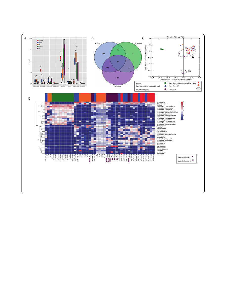
sites is shown in Figure 1A.
Table 1 Primers
Primer
Sequence
Reference
27 F
5
′AGAGTTTGATCMTGGCTCAG-3′
[
]
341
5
′-CCTAYGGGRBGCASCAG-3′
[
806
5
′-GGACTACNNGGGTATCTAAT-3′
[
TitA_341F
5
′-CGTATCGCCTCCCTCGCGCCATCAG-TAG-CCTAYGGGRBGCASCAG-3′
[
]
TitB_806R
5
′-CTATGCGCCTTGCCAGCCCGCTCAG-GGACTACNNGGGTATCTAAT-3′
[
]
1492R
5
′-GGTTACCTTGTTACGACTT-3′
[
]
Barfod et al. BMC Microbiology 2013, 13:303
Page 3 of 12
http://www.biomedcentral.com/1471-2180/13/303
In Additional file 2: Table S2 we have listed all the bac-
teria that were found, which were unique for the lung
samples and which were shared between sampling sites.
The bacterial sequences of the lung samples
If we only look at the lung samples, the most dominant
lung phyla found were Proteobacteria, Firmicutes, Acti-
nobacteria, Bacteroidetes
and Cyanobacteria. Additionally
we observed Fusobacteria and Cyanobacteria in the lung
and vaginal samples.
In order to highlight phyla variations in the lung com-
munity compared to vaginal and caecal communities, we
first we took the three lung sample types: bronchoalveo-
lar lavage fluids (BAL-plus), and BAL-minus, where the
mouse cells have been removed by a spin protocol and
finally lung tissue from the distal tip of the lung and
considered them as one ecological community. In this
lung community profile, Actinobacteria, and Proteobacteria
were clearly more abundant than in the caecum commu-
nity (KW, p < 0.0001).
Then, looking at the differences between the three
lung sample types, Firmicutes appeared (KW, p < 0.05)
more abundant in lung tissue (57%) than in BAL samples
(20%). The SR1 bacteria were found only in BAL-minus
and Lung tissue samples, but Tenericutes was observed
in all samples, except in the vaginal samples. Other
phyla observed below 0.5% abundance were Chloroflexi,
Deinococcus-Thermus, Fibrobacteres, Gemmatimonadetes,
OD1, OP10, Planctomycetes, Verrucomicrobia,
and WS3.
Comparing lung sampling methods we also found a
significant variation for Actinobacteria and Cyanobacteria,
which were largely abundant in both type of BAL commu-
nities relative to the lung tissue samples (KW, p < 0.05).
At phylum level, the composition of the lung tissue
Figure 1 Community composition. (A) Distribution of Phyla between sample types. LF-plus bronchoalveolar lavage (BAL) fluids and LF-minus is
BAL where the mouse cells have been removed. LT is lung tissue and VF is vaginal flushing, (B) Venn diagram of identified shared and unique genera
from each sampling site. All the lung type samples are considered here as one. (complete list shown in Additional file 3: Table S4), (C) The PcoA plot is
generated of the Bray-Curtis dissimilarity metric based on OTU counts and explains the largest variance between all samples (PCoA plot 1vs 3 and
PCoA plot 2 vs. 3 are attached in Additional file 4: Figure S4), (D) Heat map of even subsampled OTU table. The dendrogram is two sited hierarchal
clustered by abundance dissimilarity and the data are log transformed. Shown are only taxa, which counted for at least 0.5% of the generated
sequences. The x-axis clusters the animal samples and the y-axis the taxonomical information. * marks Vaginal subcluster S1 and ** subcluster S2.
Barfod et al. BMC Microbiology 2013, 13:303
Page 4 of 12
http://www.biomedcentral.com/1471-2180/13/303

samples appeared to be very similar to the vaginal sam-
ples except for a larger abundance of Cyanobacteria in
vaginal samples (KW, p < 0.05).
Bacterial sequences of the caecum
Looking at the caecum samples, they contained more
Firmicutes
and Bacteroidetes KW, p < 0.0001) than the
lung samples and Acidobacteria and Cyanobacteria were
absent. The phylum Bacteroidetes (29%) appeared to be
the second most abundant after the Firmicutes (59%).
The vaginal and the caecal communities only had Rumi-
nococcus
in common, a genus that was not observed in
the lung microbiota. Three genera were found in caecal
samples alone; Robinsoniella, Parasutterella and Ramli-
bacter
. The low numbers of genera detected in the caecal
samples is due to the depth of taxonomic information
obtained for these particular OTU sequences towards
the consensus lineage of the database.
Overlapping genera
For an overview comparison between the different sample
types, we have merged the results found in the different
lung communities and displayed the overlapping genera-
wit hcaecum and vagina in a venn diagram. This diagram
reflects 255 identified genera (summarized in Additional
file 3: Table S4), that covers 76% of the sequences from
BAL-plus, 68% from BAL-minus, 66% of vaginal and lung
tissue community and 27% of sequences assigned to the
caecum community (Figure 1B).
Lung samples, vaginal and caecum samples shared the
12 core genera Bacteroides, Barnesiella, Odoribacter,
Alistipes, Mucispirillum, Lactobacillus, Streptococcus,
Peptoniphilus, Roseburia, Anaerotruncus, Oscillibacter,
Pseudomonas
. We observed Parabacteroides, Eubacterium,
Marvinbryantia
, Butyricicoccus, Papillibacter, Bosea, Anae-
roplasma
, lung and caecum. The pulmonic and vaginal
community shared 103 genera (Additional file 3: Table S4).
Additionally Akkermansia was also found in the lung but
only in one caecum sample in the raw data set.
Variability in community composition between samples
obtained from the same sampling site (Beta_diversity)
To make a sample to sample comparison and illustrate the
variation between our mice we have performed a principle
coordinate analysis (PCoA) based on the Bray-Curtis
dissimilarity between OTU count metric PCoA plot
(Figure 1C), which explains the largest variance between
all samples (Additional PCoA 2 and 3 are found in
Additional file 4: Figure S4).
The caecal samples cluster together at a significant dis-
tance from lung and vaginal communities, confirmed by
the analysis of similarity, anosim (R = 0.673, p = 0.001)
The dissimilarity between the three lung communities
was found to be little due to strong cluster overlap (anosim,
R = 0.09, p = 0.05) when comparing only the lung distances.
We found large variation within the vaginal samples
resulting in a division into subcluster 1 (S1), containing
animal vaginal sample 8, 5 and 2, and subcluster 2 (S2),
vaginal sample 3,4,6,9 and 10 (anosim, R = 0.72, p = 0.001).
The separation is clearly shown in PCoA1 (Figure 1C) and
PCoA3 (Additional file 4: Figure S4). Those samples that
grouped into S1 were found to be less similar to caecum
and lung communities, whereas samples grouping into S2
appeared more closely related to the lung microbiota.
A more detailed description of the taxa responsible for
distinguishing bacterial communities in the lung, caecum
and vagina is demonstrated using a heatmap dendrogram
(Figure 1D).
We removed from the subsampled OTU table all ob-
servations accounting for less than 0.5% of the generated
sequences to visualize the taxa with main impact on the
community profile. This method provides maximal taxo-
nomic resolution of each individual animal sample and
directly reflects the PCoA plots since both analyses are
based on OTU count dissimilarities.
For the caecum samples, 27% could be assigned to a
taxonomic genus as mentioned before and the sequences
belonged to Alistipes (16%) Anaeroplasma (1.5%) and a
22 genera listed in Additional file 3: Table S4. We
observed a better taxonomic resolution on the family
level, were 77% of the reads were successful assigned.
The three major families in the caecum were Lachnospira-
ceae
(33.8%), Ruminococcaceae (15.3%) and Porphyromo-
nadaceae
(7.9%).
Vaginal samples within S1 contained between 56-97%
of Streptococcus, while vaginal samples within S2 only
had 0.2
– 10% of the gram-positive bacterium, explaining
why here appears to be such a distinction between the S1
and S2 groups. In addition to Streptococcus, notable
contributions from Acinetobacter (6.2%), Sphinogmonas
(3.3%), Enterococcus (3.1%), and Polaromonas (1.8%) were
also observed in the vaginal community.
All lung samples had representative sequences from
genera including Staphylococcus (8.3%) Massilia (2.6%),
Corynebacterium
(2.2%), Pseudomonas (2.53%), Strepto-
coccus
(2.3%) and Sphingomonas (1.7%) without signifi-
cant variation (KW, p > 0.05).
Even though the beta diversity measure indicated that
there were minimal differences between the lung com-
munities sampled using different methods, six major
genera varied significantly (KW, p < 0.05). Acinetobacter,
Pelomonas
, and Schlegella were more abundant in the
BAL-plus samples in comparison to the BAL-minus or
the lung tissue samples. Arcobacter, and Polaromonas were
highly associated with BAL-minus, whereas Brochothrix
was only found in the lung tissue samples.
Barfod et al. BMC Microbiology 2013, 13:303
Page 5 of 12
http://www.biomedcentral.com/1471-2180/13/303
Richness and diversity of sample type (Alpha diversity)
To compare the OTU diversity between sample approaches
and sampling sites, we have calculated the alpha diversity
index. There were two key points we were interested in.
First, we wanted to know if the alpha diversity of the BAL
samples was higher or lower than the diversity of the lung
tissue samples. A larger or comparable alpha
– diversity
index would indicate that the BAL samples communities
provide a representative snapshot picture of the microbial
composition of the lung. However a lower alpha-diversity
of the BAL samples would make functional assumption
based on the BAL sampling difficult since a significant
amount of taxa will not be described. Secondly, we
expected that host cell removal from the BAL-minus
material would reduce the diversity index because some
bacteria could be stronger attached to the pulmonic cell
surface than others and could be removed from the
sample by centrifugation.
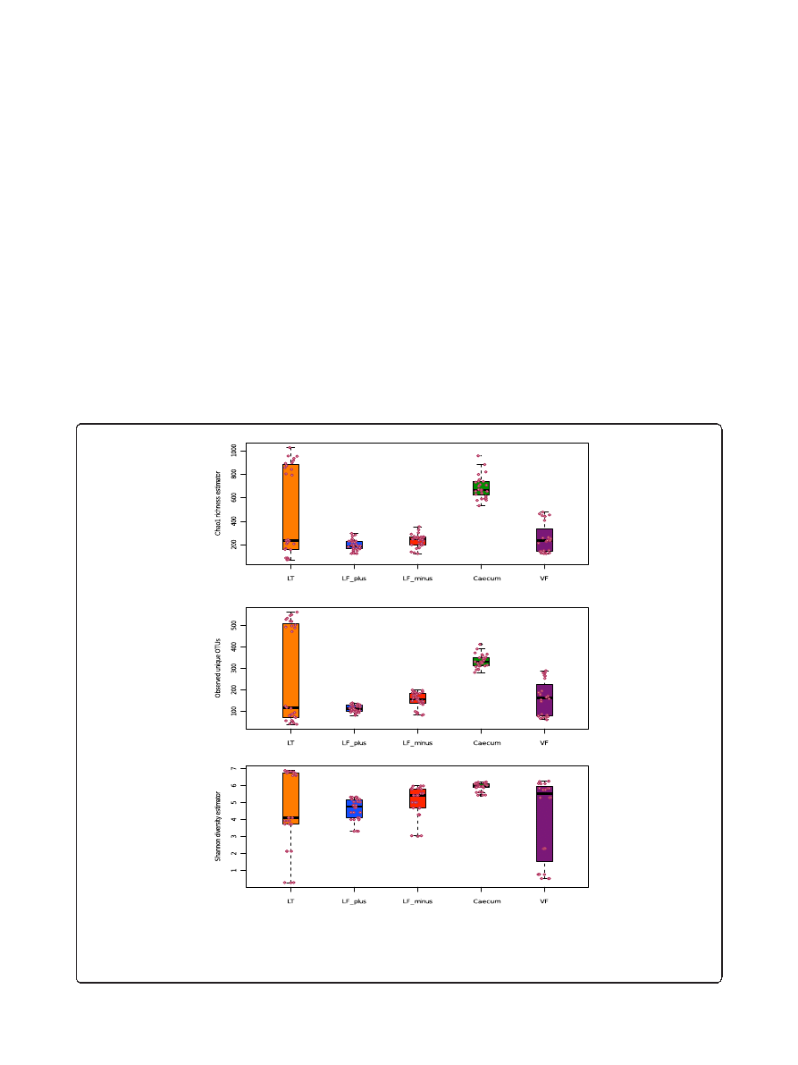
The bacterial community of the BAL-minus were in
50% of the cases (indicated by the median) richer than
the BAL-plus (Figure 2A). We found this difference to
be significant (W, p < 0.05).
There was no significant variation between the BAL-
minus and lung tissue samples. The mouse caecum
community is generally richer than all other tested
communities, except of the upper quartile of the tissue
samples. The vaginal microbiota appeared to be as rich as
the lung tissue community.
In more than half of the BAL-minus samples, more
unique OTUs were observed than in the lung tissue
material (Figure 2B). The BAL-plus samples contained
significantly less OTUs than the BAL-minus samples
A
B
C
Figure 2 Alpha diversity plots. A: Chao1 richness estimator between sample types and individual samples (circles), LF-plus is bronchoalveolar
lavage (BAL) fluids and LF-minus is BAL where the mouse cells have been removed. LT is lung tissue and VF is vaginal flushing, B: Observed unique
OTUs and C: Shannon diversity estimator between sample types (s above) and individual samples (circles). The sequences (3350) were randomly even
subsampled before calculating the alpha diversity. The boxplots show median, quartile, smallest and largest observations as well as outliers
(circles). Significant variation is indicated by * (KW, p < 0.05).
Barfod et al. BMC Microbiology 2013, 13:303
Page 6 of 12
http://www.biomedcentral.com/1471-2180/13/303

(W, p < 0.001). The variation of Chao1 and observed
OTUs comparing all pulmonic samples were significant
(KW, p < 0.01)
We observed the highest number of unique OTUs in
the caecum samples, compared to vaginal and lung tis-
sue microbiota (W, p > 0.05).
A slightly different picture was observed for the diver-
sity index (Figure 2C). In most cases the alpha diversity
of BAL-minus samples appeared to be larger than the
BAL-plus and lung tissue samples. However, the vari-
ation of diversity between all pulmonic samples was not
significant (KW, p > 0.05). The Shannon index varied
significantly when comparing both BAL-plus and BAL-
minus communities only (W, p < 0.05) and reflect the
observation of Chao1 and unique OTU sequences.
In summary, the mouse cell-free BAL samples yielded a
richer microbial community, had a larger alpha-diversity
and contained more unique OTU in comparison to the
samples with mouse cells. In addition, at least 50% of the
alpha-diversity observations the BAL-minus show larger
diversity indexes than the lung tissue samples. The upper
quartile of the lung tissue samples varied largely for all
three diversity indicators approaching larger diversity
(Shannon), richness (Chao1) and observed OTUs as found
for the caecum samples. This could be the result of non-
proper flushing or contamination during the experimen-
tal process. However, the low diversity, richness and fewer
OTUs in the lung tissue samples correspond to higher
diversity, richness and more OTUs in the matching BAL
samples. There is also a large overlap in beta-diversity
based on OTU abundance of lung tissue samples with
the BAL samples, suggesting that, a biased flushing is
more likely to be the reason, than contamination.
Bacteria found via traditional culturing of BAL
To establish any possibly cultivable part of the lung
microbiota and possible viable contaminations, we per-
formed a conventional cultivation study of BAL fluids
from 10 additional mice. Of the 40 different agar plates
under various conditions with 200
μL BAL per plate from
each of the 10 mice, we only found a few bacterial colonies
on 5 plates originating from only 4 different mice. These
bacteria colonies were all identified to be Micrococcus
luteus
with 99% probability by the Vitek2 system (Bio
Mérieux, France).
Discussion
Methodology
In this work we have sequenced the lung bacterial 16S
rRNA gene variable region V3/V4 with different methods
and compared the results to gut and vaginal bacterial
microbiome. We chose the V3/V4 region since Claesson
et. Al [21] reported that it taxonomically characterizes
microbial communities best without sequencing the entire
16S rRNA gene. Furthermore the same approach has
been applied in multiple studies to study bacterial inter-
action with lakes, plants, humans and most important
with mice [22-25].
In contrast to the general assumption, our results sug-
gest that the lower airways in mice are not sterile and
have a distinct bacterial microbiome that could probably
influence airway diseases. A classic obstacle in the in-
vestigation of the microbiota of the lungs is the likelihood
of contamination with bacteria from the upper respiratory
tract (URT). This is especially true for the study of the
human respiratory microbiome, because the procedure
used has a high risk of contamination with oral micro-
biota [7]. In our study, this is bypassed by the invasive
entry via the throat into trachea. We have extracted bac-
terial DNA from lung tissue, BAL with and without
mouse cells and vaginal flushings. Our results show that
it is possible to consistently obtain comparable sequences
from the BAL fluid to use for community studies related
the development of inflammatory disease in our mouse
model.
The use of BAL as the sample for investigations has
several advantages. The BAL sampling resembles the pro-
cedures used in humans, except that the work in animals
bypasses both URT and oral microorganisms and samples
the entire lung instead of just a local lung compartment.
The microbial community has been shown to vary
with the site of sampling in excised lung from a COPD
lung transplant [26]. The removal of mouse cells in the
BAL has the added advantage that we can use the lung
cells and determine the lung inflammatory status of the
mouse by staining and differentiating the immune cells
[27]. A limitation of our study is the lack of comparison
of our sequences with that of the upper respiratory
flora. This could possibly be obtained by performing 16S
rDNA sequencing on a matching nasal lavage sample for
each mouse. This should be done in the future.
Our lung tissue samples showed some clustering that
could indicate a sampling problem. In our study we
sampled the distal tip of the left lung lobe after the
BAL procedure was performed. The clustering could
be a result of this BAL procedure not being equally
effective between samples in the very low airways, some-
times leaving the distal tip un-flushed. This would predict
a clustering showing one community equal to the one
found in the BAL and one more rich and diverse repre-
senting the less rinsed tissue. If we were especially inter-
ested in the tissue associated microbiota, BAL should not
be performed before sampling and mouse cells should
not be removed from the BAL fluid before extraction.
Our results show that there are fewer OTUs in the
BAL-plus samples with mouse cells and that the lung
tissues samples have a large variation. This suggests that
the removal of tissues and host cells is a viable approach,
Barfod et al. BMC Microbiology 2013, 13:303
Page 7 of 12
http://www.biomedcentral.com/1471-2180/13/303

when extracting DNA for the examination of the lung
microbiome.
Another challenge when working with low bacterial
loads is the risk of contamination from the environment
or sampling procedures. Some contamination must be
expected and taken into account when interpreting data.
We believe that we have taken large precautions to
insure sterility during procedures and we have used ex-
cess controls to check that our sampling procedure or
experimental chemicals did not produce any sequences
on their own in the PCRs. Culturing of the BAL used for
DNA extraction did not yield many bacteria either. Fur-
thermore, our sequences were very consistent between
mice. This would suggest that any contamination was
either negligible or at least distributed evenly between
mice.
We did find large variation within the vaginal samples
resulting in subclustering into groups we designated S1
and S2 (Figure 1C and D). S1 (vaginal samples 2, 5 and 8)
was found to be much more distantly related to caecum
and lung communities than the S2 group, which more
closely resembled the lung microbiota. We believe this
could be the result of a possible infection in the S1 vagi-
nas, as these 3 samples contained 56-97% Streptococcus.
In the present study, we did not monitor the stage of the
estrous cycle at the time of sampling, which has been
shown to change the bacterial profile of the vagina in
animals and humans [28,29]. Mice have a daily fluctu-
ation in estrous cycle, which in part could explain the
subclustering of the vaginal microbiota. This should be
taken into account in futures studies.
There was a concern that the vaginal sampling pro-
cedure would yield a large overlap in OTUs with the gut
microbiota due to cross contamination during sampling.
This has been shown not to be the case, as we show here
there is a very little overlap between caecum and vaginal
microbiota. To our best knowledge this is the first time
that the BALB/c mouse vaginal bacterial community has
been investigated with 454 Pyrosequencing for a full com-
munity study. This promises to be useful in futures studies
of the
“inheritance” of bacterial microbiome from mother
to pup or vaginal microbiome related diseases such as
vaginosis [28,30].
We faced two main obstacles: The low DNA concen-
tration in all samples, except for the caecal material and
unspecific primer binding in the tissue samples.
To overcome the low DNA concentration we increased
the PCR cycle number. The large cycle number essentially
could amplify any kind of contamination or primer bias
such as chimeras, but we adjusted the rounds of cycles
to the crucial experimental negative controls as de-
scribed in the material and methods. Our results are
confirmed by the observed community profile of previ-
ous human lung observations (discussed in detail below)
and the low abundance of chimera (<3% of quality trimmed
sequences) [31,32].
The second obstacle was the non-specific binding of
the primers in the lung tissue sample caused by the low
amounts of bacterial DNA and large amounts of eukaryotic
nucleic acids. Since the risk of contamination barely left
space for adjustments, we chose to do a nested PCR and
amplified a
∼ 1500 bp long fragment of the 16S rRNA gene
prior amplifying the hyper variable region V3/V4. Although
both primer sets are universal and theoretically target
all bacteria and archaea, the tendency to favor certain
taxonomic groups cannot be excluded, thus one primer
set should be preferred to compare the different samples.
Therefor we were expecting a significantly different
clustering in beta-diversity of the lung tissue community
in comparison to the BAL fluids. However the differ-
ences were small supporting our methodology.
The lung has a distinct bacterial community
It is not known from where we obtain our putative bac-
terial lung microbiota however it is most likely to be in a
flux state with the environment. There is support for
this notion in the hygiene hypothesis of the development
of asthma and allergies [33]. We hypothesize that mice
obtain the bacteria from their local environment and
littermates influenced by handling by human, feed and
water. But it is also a possibility that the core lung
microbiome is established in utero, during and after
birth in the very early life, as it is suggested with gut
microbiota from human and animal studies [34-36].
The lung microbiota found via the culture independent
methods described in our study of BALB/c mice, have
been shown not to be just a subpopulation of the bacteria
found in caecum samples and did not vary significantly
between mice suggesting that a distinct lung microbiome
exists. An example of this would be the sequence for Pelo-
monas
4818 (OTU ID), which was found in all our lung
samples but not in any caecum samples. We did find 6
major genera that varied significantly between our different
sampling methods for the lung bacterial community (KW,
p < 0.05) (Additional file 5: Figure S3). Acinetobacter, Pelo-
monas
were most abundant in the BAL-plus, where both
Acinetobacter
and Pelomonas have been associated with
the human lung microbiota [4]. Arcobacter mostly found
in BAL-minus has likewise been found to also be corre-
lated with protection from skin allergy and protection
from OVA allergy in mouse models [37-39] and found in
human lungs [40]. Polaromona, Schlegella and Brochothrix
have not previously been found in BAL fluids from humans
or mice and are considered environmental bacteria. We
have found Prevotella and Veillonella spp. only in the lung
and vaginal samples. These species have been suggested to
be a distinct part of lung microbiome and mucus epithelia
Barfod et al. BMC Microbiology 2013, 13:303
Page 8 of 12
http://www.biomedcentral.com/1471-2180/13/303

in humans and the absence of Bacteroides associated
with asthma [3,41].
We have also compared the genera variation of vagi-
nal cluster S1 and S2 against all lung samples. S1 varied
significantly in 4 taxa (Figure 1C and D) Genera ob-
served <50 sequences sum counts were not considered.
This cut off value was chosen as an additional denoising
criterion necessary for sequences with high PCR cycle
number. Staphylococcus was more abundant in the pul-
monic samples (KW, p < 0.05) than in S1. Also, Anaero-
coccus
and Massilia were not observed in the S1
samples. The large abundance of Streptococcus in S1
(KW, p < 0.05) varied clearly from the lung samples.
The vaginal cluster S2 with high similarity in beta diver-
sity towards the lung samples varied in 32 genera, but
all taxa added up to less than our chosen detection
minimum of 50 sequences.
List of bacteria with possible influence on lung immunity
We wanted to identify the microbiota that possibly could
influence lung immunity in our animal model. We cre-
ated a list of interesting bacteria (prior to sequencing) at
the genus, family or species level, based on other previous
studies of both, human lung and animal models of disease.
This list is found in Additional file 2: Table S2 and
Additional file 6: Table S3. From our results we found
bacteria associated with asthma and COPD in the mouse
lung microbiome such as Lachnospiraceae and Akkerman-
sia muciniphilia
[42] and Shewanella, Comamodacea[43],
Haemophilus, Streptococcous, Fusobacteria
[3]. No indica-
tions were observed for Bartonellaceae, Globicatella,
Ralstoniacea
nor Nitrosomonadaceae from our premade
list. No OTU sequence blasted could be assigned to
Clostridium difficile
, Pseudomonas aeruginosa, Lactoba-
cillus
OTU 1865, Bacteriodales OTU 991 or Micrococcus
luteus
from our list either. Sequences from the genera
Micrococcus
we isolated did not contain enough taxo-
nomic information to differentiate between Micrococcus
luteus
found on our agar plates and other Micrococcus
species.
The putative Akkermansia muciniphilia was found in
lung and in one caecum sample and is especially inter-
esting as it is a mucin degrading bacterium and has been
shown to influence gut mucus layer thickness [44]. Recently,
it was reported that Akkermansia muciniphilia is present
in BALB/c caecum but not in fecal samples. The overall
BALB/c caecal microbiome found in our study is also con-
firmed with the dominant phyla being Firmicutes (69.99%)
and Bacteroidetes (22.07%) [45]. The presence of Akker-
mansia muciniphilia
in the lung mucus layer could be of
importance in asthma characterized by thickening of
the epithelium and increased mucus production [46].
Most of the lung-associated bacteria that we identified
in Additional file 2: Table S2 could only be found in the
mouse lung and vagina samples but not in the caecum.
Bifidobacterium animalis subsp. lactis
, and Lactobacil-
lus acidophilus NCFM
were added to the list of interest-
ing species because of their use as probiotic bacteria in
various mouse models and humans, and it would be
interesting to know whether or not these bacteria are
present in an unchallenged model. We found OTUs match-
ing Bifidobacterium animalis subsp. lactis, Bifidobacterium
longum subsp. longum
and Lactobacillus reutieri the latter
two not being on our list, in lung samples, but not in any
caecum samples. Bifidobacterium longum subsp. longum
have been found in human (meconium) and is regarded
as one of the first colonisers of the gut originating from
the mother [36]. Several strains of Lactobacillus have
been shown to modulate allergic pulmonary inflamma-
tion, whereas Lactobacillus reuteri has been shown to
reduce inflammation in BALB/c mice [47,48].
Impact on animal models of inflammatory lung disease
The influence of gut microbiota on lung immunity has
been vastly explored and several studies have linked changes
in the gut microbiome with changes in lung immunity
in mice [42,49-51]. As it is becoming clear that the micro-
biome of the animal used in a particular model influences
that animal
’s immune status and ultimately affects the
outcome of experiments, it is important to take precau-
tions in the model design. Things known to influence
gut microbiome composition in laboratory mice include
probiotics, antibiotics, stress, handling, vendor/site of breed-
ing and animal lineages [52-55] and it is possible that these
factors will affect the lung microbiota as well. Most studies
done on gut microbiota and lung immunity do not take lung
residing bacteria into account when the data are interpreted.
It is possible that the local lung effects seen could be the
results of changes in the lung as well as in the gut. In
our studies we always use age matched female mice from
the same site of breeding (lot number) and distribution
of the mice equally between groups as to avoid any lit-
termate bias. It should possibly also be noted whether
or not the mice have mothers that are sisters or not
as this also effects gut and possibly lung microbiome
in the offspring [52]. As an added layer of complexity
we should remember that the total mouse microbiota
do not only consists of bacteria but also fungi and vi-
ruses. In particular bacteriophages could influence gut
or lung microbiology and indirectly have adverse effects
on health [56].
Future studies into the lung microbiota of mice should
include a comparison between nasal lavages and BAL to
distinguish between upper and lower respiratory tract
microbiota and possibly longitudinal studies with culture
independent techniques.
Barfod et al. BMC Microbiology 2013, 13:303
Page 9 of 12
http://www.biomedcentral.com/1471-2180/13/303

Conclusions
BALB/cj mice were shown to have a lung microbiome
that was distinct from their caecal but overlapping with
their vaginal bacterial community. We have consistently
amplified bacterial DNA from mouse BAL fluid and have
shown that host DNA present in the DNA extraction step
influences the community profiles obtained and that this
needs to be taken into account when choosing methods,
performing the analyses and prior to biological interpret-
ation. Mouse models provide the means to obtain mech-
anistic insights into the lung microbiome. We believe that
the lung microbiota should be considered when working
with these mouse models of human disease and further
research is needed to reveal the contribution of the lung
microbiota to the pathogenesis of diseases such as respira-
tory disease common in infants (i.e. RSV), cystic fibrosis,
COPD and asthma.
Availability of supporting data
All supporting data are included as additional files and
all sequences used in this study are available in the NCBI
Sequence Read Archive under study accession number
SRP033710 (http://www.ncbi.nlm.nih.gov/sra).
Additional files
Additional file 1: Figure S5. Rarefaction curves. (A) Observed species
–
raw data. (B) Observed species after random even subsampling. The data
shown in (A) accounted for all sequences generated. The graphs evened
out after approx. 2000 sequences observed and revealed that the
random even subsampled OTU table (B) at a sequencing depth of 3530
will be efficient to include also the rare OTUs. The subsampled OTU table
(B) was used for the statistical analysis of this study and is the basis of
the Figures 1 and 2.
Additional file 2: Table S2. List of interesting taxa. This list shows the
distribution of lung associated taxa between sampling methods and sites.
Most of the lung-associated bacteria could only be found in the lung
and vagina samples but not in the caecum. LF-plus is bronchoalveolar
lavage (BAL) fluids and LF-minus is BAL where the mouse cells have been
removed. LT is lung tissue,VF is vaginal flushing and caecum from the
gut microbiota.
Additional file 3: Table S4. Distribution of genera between samples.
This table shows the distribution of genera between sample types. The
observations are based on the summarized subsampled OTU table (3318
OTUs) after singletons and doubletons were removed. We discriminated
between shared and unique genera of lung, vaginal and caecal environment.
Additional file 4: Figure S4. Additional PCoA 2 and 3. The axis of
PCoA plot 2 and 3 explain the 6.28%/24% and 10.42%/6.28% of the
variances respectively. Both plots show the large overlap of
bronchoalveolar lavage (BAL) fluids BAL-plus with mouse cells in BLUE,
BAL-minus (without mouse cells) in RED and lung tissue in ORANGE and
support plot 1. Only in plot 3 the caecal GREEN community overlaps with
the lung and vaginal community confirming its large distance from the
other sample sites.
Additional file 5: Figure S3. Variation in lung genus composition.
The genera shown counted up to at least 50 or more sequences in
relative abundance and vary significantly among the lung communities
(KW, p <0.05). LF-plus is bronchoalveolar lavage (BAL) fluids and LF-minus is
BAL where the mouse cells have been removed. LT is lung tissue, VF is
vaginal flushing and caecum represents gut microbiota.
Additional file 6: Table S3. Blast search
– putative species identity. For
further identification the representative sequence of each OTU of the
Qiime pipeline output were picked and blasted. OTUs were only
considered when the highest score, maximum identity and 100% query
cover uniquely matched one species. Additional subspecies information
corresponds to the best hit. It is also noted from how many different
animals and from which sampling site the OTUs were found. LF-plus is
bronchoalveolar lavage (BAL) fluids and LF-minus is BAL where the
mouse cells have been removed. LT is lung tissue, VF is vaginal flushing
and caecum from the gut microbiota.
Competing interests
The authors declare that they have no competing interests.
Authors
’ contributions
KKB conceived and designed the study, carried out the animal work and
DNA extractions, and drafted the manuscript. MR did the 16S data
generation, analysis and participated in the design of the study and
manuscript. SSC performed the cultivation and bacterial identification. KAK,
LHH, STL and SJS participated in design and coordination and helped to
draft the manuscript. All authors read and approved the final manuscript.
Acknowledgements
The Danish National Advanced Technology Foundation, Lundbæk
Foundation, Lars Andrup, Michael Guldbrandsen, Sofia Forssten, Al-Soud
Waleed, Shannon Russell and Karin Vestberg.
Author details
1
Statens Serum Institut, Artillerivej 5, 2300 Copenhagen S, Denmark.
2
National Research Centre for the Working Environment, Lersø Parkallé 105,
2100 Copenhagen O, Denmark.
3
Department of Biology, Microbiology,
University of Copenhagen, Universitetsparken 15, 2100 Copenhagen O,
Denmark.
Received: 18 July 2013 Accepted: 23 December 2013
Published: 28 December 2013
References
1.
Beck JM, Young VB, Huffnagle GB: The microbiome of the lung. Transl Res
2012, 160:258
–266.
2.
Huang YJ, Nelson CE, Brodie EL, Desantis TZ, Baek MS, Liu J, Woyke T,
Allgaier M, Bristow J, Wiener-Kronish JP, et al: Airway microbiota and
bronchial hyperresponsiveness in patients with suboptimally controlled
asthma. J Allergy Clin Immunol 2011, 127:372
–381.
3.
Hilty M, Burke C, Pedro H, Cardenas P, Bush A, Bossley C, Davies J, Ervine A,
Poulter L, Pachter L, et al: Disordered microbial communities in asthmatic
airways. PLoS One 2010, 5:e8578.
4.
Borewicz K, Pragman AA, Kim HB, Hertz M, Wendt C, Isaacson RE:
Longitudinal analysis of the lung microbiome in lung transplantation.
FEMS Microbiol Lett 2013, 339:57
–65.
5.
Turnbaugh PJ, Ley RE, Hamady M, Fraser-Liggett CM, Knight R, Gordon JI:
The human microbiome project. Nature 2007, 449:804
–810.
6.
Lozupone C, Cota-Gomez A, Palmer BE, Linderman DJ, Charlson ES,
Sodergren E, Mitreva M, Abubucker S, Martin J, Yao G, et al: Widespread
Colonization of the Lung by Tropheryma whipplei in HIV Infection. Am J
Respir Crit Care Med 2013, 187(10):1110
–1117.
7.
Morris A, Beck JM, Schloss PD, Campbell TB, Crothers K, Curtis JL, Flores SC,
Fontenot AP, Ghedin E, Huang L, et al: Comparison of the Respiratory
Microbiome in Healthy Non-Smokers and Smokers. Am J Respir Crit Care
Med 2013, 187(10):1067
–1075.
8.
Huang YJ, Charlson ES, Collman RG, Colombini-Hatch S, Martinez FD,
Senior RM: The Role of the Lung Microbiome in Health and Disease:
A National Heart, Lung and Blood Institute Workshop Report. Am J
Respir Crit Care Med 2013, 187(12):1382
–1387.
9.
Larsen ST, Hansen JS, Hansen EW, Clausen PA, Nielsen GD: Airway
inflammation and adjuvant effect after repeated airborne exposures to
di-(2-ethylhexyl)phthalate and ovalbumin in BALB/c mice. Toxicology 2007,
235:119
–129.
10.
Prince AM, Andrus L: PCR: how to kill unwanted DNA. Biotechniques 1992,
12:358
–360.
Barfod et al. BMC Microbiology 2013, 13:303
Page 10 of 12
http://www.biomedcentral.com/1471-2180/13/303

11.
Stokholm J, Schjorring S, Pedersen L, Bischoff AL, Folsgaard N, Carson CG,
Chawes B, Bonnelykke K, Molgaard A, Krogfelt KA, et al: Living with cat and
dog increases vaginal colonization with E. coli in pregnant women.
PLoS One 2012, 7:e46226.
12.
Smith B, Li N, Andersen AS, Slotved HC, Krogfelt KA: Optimising bacterial
DNA extraction from faecal samples: comparison of three methods.
Open Microbiol J 2011, 5:14
–17.
13.
Field KG, Gordon D, Wright T, Rappe M, Urback E, Vergin K, Giovannoni SJ:
Diversity and depth-specific distribution of SAR11 cluster rRNA genes
from marine planktonic bacteria. Appl Environ Microbiol 1997, 63:63
–70.
14.
Yu Y, Lee C, Kim J, Hwang S: Group-specific primer and probe sets to
detect methanogenic communities using quantitative real-time polymerase
chain reaction. Biotechnol Bioeng 2005, 89:670
–679.
15.
Neefs JM, Van de PY, De RP, Goris A, De WR: Compilation of small ribosomal
subunit RNA sequences. Nucleic Acids Res 1991, 19(Suppl):1987
–2015.
16.
Roche: Amplicon Fusion Primer Design Guidelines. Tech Bull Genome SEQ
FLX Syst 2009, 1:8.
17.
Caporaso JG, Kuczynski J, Stombaugh J, Bittinger K, Bushman FD, Costello EK,
Fierer N, Pena AG, Goodrich JK, Gordon JI, et al: QIIME allows analysis of
high-throughput community sequencing data. Nat Methods 2010,
7:335
–336.
18.
Edgar RC, Haas BJ, Clemente JC, Quince C, Knight R: UCHIME improves
sensitivity and speed of chimera detection. Bioinformatics 2011,
27:2194
–2200.
19.
Liu Z, Desantis TZ, Andersen GL, Knight R: Accurate taxonomy
assignments from 16S rRNA sequences produced by highly parallel
pyrosequencers. Nucleic Acids Res 2008, 36:e120.
20.
Development Core Team: R: A language and environment for statistical
computing. Vienna, Austria: R Foundation for Statistical Computing; 2005.
ISBN 3-900051-07-0 (2005) by R. 2005.
21.
Claesson MJ, Wang Q, O
’Sullivan O, Greene-Diniz R, Cole JR, Ross RP, O’Toole PW:
Comparison of two next-generation sequencing technologies for resolving
highly complex microbiota composition using tandem variable 16S rRNA
gene regions. Nucleic Acids Res 2010, 38:e200.
22.
Logares R, Lindstrom ES, Langenheder S, Logue JB, Paterson H,
Laybourn-Parry J, Rengefors K, Tranvik L, Bertilsson S: Biogeography of bacterial
communities exposed to progressive long-term environmental change.
ISME J 2013, 7:937
–948.
23.
Peiffer JA, Spor A, Koren O, Jin Z, Tringe SG, Dangl JL, Buckler ES, Ley RE:
Diversity and heritability of the maize rhizosphere microbiome under
field conditions. Proc Natl Acad Sci U S A 2013, 110:6548
–6553.
24.
Dong Q, Brulc JM, Iovieno A, Bates B, Garoutte A, Miller D, Revanna KV, Gao X,
Antonopoulos DA, Slepak VZ, et al: Diversity of bacteria at healthy human
conjunctiva. Invest Ophthalmol Vis Sci 2011, 52:5408
–5413.
25.
Devkota S, Wang Y, Musch MW, Leone V, Fehlner-Peach H, Nadimpalli A,
Antonopoulos DA, Jabri B, Chang EB: Dietary-fat-induced taurocholic acid
promotes pathobiont expansion and colitis in Il10
−/− mice. Nature 2012,
487:104
–108.
26.
Shehata AA, Schrodl W, Aldin AA, Hafez HM, Kruger M: The Effect of
Glyphosate on Potential Pathogens and Beneficial Members of Poultry
Microbiota In Vitro. Curr Microbiol 2012, 66(4):350
–358.
27.
Larsen ST, Matsubara S, McConville G, Poulsen SS, Gelfand EW: Ozone
increases airway hyperreactivity and mucus hyperproduction in mice
previously exposed to allergen. J Toxicol Environ Health A 2010, 73:738
–747.
28.
Srinivasan S, Liu C, Mitchell CM, Fiedler TL, Thomas KK, Agnew KJ, Marrazzo JM,
Fredricks DN: Temporal variability of human vaginal bacteria and
relationship with bacterial vaginosis. PLoS One 2010, 5:e10197.
29.
Koiter TR, Hazenberg MP, Van der SP: Regulation of the bacterial
microflora of the vagina in cyclic female rats. J Exp Zool 1977, 202:121
–128.
30.
Ling Z, Kong J, Liu F, Zhu H, Chen X, Wang Y, Li L, Nelson KE, Xia Y, Xiang C:
Molecular analysis of the diversity of vaginal microbiota associated with
bacterial vaginosis. BMC Genomics 2010, 11:488.
31.
Qiu X, Wu L, Huang H, McDonel PE, Palumbo AV, Tiedje JM, Zhou J:
Evaluation of PCR-generated chimeras, mutations, and heteroduplexes
with 16S rRNA gene-based cloning. Appl Environ Microbiol 2001, 67:880
–887.
32.
Haas BJ, Gevers D, Earl AM, Feldgarden M, Ward DV, Giannoukos G, Ciulla D,
Tabbaa D, Highlander SK, Sodergren E, et al: Chimeric 16S rRNA sequence
formation and detection in Sanger and 454-pyrosequenced PCR
amplicons. Genome Res 2011, 21:494
–504.
33.
Strachan DP: Hay fever, hygiene, and household size. BMJ 1989,
299:1259
–1260.
34.
Penders J, Stobberingh EE, van den Brandt PA, Thijs C: The role of the
intestinal microbiota in the development of atopic disorders. Allergy 2007,
62:1223
–1236.
35.
Hansen CH, Nielsen DS, Kverka M, Zakostelska Z, Klimesova K, Hudcovic T,
Tlaskalova-Hogenova H, Hansen AK: Patterns of early gut colonization
shape future immune responses of the host. PLoS One 2012, 7:e34043.
36.
Makino H, Kushiro A, Ishikawa E, Muylaert D, Kubota H, Sakai T, Oishi K,
Martin R, Ben AK, Oozeer R, et al: Transmission of intestinal
Bifidobacterium longum subsp. longum strains from mother to infant,
determined by multilocus sequencing typing and amplified fragment
length polymorphism. Appl Environ Microbiol 2011, 77:6788
–6793.
37.
Luyt CE, Brechot N, Combes A, Trouillet JL, Chastre J: Delivering antibiotics
to the lungs of patients with ventilator-associated pneumonia: an update.
Expert Rev Anti Infect Ther 2013, 11:511
–521.
38.
Hanski I, Von HL, Fyhrquist N, Koskinen K, Torppa K, Laatikainen T, Karisola P,
Auvinen P, Paulin L, Makela MJ, et al: Environmental biodiversity, human
microbiota, and allergy are interrelated. Proc Natl Acad Sci U S A 2012,
109:8334
–8339.
39.
Qiu H, Kuolee R, Harris G, Zhou H, Miller H, Patel GB, Chen W:
Acinetobacter baumannii infection inhibits airway eosinophilia and lung
pathology in a mouse model of allergic asthma. PLoS One 2011, 6:e22004.
40.
Bousbia S, Papazian L, Saux P, Forel JM, Auffray JP, Martin C, Raoult D, La SB:
Repertoire of intensive care unit pneumonia microbiota. PLoS One 2012,
7:e32486.
41.
Cardenas PA, Cooper PJ, Cox MJ, Chico M, Arias C, Moffatt MF, Cookson WO:
Upper airways microbiota in antibiotic-naive wheezing and healthy infants
from the tropics of rural Ecuador. PLoS One 2012, 7:e46803.
42.
Russell SL, Gold MJ, Hartmann M, Willing BP, Thorson L, Wlodarska M, Gill N,
Blanchet MR, Mohn WW, McNagny KM, et al: Early life antibiotic-driven
changes in microbiota enhance susceptibility to allergic asthma.
EMBO Rep 2012, 13(5):440
–447.
43.
Huang YJ, Nelson CE, Brodie EL, Desantis TZ, Baek MS, Liu J, Woyke T,
Allgaier M, Bristow J, Wiener-Kronish JP, et al: Airway microbiota and
bronchial hyperresponsiveness in patients with suboptimally controlled
asthma. J Allergy Clin Immunol 2010, 127(2):372
–381.
44.
Everard A, Belzer C, Geurts L, Ouwerkerk JP, Druart C, Bindels LB, Guiot Y,
Derrien M, Muccioli GG, Delzenne NM, et al: Cross-talk between
Akkermansia muciniphila and intestinal epithelium controls diet-induced
obesity. Proc Natl Acad Sci U S A 2013, 110(22):9066
–9071.
45.
Krych L, Hansen CH, Hansen AK, van den Berg FW, Nielsen DS:
Quantitatively Different, yet Qualitatively Alike: A Meta-Analysis of the
Mouse Core Gut Microbiome with a View towards the Human Gut
Microbiome. PLoS One 2013, 8:e62578.
46.
Lambrecht BN, Hammad H: The airway epithelium in asthma. Nat Med
2012, 18:684
–692.
47.
Forsythe P, Inman MD, Bienenstock J: Oral treatment with live
Lactobacillus reuteri inhibits the allergic airway response in mice. Am J
Respir Crit Care Med 2007, 175:561
–569.
48.
Karimi K, Inman MD, Bienenstock J, Forsythe P: Lactobacillus reuteri-induced
regulatory T cells protect against an allergic airway response in mice. Am J
Respir Crit Care Med 2009, 179:186
–193.
49.
Olszak T, An D, Zeissig S, Vera MP, Richter J, Franke A, Glickman JN, Siebert R,
Baron RM, Kasper DL, et al: Microbial exposure during early life has
persistent effects on natural killer T cell function. Science 2012, 336:489
–493.
50.
Noverr MC, Falkowski NR, McDonald RA, McKenzie AN, Huffnagle GB:
Development of allergic airway disease in mice following antibiotic
therapy and fungal microbiota increase: role of host genetics, antigen,
and interleukin-13. Infect Immun 2005, 73:30
–38.
51.
Russell SL, Gold MJ, Willing BP, Thorson L, McNagny KM, Finlay BB: Perinatal
antibiotic treatment affects murine microbiota, immune responses and
allergic asthma. Gut Microbes 2013, 71(1):30
–38.
52.
Hufeldt MR, Nielsen DS, Vogensen FK, Midtvedt T, Hansen AK: Variation in
the gut microbiota of laboratory mice is related to both genetic and
environmental factors. Comp Med 2010, 60:336
–347.
53.
Pang W, Stradiotto D, Krych L, Karlskov-Mortensen P, Vogensen FK, Nielsen DS,
Fredholm M, Hansen AK: Selective inbreeding does not increase gut
microbiota similarity in BALB/c mice. Lab Anim 2012, 46:335
–337.
54.
Hufeldt MR, Nielsen DS, Vogensen FK, Midtvedt T, Hansen AK: Family
relationship of female breeders reduce the systematic inter-individual
variation in the gut microbiota of inbred laboratory mice. Lab Anim 2010,
44:283
–289.
Barfod et al. BMC Microbiology 2013, 13:303
Page 11 of 12
http://www.biomedcentral.com/1471-2180/13/303

55.
Bangsgaard Bendtsen KM, Krych L, Sorensen DB, Pang W, Nielsen DS,
Josefsen K, Hansen LH, Sorensen SJ, Hansen AK: Gut microbiota
composition is correlated to grid floor induced stress and behavior in
the BALB/c mouse. PLoS One 2012, 7:e46231.
56.
Duerkop BA, Clements CV, Rollins D, Rodrigues JL, Hooper LV: A composite
bacteriophage alters colonization by an intestinal commensal bacterium.
Proc Natl Acad Sci U S A 2012, 109:17621
–17626.
doi:10.1186/1471-2180-13-303
Cite this article as: Barfod et al.: The murine lung microbiome in relation
to the intestinal and vaginal bacterial communities. BMC Microbiology
2013 13:303.
Submit your next manuscript to BioMed Central
and take full advantage of:
•
Convenient online submission
•
Thorough peer review
•
No space constraints or color figure charges
•
Immediate publication on acceptance
•
Inclusion in PubMed, CAS, Scopus and Google Scholar
•
Research which is freely available for redistribution
Submit your manuscript at
www.biomedcentral.com/submit
Barfod et al. BMC Microbiology 2013, 13:303
Page 12 of 12
http://www.biomedcentral.com/1471-2180/13/303
Document Outline
- Abstract
- Background
- Methods
- Results
- Over all sequence quality and results from all sample types
- The bacterial sequences of the lung samples
- Bacterial sequences of the caecum
- Overlapping genera
- Variability in community composition between samples obtained from the same sampling site (Beta_diversity)
- Richness and diversity of sample type (Alpha diversity)
- Bacteria found via traditional culturing of BAL
- Discussion
- Conclusions
- Additional files
- Competing interests
- Authors’ contributions
- Acknowledgements
- Author details
- References
Wyszukiwarka
Podobne podstrony:
Metabolic Activities of the Gut Microora in Relation to Cancer
73 Positioning in Relation to the Ball
Vergauwen David Toward a “Masonic musicology” Some theoretical issues on the study of Music in rela
The Gospel of St John in Relation to the Other Gospels esp that of St Luke A Course of Fourteen Lec
Ando Individual Subjective Preference Of Listeners To Vocal Music Sources In Relation To The Subseq
Judaism's Transformation to Modernization in Relation to Ame
Ando Dissimilarity Judgments In Relation To
Rykała, Andrzej Origin and geopolitical determinants of Protestantism in Poland in relation to its
Mobile Multimedia In Context To Atm Transport And Gsm Gprs Mobile Access Networks
Bacterial Resistance to Microbicides in the Healthcare Environment
Guide to the properties and uses of detergents in biology and biochemistry
A Guide to the Law and Courts in the Empire
79 1111 1124 The Performance of Spray Formed Tool Steels in Comparison to Conventional
The influence of British imperialism and racism on relationships to Indians
Guide to the properties and uses of detergents in biology and biochemistry
A Guide to the Law and Courts in the Empire
Spatial organization of intestinal microbiota in the mouse ascending colon
więcej podobnych podstron