
Neural Integration I:
Sensory Pathways and the
Somatic Nervous System
15
Did you know...?
The highest concentrations of touch receptors in
humans are in the fingertips, toes, lips, and
tongue.
Learning Outcomes
After completing this chapter, you should be able to do the following:
15-1
Specify the components of the afferent and efferent divisions
of the nervous system, and explain what is meant by the
somatic nervous system.
15-2
Explain why receptors respond to specific stimuli, and how the
organization of a receptor affects its sensitivity.
15-3
Identify the receptors for the general senses, and describe how
they function.
15-4
Identify the major sensory pathways, and explain how it is
possible to distinguish among sensations that originate in
different areas of the body.
15-5
Describe the components, processes, and functions of the
somatic motor pathways, and the levels of information
processing involved in motor control.
Clinical Notes
Assessment of Tactile Sensitivities p. 513
Cerebral Palsy p. 522
Amyotrophic Lateral Sclerosis p. 524
Anencephaly p. 525
Chapter 15
Neural Integration I: Sensory Pathways and the Somatic Nervous System
507
15
NER
V
OUS
Higher-Order Functions
Memory, learning, and
intelligence may
influence interpretation
of sensory information
and nature of motor
activities
Conscious and
subconscious
motor centers
in brain
Sensory
pathways
Skeletal
muscles
Somatic
Nervous
System (SNS)
General
sensory
receptors
Visceral effectors
(smooth muscles,
glands, cardiac
muscle, adipocytes,
etc.)
CHAPTER 16
CHAPTER 15
Autonomic
Nervous
System (ANS)
Motor
pathways
Sensory
processing
centers in
brain
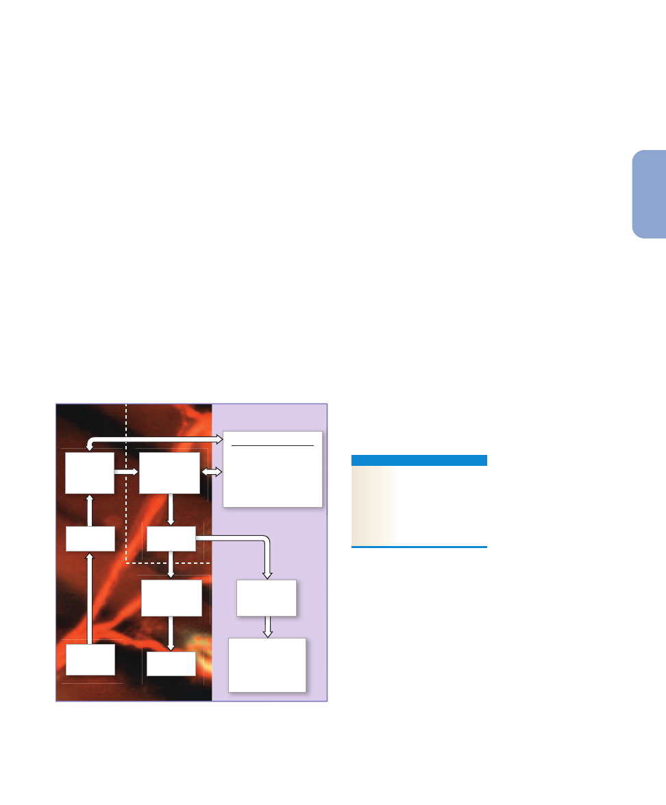
Figure 15–1
An Overview of Neural Integration. This figure
illustrates the relationships between Chapters 15 and 16 and
indicates the major topics considered in this chapter.
An Introduction to Sensory
Pathways and the Somatic
Nervous System
This chapter examines how the nervous system works as an
integrated unit. It considers sensory receptors, sensory pro-
cessing centers in the brain, and conscious and subconscious
motor functions. The left-hand portion of
Figure 15–1
pro-
vides an overview of the topics we will cover in this chapter.
Our discussion will focus on the “general senses” that provide
information about the body and its environment. The “special
senses”—smell, taste, sight, equilibrium (balance), and hear-
ing—will be considered in Chapter 17.
15-1
Sensory information from all
parts of the body is routed to the
somatosensory cortex
Specialized cells called sensory receptors monitor specific con-
ditions in the body or the external environment. When stim-
ulated, a receptor passes information to the CNS in the form
of action potentials along the axon of a sensory neuron. Such
axons are parts of sensory pathways—the nerves, nuclei, and
tracts that deliver somatic and visceral sensory information to
their final destinations inside the CNS. Taken together, the re-
ceptors, sensory neurons, and sensory pathways constitute
the afferent division of the nervous system.
l
p. 387
Somatic and visceral sensory information often travels
along the same pathway. Somatic sensory information is dis-
tributed to sensory processing centers in the brain—either the
primary sensory cortex of the cerebral hemispheres or appro-
priate areas of the cerebellar hemispheres. Visceral sensory
information is distributed primarily to reflex centers in the
brain stem and diencephalon.
In this chapter we consider the somatic motor portion of
the efferent division—the nuclei, motor tracts, and motor
neurons that control peripheral effectors. Somatic motor
commands—whether they arise at the conscious or subcon-
scious levels—travel from motor centers in the brain along
somatic motor pathways, which consist of motor nuclei, tracts,
and nerves. The motor neurons and pathways that control
skeletal muscles form the somatic nervous system (SNS).
Chapter 16 begins with a discussion of the visceral motor
portion of the efferent division. All visceral motor commands
are carried into the PNS by the autonomic nervous system
(ANS). Both somatic and visceral motor commands may be
issued in response to arriving sensory information, but these
commands may be modified on the basis of planning, memo-
ries, and learning—the so-called higher-order functions of the
brain that we will consider at the close of Chapter 16.
C H E C K P O I N T
1. What do we call the body’s specialized cells that
monitor specific internal or external conditions?
2. Is it possible for somatic motor commands to arise at
the subconscious level?
See the blue Answers tab at the end of the book.
15-2
Sensory receptors connect our
internal and external environments
with the nervous system
Sensory receptors are specialized cells or cell processes that
provide your central nervous system with information about
conditions inside or outside the body. The term general
senses is used to describe our sensitivity to temperature,
pain, touch, pressure, vibration, and proprioception. General
sensory receptors are distributed throughout the body, and
they are relatively simple in structure. Some of the informa-
tion they send to the CNS reaches the primary sensory cortex
and our awareness. As noted in Chapter 12, sensory informa-
tion is interpreted on the basis of the frequency of arriving ac-
tion potentials.
l
p. 424
For example, when pressure
sensations are arriving, the harder the pressure, the higher the
frequency of action potentials. The arriving information is
called a sensation. The conscious awareness of a sensation is
called a perception.
The special senses are olfaction (smell), vision (sight),
gustation (taste), equilibrium (balance), and hearing. These
sensations are provided by receptors that are structurally
more complex than those of the general senses. Special sen-
sory receptors are located in sense organs such as the eye or
ear, where the receptors are protected by surrounding tissues.
The information these receptors provide is distributed to spe-
cific areas of the cerebral cortex (the auditory cortex, the vis-
ual cortex, and so forth) and to centers throughout the brain
stem. We will consider the special senses in Chapter 17.
Sensory receptors represent the interface between the
nervous system and the internal and external environments.
A sensory receptor detects an arriving stimulus and translates
it into an action potential that can be conducted to the CNS.
This translation process is called transduction. If transduction
does not occur, then as far as you are concerned, the stimulus
doesn’t exist. For example, bees can see ultraviolet light you
can’t see, and dogs can respond to sounds you can’t hear. In
each case the stimuli are there—but your receptors cannot
detect them.
In the rest of this section we examine the basic concepts
of receptor function and sensory processing. We begin by
considering how receptors detect stimuli.
The Detection of Stimuli
Each receptor has a characteristic sensitivity. For example, a
touch receptor is very sensitive to pressure but relatively in-
sensitive to chemical stimuli, whereas a taste receptor is sen-
sitive to dissolved chemicals but insensitive to pressure. This
feature is called receptor specificity.
Specificity may result from the structure of the receptor
cell, or from the presence of accessory cells or structures that
shield the receptor cell from other stimuli. The simplest re-
ceptors are the dendrites of sensory neurons. The branching
tips of these dendrites, called free nerve endings, are not pro-
tected by accessory structures. Free nerve endings extend
through a tissue the way grass roots extend into the soil. They
can be stimulated by many different stimuli and therefore ex-
hibit little receptor specificity. For example, free nerve end-
ings that respond to tissue damage by providing pain
sensations may be stimulated by chemical stimulation, pres-
sure, temperature changes, or trauma. Complex receptors,
such as the eye’s visual receptors, are protected by accessory
cells and connective tissue layers. These cells are seldom ex-
posed to any stimulus other than light and so provide very
specific information.
The area monitored by a single receptor cell is its receptive
field (
Figure 15–2
). Whenever a sufficiently strong stimulus
arrives in the receptive field, the CNS receives the informa-
tion “stimulus arriving at receptor X.” The larger the recep-
tive field, the poorer your ability to localize a stimulus. A
touch receptor on the general body surface, for example, may
have a receptive field 7 cm (2.5 in.) in diameter. As a result,
you can describe a light touch there as affecting only a gen-
eral area, not an exact spot. On the tongue or fingertips,
where the receptive fields are less than a millimeter in diam-
eter, you can be very precise about the location of a stimulus.
An arriving stimulus can take many forms. It may be a
physical force (such as pressure), a dissolved chemical, a
sound, or light. Regardless of the nature of the stimulus, how-
ever, sensory information must be sent to the CNS in the form
of action potentials, which are electrical events.
As noted earlier, transduction is the translation of an ar-
riving stimulus into an action potential by a sensory receptor.
Transduction begins when a stimulus changes the transmem-
brane potential of the receptor cell. This change, called a
receptor potential, is either a graded depolarization or a graded
hyperpolarization. The stronger the stimulus, the larger the
receptor potential.
The typical receptors for the general senses are the den-
drites of sensory neurons, and the sensory neuron is the re-
ceptor cell. Any receptor potential that depolarizes the
plasma membrane will bring the membrane closer to thresh-
old. A depolarizing receptor potential in a neural receptor is
called a generator potential.
Sensations of taste, hearing, equilibrium, and vision are
provided by specialized receptor cells that communicate with
508
Unit 3
Control and Regulation
Receptive
field 1
Receptive
field 2
Figure 15–2
Receptors and Receptive Fields. Each receptor cell
monitors a specific area known as the receptive field.

Chapter 15
Neural Integration I: Sensory Pathways and the Somatic Nervous System
509
15
NER
V
OUS
sensory neurons across chemical synapses. The receptor cells
develop graded receptor potentials in response to stimula-
tion, and the change in membrane potential alters the rate of
neurotransmitter release at the synapse. The result is a depo-
larization or hyperpolarization of the sensory neuron. If suf-
ficient depolarization occurs, an action potential appears in
the sensory neuron. In this case, the receptor potential and
the generator potential occur in different cells: The receptor
potential develops in the receptor cell, and the generator po-
tential appears later, in the sensory neuron.
Whenever a sufficiently large generator potential appears,
action potentials develop in the axon of a sensory neuron. For
reasons discussed in Chapter 12, the greater the degree of sus-
tained depolarization at the axon hillock, the higher the fre-
quency of action potentials in the afferent fiber.
l
p. 424
The arriving information is then processed and interpreted by
the CNS at the conscious and subconscious levels.
The Interpretation of Sensory
Information
Sensory information that arrives at the CNS is routed accord-
ing to the location and nature of the stimulus. Previous chap-
ters emphasized the fact that axons in the CNS are organized
in bundles with specific origins and destinations. Along sen-
sory pathways, a series of neurons relays information from
one point (the receptor) to another (a neuron at a specific site
in the cerebral cortex). For example, sensations of touch,
pressure, pain, and temperature arrive at the primary sensory
cortex; visual, auditory, gustatory, and olfactory sensations
reach the visual, auditory, gustatory, and olfactory regions of
the cortex, respectively.
The link between peripheral receptor and cortical neuron
is called a labeled line. Each labeled line consists of axons car-
rying information about one modality, or type of stimulus
(touch, pressure, light, sound, and so forth). The CNS inter-
prets the modality entirely on the basis of the labeled line over
which it arrives. As a result, you cannot tell the difference be-
tween a true sensation and a false one generated somewhere
along the line. For example, when you rub your eyes, you
commonly see flashes of light. Although the stimulus is me-
chanical rather than visual, any activity along the optic nerve
is projected to the visual cortex and experienced as a visual
perception.
The identity of the active labeled line indicates the type
of stimulus. Where it arrives within the sensory cortex deter-
mines its perceived location. For example, if activity in a la-
beled line that carries touch sensations stimulates the facial
region of your primary sensory cortex, you perceive a touch
on the face. All other characteristics of the stimulus—its
strength, duration, and variation—are conveyed by the fre-
quency and pattern of action potentials. The translation of
complex sensory information into meaningful patterns of ac-
tion potentials is called sensory coding.
Some sensory neurons, called tonic receptors, are always
active. The frequency with which these receptors generate ac-
tion potentials indicates the background level of stimulation.
When the stimulus increases or decreases, the rate of action
potential generation changes accordingly. Other receptors are
normally inactive, but become active for a short time when-
ever a change occurs in the conditions they are monitoring.
These receptors, called phasic receptors, provide informa-
tion about the intensity and rate of change of a stimulus. Re-
ceptors that combine phasic and tonic coding can convey
extremely complicated sensory information.
Adaptation
Adaptation is a reduction in sensitivity in the presence of a
constant stimulus. You seldom notice the rumble of the tires
when you ride in a car, or the background noise of the air con-
ditioner, because your nervous system quickly adapts to stim-
uli that are painless and constant. Peripheral adaptation occurs
when the level of receptor activity changes. The receptor re-
sponds strongly at first, but thereafter its activity gradually de-
clines, in part because the size of the generator potential
gradually decreases. This response is characteristic of phasic
receptors, which are hence also called fast-adapting
receptors. Temperature receptors (thermoreceptors) are phasic
receptors; you seldom notice room temperature unless it
changes suddenly. Tonic receptors show little peripheral adap-
tation and so are called slow-adapting receptors. Pain recep-
tors (nociceptors) are slow-adapting receptors, which is one
reason why pain sensations remind you of an injury long after
the initial damage has occurred.
Adaptation also occurs along sensory pathways inside the
CNS. For example, a few seconds after you have been exposed
to a new smell, awareness of the stimulus virtually disappears,
although the sensory neurons are still quite active. This process
is known as central adaptation. Central adaptation generally in-
volves the inhibition of nuclei along a sensory pathway.
Peripheral adaptation reduces the amount of information
that reaches the CNS. Central adaptation at the subconscious
level further restricts the amount of detail that arrives at the
cerebral cortex. Most of the incoming sensory information is
processed in centers along the spinal cord or brain stem at the
subconscious level. Although this processing can produce re-
flexive motor responses, we are seldom consciously aware of
either the stimuli or the responses.
The output from higher centers can increase receptor
sensitivity or facilitate transmission along a sensory pathway.
The reticular activating system in the mesencephalon helps
focus our attention and thus heightens or reduces our aware-
ness of arriving sensations.
l
p. 475
This adjustment of

510
Unit 3
Control and Regulation
sensitivity can occur under conscious or subconscious direc-
tion. When you “listen carefully,” your sensitivity and aware-
ness of auditory stimuli increase. Output from higher centers
can also inhibit transmission along a sensory pathway. Such
inhibition occurs when you enter a noisy factory or walk
along a crowded city street, as you automatically tune out the
high level of background noise.
Now that we have examined the basic concepts of recep-
tor function and sensory processing, we consider how those
concepts apply to the general senses.
The
A
&
P Top 100
#51
Stimulation of a receptor produces action potentials
along the axon of a sensory neuron. The frequency or
pattern of action potentials contains information about the
strength, duration, and variation of the stimulus. Your
perception of the nature of that stimulus depends on the
path it takes inside the CNS.
C H E C K P O I N T
3. Define adaptation.
4. Receptor A has a circular receptive field with a
diameter of 2.5 cm. Receptor B has a circular receptive
field 7.0 cm in diameter. Which receptor provides
more precise sensory information?
See the blue Answers tab at the end of the book.
15-3
General sensory receptors can
be classified by the type of
stimulus that excites them
Receptors for the general senses are scattered throughout the
body and are relatively simple in structure. The simple classi-
fication scheme introduced in Chapter 12 divides them into
exteroceptors, proprioceptors, and interoceptors.
l
p. 391
Exteroceptors provide information about the external environ-
ment; proprioceptors report the positions of skeletal muscles
and joints; interoceptors monitor visceral organs and functions.
A more detailed classification system divides the general
sensory receptors into four types by the nature of the stimulus
that excites them: nociceptors (pain), thermoreceptors (tem-
perature), mechanoreceptors
(physical distortion), and
chemoreceptors (chemical concentration). Each class of recep-
tors has distinct structural and functional characteristics. The
difference between a somatic receptor and a visceral receptor
is its location, not its structure. A pain receptor in the gut
looks and acts like a pain receptor in the skin, but the two sen-
sations are delivered to separate locations in the CNS. How-
ever, proprioception is a purely somatic sensation—there are
no proprioceptors in the visceral organs of the thoracic and ab-
dominopelvic cavities. Your mental map of your body doesn’t
include these organs; you cannot tell, for example, where your
spleen, appendix, or pancreas is at the moment. The visceral
organs also have fewer pain, temperature, and touch receptors
than one finds elsewhere in the body, and the sensory informa-
tion you receive is poorly localized because the receptive fields
are very large and may be widely separated.
Although general sensations are widely distributed in the
CNS, most of the processing occurs in centers along the sen-
sory pathways in the spinal cord or brain stem. Only about 1
percent of the information provided by afferent fibers reaches
the cerebral cortex and our awareness. For example, we usu-
ally do not feel the clothes we wear or hear the hum of the en-
gine when riding in a car.
Nociceptors
Pain receptors, or nociceptors (noxa, harm), are especially
common in the superficial portions of the skin, in joint cap-
sules, within the periostea of bones, and around the walls of
blood vessels. Other deep tissues and most visceral organs
have few nociceptors. Pain receptors are free nerve endings
with large receptive fields (
Figure 15–2
). As a result, it is often
difficult to determine the exact source of a painful sensation.
Nociceptors may be sensitive to (1) extremes of temper-
ature, (2) mechanical damage, and (3) dissolved chemicals,
such as chemicals released by injured cells. Very strong stim-
uli, however, will excite all three receptor types. For that rea-
son, people describing very painful sensations—whether
caused by acids, heat, or a deep cut—use similar descriptive
terms, such as “burning.”
Stimulation of the dendrites of a nociceptor causes depo-
larization. When the initial segment of the axon reaches
threshold, an action potential heads toward the CNS.
Two types of axons—Type A and Type C fibers—carry
painful sensations.
l
p. 413
Myelinated Type A fibers carry
sensations of fast pain, or prickling pain. An injection or a
deep cut produces this type of pain. These sensations very
quickly reach the CNS, where they often trigger somatic re-
flexes. They are also relayed to the primary sensory cortex
and so receive conscious attention. In most cases, the arriving
information permits the stimulus to be localized to an area
several inches in diameter.
Slower, Type C fibers carry sensations of slow pain, or
burning and aching pain. These sensations cause a generalized
activation of the reticular formation and thalamus. The indi-
vidual becomes aware of the pain but has only a general idea
of the area affected.
Pain receptors are tonic receptors. Significant peripheral
adaptation does not occur, and the receptors continue to respond
as long as the painful stimulus remains. Painful sensations cease

Chapter 15
Neural Integration I: Sensory Pathways and the Somatic Nervous System
511
15
NER
V
OUS
only after tissue damage has ended. However, central adapta-
tion may reduce the perception of the pain while pain recep-
tors remain stimulated. This effect involves the inhibition of
centers in the thalamus, reticular formation, lower brain
stem, and spinal cord.
An understanding of the origins of pain sensations and an
ability to control or reduce pain levels have always been among
the most important aspects of medical treatment. After all, it is
usually pain that induces someone to seek treatment; conditions
that are not painful are typically ignored or tolerated. Although
we often use the term pain pathways, it is becoming clear that
pain distribution and perception are extremely complex—more
so than had previously been imagined.
The sensory neurons that bring pain sensations into the
CNS release glutamate and/or substance P as neurotransmit-
ters. These neurotransmitters produce facilitation of neurons
along the pain pathways. As a result, the level of pain experi-
enced (especially chronic pain) can be out of proportion to
the amount of painful stimuli or the apparent tissue damage.
This effect may be one reason why people differ so widely in
their perception of the pain associated with childbirth,
headaches, or back pain. This facilitation is also presumed to
play a role in phantom limb pain (discussed shortly); the sen-
sory neurons may be inactive, but the hyperexcitable in-
terneurons may continue to generate pain sensations.
The level of pain felt by an individual can be reduced by
the release of endorphins and enkephalins within the CNS. As
noted in Chapter 12, endorphins and enkephalins are neuro-
modulators whose release inhibits activity along pain path-
ways in the brain.
l
p. 418
These compounds, structurally
similar to morphine, are found in the limbic system, hypo-
thalamus, and reticular formation. The pain centers in these
areas also use substance P as a neurotransmitter. Endorphins
bind to the presynaptic membrane and prevent the release of
substance P, thereby reducing the conscious perception of
pain, although the painful stimulus remains.
Tips
&
Tricks
The P in substance P stands for peptide and is involved with
pain, which it transmits peripherally.
Thermoreceptors
Temperature receptors, or thermoreceptors, are free nerve
endings located in the dermis, in skeletal muscles, in the liver,
and in the hypothalamus. Cold receptors are three or four times
more numerous than warm receptors. No structural differences
between warm and cold thermoreceptors have been identified.
Temperature sensations are conducted along the same
pathways that carry pain sensations. They are sent to the
reticular formation, the thalamus, and (to a lesser extent) the
primary sensory cortex. Thermoreceptors are phasic recep-
tors: They are very active when the temperature is changing,
but they quickly adapt to a stable temperature. When you en-
ter an air-conditioned classroom on a hot summer day or a
warm lecture hall on a brisk fall evening, the temperature
change seems extreme at first, but you quickly become com-
fortable as adaptation occurs.
Mechanoreceptors
Mechanoreceptors are sensitive to stimuli that distort their
plasma membranes. These membranes contain mechanically
gated ion channels whose gates open or close in response to
stretching, compression, twisting, or other distortions of the
membrane. There are three classes of mechanoreceptors:
1.
Tactile receptors provide the closely related sensations
of touch, pressure, and vibration. Touch sensations pro-
vide information about shape or texture, whereas pres-
sure sensations indicate the degree of mechanical
distortion. Vibration sensations indicate a pulsing or os-
cillating pressure. The receptors involved may be special-
ized in some way. For example, rapidly adapting tactile
receptors are best suited for detecting vibration. But your
interpretation of a sensation as touch rather than pressure
is typically a matter of the degree of stimulation, and not
of differences in the type of receptor stimulated.
2.
Baroreceptors (bar-o
¯-re-SEP-torz; baro-, pressure) detect
pressure changes in the walls of blood vessels and in por-
tions of the digestive, reproductive, and urinary tracts.
3.
Proprioceptors monitor the positions of joints and mus-
cles. They are the most structurally and functionally
complex of the general sensory receptors.
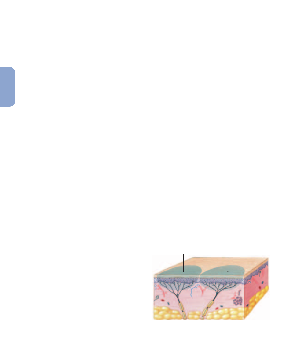
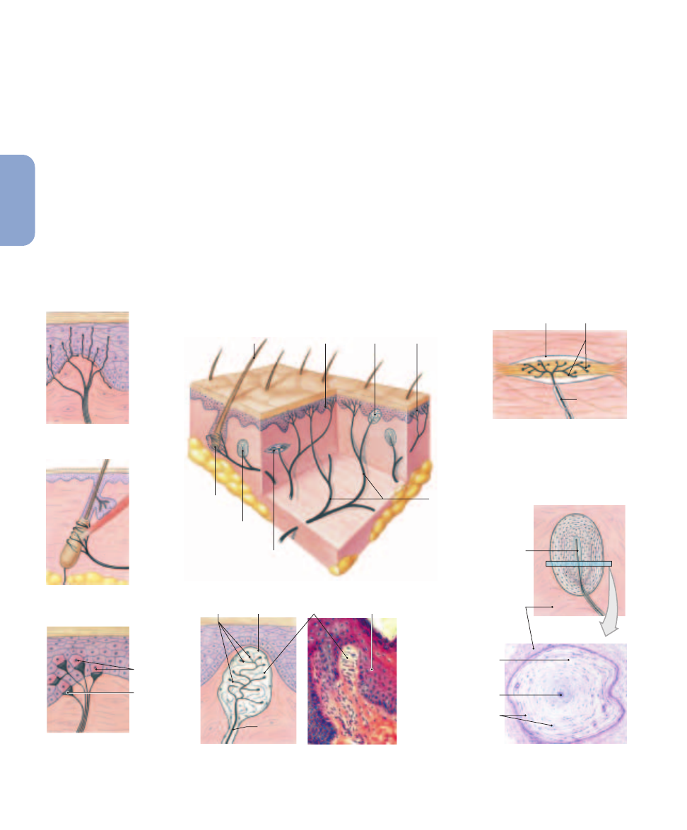
Tactile Receptors
Fine touch and pressure receptors provide detailed infor-
mation about a source of stimulation, including its exact lo-
cation, shape, size, texture, and movement. These receptors
are extremely sensitive and have relatively narrow receptive
fields. Crude touch and pressure receptors provide poor lo-
calization and, because they have relatively large receptive
fields, give little additional information about the stimulus.
Tactile receptors range in complexity from free nerve
endings to specialized sensory complexes with accessory cells
and supporting structures.
Figure 15–3
shows six types of tac-
tile receptors in the skin:
1.
Free nerve endings sensitive to touch and pressure are sit-
uated between epidermal cells (
Figure 15–3a
). There ap-
pear to be no structural differences between these
receptors and the free nerve endings that provide temper-
ature or pain sensations. These are the only sensory re-
512
Unit 3
Control and Regulation
Dendrite
Dendrite
Accessory
cells
Layers of collagen
fibers separated
by fluid
Dermis
Hair
Tactile discs
(innervating
Merkel cell)
Tactile
corpuscle
Sensory
nerves
Ruffini
corpuscle
Merkel cells
Tactile disc
Capsule
Lamellated
corpuscle
Root hair
plexus
(a) Free nerve endings
(d) Tactile corpuscle
(f) Ruffini corpuscle
(b) Root hair plexus
Free
nerve
ending
(c) Merkel cells and
tactile discs
(e) Lamellated corpuscle
Dermis
Tactile corpuscle
Dendrites
Epidermis
Capsule
Afferent
fiber
Dendrites
Afferent
fiber
LM
330
LM
75
Figure 15–3
Tactile Receptors in the Skin.
ceptors on the corneal surface of the eye, but in other por-
tions of the body surface, more specialized tactile recep-
tors are probably more important. Free nerve endings
that provide touch sensations are tonic receptors with
small receptive fields.
2.
Wherever hairs are located, the nerve endings of the root
hair plexus monitor distortions and movements across
the body surface (
Figure 15–3b
). When a hair is dis-
placed, the movement of the follicle distorts the sensory
dendrites and produces action potentials. These recep-
tors adapt rapidly, so they are best at detecting initial con-
tact and subsequent movements. Thus, you generally feel
your clothing only when you move or when you con-
sciously focus on tactile sensations from the skin.
3.
Tactile discs, or Merkel (MER-kel) discs, are fine touch
and pressure receptors (
Figure 15–3c
). They are extremely
sensitive tonic receptors, with very small receptive fields.
The dendritic processes of a single myelinated afferent
fiber make close contact with unusually large epithelial
cells in the stratum germinativum of the skin; these Merkel
cells were described in Chapter 5.
l
p. 160
4.
Tactile corpuscles, or Meissner (MIS-ner) corpuscles,
perceive sensations of fine touch and pressure and low-
frequency vibration. They adapt to stimulation within a
second after contact. Tactile corpuscles are fairly large
structures, measuring roughly 100 mm in length and
50 mm in width. These receptors are most abundant in
the eyelids, lips, fingertips, nipples, and external geni-
talia. The dendrites are highly coiled and interwoven, and
they are surrounded by modified Schwann cells. A fi-
brous capsule surrounds the entire complex and anchors
it within the dermis (
Figure 15–3d
).

Chapter 15
Neural Integration I: Sensory Pathways and the Somatic Nervous System
513
15
NER
V
OUS
Tips
&
Tricks
To remember that Meissner corpuscles perceive pressure
sensations, associate the m and ss in “Meissner” with
massage.
5.
Lamellated (LAM-e-lat-ed; lamella, a little thin plate)
corpuscles, or pacinian (pa-SIN-e-an) corpuscles, are sen-
sitive to deep pressure. Because they are fast-adapting re-
ceptors, they are most sensitive to pulsing or
high-frequency vibrating stimuli. A single dendrite lies
within a series of concentric layers of collagen fibers and
supporting cells (specialized fibroblasts) (
Figure 15–3e
).
The entire corpuscle may reach 4 mm in length and 1 mm
in diameter. The concentric layers, separated by intersti-
tial fluid, shield the dendrite from virtually every source
of stimulation other than direct pressure. Lamellated cor-
puscles adapt quickly because distortion of the capsule
soon relieves pressure on the sensory process. Somatic
sensory information is provided by lamellated corpuscles
located throughout the dermis, notably in the fingers,
mammary glands, and external genitalia; in the superfi-
cial and deep fasciae; and in joint capsules. Visceral sen-
sory information is provided by lamellated corpuscles in
mesenteries, in the pancreas, and in the walls of the ure-
thra and urinary bladder.
6.
Ruffini (roo-FE-ne) corpuscles are also sensitive to pres-
sure and distortion of the skin, but they are located in the
reticular (deep) dermis. These receptors are tonic and
show little if any adaptation. The capsule surrounds a
core of collagen fibers that are continuous with those of
the surrounding dermis (
Figure 15–3f
). In the capsule, a
network of dendrites is intertwined with the collagen
fibers. Any tension or distortion of the dermis tugs or
twists the capsular fibers, stretching or compressing the
attached dendrites and altering the activity in the myeli-
nated afferent fiber.
Our sensitivity to tactile sensations may be altered by in-
fection, disease, or damage to sensory neurons or pathways.
As a result, mapping tactile responses can sometimes aid clin-
ical assessment. Sensory losses with clear regional boundaries
indicate trauma to spinal nerves. For example, sensory loss
within the boundaries of a dermatome can help identify the
affected spinal nerve or nerves.
l
p. 438
Tickle and itch sensations are closely related to the sen-
sations of touch and pain. The receptors involved are free
nerve endings, and the information is carried by unmyeli-
nated Type C fibers. Tickle sensations, which are usually (but
not always) described as pleasurable, are produced by a light
touch that moves across the skin. Psychological factors are in-
volved in the interpretation of tickle sensations, and tickle
sensitivity differs greatly among individuals. Itching is prob-
ably produced by the stimulation of the same receptors. Spe-
cific “itch spots” can be mapped in the skin, the inner surfaces
of the eyelids, and the mucous membrane of the nose. Itch
sensations are absent from other mucous membranes and
from deep tissues and viscera. Itching is extremely unpleas-
ant, even more unpleasant than pain. Individuals with ex-
treme itching will scratch even when pain is the result. Itch
receptors can be stimulated by the injection of histamine or
proteolytic enzymes into the epidermis and superficial der-
mis. The precise receptor mechanism is unknown.
Applying the base of a tuning fork to the skin tests
vibration receptors. Damage to an individual spinal nerve
produces insensitivity to vibration along the paths of the
related sensory nerves. If the sensory loss results from spinal
cord damage, the injury site can typically be located by
walking the tuning fork down the spinal column, resting its
base on the vertebral spines.
Descriptive terms are used to indicate the degree of
sensitivity in the area. Anesthesia implies a total loss of
sensation; the individual cannot perceive touch, pressure,
pain, or temperature sensations in that area. Hypesthesia is a
reduction in sensitivity, and paresthesia is the presence of
abnormal sensations such as the pins-and-needles sensation
when an arm or leg “falls asleep” as a result of pressure on a
peripheral nerve.
Assessment of Tactile Sensitivities
Regional sensitivity to light touch can be checked by gentle
contact with a fingertip or a slender wisp of cotton. The
two-point discrimination test provides a more detailed
sensory map of tactile receptors. Two fine points of a bent
paper clip or another object are applied to the skin surface
simultaneously. The subject then describes the contact.
When the points fall within a single receptive field, the indi-
vidual will report only one point of contact. A normal indi-
vidual loses two-point discrimination at 1 mm (0.04 in.) on
the surface of the tongue, at 2–3 mm (0.08–0.12 in.) on the
lips, at 3–5 mm (0.12–0.20 in.) on the backs of the hands
and feet, and at 4–7 cm (1.6–2.75 in.) over the general body
surface.
C L I N I C A L N O T E

514
Unit 3
Control and Regulation
Baroreceptors
Baroreceptors monitor changes in pressure in an organ. A
baroreceptor consists of free nerve endings that branch
within the elastic tissues in the wall of a distensible organ,
such as a blood vessel or a portion of the respiratory, diges-
tive, or urinary tract. When the pressure changes, the elastic
walls of the tract recoil or expand. This movement distorts
the dendritic branches and alters the rate of action potential
generation. Baroreceptors respond immediately to a change
in pressure, but they adapt rapidly, and the output along the
afferent fibers gradually returns to normal.
Baroreceptors monitor blood pressure in the walls of ma-
jor vessels, including the carotid artery (at the carotid sinus)
and the aorta (at the aortic sinus). The information plays a
major role in regulating cardiac function and adjusting blood
flow to vital tissues. Baroreceptors in the lungs monitor the
degree of lung expansion. This information is relayed to the
respiratory rhythmicity centers, which set the pace of respira-
tion. Comparable stretch receptors at various sites in the di-
gestive and urinary tracts trigger a variety of visceral reflexes,
including those of urination and defecation. We will describe
those baroreceptor reflexes in chapters that deal with specific
physiological systems.
Proprioceptors
Proprioceptors monitor the position of joints, the tension in
tendons and ligaments, and the state of muscular contraction.
There are three major groups of proprioceptors:
1.
Muscle Spindles. Muscle spindles monitor skeletal muscle
length and trigger stretch reflexes.
l
p. 451
2.
Golgi Tendon Organs. Golgi tendon organs are similar in
function to Ruffini corpuscles but are located at the junc-
tion between a skeletal muscle and its tendon. In a Golgi
tendon organ, dendrites branch repeatedly and wind
around the densely packed collagen fibers of the tendon.
These receptors are stimulated by tension in the tendon;
they thus monitor the external tension developed during
muscle contraction.
3.
Receptors in Joint Capsules. Joint capsules are richly in-
nervated by free nerve endings that detect pressure, ten-
sion, and movement at the joint. Your sense of body
position results from the integration of information
from these receptors with information provided by
muscle spindles, Golgi tendon organs, and the receptors
of the inner ear.
Proprioceptors do not adapt to constant stimulation, and
each receptor continuously sends information to the CNS. A
relatively small proportion of the arriving proprioceptive in-
formation reaches your awareness; most proprioceptive infor-
mation is processed at subconscious levels.
Chemoreceptors
Specialized chemoreceptive neurons can detect small changes
in the concentration of specific chemicals or compounds. In
general, chemoreceptors respond only to water-soluble and
lipid-soluble substances that are dissolved in the surrounding
fluid. These receptors exhibit peripheral adaptation over a per-
iod of seconds, and central adaptation may also occur.
The chemoreceptors included in the general senses do
not send information to the primary sensory cortex, so we are
not consciously aware of the sensations they provide. The ar-
riving sensory information is routed to brain stem centers
that deal with the autonomic control of respiratory and car-
diovascular functions. Neurons in the respiratory centers of
the brain respond to the concentration of hydrogen ions (pH)
and levels of carbon dioxide molecules in the cerebrospinal
fluid. Chemoreceptive neurons are also located in the carotid
bodies, near the origin of the internal carotid arteries on each
side of the neck, and in the aortic bodies, between the major
branches of the aortic arch. These receptors monitor the pH
and the carbon dioxide and oxygen levels in arterial blood.
The afferent fibers leaving the carotid or aortic bodies reach
the respiratory centers by traveling within cranial nerves IX
(glossopharyngeal) and X (vagus).
C H E C K P O I N T
5. List the four types of general sensory receptors, and
identify the nature of the stimulus that excites each
type.
6. Identify the three classes of mechanoreceptors.
7. What would happen to you if the information from
proprioceptors in your legs were blocked from
reaching the CNS?
See the blue Answers tab at the end of the book.
15-4
Separate pathways carry
somatic sensory and visceral
sensory information
A sensory neuron that delivers sensations to the CNS is often
called a first-order neuron. The cell body of a first-order gen-
eral sensory neuron is located in a dorsal root ganglion or cra-
nial nerve ganglion. In the CNS, the axon of that sensory neuron
synapses on an interneuron known as a second-order neuron,
which may be located in the spinal cord or brain stem. If the sen-
sation is to reach our awareness, the second-order neuron
synapses on a third-order neuron in the thalamus. Somewhere
along its length, the axon of the second-order neuron crosses
Chapter 15
Neural Integration I: Sensory Pathways and the Somatic Nervous System
515
15
NER
V
OUS
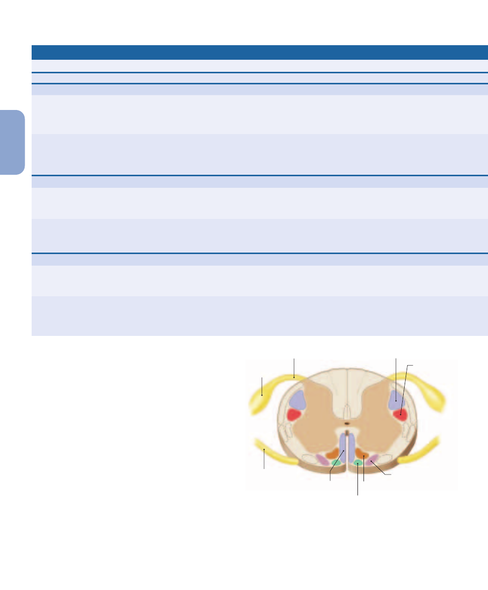
Fasciculus gracilis
Posterior column
pathway
Posterior spinocerebellar tract
Anterior spinocerebellar tract
Spinocerebellar
pathway
Lateral spinothalamic tract
Anterior spinothalamic tract
Spinothalamic
pathway
Dorsal root
Dorsal root
ganglion
Ventral root
Fasciculus cuneatus
Figure 15–4
Sensory Pathways and Ascending Tracts in the Spinal Cord. A cross-sectional view of the spinal cord indicating the
locations of the major ascending (sensory) tracts. For information about these tracts, see Table 15–1. Descending (motor) tracts (identified in
Figure 15–8) are shown in dashed outline.
over to the opposite side of the CNS. As a result, the right side
of the thalamus receives sensory information from the left side
of the body, and the left side of the thalamus receives sensory in-
formation from the right side of the body.
The axons of the third-order neurons ascend without
crossing over and synapse on neurons of the primary sensory
cortex of the cerebral hemisphere. As a result, the right cere-
bral hemisphere receives sensory information from the left
side of the body, and the left cerebral hemisphere receives sen-
sations from the right side. The reason for this crossover is
unknown. Although it has no apparent functional benefit,
crossover occurs along sensory and motor pathways in all
vertebrates.
Somatic Sensory Pathways
Somatic sensory pathways carry sensory information from
the skin and musculature of the body wall, head, neck, and
limbs. We will consider three major somatic sensory path-
ways: (1) the posterior column pathway, (2) the spinothalamic
pathway, and (3) the spinocerebellar pathway. These pathways
utilize pairs of spinal tracts, symmetrically arranged on oppo-
site sides of the spinal cord. All the axons within a tract share
a common origin and destination.
Figure 15–4
indicates the relative positions of the spinal
tracts involved. Note that tract names often give clues to their
function. For example, if the name of a tract begins with
spino-, the tract must start in the spinal cord and end in the
brain. It must therefore be an ascending tract that carries sen-
sory information. The rest of the name indicates the tract’s
destination. Thus, a spinothalamic tract begins in the spinal
cord and carries sensory information to the thalamus.
If, on the other hand, the name of a tract ends in -spinal,
the tract ends in the spinal cord and starts in a higher center
of the brain. It must therefore be a descending tract that car-
ries motor commands. The first part of the name indicates the
nucleus or cortical area of the brain where the tract originates.
For example, a corticospinal tract carries motor commands
from the cerebral cortex to the spinal cord. Such tracts will be
considered later in the chapter.
The Posterior Column Pathway
The posterior column pathway carries sensations of highly
localized (“fine”) touch, pressure, vibration, and propriocep-
tion (
Figure 15–5a
). This pathway, also known as the dorsal
column/medial lemniscus, begins at a peripheral receptor and
ends at the primary sensory cortex of the cerebral hemi-
spheres. The spinal tracts involved are the left and right
fasciculus gracilis (gracilis, slender) and the left and right
fasciculus cuneatus (cuneus, wedge-shaped). On each side
of the posterior median sulcus, the fasciculus gracilis is me-
dial to the fasciculus cuneatus.
The axons of the first-order neurons reach the CNS
within the dorsal roots of spinal nerves and the sensory roots
of cranial nerves. The axons ascending within the posterior
column are organized according to the region innervated. Ax-
ons carrying sensations from the inferior half of the body as-
cend within the fasciculus gracilis and synapse in the nucleus
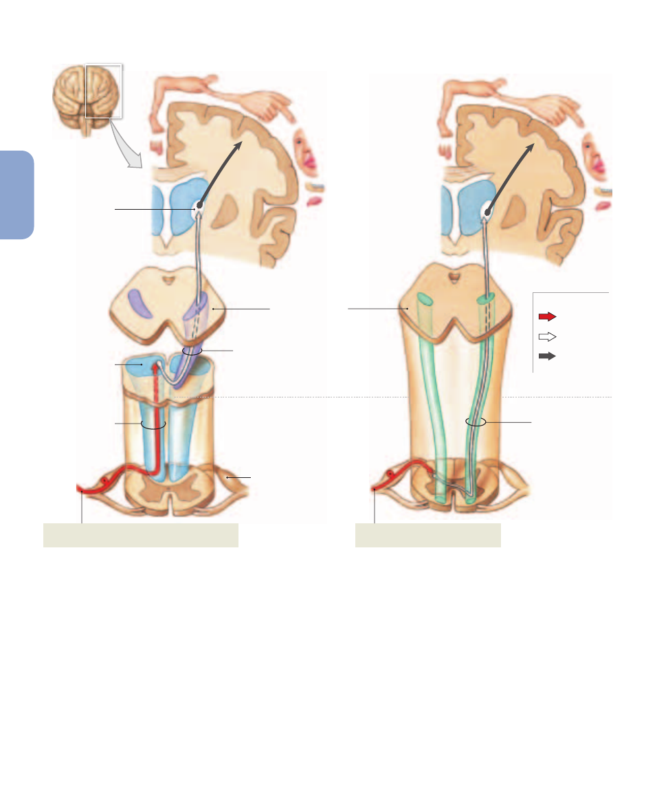
516
Unit 3
Control and Regulation
Axon of first-
order neuron
Second-order
neuron
Third-order
neuron
MESENCEPHALON
Nucleus gracilis
and nucleus
cuneatus
MEDULLA OBLONGATA
SPINAL CORD
Dorsal root
ganglion
Fasciculus gracilis
and fasciculus
cuneatus
Fine-touch, vibration, pressure, and proprioception
sensations from right side of body
Sensory homunculus of
left cerebral hemisphere
Ventral nuclei
in thalamus
Medial
lemniscus
(a) Posterior column pathway
Crude touch and pressure sensations
from right side of body
Anterior
spinothalamic
tract
Sensory homunculus of
left cerebral hemisphere
(b) Anterior spinothalamic tract
KEY
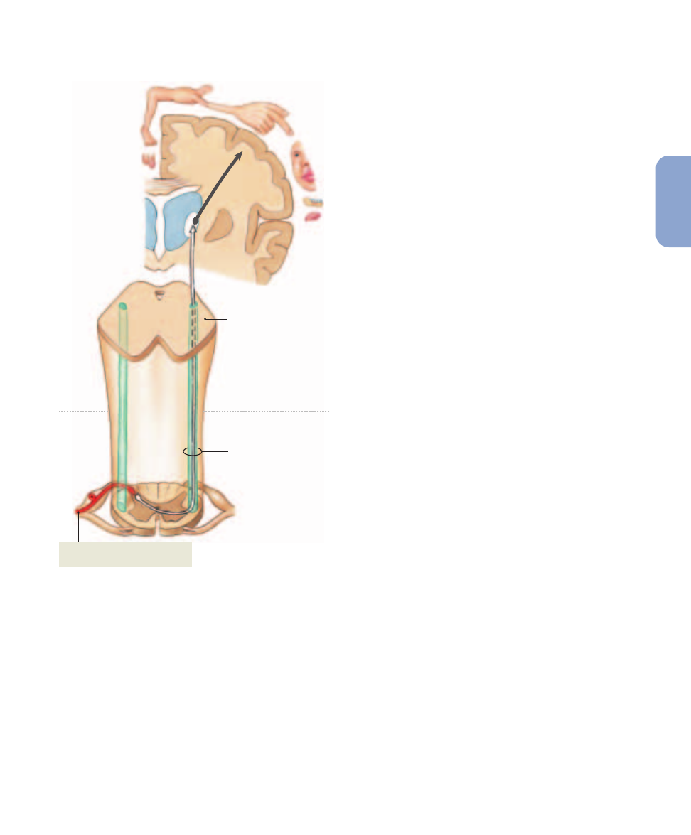
Figure 15–5
The Posterior Column Pathway, and the Spinothalamic Tracts of the Spinothalamic Pathway. For clarity, only the
pathways for sensations originating on the right side of the body are shown.
(a)
The posterior column pathway delivers fine touch, vibration,
and proprioception information to the primary sensory cortex on the opposite side of the body.
(b)
The anterior spinothalamic tracts carry
sensations of crude touch and pressure to the primary sensory cortex on the opposite side of the body.
(c)
The lateral spinothalamic tracts carry
sensations of pain and temperature to the primary sensory cortex on the opposite side of the body.
gracilis of the medulla oblongata. Axons carrying sensations
from the superior half of the trunk, upper limbs, and neck as-
cend in the fasciculus cuneatus and synapse in the nucleus
cuneatus.
l
p. 471
Axons of the second-order neurons of the nucleus gracilis
and nucleus cuneatus ascend to the thalamus. As they ascend,
these axons cross over to the opposite side of the brain stem.
The crossing of an axon from the left side to the right side, or
from the right side to the left side, is called decussation.
Once on the opposite side of the brain, the axons enter a tract
called the medial lemniscus (lemniskos, ribbon). As it as-
cends, the medial lemniscus runs alongside a smaller tract
that carries sensory information from the face, relayed from
the sensory nuclei of the trigeminal nerve (N V).
The axons in these tracts synapse on third-order neurons
in one of the ventral nuclei of the thalamus.
l
p. 475
These
Chapter 15
Neural Integration I: Sensory Pathways and the Somatic Nervous System
517
15
NER
V
OUS
Lateral
spinothalamic
tract
Pain and temperature sensations
from right side of body
(c) Lateral spinothalamic tract
Sensory homunculus of
left cerebral hemisphere
MEDULLA OBLONGATA
SPINAL CORD
MESENCEPHALON
nuclei sort the arriving information according to (1) the na-
ture of the stimulus and (2) the region of the body involved.
Processing in the thalamus determines whether you perceive
a given sensation as fine touch, or as pressure or vibration.
Our ability to localize the sensation—to determine pre-
cisely where on the body a specific stimulus originated—
depends on the projection of information from the thalamus
to the primary sensory cortex. Sensory information from the
toes arrives at one end of the primary sensory cortex, and in-
formation from the head arrives at the other. When neurons
in one portion of your primary sensory cortex are stimulated,
you become aware of sensations originating at a specific loca-
tion. If your primary sensory cortex were damaged or the pro-
jection fibers were cut, you could detect a light touch but
would be unable to determine its source.
The same sensations are reported whether the cortical
neurons are activated by axons ascending from the thalamus
or by direct electrical stimulation. Researchers have electri-
cally stimulated the primary sensory cortex in awake individ-
uals during brain surgery and asked the subjects where they
thought the stimulus originated. The results were used to cre-
ate a functional map of the primary sensory cortex. Such a
map, three of which are shown in
Figure 15–5
, is called a
sensory homunculus (“little man”).
The proportions of the sensory homunculus are very dif-
ferent from those of any individual. For example, the face is
huge and distorted, with enormous lips and tongue, whereas
the back is relatively tiny. These distortions occur because
the area of sensory cortex devoted to a particular body region
is proportional not to the region’s absolute size, but to the
number of sensory receptors it contains. In other words, many
more cortical neurons are required to process sensory infor-
mation arriving from the tongue, which has tens of thou-
sands of taste and touch receptors, than to analyze sensations
originating on the back, where touch receptors are few and
far between.
The Spinothalamic Pathway
The spinothalamic pathway provides conscious sensations
of poorly localized (“crude”) touch, pressure, pain, and tem-
perature. In this pathway, the axons of first-order sensory
neurons enter the spinal cord and synapse on second-order
neurons within the posterior gray horns. The axons of these
interneurons cross to the opposite side of the spinal cord be-
fore ascending. This pathway includes relatively small tracts
that deliver sensations to reflex centers in the brain stem
as well as larger tracts that carry sensations destined for
the cerebral cortex. We will ignore the smaller tracts in this
discussion.
Sensations bound for the cerebral cortex ascend within
the anterior or lateral spinothalamic tracts. The anterior
spinothalamic tracts carry crude touch and pressure sen-
sations (
Figure 15–5b
), whereas the lateral spinothalamic
tracts carry pain and temperature sensations (
Figure 15–5c
).
These tracts end at third-order neurons in the ventral nu-
cleus group of the thalamus. After the sensations have been
sorted and processed, they are relayed to the primary sen-
sory cortex.
The perception that an arriving stimulus is painful rather
than cold, hot, or vibrating depends on which second-order
and third-order neurons are stimulated. The ability to local-
ize that stimulus to a specific location in the body depends
on the stimulation of an appropriate area of the primary sen-
sory cortex. Any abnormality along the pathway can result in
518
Unit 3
Control and Regulation
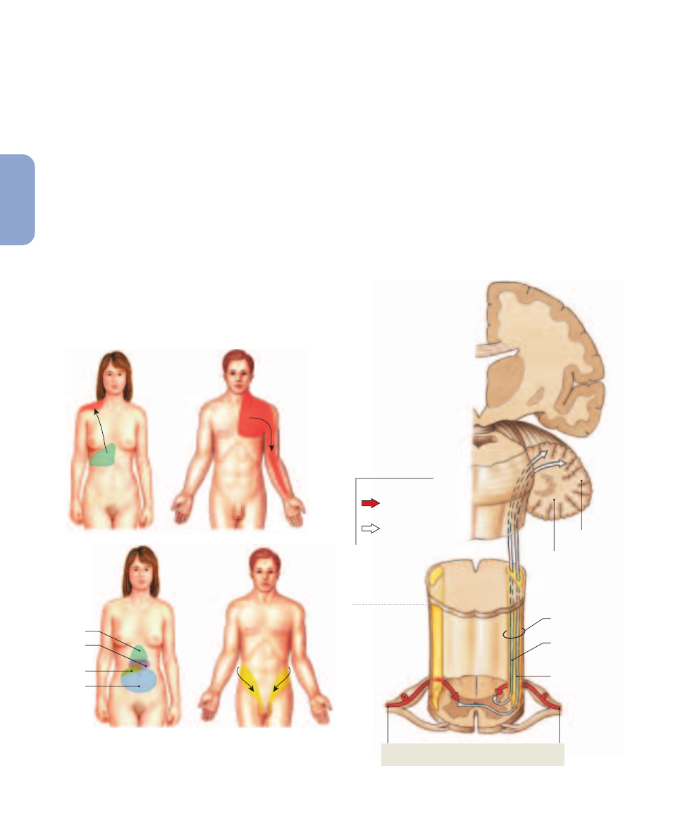
Liver and
gallbladder
Heart
Ureters
Stomach
Colon
Small
intestine
Appendix
Figure 15–6
Referred Pain. Pain sensations from visceral organs
are often perceived as involving specific regions of the body surface
innervated by the same spinal segments. Each region of perceived
pain is labeled according to the organ at which the pain originates.
inappropriate sensations or inaccurate localization of the
source. Consider these examples:
•
An individual can experience painful sensations that are
not real. For example, a person may continue to
experience pain in an amputated limb. This phantom limb
pain is caused by activity in the sensory neurons or
interneurons along the spinothalamic pathway. The
neurons involved were once part of the labeled line that
monitored conditions in the intact limb. These labeled
lines and pathways are developmentally programmed;
even individuals born without limbs can have phantom
limb pain.
•
An individual can feel pain in an uninjured part of the
body when the pain actually originates at another
location. For example, strong visceral pain sensations
arriving at a segment of the spinal cord can stimulate
interneurons that are part of the spinothalamic pathway.
Activity in these interneurons leads to the stimulation of
CEREBELLUM
PONS
Arbor vitae
Spinocerebellar
pathway
Posterior
spinocerebellar
tract
Anterior
spinocerebellar
tract
Proprioceptive input from Golgi tendon organs,
muscle spindles, and joint capsules
MEDULLA
OBLONGATA
SPINAL
CORD
KEY
Axon of first-
order neuron
Second-order
neuron
Figure 15–7
The Spinocerebellar Pathway.
the primary sensory cortex, so the individual feels pain
in a specific part of the body surface. This phenomenon
is called referred pain. Two familiar examples are
(1) the pain of a heart attack, which is frequently felt in
the left arm, and (2) the pain of appendicitis, which is
generally felt first in the area around the navel and then
in the right lower quadrant. These and additional
examples are shown in
Figure 15–6
.
The Spinocerebellar Pathway
The cerebellum receives proprioceptive information about
the position of skeletal muscles, tendons, and joints along
the spinocerebellar pathway (
Figure 15–7
). This infor-
Chapter 15
Neural Integration I: Sensory Pathways and the Somatic Nervous System
519
15
NER
V
OUS
cessing and sorting center for visceral sensory information; it has
extensive connections with the various cardiovascular and respi-
ratory centers as well as with the reticular formation.
The dorsal roots of spinal nerves T
1
–L
2
carry visceral sen-
sory information provided by receptors in organs located be-
tween the diaphragm and the pelvic cavity. The dorsal roots of
spinal nerves S
2
–S
4
carry visceral sensory information from or-
gans in the inferior portion of the pelvic cavity, including the
last portion of the large intestine, the urethra and base of the
urinary bladder, and the prostate gland (males) or the cervix of
the uterus and adjacent portions of the vagina (females).
The first-order neurons deliver the visceral sensory infor-
mation to interneurons whose axons ascend within the
spinothalamic pathway. Most of the sensory information is de-
livered to the solitary nucleus, and because it never reaches the
primary sensory cortex we remain unaware of these sensations.
C H E C K P O I N T
8. As a result of pressure on her spinal cord, Jill cannot
feel fine touch or pressure on her lower limbs. Which
spinal tract is being compressed?
9. Which spinal tract carries action potentials generated
by nociceptors?
10. Which cerebral hemisphere receives impulses
conducted by the right fasciculus gracilis?
See the blue Answers tab at the end of the book.
15-5
The somatic nervous system is
an efferent division that controls
skeletal muscles
Motor commands issued by the CNS are distributed by the
somatic nervous system (SNS) and the autonomic nervous
system (ANS). The somatic nervous system, also called the
somatic motor system, controls the contractions of skeletal
muscles. The output of the SNS is under voluntary control.
The autonomic nervous system, or visceral motor system, con-
trols visceral effectors, such as smooth muscle, cardiac mus-
cle, and glands. We will examine the organization of the ANS
in Chapter 16; our interest here is the structure of the SNS.
Throughout this discussion we will use the terms motor neu-
ron and motor control to refer specifically to somatic motor
neurons and pathways that control skeletal muscles.
Somatic motor pathways always involve at least two mo-
tor neurons: an upper motor neuron, whose cell body lies in
a CNS processing center, and a lower motor neuron, whose
cell body lies in a nucleus of the brain stem or spinal cord.
The upper motor neuron synapses on the lower motor neu-
ron, which in turn innervates a single motor unit in a skeletal
muscle. Activity in the upper motor neuron may facilitate or
mation does not reach our awareness. The axons of first-
order sensory neurons synapse on interneurons in the
dorsal gray horns of the spinal cord. The axons of these
second-order neurons ascend in one of the spinocerebel-
lar tracts:
•
The posterior spinocerebellar tracts contain axons that
do not cross over to the opposite side of the spinal cord.
These axons reach the cerebellar cortex via the inferior
cerebellar peduncle of that side.
•
The anterior spinocerebellar tracts are dominated by
axons that have crossed over to the opposite side of the
spinal cord, although they do contain a significant
number of uncrossed axons as well. The sensations
carried by the anterior spinocerebellar tracts reach the
cerebellar cortex via the superior cerebellar peduncle.
Interestingly, many of the axons that cross over and
ascend to the cerebellum then cross over again within the
cerebellum, synapsing on the same side as the original
stimulus. The functional significance of this “double
cross” is unknown.
The information carried by the spinocerebellar pathway
ultimately arrives at the Purkinje cells of the cerebellar cortex.
l
p. 472
Proprioceptive information from each part of the
body is relayed to a specific portion of the cerebellar cortex.
We will consider the integration of proprioceptive informa-
tion and the role of the cerebellum in somatic motor control
in a later section.
Table 15–1 reviews the somatic sensory pathways dis-
cussed in this section.
The
A
&
P Top 100
#52
Most somatic sensory information is relayed to the
thalamus for processing. A small fraction of the arriving
information is projected to the cerebral cortex and reaches
our awareness.
Visceral Sensory Pathways
Visceral sensory information is collected by interoceptors
monitoring visceral tissues and organs, primarily within the
thoracic and abdominopelvic cavities. These interoceptors in-
clude nociceptors, thermoreceptors, tactile receptors, barore-
ceptors, and chemoreceptors, although none of them are as
numerous as they are in somatic tissues. The axons of the
first-order neurons usually travel in company with auto-
nomic motor fibers innervating the same visceral structures.
Cranial nerves V, VII, IX, and X carry visceral sensory infor-
mation from the mouth, palate, pharynx, larynx, trachea, esoph-
agus, and associated vessels and glands.
l
pp. 494–498
This
information is delivered to the solitary nucleus, a large nucleus
in the medulla oblongata. The solitary nucleus is a major pro-
520
Unit 3
Control and Regulation
inhibit the lower motor neuron. Activation of the lower motor
neuron triggers a contraction in the innervated muscle. Only
the axon of the lower motor neuron extends outside the CNS.
Destruction of or damage to a lower motor neuron eliminates
voluntary and reflex control over the innervated motor unit.
Conscious and subconscious motor commands control
skeletal muscles by traveling over three integrated motor
pathways: the corticospinal pathway, the medial pathway, and
the lateral pathway.
Figure 15–8
indicates the positions of the
associated motor (descending) tracts in the spinal cord. Ac-
tivity within these motor pathways is monitored and adjusted
by the basal nuclei and cerebellum. The output of these cen-
ters stimulates or inhibits the activity of either (1) motor nu-
clei or (2) the primary motor cortex.
The Corticospinal Pathway
The corticospinal pathway (
Figure 15–9
), sometimes called
the pyramidal system, provides voluntary control over skeletal
muscles. This system begins at the pyramidal cells of the pri-
TABLE 15–1
Principal Ascending (Sensory) Pathways
Location of Neuron Cell Bodies
Pathway/Tract
Sensation(s)
First-Order
Second-Order
Third-Order
POSTERIOR COLUMN PATHWAY
Fasciculus gracilis
Proprioception and fine touch,
Dorsal root ganglia of
Nucleus gracilis of
Ventral nuclei
ventral pressure, and vibration
inferior half of body;
medulla oblongata;
of thalamus
from inferior half of body
axons enter CNS in
axons cross over before
dorsal roots and join
entering medial
fasciculus gracilis
lemniscus
Fasciculus cuneatus
Proprioception and fine touch,
Dorsal root ganglia of
Nucleus cuneatus of
As above
ventral pressure, and vibration
superior half of body;
medulla oblongata;
from superior half of body
axons enter CNS in
axons cross over before
dorsal roots and join
entering medial
fasciculus cuneatus
lemniscus
SPINOTHALAMIC PATHWAY
Lateral spinothalamic tracts
Pain and temperature
Dorsal root ganglia;
Interneurons in posterior
Ventral nuclei
axons enter CNS in
gray horn; axons enter
of thalamus
dorsal roots
lateral spinothalamic
tract on opposite side
Anterior spinothalamic tracts
Crude touch and pressure
As above
Interneurons in posterior
As above
gray horn; axons enter
anterior spinothalamic
tract on opposite side
SPINOCEREBELLAR PATHWAY
Posterior spinocerebellar tracts
Proprioception
Dorsal root ganglia;
Interneurons in posterior
Not present
axons enter CNS in
gray horn; axons enter
dorsal roots
posterior spinothalamic
tract on same side
Anterior spinocerebellar tracts
Proprioception
As above
Interneurons in same
Not present
spinal section; axons
enter anterior
spinocerebellar tract on
the same or opposite side
Anterior
corticospinal
tract
Dorsal root
ganglion
Dorsal root
Tectospinal tract
Lateral corticospinal tract
Reticulospinal tract
Vestibulospinal tract
Rubrospinal
tract
Ventral root
Figure 15–8
Descending (Motor) Tracts in the Spinal Cord. A
cross-sectional view indicating the locations of the major descending
(motor) tracts that contain the axons of upper motor neurons. The
origins and destinations of these tracts are listed in Table 15–2.
Sensory tracts (shown in Figure 15–4) appear in dashed outline.
Chapter 15
Neural Integration I: Sensory Pathways and the Somatic Nervous System
521
15
NER
V
OUS
mary motor cortex.
l
p. 484
The axons of these upper mo-
tor neurons descend into the brain stem and spinal cord to
synapse on lower motor neurons that control skeletal muscles.
In general, the corticospinal pathway is direct: The upper mo-
tor neurons synapse directly on the lower motor neurons.
However, the corticospinal pathway also works indirectly, as it
innervates centers of the medial and lateral pathways.
The corticospinal pathway contains three pairs of de-
scending tracts: (1) the corticobulbar tracts, (2) the lateral cor-
ticospinal tracts, and (3) the anterior corticospinal tracts.
These tracts enter the white matter of the internal capsule, de-
scend into the brain stem, and emerge on either side of the
mesencephalon as the cerebral peduncles.
The Corticobulbar Tracts
Axons in the corticobulbar (kor-ti-ko
¯-BUL-bar; bulbar, brain
stem) tracts synapse on lower motor neurons in the motor nu-
clei of cranial nerves III, IV, V, VI, VII, IX, XI, and XII. The cor-
ticobulbar tracts provide conscious control over skeletal
muscles that move the eye, jaw, and face, and some muscles of
the neck and pharynx. The corticobulbar tracts also innervate
the motor centers of the medial and lateral pathways.
The Corticospinal Tracts
Axons in the corticospinal tracts synapse on lower motor neu-
rons in the anterior gray horns of the spinal cord. As they de-
scend, the corticospinal tracts are visible along the ventral
surface of the medulla oblongata as a pair of thick bands, the
pyramids. Along the length of the pyramids, roughly 85 per-
cent of the axons cross the midline (decussate) to enter the de-
scending lateral corticospinal tracts on the opposite side of
Location of Neuron Cell Bodies
Final Destination
Site of Crossover
Primary sensory cortex
Axons of second-order neurons
on side opposite stimulus
before entering the medial
lemniscus
As above
As above
Primary sensory cortex
Axons of second-order
on side opposite stimulus
neurons at level of entry
As above
As above
Cerebellar cortex
None
on side of stimulus
Cerebellar cortex on side
Axons of most second-order
opposite (and side of)
neurons cross over before
stimulus
entering tract; many re-cross
at cerebellum
Corticobulbar
tract
Pyramids
Cerebral peduncle
MESENCEPHALON
Motor homunculus on primary motor
cortex of left cerebral
hemisphere
Decussation
of pyramids
Anterior
corticospinal
tract
SPINAL CORD
To
skeletal
muscles
To
skeletal
muscles
MEDULLA
OBLONGATA
Motor nuclei
of cranial
nerves
Motor nuclei
of cranial
nerves
Axon of first-
order neuron
Second-order
neuron
KEY
To
skeletal
muscles
Lateral
corticospinal
tract
Figure 15–9
The Corticospinal Pathway. The corticospinal
pathway originates at the primary motor cortex. The corticobulbar
tracts end at the motor nuclei of cranial nerves on the opposite side
of the brain. Most fibers in this pathway cross over in the medulla
and enter the lateral corticospinal tracts; the rest descend in the
anterior corticospinal tracts and cross over after reaching target
segments in the spinal cord.

522
Unit 3
Control and Regulation
the spinal cord. The other 15 percent continue uncrossed along
the spinal cord as the anterior corticospinal tracts. At the
spinal segment it targets, an axon in the anterior corticospinal
tract crosses over to the opposite side of the spinal cord in the
anterior white commissure before synapsing on lower motor
neurons in the anterior gray horns.
The Motor Homunculus
The activity of pyramidal cells in a specific portion of the pri-
mary motor cortex will result in the contraction of specific
peripheral muscles. The identities of the stimulated muscles
depend on the region of motor cortex that is active. As in the
primary sensory cortex, the primary motor cortex corre-
sponds point by point with specific regions of the body. The
cortical areas have been mapped out in diagrammatic form,
creating a motor homunculus.
Figure 15–9
shows the motor
homunculus of the left cerebral hemisphere and the corti-
cospinal pathway controlling skeletal muscles on the right
side of the body.
The proportions of the motor homunculus are quite differ-
ent from those of the actual body, because the motor area de-
voted to a specific region of the cortex is proportional to the
number of motor units involved in the region’s control, not to
its actual size. As a result, the homunculus provides an indica-
tion of the degree of fine motor control available. For example,
the hands, face, and tongue, all of which are capable of varied
and complex movements, appear very large, whereas the trunk
is relatively small. These proportions are similar to those of the
sensory homunculus (
Figure 15–5
). The sensory and motor ho-
munculi differ in other respects because some highly sensitive
regions, such as the sole of the foot, contain few motor units,
and some areas with an abundance of motor units, such as the
eye muscles, are not particularly sensitive.
C L I N I C A L N O T E
Cerebral Palsy
The term cerebral palsy refers to a
number of disorders that affect voluntary motor
performance; they appear during infancy or childhood and
persist throughout the life of the affected individual. The
cause may be trauma associated with premature or
unusually stressful birth, maternal exposure to drugs
(including alcohol), or a genetic defect that causes the
improper development of motor pathways. Problems
during labor and delivery may produce compression or
interruption of placental circulation or oxygen supplies. If
the oxygen concentration in fetal blood declines
significantly for as little as 5–10 minutes, CNS function can
be permanently impaired. The cerebral cortex, cerebellum,
basal nuclei, hippocampus, and thalamus are likely targets,
producing abnormalities in motor skills, posture and
balance, memory, speech, and learning abilities.
The Medial and Lateral Pathways
Several centers in the cerebrum, diencephalon, and brain
stem may issue somatic motor commands as a result of pro-
cessing performed at a subconscious level. These centers and
their associated tracts were long known as the extrapyramidal
system (EPS), because it was thought that they operated inde-
pendently of, and in parallel with, the pyramidal system (cor-
ticospinal pathway). This classification scheme is both
inaccurate and misleading, because motor control is inte-
grated at all levels through extensive feedback loops and in-
terconnections. It is more appropriate to group these nuclei
and tracts in terms of their primary functions: The compo-
nents of the medial pathway help control gross movements
of the trunk and proximal limb muscles, whereas those of the
lateral pathway help control the distal limb muscles that
perform more precise movements.
The medial and lateral pathways can modify or direct skele-
tal muscle contractions by stimulating, facilitating, or inhibiting
lower motor neurons. It is important to note that the axons of
upper motor neurons in the medial and lateral pathways
synapse on the same lower motor neurons innervated by the
corticospinal pathway. This means that the various motor path-
ways interact not only within the brain, through interconnec-
tions between the primary motor cortex and motor centers in
the brain stem, but also through excitatory or inhibitory inter-
actions at the level of the lower motor neuron.
The Medial Pathway
The medial pathway is primarily concerned with the control
of muscle tone and gross movements of the neck, trunk, and
proximal limb muscles. The upper motor neurons of the me-
dial pathway are located in the vestibular nuclei, the superior
and inferior colliculi, and the reticular formation.
The vestibular nuclei receive information, over the
vestibulocochlear nerve (N VIII), from receptors in the inner
ear that monitor the position and movement of the head.
These nuclei respond to changes in the orientation of the head,
sending motor commands that alter the muscle tone, exten-
sion, and position of the neck, eyes, head, and limbs. The pri-
mary goal is to maintain posture and balance. The descending
fibers in the spinal cord constitute the vestibulospinal tracts.
The superior and inferior colliculi are located in the
tectum, or roof of the mesencephalon (
Figure 14–8
, p. 474).
The colliculi receive visual (superior) and auditory (inferior)
sensations. Axons of upper motor neurons in the colliculi de-
scend in the tectospinal tracts. These axons cross to the op-
posite side immediately, before descending to synapse on
lower motor neurons in the brain stem or spinal cord. Axons
in the tectospinal tracts direct reflexive changes in the posi-
tion of the head, neck, and upper limbs in response to bright
lights, sudden movements, or loud noises.

Chapter 15
Neural Integration I: Sensory Pathways and the Somatic Nervous System
523
15
NER
V
OUS
The reticular formation is a loosely organized network of
neurons that extends throughout the brain stem.
l
p. 470
The reticular formation receives input from almost every as-
cending and descending pathway. It also has extensive inter-
connections with the cerebrum, the cerebellum, and brain stem
nuclei. Axons of upper motor neurons in the reticular forma-
tion descend into the reticulospinal tracts without crossing to
the opposite side. The effects of reticular formation stimulation
are determined by the region stimulated. For example, the
stimulation of upper motor neurons in one portion of the retic-
ular formation produces eye movements, whereas the stimula-
tion of another portion activates respiratory muscles.
The Lateral Pathway
The lateral pathway is primarily concerned with the control
of muscle tone and the more precise movements of the distal
parts of the limbs. The upper motor neurons of the lateral
pathway lie within the red nuclei of the mesencephalon.
l
p. 475
Axons of upper motor neurons in the red nuclei cross
to the opposite side of the brain and descend into the spinal
cord in the rubrospinal tracts (ruber, red). In humans, the
rubrospinal tracts are small and extend only to the cervical
spinal cord. There they provide motor control over distal
muscles of the upper limbs; normally, their role is insignifi-
cant as compared with that of the lateral corticospinal tracts.
However, the rubrospinal tracts can be important in main-
taining motor control and muscle tone in the upper limbs if
the lateral corticospinal tracts are damaged.
Table 15–2 reviews the major descending (motor) tracts
discussed in this section.
The Basal Nuclei and Cerebellum
The basal nuclei and cerebellum are responsible for coordi-
nation and feedback control over muscle contractions,
whether those contractions are consciously or subcon-
sciously directed.
The Basal Nuclei
The basal nuclei provide the background patterns of move-
ment involved in voluntary motor activities. For example,
they may control muscles that determine the background po-
sition of the trunk or limbs, or they may direct rhythmic cy-
cles of movement, as in walking or running. These nuclei do
not exert direct control over lower motor neurons. Instead,
they adjust the activities of upper motor neurons in the vari-
ous motor pathways based on input from all portions of the
cerebral cortex, as well as from the substantia nigra.
TABLE 15–2
Principal Descending (Motor) Pathways
Location of Upper
Tract
Motor Neurons
Destination
Site of Crossover
Action
CORTICOSPINAL PATHWAY
Corticobulbar tracts
Primary motor cortex
Lower motor neurons
Brain stem
Conscious motor control
(cerebral hemisphere)
of cranial nerve
of skeletal muscles
nuclei in brain stem
Lateral corticospinal tracts
As above
Lower motor neurons
Pyramids of medulla
As above
of anterior gray horns
oblongata
of spinal cord
Anterior corticospinal tracts
As above
As above
Level of lower motor
As above
neuron
MEDIAL PATHWAY
Vestibulospinal tracts
Vestibular nuclei
As above
None (uncrossed)
Subconscious regulation of
(at border of pons
balance and muscle tone
and medulla oblongata)
Tectospinal tracts
Tectum (mesencephalon:
Lower motor neurons
Brain stem
Subconscious regulation of
superior and inferior
of anterior gray horns
(mesencephalon)
eye, head, neck, and upper
colliculi)
(cervical spinal cord only)
limb position in response to
visual and auditory stimuli
Reticulospinal tracts
Reticular formation
Lower motor neurons
None (uncrossed)
Subconscious regulation of
(network of nuclei in
of anterior gray horns
reflex activity
brain stem)
of spinal cord
LATERAL PATHWAY
Rubrospinal tracts
Red nuclei of
As above
Brain stem
Subconscious regulation of
mesencephalon
(mesencephalon)
upper limb muscle tone and
movement

524
Unit 3
Control and Regulation
The basal nuclei adjust or establish patterns of movement
via two major pathways:
1.
One group of axons synapses on thalamic neurons, whose
axons extend to the premotor cortex, the motor associa-
tion area that directs activities of the primary motor cor-
tex. This arrangement creates a feedback loop that
changes the sensitivity of the pyramidal cells and alters the
pattern of instructions carried by the corticospinal tracts.
2.
A second group of axons synapses in the reticular forma-
tion, altering the excitatory or inhibitory output of the
reticulospinal tracts.
Two distinct populations of neurons exist: one that stim-
ulates neurons by releasing acetylcholine (ACh), and another
that inhibits neurons through the release of gamma aminobu-
tyric acid (GABA). Under normal conditions, the excitatory
interneurons are kept inactive, and the tracts leaving the basal
nuclei have an inhibitory effect on upper motor neurons. In
Parkinson disease, the excitatory neurons become more ac-
tive, leading to problems with the voluntary control of move-
ment.
l
p. 484
If the primary motor cortex is damaged, the individual
loses the ability to exert fine control over skeletal muscles.
However, some voluntary movements can still be con-
trolled by the basal nuclei. In effect, the medial and lateral
pathways function as they usually do, but the corticospinal
pathway cannot fine-tune the movements. For example, after
damage to the primary motor cortex, the basal nuclei can still
receive information about planned movements from the pre-
frontal cortex and can perform preparatory movements of the
trunk and limbs. But because the corticospinal pathway is in-
operative, precise movements of the forearms, wrists, and
hands cannot occur. An individual in this condition can
stand, maintain balance, and even walk, but all movements
are hesitant, awkward, and poorly controlled.
The Cerebellum
The cerebellum monitors proprioceptive (position) sensa-
tions, visual information from the eyes, and vestibular (bal-
ance) sensations from the inner ear as movements are
under way. Axons within the spinocerebellar tracts deliver
proprioceptive information to the cerebellar cortex. Visual
information is relayed from the superior colliculi, and bal-
ance information is relayed from the vestibular nuclei. The
output of the cerebellum affects upper motor neuron activ-
ity in the corticospinal, medial, and lateral pathways.
All motor pathways send information to the cerebellum
when motor commands are issued. As the movement pro-
ceeds, the cerebellum monitors proprioceptive and vestibular
information and compares the arriving sensations with those
experienced during previous movements. It then adjusts the
activities of the upper motor neurons involved. In general,
any voluntary movement begins with the activation of far
more motor units than are required—or even desirable. The
cerebellum acts like a brake, providing the inhibition needed
to minimize the number of motor commands used to perform
the movement. The pattern and degree of inhibition changes
from moment to moment, and this makes the movement effi-
cient, smooth, and precisely controlled.
The patterns of cerebellar activity are learned by trial and er-
ror, over many repetitions. Many of the basic patterns are estab-
lished early in life; examples include the fine balancing
adjustments you make while standing and walking. The ability
to fine-tune a complex pattern of movement improves with prac-
tice, until the movements become fluid and automatic. Consider
the relaxed, smooth movements of acrobats, golfers, and sushi
chefs. These people move without thinking about the details of
their movements. This ability is important, because when you
concentrate on voluntary control, the rhythm and pattern of the
movement usually fall apart as your primary motor cortex starts
overriding the commands of the basal nuclei and cerebellum.
C L I N I C A L N O T E
Amyotrophic Lateral Sclerosis
Amyotrophic lateral
sclerosis (ALS) is a progressive, degenerative disorder that
affects motor neurons in the spinal cord, brain stem, and
cerebral hemispheres. The degeneration affects both upper
and lower motor neurons. A defect in axonal transport is
thought to underlie the disease. Because a motor neuron
and its dependent muscle fibers are so intimately related,
the destruction of CNS neurons causes atrophy of the
associated skeletal muscles. It is commonly known as Lou
Gehrig disease, named after the famous New York
Yankees player who died of the disorder. Noted physicist
Stephen Hawking is also afflicted with this condition.
Levels of Processing and Motor Control
All sensory and motor pathways involve a series of synapses,
one after the other. Along the way, the information is distrib-
uted to processing centers operating at the subconscious
level. Consider what happens when you stumble—you often
recover your balance even as you become aware that a prob-
lem exists. Long before your cerebral cortex could assess the
situation, evaluate possible responses (shift weight here,
move leg there, and so on), and issue appropriate motor com-
mands, monosynaptic and polysynaptic reflexes, perhaps ad-
justed by the brain stem and cerebellum, successfully
prevented a fall. This is a general pattern; spinal and cranial
reflexes provide rapid, involuntary, preprogrammed re-
sponses that preserve homeostasis over the short term. Vol-
untary responses are more complex and require more time to
prepare and execute.

Chapter 15
Neural Integration I: Sensory Pathways and the Somatic Nervous System
525
15
NER
V
OUS
C L I N I C A L N O T E
Anencephaly
Although it may seem strange, physicians
generally take newborn infants into a dark room and shine
a light against the skull. They are checking for anencephaly
(an-en-SEF-uh-le¯) a rare condition in which the brain fails
to develop at levels above the mesencephalon or lower
diencephalon.
In most such cases, the cranium also fails to develop,
and diagnosis is easy, but in some cases, a normal skull
forms. In such instances, the cranium is empty and
translucent enough to transmit light. Unless the condition
is discovered right away, the parents may take the infant
home, unaware of the problem. All the normal behavior
patterns expected of a newborn are present, including
suckling, stretching, yawning, crying, kicking, sticking
fingers in the mouth, and tracking movements with the
eyes. However, death will occur naturally within days or
months.
This tragic condition provides a striking demonstration of
the role of the brain stem in controlling complex motor
patterns. During normal development, these patterns become
incorporated into variable and versatile behaviors as control
centers and analytical centers appear in the cerebral cortex.
Cranial and spinal reflexes control the most basic motor
activities. Integrative centers in the brain perform more elab-
orate processing, and as we move from the medulla oblongata
to the cerebral cortex, the motor patterns become increas-
ingly complex and variable. The most complex and variable
motor activities are directed by the primary motor cortex of
the cerebral hemispheres.
During development, the spinal and cranial reflexes are
the first to appear. More complex reflexes and motor patterns
develop as CNS neurons multiply, enlarge, and interconnect.
The process proceeds relatively slowly, as billions of neurons
establish trillions of synaptic connections. At birth, neither
the cerebral nor the cerebellar cortex is fully functional. The
behavior of newborn infants is directed primarily by centers
in the diencephalon and brain stem.
The
A
&
P Top 100
#53
Neurons of the primary motor cortex innervate motor
neurons in the brain and spinal cord responsible for
stimulating skeletal muscles. Higher centers in the brain can
suppress or facilitate reflex responses; reflexes can
complement or increase the complexity of voluntary
movements.
C H E C K P O I N T
11. What is the anatomical basis for the fact that the left
side of the brain controls motor function on the right
side of the body?
12. An injury involving the superior portion of the motor
cortex affects which region of the body?
13. What effect would increased stimulation of the motor
neurons of the red nucleus have on muscle tone?
See the blue Answers tab at the end of the book.
Related Clinical Terms
amyotrophic lateral sclerosis (ALS): A progressive, degenerative
disorder affecting motor neurons of the spinal cord, brain stem,
and cerebral hemispheres.
anencephaly: A rare condition in which the brain fails to develop at
levels above the mesencephalon or inferior part of the
diencephalon.
anesthesia: A total loss of sensation.
cerebral palsy: A disorder that affects voluntary motor performance
and arises in infancy or childhood as a result of prenatal trauma,
drug exposure, or a congenital defect.
hypesthesia: A reduction in sensitivity.
paresthesia: The presence of abnormal sensations, such as tingling,
prickling, or burning.
two-point discrimination test: A diagnostic test in which two stimuli
are applied simultaneously to the skin to assess tactile sensitivities.
Chapter Review
Study Outline
An Introduction to Sensory Pathways and the Somatic
Nervous System
p. 507
1. The nervous system works as an integrated unit. This chapter
considers sensory receptors, sensory processing centers in the
brain, and conscious and subconscious motor functions.
(Figure 15–1)
15-1
Sensory information from all parts of the body is
routed to the somatosensory cortex
p. 507
2. The brain, spinal cord, and peripheral nerves continuously
communicate with each other and with the internal and
external environments. Information arrives via sensory
receptors and ascends within the afferent division, while

526
Unit 3
Control and Regulation
motor commands descend and are distributed by the efferent
division. (Figure 15–1)
15-2
Sensory receptors connect our internal and external
environments with the nervous system
p. 507
3. A sensory receptor is a specialized cell or cell process that
monitors specific conditions within the body or in the external
environment. Arriving information is called a sensation;
awareness of a sensation is a perception.
4. The general senses are our sensitivity to pain, temperature,
touch, pressure, vibration, and proprioception. Receptors for
these senses are distributed throughout the body. Special senses,
located in specific sense organs, are structurally more complex.
5. Each receptor cell monitors a specific receptive field. Transduction
begins when a large enough stimulus depolarizes the receptor
potential or generator potential to the point where action potentials
are produced. (Figure 15–2)
6. Tonic receptors are always active. Phasic receptors provide
information about the intensity and rate of change of a stimulus.
Adaptation is a reduction in sensitivity in the presence of a
constant stimulus. Tonic receptors are slow-adapting receptors,
while phasic receptors are fast-adapting receptors.
15-3
General sensory receptors can be classified by the type
of stimulus that excites them
p. 510
7. Three types of nociceptor found in the body provide
information on pain as related to extremes of temperature,
mechanical damage, and dissolved chemicals. Myelinated
Type A fibers carry fast pain. Slower Type C fibers carry
slow pain.
8. Thermoreceptors are found in the dermis. Mechanoreceptors
are sensitive to distortion of their membranes, and include
tactile receptors, baroreceptors, and proprioceptors. There
are six types of tactile receptors in the skin, and three kinds of
proprioceptors. Chemoreceptors include carotid bodies and
aortic bodies. (Figure 15–3)
15-4
Separate pathways carry somatic sensory and visceral
sensory information
p. 514
9. Sensory neurons that deliver sensation to the CNS are referred
to as first-order neurons. These synapse on second-order
neurons in the brain stem or spinal cord. The next neuron in
this chain is a third-order neuron, found in the thalamus.
10. Three major somatic sensory pathways carry sensory information
from the skin and musculature of the body wall, head, neck, and
limbs: the posterior column pathway, the spinothalamic pathway,
and the spinocerebellar pathway. (Figure 15–4)
11. The posterior column pathway carries fine touch, pressure,
and proprioceptive sensations. The axons ascend within the
fasciculus gracilis and fasciculus cuneatus and relay
information to the thalamus via the medial lemniscus. Before
the axons enter the medial lemniscus, they cross over to the
opposite side of the brain stem. This crossing over is called
decussation. (Figure 15–5; Table 15–1)
12. The spinothalamic pathway carries poorly localized
sensations of touch, pressure, pain, and temperature. The
axons involved decussate in the spinal cord and ascend within
the anterior and lateral spinothalamic tracts to the ventral
nuclei of the thalamus. Abnormalities along the spinothalamic
pathway can lead to referred pain, inaccurate localizations of
the source of pain. (Figures 15–5, 15–6; Table 15–1)
13. The spinocerebellar pathway, including the posterior and
anterior spinocerebellar tracts, carries sensations to the
cerebellum concerning the position of muscles, tendons, and
joints. (Figure 15–7; Table 15–1)
14. Visceral sensory pathways carry information collected by
interoceptors. Sensory information from cranial nerves V, VII,
IX, and X is delivered to the solitary nucleus in the medulla
oblongata. Dorsal roots of spinal nerves T
1
–L
2
carry visceral
sensory information from organs between the diaphragm and
the pelvic cavity. Dorsal roots of spinal nerves S
2
–S
4
carry
sensory information from more inferior structures.
15-5
The somatic nervous system is an efferent division
that controls skeletal muscles
p. 519
15. Somatic motor (descending) pathways always involve an
upper motor neuron (whose cell body lies in a CNS
processing center) and a lower motor neuron (whose cell
body is located in a nucleus of the brain stem or spinal cord).
(Figure 15–8)
16. The neurons of the primary motor cortex are pyramidal cells.
The corticospinal pathway provides voluntary skeletal muscle
control. The corticobulbar tracts terminate at the cranial nerve
nuclei; the corticospinal tracts synapse on lower motor
neurons in the anterior gray horns of the spinal cord. The
corticospinal tracts are visible along the medulla as a pair of
thick bands, the pyramids, where most of the axons decussate
to enter the descending lateral corticospinal tracts. Those that
do not cross over enter the anterior corticospinal tracts. The
corticospinal pathway provides a rapid, direct mechanism for
controlling skeletal muscles. (Figure 15–9; Table 15–2)
17. The medial and lateral pathways include several other
centers that issue motor commands as a result of processing
performed at a subconscious level. (Table 15–2)
18. The medial pathway primarily controls gross movements of
the neck, trunk, and proximal limbs. It includes the
vestibulospinal, tectospinal, and reticulospinal tracts. The
vestibulospinal tracts carry information related to
maintaining balance and posture. Commands carried by the
tectospinal tracts change the position of the head, neck,
and upper limbs in response to bright lights, sudden
movements, or loud noises. Motor commands carried by the
reticulospinal tracts vary according to the region
stimulated. (Table 15–2)
19. The lateral pathway consists of the rubrospinal tracts, which
primarily control muscle tone and movements of the distal
muscles of the upper limbs. (Table 15–2)
20. The basal nuclei adjust the motor commands issued in other
processing centers and provide background patterns of
movement involved in voluntary motor activities.
21. The cerebellum monitors proprioceptive sensations, visual
information, and vestibular sensations. The integrative
activities performed by neurons in the cortex and nuclei of the
cerebellum are essential for the precise control of movements.
22. Spinal and cranial reflexes provide rapid, involuntary,
preprogrammed responses that preserve homeostasis.
Voluntary responses are more complex and require more time
to prepare and execute.
23. During development, the spinal and cranial reflexes are first to
appear. Complex reflexes develop over years, as the CNS
matures and the brain grows in size and complexity.
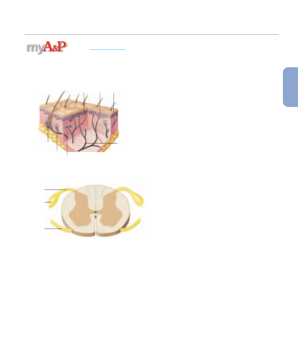
Chapter 15
Neural Integration I: Sensory Pathways and the Somatic Nervous System
527
15
NER
V
OUS
Review Questions
See the blue Answers tab at the end of the book.
Access
more review material online at
myA&P™ (
www.myaandp.com
). There,
you’ll find chapter guides, chapter quizzes,
practice tests, animations, flashcards, a glossary with pronunciations,
Interactive Physiology
®
(IP) exercises and quizzes, and more to
help you succeed in the course
.
LEVEL 1
Reviewing Facts and Terms
1. Identify the tactile receptors of the skin in the following diagram.
(a) ___________
(b) ___________
(c) ___________
(d) ___________
(e) ___________
(f) ___________
2. Shade in all the ascending sensory tracts in the following
diagram of the spinal cord.
my
a
b
Sensory
nerves
f
e
d
c
Dorsal root
Dorsal root
ganglion
Ventral root
3. The larger the receptive field, the
(a) larger the stimulus needed to stimulate a sensory receptor.
(b) fewer sensory receptors there are.
(c) harder it is to locate the exact point of stimulation.
(d) larger the area of the somatosensory cortex in the brain that
deals with the area.
(e) closer together the receptor cells.
4. The CNS interprets information entirely on the basis of the
(a) number of action potentials that it receives.
(b) kind of action potentials that it receives.
(c) line over which sensory information arrives.
(d) intensity of the sensory stimulus.
(e) number of sensory receptors that are stimulated.
5. The area of sensory cortex devoted to a body region is relative to the
(a) size of the body area.
(b) distance of the body area from the brain.
(c) number of motor units in the area of the body.
(d) number of sensory receptors in the area of the body.
(e) size of the nerves that serve the area of the body.
6. ___________ are receptors that are normally inactive, but
become active for a short time whenever there is a change in
the condition that they monitor.
7. Identify six types of tactile receptors located in the skin, and
describe their sensitivities.
8. What three types of mechanoreceptors respond to stretching,
compression, twisting, or other distortions of the plasma
membrane?
9. What are the three major somatic sensory pathways, and what
is the function of each pathway?
10. Which three pairs of descending tracts make up the
corticospinal pathway?
11. Which three motor pathways make up the medial pathway?
12. What are the two primary functional roles of the cerebellum?
13. The corticospinal tract
(a) carries motor commands from the cerebral cortex to the
spinal cord.
(b) carries sensory information from the spinal cord to the brain.
(c) starts in the spinal cord and ends in the brain.
(d) does all of these.
14. What three steps are necessary for transduction to occur?
LEVEL 2
Reviewing Concepts
15. Differentiate between a tonic receptor and a phasic receptor.
16. What is a motor homunculus? How does it differ from a
sensory homunculus?
17. Describe the relationship among first-, second-, and third-
order neurons.
18. Damage to the posterior spinocerebellar tract on the left side
of the spinal cord at the L
1
level would interfere with the
coordinated movement of which limb(s)?
19. What effect does injury to the primary motor cortex have on
peripheral muscles?
20. By which structures and in which part of the brain is the level
of muscle tone in the body’s skeletal muscles controlled? How
is this control exerted?
21. Explain the phenomenon of referred pain in terms of labeled
lines and organization of sensory tracts and pathways.
LEVEL 3
Critical Thinking and Clinical Applications
22. Kelly is having difficulty controlling her eye movements and
has lost some control of her facial muscles. After an
examination and testing, Kelly’s physician tells her that her
cranial nerves are perfectly normal but that a small tumor is
putting pressure on certain fiber tracts in her brain. This
pressure is the cause of Kelly’s symptoms. Where is the tumor
most likely located?
23. Clarence, a construction worker, suffers a fractured skull when a
beam falls on his head. Diagnostic tests indicate severe damage to
the motor cortex. His wife is anxious to know if he will ever be able
to move or walk again. What would you tell her?
24. Phil had to have his arm amputated at the elbow after an
accident. He tells you that he can sometimes still feel pain in
his fingers even though the hand is gone. He says this is
especially true when he bumps the stub. How can this be?
Wyszukiwarka
Podobne podstrony:
Fundamentals of Anatomy and Physiology 8e A01 MART5891 08 SE ESHT
Fundamentals of Anatomy and Physiology 8e DES MART5891 08 SE DE#2DDEA
Fundamentals of Anatomy and Physiology 8e DES MART5891 08 SE C0#2DDE9
Fundamentals of Anatomy and Physiology 8e M27 MART5891 08 SE C27
Fundamentals of Anatomy and Physiology 8e A01 MART 5891 08 SE FM
Fundamentals of Anatomy and Physiology 8e Z03 MART 5891 08 SE ANS
Fundamentals of Anatomy and Physiology 8e Z05 MART 5891 08 SE PCRED
Fundamentals of Anatomy and Physiology 8e Z02 MART 5891 08 SE App
Fundamentals of Anatomy and Physiology 8e Z07 MART 5891 08 SE INDX
Fundamentals of Anatomy and Physiology 8e Z08 MART 5891 08 SE END
Fundamentals of Anatomy and Physiology 8e Z04 MART 5891 08 SE GLOS
Fundamentals of Anatomy and Physiology 8e ZO6 MART 5891 08 SE S#2DE0F
Fundamentals of Anatomy and Physiology Glossary 2
Fundamentals of Anatomy and Physiology 22 Chapter
Fundamentals of Anatomy and Physiology FM
Fundamentals of Anatomy and Physiology Appendix III
Fundamentals of Anatomy and Physiology Appendix I
Fundamentals of Anatomy and Physiology Appendix II
Fundamentals of Anatomy and Physiology ENDPAP
więcej podobnych podstron