Guzy pochodzenia mezodermalnego
W grupie nowotworów pochodzenia mezodermalnego mieszczą się guzy wywodzące się ze zróżnicowanych tkanek, pochodzących ze środkowego listka zarodkowego, bądź też z wielopotencjalnych komórek mezenchymalnych. W piśmiennictwie onkologicznym dużą część tych nowotworów ujmuje się w niezbyt dokładnie zdefiniowanej grupie „guzów tkanek miękkich” (ang. soft tissue tumours). Do guzów tkanek miękkich nie należą na przykład nowotwory rozwijające się w kościach, ale zalicza się doń nowotwory obwodowego układu nerwowego.
Cechami charakterystycznymi guzów mezenchymalnych m.in. są:
tworzenie włokien srebrochłonnych (w rozrostach nabłonkowych komórki raka niszczą podścielisko)
niezbyt wyraźna róznica miedzy postaciami rozostów łagodnych, odczynowych, półzłosliwych i złośliwych
brak pojęcia „mięsaka przedinwazyjnego”
duża róznosrodność immunofenotypowa i morfologiczna tych rozrostów
Nowotwory mezenchymalne (zwłaszcza złośliwe) występują u ludzi znacznie rzadziej niż nowotwory nabłonkowe, a ich biologiczne właściwości są mniej poznane. W przypadku nowotworów złośliwych mięsaki stanowią ok. 1/10 nowotworów złosliwych występujacych u ludzi dorosłych (reszta to głownie raki) oraz ok. 1/3 nowotworów złosliwych wieku dzieciecego (reszta to głownie białaczki, chłoniaki i glejaki)
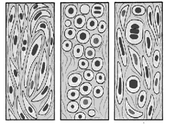
Przy wstepnej diagnostyce histopatologicznej tej grupy guzów (zwłaszcza złośliwych) często stosuje się roboczy podział na nowotwory drobnookrągłokomórkowe, nowotwory wrzecionowatokomórkowe i nowotwory polimorficznokomórkowe.
sarcomata fusocellularia |
sarcomata globocelullaria |
sarcomata polymorphocellularia |
|
||
leiomyosarcoama |
rhabdomyosarcoma embryonale |
fibrohistiocytoma malignum |
fibrosarcoma |
haemangosarcoma |
osteosarcoma extraskeletale |
sarcoma synoviale |
sarcoma Ewing |
inne nowotwory poliklonalne |
schwannoma malignum |
neuroepitheloma malignum |
|
|
liposarcoma globocellulare |
|
|
neuroblastoma |
|
Róznicowanie z: |
||
carcinoma planoepitheliale fusocellurare |
lymphoma malignum |
lymphoma malignum |
|
carcinoma anaplasticum |
carcinoma anaplasticum |
Szczegółowsza diagnostyka wymaga zwykle złozonych odczynów histochemicznych i immunohistochemicznych, czasem badań elektronowomikroskopowych
Nowotwory tkanek łacznych
WORLD HEALTH ORGANIZATION'S HISTOLOGICAL CLASSIFICATION OF SOFT TISSUE TUMORS (1995)
1 Fibrous tissue tumor
Benign
Fibroma
Keloid
Nodular fasciitis
Proliferative fasciitis
Proliferative myositis
Elastofibroma
Fibrous hamartoma of infancy
Myofibromatosis, solitary and multicentric
Fibromatosis colli
Calcifying aponeurotic fibroma
Hyaline fibromatosis
Fibromatosis
Superficial fibromatosis
Palmar and plantar fibromatosis
Infantile digital fibromatosis (digital fibroma)
Deep fibromatosis
Abdominal fibromatosis (desmoid tumor)
Extraabdominal fibromatosis (desmoid tumor)
Intraabdominal and mesenteric fibromatosis
Infantile fibromatosis
Malignant
Fibrosarcoma
Adult fibrosarcoma
Congenital or infantile fibrosarcoma
WORLD HEALTH ORGANIZATION'S HISTOLOGICAL CLASSIFICATION OF SOFT TISSUE TUMORS (1995) CONTINUED
2. Fibrohistiocytic tumors
Benign
Fibrous histiocytoma
Cutaneous histiocytoma (dermatofibroma)
Deep histiocytoma
Juvenile xanthogranuloma
Reticulohistiocytoma
Xanthoma
Intermediate
Atypical fibroxanthoma
Dermatofibrosarcoma protuberans
Pigmented dermatofibrosarcoma protuberans (Bednar tumor)
Giant cell fibroblastoma
Plexiform fibrohistiocytic tumor
Angiomatoid fibrous histiocytoma
Malignant
Malignant fibrous histiocytoma
Storiform-pleomorphic
Myxoid
Giant cell
Xanthomatous (inflammatory)
3. Lipomatous tumors
Benign
Lipoma
Lipoblastoma (fetal lipoma)
Lipomatosis
Angiolipoma
Spindle cell lipoma
Pleomorphic lipoma
Angiomyolipoma
Myelolipoma
Hibernoma
Atypical lipoma
Malignant
Well-differentiated liposarcoma
Lipomalike
Sclerosing
Inflammatory
Myxoid liposarcoma
Round cell (poorly differentiated myxoid) liposarcoma
Pleomorphic liposarcoma
Dedifferentiated liposarcoma
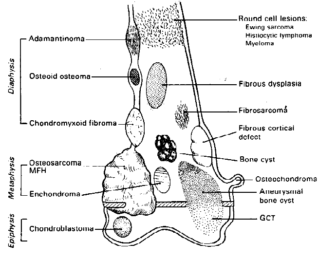
WORLD HEALTH ORGANIZATION'S HISTOLOGICAL CLASSIFICATION OF BONE TUMORS (1995)
Bone-forming tumors (19.3%)
Benign
Osteoma
Osteoid osteoma and osteoblastoma (benign osteoblastoma)
Malignant
Osteosarcoma (conventional)
Teleangiectaic
Small cell
Fibrohistiocytic
Anaplastic
Well-differentiated intramedullary
Juxtacortical osteosarcoma (Parosteal osteogenic sarcoma)
Periosteal osteosarcoma
Osteosarcoma of the jaw
Osteosarcoma in Paget disease
Cartilage-forming tumors (20.9%)
Benign
Chondroma
Osteochondroma (osteocartilagineus exostosis)
Chondroblastoma (benign chondroblastoma, epiphyseal chondroblastoma)
Chondromyxoid fibroma
Malignant
Chondrosarcoma
Juxtacortical chondrosarcoma
Mesenchymal chondrosarcoma
Giant cell tumor (osteoclastoma, grade I, II and III) (9.8%)
Marrow tumors (41.4%)
Malignant
Ewing's tumor (primitive neuroectodermal tumor-PNET)
Malignant lymphoma and related tumors
Primary large cell malignant lymphoma
Burkitt's lymphoma
Hodgkin's disease
Acute leucaemia
Plasma cell myeloma
Vascular Tumors (1.6%)
Benign
Haemangioma
Massive osteolysis (Gorham's diserase)
Lymphangioma
Glomus tumor
Intermaediate or indeterminate
Haemangioendothelioma
Haemangiopericytoma
Malignant
Angiosarcoma
WORLD HEALTH ORGANIZATION'S HISTOLOGICAL CLASSIFICATION OF BONE TUMORS (1995) (continued)
Other connective tissue tumors (3.8%)
Benign
Solitary fibrous tumor
Desmoplastic fibroma
Infantile myofibromatosis
leiomyoma
lipoma
Malignant
Fibrosarcoma
Malignant fibrous histiocytoma
leiomyosarcoma
liposarcoma
Other primary tumors of bone (3.1%)
Chordoma
Adamantinoma of long bones
Peripherial nerve tumors
Neurilemmoma
Neurofibroma and von Recklinghausen's disease
Malignant peripherial nerve sheath tumor
Xanthoma of bone
Neurilemmoma
Tumor like lesions
Solitary bone cyst (simple bone cyst)
Aneurysmal bone cyst
Ganglion cyst of bone (Juxta-articular bone cyst)
Metaphysel fibrous defect (non-ossifyig fibroma)
Fibrous dysplasia
Myositis ossificans
Langerhans' cell granulomatosis
Eosinophilic granuloma
Brown tumor in hyperparathyreoidism
Nowotwory innych tkanek „miękkich”
WORLD HEALTH ORGANIZATION'S HISTOLOGICAL CLASSIFICATION OF SOFT TISSUE TUMORS (1995) CONTINUED
4. Smooth muscle tumors
Benign
Leiomyoma
Angiomyoma
Epithelioid leiomyoma
Leiomyomatosis peritoneales disseminata
Malignant
Leiomyosarcoma
Epithelioid leiomyosarcoma
5. Skeletal muscle tumor
Benign
Rhabdomyoma
Adult
Genital
Fetal
Malignant
Rhabdomyosarcoma
Embryonal rhabdomyosarcoma
Botryoid rhabdomyosarcoma
Spindle cell rhabdomyosarcoma
Alveolar rhabdomyosarcoma
Pleomorphic rhabdomyosarcoma
Rhabdomyosarcoma with ganglionic differentiation (ectomesenchymoma)
6. Endothelial tumors of blood and lymph vessels
Benign
Papillary endothelial hyperplasia
Hemangioma
Capillary hemangioma
Cavernous hemangioma
Venous hemangioma
Epithelioid hemangioma (angiolymphoid hyperplasia, histiocytoid hemangioma)
Pyogenic granuloma (granulation tissue type hemangioma)
Acquired tuffed hemangioma (angioblastoma)
Lymphangioma
Lymphangiomyoma
Lymphangiomyomatosis
Angiomatosis
Lymphangiomatosis
Intermediate: Hemangioendothelioma
Spindle cell hemangioendothelioma
Endovascular papillary angioendothelioma (Dąbska tumor)
Epithelioid hemangioendothelioma
Malignant
Angiosarcoma
Lymphangiosarcoma
Kaposi's sarcoma
WORLD HEALTH ORGANIZATION'S HISTOLOGICAL CLASSIFICATION OF SOFT TISSUE TUMORS (1995) CONTINUED
7. Perivascular tumors
Benign
Benign hemangiopericytoma
Glomus tumor
Malignant
Malignant hemangiopericytoma
Malignant glomus tumor
8. Synovial tumors
Benign
Tenosynovial giant cell tumor
Localized
Diffuse (extraarticular pigmented villonodular synovitis)
Malignant
Malignant tenosynovial giant cell tumor
9. Mesothelial tumors
Benign
Solitary fibrous tumor of pleura and peritoneum (localized fibrous mesothelioma)
Multicystic mesothelioma
Adenomatoid tumor
Well-differentiated papillary mesothelioma
Malignant
Malignant solitary fibrous tumor of pleura and peritoneum (malignant localized fibrous mesothelioma)
Diffuse mesothelioma
Epithelial
Spindled (sarcomatoid)
Biphasic
10. Neural tumors
Benign
Traumatic neuroma
Morton's neuroma
Neuromuscular hamartoma
Nerve sheath ganglion
Schwannoma (neurilemoma)
Plexiform schwannoma
Cellular schwannoma
Degenerated (ancient) schwannoma
Neurofibroma
Diffuse
Plexiform
Pacinian
Epithelioid
Granular cell tumor
Melanocytic schwannoma
Neurothekeoma (nerve sheath myxoma)
Ectopic meningioma
Ectopic ependymoma
Ganglioneuroma
Pigmented neuroectodermal tumor of infancy (retinal enlage tumor, melanotic progonoma)
WORLD HEALTH ORGANIZATION'S HISTOLOGICAL CLASSIFICATION OF SOFT TISSUE TUMORS (1995) CONTINUED
Malignant
Malignant peripheral nerve sheath tumor (MPNST) (malignant schwannoma, neurofibrosarcoma)
Malignant peripheral nerve sheath tumor with rhabdomyosarcoma (malignant triton tumor)
Malignant peripheral nerve sheath tumor with glandular differentiation
Epithelioid malignant peripheral nerve sheath tumor
Malignant granular cell tumor
Clear cell sarcoma (malignant melanoma of soft parts)
Malignant melanotic
Neuroblastoma
Ganglioneuroblastoma
Neuroepithelioma (peripheral neuroectodermal tumor, peripheral neuroblastoma)
11 . Paraganglionic tumors
Benign
Paraganglioma
Malignant
Malignant paraganglioma
12. Cartilage and bone tumors
Benign
Panniculitis ossificans
Myositis ossificans
Fibrodysplasia (myositis) ossificans progressiva
Extraskeletal chondroma
Extraskeletal osteochondroma
Extraskeletal osteoma
Malignant
Extraskeletal chondrosarcoma
Well-differentiated chondrosarcoma
Myxoid chondrosarcoma
Mesenchymal chondrosarcoma
Dedifferentiated chondrosarcoma
Extraskeletal osteosarcoma
13. Pluripotential mesenchymal tumors
Benign
Mesenchymoma
Malignant
Malignant mesenchymoma
WORLD HEALTH ORGANIZATION'S HISTOLOGICAL CLASSIFICATION OF SOFT TISSUE TUMORS (1995) CONTINUED
14. Miscellaneous tumors
Benign
Congenital granular cell tumor
Tumoral calcinosis
Myxoma
Cutaneous
Intramuscular
Angiomyxoma
Amyloid tumor
Parachordoma
Ossifying fibromyxoid tumor
Juvenile angiofibroma
Inflammatory myofibroblastic tumor (inflammatory fibrosarcoma)
Malignant
Alveolar soft part sarcoma
Epithelioid sarcoma
Extraskeletal Ewing sarcoma
"Synovial" sarcoma
Monophasic fibrous type
Malignant (extrarenal) rhabdoid tumor
Desmoplastic small cell tumor of children and young adults
15. Unclassified tumors
Gastrointestinal stromal tumor (GIST)- guz o niejasnej histogenezie, i bardzo zróznicowaj dynamice (na ogół o złośliwym przebiegu), rosnacy w ścianie żoładka i jelit. Dawniej uznawano go za guz pochodzenia mięśniowego lub nerwowego. Uważa się, że wywodzi się ze specjalnych komórek nerwowychukładu autonomicznego (gastroiintestinal pacemaker cells) lub niedojrzałych komórek mezenchymalnych
Wyszukiwarka
Podobne podstrony:
wyklad 3 now4, Patomorfologia, Zmiany postępowe i nowotwory
wyklad 3 now5, Patomorfologia, Zmiany postępowe i nowotwory
Nowotwory nabłonkowe, Patomorfologia, Zmiany postępowe i nowotwory
nowotwory5, Patomorfologia, Zmiany postępowe i nowotwory
zab w krążeniu +zmiany postępowe, Patomorfologia, Zmiany postępowe i nowotwory
CHOROBY MIELO – LIMFO – (RETIKULO) – PROLIFERACYJNE, Patomorfologia, Zmiany postępowe i nowotwory
TNM, Patomorfologia, Zmiany postępowe i nowotwory
Wykład, Patomorfologia, Zmiany postępowe i nowotwory
pytania patomorfo koło 2 postepowe nowotwory
zmiany postępowe i wsteczne transformacja nowotworowa
09 ZMIANY POSTĘPOWE I GUZY NIENOWOTWOROWEid 7800 ppt
patomorfa charakterystyka wybranych nowotworów(2)
ZMIANY POSTĘPOWE, Wykłady
Zmiany przednowotworowe i nowotwory nabonkowe
Zmiany postępowe 3(1), ★ materiały rok III wety, rok III, ANATOMIA PATOLOGICZNA, II KOŁO
patomorfa charakterystyka wybranych nowotworów (1)
więcej podobnych podstron