26
Biofunctionalized Nanoparticles for Surface-Enhanced Raman
Scattering and Surface Plasmon Resonance
Mahnaz El-Kouedi and Christine D. Keating
26.1
Overview
26.1.1
Introduction
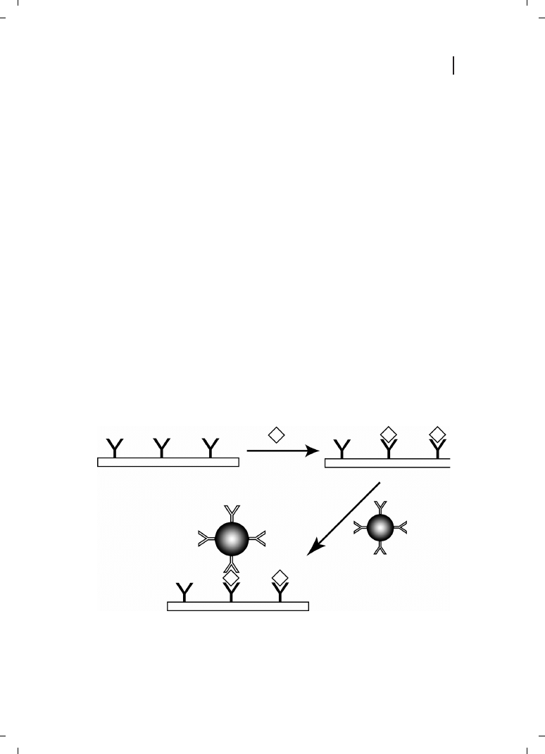
Nano- and microparticle–biomolecule conjugates have been used as amplification tags in
a wide variety of biosensing schemes [1]. The general format for many of these assays,
which are analogous to fluorescently-detected sandwich immunoassays, is illustrated in
Figure 26.1. A sensing surface is derivatized with a biorecognition element (such as an
antibody or oligonucleotide) which is capable of selectively binding the desired analyte
from solution. Detection is achieved by completing the “sandwich” with a second selective
429
Figure 26.1
Typical bioconjugate-based sandwich bioassay. A surface functionalized with biorecognition
chemistry (in this case an antibody) is exposed to analyte (diamond), after which a secondary antibody-
labeled particle binds. The particle amplifies the sensor response, thereby increasing sensitivity.
Nanobiotechnology. Edited by Christof Niemeyer, Chad Mirkin
Copyright
c 2004 WILEY-VCH Verlag GmbH & Co. K aA, Weinheim
ISBN 3-527-30658-7
G

biorecognition molecule, this time tagged with a particle. This chapter will focus on metal
nanoparticle bioconjugates in two surface-based optical sensing platforms : surface plas-
mon resonance (SPR) and surface-enhanced Raman scattering (SERS) [2–4]. In both tech-
niques, the nanoscale structure at a metal–dielectric interface is critically important to suc-
cessful sensing. We will discuss the application of metal nanoparticle bioconjugates in
sensing strategies based on refractive index changes (optical extinction and surface plas-
mon resonance) and electromagnetic field enhancement of vibrational scattering (SERS).
26.1.2
Applications in SPR
In recent years, a wide variety of analytical techniques has been developed based on
changes associated with the SPR of gold and silver nanoparticles and thin films. The
SPR is a consequence of the collective oscillations of metal valence electrons resulting
in a strong absorption peak that can be used to monitor changes in the surrounding me-
dium such as biomolecule adsorption events at the metal surface, or nanoparticle aggre-
gation [3, 4]. Analytical plasmon resonance techniques can be divided into two classes :
1. extinction-based methods, in which changes in the visible or near-infra-red (IR) trans-
mission are monitored, typically for collections of nanoparticles; and
2. reflectivity-based methods, in which changes in the angle-dependent intensity of re-
flected light are monitored for planar metal films or gratings.
26.1.2.1
Nanoparticle Substrates
An introduction to aggregation-based bioassays can be found in a review by Englebienne
[5]. Briefly, metal nanoparticle aggregation results in a red shift of the plasmon resonance
absorption, accompanied by a decrease in peak intensity due to aggregate sedimentation.
Aggregation of “bare”, charge-stabilized nanoparticles can be induced by the simple addi-
tion of high concentrations of salt; bioconjugates can be selectively crosslinked by the
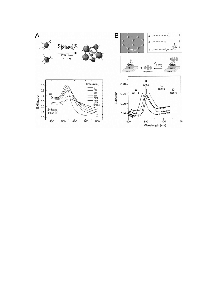
presence of analyte. For example, Mirkin and coworkers reported a DNA sensor based
on the dramatic redshift in absorbance upon hybridization-driven gold nanoparticle aggre-
gation [6, 7] (Figure 26.2A). Recently, Englebienne has used colloidal nanoparticles for the
high-throughout screening of proteins, and has determined ligand–protein interactions
for 30 antibody–antigen pairs [8]. For Au nanoparticles, optical changes occur in the visi-
ble; to monitor bioassays in whole blood, Hirsch et al. have used SiO
2
core/Au shell bio-
conjugates, which absorb in the near-IR where interference from cell and tissue absorp-
tion is minimal [9].
Bioconjugate aggregation is not a requirement for analyte detection; it is also possible to
monitor analyte binding through small shifts in the plasmon resonance absorbance due to
changes in the refractive index surrounding the particle [3, 10, 11]. Indeed, Van Duyne
and coworkers have taken advantage of changes in this localized SPR effect (LSPR) to
monitor protein binding to triangular Ag nanoparticles bound to a glass substrate (Figure
26.2B) [12]. In this investigation,
Z50 nm-high, 100 nm-wide nanoparticles were prepared
by nanosphere lithography [13] and derivatized with biotin for streptavidin detection. The
binding of 100 nM streptavidin to biotinylated prisms results in an impressive 27-nm red-
430
26 Biofunctionalized Nanoparticles for Surface-Enhanced Raman Scattering and Surface Plasmon Resonance
shift of the plasmon resonance band. Low picomolar/high femtomolar limits of detection
were reported for streptavidin binding; samples were measured dry, in a N
2
environment.
These authors also demonstrated that the LSPR response could be amplified by using bio-
tin-modified colloidal Au nanoparticles in a sandwich format [12]. Recent improvements
have enabled real-time LSPR analysis of antibody binding in physiological buffer, with a
detection limit of
Z700 pM [14].
26.1.2.2
Planar Substrates
SPR techniques based on the planar Au substrate configuration have been utilized exten-
sively for the detection of biomolecules, and have become increasingly popular since the
development of the first commercial BIAcore instrument in 1990 [2, 15]. SPR detection is
based on a refractive index change near a thin noble metal film upon analyte binding. The
conventional SPR system is based on the Kretschmann configuration. In this configura-
tion, a metal film (usually
Z50 nm of Au) is evaporated either directly onto a hemisphe-
rical prism or evaporated onto a glass slide and index matched to the prism [2, 16]. Under
conditions of total internal reflection, p-polarized light is used to illuminate the metal film
431
26.1 Overview
Figure 26.2
(A) Aggregation-based solution assay
for the detection of DNA as utilized by Mirkin et al.
[6]. A solution of DNA-coated nanoparticles
changes color from red to blue upon the addition of
a complementary DNA linker sequence, due to
DNA duplex formation and subsequent particle
aggregation (top). The aggregation results in the
shift and broadening of the plasmon resonance
peak, which can be monitored using absorbance
spectroscopy (bottom). (Reprinted with permission
from ACS;
c 2000.) (B) Localized surface plasmon
resonance (LSPR) biosensor utilized by Van Duyne
et al. [12]. Triangular nanoparticles synthesized
using nanosphere lithography techniques were
functionalized with a biotinylated SAM layer. Strep-
tavidin was then detected by looking at the shift in
the Ag plasmon resonance band. In the lower panel,
curve (A) unfunctionalized Ag; (B) Ag with ami-
nated SAM; (C) nanoparticles after biotin attach-
ment; (D) streptavidin detection shift. (Reprinted
with permission from ACS;
c 2002.)

through the prism. At the plasmon resonant angle, the light induces the collective oscilla-
tions of the valence electrons of the metal film, resulting in a decrease in the reflectivity.
The position of this resonance angle depends strongly on the refractive index at the metal
interface; changes in the resonant angle or in the intensity of reflected light at a fixed
angle can be used to detect binding events at the substrate surface.
Although one advantage of SPR is the possibility of label-free detection, in many cases
conventional SPR lacks the desired sensitivity for biosensing. SPR response can be ampli-
fied by using a sandwich geometry, where the change in refractive index due to analyte
binding is enhanced by the binding of a macromolecule or particle with either large
MW or high refractive index (see Figure 26.1). Examples include protein–DNA multilayers
[17], liposomes [16], and polymer beads [18, 19]. We will focus on Au nanoparticle biocon-
jugates as amplification tags, an excellent introduction to which subject can be found in
Ref. 20.
Large perturbations in the SPR response upon Au nanoparticle binding are thought to
result largely from the particles acting as roughness features, enabling nonradiative sur-
face plasmons propagating in the thin film to become radiative modes [20]. Au nanopar-
ticle-amplified SPR has been used for detection of human serum albumin (HAS), using
30-nm diameter Au :protein bioconjugates [21], and also for the detection of immunoglo-
bulins [22, 23]. A thousand-fold enhancement was observed for HAS detection as com-
pared to the unamplified binding event [21]. Au :IgG bioconjugate binding was monitored
in real time by Gu et al., who observed SPR response saturation in minutes [22].
Lyon et al. used 11-nm diameter colloidal Au for detection of human IgG using a sand-
wich immunoassay such as that described in Figure 26.1 [23]. For a 6.7 pM solution of
IgG, a 0.33¯ shift was observed in the resonance angle; given that angle shifts
J0.005¯
are detectable, these authors reasoned that actual detection limits may be much lower
than picomolar [23]. The experiments showed a “quasi-linear” relationship between the
particle coverage and the shift of the plasmon resonance angle, such that SPR shift can
be related to antigen concentration.
Some experimental and theory papers have addressed the impact of nanoparticles on
the SPR response of thin metal films. The electromagnetic coupling efficiency and
hence the amplification efficiency of the Au tags typically decreases with increasing se-
paration [3, 4,24]. This is demonstrated by the reduced SPR shift for the protein detection
experiments as compared to the shifts observed for the binding of Au nanoparticles on a
mercaptoethylamine self-assembled monolayer (SAM) [23]. Leung et al. stressed the im-
portance of nanoparticle film morphology, predicting that clustering of the nanoparticles
would lead to greater signal amplification [25]. Fendler and coworkers have examined the
SPR response from Au and Ag thin films upon binding 10-nm Au or 30-nm diameter Ag
nanoparticles with both experiments and modeling [26], while Lyon et al. have investigated
the effect of Au nanoparticle size on SPR shift for particles 30–60 nm in diameter [27]. At
constant particle coverages, greater shifting and broadening of the plasmon peak was ob-
served with increasing particle size. From surface coverage data, these authors concluded
that larger particles provide greater sensitivity, but at the same time decrease the maxi-
mum concentration that can be quantified [27].
Recently, Au particle-amplified SPR has been used to increase the sensitivity for the de-
tection of short oligonucleotide sequences 1000-fold over the unamplified experiment [28].
432
26 Biofunctionalized Nanoparticles for Surface-Enhanced Raman Scattering and Surface Plasmon Resonance
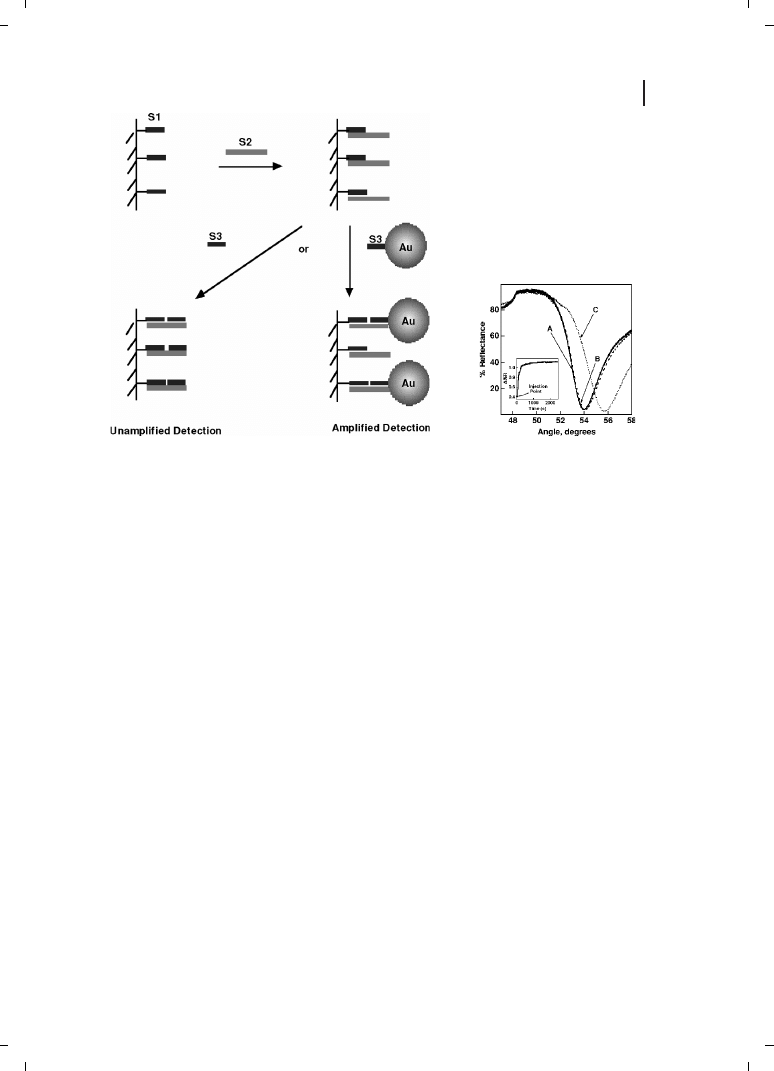
In these experiments, a sandwich hybridization assay analogous to the immunoassay of
Figure 26.1 was employed, in which three DNA strands were used (Figure 26.3). Submo-
nolayer coverage of a DNA oligonucleotide that was half complementary to the analyte se-
quence, was attached to the sensor surface. The analyte was then introduced, followed by
binding of the Au amplification tags. Upon binding of the nanoparticles to the surface, a
large perturbation to the resonance angle was observed (Figure 26.3). Binding reversibility
was demonstrated using thermal denaturation and enzyme cleavage to remove bound
DNA and DNA :Au bioconjugates [28].
He et al. also demonstrated the feasibility of using imaging SPR and Au particle ampli-
fication to detect hybridization events in an array format with
Z10 pM detection limits
and a five orders of magnitude dynamic range [28]. These experiments show promise
for particle amplified SPR in array-based DNA analysis. A key challenge will be reducing
the background arising from nonspecific binding of bioconjugates to the metal film; it is
this nonspecific background that currently limits the sensitivity of the technique. In the
absence of nonspecific binding, substantial improvements in detection limits are ex-
pected.
433
26.1 Overview
Figure 26.3
Particle-amplified SPR for the detec-
tion of DNA as demonstrated by He et al. [28]. DNA
sequence 1 (S1) is attached to a thin gold film
evaporated onto a glass slide (curve A). The analyte
sequence S2 binds to the surface, resulting in a very
small shift in the SPR response (curve B). A large
shift was observed upon the binding of sequence S3
attached to Au nanoparticle amplification tags, as
shown in curve C. Inset shows real-time monitoring
of the change in reflectivity at 53.2¯ after exposure to
S3 :Au particles. (Reprinted with permission from
ACS;
c 2002.)

26.1.3
Applications in SERS
SERS refers to the large signal enhancements observed for molecules adsorbed at rough-
ened metal surfaces, particularly Ag and Au [4]. Two phenomena give rise to enhance-
ments :
1. Local amplification of the electromagnetic field near roughness features in free elec-
tron metals. This portion of the SERS enhancement can be theoretically modeled,
and has been reported to account for the bulk of the observed enhancements [4].
2. For molecules chemisorbed to the metal surface, an additional chemical effect is ob-
served. This effect has been described by Campion and others [4, 29]. In combination,
these effects can give rise to experimentally observed enhancements sufficient to per-
mit single molecule detection [30].
An extensive literature exists describing SERS and resonant-SERS (or SERRS) of various
biomolecules [4, 30]. In SERRS, the laser line used for excitation is resonant with an op-
tical absorbance of the molecule, leading to an additional enhancement in scattering from
vibrations coupled to the electronic transition. In nearly all cases, proteins are added to
colloidal Ag sols to induce aggregation or are added immediately after aggregation has
been induced by addition of salt. The resulting Ag nanoparticle–protein aggregates are
ill-defined but often give rise to very intense SERS spectra. Due in part to the success
of this approach, very few studies of well-defined nanoparticle bioconjugates have ap-
peared. SERS studies of protein and nucleic acid bioconjugates are described separately,
as these molecules have very different binding modalities to metal nanoparticles.
26.1.3.1
Proteins
Unlike many SPR and LSPR approaches, the goal of protein SERS has often been to elu-
cidate binding chemistry and active site structure–function rather than to detect the pro-
tein of interest. The bioactivity of adsorbed protein molecules is of great importance, par-
ticularly given the propensity of proteins to denature on solid surfaces [31–33]. Several
groups have addressed the issue of bioactivity for adsorbed enzymes in SERS studies.
The characteristic vibrational frequencies of heme chromophores present in many en-
zymes enable spectroscopic determination of enzyme stability, orientation, and charge
state. Several groups have used the heme spin state marker bands to monitor conforma-
tional changes in proteins adsorbed to aggregated Ag sols. Although early studies at Ag
electrodes [32] and BH
4
-reduced Ag sols [33] indicated some protein denaturation at the
metal surface, later investigations for cytochrome c (Cc) and several cytochromes P450
showed the retention of native spin states, which suggested retention of native protein
structure [34–36]. The principal difference in the latter studies was the use of Ag sols pre-
pared by citrate reduction of AgNO
3
[37]; such particles have an adsorbed layer of citrate
which seems to increase their biocompatibility. Citrate-reduced Ag nanoparticles were also
used in a study by Broderick et al. which showed that the nonheme iron enzyme chloro-
catechol dioxygenase retained 60–85 % of its activity when adsorbed to aggregated colloi-
dal Ag nanoparticles. These authors were able to observe enzymatic turnover at the Ag
surface via SERRS [38].
434
26 Biofunctionalized Nanoparticles for Surface-Enhanced Raman Scattering and Surface Plasmon Resonance

Well-defined, discrete protein–nanoparticle bioconjugates were not employed in any of
the aforementioned SERS investigations, and indeed are rarely used for SERS. The prin-
cipal reason for this is that it is simply not necessary to prepare discrete bioconjugates in
order to acquire SERS spectra. In addition to the ease of acquiring SERS spectra for bio-
molecules adsorbed to colloidal Ag aggregates or other roughened Ag substrates, another
important reason for the relative dearth of biomolecule–nanoparticle conjugate SERS
studies is the relative difficulty of handling colloidal Ag sols, the substrate of choice
for biomolecule SERS. To our knowledge, the first use of SERS to characterize well-de-
fined bioconjugates was by Ahern and Garrell in 1991 [39]. For protein–Ag nanoparticle
conjugates, these authors observed a time-dependent increase in SERS signal for
proteins adsorbed to colloidal nanoparticles over the course of several days, consistent
with conformational changes of the protein. No SERS signal was observed for Au
nanoparticle bioconjugates [39]. Despite the poor SERS activity of isolated nanometer
Au spheres, Au bioconjugates are more stable than their Ag counterparts. Several
researchers have described methods for acquiring SERS for Au nanoparticle bio-
conjugates.
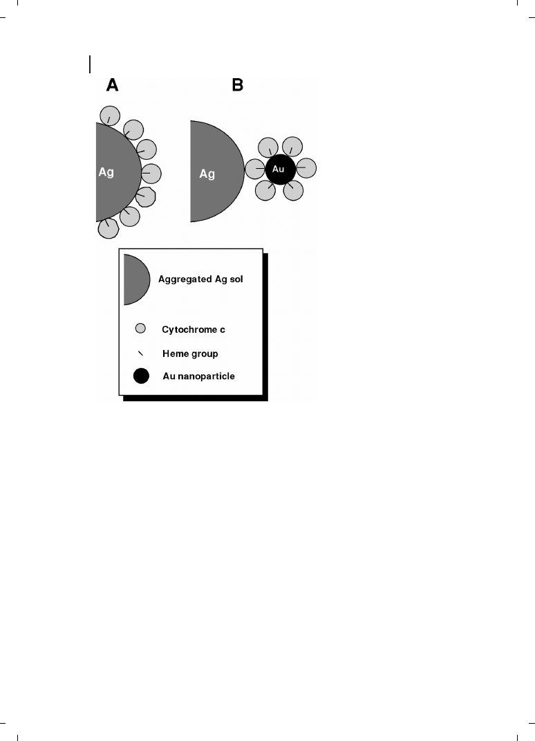
Keating et al. have demonstrated that preconjugation of Cc to colloidal Au nanoparticles
provided greater stability and control over protein orientation as compared to direct ad-
sorption onto aggregated Ag sols [40]. Well-defined Cc :Au nanoparticle conjugates were
prepared and SERRS spectra for the Cc :Au bioconjugates were compared to spectra for
Cc alone. The two geometries investigated in these studies are shown in Figure 26.4.
Ag :Cc and Ag :Cc :Au samples were prepared by adsorption of Cc or Cc :Au conjugates,
respectively, to aggregated colloidal Ag. When Cc adsorbs to negatively charged surfaces,
such as colloidal metal nanoparticles, the heme group is located near the metal surface.
Thus, in Cc :Au bioconjugates, the heme chromophore faces in towards the Au, and re-
mains so upon adsorption of the bioconjugates to the surface of aggregated Ag sol. In con-
trast, Cc directly adsorbed to aggregated Ag nanoparticles binds with its heme close to the
Ag surface (Figure 26.4). SERRS spectra for Ag :Cc :Au and Ag :Cc samples were closer in
intensity than expected based on heme proximity to the Ag surface, due to the presence of
the Au nanoparticles. In addition, preconjugation to colloidal Au led to reproducible pro-
tein orientation on the surface and increased resistance to conformational changes indi-
cated by the spin state marker bands [40].
These Ag :Cc :Au sandwiches provided an opportunity to probe the heightened
electromagnetic fields that had long been predicted between closely spaced metal
particles [4, 30]. SERRS spectra were acquired under identical conditions for
Ag :Cc :Au and Ag :Cc at several wavelengths in the visible spectrum [41]. Electromagnetic
coupling between the Au nanoparticles and the aggregated Ag surface was evidenced
as increased relative intensity for Ag :Cc :Au versus Ag :Cc at wavelengths matched to
the optical absorbance maximum of the Ag :Cc :Au sandwich. Keating et al. also prepared
Au :Cc :Au and Ag :Cc :Ag sandwiches; scattering intensities for the Ag :Cc :Ag sandwiches
exceeded that for directly adsorbed Cc, despite the greater distance between the heme
chromophore and the aggregated Ag surface for the sandwiches. These studies demon-
strated strong electromagnetic coupling between the bioconjugated metal nanoparticles
and the aggregated Ag surface, despite the intervening protein layer (the Cc diameter is
Z34 ) [41].
435
26.1 Overview
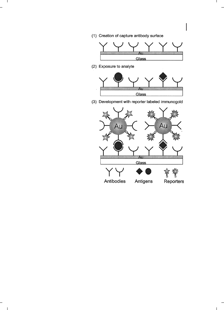
Porter and coworkers have reported using antibody–Au bioconjugates in a SERS-based
immunoassay [42]. In these studies, a submonolayer of small-molecule Raman probes was
adsorbed to the Au nanoparticle surface prior to adsorbing antibody molecules; this ap-
proach is illustrated in Figure 26.5. A capture antibody is attached to a planar Au sub-
strate. After analyte binding, Raman-tagged secondary antibody–Au bioconjugates are
added. SERS spectra show the characteristic fingerprint spectra from adsorbed Raman
probes, enabling identification of the analyte. By using different Raman probe molecules
for different antibodies, two immunoassays could be performed simultaneously. Raman
spectra provide narrower bandwidths and a much greater complexity as compared to fluor-
escence spectra; thus, many more Raman-based dyes can be envisioned. Hence, this bio-
conjugate SERS-based methodology has potential for multiplexed immunoassays, and de-
tection limits were estimated at
Z2 nM [42]. Further improvements may be possible by
using Raman probes for which resonant enhancement is possible at the SERS excitation
wavelength.
436
26 Biofunctionalized Nanoparticles for Surface-Enhanced Raman Scattering and Surface Plasmon Resonance
Figure 26.4
Schematic depiction of the
two types of SERS samples investigated
by Keating et al. [40]. In (A), Cc is directly
adsorbed on aggregated Ag nanoparti-
cles (Ag :Cc), while in (B) the Cc :Au
bioconjugates are adsorbed to aggre-
gated Ag, resulting in a greater separa-
tion between the SERS-active Ag surface
and the heme chromophore (Ag :Cc :Au).
(Reprinted with permission from ACS;
c1998.)
26.1.3.2
Nucleic Acids
Several groups have studied DNA binding to colloidal Au and Ag particles via SERS [43–
45]. Murphy and coworkers explored the binding of intrinsically bent double-stranded
DNA sequences to 14 nm-diameter colloidal Au spheres as a means of understand the
binding of these sequences to proteins of similar size [45]. Other investigations have
been aimed at ultrasensitive DNA detection. For example, Kneipp et al. have demonstrated
single molecule detection and identification of single DNA bases on colloidal Ag aggre-
gates [43].
Smith and coworkers have developed a SERRS-based detection strategy for ultrasensi-
tive detection of dye-labeled, single-stranded and double-stranded DNA via SERRS [44].
These authors have sought to improve sensitivity and reduce the standard deviations in
signals between experiments by controlling DNA adsorption on the Ag surface. The ne-
gative charge on the DNA backbone is electrostatically repelled from the negatively
charged colloidal Ag particles. Smith and coworkers addressed this by both incorporating
437
26.1 Overview
Figure 26.5
SERS immunoassay format
used by Porter et al. [42]. Amplification
tags comprised of Au nanoparticles
coated with both small molecule Raman
tags and antibodies for selective biore-
cognition of analyte. Sandwich immu-
noassays were probed by SERS. (Rep-
rinted with permission from ACS;
c
1999.)
positively charged propargylamino-modified deoxyuridine bases and mixing the modified
oligonucleotides with a positively charged polymer, spermine, prior to addition to colloidal
Ag nanoparticles. The resulting aggregated Ag suspension yields strong SERRS signals
for oligonucleotide-bound dye molecules, and enabled detection limits as low as 8
q
10
–13
M [44]. This approach has been used to detect PCR products using SERRS-active
primers which contained both a 5
l dye moiety as well as a propynyl-modified sequence
to improve adsorption [46].
In 1996, the Mirkin/Letsinger [47] and Alivisatos [48] groups each published methods
for single-point attachment of oligonucleotides to Au nanoparticles via self-assembly of
thiol groups covalently attached to the 3
l or 5l terminus of the DNA. These DNA–Au bio-
conjugates have found a wide range of applications in biosensing [49–51], but have only
recently begun to be used in SERS experiments. Franzen and coworkers have used SERS
to monitor DNA–Au bioconjugate monolayer morphology and surface aggregation [52].
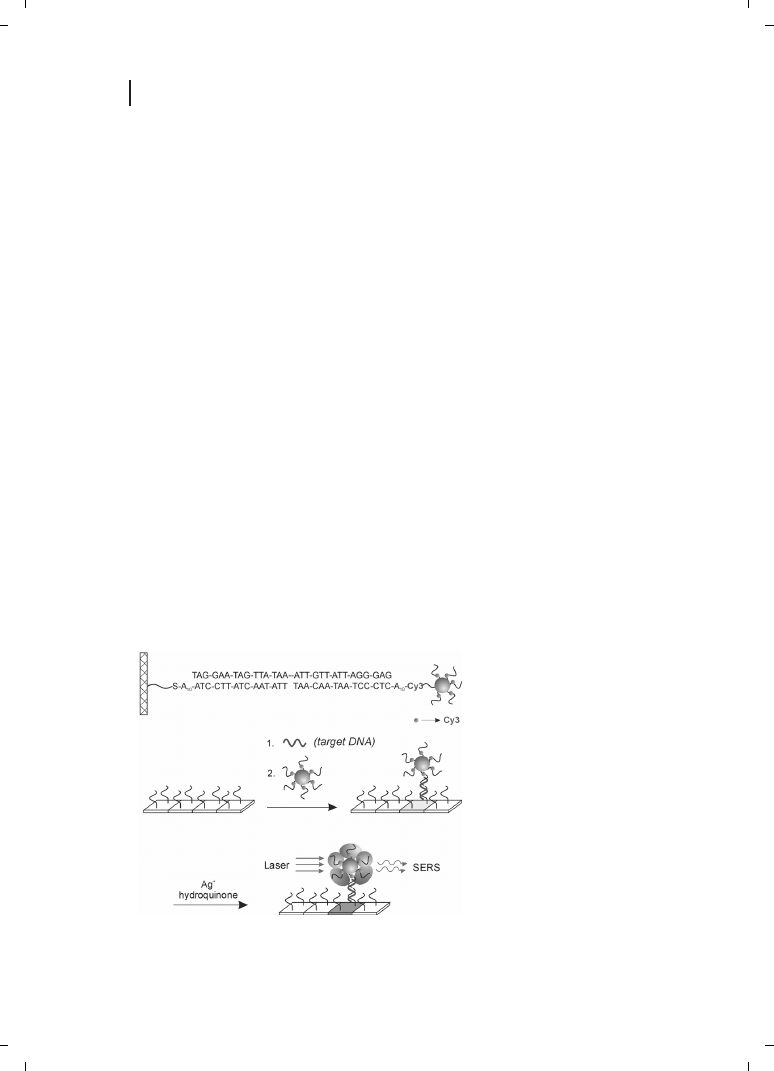
The potential of Raman probes as tags for multiplexing was exploited recently by the
Mirkin group to prepare an elegant multianalyte DNA sensor based on SERRS (Figure
26.6) [53]. In these studies, dye molecules were incorporated into the oligonucleotide se-
quence prior to assembly of the DNA–Au bioconjugates. A sandwich hybridization assay
was performed on a chip surface, such that bioconjugates attached to the surface only
when the analyte sequence was present. Rather than measure the Raman signal directly
from the surface-bound (dye-harboring) bioconjugates, Mirkin and colleagues chemically
reduced Ag onto the Au particles. This process resulted in a much more SERS-active sub-
strate as compared to the 13-nm Au particles alone. The resulting Raman spectra showed
good discrimination between six different bioconjugates, as well as detection limits of 20
q 10
–15
M. In addition to outstanding sensitivity and multiplexing capability, this ap-
proach benefits from the unusual selectivity of DNA–Au bioconjugate hybridization,
which permits discrimination of single base mismatches by temperature or salt stringency
assays [51, 52].
Mulvaney et al. recently reported a novel SERS-based nanoprobe, in which Raman-ac-
tive molecules on the surface of Au or Ag nanospheres were protected by a glass shell
438
26 Biofunctionalized Nanoparticles for Surface-Enhanced Raman Scattering and Surface Plasmon Resonance
Figure 26.6
SERS-based DNA and
RNA detection strategy reported by
Mirkin et al. [53]. DNA–Au bioconju-
gates incorporated Raman-active dye
molecules at the 3
l, thiol-terminated
end of the oligonucleotides. These
probes were selectively assembled
onto the DNA chip surface via sand-
wich hybridization assays, followed by
chemical reduction of Ag metal se-
lectively onto the Au bioconjugates.
SERS was then used to detect and
identify the Raman label, and hence
the DNA sequence, present in each
spot. (Reprinted with permission
from AAAS;
c 2002.)

[54]. Such particles could potentially be coupled to biomolecules of interest and used as
Raman tags in multiplexed bioassays.
26.2
Methods
26.2.1
Planar SPR Substrate Preparation
An excellent resource for SPR is the website of Professor Corn’s research group at the
University of Wisconsin, as this provides – among other things – software for Fresnel cal-
culations [55]. The preparation of SPR substrates begins with the vacuum evaporation of
an Au thin film (approximately 40–50 nm thickness) onto freshly cleaned glass substrates.
The surface is then derivatized for selective biomolecule attachment. Different attachment
chemistries have been developed, including the use of commercially available chips that
include a coupling matrix [15]. These polymer matrices – which are usually a functiona-
lized dextran – are not generally amenable to particle amplification, as the nanoparticles
are usually too large to fit into the pores of the matrix. Instead, alkanethiol-based self-as-
sembled monolayers and biotin–streptavidin attachment chemistries are favored for par-
ticle-amplified SPR. Streptavidin can be physisorbed onto the Au substrate, and biotiny-
lated DNA strands or proteins can then be attached to the substrate surface. The adsorp-
tion of a carboxy- or amino-terminated self-assembled monolayer followed by protein at-
tachment using carbodiimide chemistry (EDC/NHS), has also been successful [56].
26.2.2
Metal Nanoparticles
Recipes for the synthesis of colloidal Au particles of various sizes can be found in Ref. [57].
A slight modification of these protocols for monodisperse 12 nm-diameter Au nano-
spheres leads to an approximately four-fold increase in particle concentration [58]. The
most popular Ag nanoparticle preparation method for SERS is the citrate reduction pro-
tocol published by Lee and Meisel in 1982 [37]. Particles prepared following this protocol
are typically quite polydisperse but give rise to excellent SERS enhancements. Other Ag
particle recipes include EDTA reduction [41] and BH
4
reduction [33]. Synthetic methods
also exist for preparation of core-shell nanoparticles including Au core-Ag shell [59] and
silica core–Au shell [60]. Au and Ag nanoparticles are commercially available from Ted
Pella, Inc. (www.tedpella.com), with or without adsorbed antibodies or streptavidin.
26.2.3
Bioconjugates
Protein–nanoparticle conjugates, prepared by direct adsorption of proteins to colloidal Au,
have been used for decades as electron-dense markers in transmission electron micro-
scopy experiments [57]. Detailed methods for preparation of protein–Au conjugates are
available [57]. A typical protocol begins with a flocculation assay, which determines the
439
26.2 Methods

concentration of protein necessary to stabilize nanoparticles from salt-induced aggrega-
tion. This procedure has been described in several references, among them the excellent
books by Hyat [40, 57, 61].
DNA–nanoparticle conjugates, prepared by self-assembly of 5
l or 3l terminal thiol- mod-
ified oligonucleotides were introduced by the Mirkin–Letsinger and Alivisatos groups in
1996 [47, 48]. Several useful references on preparation, handling, and characterization
of these bioconjugates have appeared, including their separation by gel electrophoresis
and factors influencing hybridization efficiency [62–64].
26.2.4
General Comments
Other methods are useful for coupling biomolecules to particles that have different sur-
face chemistries. For example, Au nanoclusters with phosphine ligands are commercially
available as “nanogold” and undecagold” from Nanoprobes, Inc. (www.nanoprobes.com).
These particles are covalently modified with reactive groups for use in coupling reactions
with biomolecules of interest.
Researchers new to colloidal metal particles often have difficulty with particle stability.
Metal nanoparticles are stabilized against irreversible aggregation by charge repulsion due
to adsorbed ions and/or steric hindrance due to large adsorbates (e. g., biomolecules). The
addition of even small amounts of salts to charge-stabilized nanoparticles can lead to irre-
versible aggregation, as evidenced by a dramatic color change and ultimately precipitation
of aggregates from solution. All glassware must be scrupulously clean, and all H
2
O must
be deionized. The addition of biomolecules for particle derivatization must be carried out
with care in order to prevent nanoparticle aggregation due, for example, to the buffer so-
lution in which the biomolecules are dissolved, or to crosslinking by the biomolecules
themselves [61].
26.3
Future Outlook
Recent years have seen great strides in controlling the size, shape, and monodispersity of
colloidal metal particles. It is now possible to produce metal nanoparticles with highly
controlled optical properties based on particle size, shape, and composition. Such optical
probes will be increasingly employed in ultrasensitive detection strategies such as those
described here. Triangular or rod-shaped nanoparticles are particularly attractive for
LSPR assays, and may also find application in SERS bioconjugates. For example, these
particles could lead to higher SERS intensities for Raman-tagged metal core/SiO
2
shell na-
noparticles. Arrays of Ag nanoparticles prepared by nanosphere lithography such as those
used for LSPR have recently been shown to give SERS enhancements
j10
8
[65], and may
find application in SERS-based biosensing.
The work described here demonstrates the flexibility of well-defined particle–biomole-
cule conjugates as amplification tags in a wide variety of sensing formats. Indeed, such
conjugates are also finding application in biosensor strategies ranging from light scatter-
ing [66] to QCM [67] to electrical detection [51]. Regardless of application details, the pre-
440
26 Biofunctionalized Nanoparticles for Surface-Enhanced Raman Scattering and Surface Plasmon Resonance

paration of stable, well-defined bioconjugates is critically important. Challenges include
nonspecific binding of nanoparticles, which can limit the sensitivity and dynamic range
of bioconjugate-based assays. Results from the Mirkin group indicate that excellent rejec-
tion of nonspecific binding can be achieved [50, 51]. In addition, the long-term stability of
bioconjugates becomes important for commercial applications. It is encouraging to note
that bioactive Au–protein conjugates can be purchased, stored for months, and used suc-
cessfully in tissue staining for transmission electron microscopy; this bodes well for im-
provements in stability for the many novel bioconjugates now being prepared in labora-
tories around the world.
We expect vigorous research and increasing commercial applications for nanoparticle–
biomolecule conjugates in the coming years. Nanoparticle-amplified SPR has tremendous
potential for real-time sensing as well as coupling with spatially-patterned surfaces, such
as DNA microarrays, for multiplexing. The most successful SERS strategies in the im-
mediate future will likely be those that, like the work of Porter and of Mirkin, detect ana-
lytes indirectly by sensing a SERS-active label. SERS has tremendous potential for ultra-
sensitive multiplexing, and may find application in, for example, medical diagnostics and
the analysis of gene expression.
441
References
References
[1]
(a) Niemeyer, C. M., Angew. Chem. Int. Ed.
2001, 40, 4128–4158. (b) Storhoff, J. J.,
Mirkin, C. A., Chem. Rev. 1999, 99, 1849–
1862. (c) Bangs, L. B., Pure Appl. Chem.
1996, 68, 1873–1879.
[2]
(a) Raether, H., Surface Plasmons on Smooth
and Rough Surfaces and on Gratings, Vol.
111, Springer-Verlag, Berlin, 1998. (b)
Sambles, J. R., Bradbery, G. W., Yang, F.,
Contemp. Phys. 1991, 32, 173–183. (c)
Homola, J., Yee, S. S., Gauglitz, G., Sens.
Actuators B 1999, 54, 3–15.
[3]
(a) Kelly K. L., Coronado, E., Zhao L. L.,
Shatz, G. C., J. Phys. Chem. B 2002, 107,
668–677. (b) Mulvaney, P., Langmuir 1996,
12, 788–800. (c) Khlebtsov, N. G., Boga-
tyrev, V. A., Dykman, L. A., Melnikov, A. G.,
J. Coll. Interfac. Sci. 1996, 180, 436–445.
[4]
(a) Brandt, E. S., Cotton, T. M., in : Rossiter,
B. W., Baetzold, R. C. (eds), Surface-En-
hanced Raman Scattering, 2nd edn., John
Wiley Sons, New York, 1993, pp. 633–718.
(b) Campion, A., Kambhampati, P., Chem.
Soc. Rev. 1998, 27, 241–250. (c) Chang,
R. K., Furtak, T. E., Surface Enhanced
Raman Scattering, Plenum Press, New
York, 1982.
[5]
Englebienne, P., J. Mater. Chem. 1999, 9,
1043–1054.
[6]
Storhoff, J. J., Lazarides, A. A., Mucic, R. C.,
Mirkin, C. A., Letsinger, R. L., Shatz, G. C.,
J. Am. Chem. Soc. 2000, 122, 4640–4650.
[7]
Elghanian, R., Storhoff, J. J., Mucic, R. C.,
Letsinger, R. L., Mirkin, C. A., Science 1997,
277, 1078–1081.
[8]
Englebienne, P., Van Noonacker, A., Ver-
has, M., Analyst 2001, 126, 1645–1651.
[9]
Hirsch, L. R., Jackson, J. B., Lee, A., Halas,
N. J., West, J. L., Anal. Chem. 2003, 75,
2377–2381.
[10]
Englebienne, P., Analyst 1998, 123, 1599–
1603.
[11]
Xu, H., Kll, M., Sens. Actuators B 2002, 87,
244–249.
[12]
Haes, A. J., Van Duyne, R. P., J. Am. Chem.
Soc. 2002, 124, 10596–10604.
[13]
Haynes, C. L., Van Duyne, R. P., J. Phys.
Chem. B 2001, 105, 5599–5611.
[14]
Riboh, J. C., Haes, A. J., McFarland, A. D.,
Yonzon, C. R., Van Duyne, R. P., J. Phys.
Chem. B 2003, 107, 1772–1780.

442
26 Biofunctionalized Nanoparticles for Surface-Enhanced Raman Scattering and Surface Plasmon Resonance
[15]
http ://www.biacore.com, March 2003.
[16]
Wink, T., van Suilen, S. J., Bult, A., van
Bennekom, W. P., Anal. Chem. 1998, 70,
827–832.
[17]
Jordan, C. E., Frutos, A. G., Thiel, A. J.,
Corn, R. M., Anal. Chem. 1997, 69, 4939–
4947.
[18]
Kubitschko, A., Spinke, J., Brükner, T.,
Pohl, S., Oranth, N., Anal. Biochem. 1997,
253, 112–122.
[19]
Stella, B., Arpicco, S., Peracchia, M. T.,
Desmaéle, D., Hoebeke, J., Renoir, M.,
D’Angelo, J., Cattel, L., Couvreur, P.,
J. Pharm. Sci. 2000, 89, 1452–1464.
[20]
Natan, M. J., Lyon, A. L. Surface Plasmon
Resonance Biosensing with Colloidal Au
Amplification, in : Feldheim, D. L., Foss,
Jr., C. A. (eds), Metal Nanoparticles : Synth-
esis, Characterization, and Applications,
Marcel Dekker : New York, 2002, pp.
183–205.
[21]
Buckel, P. E., Davies, R. J., Kinning, T.,
Yeung, D., Edwards, P. R., Pollard-Knight,
D., Lowe, C. R., Biosensors Bioelectron. 1993,
8, 355–363.
[22]
Gu, J. H., Lu, H., Chem, Y. W., Liu, L. Y.,
Wang, P., Ma, J. M., Lu, Z. H., Supramolec.
Sci. 1998, 5, 695–698.
[23]
Lyon, L. A., Musick, M. D., Natan, M. J.,
Anal. Chem. 1998, 70, 5177–5183.
[24]
Sandrock, M. L., Foss, C. A., Jr., J. Phys.
Chem B 1999, 103, 11398–11406.
[25]
Leung, P.-T., Pollard-Knight, D., Malan,
G. P., Finlan, M. F., Sens. Actuat. B 1994,
22, 175–180.
[26]
(a) Hutter, E., Cha, S., Liu, J. F., Park, J., Yi,
J., Fendler, J. H., Roy, D., J. Phys. Chem. B
2001, 105, 8–12. (b) Chah, S., Hutter, E.,
Roy, D., Fendler, J. H., Yi, J., Chem. Phys.
2001, 272, 127–136.
[27]
Lyon, L. A., Pea, D. J., Natan, M. J., J. Phys.
Chem. B 1999, 103, 5826–5831.
[28]
He, L., Musick, M. D., Nicewarner, S. R.,
Salinas, F. G., Benkovic, S. J., Natan,
M. J., Keating, C. D., J. Am. Chem. Soc.
2000, 122, 9071–9077.
[29]
Campion, A., Ivanecky III, J. E., Child,
C. M., Foster, M., J. Am. Chem. Soc. 1995,
117, 11807–11808.
[30]
(a) Kneipp, K., Kneipp, H., Itzkan, I., Da-
sari, R. R., Feld, M. J., Phys. Condens. Matter
2002, 14, R597–R624. (b) Kneipp, K.,
Kneipp, H., Itzkan, I., Dasari, R. R., Feld,
M., Chem. Rev. 1999, 99, 2957–2975.
[31]
(a) Holt, R. E., Cotton, T. M., J. Am. Chem.
Soc. 1989, 111, 2815–2821. (b) Holt, R. E.,
Cotton, T. M., J. Am. Chem. Soc. 1987, 109,
1841–1845.
[32]
Cotton, T. M., Schultz, S. G., Van Duyne,
R. P., J. Am. Chem. Soc. 1980, 102, 7962–
7965.
[33]
Smulevich, G., Spiro, T. G., J. Phys. Chem.
1985, 89, 5168–5173.
[34]
MacDonald, I. D. G., Smith, W. E., Lang-
muir 1996, 12, 706–713.
[35]
Rospendowshi, B. N., Kelly, K., Wolf, C. R.,
Smith, W. E., J. Am. Chem. Soc. 1991, 113,
1217–1225.
[36]
Kelly, K., Rospendowski, B. N., Smith,
W. E., Wolf, C. R., FEBS Lett. 1987, 222,
120–124.
[37]
Lee, P. V., Meisel, D., J. Phys. Chem. 1982,
86, 3391–3395.
[38]
Broderick, J. B., Natan, M. J., O’Halloran,
T. V., Van Duyne, R. P., Biochemistry 1993,
32, 13771–13776.
[39]
Ahern, A. M., Garrell, R., Langmuir 1991, 7,
254–261.
[40]
Keating, C. D., Kovaleski, K. M., Natan,
M. J., J. Phys. Chem. B 1998, 102, 9404–
9413.
[41]
Keating, C. D., Kovaleski, K. M., Natan,
M. J., J. Phys. Chem. B 1998, 102, 9414–
9425.
[42]
Ni, J., Lipert, J., Dawson, C. B., Porter,
M. D., Anal. Chem. 1999, 71, 4903–4908.
[43]
Kneipp, K., Kneipp, H., Kartha, V. B.,
Manoharan, R., Deinum, G., Itzkan, I.,
Dasari, R. R., Feld, M. S., Phys. Rev. E 1998,
57, R6281–R6284.
[44]
Graham, D., Smith, W. E., Linacre, A. M. T.,
Munro, C. H., Watson, N. D., White, P. C.,
Anal. Chem. 1997, 69, 4703–4707.
[45[
Gearheart, L. A., Ploehn, H. J., Murphy,
C. J., J. Phys. Chem. B 2001, 105, 12609–
12615.
[46]
Graham, D., Mallinder, B. J., Whitcomb,
D., Watson, N. D., Smith, W. E., Anal.
Chem. 2002, 74, 1069–1074.
[47]
Mirkin, C. A., Letsinger, R. L., Mucic, R. C.,
Storhoff, J. J., Nature 1996, 382, 607–609.
[48]
Alivisatos, P. A., Johnsson, K. P., Peng, X.,
Wilson, T. E., Loweth, C. J., Bruchez,
M. P. J., Schultz, P. G., Nature 1996, 382,
609–611.
[49]
(a) Storhoff, J. J., Elghanian, R., Mucic,
R. C., Mirkin, C. A., Letsinger, R. L., J. Am.
Chem. Soc. 1998, 120, 1959–1964. (b) Rey-

443
References
nolds, III, R. A., Mirkin, C. A., Letsinger,
R. L., J. Am. Chem. Soc. 2000, 122, 3795–
3796.
[50]
Taton, T. A., Mirkin, C. A., Letsinger, R. L.,
Science 2000, 289, 1757–1760.
[51]
Park, S.-J., Taton, T. A., Mirkin, C. A.,
Science, 2002, 295, 1503–1506.
[52]
Sauthier, M. L., Carroll, R. L., Gorman,
C. B., Franzen, S., Langmuir 2002, 18,
1825–1830.
[53]
Cao, Y. C., Jin, R., Mirkin, C. A., Science
2002, 297, 1536–1540.
[54]
Mulvaney, S. P., Musick, M. D., Keating,
C. D., Natan, M. J., Langmuir 2003, 19,
4784–4790.
[55]
www.corninfo.chem.wisc.edu, accessed
April 2003.
[56]
www.corninfo.chem.wisc.edu/writings/sur-
facechem.html, accessed April 2003.
[57]
Colloidal Gold : Principles, Methods, and
Applications, Hayat, M. A. (ed.), Academic
Press, San Diego, 1989, Vols. 1–3.
[58]
Grabar, K. C., Freeman, R. G., Hommer,
M. B., Natan, M. J., Anal. Chem. 1995, 67,
735–743.
[59]
Cao, Y., Jin, R., Mirkin, C. A., J. Am. Chem.
Soc. 2001, 123, 7961–7962.
[60]
Oldenburg, S. J., Averitt, R. D., Westcott,
S. L., Halas, N. J., Chem. Phys. Lett. 1998,
288, 243–247.
[61]
Keating, C. D., Musick, M. D., Keefe, M. H.,
Natan, M. J., J. Chem. Ed. 1999, 76, 949–
956.
[62]
Demers, L. M., Mirkin, C. A., Mucic, R. C.,
Reynolds, III, R. A., Letsinger, R. L., El-
ghanian, R., Viswanadham, G., Anal.
Chem. 2000, 72, 5535–5541.
[63]
Nicewarner-Pena, S. R., Raina, S., Good-
rich, G. P., Fedoroff, N. V., Keating,
C. D.,
J. Am. Chem. Soc. 2002, 124, 7314–7323.
[64]
(a) Zanchet, D., Micheel, C. M., Parak,
W. J., Gerion, D., Williams, S. C., Alivisatos,
A. P., J. Chem. Phys. B 2002, 106, 11758–
11763. (b) Zanchet, D., Michael, C. M.,
Parak, W. J., Gerion, D., Alivisatos, A. P.,
Nano Lett. 2001, 1, 32–35. (c) Parak, W. J.,
Pellegrino, T., Micheel, C. M., Gerion, D.,
Williams, S. C., Alivisatos, A. P., Nano Lett.
2003, 3, 33–36.
[65]
Haynes, C. L., Van Duyne, R. P., J. Phys.
Chem B 2003, 107, 7426–7433.
[66]
Taton, T. A., Lu, G., Mirkin, C. A., J. Am.
Chem. Soc. 2001, 123, 5164–5165.
[67]
(a) Liu, T., Tang, J., Zhao, H., Deng, Y.,
Jiang, L., Langmuir 2002, 18, 5624–5626.
(b) Zhao, H. Q., Lin, L., Li, J. R., Tang, J. A.,
Duan, M. X., Jiang, L., J. Nanopart. Res.
2001, 3, 321–323.
Wyszukiwarka
Podobne podstrony:
03 Antibody conjugated magnetic PLGA nanoparticles for diagnosis and treatment of breast cancer
GUIDELINES FOR WRITING AND PUBLISHING SCIENTIFIC PAPERS
Guidelines for Persons and Organizations Providing Support for Victims of Forced Migration
steel?rgoes guidelines for master and co
for love and sex (2)
Get Set for Media and Cultural Studies
Improvements in Fan Performance Rating Methods for Air and Sound
Preparing for Death and Helping the Dying Sangye Khadro
Supply chain for cheese and desserts
Conditioning for Sports and Martial Arts
For Health and Strenght
Jig For Frame And Panel Gluing
10 129 139 New Tool Steel for Warm and Hot Forging
Supply chain for vegetables and fruits
Check your Vocabulary for Banking and Finance
więcej podobnych podstron