
DOI: 10.1126/science.1090124
, 242 (2004);
304
Science
, et al.
Pascale Cossart
Bacterial Invasion: The Paradigms of Enteroinvasive Pathogens
This copy is for your personal, non-commercial use only.
colleagues, clients, or customers by
, you can order high-quality copies for your
If you wish to distribute this article to others
following the guidelines
can be obtained by
Permission to republish or repurpose articles or portions of articles
):
March 7, 2012
www.sciencemag.org (this infomation is current as of
The following resources related to this article are available online at
http://www.sciencemag.org/content/304/5668/242.full.html
version of this article at:
including high-resolution figures, can be found in the online
Updated information and services,
http://www.sciencemag.org/content/304/5668/242.full.html#related
found at:
can be
related to this article
A list of selected additional articles on the Science Web sites
332 article(s) on the ISI Web of Science
cited by
This article has been
http://www.sciencemag.org/content/304/5668/242.full.html#related-urls
100 articles hosted by HighWire Press; see:
cited by
This article has been
http://www.sciencemag.org/cgi/collection/microbio
Microbiology
subject collections:
This article appears in the following
registered trademark of AAAS.
is a
Science
2004 by the American Association for the Advancement of Science; all rights reserved. The title
Copyright
American Association for the Advancement of Science, 1200 New York Avenue NW, Washington, DC 20005.
(print ISSN 0036-8075; online ISSN 1095-9203) is published weekly, except the last week in December, by the
Science
on March 7, 2012
Downloaded from

107. T. C. Pierson, R. W. Doms, Curr. Top. Microbiol.
Immunol. 281, 1 (2003).
108. F. Reggiori, H. R. Pelham, Nature Cell Biol. 4, 117 (2002).
109. L. D. Hernandez, L. R. Hoffman, T. G. Wolfsberg, J. M.
White, Annu. Rev. Cell Dev. Biol. 12, 627 (1996).
110. We thank J. Heuser, J. Kartenbeck, andN. Pante for
electron micrographs, andA. Tagawa andL. Ellgaard
for critical reading of the manuscript. Supported by
grants from the Swiss National Science Foundation,
the Swiss Federal Institute of Technology (SEP), and
the European Union (Euro Gene Drug) (A.H.) andby
the Human Frontiers Science Program (A.E.S.).
R E V I E W
Bacterial Invasion: The Paradigms of
Enteroinvasive Pathogens
Pascale Cossart
1
* and Philippe J. Sansonetti
2
*
Invasive bacteria actively induce their own uptake by phagocytosis in normally
nonphagocytic cells and then either establish a protected niche within which they
survive and replicate, or disseminate from cell to cell by means of an actin-based
motility process. The mechanisms underlying bacterial entry, phagosome matura-
tion, and dissemination reveal common strategies as well as unique tactics evolved
by individual species to establish infection.
To establish and maintain a successful infec-
tion, microbial pathogens have evolved a va-
riety of strategies to invade the host, avoid or
resist the innate immune response, damage
the cells, and multiply in specific and nor-
mally sterile regions. Based on their capacity
to deal with these critical issues, bacteria can
be grouped in different categories. Here we
review the so-called invasive bacteria, i.e.,
bacteria that are able to induce their own
phagocytosis into cells that are normally
nonphagocytic. We focus on the tactics used
by enteroinvasive bacteria to trigger their
uptake by epithelial cells and discuss their
intracellular life-styles. The mechanisms of
entry and life-styles of other intracellular patho-
gens have been reviewed elsewhere (1– 4).
During phagocytosis by phagocytes,
bacteria play a passive role. In contrast,
during bacterial-induced phagocytosis, the
bac-
terium is the key and active player in the
complex interplay between the invading
microbe and the host cell (5 ). Another im-
portant component is the cytoskeleton,
whose plasticity is critical and optimally
exploited. After internalization, some bac-
teria remain in a vacuole, in which they
replicate. They prevent the normal matura-
tion and trafficking of the phagosome and
impair its normal bacteriolytic activities.
Other bacteria escape from the vacuole and
replicate in the cytosol. In some cases, they
also move and disseminate by means of an
actin-based motility process.
How the cell senses the bacterial intruders
and adjusts its transcription and translation
programs to its new life with a parasite is
an important issue. Apoptosis and anti-
apoptosis, as well as cell cycle– and inflam-
mation-related signaling pathways, are repro-
grammed after infection to help the cell
to survive the stress induced by the infection.
The success of an infection depends on the
messages that the two players—the bacterium
and the cell—send to each other. At each step of
the infectious process, the bacterium exploits the
host cell machinery to its own profit.
Entry Mechanisms
To enter nonphagocytic cells such as intesti-
nal epithelial cells, some microbial pathogens
express a surface protein able to bind eukary-
otic surface receptors often involved in cell-
matrix or cell-cell adherence. Expression of
this protein leads to the formation of a vacu-
ole that engulfs the bacterium through a “zip-
pering” process in which relatively modest
cytoskeletal rearrangements and membrane
extensions occur in response to engagement
of the receptor. The initial interactions be-
tween the bacterial protein and its receptor
trigger a cascade of signals, including protein
phosphorylations and/or recruitment of adap-
tors and effectors, and activation of cytoskel-
eton components that culminate in phagocyt-
ic cup closure and bacterial internalization.
Other pathogens have devised mechanisms to
bind a protein that can itself act as a bridge
between the bacterium and a transmembrane
receptor, which then mediates the entry pro-
cess. Finally, pathogens can also bypass the
first step of adhesion and interact directly
with the cellular machinery that regulates the
actin cytoskeleton dynamics by injecting ef-
fectors through a dedicated secretory system.
The effector molecules cause massive cy-
toskeletal changes that trigger the formation
of a macropinocytic pocket, loosely bound to
the bacterial body.
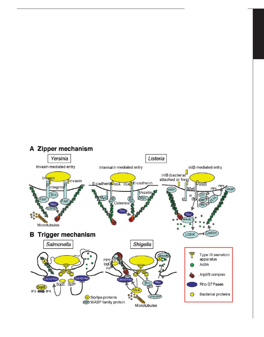
The Zipper Mechanism of Entry
Yersinia pseudotuberculosis and Listeria
monocytogenes both harness transmembrane
cell-adhesion proteins as receptors for entry
into mammalian cells (Figs. 1A and 2A).
Entry can be divided into three successive
steps: (i) Contact and adherence. This step is
independent of the actin cytoskeleton and
involves only the bacterial ligand and its
receptor. It leads to receptor clustering. (ii)
Phagocytic cup formation. This step is trig-
gered by the transient signals occurring
after formation of the first ligand-receptor
complexes and propagating around the in-
vading microbe. These signals induce actin
polymerization and membrane extension.
(iii) Phagocytic cup closure and retraction,
and actin depolymerization.
The Yersinia outer-membrane protein in-
vasin binds to integrin receptors that have the

1
chain and are normally implicated in ad-
herence of cells to the extracellular matrix
(6). Invasin does not possess the RGD motif
present in fibronectin, but both proteins inter-
act with integrins by a structurally similar
domain. Invasin has a higher affinity for in-
tegrins and can oligomerize, inducing inte-
grin clustering and efficient downstream sig-
naling. The cytoplasmic tail of the

1
chain,
which normally interacts with the cytoskele-
ton in focal complexes of adhesion plaques,
is critical for entry, but surprisingly, alter-
ations of this domain that impair interaction
with the cytoskeleton increase internaliza-
tion. Thus, a lower affinity of the integrin for
the cytoskeleton could allow higher mobility
of the receptors in the membrane.
Activation of integrins leads to tyrosine-
phosphorylation events required for entry.
The tyrosine kinase FAK (focal adhesion ki-
nase) is the most attractive candidate for
transmitting a signal from clustered integrins
to the cytoskeleton, because the

1
-chain cy-
toplasmic domain binds to FAK, and domi-
nant-inhibitory mutations in FAK strongly
impair invasin-mediated uptake (7). Src,
phosphoinositide 3-kinase (PI 3-kinase), and
1
Unite´ des Interactions Bacte´ries-Cellules, INSERM
Unite´ 604,
2
Unite´ de Pathoge´nie Microbienne Mo-
leculaire, INSERM Unite´ 389, De´partement de Biologie
Cellulaire et Infection, Institut Pasteur, 28 Rue du
Docteur Roux, Paris 75015, France.
*To whom correspondence should be addressed. E-
mail: pcossart@pasteur.fr (P.C.); psan.son@pasteur.fr
(P.J.S.)
C
E L L U L A R
I
N V A S I O N S
9 APRIL 2004 VOL 304 SCIENCE www.sciencemag.org
242
S
PECIAL
S
ECTION
on March 7, 2012
Downloaded from
Rac are also involved in invasin-mediated
uptake. Why there is a requirement for phos-
phoinositide 3-kinase is unknown. Efficient
entry involves a Rac1-Arp2/3 pathway which
may involve N-WASP (8–10). The local
concentration of phosphatidylinositol 4,5-
bisphosphate [(PIP
2
, PI(4,5)P
2
] is critical for
entry, and Arf6 may play a role in activa-
tion of phosphoinositol-4-phosphate-5-kinase
(PIP
5
kinase) and control of cytoskeleton re-
arrangements and membrane traffic involved
in closure of the phagocytic cup (11).
Several surface proteins contribute to entry
of L. monocytogenes into nonphagocytic cells in
vitro (12). The best-characterized protein, in-
ternalin (InlA), is a surface protein that is co-
valently anchored to the cell wall and belongs to
a large family of leucine-rich repeat (LRR) pro-
teins. As for invasin, coating of latex beads with
internalin promotes their entry, thus facilitating
dissection of the specific pathway. Entry of Lis-
teria into cells involves interaction between the
LRR region of internalin and the first ectodo-
main of human E-cadherin, a transmembrane
glycoprotein normally involved in homophilic
E-cadherin–E-cadherin interactions at adherens
junctions of polarized epithelial cells. The LRR
domain surrounds the first ectodomain of E-
cadherin (13). This weak-affinity interaction
cannot take place if proline-16 is changed into
glutamic acid, as in murine E cadherin (14).
Formation and maintenance of adherens junc-
tions require the integrity of the E-cadherin cy-
toplasmic domain that binds catenins (
␣, , and
p120 catenins), which interact with the cell actin
cytoskeleton (15). Similarly, entry of Listeria
into cells requires the terminal 35 amino acids of
E-cadherin. The latter binds to
-catenin, which
recruits
␣-catenin, which in turn interacts with
actin. Actin polymerization during internalin-
mediated entry is Rac dependent and mediated
by Arp2/3, but how Arp2/3 is activated is un-
known (16). Entry also requires an unconven-
tional myosin, myosinVIIa, and its ligand veza-
tin (17). These two proteins probably play a role
in the dynamics of the phagocytic cup. How the
tension generated by the myosin motor is cou-
pled to actin polymerization required for entry
has not been established.
The second well-characterized L. mono-
cytogenes invasion protein is InlB (12, 18,
19). This surface protein belongs to the
LRR family of proteins and is only loosely
attached by its C-terminal repeats to the
bacterial surface, where it interacts with
lipotechoic acids. Soluble InlB can reasso-
ciate with the bacterial surface of an InlB
mutant and promote entry.
InlB interacts with three cellular ligands (12,
18). The most relevant one is Met, a transmem-
brane receptor tyrosine kinase that upon interac-
tion with its normal ligand, the hepatocyte
growth factor (HGF), dimerizes and elicits phos-
phorylation on two critical residues that act as
docking sites to recruit signaling and adaptor
molecules (20). Met binding to the concave sur-
face of the InlB LRRs also leads to its transient
phosphorylation and to the recruitment and phos-
phorylation of the adaptor proteins Cbl, Gab1,
and Shc, and activation of PI 3-kinase with the
generation of PIP
3
at the plasma membrane (21).
Optimal activity of Met requires the presence of
glycosaminoglycans (GAGs) on the cell surface,
probably promoting oligomerization of the
growth factor and/or its protection from extra-
cellular proteases. GAGs also increase Listeria
InlB-dependent entry into the target cell. Heparin
can detach InlB from the bacterial surface, rein-
Fig. 1. Mechanisms usedby bacteria to enter cells. (A) The zipper mechanism usedby Yersinia and Listeria. (B) The trigger mechanism usedby
Salmonella and Shigella.
C
E L L U L A R
I
N V A S I O N S
www.sciencemag.org SCIENCE VOL 304 9 APRIL 2004
243
S
PECIAL
S
ECTION
on March 7, 2012
Downloaded from
forcing the hypothesis that InlB may act as a
soluble protein. Thus, InlB mimics HGF, the
normal Met ligand, and similarly to growth fac-
tors, soluble InlB induces actin-rich membrane
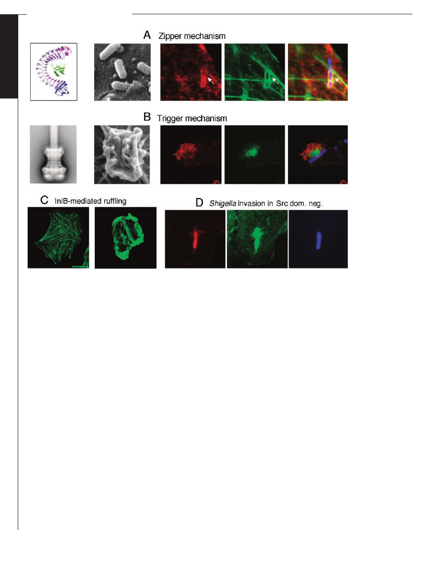
ruffles (Fig. 2C).
InlB also interacts with gC1qR/p32, a
ubiquitous protein first identified as the
receptor for the globular part of the comple-
ment component C1q (22). However, the sub-
cellular location and function of gC1qR re-
main controversial, and its role in cell entry
remains to be clarified.
Contact between Met and InlB, present on
the bacterium or released from its surface, ini-
tiates actin nucleation and polymerization via the
small guanosine triphosphatase (GTPase) Rac,
WAVE, and the Arp2/3 complex (19, 23). Actin
filament elongation, which provides the driving
force for membrane extension around the bacte-
rium, involves VASP, which may act as an
anticapping protein at the barbed ends. Cofilin also
participates in this process. This protein increases
actin turnover by triggering actin depolymerization
at pointed ends of actin filaments and by creating
new free ends for polymerization by severing actin
filaments. In the initial steps of cell entry, cofilin
activity is modulated by LIM kinase. Then progres-
sive accumulation of cofilin on filaments favors
filament disassembly and retraction of the phago-
cytic cup. Thus, the InlB-Met interactions probably
elicit both a Rac-WAVE-ARP2/3 and a Rac-PAK-
LIM-kinase-cofilin cascade. It is still unknown
how Rac is activated downstream of Met. The role
of PI 3-kinase is also unknown. The working hy-
pothesis is that, as in phagocytosis, PI 3-kinase
facilitates cup closure, probably by recruiting mem-
brane vesicles and actin regulators. It may also
induce sustained activation of Rac.
InlB is thus a strong signaling protein that by
itself acts as an invasin but may also potentiate
other bacterial factors involved in Listeria entry
and tissue tropism, such as internalin. Other pro-
teins such as the autolysins Ami, Auto, and ActA
contribute to Listeria adherence and entry (24).
In addition, listeriolysin O (LLO), a pore-form-
ing, cholesterol-dependent cytolysin involved
mainly in escape from the internalization vacu-
ole (25, 26) and that, like other toxins, interacts
with lipid rafts (27),
allows entry of extra-
cellular calcium and
stimulates entry (28).
Even in the absence
of LLO, both interna-
lin- and InlB-mediat-
ed entry are depen-
dent on the presence
of raft microdomains,
suggesting that for en-
try, Listeria take ad-
vantage of raft mi-
crodomains,
which
are known to be en-
riched
in
receptors
and signaling mole-
cules.
Interestingly,
cholesterol depletion
does not affect the in-
ternalin-
and
InlB-
mediated pathways at
the same step of the
entry process (29).
The Trigger
Mechanism of
Entry
Both
Shigella
and
Salmonella use this
mechanism to enter
the cell (Fig. 1B and
Fig. 2B). Contact be-
tween
bacteria
and
cells is mediated by
the type III secretory
system (TTSS) (Fig.
1). The TTSS allows
direct
activation
of
components of the cy-
toskeleton by delivery
of dedicated bacterial effectors. In Salmonella, the
TTSS is encoded by a chromosomal patho-
genicity island (SPI-1) and in Shigella by a
plasmid-located pathogenicity island (PAI).
These PAIs encode the structural components
of the TTSS and some of their dedicated
effectors. Two of these components (i.e.,
SipB/C in Salmonella, IpaB/C in Shigella)
form a pore, or translocator, that delivers the
effectors into the cell cytoplasm, creating a
continuum between the bacterial and eukary-
otic cytoplasms (30, 31).
The interaction of bacteria with their epithe-
lial cell target occurs in four successive stages:
1) A pre-interaction stage. At 37°C, the ef-
fector molecules stored in the bacterial cyto-
plasm are associated with dedicated chaperones,
whose major role is to avoid premature associa-
tion of the effector molecules and their proteo-
lytic degradation (32). In exponentially growing
bacteria, the TTSSs are properly assembled, but
the secretion of effector proteins is repressed
until the bacterium establishes contact with its
cell target.
Fig. 2. The zipper andthe trigger mechanisms. (A): Zipper mechanism. From left to right: x-ray structure of internalin interacting
with E-cadherin [reprinted from (13) with permission from Elsevier]; scanning electron micrograph of Listeria entering into Caco2
cells; immunofluorescence images of Listeria entering into Vero cells (red: Met; green, actin; and blue: bacteria). (B) Trigger
mechanism. From left to right: Reconstitution of the TTSS; scanning electron micrograph of Shigella entering into cells;
immunofluorescence images of Shigella entering into Caco 2 cells (red: cortactin; green: actin; and blue: bacteria). (C) InlB-mediated
ruffling. Control cells andcells ruffling upon incubation with soluble InlB (green: actin). (D) Shigella entering into Src dominant-
negative cells (red: cortactin; green: actin; and blue: bacteria). Src-dependent tyrosine phosphorylation of cortactin is essential to
trigger massive extension of actin filaments at a distance from the entry focus; thus, cells expressing a Src dominant-negative
construct form inefficient entry foci with limitedactin polymerization tightly aroundthe entry vacuole.
C
E L L U L A R
I
N V A S I O N S
9 APRIL 2004 VOL 304 SCIENCE www.sciencemag.org
244
S
PECIAL
S
ECTION
on March 7, 2012
Downloaded from

2) An interaction stage. This stage encom-
passes complex events leading to the formation
of a signaling platform. A recognition event is
likely to take place at the tip of the TTSS,
activating the secretory process via a retroactive
signaling, possibly involving an adenosine
triphosphatase in the TTSS basal body (33). In
Shigella, the high-affinity binding of IpaB to
CD44 —the hyaluronic acid receptor that is
strongly expressed on the basolateral membrane
of intestinal epithelial cells and on the surface of
many other cell types, including cells of myeloid
lineage—may be a key step in achieving tran-
sient adherence to the cell surface, activation of
the secretory machinery, and insertion of the
IpaB/C translocon into the eukaryotic cell mem-
brane. Consistent with the association of CD44
with cholesterol and sphingolipid-rich mem-
brane rafts, this step of the interaction is depen-
dent on intact rafts (34). Cholesterol extraction
disrupts binding to and entry into epithelial cells,
and IpaB and CD44 segregate in these rafts.
Similarly, in Salmonella, the protein components
of the SipB/C translocon also segregate in rafts.
The initial interaction may take place in these
membrane subdomains because (i) the targeted
receptor is enriched in rafts; (ii) the lipid com-
position of rafts is optimal for the formation of
the pore and translocon, in a way similar to the
cholesterol dependence of several hemolysins
(27); and (iii) these domains are enriched in
signaling molecules such as tyrosine kinases of
the src family.
3) The formation of a macropinocytic pocket.
This stage involves localized but massive rear-
rangements of the cell surface, characterized by
the formation of intricate filopodial and lamelli-
podial structures that appear similar in Salmonel-
la and Shigella. Rearrangements of the actin
cytoskeleton largely account for the formation of
the entry focus. At the early stage of Shigella
entry, VirA, a plasmid-encoded protein secreted
through the TTSS, induces local destabilization
of the microtubules that results in their depoly-
merization (35). The latter affects the early
events of actin rearrangement through the deac-
tivation of RhoA, leading to Rac1 activation and
formation of Rac1-IRSp53-WAVE2 complex
that recruits Arp2/3. IpaC in Shigella (36) and
SipC in Salmonella (37) initiate actin nucle-
ation through their C-terminal domain, which
is exposed to the cytoplasm of the eukaryotic
cell, via the IpaB/C or SipB/C pore. The mech-
anism of initial actin nucleation, however, re-
mains uncertain. SipC can nucleate actin alone
in vitro (37), but IpaC requires activation of
Cdc42 and Rac 1 (36).
Massive extension of the actin filaments that
form entry foci seems to respond to different
mechanisms in Salmonella and Shigella. In Sal-
monella, the translocated SopE proteins (SopE1
and SopE2) act as exchange factors for the
Cdc42 and Rac-1 GTPases, thus massively
boosting the initial nucleation event (38). More-
over, SopB/SigD, a TTSS-secreted phosphati-
dylinositol phosphatase (39), stimulates actin re-
arrangements and mediates bacterial entry,
whereas SipA binds and stabilizes actin fila-
ments (40). Shigella has evolved a similar pro-
cess of boosting cytoskeletal rearrangements, al-
though through different molecular mechanisms.
The C-terminal domain of IpaC is central to the
activation of Cdc42 and Rac-1, which is quickly
followed by activation of the tyrosine kinase
c-src upon contact with IpaC (41), recruitment of
cortactin to the membrane upon its c-src–medi-
ated tyrosine phosphorylation, and further mas-
sive actin polymerization in the vicinity of the
original actin cup via the Arp2/3 complex (42)
(Fig. 2C). This process is amplified by IpgD, a
Shigella homolog of SopB/SigD. IpgD expresses
a phospatidylinositol phosphatase activity that
hydrolyzes PI(4,5)P2 into PI(5)P [phosphatidyl-
inositol 5-phosphate], thus disconnecting the ac-
tin subcortical cytoskeleton from the membrane
and favoring actin dynamics at the entry site
(43). The Abl family of tyrosine kinases is also
involved in Shigella entry through phosphoryl-
ation of the adaptor molecule Crk (44).
4) Actin depolymerization and closing of
the macropinocytic pocket. This final stage is
similar in Shigella and Salmonella, despite
important differences between the effectors
involved and the molecular mechanisms ex-
ploited. In the case of Salmonella, SptP, a
TTSS-secreted protein, has two activities: (i)
a tyrosine-phosphatase activity that regulates
activity of the mitogen-activated protein ki-
nase (MAPK) induced by entry; and (ii) a
GAP (GTPase-activating protein) activity on
Cdc42 and Rac that antagonizes the activity
of SopE, thus leading to shrinking of the
entry focus by blocking further actin poly-
merization (45). It may seem strange that
proteins of opposite functions are injected
simultaneously into the target cell. Recent
evidence indicates that, despite equivalent
amounts delivered by the TTSS, SopE is
rapidly degraded through a proteasome-
dependent pathway, whereas SptP is more
stable (46). In the case of Shigella, IpaA, a
TTSS-secreted protein, binds the N-terminal
head domain of vinculin, a key protein in the
formation of cell-adherence plaques, and in-
duces actin depolymerization (47).
Intracellular Life-Styles
After internalization, bacteria remain in a
vacuole or escape to the cytosol, where they
replicate. Some intracytosolic bacteria may
also move by a process of polarized actin
polymerization that takes place at one pole of
the bacterium and provides the force for bac-
terial locomotion inside the cytosol and to-
ward neighboring cells.
The Vacuole as an Intracellular
Replication Compartment
Bacteria that replicate inside the internalization
vacuole have developed an impressive array of
strategies (4) aimed at surviving in a hostile and
changing environment characterized by poor nu-
trient content, progressive decrease of the pH,
and delivery of antibacterial peptides and lyso-
somal enzymes as late endosomes mature to
lysosomes. In macrophages, these conditions are
even more drastic and exacerbated by the deliv-
ery of reactive oxygen and nitrogen intermedi-
ates. Two major strategies can be recognized,
although a given species may use a combination
of both: (i) Bacteria may adapt to and eventually
resist these hostile conditions, thus developing a
state of metabolic adaptation to the stress im-
posed by these conditions; (ii) alternatively, bac-
teria may alter the biogenesis and dynamics of
their vacuolar compartment, thus creating for
themselves a less hostile niche that is permissive
for their survival and growth. Salmonella repre-
sents a paradigm of the complex combination of
these two survival and growth strategies (Fig. 3).
After a few hours of invasion, bacteria reside in
an atypical acidic compartment called the SCV
(Salmonella containing vacuole), which is nei-
ther a late nor an early endosome (48). How
bacteria redirect the fate of this compartment
away from the normal phagosomal pathway in-
volves transient acquisition of rab5, PI3-kinase,
EEA1, and finally rab7 (49). In addition, merg-
ing of the SCV with the endoplasmic reticulum
appears to contribute to early SCV maturation
(50) and membranes of the trans-Golgi network
surround the SCV at late times of infection (51),
suggesting interactions with both the endocytic
and the biosynthetic pathway. Numerous bacte-
rial genes are required for survival and replica-
tion. A key role is played by the SPI2 effector
SifA—a protein required for the formation of
Sifs, filaments enriched in lysosomal glycopro-
teins (Lgps), and extensions of the SCV, in
epithelial cells (52). The function of SifA may be
to mediate the recruitment of vesicles and in-
crease the SCV membrane surface area to ac-
commodate replicating bacterial cells.
Life in the Cytosol and Actin-Based
Intra- and Intercellular Motility
Some intracellular pathogens able to induce
their own phagocytosis into epithelial cells
escape from the internalization vacuole, rep-
licate in the cytosol, and move by recruiting
and polymerizing actin (53) (Fig. 4). Actin
polymerization at one pole of the bacterium
provides the energy for movement and en-
ables the bacteria to reach the plasma mem-
brane, where they form protrusions that are
endocytosed by neigboring cells, allowing
the formation of a two-membrane vacuole,
cell to cell spread, and tissue dissemination.
For Listeria, escape from the vacuole is me-
diated by a pore-forming toxin called listerioly-
sin O (LLO), a potent signaling molecule that
activates nuclear factor
B (NF-B) and a vari-
ety of other pathways (25). Intracytosolic repli-
cation requires expression of a sugar-uptake
system, which is absent in the nonpathogenic
C
E L L U L A R
I
N V A S I O N S
www.sciencemag.org SCIENCE VOL 304 9 APRIL 2004
245
S
PECIAL
S
ECTION
on March 7, 2012
Downloaded from
species L. innocua (25). Actin recruitment by
Listeria and polymerization are triggered by the
surface protein ActA, which recruits and acti-
vates the seven-protein Arp2/3 complex, hence
generating a dendritic network of branched actin
filaments (54). Modulation and control of actin-
based movements involve several other proteins:
(i) cofilin; (ii) capping protein, which caps the
barbed ends of actin filaments; (iii) profilin,
which binds to monomeric actin and, in complex
with actin, to actin-filament barbed ends, hence
providing actin monomers to growing barbed
ends; (iv)
␣-actinin, which cross-links actin fil-
aments; and (v) VASP, which binds to ActA and
F-actin and modulates branch density and move-
ment. Shigella, after escaping from the vacuole
upon the action of IpaB, expresses on its surface
an outer-membrane protein called IcsA/VirG.
This protein, which is unrelated to ActA, recruits
the cellular protein called N-WASP (55, 56).
Cellular N-WASP is functionally and structural-
ly related to bacterial ActA and can recruit and
activate the Arp2/3 complex, highlighting how
bacteria may either mimic or recruit mammalian
proteins to harness eukaryotic pathways (5).
Even though Rickettsia is not an enteroinva-
sive microorganism, it is worth mentioning that
after its escape into the cytoplasm, it forms actin
tails made of long, unbranched actin fila-
ments, which differ from those generated by
ActA or IcsA/N-WASP (Fig. 4). Similar to
proteins of the WASP family, the bacterial
surface protein involved, RickA (57 ), is
composed of three regions, with a central
proline-rich region and a C-terminal part
that recruits Arp2/3. Because Arp2/3 gener-
ates a network of branched actin filaments,
the discovery that RickA activates Arp2/3 in
vitro and is recruited on the McRettsial sur-
face was unexpected, providing a new tool
to address Arp2/3 regulation.
Cell Responses to Intracellular Pathogens
In addition to the transient posttranslational
modifications occurring upon entry, intracel-
lular bacteria induce drastic changes in the
pattern of transcription and translation of in-
fected cells. This is particularly true for in-
testinal epithelial cells that, upon invasion by
Salmonella or Shigella, behave as sentinels
by inducing a transcriptional program whose
major function is to up-regulate innate im-
mune defense mechanisms (58). This pro-
gram occurs largely in response to the induc-
tion of NF-
B that regulates a large portion
of the pro-inflammatory genes. The pro-
inflammatory program of epithelial cells—in
contrast to the outside-in signaling pathway
that Toll-like receptors mediate in phagocytic
cells, in the presence of bacterial PAMPs
(pathogen-associated molecular patterns)—
appears to be mediated by an intracellular
sensing system involving cytosolic proteins
of the Nod family (59). Nod1 is prevalent in
intestinal epithelial cells and shows specific
recognition for muropeptides originating
from the peptidoglycan of Gram-negative mi-
croorganisms (60, 61). Another cytosolic pro-
tein, Nod2, recognizes peptidoglycans from
any bacterial species, essentially because it is
able to recognize muramyl-dipeptide, a struc-
ture common to all peptidoglycans.
Through their capacity to regulate gene tran-
scription and by other pathways, intracellular
bacteria can take over the fate of their host cell.
Among the most striking paradigms are bacteria
that manipulate cell apoptotic processes. Three
major pathways have so far been identified: (i)
Intracellular Shigella and Salmonella, respec-
tively, secrete IpaB and SipB through their
TTSS. These two proteins activate the pro-apop-
totic cysteine protease caspase-1, which causes
apoptotic death of infected macrophages while
also initiating an inflammatory response through
processing or maturation of two potent pro-in-
flammatory cytokines, interleukin-1
 (IL-1)
and IL-18 (62, 63). (ii) Yersinia translocate plas-
mid-encoded Yop proteins, one of which, YopP/
YopJ, binds to and neutralizes the activity of a
MAPK kinase, thereby blocking the activation of
NF-
B, an essential system supporting cell sur-
vival (64). (iii) The third pathway, although not
yet clearly described in enteroinvasive bacteria,
is worth mentioning. Upon interaction of Neis-
seria gonorrhoeae with epithelial cells, the se-
creted protein PorB causes Ca
2
⫹
fluxes that ac-
tivate caspases, and consequently cell apoptosis
(65). PorB creates mitochondrial pores, thus in-
ducing apoptosis through the release of cyto-
chrome c. Finally, epithelial cells infected by
Shigella undergo activation of their connexin-
constituted hexameric hemichannels. The infect-
ed cells release ATP, which acts as a paracrine
mediator activating Ca
2
⫹
fluxes in neighboring
cells, thus increasing their competence for bac-
terial invasion and cell-to-cell spread (66).
Fig. 3. Intracellular life-styles. Schematic representation of the Salmonella-containing vacuole (see
text). Listeria and Shigella lyse the vacuole andmove in the cytosol by an actin-basedmotility
process mediatedby ActA or IcsA/VirG, which interact with Arp2/3 or N-WASP andArp2/3,
respectively. EE: early endosome; LE: late endosome; Ly: lysosome; ER: endoplasmic reticulum
Fig. 4. Actin-basedmotility of Listeria, Rickettsia,
and Shigella. Electron micrographs of actin tails
labeledwith fragment S1 of myosin (69) [reprint-
edwith permission from Journal of Cell Science].
C
E L L U L A R
I
N V A S I O N S
9 APRIL 2004 VOL 304 SCIENCE www.sciencemag.org
246
S
PECIAL
S
ECTION
on March 7, 2012
Downloaded from
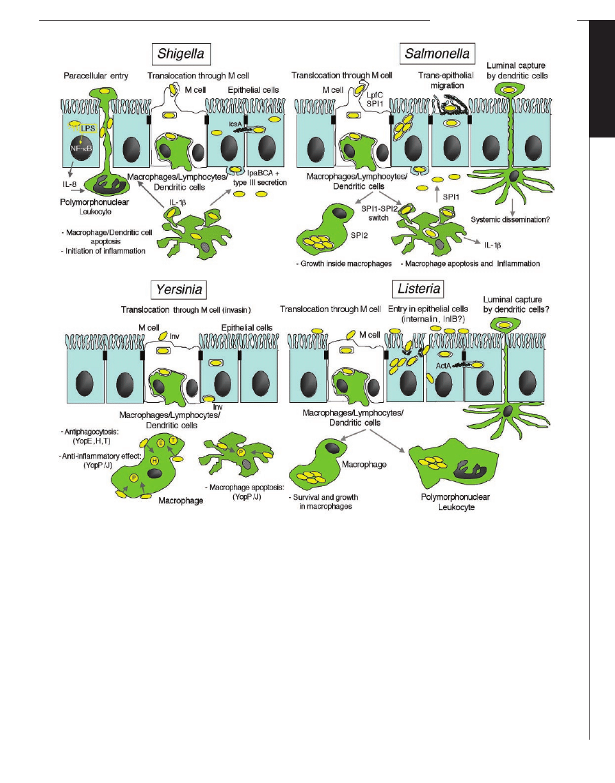
Fig. 5. The invasive strategies of enteroinvasive pathogens. Intestinal
epithelial cells (IECs) maintain a physical barrier against commensal flora,
although specializedsites such as the follicle-associatedepithelium (FAE)
allow constant sampling of the luminal flora through M cells. Translo-
catedbacteria thus exposedto macrophages, dendritic cells (DCs), andB
lymphocytes are captured, killed, processed, and presented to the im-
mune system. Invasive pathogens take advantage of this route to cross
the epithelial barrier. Once translocated, bacteria must survive attack by
macrophages. The four bacterial species consideredhave solvedthis issue
differently: L. monocytogenes are phagocytosedbut escape into the
cytoplasm, andthus avoidbeing killedin lysosomal compartments.
Yersinia adopt an antiphagocytic strategy by intracellular injection of
YopE, H, and T that inactivate the actin cytoskeleton. In addition, they
adopt an anti-inflammatory strategy, with YopP/J blocking tumor necro-
sis factor–
␣ production, which prevents further local recruitment of
predators such as monocytes and polymorphonuclear leukocytes. Alter-
natively, phagocytosed Yersinia may cause YopP/J-dependent apoptosis
of their host cell. Shigella not only cause apoptosis of macrophages and
monocytes, thus ensuring their own survival, but also trigger early
mucosal inflammation through the release of mature IL-1
 andIL-18,
which disrupts epithelial impermeability and facilitates bacterial spread
at a distance. Finally, Salmonella remodel their phagosomes, thus avoid-
ing its transition to a lysosome andcreating an intracellular niche
that allows their efficient replication; this Spi2-dependent process
is an alternative to the Spi1-dependent apoptotic killing of macrophages
similar to that causedby Shigella. Having crossedthe epithelial barrier
andcircumventedthe threat of phagocytosis, the bacterial species
consideredhere proceedalong different pathways. L. monocytogenes
disseminate systemically, possibly inside circulating monocytes and DCs.
Yersinia may invade IECs through their basolateral pole, a process medi-
atedby invasin; they also cause local andmesenteric abscesses in local
andloco-regional lymphoidstructures. Shigella proceeds to TTSS/Ipa-
dependent entry into epithelial cells followed by escape into the cyto-
plasm, intracellular motility, andcell-to-cell spread, thus establishing the
infectious process at the mucosal level, without extensive systemic
dissemination. Salmonella may, like Shigella, enter IECs through their
basolateral pole in a TTSS/Sop-dependent manner. Alternative routes of
invasion involve IECs directly, away from the FAE. In particular, invasion
by L. monocytogenes is mediatedby internalin (InlA) andpossibly InlB. In
addition, Salmonella are able to dislocate the brush border cytoskeleton
andcause an apical entry ruffle. Shigella and Yersinia seem unable to
disrupt the epithelial barrier from a luminal position unless massive
inocula are used. A third process of translocation may involve DCs
crawling between IECs or sending pseudopods to capture luminal bacte-
ria andretract in a subepithelial position. Salmonella are able to trans-
locate in this way, possibly followedby systemic diffusion of Salmonella-
loaded DCs. It is not yet clear whether this type of translocation occurs
in the other invasive species.
C
E L L U L A R
I
N V A S I O N S
www.sciencemag.org SCIENCE VOL 304 9 APRIL 2004
247
S
PECIAL
S
ECTION
on March 7, 2012
Downloaded from

Bacterial Invasion: In Vivo Veritas
A major issue is to validate, in vivo, the molec-
ular and cellular events analyzed in vitro. If one
focuses on invasion of the intestinal barrier, it is
clear that L. monocytogenes, Shigella, Salmonel-
la, and Yersinia, despite their shared capacity to
invade epithelial cells in vitro, differ with regard
to (i) the capacity to disrupt, invade, and even-
tually cause the inflammatory destruction of the
epithelium; and (ii) the possibility of proceeding
to systemic dissemination and possibly coloni-
zation of organs at a distance.
A major handicap to studying the respective
invasive phenotypes in vivo has been the lack
of a mouse model simulating the intestinal and
systemic diseases observed in humans (67).
This was particularly the case for L. monocyto-
genes, until a transgenic mouse line expressing
the human E-cadherin receptor of internalin
became available, thus unlocking the transintes-
tinal route for this pathogen, i.e., via invasion of
enterocytes (68). A relevant animal model has
yet to be found for Shigella because, unlike
infected humans, mice do not undergo exten-
sive invasion and inflammatory destruction
of their rectal and colonic mucosae. Despite
these limitations, a picture is emerging (Fig.
5) concerning the various strategies used by
these pathogens.
In conclusion, although current work aims
to elucidate the in vivo relevance of the now
well-understood mechanisms used by inva-
sive bacteria in vitro, future efforts should
focus on understanding both bacterial and
host cell transcription and translation pro-
grams during infection, in various cells and
tissues. This information should provide
vital clues in the ongoing battle against
bacterial disease and for elaborating new
therapeutic strategies.
References and Notes
1. C. R. Roy, L. G. Tilney, J. Cell Biol. 158, 415 (2002).
2. K. A. Fields, T. Hackstadt, Annu. Rev. Cell Dev. Biol.
18, 221 (2002).
3. C. R. Hauck, T. F. Meyer, Curr. Opin. Microbiol. 6, 43
(2003).
4. C. C. Scott, R. J. Botelho, S. Grinstein, J. Membr. Biol.
193, 137 (2003).
5. B. B. Finlay, P. Cossart, Science 276, 718 (1997).
6. R. R. Isberg, P. Barnes, J. Cell Sci. 114, 21 (2001).
7. M. A. Alrutz, R. R. Isberg, Proc. Natl. Acad. Sci. U.S.A.
95, 13658 (1998).
8. M. A. Alrutz et al., Mol. Microbiol. 42, 689 (2001).
9. K. McGee, M. Zettl, M. Way, M. Fallman, FEBS Lett.
509, 59 (2001).
10. K. McGee, P. Holmfeldt, M. Fallman, FEBS Lett. 533,
35 (2003).
11. K. W. Wong, R. R. Isberg, J. Exp. Med. 198, 603 (2003).
12. P. Cossart, J. Pizarro-Cerda, M. Lecuit, Trends Cell Biol.
13, 23 (2003).
13. W. D. Schubert et al., Cell 111, 825 (2002).
14. M. Lecuit et al., EMBO J. 18, 3956 (1999).
15. M. Lecuit et al., Proc. Natl. Acad. Sci. U.S.A. 97,
10008 (2000).
16. S. Sousa, M. Lecuit, P. Cossart, in preparation.
17. S. Sousa et al., J. Cell Sci., in press.
18. H. Bierne, P. Cossart, J. Cell Sci. 115, 3357 (2002).
19. H. Bierne et al., in preparation.
20. Y. Shen, M. Naujokas, M. Park, K. Ireton, Cell 103, 501
(2000).
21. K. Ireton et al., Science 274, 780 (1996).
22. L. Braun, B. Ghebrehiwet, P. Cossart, EMBO J. 19,
1458 (2000).
23. H. Bierne et al., J. Cell Biol. 155, 101 (2001).
24. O. Dussurget, J. Pizarro-Cerda, P. Cossart, Annu. Rev.
Microbiol., in press.
25. J. A. Vazquez-Boland et al., Clin. Microbiol. Rev. 14,
584 (2001).
26. A. L. Decatur, D. A. Portnoy, Science 290, 992 (2000).
27. F. G. Van der Goot, T. Harder, Semin. Immunol. 13,
89 (2001).
28. S. Dramsi, P. Cossart, Infect. Immun. 71, 3614 (2003).
29. S. Seveau, S. Giroux, H. Bierne, P. Cossart, in
preparation.
30. J. E. Galan, Annu. Rev. Cell Dev. Biol. 17, 53 (2001).
31. P. J. Sansonetti, FEMS Microbiol. Rev. 25, 3 (2001).
32. C. Parsot, C. Hamiaux, A. L. Page, Curr. Opin. Micro-
biol. 6, 7 (2003).
33. A. Blocker, K. Komoriya, S. Aizawa, Proc. Natl. Acad.
Sci. U.S.A. 100, 3027 (2003).
34. F. Lafont, G. Tran Van Nhieu, K. Hanada, P. San-
sonetti, F. G. van der Goot, EMBO J. 21, 4449 (2002).
35. S. Yoshida et al., EMBO J. 21, 2923 (2002).
36. G. Tran Van Nhieu, E. Caron, A. Hall, P. J. Sansonetti,
EMBO J. 18, 3249 (1999).
37. R. D. Hayward, V. Koronakis, EMBO J. 18, 4926 (1999).
38. W. D. Hardt, L. M. Chen, K. E. Schuebel, X. R. Bustelo,
J. E. Galan, Cell 93, 815 (1998).
39. F. A. Norris, M. P. Wilson, T. S. Wallis, E. E. Galyov,
P. W. Majerus, Proc. Natl. Acad. Sci. U.S.A. 95, 14057
(1998).
40. D. Zhou, M. S. Mooseker, J. E. Galan, Proc. Natl. Acad.
Sci. U.S.A. 96, 10176 (1999).
41. J. Mounier, P. J. Sansonetti, G. Tran Van Nhieu, in
preparation.
42. L. Bougne`res et al., in preparation.
43. K. Niebuhr et al., EMBO J. 21, 5069 (2002).
44. E. A. Burton, R. Plattner, A. M. Pendergast, EMBO J.
22, 5471 (2003).
45. C. E. Stebbins, J. E. Galan, Mol. Cell 6, 1449 (2000).
46. T. Kubori, J. E. Galan, Cell 115, 333 (2003).
47. R. Bourdet-Sicard et al., EMBO J. 18, 5853 (1999).
48. D. W. Holden, Traffic 3, 161 (2002).
49. S. Meresse, O. Steele-Mortimer, B. B. Finlay, J. P.
Gorvel, EMBO J. 18, 4394 (1999).
50. O. Steele-Mortimer, S. Meresse, J. P. Gorvel, B. H. Toh,
B. B. Finlay, Cell. Microbiol. 1, 33 (1999).
51. S. P. Salcedo, D. W. Holden, EMBO J. 22, 5003 (2003).
52. F. Garcia-del Portillo, M. B. Zwick, K. Y. Leung, B. B.
Finlay, Proc. Natl. Acad. Sci. U.S.A. 90, 10544 (1993).
53. F. Frischknecht, M. Way, Trends Cell Biol. 11, 30 (2001).
54. M. D. Welch et al., Science 281, 105 (1998).
55. T. Suzuki, H. Miki, T. Takenawa, C. Sasakawa, EMBO J.
17, 2767 (1998).
56. C. Egile et al., J. Cell Biol. 146, 1319 (1999).
57. E. Gouin et al., Nature 427, 457 (2004).
58. M. F. Kagnoff, in Microbial Pathogenesis and the
Intestinal Epithelial Cell, G. A. Hecht, Ed. (American
Society for Microbiology, Washington, DC, 2003).
59. S. E. Girardin et al., EMBO Rep. 2, 736 (2001).
60. S. E. Girardin et al., Science 300, 1584 (2003).
61. M. Chamaillard et al., Nature Immunol. 4, 702 (2003).
62. H. Hilbi et al., J. Biol. Chem. 273, 32895 (1998).
63. D. Hersh et al., Proc. Natl. Acad. Sci. U.S.A. 96, 2396
(1999).
64. K. Orth et al., Science 285, 1920 (1999).
65. A. Muller et al., EMBO J. 18, 339 (1999).
66. G. Tran Van Nhieu et al., Nature Cell Biol. 5, 720 (2003).
67. M. Lecuit, P. Cossart, Trends Mol. Med. 8, 537 (2002).
68. M. Lecuit et al., Science 292, 1722 (2001).
69. E. Gouin et al., J. Cell Sci. 112, 1697 (1999).
70. We thank E. Veiga, E. Gouin, andO. Dussurget for
help with manuscript preparation andH. Bierne, L.
Bougne`res, andG. Tran van Nhieu for generously pro-
viding unpublished figures. The authors are internation-
al Scholars of the HowardHughes Medical Institute.
R E V I E W
Intracellular Parasite Invasion Strategies
L. D. Sibley
Intracellular parasites use various strategies to invade cells and to subvert cellular
signaling pathways and, thus, to gain a foothold against host defenses. Efficient cell
entry, ability to exploit intracellular niches, and persistence make these parasites
treacherous pathogens. Most intracellular parasites gain entry via host-mediated
processes, but apicomplexans use a system of adhesion-based motility called “glid-
ing” to actively penetrate host cells. Actin polymerization–dependent motility
facilitates parasite migration across cellular barriers, enables dissemination within
tissues, and powers invasion of host cells. Efficient invasion has brought widespread
success to this group, which includes Toxoplasma, Plasmodium, and Cryptosporidium.
Parasites exist in virtually every conceivable
niche, but none is so specialized as that of the
obligate intracellular parasite, which must
gain entry into the cells of its host to survive.
Most intracellular parasites are protozoans,
many of which are responsible for lethal and
debilitating diseases in animals and humans.
Our defenses present an array of barriers to
infection, including skin, mucosa, connective
tissue, and an active surveillance system to
detect and destroy foreign objects. Overcom-
ing these defenses and breaching the final
barrier imposed by the cell membrane is a
formidable challenge. By entering into the
confines of a host cell, the parasite assures
itself of both a ready source of nutrients and
a potential means to avoid immune clearance.
Parasites that practice this life-style have typ-
ically given up the capacity for extracellular
growth, which leaves them vulnerable if en-
try is impeded. Defining how parasites gain
entry into their host cells is thus important for
rational design of improved therapies. Para-
sites are among the earliest branching eu-
karyotes (1); their study expands our knowl-
C
E L L U L A R
I
N V A S I O N S
9 APRIL 2004 VOL 304 SCIENCE www.sciencemag.org
248
S
PECIAL
S
ECTION
on March 7, 2012
Downloaded from
Wyszukiwarka
Podobne podstrony:
Dr Who Target 156 The Paradise Of Death # Barry Letts
The Paradise War Stephen R Lawhead
Stableford, Brian Hooded Swan 02 The Paradise Game
The Paradise of Death
Divine Invasion, The
The Paradise Game Brian Stableford
Lactic Acid Bacteria in the Treatment of Acute 12
Brian Stableford Hooded Swan 4 The Paradise Game
The Paradise Game Brian Stableford
The History of Great Britain - Chapter One - Invasions period (dictionary), filologia angielska, The
Evolution in Brownian space a model for the origin of the bacterial flagellum N J Mtzke
D Day The Invasion of Normandy
Analysis of the?y of Pigs Invasion
The History of Great Britain Chapter One Invasions period
Burnout Paradise The Ultimate Box
The Sims 3 Rajska Wyspa ( Island Paradise )Torrent
Robert Silverberg The Martian Invasion Journals of Henry James
098 Doctor Who The Invasion
Clark The Industrial Arts Paradigm
więcej podobnych podstron