
Introduction
Mesenchymal stem cells (MSCs) are non-
hematopoietic cells, which reside in the bone mar-
row together with better known and characterized
class of stem cells - hematopoietic stem cells. They
were first described by Fridenstein et al. in 1976, as
the clonal, plastic adherent cells, being a source of
the osteoblastic, adipogenic and chondrogenic cell
lines [38]. The interest in MSCs rapidly grows with
expanding knowledge about their exceptional char-
acteristics and usefulness in the clinic. This review
describes the latest data about MSC biology and
behavior in vitro, as well as in vivo. It presents also
molecular features of MSCs and their broad use in
various clinical settings.
Sources of MSCs
The main source of MSCs is the bone marrow. These
cells constitute, however, only a small percentage of
the total number of bone marrow populating cells. Pit-
tenger et al. showed that only 0.01% to 0.001% of
mononuclear cells isolated on density gradient
(ficoll/percoll) give rise to plastic adherent fibroblast-
like colonies [96]. The number of MSCs isolated from
this tissue may vary in terms of the yield and the qual-
ity, even when the cells are obtained from the same
donor [95].
Apart from the bone marrow, MSCs are also locat-
ed in other tissues of the human body. There is an
increasing number of reports describing their presence
in adipose tissue [43], umbilical cord blood, chorionic
villi of the placenta [54], amniotic fluid [122], periph-
eral blood [133], fetal liver [11], lung [57], and even in
exfoliated deciduous teeth [85].
The amount of MSCs decreases with age [36] and
infirmity [56]. The greatest number of MSCs is found
in neonates, than it is reduced during the lifespan to
FOLIA HISTOCHEMICA
ET CYTOBIOLOGICA
Vol. 44, No. 4, 2006
pp. 215-230
Mesenchymal stem cells: characteristics and clinical
applications
Sylwia Bobis, Danuta Jarocha and Marcin Majka
Department of Transplantation, Polish-American Institute of Pediatrics, Jagiellonian University Medical
College, Cracow, Poland
Abstract: Mesenchymal stem cells (MSCs) are bone marrow populating cells, different from hematopoietic stem cells,
which possess an extensive proliferative potential and ability to differentiate into various cell types, including: osteo-
cytes, adipocytes, chondrocytes, myocytes, cardiomyocytes and neurons. MSCs play a key role in the maintenance of
bone marrow homeostasis and regulate the maturation of both hematopoietic and non-hematopoietic cells. The cells are
characterized by the expression of numerous surface antigens, but none of them appears to be exclusively expressed on
MSCs. Apart from bone marrow, MSCs are located in other tissues, like: adipose tissue, peripheral blood, cord blood,
liver and fetal tissues. MSCs have been shown to be powerful tools in gene therapies, and can be effectively transduced
with viral vectors containing a therapeutic gene, as well as with cDNA for specific proteins, expression of which is
desired in a patient. Due to such characteristics, the number of clinical trials based on the use of MSCs increase. These
cells have been successfully employed in graft versus host disease (GvHD) treatment, heart regeneration after infarct,
cartilage and bone repair, skin wounds healing, neuronal regeneration and many others. Of special importance is their use
in the treatment of osteogenesis imperfecta (OI), which appeared to be the only reasonable therapeutic strategy. MSCs
seem to represent a future powerful tool in regenerative medicine, therefore they are particularly important in medical
research.
Key words: Mesenchymal stem cells (MSCs) - Osteogenesis imperfecta - Gene therapy
Correspondence: M. Majka, Dept. Transplantation, Polish-
American Institute of Pediatrics, Wielicka 265, 30-663 Kraków,
Poland; e-mail: mmajka@cm-uj.krakow.pl
Review article

about one-half at the age of 80 [36]. As for circulating
fetal MSCs, the highest number is detected in the first
trimester and declines during the second trimester to
about 0.0001% and further to 0.00003% of nucleated
cells in cord blood [11].
Surface markers on MSCs
MSCs constitute a heterogeneous population of cells,
in terms of their morphology, physiology and expres-
sion of surface antigens. Up to now, no single specific
marker has been identified. MCSs express a large
number of adhesion molecules, extracellular matrix
proteins, cytokines and growth factor receptors, asso-
ciated with their function and cell interactions within
the bone marrow stroma [28]. They also express a
wide variety of antigens characteristic for other cell
types, as confirmed by advanced molecular tech-
niques, including serial analysis of gene expression
[111] and DNA microarray [61]. The population of
MSCs isolated from bone marrow express: CD44,
CD105 (SH2; endoglin), CD106 (vascular cell adhe-
sion molecule; VCAM-1), CD166, CD29, CD73 (SH3
and SH4), CD90 (Thy-1), CD117, STRO-1 and Sca-1
[5, 7, 21, 26, 44, 160]. Interestingly, the observations
made by Bonyadi et al. [8] present late-onset osteo-
porosis in mice lacking Sca-1. Parallelly, MSCs do not
possess markers typical for hematopoietic and
endothelial cell lineages: CD11b, CD14, CD31, CD33,
CD34, CD133 and CD45 [96]. The absence of CD14,
CD34 and CD45 antigens on their surface create the
basis to distinguish them from the hematopoietic pre-
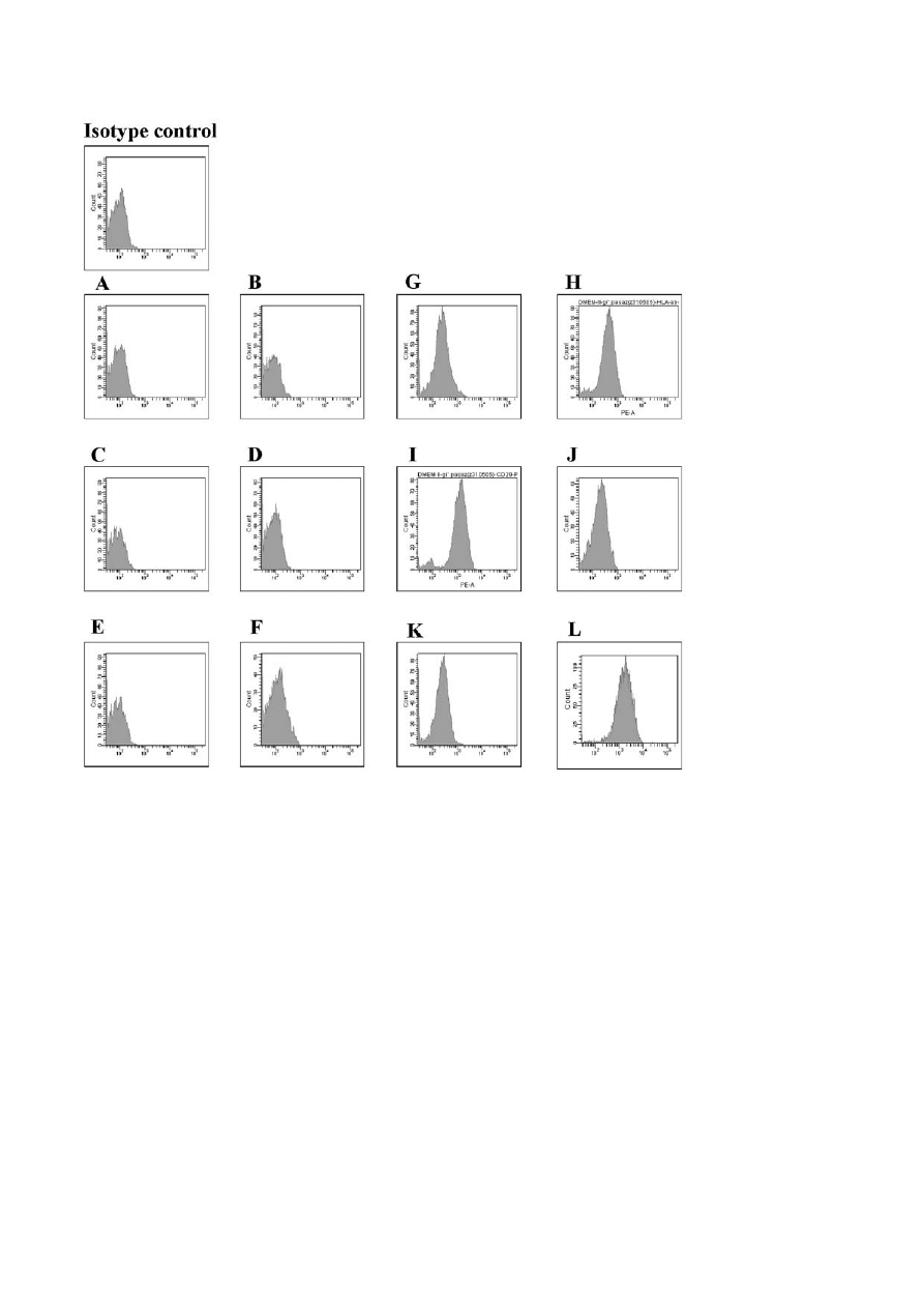
cursors [5]. In Figure 1 we present the phenotype char-
acteristic of the 2nd passage BM-MSCs. This data
from our laboratory confirm the standard description
of these cells.
MSCs are also known to express a set of receptors
associated with matrix- and cell-to-cell adhesive inter-
actions, like integrins
α
V
β3 and α
V
β5, ICAM-1,
ICAM-2, LFA-3 and L-selectin [21, 96, 7].
There have been studies to find an accurate combi-
nation of a limited number of antigens in order to iso-
late pure population of MSCs from a tissue. From the
data available up to now, several options have been
proposed in this context. One of them suggests that the
co-expression of CD105 and CD73 could be sufficient
[96]. Another one implies that the expression of
CD166 and CD105 makes it possible to separate the
earliest precursors of MSCs from more mature cells
[2]. In turn, examination of the CFU-F obtained from
bone marrow stroma demonstrated that the MSCs frac-
tion may by identified by several markers, including
STRO-1, Thy-1, CD49a, CD10, Muc18/CD146, as
well as with the antibodies to receptors for PDGF
(platelet derived growth factor) and EGF (epithelial
growth factor) [5, 8, 26, 44, 96, 98].
Although MSCs have been described by a subset of
surface antigens, little is known about fresh or nonex-
panded MSCs, mostly because of their very low fre-
quency in adult bone marrow [7]. The findings by
Boiret et al. [7] showed that the most discriminative
markers for MSCs examined after short time of adher-
ence (1-3 days) were: CD73 and CD49a, as all the
CFU-F-colonies (100%) were CD73- and most
(95.2%) were CD49a- positive. Interestingly, these
data did not confirm that CD105 and CDw90 could be
selective markers for MSCs, as only 45.4% and 49%
of the CFU-F were positive for these molecules,
respectively [7]. Furthermore, the authors checked the
surface protein expression on freshly isolated bone
marrow MSCs, showing, as found previously, that
CD73 and CD49a were the most extensively expressed
antigens in CFU-F-enriched subset. These results
stand in opposition with the popular description of
MSC phenotype, which postulated the STRO-1 anti-
gen to be exclusively expressed by primitive mes-
enchymal precursors [26, 44].
However, the presence of some antigens may
change in vitro, due to specific culture conditions and
the duration prior to individual passages [22]. Interest-
ingly, some antigens may be found on freshly isolated
MSCs, but their expression disappears in culture. Such
a phenomenon was observed in case of CD34 antigen.
This molecule was expressed by MSCs obtained from
mouse fetal lungs, but could not be found in in vitro
cultures of MSCs [36]. This would suggest that the
expression of that molecule vanishes during the matu-
ration process. Similar results were obtained in case of
chemokine receptor expression on human MSCs [49].
The second passage BMSCs expressed: CCR1, CCR7,
CCR9, CXCR4, CXCR5 and CXCR6. At the 12-16th
passage, there was no expression of any of those mol-
ecules, which was also confirmed by a disability of the
cells to migrate towards specific chemokine attrac-
tants. Moreover, the loss of these receptors' expression
was accompanied by a decrease in the expression of
adhesion molecules - ICAM-1, ICAM-2, VCAM-1
and CD157. Moreover, the alteration in BM-MSCs
phenotype was associated with increasing cell cycle
arrest and induction of the apoptotic pathway [49].
The change in antigen expression has been also
described for MSCs undergoing differentiation
process. As an example, the CD166 antigen (activated
leukocyte cell adhesion molecule) has been presented
on undifferentiated MSCs but was absent from the
cells that underwent osteogenic differentiation [10].
Furthermore, the cell clones derived from different tis-
sues may slightly differ in terms of cell surface mole-
cules. A survey investigating the antigen profile on
MSCs isolated from adipose tissue revealed that in
majority these cells are very much alike as bone mar-
row-derived MSCs [64]. However, in a small number
216
S. Bobis et al.
of surface proteins, the two populations differ. The adi-
pose tissue MSCs were shown to possess additionally
CD49d [64], CD62e and CD31 [43].
Basic biology and functions of MSCs
Human MSCs are known to constitute a heterogeneous
population of cells and their properties and functional-
ity depend on the environmental characteristics. MSCs
can be expanded in culture were they give rise to
fibroblastic colonies (CFU-F). The CFU-F units are
well documented to possess an extended proliferative
potential in vitro [22]. Studies in rodents with
3
[H]-
thymidine labeling demonstrated that CFU-F are
essentially in a noncycling state in vivo [133]. The
number of colonies obtained from bone marrow aspi-
rates differs among species, as well as throughout the
culture conditions used in each individual experiment.
Colony formation by MSCs derived from adult human
BM is feeder cell independent, while the rodent cells
require a source of irradiated feeder cells to achieve
maximal plating efficiency [9, 97].
The cultures of MSCs are, however, not complete-
ly explored. Former studies claimed that MSCs isolat-
ed from bone marrow comprise a single phenotypic
population forming symmetric, spindle-shaped
colonies (homology up to 98%) [96]. More recent
studies, however, indicate that single-cell derived
colonies are morphologically heterogeneous, contain-
ing at least two different cell types: small spindle-
shaped cells and large cuboidal or flattened cells [9,
55]. In terms of proliferative potential, the cells have
been also described as small rapidly-renewing, and
large slowly-renewing [102]. Contrastingly, the work
217
Mesenchymal stem cells
Fig. 1. Phenotype of bone marrow derived MSCs after two passages. Cells were cultured in DMEM with 10% FBS. A - CD14, B - CD33,
C - CD133, D - CD45, E - CD34, F - HLA-DR, G - CD105, H - HLA-ABC, I - CD29, J - CD44, K - CD166, L - CD73. Flow cytometry.

performed by Colter et al. [19] describes the popula-
tion of small and agranular cells (RS-1) within station-
ary culture of MSCs with a low capacity to generate
colonies and non-reactive to the cell cycle-specific
antigen Ki-67. That cell subpopulation was shown,
however, to be responsible for the capacity of the
whole population of MSCs to expand in culture. Fur-
thermore, it was speculated that RS cells may cycle
under stimulation by factors secreted by the more
mature MSCs. These cells were, thus, proposed to rep-
resent an ex vivo subset of recycling uncommitted
mesenchymal stem cells [19].
Nevertheless, the latest findings show that MSC
colonies contain as much as three types of cells. The
third fraction was described to be composed of very
small rapidly self-renewing cells [20], which are
reported as the earliest progenitors and possess the
greatest potential for multilineage differentiation. The
examination of these cells revealed that they were
about 7
μm in diameter and had a high nucleus-to-
cytoplasm ratio. They could be also distinguished from
more mature cells by the presence of specific surface
epitopes and expressed proteins, like vascular endothe-
lial growth factor receptor-2, tyrosine kinase receptor,
transferrin receptor and annexin II (lipocortin 2). Some
of the rapidly renewing cells contained also other
markers, like c-kit (CD117), multidrug resistance epi-
tope and epithelial membrane antigen. Interestingly,
these cells were negative for STRO-1, an antigen orig-
inally considered as a marker for MSCs [26].
MSCs play a significant role in bone marrow
microenvironment. The major function of these cells is
to create a tissue framework, which assures a mechan-
ical support for hematopoietic cell system. They
secrete a number of extracellular matrix proteins,
including fibronectin, laminin, collagen and proteogly-
cans [28]. Moreover, MSCs produce hematopoietic
and non-hematopoietic growth factors, chemokines
and cytokines, thereby participating in the regulation
of hemopoiesis. MSCs secrete: IL-1a, IL-1b, IL-6, IL-
7, IL-8, IL-11, IL-14, IL-15, macrophage colony-stim-
ulating factor, granulocyte-macrophage colony-stimu-
lating factor (GM-SCF), leukemia inhibitory factor,
stem cell factor (SCF), fetal liver tyrosine kinase-3,
thrombopoietin and hepatocyte growth factor (HGF)
[7, 20, 22, 44, 64]. Some of these proteins are pro-
duced by quiescent cells, whereas the others after stim-
ulation. The involvement of MSCs in hematopoiesis is
additionally consolidated by their presence in fetal
liver and bone marrow just prior to the onset of defin-
itive hemopoiesis at those sites [11, 80]. An animal
model study confirmed that human MSCs marked with
GFP and transplanted into the tibia of NOD/SCID
mice, integrated into the functional components of
hematopoietic microenvironment and actively partici-
pated in the hematopoietic cell development [86]. Dur-
ing 4 to 10 weeks after transplantation, GFP-MSCs
differentiated into pericytes, myofibroblasts, stromal
cells, osteocytes and endothelial cells. This led to the
increase in the number of functionally and phenotypi-
cally primitive human hematopoietic cells in murine
bone marrow microenvironment. The engrafted cells
supported human hematopoiesis via secreted factors
and by physical interactions with primitive hematopoi-
etic cells [86]. Other studies showed that cotransplan-
tation of human MSCs and HSCs resulted in increased
chimerism or/and accelerated hematopoietic recovery
in animal models and in humans [36, 67, 71]. More-
over, MSCs are known to produce a variety of
cytokines that are involved in homing (stromal derived
factor-1 - SDF-1) or proliferation and differentiation of
hematopoietic cells (GM-CSF, SCF, IL-6) [48]. It has
been proposed that several chemokine axes are
involved in maintaining bone marrow homeostasis,
and that some chemokines, which MSCs possess the
receptors for, like CCR9 and CXCR4 may operate in
an autocrine manner, similarly as it is in case of HSCs
[49].
Among other well known biological activities of
MSCs, it is worth to emphasize their immunomodula-
tory functions. These cells are able to inhibit respons-
es of alloreactive T lymphocytes. They express neither
MHC class II molecules nor costimulatory receptors
(CD80, CD86) on their surface, therefore they do not
exhibit antigen-presenting cell activities [3, 36]. The
addition of interferon-
γ (IFN-γ) to the cultures of
MSCs enhances the expression of MHC class I and
triggers the expression of MHC class II, but not of the
costimulatory molecules. [36]. It has been well estab-
lished that MSCs from various species can exert pro-
found immunosupression by inhibiting T-cell respons-
es to polyclonal stimuli [29] and to their cognate pep-
tide [69]. The inhibition did not seem to be antigen
specific and targeted both primary and secondary T-
cell responses [69]. The inhibitory effect was shown to
be directed mostly at the level of cell proliferation. T
cells stimulated in the presence of MSCs were arrest-
ed in the G1 phase as a result of cyclin D downregula-
tion [41]. The suppression, however, was not apoptot-
ic and could be reversed. In the absence of MSCs and
with appropriate stimuli, T cells continue to proliferate
[29]. The precise mechanism by which MSCs modu-
late immunological response is still to be clarified, but
overall data suggest that soluble factors as well as cell
contact mediated mechanisms are involved. Blocking
experiments with the use of neutralizing monoclonal
antibodies against transforming growth factor-
β
(TGF-
β) and HGF suggest that these factors are at
least in part responsible for the inhibitory effects
caused by MSCs [29]. Moreover, MSCs can affect
other cells participating in immune response like B
cells [41] and dendritic cells [63].
218
S. Bobis et al.

Circulation and niches of MSCs
Little is known about the nature and localization of
undifferentiated multipotent MSCs. These cells may
be found in various tissues in special places called
'stem cell niches', which serve as stem cells reservoirs.
They remain quiescent and possess the capacity for
self-renewal after an injury, disease or aging [96]. The
stem cell niche hypothesis for the bone marrow cells
was developed by Schofield, who suggested that cer-
tain microenvironmental conditions of the marrow
stroma could maintain the stem cells in a primitive,
quiescent state [112]. The investigation of anatomical
distribution of MSCs within bone marrow revealed
that the cells are located in a close association with
endosteum [44]. Such places, therefore, could be
regarded as potential niches for MSCs. The findings
are, however, based on the STRO-1
+
stromal cell pop-
ulation, and the identification of MSCs expressing
other specific markers, may change this picture.
The question how MSCs maintain their undifferen-
tiated state within the niche is not completely resolved.
However, there are some findings indicating that MSC
decision to differentiate or to stay quiescent is regulat-
ed by Wnt family members, which support undifferen-
tiated state of MSCs, as well as their inhibitors, like:
Dickkopf-1 (Dkk1), Frizzled b-1 (Frzb-1) or sFRP1
[106]. Wnt signaling is known to prevent differentia-
tion process by inducing high levels of oct-3/4, rex-1
and the homeodomain transcription factor Nanog
[106]. Apart from Wnt- and Dkk1-mediated signaling,
also Notch, Hedgehog and BMP-pathways play a role
in proliferation and differentiation of stem cells.
Therefore, it can be speculated, that at least some of
these factors are also important for MSCs growth in
their niche.
After particular stimuli, a stem cell may leave its
niche and circulate in blood [35]. The cell must be
afterwards attracted to another site, where under spe-
cific microenvironmental circumstances is able to
enter its differentiation program [127]. The study on
MSC homing indicates that the expression of
chemokine receptors, as quoted previously, help them
in trafficking to various tissues, including bone mar-
row [76]. Among them, a pivotal role is played by
CXCR4, the receptor for SDF-1, which, inter alia, is
produced by stromal cells. Many findings confirm the
extensive multi-organ homing ability of MSCs. In
murine model, circulating mesenchymal progenitors,
detected in bloodstream, were able to migrate and col-
onize various tissues [39]. Similar results were
obtained in humans [101]. Moreover, these cells were
present in the blood of breast cancer patients after
growth factor-induced mobilization of hematopoietic
stem cells [35]. These data suggest that adequate stim-
uli may mobilize and release quiescent MSCs residing
in a tissue. Additionally, a subset of quiescent cells
(5-10%) was identified in cultures of mesenchymal
cells isolated from cord blood, suggesting that uncom-
mitted mesenchymal progenitors circulate during ges-
tation, and travel from fetal sites into other tissues
early during development [80]. As another example,
MSCs were described to locally migrate to injured
sites, to support the regeneration process. Such cases
were documented in cartilage repair [14, 40], muscle
[23] and heart [110] regeneration, migration through-
out forebrain and cerebellum [68] and differentiation
into osteoblasts in regenerating bone [50, 51]. The
homing capacity of MSCs may decrease after exten-
sive culturing in vitro. A study based on syngeneic
mouse model revealed that primary bone marrow-
derived MSCs were able to home efficiently to the
bone marrow and spleen, whereas culture-expanded
MSCs had lost this capacity after 24-48 hours in cul-
ture [36]. It might be speculated, therefore, that in vitro
propagation of bone marrow-derived MSCs dramati-
cally decreases their homing to bone marrow and
spleen.
Growth and expansion of MSCs
Various protocols have been developed to grow and
expand MSCs. Cells which initially adhere to the tis-
sue culture plastic, display fibroblastic appearance and
develop into symmetrical colonies between 5 and 7
days after plating. Human MSCs proliferate most rap-
idly and maximally retain their multipotential ability
when cultured at relatively low densities [107]. These
cells may be seeded at the range from 1×10
4
to 0.4×10
6
cells/cm
2
[37, 82]. The initial culture concentration
affects not only growth of MSCs but also their mor-
phology [121]. When the cells are grown at a low den-
sity, they mostly display a spindle-like shape, but when
they reach confluence and start to grow in several lay-
ers, the cells become flat with torn ends.
In vitro growth of MSCs is characterized by the
occurrence of three phases, similarly to other progeni-
tor cells: (i) an opening lag phase, which lasts for 3-4
days, followed by (ii) a rapid expansion (log phase)
and closes with (iii) a stationary phase [9, 20]. The last
stage does not rely on cell contact inhibition and
replating the cells triggers their growth for approxi-
mately five more passages [20]. Prockop et al. [42]
suggests that the shift between different stages is reg-
ulated mainly by the expression of Dickkopf-1 (Dkk-
1) and Wnt5a genes, which play opposite roles. The
greatest expression of Dkk-1 appears during the log
phase and shortens the former stage by inhibition of
Wnt5a expression, whereas Wnt5a protein level
becomes maximal during the stationary phase.
Under optimal conditions, MSCs can be maintained
in culture for 20-30 population doublings and still
retain their capacity for differentiation [37]. More
219
Mesenchymal stem cells
recent studies show that these cells are able to grow
and divide for even more than 50 population doublings
[98]. This indicates a great proliferative potential of
these cells. Examination of the cell cycle profile of
MSCs revealed that about 10% of these cells occurs in
phases S, G2 and M of the cell cycle, while the vast
majority of the cells remain in the G0/G1 phase [21].
In genomic assays, MSCs maintain a normal kary-
otype and telomerase activity, even at passage 12 [96].
However, extensive subcultivation of MSCs impairs
their functionality and the cells display evident signs
of senescence and/or apoptosis [21].
Proliferation of MSCs is influenced by a number of
cytokines and growth factors. The list of hormones and
other molecules involved in the regulation of CFU-F
proliferation in vitro is growing. PDGF and fibroblast
growth factor-2 (FGF-2) have been shown to be potent
mitogens for CFU-F [9], and EGF exerts the same
effect on STRO-1 enriched population of MSCs [9].
Opposite results can be obtained after addition of inter-
feron-alpha and interleukin 4 to the culture [9, 61].
Both cytokines inhibit colony formation stimulated by
the combination of EGF and PDGF in a dose-depend-
ent manner. Additionally, it was demonstrated that
binding of heparin-binding epidermal growth factor
(HB-EGF) to its receptor HER-1 on MSCs, consoli-
dates proliferation and prevents differentiation of these
cells induced by conditioning [70]. Thus, it can be
speculated that the HB-EGF/HER-1 axis is important
for MSC renewal and differentiation. The proliferative
activity of MSCs was shown to be directly proportion-
al to their differentiation potential [97].
Differentiation potential of MSCs
It is still not clear if there is one multipotent MSC that
gives rise to each cell of mesenchymal origin, or a
mixture of progenitor cells committed to different cell
lineages. In vitro and animal implant studies did not
solve this problem up to date, showing different, often
opposite results [9, 25]. In earlier studies it was
believed that MSCs could differentiate only into tis-
sues of mesodermal origin. Recently, according to
large-scale studies on MSC biology, this dogma has
been changed. Successful differentiation has been
achieved in a variety of cell lineages, including
osteoblasts, chondrocytes, adipocytes (Fig. 2), fibro-
blasts, myoblasts and cardiomyocytes, hepatocytes,
tenocytes, cenocytes, and even neurons [33, 62, 80, 96,
128]. However, some scientists hypothesize that gen-
erating cells of origin different than mesodermal, is
due to specific reprogramming process of gene expres-
sion in MSCs [105] or occurs as a result of particular
soluble factor activity [117]. According to the former
hypothesis, it was believed that MSCs undergo a
process called 'stem cells plasticity', changing their lin-
eage commitment. One of such theories, termed 'sto-
chastic repression/induction model, claims that differ-
entiation potential observed for various sets of MSCs
arises from a series of gene silencing events occurring
during development [27]. This results in the appear-
ance of diverse MSC populations capable of express-
ing different cell-commitment genes. However, the
data from other investigators rebut a statement about
MSC plasticity [100]. In addition, there are assumptions
that the observed change in MSC phenotype results
from spontaneous fusion of those cells with other line-
age cells [97, 116]. Other authors describe the presence
of cell population similar to MSCs called multipotent
adult progenitor cell (MAPC) in adult bone marrow
[62], which can be obtained together with MSCs during
isolation. Culturing MAPCs in specific conditioning
media leads to their differentiation into cells derived
typically from the three germ layers: ectoderm, meso-
derm and endoderm, as also confirmed in the animal
models. It is not clear what is the relationship between
MSCs and MAPCs. It can be speculated, that MAPCs
are either MSC progenitors or just compose an artificial
cell population arisen in in vitro culture [22]. Besides
that, MSCs were shown to express genes specific for
both: ectodermal and mesodermal cells, and even for
terminally differentiated cells, like neurons and
osteoblasts [33]. This data was confirmed using RT-
PCR and DNA microarray techniques.
220
S. Bobis et al.
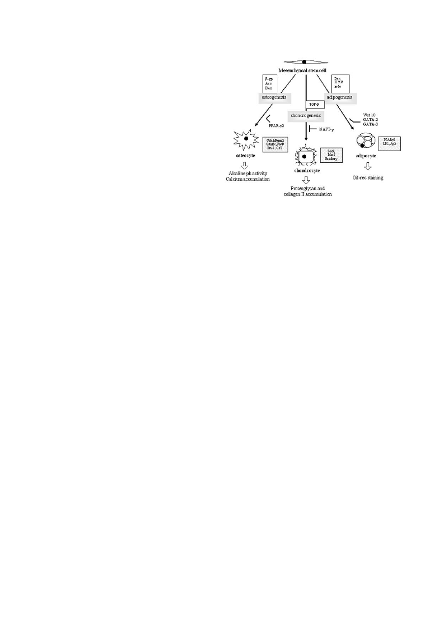
Fig. 2. The scheme of MSC differentiation into the three mes-
enchymal lineages: osteocytes, chondrocytes and adipocytes. The
upper boxes contain inducing factors for each of these pathways,
and the lower ones - the major transcription factors (shadowed).
Ways to identify differentiated cells are pointed by empty arrows.
Abbreviations:
β-gp - β-glycerophosphate; Asc - ascorbic acid;
Dex - dexamethasone; TGF-
β - transforming growth factor-β;
IBMX - isobutylmethylxanthine; indo - indomethacin; PPAR
γ2 -
peroxisome proliferation-activated receptor
γ2; NAFT-p - nuclear
factor of activated T cell; LPL - lipoprotein lipase; aP2 - fatty acid-
binding protein.

However, not all the adherent CFU-F colonies
obtained from the bone marrow aspirates display
pluripotent capacity for differentiation. Pittenger et al.
[96] reported that approximately one-third of them
might be successfully directed to the osteogenic, chon-
drogenic and adipogenic lineages. In vitro differentia-
tion into particular cell lineage demands treating the
cells with a proper mixture of specific differentiating
factors. It must be mentioned that basal nutrients, cell
density, spatial organization, mechanical forces,
growth factors and cytokines, all play a role in MSC
differentiation. To achieve efficient outcome of the
process, each factor should be optimized. Interesting-
ly, the same factor may launch differentiation to
diverse cell lineages in cell cultures derived from var-
ious species. For example, dexametasone is an estab-
lished factor that triggers the differentiation towards
osteoblastic cell lineage in human MSCs [59], where-
as in mouse-derived MSCs it causes adipocyte forma-
tion [24]. Contrariwise, recombinant human bone mor-
phogenetic protein 2 (rhBMP-2) at low doses induces
mouse MSCs into osteogenic lineage [25], but to
obtain the same effect on human MSCs, very high
doses of this factor are required [60]. Apart from that,
the downstream molecular events are very much alike
in various species, which was demonstrated for osteo-
genesis. Both, human and mouse MSCs involve the
transcription factor Cbfa1/Runx2 in this process [32].
It is also known that MSCs synthesize and secrete spe-
cific cytokines and growth factors, and the induction
into each differentiation pathway involves modulation
of their production, as well as regulation of particular
signal-transduction pathway proteins [58]. Moreover,
the cell density has been also shown to be a critical
parameter for differentiation [19, 102]. When MSCs
are seeded at low density, they proliferate and secrete
Dkk1, which favors the undifferentiating phenotype of
the cells. On the contrary, when the cells reach conflu-
ence, Wnt-5a expression abrogates the effect of Dkk1
[42].
It has also been reported that the differentiation
potential may differ in the relation to the source of
MSCs. This statement, however, have as many pros as
cons. According to one study, MSCs derived from adi-
pose tissue possess the impaired ability to differentiate
into both osteoblasts and chondrocytes [55]. Other sci-
entists, on the contrary, demonstrated that MSCs iso-
lated from fat display the same characteristics as
MSCs from bone marrow and might be alternatively
used for clinical trials [66].
In order to obtain osteoblastic cell line, the conflu-
ent monolayer of MSCs should be incubated with a
mixture containing
β
-glycerophosphate, ascorbic acid
and dexamethasone, throughout the period of 2-3
weeks [27]. Participation of bone morphogenetic pro-
teins (BMPs) in bone formation process has been also
postulated, although different BMPs play different
roles [15, 60, 71, 89]. Other important factors involved
in osteogenic regulation are: TGF, insulin-like growth
factor (IGF), brain-derived growth factor (BDGF),
FGF-2, leptin and parathyroid hormone related pep-
tide (PTHrP) [79]. These proteins regulate secretion
of matrix proteins and the expression of signals nec-
essary for bone remodeling through osteoclast activa-
tion. Among the transcription factors committed to
osteogenesis, pivotal roles are attributed to
Cbfa1/Runx2, Osterix,
ΔFosB, Fra-1, Aj18 and Osf1.
Apart from them, Msx2, Dlx5 and TWIST were
shown to take part in this process [132]. As it was
documented, Cbfa1/Runx2 is necessary for osteoblast
formation, but only Dlx5 allows distinguishing the
mineralized osteoblasts. Progression of osteogenesis
might be measured through alkaline phosphatase
activity and calcium accumulation (Fig. 3B) [96].
Human MSCs were shown to possess a great poten-
tial to differentiate into osteoblasts, which was main-
tained for up to 40 doublings in culture, even after
cryopreservation [9].
Chondrogenesis in turn is classically carried out in
micro-mass cultures of MSCs after addition of TGF-
β.
Among TGF-
β
family members, the most important
role in chondrogenesis play BMPs and cartilage-
derived morphogenetic proteins (CDMPs) [27]. Apart
from BMP signaling, cooperation between BMPs and
members of Hedgehog family (Hh) has been reported
[129]. A regulatory role in this process has been
attributed to the proteins from Wnt family. Among
them, Wnt-4 and Wnt-14 were shown to display high
expression at sites of future joint development [34],
whereas Wnt-7a was shown to inhibit chondrogenesis
[53]. Additionally, as recent data indicate, the signal-
ing triggered by the FGF receptor 3 is sufficient to
induce chondrogenic differentiation [48]. TGF-
β and
related cytokines exhibit the ability to induce signal
transduction pathways specific for chondrogenesis,
mostly via activation of mitogen-acivated protein
(MAP) kinases such as: ERK-1, p38, PKC and Jun
[108], whereas FGF receptor acts through Smad pro-
tein signaling [48]. The activation leads to induction
of specific transcription factors expression. The most
important roles play Sox9, Msx2 [109], and Brachury
[48]. They were shown to activate the expression of
chondrocyte-specific genes, like aggrecan and colla-
gen II. Participation in this process has been also
shown for Hox, Pax and Forkhead. Chondrogenic
formation, except from morphological changes, may
be verified by histological testing for the presence of
proteoglycan in the extracellular matrix and collagen
type II chains, which are typical of articular cartilage
[96]. Inhibitory effect on chondrogenesis may be
achieved through nuclear factor of activated T cell -
NAFT-p activity [99].
221
Mesenchymal stem cells
In vitro adipogenesis (Fig. 3C) can be induced by
treating MSCs with a hormonal cocktail containing
dexamethasone, isobutyl methyl xanthine (IBMX) and
indomethacin [25, 27]. The differentiation might be
confirmed using oil-red staining technique and con-
trolling the expression of specific proteins, such as
peroxisome proliferation-activated receptor
γ2
(PPAR
γ2), lipoprotein lipase (LPL), and the fatty acid-
binding protein aP2 [96]. Inhibition of adipogenesis
can by accomplished by the induction of Wnt10b
[103], GATA-2 and GATA-3 [120].
An interesting role in MSCs differentiation toward
osteoblastic versus adipogenic cell lineage is played
by BMP proteins. The BMP-2 as well as bFGF have
been shown to synergistically enhance in vivo bone
formation by MSCs [60, 89]. Selective blocking of the
BMP receptor type 1B (BMPR-1R) resulted in the dif-
ferentiation into adipocytes, which would likewise
suggest that the expression of this receptor is required
for osteocyte formation. Conversely, overexpression
of BMPR-1A blocked adipogenic differentiation and
prompted osteoblastic generation [13]. The findings
indicate that changes in the BMP receptor levels are
intrinsic factors for the commitment into adipogenic or
osteoblastic cell line. Additionally, adipocyte tran-
scription factor -PPAR
γ2 was demonstrated to repress
osteogenesis [75].
Apart from factors inducing differentiation into the
three cell lines described above, the molecules pro-
moting other cell lineage formation, like myocardium
and even neurons, have been identified, but they are
not completely defined so far [33, 45, 80]. Moreover,
MSCs cultured in each of differentiation conditions
produce autocrine and paracrine factors that might be
essential for lineage progression [96].
Clinical application of MSCs
The specific characteristics of MSCs, including their
extensive proliferative potential and the ability to dif-
ferentiate into various cell types, like bone, fat and car-
tilage, make them an attractive tool in regenerative
medicine. This is especially evident in such fields as
cellular biology and gene therapy, resulting in consid-
erable increase in the number of clinical trials based on
the use of MSCs. Apparently, these cells might be sim-
ply isolated from various tissues and expanded in cul-
ture in large numbers that gives the opportunity to cre-
ate a tissue-engineered constructs containing these
cells and re-introduce them into a patient [104, 124,
131]. Full healing is a complex process and demands
integration of the regenerated tissue with the surround-
ing host tissues and differentiation through the natural
signaling pathways. As it was documented, MSCs pos-
sess the capacity to engraft into various tissues and
organs when infused systematically, and this engraft-
ment has been shown to be stable in the long term [28,
31]. Even more, MSCs infused to the peripheral circu-
lation have the ability to migrate to a specific site of
222
S. Bobis et al.
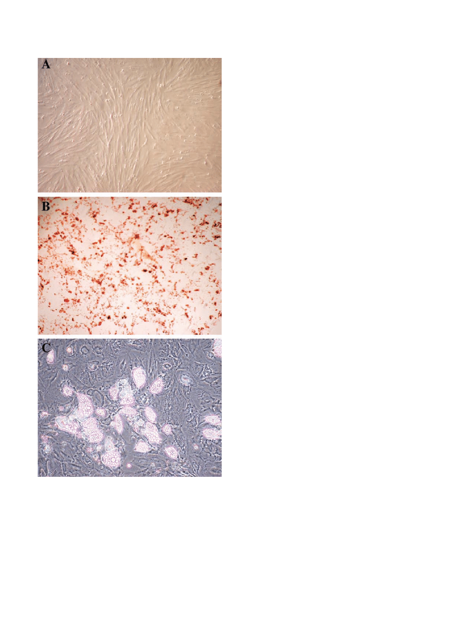
Fig. 3. Bone marrow derived MSCs. Cells were cultured in
MesenCult Basal Medium for three passages and then differentia-
tion was started. A - Cultured MSCs; B - MSCs after 20 days of
osteogenic differentiation (alkaline phosphatase staining); C -
MSCs after 20 days of adipogenic differentiation (note adipocytes
containing lipid droplets).

injury. This phenomenon has been reported in animal
models of bone fracture, cerebral ischemia and
myocardial infarction [110, 125]. In one study, the
authors managed to localize MSCs transplanted to
neonatal mice, using the whole body imaging tech-
nique [88]. On the 7th day post injection, the cells pre-
sented a wide distribution throughout the body of the
recipient mice. 18 days later, the majority of infused
cells were found in lungs and liver, and a very small
population was present in other tissues. Finally, 35
days post infusion, a significant number of the cells
was located in bones, indicating that these cells may
participate in bone formation [88]. Interesting results
were delivered by Prockop et al. [76], who examined
the MSC engraftment efficiency in various tissues in
immunodeficient mice, using a sensitive RT-PCR
method. The engraftment appeared to be at a very low
level, and varied in different tissues. Interestingly, the
survey revealed the presence of a subpopulation of
small size MSCs - rapidly-self renewing MCSs (RS-
MSCs), which engrafted preferentially in comparison
to a larger, slowly renewing MSCs (SR-MSCs). The
two subpopulations varied not only in terms of differ-
entiation potential but also in the surface epitopes. The
more effective engraftment of RS-MSCs might be par-
tially explained by their expression of CXCR4 and
CXCR1, which are known to be involved in the traf-
ficking of MSCs [76].
MSCs have been also proposed to be an excellent
potential tool for gene therapies. They can be subject-
ed to various genetic modifications, such as transduc-
tion with viral vectors carrying a therapeutic gene or
cDNA for special proteins, serving as molecular trans-
mitters. In a mouse model, the genetically modified
MSCs implanted in an ectopic site and subsequently
transplanted to a secondary donor, showed about 74%
stable gene transfer efficiency [31]. They could be
therefore useful in delivering particular genes into
organs or a tissue of special need. Furthermore, there
have been clinical studies in humans with MSCs trans-
fected with viral vectors containing the gene for coag-
ulation factor VII or IX, in case of haemophilia treat-
ment [18]. These cells are also metabolically active
and may serve as a suitable source secreting therapeu-
tic proteins, such as defective enzymes [123]. When
successful, this approach could bring outstanding
results in tissue and body repair.
One of the fields for MSC use in regenerative med-
icine is the treatment of bone defects. First approach to
bone repair relied on biodegradable scaffolds impreg-
nated with recombinant BMPs, and was designed to
induce bone formation through the recruitment of local
MSCs [71]. This project was successfully accom-
plished in an animal model (Lewis rats), showing that
MSCs attracted to BMP-2 are able to regenerate the
injured bone. Such approach was made also by Petite
et al. [94], who managed to heal large segmental bone
defect in sheep. The results were, however, not com-
pletely satisfying because the amount and the quality
of regenerated bone remained disappointing. As anoth-
er example, MSCs were activated through the intra-
muscular injection of adenovirus-mediated hBMP-2
gene transfer in nude mice, which resulted in local
MSC proliferation and differentiation [78]. Further-
more, a portion of implanted cells were competent
themselves to respond to the factors in an autocrine or
paracrine way. The bone healing using MSCs might be
improved with the use of other specific cytokines, like
IGF, PDGF and FGF [15].
With reference to numerous clinical trials using
MSCs, a special attention ought to be paid toward
osteogenesis imperfecta (OI) treatment. This is a
genetic disorder resulting from mutations in collagen I
gene, causing many abnormalities especially in bone
structure [52]. There have been over 150 mutations
responsible for the OI outcome identified, affecting
COL1A1 and COL1A2 genes [84]. As collagen is the
major protein of the extracellular matrix of the bone,
the patients with OI suffer from frequent and numer-
ous fractures, progressive deformities of limbs and
spine, retarded bone growth and short stature [52].
Therefore, a treatment strategy for OI is mainly aimed
at improving bone strength through ameliorating the
structural integrity of collagen [52]. Among therapies
applied to OI, only cell and gene regimens brought
positive effect and seem to be the only reasonable
tools.
The cell therapy approach targeted to osteoblast
formation from MSCs was first investigated on murine
models. MSCs isolated from transgenic mice were
transplanted into irradiated recipient mice [93]. The
location of these cells was inspected 1-5 months after
cell infusion. According to the results, 1.5%-12% of
the cells were found in various tissues, including bones
[93]. Other studies were performed using immunodefi-
cient SCID mouse model, confirming the homing
capacity of hMSCs to the bone marrow and the ability
to differentiate into osteoblasts in vivo [92].
The first steps in therapeutic approach using MSC
transplantation in OI patients were done by Horwitz et
al. in 1999 [51]. Allogenic unmanipulated bone mar-
row from HLA-identical or single-antigen-mis-
matched siblings was transplanted to three children
with OI. The therapeutic outcome was successful
(1.5%-2% of engraftment), showing donor-derived
MSCs located in the bone marrow of the recipient.
Bone marrow MSCs were able to give rise to properly
functioning osteoblasts, resulting in the increase in
bone mineral content, as well as the improvement in
growth velocity and the reduction of bone fracture fre-
quencies [51]. Encouraged by the results, the authors
performed next trials [52]. Bone marrow was obtained
223
Mesenchymal stem cells

from allogenic, HLA-compatible, sibling donors and
was given twice to each patient. Among the five chil-
dren enrolled in this study, three appeared chimeric
and showed donor osteoblast engraftment. As a result,
those children gained significant increase in total body
length with a median of 7.5 cm, measured 6 months
after transplantation, in comparison to 1.25 cm for
control patients. Moreover, the bone mineral content
improved by 45% to 77% of the baseline values. The
number of fractures, visualised by radiography,
declined from an average of 10 during 6 months before
treatment, to 2. Unfortunately, the follow-up study
demonstrated that the growth ratio either decreased or
remained unchanged. In contrast, bone mineralization
continued to increase [52].
Better results were obtained when purified popula-
tion of MSCs was used for grafting. Such a survey was
performed by Horwitz et al. in 2002 [50], demonstrat-
ing the successful engraftment of MSCs. The study
enrolled six children, each of them received two infu-
sions of the allogenic cells. MSCs were transduced
with the LNc8 or G1PLII retroviral vectors, in order to
localize the engrafted cells in patients. The vectors
contained either the neomycine phosphotransferase
gene (neo
R
) or nonexpressing
β-galactosidase (β-gal)
and neo
R
sequences, respectively. The transduction
efficiency was in a range from 2% to 25%. The cells
expressing G1PLII marker were detected in five
patients, at least at one site. The localization included
bones, skin and marrow stroma and brought a positive
healing effect expressed as the acceleration of growth
velocity, in a range from 60% to 94% of the predicted
values for age- and sex-matched healthy children [50].
Furthermore, there has been a novel clinical trail of
in utero MSC transplantation in patient with severe OI
[73]. Allogenic, HLA-incompatible MSCs obtained
from a human male fetal liver, were injected to the
umbilical vein at the week 32 of gestation, in a total
number of 6.5×10
6
cells. After a baby-girl delivery, a
centromeric XY-chromosome-specific probe revealed
0.3% of the donor cells. Interestingly, when examining
whole male genome, the detection of Y chromosome-
positive cells showed 7.4% of the engraftment. There
was no immunoreactivity against transplanted cell
detected, indicating the safety of the study. The out-
come was outstanding, demonstrating the improve-
ment of bone mineralization from 48% at 3 months to
56% at 12 months and 76% at 22 months, in compari-
son to age-matched controls. However, this increase
may be partially attributed to pamidronate treatment,
started from the 4th month. The follow-up revealed only
three fractures during the first two years, normal psy-
chomotor development and correct growth tendency.
A new approach toward OI treatment has been
developed with the occurrence of gene therapy. In the
picture of the disease, the product of mutant allele
interferes with the peptide produced by normal allele,
resulting in abnormal collagen fibril formation. The
gene therapy therefore, should be first directed toward
silencing of the mutant allele expression, and then
replacing the mutated gene. This can be achieved
either by degradation of the mutant mRNA or by dis-
ruption of the mutant gene [12]. However, the treat-
ment strategy might be complicated by the genetic het-
erogeneity of the disease and the fact, that most OI
mutations are dominant-negative.
Gene therapy trial combined with the use of MSCs
was performed by the Russel's group [12], who per-
formed ex vivo genetic modification of autological
MSCs from OI patients. The cells were targeted with
viral vector AAV-COLe1INpA that was designed to
disrupt exon 1 of the chromosomal COL1A1 gene, by
inserting an inactivating cassette. This would change
the mutated gene into a null form, eliminating the pro-
duction of abnormal collagen chains, thus leading to
mild disease symptoms. The results demonstrated that
31% to 90% of the positively selected MSC clones
(0.06% to 0.23% of unselected MSCs) underwent gene
targeting at one allele of COL1A1 gene. There were
very similar targeting frequencies at mutant and wild-
type alleles, suggesting that there was no allele prefer-
ence in this process. Furthermore, very similar target-
ing frequencies in a range of 90% were observed in
polyclonal, as well as in monoclonal cell population.
Gene modification improved collagen processing, sta-
bility and structure, thus preventing pro-collagen pep-
tide retention within the cells. Moreover, the diameter
of collagen fibrils, as well as the melting temperature
was dramatically improved, resembling the values
obtained for wild-type cells. The targeted cells were
also tested for bone and fat formation ability in vivo,
demonstrating their multilineage potential.
Another great challenge for tissue engineering
using MSCs is the treatment of cartilage lesions. The
first reports handling this issue come from Wakitani et
al. [124], who filled mechanically induced full-thick-
ness lesions in New Zealand white rabbits with colla-
gen sponges saturated with MSCs. These cells differ-
entiated into active chondrocytes that produced carti-
lagineous matrix. However, there were some draw-
backs in the first experiment: a discontinuity between
the host tissue and the new tissue, as well as the pro-
gressive thinning of the repaired tissue was observed
[14]. Other scientists successfully performed the carti-
lage differentiation in knee joints, using MSCs stimu-
lated with BMP-2 and IGF-1 [40], whereas unstimu-
lated MSCs failed to induce chondrogenesis under the
same circumstances [89]. It is also worth to itemize
that pro-inflammatory cytokines, which are expressed
in abundance in pathological situations, effectively
inhibit BMP-mediated chondrocyte response. Never-
theless, there have been reports of MSC differentiation
224
S. Bobis et al.

into tendon [131], as well as trials for vertebral disc
regeneration with the use of scaffolds [104]. Those
animal model results seem to be very promising, how-
ever, further studies are needed before their application
to humans.
Further example of potential clinical MSC useful-
ness is the possibility to accelerate the reconstitution of
hematopoiesis in patients after myeloablative
chemotherapy or radiotherapy. Such approach seems
to successfully attenuate graft versus host disease
(GvHD) after hematopoietic stem cell transplantation.
The stromal support has been well documented to be
essential for hematopoiesis and the cell-cell interac-
tions in the marrow microenvironment are critical for
normal hematopoietic function [123]. In a mouse
model, MSC infusion not only prevented the occur-
rence of graft failure, but also had an immunomodula-
tory effect [39]. Moreover, preliminary reports of co-
transplantation of MSCs and HSCs from HLA-identi-
cal siblings showed the reduction in acute and chronic
GvHD [72]. It was demonstrated that addition of
MSCs to the grafting material significantly accelerated
reconstitution of hematopoiesis in autologic and allo-
genic transplantations. This was observed especially in
umbilical cord blood transplantation, both haploidenti-
cal and from unrelated donors [36]. In one case report,
a patient with acute lymphoblastic leukemia, who
developed severe GvHD after allogenic HSC trans-
plantation and did not respond to the applied therapy,
was cured by the use of haploidentical MSCs. The
cells were given twice and no toxicity after infusion
was observed. The outcome indicated that MSCs had a
striking immunosuppressive effect and caused a rapid
healing of damaged gut epithelium. Additionally, the
patient had no minimal residual disease in blood and
bone marrow one year after transplantation [74].
In addition, there are also observations indicating
the usefulness of MSC transplantation in myocardium
regeneration after myocardial infarction. Among all
bone marrow-derived cell populations, only MSCs
were shown to be able to differentiate into cardiomy-
ocytes in vitro [45]. Murine model studies using 5-aza-
cytidine to induce cardiomyocyte differentiation con-
firmed at the molecular level that this cell type could
originate from MSCs [80]. The cells not only con-
tained myotube-like structures and myofilaments, but
were also positively stained for the cardiomyocyte-
specific markers, such as sarcomeric myosin, desmin
and actinin, and showed the expression of cardiomy-
ocyte-specific genes and transcription factors [80].
The same effect was obtained for human MSCs [128].
Prompted by in vitro studies, scientists performed in
vivo experiments. Wang et al. [126] demonstrated that
murine MSCs participate in the formation of new car-
diomyocytes in the normal, uninjured heart. Immuno-
histochemistry executed 4 weeks after injection
proved that donor-derived MSCs were present in the
heart, expressing cardiac markers. The same potential
was demonstrated for human MSCs, which were
injected into the heart of SCID mice. Although the
cells engrafted in small percentage (0.44%), they were
positive for cardiac markers [118]. When used in ani-
mal models for cardiac damage, MSCs successfully
colonized the injured tissue and transformed into prop-
erly active cardiac cells [119]. Spectacular results were
obtained when MSCs transplanted into injured heart
were transduced with a virus encoding Akt - an anti-
apoptotic gene prolonging cell survival, which pre-
vented the pathological remodeling of the left ventricle
after infarction. Approximately 80% of the injured
myocardium regenerated and the cardiac function was
completely restored [81]. Besides improving cardiac
function, MSCs were shown to be able to increase the
ventricular wall mass [113]. Furthermore, local admin-
istration of MSCs to the heart generated de novo
myocardial formation, giving the hope of the use of
these cells in the treatment of coronary heart disease
[90]. The injection of MSCs into infarct zone of
patients with myocardial infarction appeared to be
beneficial for the general heart functionality [115].
Promising results have been also obtained when
using MSCs in neuronal lesion treatment. Previous
studies showed that MSC transplantation improves
recovery after stroke or traumatic brain injury [16].
Additionally, in in vitro co-cultures of MSCs and neu-
ral stem cells, preferential neuronal differentiation has
been observed [77]. Moreover, grafts of MSCs in ani-
mal models have been shown to promote remyelina-
tion [1] as well as partial recovery of function [17].
After direct injection of MSCs into rodent brain, the
cells migrated within the brain and differentiated into
GFAP
+
glial populations [4]. The transplantation of
MSCs into infarcted brain led to the reduction of cell
death and the increase in cell proliferation. Moreover,
MSCs were demonstrated to be able to produce even
myelinating Schwann-like cells, with the typical spin-
dle-shaped morphology and the expression of specific
markers, such as LNGFR, Krox-20, CD104 and S100
[65]. Testing these cells in vivo, by means of transplan-
tation to autologous muscle conduit with 2 cm gap in rat
sciatic nerve, showed their capacity to colonize the
lesion site and regenerate the damaged nerve. The cells
were able to myelinate more than one axon in some
cases, similarly as it is in CNS [65]. In a different set of
experiments, MSCs transplanted into a subtotal cervical
hemisection in adult female rats, were able to integrate
efficiently into the injury site. Moreover, immunohisto-
chemical analysis showed marked axonal growth, indi-
cating that these cells enhance axonal growth after
spinal cord injury. Interestingly, the recovery levels
strongly depended on the human donor and even varied
from lot to lot of MSCs isolated fraction [87].
225
Mesenchymal stem cells

The list of reports indicating that MSCs contribute
to tissue repair in vivo enlarges. There are examples of
MSC utilization in the repair of kidney [47], muscle
[23] and lung [91]. The cells were also found to pro-
mote angiogenesis [46], and were used in chronic skin
wound treatment [6]. The implantation of MSCs
together with occlusive dressing and subsequent epi-
dermal grafts significantly accelerated wound healing
and decreased the risk of amputation in endangered
patients [130].
Clinical trials based on MSCs can omit many of the
limitations associated with the use of embryonic stem
cells (ES). Unlike ES, MSC are not immunogenic,
when used autologically, they do not induce immune
rejection and are also less probable to trigger teratoma
formation, not to mention the ethical concerns.
Unfortunately, there are also some drawbacks con-
cerning the use of MSCs. Firstly, according to some
observations MSCs fused with endogenous differenti-
ated cells and formed tetraploid cells in vivo, although
such an event seems to be extremely rare [114].
Secondly, MSCs were shown to permit tumor growth
in allogenic recipients [30] in animal models. A further
question arises, whether the grafted MSCs can main-
tain their undifferentiated state, thus supporting the
therapeutic effect on a long term basis.
Concluding remarks
It seems well-founded that MSCs constitute a superb
potential tool in regenerative medicine and gene thera-
py approaches. They possess an extensive proliferative
potential and are able to differentiate into various cell
lineages. Due to these important features, the use of
MSCs in clinical trials increases. It has been docu-
mented that these cells engraft successfully in patients
and cause beneficial effects. After learning more about
their properties, it will be possible to start new, more
advanced and better treatment strategies for various
diseases, even those, which seem to be incurable at
present. Moreover, knowing that each patient is genet-
ically different and may give different response to a
treatment, and carry variable predisposition to differ-
ent diseases, specifically targeted strategies using
autologous MSCs, may be designed. However, it is
still a long way to go before using these cells as a rou-
tinely applied therapy in clinics.
Acknowledgments: This study was supported by research grant
PZB-KBN 2/P05C/029/26 from the National Committee of Scien-
tific Research.
References
[ 1] Akiyama Y, Radtke C, Kocsis JD (2002) Remyelination of the
rat spinal cord by transplantation of identified bone marrow
stromal cells. J Neurosci 22: 6623-6630
[ 2] Alsalameh S, Amin R, Gemba T, Lotz M (2004) Identification
of mesenchymal progenitor cells in normal and osteoarthritic
human articular cartilage. Arthritis Rheum 50: 1522-1532
[ 3] Angoulvant D, Clerc A, Benchalal S, Galambrun C, Farre A,
Bertrand Y, Eljaafari A (2004) Human mesenchymal stem
cells suppress induction of cytotoxic response to alloantigens.
Biorheology 41: 469-476
[ 4] Azizi SA, Stokes D, Augelli BJ, DiGirolamo C, Prockop DJ
(1998) Engraftment and migration of human bone marrow
stromal cells implanted in the brains of albino rats-similarities
to astrocyte grafts. Proc Natl Acad Sci USA 95: 3908
[ 5] Baddoo M, Hill K, Wilkinson R, Gaupp D, Hughes C, Kopen
GC, Phinney DG (2003) Characterization of mesenchymal
stem cells isolated from murine bone marrow by negative
selection. J Cell Biochem 89: 1235-1249
[ 6] Badiavas EV, Falanga V (2003) Treatment of chronic wounds
with bone marrow-derived cells. Arch Dermatol 139: 510-516
[ 7] Boiret N, Rapatel C, Veyrat-Masson R, Guillouard L, Guérin
J-J, Pigeon P, Descamps S, Boisgard S, Berger MG (2005)
Characterization of nonexpanded mesenchymal progenitor
cells from normal adult human bone marrow. Exp Hematol
33: 219-225
[ 8] Bonyadi M, Waldman SD, Lui D, Aubin JE Grynpas MD,
Stanford WL (2003) Mesenchymal progenitor self-renewal
deficiency leads to age-dependent osteoporosis in Sca-1/Ly-
6A null mice. Proc Natl Acad Sci USA 100: 5840-5845
[ 9] Bruder SP, Jaiswal N, Haynesworth SE (1997) Growth kinet-
ics, self-renewal, and the osteogenic potential of purified
human mesenchymal stem cells during extensive subcultiva-
tion and following cryopreservation. J Cell Biochem 64: 278-
294
[10] Bruder SP, Ricalton NS, Boynton RE, Connolly TJ, Jaiswal
N, Zaia J, Barry FP (1998) Mesenchymal stem cells surface
antigen SB-10 corresponds to activated leukocyte cell adhe-
sion molecule and is involved in osteogenic differentiation. J
Bone Miner Res 13: 655-663
[11] Campagnoli C, Roberts IA, Kumar S, Bennet PR, Bellan-
tuono I, Fisk NM (2001) Identification of mesenchymal
stem/progenitor cells in human first-trimester fetal blood,
liver and bone marrow. Blood 98: 2396-2402
[12] Chamberlain JR, Schwarze U, Wang PR, Hirata RK, Hanken-
son KD, Pace JM, Undrewood RA, Song KM, Sussman M,
Byers PH, Russel DW (2004) Gene targeting in stem cells
from individuals with osteogenesis imperfecta. Science 303:
1198-1201
[13] Chen D, Ji X, Harris MA, Feng JQ, Karsenty G, Celeste AJ,
Rosen V, Mundy GR, Harris SE (1998) Differential roles for
bone marrow morphogenic protein (BMP) receptor type IB
and IA in differentiation and specification of mesenchymal
precursor cells to osteoblast and adipocyte lineages. J Cell
Biol 142: 295-305
[14] Caplan AI, Elyaderani M, Mochizuki Y, Wakitani S, Goldberg
VM (1997) Principles of cartilage repair and regeneration.
Clin Orthop Relat Res 342: 254-569
[15] Cheng H, Jiang W, Phillips FM, Haydon RC, Peng Y, Zhou L,
Luu HH, An N, Breyer B, Vanichakarn P, Szatkowski JP, Park
JY, He TC (2003) Osteogenic activity of the fourteen types of
human bone morphogenic proteins (BMPs). J Bone Surg Am
85: 1544-1552
[16] Chopp M, Li Y (2002) Treatment of neural injury with mar-
row stromal cells. Lancet Neurol 1: 92-100
[17] Chopp M, Zhang XH, Li Y, Wang L, Chen J, Lu D, Lu M,
Rosenblum M (2000) Spinal cord injury in rat: treatment with
bone marrow stromal cell transplantation. NeuroReport 11:
3001-3005
[18] Chuah MK, Van Damme A, Zwinnen H, Goovaerts I,
Vanslembrouck V, Collen D, Vandendriessche T (2000) Long-
term persistence of human bone marrow stromal cells trans-
226
S. Bobis et al.

duced with factor VIII- retroviral vectors and transient pro-
duction of therapeutic levels of human factor VIII in non-
myeloablated immunodeficient mice. Human Gene Ther 11:
729-738
[19] Colter DC, Class R, DiGirolamo CM, Prockop DJ (2000)
Rapid expansion of recycling stem cells in cultures of plastic-
adherent cells from human bone marrow. Proc Natl Acad Sci
USA 97: 3213-3218
[20] Colter DJ, Sekiya I, Prockop DJ (2001) Identification of a
subpopulation of rapidly self-renewing and multipotential
adult stem cells in colonies of human marrow stromal cells.
Proc Natl Acad Sci USA 98: 7841-7845
[21] Cognet PA, Minguell JJ (1999) Phenotypical and functional
properties of human bone marrow mesenchymal progenitor
cells. J Cell Physiol 181: 67-73
[22] Dazzi F, Ramasamy R, Glennie S, Jones SP, Roberts I (2006)
The role of mesenchymal stem cells in haemopoiesis. Blood
Rev 20: 161-171
[23] De Bari C, Dell'Accio F, Vandenabeele F, Vermeesch JR, Ray-
mackers JM, Luyten FP (2003) Skeletal muscle repair by
adult human mesenchymal stem cells from synovial mem-
brane. J Cell Biol 160: 909-918
[24] Dennis JE, Caplan AI (1996) Differentiation potential of con-
ditionally immortalized mesenchymal progenitor cells from
adult marrow of H-2K
b
-tsA58 transgenic mouse. J Cell Phys-
iol 167: 523-538
[25] Dennis JE, Merriam A, Awadallah A, Yoo JU, Johnstone B,
Caplan AI (1999) A quadripotential mesenchymal progenitor
cell isolated from the marrow of an adult mouse. J Bone
Miner Res 14: 1-10
[26] Dennis JE, Carbillet JP, Caplan AI, Charbord P (2002) The
STRO-1+ marrow cell population is multipotential. Cells Tis-
sues Organs 170: 73-82
[27] Dennis JE, Charbord P (2002) Origin and differentiation of
human and murine stroma. Stem Cells 20: 205-214
[28] Devine SM, Hoffman R (2000) Role of mesenchymal stem
cell in hematopoietic stem cell transplantation. Curr Opin
Hematol 7: 358-363
[29] Di Nicola M, Carlo-Stella C, Magni M, Milanesi M, Longoni
PD, Matteucci P, Grisanti S, Gianni AM (2002) Human bone
marrow stromal cells suppress T-lymphocyte proliferation
induced by cellular or nonspecific mitogenic stimuli. Blood
99: 3838-3843
[30] Djouad F, Plence P, Bony C, Tropel P, Apparailly F, Sany J,
Noel D, Jorgensen C (2003) Immunosupressive effect of mes-
enchymal stem cells favors tumor growth in allogenic ani-
mals. Blood 102: 3837-3844
[31] Drize NJ, Surin VL, Gan OI, Deryugina EI, Chertkov JL
(1992) Gene therapy model for stromal precursor cells of
hematopoietic microenvironment. Leukemia 3: 174S
[32] Ducy P, Zhang R, Geoffroy V, Ridall AL, Karsenty G (1997)
Osf2/Cbfa1: a transcriptional activator of osteoblast differen-
tiation. Cell 89: 747-754
[33] Egusa H, Schweizer FE, Wang CC, Matsuka Y, Nishimura I
(2005) Neuronal differentiation of bone marrow-derived stro-
mal stem cells involves suppression of discordant phenotypes
through gene silencing. J Biol Chem 280: 23691-23697
[34] Enomoto-Iwamoto M, Kitagaki J, Koyama E, Tamamura Y,
Wu C, Kanatani N, Koike T, Okada H, Komori T, Yoneda T,
Church V, Francis-West PH, Kurisu K, Nohno T, Pacifici M,
Iwamoto M (2002) The Wnt antagonist Frzb-1 regulates
chondrocytes maturation and long bone development during
limb skeletogenesis. Dev Biol 251: 142-156
[35] Fernandez M, Simon V, Herrera G, Cao C, Del Favero H,
Minguell JJ (1997) Detection of stromal cells in peripheral
blood progenitor cell collections from breast cancer patients.
Bone Marrow Transplant 20: 265-271
[36] Fibbe WE, Noort WA (2003) Mesenchymal stem cells and
hematopoietic stem cells transplantation. Ann NY Acad Sci
996: 235-244
[37] Friedenstein AJ, Chailakhjan RK, Lalykina KS (1970) The
development of fibroblast colonies in monolayer cultures of
guinea-pig bone marrow and spleen cells. Cell Tissue Kinet 3:
393-403
[38] Friedenstein AJ, Gorskaja U, Kalugina NN (1976) Fibroblast
precursors in normal and irradiated mouse hematopoietic
organs. Exp Hematol 4: 267-274
[39] Gao J, Dennis JE, Muzic RF, Lundberg M, Caplan AI (2001)
The dynamic in vivo distribution of bone-marrow-derived
mesenchymal stem cells after infusion. Cell Tissues Organs
169: 10-20
[40] Gelse K, von der Mark K, Aigner T, Park J, Schneider H
(2003) Articular cartilage repair by gene therapy using growth
factor-producing mesenchymal cells. Arthritis Rheum 48:
430-441
[41] Glennie S, Soeiro I, Dyson PJ, Lam EW, Dazzi F (2005) Bone
marrow mesenchymal stem cells induce division arrest aner-
gy of activated T cells. Blood 105: 2821-2827
[42] Gregory CA, Singh H, Perry AS, Prockop DJ (2003) Wnt sig-
nalling inhibitor Dkk-1 is required for re-entry into the cell
cycle of human adult stem cells from bone marrow stroma
(hMSCs). J Biol Chem 278: 28067-28078
[43] Gronthos S, Franklin DM, Leddy HA, Robey PG, Storms
RW, Gimble JM (2001) Surface protein characterization of
human adipose tissue-derived stromal cells. J Cell Physiol
189: 54-63
[44] Gronthos S, Zannettino AC, Hay DJ, Shi S, Graves SE,
Kortesidis A, Simmonos PJ (2003) Molecular and cellular
characterization of highly purified stromal stems derived
from human bone marrow. J Cell Sci 116: 1827-1835
[45] Heng BC, Haider HK, Sim EK, Cao T, Ng SC (2004) Strate-
gies for directing the differentiation of stem cells into car-
diomyogenic lineage in vitro. Cardiovasc Res 62: 34-42
[46] Hernigou P, Beaujean F (2002) Treatment of osteonecrosis
with autologous bone marrow grafting. Clin Orthop Relat Res
405: 14-23
[47] Herrera MB, Bussolati B, Bruno S, Fonsato V, Romanazzi
GM, Camussi G (2004) Mesenchymal stem cells contribute to
the renal repair of acute tubular epithelial injury. Int J Mol
Med 14: 1035-1041
[48] Hoffman A, Czichos S, Kaps C, Bachner D, Mayer H, Zilber-
man Y, Turgeman G, Pelled G, Gross G, Gazit D (2002) The
T-box transcription factor Brachury mediates cartilage devel-
opment in mesenchymal stem cell line C3H10T1/2. J Cell Sci
115: 769-781
[49] Honczarenko M, Lee Y, Skierkowski M, Ghiran I, Glodek
AM, Silberstein LE (2006) Human bone marrow stromal cells
express a distinct set of biologically functional chemokine
receptors. Stem Cells 24: 1030-1041
[50] Horwitz EM, Gordon PL, Koo WK, Marx JC, Neel MD,
McNall RY, Muul L, Hofmann T (2002) Isolated allogenic
bone-marrow-derived mesenchymal cells engraft and stimu-
late growth in children with osteogenesis imperfecta: impli-
cations for cell therapy of bone. Proc Natl Acad Sci USA 99:
8932-8937
[51] Horwitz EM, Prockop DJ, Fitzpatrick LA, Koo WWK, Gor-
don PL, Neel M, Sussman M, Orchard P, Marx JC, Pyeritz
RE, Brenner MK (1999) Transplantability and therapeutic
effects of bone marrow-derived mesenchymal cells in chil-
dren with osteogenesis imperfecta. Nat Med 5: 309-313
[52] Horwitz EM, Prockop DJ, Gordon PL, Koo WWK, Flitz-
patrick A, Neel MD, McMarville ME, Orchard PJ, Pyeritz
RE, Brenner MK (2001) Clinical responses to bone marrow
transplantation in children with severe osteogenesis imperfec-
ta. Blood 97: 1227-1231
227
Mesenchymal stem cells

[53] Hwang SG, Ryu JH, Kim IC, Jho EH, Jung HC, Kim K, Kim
SJ, Chun JS (2004) Wnt-7a causes loss of differentiated phe-
notype and inhibits apoptosis of articular chondrocytes via
different mechanisms. J Biol Chem 279: 26597-26604
[54] Igura K, Zhang X, Takahashi K, Mitsuru A, Yamaguchi S,
Takashi TA (2004) Isolation and characterization of mes-
enchymal progenitor cells from chorionic villi of human pla-
centa. Cytotherapy 6: 543-553
[55] Im G, Shin Y, Lee K (2005) Do adipose tissue-derived mes-
enchymal stem cells have the same osteogenic and chondro-
genic potential as bone marrow-derived cells? Osteoarthritis
Cartilage 13: 845-853
[56] Inoue K, Ohgushi H, Yoshikawa T, Okumura M, Sempuku T,
Tamai S, Dohi Y (1997) The effect of aging on bone forma-
tion in porous hydroxyapatite: biochemical and histological
analysis. J Bone Miner Res 12: 989-994
[57] in 't Anker PS, Noort WA, Scherjon SA, Kleijburg-van der
Keur C, Kruisselbrink AB, van Bezooijen RL, Beekhuizen W,
Willemze R, Kanhai HH, Fibbe WE (2003) Mesenchymal
stem cells in human second-trimester bone marrow, liver,
lung, and spleen exhibit a similar immunophenotype but a
heterogeneous multilineage differentiation potential. Haema-
tologica 88: 845-852
[58] Jaiswal N, Bruder SP (1997) Human osteoblastic cells secrete
paracrine factors which regulate differentiation of osteogenic
precursors in marrow. Trans Orthop Res Soc 22: 524
[59] Jaiswal N, Haynesworth SE, Caplan AI, Bruder SP (1997)
Osteogenic differentiation of purified, culture-expanded human
mesenchymal stem cells in vitro. J Cell Biochem 64: 295-312
[60] Jaiswal N et al. (2000) Differentiation of human mesenchy-
mal stem cells by transforming growth factor
β superfamily:
expression of osteoblast phenotype by BMP-2 and BMP-4. J
Bone Miner Res 14: 240
[61] Jeong JA, Hong SH, Gang EJ, Ahn C, Hwang SH, Yang IH,
Han H, Kim H (2005) Differential gene expression profiling
of human umbilical cord blood-derived mesenchymal stem
cells by DNA microarray. Stem Cells 23: 584-593
[62] Jiang Y, Jahagirdar BN, Reinhardt RL, Shwartz RE, Keene
CD, Oritz-Gonzalez XR, Reyes M, Lenvik T, Lund T, Black-
stad M, Du J, Aldrich S, Lisberg A, Low W, Largaespada DA,
Verfaillie CM (2002) Pluripotency of mesenchymal stem
cells derived from adult marrow. Nature 418: 41-49
[63] Jiang XX, Zhang Y, Liu B, Zhang SX, Wu Y, Yu XD, Mao N
(2005) Human mesenchymal stem cells inhibit differentiation
and function of monocyte-derived dendritic cells. Blood 105:
4120-4126
[64] Katz AJ, Tholpady A, Tholpady SS, Shang H, Ogle RC
(2005) Cell surface and transcriptional characterization of
human adipose-derived adherent stromal (hADAS) cells.
Stem Cells 23: 412-423
[65] Keilhoff G, Goihl A, Langnäse K, Fansa H (2006) Transdif-
ferentiation of mesenchymal stem cells into Schwann cell-
like myelinating cells. Eur J Cell Biol 85: 11-24
[66] Kim SJ, Cho HH, Kim YJ, Seo SY, Kim HN, Lee JB, Kim JH,
Chung JS, Jung JS (2005) Human adipose stromal cells
expanded in human serum promote engraftment of human
peripheral blood hematopoietic stem cells in NOD/SCID
mice. Biochem Biophys Res Commun 329: 25-31
[67] Koc ON, Gerson SL, Cooper BW, Dyhouse SM,
Haynesworth SE, Caplan AI, Lazarus HM (2000) Rapid
hematopoietic recovery after coinfusion of autologous-blood
stem cells and culture-expanded marrow mesenchymal stem
sells in advanced breast cancer patients receiving high-dose
chemotherapy. J Clin Oncol 18: 307-316
[68] Kopen GC, Prockop DJ, Phinney DG (1999) Marrow stro-
mal cells migrate throughout forebrain and cerebellum, and
they differentiate into astrocytes after injection into neonatal
mouse brains. Proc Natl Acad Sci USA 69: 10711-10716
[69] Krampera M, Glennie S, Dyson J, Scott D, Laylor R, Simp-
son E, Dazzi F (2003) Bone marrow mesenchymal stem cells
inhibit the response of na?ve and memory antigen-specific T
cells to their cognate peptide. Blood 101: 3722-3729
[70] Krampera M, Pasini A, Rigo A, Scupoli MT, Tecchio C,
Malpeli G, Scarpa A, Dazzi F, Pizzolo G, Vinante F (2005)
HB-EGF/HER-1 signaling in bone marrow mesenchymal
stem cells: inducing cell expansion and preventing reversibly
multi-lineage differentiation. Blood 106: 59-66
[71] Lane JM, Yasko AW, Tomin E, Cole BJ, Waller S, Browne M,
Turek T, Gross J (1999) Bone marrow and recombinant
human bone morphogenic protein-2 in osseous repair. Clin
Orthop Relat Res 361: 216-227
[72] Lazarus HM, Koc ON, Devine SM, Curtin P, Maziarz RT,
Holland HK, Shpall EJ, McCarthy P, Atkinson K, Cooper
BW, Gerson SL, Laughlin MJ, Loberiza FR Jr (2005)
Cotransplantation of HLA-identical sibling culture-expanded
mesenchymal stem cells and hematopoietic stem cells in
hematologic malignancy patients. Biol Blood Marrow Trans-
plant 11: 389-398
[73] Le Blanc K, Gotherstrom C, Ringden O, Hassan M, McMa-
hon R, Horwitz E, Anneren G, Axelsson O, Nunn J, Ewald U,
Norden-Lindeberg S, Jansson M, Dalton A, Astrom E, West-
gren M (2005) Fetal mesenchymal stem-cell engraftment in
bone after in utero transplantation in a patient with severe
osteogenesis imperfecta. Transplantation 79: 1607-1614
[74] Le Blanc K, Rasmusson I, Sundberg B, Götherström C, Has-
san M, Uzunel M, Ringdén O (2004) Treatment of severe
acute graft-versus-host disease with third party haploidentical
mesenchymal stem cells. Lancet 363: 1439-1441
[75] Lecka-Czernik B, Gubrij I, Moerman EJ, Kajkenova O, Lip-
schitz DA, Manolagas SC, Jilka RL (1999) Inhibition of
Osf2/Cbfa1 expression and terminal osteoblast differentiation
by PPAR-
γ2. J Cell Biochem 74: 357-371
[76] Lee RH, Hsu SC, Munoz J, Jung JS, Lee NR, Pochampally R,
Prockop DJ (2006) A subset of human rapidly-self renewing
marrow stromal cells (MSCs) preferentially engraft in mice.
Blood 107: 2153-61
[77] Lou S, Gu P, Chen F, He C, Wang M, Lu C (2003) The effect
of bone marrow stromal cells on neuronal differentiation of
mesencephalic neural stem cells in Sprague-Dawley rats.
Brain Res 968: 114-121
[78] Lou J, Xu F, Merkel K, Manske P (1999) Gene therapy: ade-
novirus-mediated human bone morphogenic protein-2 gene
transfer induces mesenchymal progenitor cell proliferation
and differentiation in vitro and bone formation in vivo. J
Orthop Res 17: 43-50
[79] Mackie EJ (2003) Osteoblasts: novel roles in orchestration
of skeletal architecture. Int J Biochem Cell Biol 35: 1301-
1305
[80] Makino S, Fukuda K, Miyoshi S, Konishi F, Kodama H, Pan
J, Sano M, Takahashi T, Hori S, Abe H, Hata J, Umezawa A,
Ogawa S (1999) Cardiomyocytes can be generated from mar-
row stromal cells in vitro. J Clin Invest 103: 697-705
[81] Magi AA, Noiseux N, Kong D, He H, Rezvani M, Ingwall JS,
Dzau VJ (2003) Mesenchymal stem cells modified with Akt
prevent remodelling and restore performance of infarcted
hearts. Nat Med 9: 1195-1201
[82] McBride C, Gaupp D, Phinney DG (2003) Quantifying levels
of transplanted murine and human mesenchymal stem cells in
vivo by real-time PCR. Cytotherapy 5: 7-18
[83] Mendes SC, Robin C, Dzierzak E (2005) Mesenchymal pro-
genitor cells localize within hematopoietic sites throughout
ontogeny. Development 132: 1127-1136
[84] Millington-Ward S, McMahon HP, Allen D, Tuohy G, Kiang
AS, Palfi A, Kenna PF, Humphries P, Farrar GJ (2004) RNAi
of Col1A1 in mesenchymal progenitor cells. Eur J Hum
Genet 12: 864-866
228
S. Bobis et al.

[85] Miura M, Gronthos S, Zhao M, Lu B, Fisher LW, Robey PG,
Shi S (2003) SHED: stem cells from human exfoliated decid-
uous teeth. Proc Natl Acad Sci USA 100: 5807-5812
[86] Muguruma Y, Yahata T, Miyatake H, Sato T, Uno T, Itoh J,
Kato S, Ito M, Hotta T, Ando K (2006) Reconstitition of the
functional human hematopietic microenvironment derived
from human mesenchymal stem cells in the murine bone mar-
row compartment. Blood 107: 1878-87
[87] Neuhuber B, Himes BT, Shumsky JS, Gallo G, Fisher I
(2005) Axon growth and recovery of function supported by
human bone marrow stromal cells in the injured spinal cord
exhibit donor variations. Brain Res 1035: 73-85
[88] Niyibizi C, Wang S, Mi Z, Robbins PD (2004) The fate of
mesenchymal stem cells transplanted into immunocompetent
neonatal mice: implications for skeletal gene therapy via stem
cells. Mol Ther 9: 1525-2016
[89] Noël D, Gazit D, Bouquet C, Apparailly F, Bony C, Plence P,
Millet V, Turgeman G, Perricaudet M, Sany J, Jorgensen C
(2004) Short-term BMP-2 expression is sufficient for in vivo
osteo-chondral differentiation of mesenchymal stem cells.
Stem Cells 22: 74-85
[90] Orlic D, Kajstura J, Chimenti S, Jakoniuk I, Anderson SM, Li
B, Pickel J, McKay R, Nadal-Ginard B, Bodine DM, Leri A,
Anversa P (2001) Bone marrow cells regenerate infarcted
myocardium. Nature 410: 701-705
[91] Ortiz LA, Gambelli F, McBride C, Gaupp D, Baddoo M,
Kaminski N, Phinney DG (2003) Mesenchymal stem cell
engraftment in lung is enchanced in response to bleomycin
exposure and ameliorates its fibrotic effects. Proc Natl Acad
Sci USA 100: 8407-8411
[92] Oyama M, Tatlock A, Fukuta S, Kavalkovich K, Nishimura
K, Johnstone B, Robbins PD, Evans CH, Niyibizi C (1999)
Retrovirally transduced bone marrow stromal cells isolated
from a mouse model of human osteogenesis imperfecta (oim)
persist in bone and retain the ability to form cartilage and
bone after extended passaging. Gene Ther 6: 321-329
[93] Pereira RF, Halford KW, O'Hara MD, Leeper DB, Sokolov
BP, Pollard MD, Bagasra O, Prockop DJ (1995) Cultures of
adherent cells from marrow can serve as long-lasting precur-
sor cells for bone, cartilage and lung in irradiated mice. Proc
Natl Acad Sci USA 92: 4857-4861
[94] Petite H, Viateau V, Bensaid W, Meunier A, de Pollak C, Bour-
guignon M, Oudina K, Sedel L, Guillemin G (2000) Tissue-
engineered bone regeneration. Nat Biotechnol 18: 959-963
[95] Phinney DG, Kopen G, Isaacson RL, Prockop DJ (1999) Plas-
tic adherent stromal cells from the bone marrow of common-
ly used strains of inbred mice: variations in yield, growth, and
differentiation. J Cell Biochem 72: 570-585
[96] Pittenger MF, Mackay AM, Beck SC, Jaiswal RK, Douglas R,
Mosca JD, Moorman MA, Simoneti DW, Craig S, Marshak
DR (1999) Multilineage potential of adult human mesenchy-
mal stem cells. Science 284: 143-147
[97] Prockop DJ, Gregory CA, Spees JL (2003) One strategy for
cell and gene therapy: harnessing the power of adult stem
cells to repair tissues. Proc Natl Acad Sci USA 100,
Suppl:11917-11923
[98] Ramalho-Santos M, Yoon S, Matsuzaki Y, Mulligan RC, Mel-
ton DA (2002) "Stemness": transcriptional profiling of
embryonic and adult stem cells. Science 298: 597-600
[99] Ranger AM, Gerstenfeld LC, Wang J, Kon T, Bae H, Gravallese
EM, Glimcher MJ, Glimcher LH (2000) The nuclear factor of
activated T cells (NAFT) transcription factor NAFTp (NAFTc2)
is a repressor of chondrogenesis. J Exp Med 191: 9-22
[100] Ratajczak MZ, Kucia M, Reca R, Majka M, Janowska-Wiec-
zorek A, Ratajczak J (2004) Stem-cell plasticity revisited:
CXCR4-positive cells expressing mRNA for early muscle,
liver and neural cells 'hide out' in the bone marrow. Leukemia
18: 29-40
[101] Reading L, Still K, Bishop N, Scutt A (2000) Peripheral
blood as an alternative source of mesenchymal stem cells.
Bone 26, Suppl: 9S
[102] Reyes M, Lund T, Lenvik T, Aguiar D, Koodie L, Verfaillie
CM (2001) Purification and ex vivo expansion of postnatal
human marrow mesodermal progenitor cells. Blood 98:
2615-2625
[103] Ross SE, Hemati N, Longo KA, Bennett CN, Lucas PC,
Erickson RL, MacDougald OA (2000) Inhibition of adipoge-
nesis by Wnt signaling. Science 289: 950-953
[104] Sakai D, Mochida J, Yamamoto Y, Nomura T, Okuma M,
Nishimura K, Nakai T, Ando K, Hotta T (2003) Transplanta-
tion of mesenchymal stem cells embedded in Atelocollagen®
gel to the intervertebral disc: a potential therapeutic model
for disc degeneration. Biomaterials 24: 3531-3541
[105] Sato Y, Araki H, Kato J, Nakamura K, Kawano Y, Kobune M,
Sato T, Miyanishi K, Takayama T, Takahashi M, Takimoto R,
Iyama S, Matsunaga T, Ohtani S, Matsuura A, Hamada H,
Niitsu Y (2005) Human mesenchymal stem cells xenograft-
ed directly to rat liver differentiated into human hepatocytes
without fusion. Blood 106: 756-763
[106] Sato N, Meijer L, Skaltsounis L, Greengard P, Brivanolu AH
(2004) Maintenance of pluripotency in human and mouse
embryonic stem cells through activation of Wnt signaling by
a pharmacological GSK-3-specific inhibitor. Nat Med 10:
55-63
[107] Sekiya I, Colter DC, Prockop DJ (2001) BMP-6 enhances
chondrogenesis in a subpopulation of human marrow stromal
cells. Biochem Biophys Res Commun 284: 411-418
[108] Sekiya I, Vouristo TJ, Larson BL, Prockop DJ (2002) in vitro
cartilage formation by human adult stem cells from bone
marrow stroma defines the sequence of cellular and molecu-
lar events during chondrogenesis. Proc Natl Acad Sci USA
99: 4397-4402
[109] Semba I, Nonaka K, Takahashi I, Takahashi K, Dashner R,
Shum L, Nuckolls GH, Slavkin HC (2000) Positionally-
dependent chondrogenesis induced by BMP4 is co-regulated
by Sox9 and Msx2. Dev Dyn 217: 401-414
[110] Shake JG, Gruber PJ, Baumgartner WA, Senechal G, Meyers
J, Redmond JM, Pittenger MF, Martin BJ (2002) Mes-
enchymal stem cell implantation in a swine myocardial
infarct model: engraftment and functional effects. Ann Tho-
rac Surg 73: 1919-1956
[111] Silva WA Jr, Covas DT, Panepucci RA, Proto-Siqueira R,
Siufi JL, Zanette DL, Santos AR, Zago MA (2003) The pro-
file of gene expression of human marrow mesenchymal stem
cells. Stem Cells 21: 661-669
[112] Shofield R (1978) The relationship between the hemopoietic
stem cell and the spleen colony-forming cells: a hypothesis.
Blood Cells 4: 7-25
[113] Smits AM, van Vliet P, Hassink RJ, Goumans MJ, Doeven-
dans PA (2005) The role of stem cells in cardiac regenera-
tion. J Cell Mol Med 9: 25-36
[114] Spees JL, Olson SD, Ylostalo J, Lynch PJ, Smith J, Perry A,
Peister A, Wang MY, Prockop DJ (2003) Differentiation, cell
fusion, and nuclear fusion during ex vivo repair of epithelium
by human adult stem cells from bone marrow stroma. Proc
Natl Acad Sci USA 100: 2397-2402
[115] Stamm C, Westphal B, Kleine HD, Petzsch M, Kittner C,
Klinge H, Schumichen C, Nienaber CA, Freund M, Steinhoff
G (2003) Autologous bone-marrow stem-cell transplantation
for myocardial regeneration. Lancet 361: 45-46
[116] Terada M, Hamazaki T, Oka M, Hoki M, Mastalerz DM,
Nakano Y, Meyer EM, Morel L, Petersen BE, Scott EW
(2002) Bone marrow cells adopt the phenotype of other cells
by spontaneous cell fusion. Nature 416: 542-545
[117] Togel F, Hu Z, Weiss K, Isaac J, Lange C, Westenfelder C
(2005) Administered mesenchymal stem cells protect
229
Mesenchymal stem cells

against ischemic acute renal failure through differentiation-
independent mechanisms. Am J Physiol Renal Physiol 289:
F31-F42
[118] Toma C, Pittenger MF, Cahill KS, Byrne BJ, Kessler PD
(2002) Human mesenchymal stem cells differentiate to a car-
diomyocyte phenotype in the adult murine heart. Circulation
105: 93-98
[119] Tomita S, Li RK, Weisel RD, Mickle DA, Kim EJ, Sakai T,
Jia ZQ (1999) Autologous transplantation of bone marrow
cells improves damaged heart function. Circulation 100:
II247-II259
[120] Tong Q, Dalgin G, Xu H, Ting CN, Leiden JM, Hotamislig-
il GS (2000) Function of GATA transcription factors in
preadipocyte-adipocyte transition. Science 290: 134-138
[121] Tropel P, Noel D, Platet N, Legrand P, Benabid AL, Berger F
(2004) Isolation and characterization of mesenchymal stem
cells from adult mouse bone marrow. Exp Cell Res 295: 395-
406
[122] Tsai MS, Lee JL, Chang YJ, Hwang SM (2004) Isolation of
human multipotent mesenchymal stem cells from second-
trimester amniotic fluid using a novel two-stage culture pro-
tocol. Hum Reprod 19: 1450-1456
[123] Verfaillie CM (1998) Adhesion receptors as regulators of
hematopoietic process. Blood 92: 2609-2612
[124] Wakitani S, Goto T, Pineda SJ, Young RG, Mansour JM,
Caplan AI, Goldberg VM (1994) Mesenchymal cell-based
repair of large, full-thickness defects of articular cartilage. J
Bone Joint Surg Am 76: 579-592
[125] Wang L, Li Y, Chen X, Chen J, Gautam SC, Xu Y, Chopp M
(2002) MCP-1, MIP-1, IL-8 and ischemic cerebral tissue
enchance human bone marrow stromal cell migration in
interface culture. Hematology 7: 113-117
[126] Wang SJ, Shum-Tim D, Galipeau J, Chedrawy E, Eliopoulus
N, Chiu RC (2000) Marrow stromal cells for cellular car-
diomyoplasty: feasibility and potential clinical advantages. J
Thorac Cardiovasc Surg 120: 999-1005
[127] Watt FM, Hogan BL (2000) Out of Eden: stem cells and their
niches. Science 287: 1427-1230
[128] Xu W, Zhang X, Qian H, Zhu W, Sun X, Hu J, Zhou H, Chen
Y (2004) Mesenchymal stem cells from adult human bone
marrow differentiate into a cardiomyocyte phenotype in
vitro. Exp Biol Med (Maywood) 229: 623-631
[129] Yamaguchi A, Komori T, Suda T (2000) Regulation of
osteoblast differentiation mediated by bone-morphogenic
proteins, hedgehoghs, and Cbfa1. Endocr Rev 21: 393-411
[130] Yamaguchi Y, Sumikawa Y, Yoshida S, Kubo T, Yoshikawa
K, Itami S (2005) Prevention of amputation caused by rheu-
matic diseases following a novel therapy of exposing bone
marrow, occlusive dressing and subsequent epidermal graft-
ing. Br J Dermatol 152: 664-672
[131] Young RH, Butler DL, Weber W, Caplan AI, Gordon SL,
Fink DJ (1998) Use of mesenchymal stem cells in a collagen
matrix for Achilles tendon repair. J Orthop Res 6: 406
[132] Zelzer E, Olsen BR (2003) The genetic basis for skeletal dis-
eases. Nature 423: 343-348
[133] Zvaifler NJ, Marinova-Mutafchieva L, Adams G, Edwards
CJ, Moss J, Burger JA, Maini RN (2000) Mesenchymal pre-
cursor cells in the blood of normal individuals. Arthritis Res
2: 477-488
Received: May 23, 2006
Accepted after revision: July 7, 2006
230
S. Bobis et al.
Wyszukiwarka
Podobne podstrony:
więcej podobnych podstron