Primary care/gastroenterology
Diseases of the liver, pancreas and biliary system affect a
substantial proportion of the world’s population and involve
doctors and health care workers across many disciplines. The aim
of the ABC of Liver, Pancreas and Gall Bladder is to provide an
overview of these diseases. To this end it contains helpful
algorithms for diagnosing and treating common diseases.
Information on treatment and prognosis for rarer conditions are
also discussed, making this a comprehensive yet concise
introduction to the subject.
Contents include:
●
gallstone disease
●
acute and chronic viral hepatitis
●
causes of parenchymal liver disease
●
portal hypertension
●
liver tumours
●
liver abscesses and hydatid disease
●
acute and chronic pancreatitis
●
pancreatic tumours
●
liver and pancreatic trauma
●
transplantation of the liver and pancreas.
Written by a leading expert in the field, this book enables the busy
clinician to keep abreast of advances in diagnosis and
management of all conditions and provides the essential
information for medical and nursing students, GPs and junior
hospital doctors in general medical and surgical training.
www.bmjbooks.com
AB
C
OF LIVER, P
ANCREAS AND GALL BLADDER
Beckingham
ABC
OF
LIVER, PANCREAS
AND GALL
BLADDER
Edited by
IJ Beckingham

ABC OF
LIVER, PANCREAS AND GALL BLADDER

This Page Intentionally Left Blank

ABC OF LIVER, PANCREAS AND GALL
BLADDER
Edited by
I J BECKINGHAM
Consultant Hepatobiliary and Laparoscopic Surgeon, Queen’s Medical Centre, Nottingham

© BMJ Books 2001
BMJ Books is an imprint of the BMJ Publishing Group
All rights reserved. No part of this publication may be reproduced, stored in a retrieval system, or transmitted, in any form or by
any means, electronic, mechanical, photocopying, recording and/or otherwise, without the prior written permission of the
publishers.
First published in 2001
by BMJ Books, BMA House, Tavistock Square,
London WC1H 9JR
www.bmjbooks.com
British Library Cataloguing in Publication Data
A catalogue record for this book is available from the British Library
ISBN 0 7279 1531 2
Cover design by Marritt Associates, Harrow, Middlesex
Typeset by FiSH Books and BMJ Electronic Production
Printed and bound in Spain by GraphyCems, Navarra

Investigation of liver and biliary disease
Other causes of parenchymal liver disease
Portal hypertension -2. Ascites, encephalopathy, and other conditions
Liver abscesses and hydatid disease
Transplantation of the liver and pancreas
v
Contents

This Page Intentionally Left Blank

I J Beckingham
Consultant Hepatobiliary and Laparoscopic Surgeon,
Queen’s Medical Centre, Nottingham
P C Bornman
Professor of Surgery, University of Cape Town, South
Africa
J E J Krige
Associate Professor of Surgery, Groote Schuur
Hospital, Cape Town, South Africa
J P A Lodge
Consultant Hepatobiliary and Transplant Surgeon, St
James Hospital, Leeds
K R Prasad
Senior Transplant Fellow, St James Hospital, Leeds
S D Ryder
Consultant Hepatologist, Queen’s Medical Centre,
Nottingham
vii
Contributors

This Page Intentionally Left Blank

Diseases of the Liver, Pancreas and biliary system affect a substantial proportion of the worlds
population and involve doctors and health care workers across many disciplines. Many of these
diseases produce great misery and distress and are economically important requiring much time
off work. The aim of this series was to provide an overview of these diseases and enable the busy
clinician to keep abreast of advances in diagnosis and management of not only the common but
also the rarer, but none the less important, conditions.
ix
Preface

This Page Intentionally Left Blank
1 Investigation of liver and biliary disease
I J Beckingham, S D Ryder
Jaundice is the commonest presentation of patients with liver
and biliary disease. The cause can be established in most cases
by simple non-invasive tests, but many patients will require
referral to a specialist for management. Patients with high
concentrations of bilirubin ( > 100 ìmol/l) or with evidence of
sepsis or cholangitis are at high risk of developing
complications and should be referred as an emergency because
delays in treatment adversely affect prognosis.
Jaundice
Hyperbilirubinaemia is defined as a bilirubin concentration
above the normal laboratory upper limit of 19 ìmol/l. Jaundice
occurs when bilirubin becomes visible within the sclera, skin,
and mucous membranes, at a blood concentration of around
40 ìmol/l. Jaundice can be categorised as prehepatic, hepatic,
or posthepatic, and this provides a useful framework for
identifying the underlying cause.
Around 3% of the UK population have hyperbilirubinaemia
(up to 100 ìmol/l) caused by excess unconjugated bilirubin, a
condition known as Gilbert’s syndrome. These patients have
mild impairment of conjugation within the hepatocytes. The
condition usually becomes apparent only during a transient rise
in bilirubin concentration (precipitated by fasting or illness) that
results in frank jaundice. Investigations show an isolated
unconjugated hyperbilirubinaemia with normal liver enzyme
activities and reticulocyte concentrations. The syndrome is often
familial and does not require treatment.
Prehepatic jaundice
In prehepatic jaundice, excess unconjugated bilirubin is
produced faster than the liver is able to conjugate it for
excretion. The liver can excrete six times the normal daily load
before bilirubin concentrations in the plasma rise.
Unconjugated bilirubin is insoluble and is not excreted in the
urine. It is most commonly due to increased haemolysis—for
example, in spherocytosis, homozygous sickle cell disease, or
thalassaemia major—and patients are often anaemic with
splenomegaly. The cause can usually be determined by further
haematological tests (red cell film for reticulocytes and
abnormal red cell shapes, haemoglobin electrophoresis, red cell
antibodies, and osmotic fragility).
Hepatic and posthepatic jaundice
Most patients with jaundice have hepatic (parenchymal) or
posthepatic (obstructive) jaundice. Several clinical features may
help distinguish these two important groups but cannot be
relied on, and patients should have ultrasonography to look for
evidence of biliary obstruction.
The most common intrahepatic causes are viral hepatitis,
alcoholic cirrhosis, primary biliary cirrhosis, drug induced
jaundice, and alcoholic hepatitis. Posthepatic jaundice is most
often due to biliary obstruction by a stone in the common bile
duct or by carcinoma of the pancreas. Pancreatic pseudocyst,
chronic pancreatitis, sclerosing cholangitis, a bile duct stricture,
or parasites in the bile duct are less common causes.
In obstructive jaundice (both intrahepatic cholestasis and
extrahepatic obstruction) the serum bilirubin is principally
conjugated. Conjugated bilirubin is water soluble and is
Box 1.1 History that should be taken from patients
presenting with jaundice
x
Duration of jaundice
x
Previous attacks of jaundice
x
Pain
x
Chills, fever, systemic symptoms
x
Itching
x
Exposure to drugs (prescribed and illegal)
x
Biliary surgery
x
Anorexia, weight loss
x
Colour of urine and stool
x
Contact with other jaundiced patients
x
History of injections or blood transfusions
x
Occupation
Box1.2 Examination of patients with jaundice
x
Depth of jaundice
x
Scratch marks
x
Signs of chronic liver disease:
Palmar erythema
Clubbing
White nails
Dupuytren’s contracture
Gynaecomastia
x
Liver:
Size
Shape
Surface
x
Enlargement of gall bladder
x
Splenomegaly
x
Abdominal mass
x
Colour of urine and stools
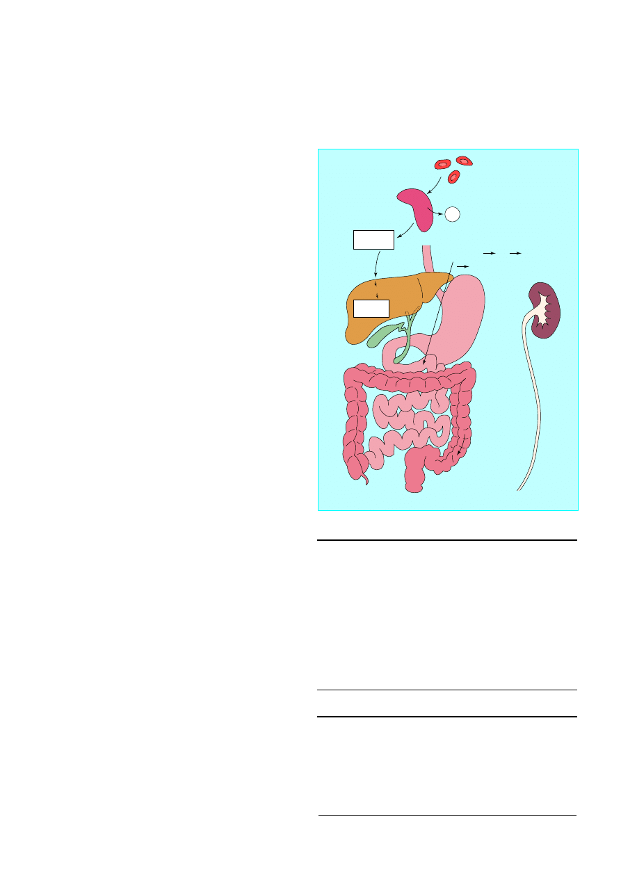
Old red blood cells
Spleen
Fe
2
+
Haem
Unconjugated
bilirubin
Conjugated
bilirubin
Bile
canaliculi
Bile
ducts
Small amount of reduced
bilirubin reabsorbed into
portal vein liver
systemic blood supply
kidneys
Bilirubin
reduced by
gut bacteria
to:
Stercobilinogen
Faeces
Terminal
ileum
Colon
Liver
Kidney
Urobilinogen
Hepatocytes
Albumin
Duodenum
Figure 1.1 Bilirubin pathway
1
excreted in the urine, giving it a dark colour (bilirubinuria). At
the same time, lack of bilirubin entering the gut results in pale,
“putty” coloured stools and an absence of urobilinogen in the
urine when measured by dipstick testing. Jaundice due to
hepatic parenchymal disease is characterised by raised
concentrations of both conjugated and unconjugated serum
bilirubin, and typically stools and urine are of normal colour.
However, although pale stools and dark urine are a feature of
biliary obstruction, they can occur transiently in many acute
hepatic illnesses and are therefore not a reliable clinical feature
to distinguish obstruction from hepatic causes of jaundice.
Liver function tests
Liver function tests routinely combine markers of function
(albumin and bilirubin) with markers of liver damage (alanine
transaminase, alkaline phosphatase, and ã-glutamyl transferase).
Abnormalities in liver enzyme activities give useful information
about the nature of the liver insult: a predominant rise in
alanine transaminase activity (normally contained within the
hepatocytes) suggests a hepatic process. Serum transaminase
activity is not usually raised in patients with obstructive
jaundice, although in patients with common duct stones and
cholangitis a mixed picture of raised biliary and hepatic enzyme
activity is often seen.
Epithelial cells lining the bile canaliculi produce alkaline
phosphatase, and its serum activity is raised in patients with
intrahepatic cholestasis, cholangitis, or extrahepatic obstruction;
increased activity may also occur in patients with focal hepatic
lesions in the absence of jaundice. In cholangitis with
incomplete extrahepatic obstruction, patients may have normal
or slightly raised serum bilirubin concentrations and high
serum alkaline phosphatase activity. Serum alkaline
phosphatase is also produced in bone, and bone disease may
complicate the interpretation of abnormal alkaline phosphatase
activity. If increased activity is suspected to be from bone, serum
concentrations of calcium and phosphorus should be measured
together with 5
¢
-nucleotidase or ã-glutamyl transferase activity;
these two enzymes are also produced by bile ducts, and their
activity is raised in cholestasis but remains unchanged in bone
disease.
Occasionally, the enzyme abnormalities may not give a clear
answer, showing both a biliary and hepatic component. This is
usually because of cholangitis associated with stones in the
common bile duct, where obstruction is accompanied by
hepatocyte damage as a result of infection within the biliary
tree.
Plasma proteins and coagulation
factors
A low serum albumin concentration suggests chronic liver
disease. Most patients with biliary obstruction or acute hepatitis
will have normal serum albumin concentrations as the half life
of albumin in plasma is around 20 days and it takes at least 10
days for the concentration to fall below the normal range
despite impaired liver function.
Coagulation factors II, V, VII, and IX are synthesised in the
liver. Abnormal clotting (measured as prolongation of the
international normalised ratio) occurs in both biliary
obstruction and parenchymal liver disease because of a
combination of poor absorption of fat soluble vitamin K (due to
absence of bile in the gut) and a reduced ability of damaged
hepatocytes to produce clotting factors.
Box 1.3 Drugs that may cause liver damage
Analgesics
x
Paracetamol
x
Aspirin
x
Non-steroidal anti-inflammatory drugs
Cardiac drugs
x
Methyldopa
x
Amiodarone
Psychotropic drugs
x
Monoamine oxidase inhibitors
x
Phenothiazines (such as chlorpromazine)
Others
x
Sodium valproate
x
Oestrogens (oral contraceptives and hormone replacement
therapy)
The presence of a low serum albumin
concentration in a jaundiced patient
suggests a chronic disease process
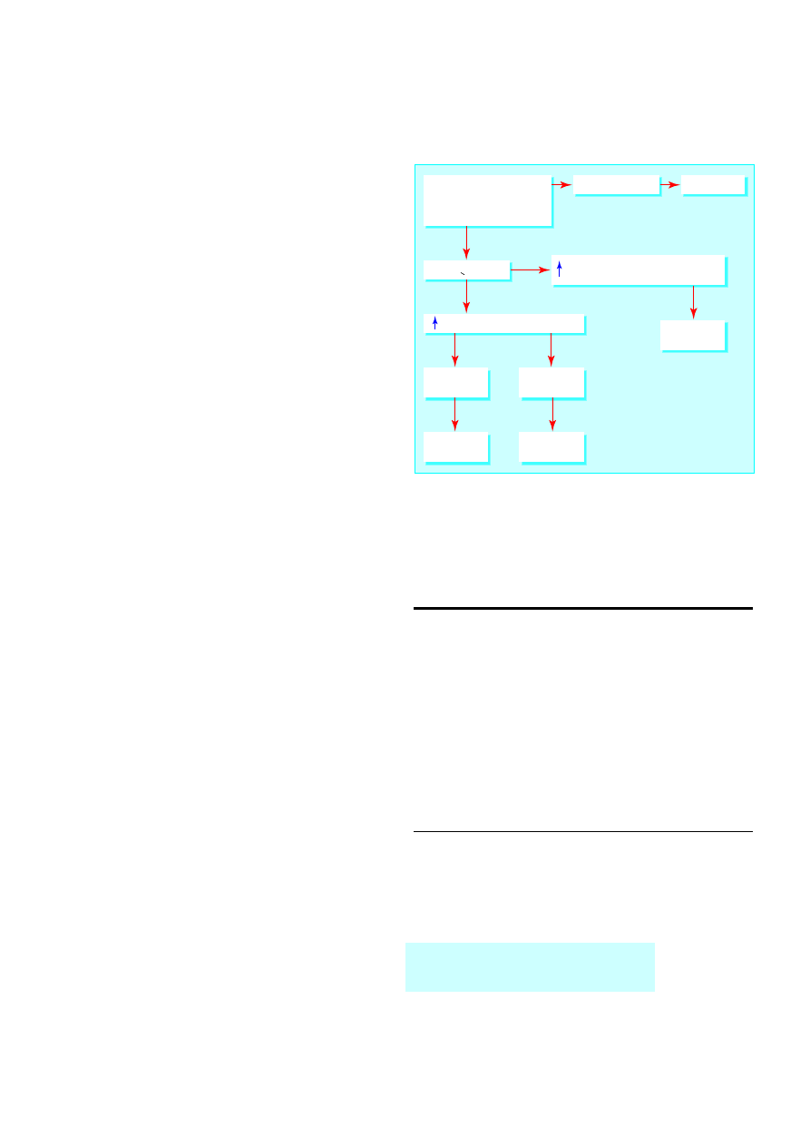
Initial consultation send results of
• Liver function tests
• Hepatitis A IgM
• Hepatitis B surface antigen
Bilirubin <100
µ
mol/l
Alanine transaminase = hepatitis
Hepatitis A IgM
positive
Hepatitis A IgM
negative
Treat for
hepatitis A
Refer
Alkaline phosphatase
g
-glutamyltransferase -
cholestasis/obstruction
Bilirubin >100
µ
mol/l
Urgent referral
Refer
Figure 1.2 Guide to investigation and referral of patients with jaundice in
primary care
ABC of Liver, Pancreas, and Gall Bladder
2
Serum globulin titres rise in chronic hepatitis and cirrhosis,
mainly due to a rise in the IgA and IgG fractions. High titres of
IgM are characteristic of primary biliary cirrhosis, and IgG is a
hallmark of chronic active hepatitis. Ceruloplasmin activity
(ferroxidase, a copper transporting globulin) is reduced in
Wilson’s disease. Deficiency of á
1
antitrypsin (an enzyme
inhibitor) is a cause of cirrhosis as well as emphysema. High
concentrations of the iron carrying protein ferritin are a marker
of haemochromatosis.
Autoantibodies are a series of antibodies directed against
subcellular fractions of various organs that are released into the
circulation when cells are damaged. High titres of
antimitochondrial antibodies are specific for primary biliary
cirrhosis, and antismooth muscle and antinuclear antibodies are
often seen in autoimmune chronic active hepatitis. Antibodies
against hepatitis are discussed in detail in a future article on
hepatitis.
Imaging in liver and biliary disease
Plain radiography has a limited role in the investigation of
hepatobiliary disease. Chest radiography may show small
amounts of subphrenic gas, abnormalities of diaphragmatic
contour, and related pulmonary disease, including metastases.
Abdominal radiographs can be useful if a patient has calcified
or gas containing lesions as these may be overlooked or
misinterpreted on ultrasonography. Such lesions include
calcified gall stones (10-15% of gall stones), chronic calcific
pancreatitis, gas containing liver abscesses, portal venous gas,
and emphysematous cholecystitis.
Ultrasonography is the first line imaging investigation in
patients with jaundice, right upper quadrant pain, or
hepatomegaly. It is non-invasive, inexpensive, and quick but
requires experience in technique and interpretation.
Ultrasonography is the best method for identifying gallbladder
stones and for confirming extrahepatic biliary obstruction as
dilated bile ducts are visible. It is good at identifying liver
abnormalities such as cysts and tumours and pancreatic masses
and fluid collections, but visualisation of the lower common bile
duct and pancreas is often hindered by overlying bowel gas.
Computed tomography is complementary to ultrasonography
and provides information on liver texture, gallbladder disease,
bile duct dilatation, and pancreatic disease. Computed
tomography is particularly valuable for detecting small lesions
in the liver and pancreas.
Cholangiography identifies the level of biliary obstruction
and often the cause. Intravenous cholangiography is rarely used
now as opacification of the bile ducts is poor, particularly in
jaundiced patients, and anaphylaxis remains a problem.
Endoscopic retrograde cholangiopancreatography is advisable
when the lower end of the duct is obstructed (by gall stones or
carcinoma of the pancreas). The cause of the obstruction (for
example, stones or parasites) can sometimes be removed by
endoscopic retrograde cholangiopancreatography to allow
cytological or histological diagnosis.
Percutaneous transhepatic cholangiography is preferred for
hilar obstructions (biliary stricture, cholangiocarcinoma of the
hepatic duct bifurcation) because better opacification of the
ducts near the obstruction provides more information for
planning subsequent management. Obstruction can be relieved
by insertion of a plastic or metal tube (a stent) at either
endoscopic retrograde cholangiopancreatography or
percutaneous transhepatic cholangiography.
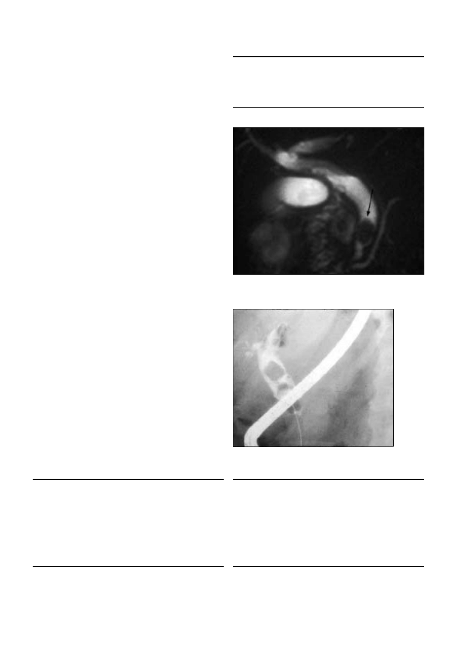
Magnetic resonance cholangiopancreatography allows
non-invasive visualisation of the bile and pancreatic ducts. It is
Table 1.1 Autoantibody and immunoglobulin characteristics
in liver disease
Autoantibodies
Immunoglobulins
Primary biliary
cirrhosis
High titre of
antimitochondrial antibody
in 95% of patients
Raised IgM
Autoimmune
chronic active
hepatitis
Smooth muscle antibody in
70%, antinuclear factor in
60%, Low antimitochondrial
antibody titre in 20%
Raised IgG in all
patients
Primary
sclerosing
cholangitis
Antinuclear cytoplasmic
antibody in 30%
Ultrasonography is the most useful initial
investigation in patients with jaundice
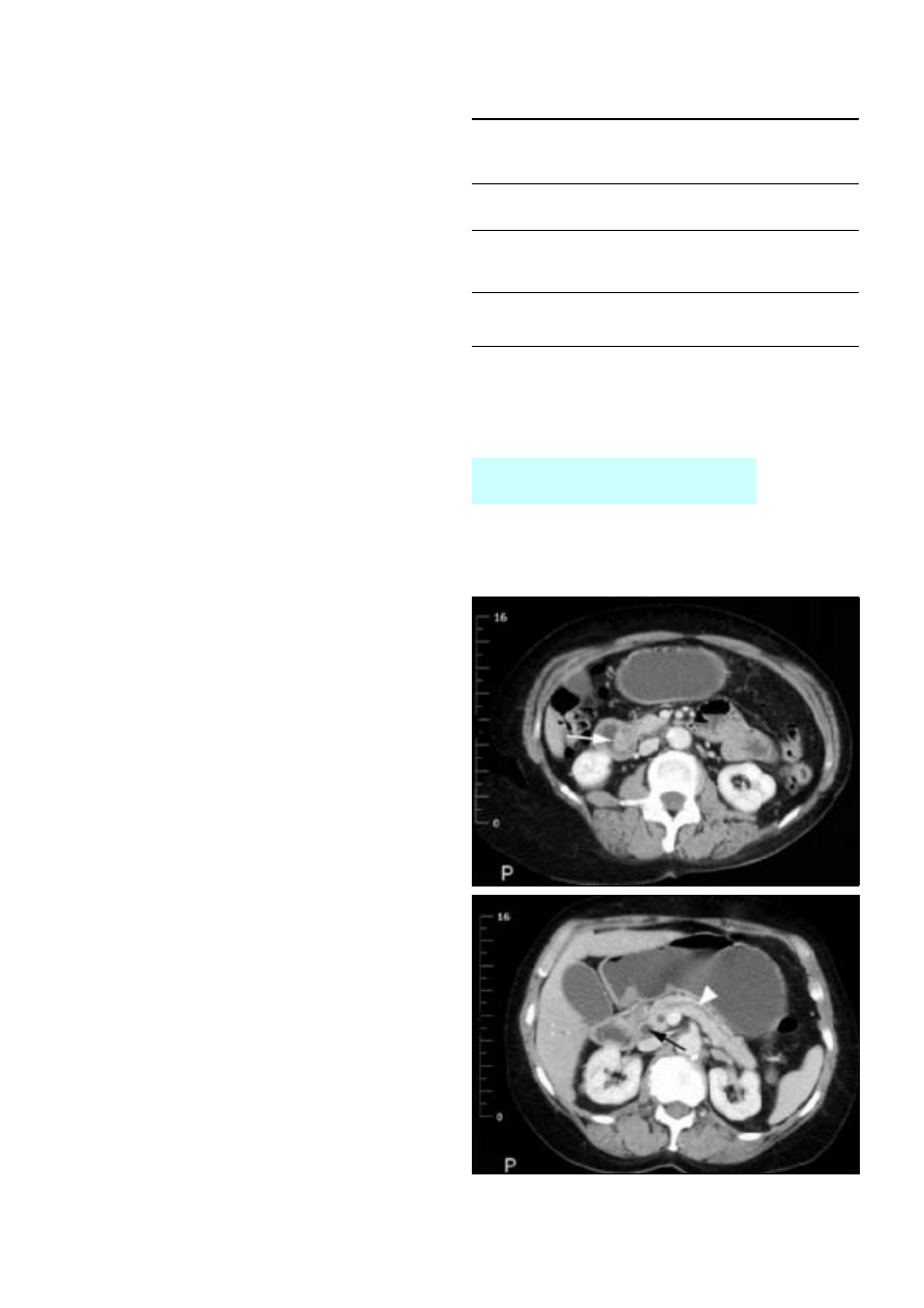
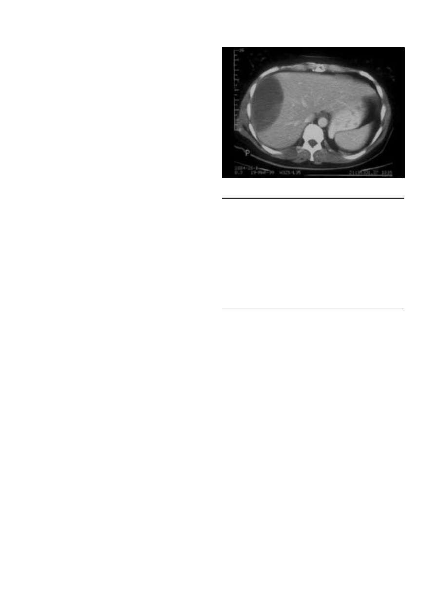
Figure 1.3 Computed tomogram of ampullary carcinoma (white arrow)
causing obstruction of the bile duct (black arrow, bottom) and pancreatic
ducts (white arrowhead)
Investigation of liver and biliary disease
3
superseding most diagnostic endoscopic
cholangiopancreatography as faster magnetic resonance
imaging scanners become more widely available.
Liver biopsy
Percutaneous liver biopsy is a day case procedure performed
under local anaesthetic. Patients must have a normal clotting
time and platelet count and ultrasonography to ensure that the
bile ducts are not dilated. Complications include bile leaks and
haemorrhage, and overall mortality is around 0.1%. A
transjugular liver biopsy can be performed by passing a special
needle, under radiological guidance, through the internal
jugular vein, the right atrium, and inferior vena cava and into
the liver though the hepatic veins. This has the advantage that
clotting time does not need to be normal as bleeding from the
liver is not a problem. Liver biopsy is essential to diagnose
chronic hepatitis and establish the cause of cirrhosis.
Ultrasound guided liver biopsy can be used to diagnose liver
masses. However, it may cause bleeding (especially with liver cell
adenomas), anaphylactic shock (hydatid cysts), or tumour
seeding (hepatocellular carcinoma or metastases). Many lesions
can be confidently diagnosed by using a combination of
imaging methods (ultrasonography, spiral computed
tomography, magnetic resonance imaging, nuclear medicine,
laparoscopy, and laparoscopic ultrasonography). When
malignancy is suspected in solitary lesions or those confined to
one half of the liver, resection is the best way to avoid
compromising a potentially curative procedure.
Summary points
x
An isolated raised serum bilirubin concentration is usually due to
Gilbert’s syndrome, which is confirmed by normal liver enzyme
activities and full blood count
x
Jaundice with dark urine, pale stools, and raised alkaline
phosphatase and ã-glutamyl transferase activity suggests an
obstructive cause, which is confirmed by presence of dilated bile
ducts on ultrasonography
x
Jaundice in patients with low serum albumin concentration suggests
chronic liver disease
x
Patients with high concentrations of bilirubin ( > 100 ìmol/l) or
signs of sepsis require emergency specialist referral
x
Imaging of the bile ducts for obstructive jaundice is increasingly
performed by magnetic resonance cholangiopancreatography, with
endoscopy becoming reserved for therapeutic interventions
Figure 1.4 Subcapsular haematoma: a complication of liver biopsy
ABC of Liver, Pancreas, and Gall Bladder
4
2 Gallstone disease
I J Beckingham
Gall stones are the most common abdominal reason for
admission to hospital in developed countries and account for
an important part of healthcare expenditure. Around 5.5
million people have gall stones in the United Kingdom, and
over 50 000 cholecystectomies are performed each year.
Types of gall stone and aetiology
Normal bile consists of 70% bile salts (mainly cholic and
chenodeoxycholic acids), 22% phospholipids (lecithin), 4%
cholesterol, 3% proteins, and 0.3% bilirubin. Cholesterol or
cholesterol predominant (mixed) stones account for 80% of all
gall stones in the United Kingdom and form when there is
supersaturation of bile with cholesterol. Formation of stones is
further aided by decreased gallbladder motility. Black pigment
stones consist of 70% calcium bilirubinate and are more
common in patients with haemolytic diseases (sickle cell
anaemia, hereditary spherocytosis, thalassaemia) and cirrhosis.
Brown pigment stones are uncommon in Britain
(accounting for < 5% of stones) and are formed within the
intraheptic and extrahepatic bile ducts as well as the gall
bladder. They form as a result of stasis and infection within the
biliary system, usually in the presence of Escherichia coli and
Klebsiella spp,
which produce â glucuronidase that converts
soluble conjugated bilirubin back to the insoluble unconjugated
state leading to the formation of soft, earthy, brown stones.
Ascaris lumbricoides
and Opisthorchis senensis have both been
implicated in the formation of these stones, which are common
in South East Asia.
Clinical presentations
Biliary colic or chronic cholecystitis
The commonest presentation of gallstone disease is biliary pain.
The pain starts suddenly in the epigastrium or right upper
quadrant and may radiate round to the back in the
interscapular region. Contrary to its name, the pain often does
not fluctuate but persists from 15 minutes up to 24 hours,
subsiding spontaneously or with opioid analgesics. Nausea or
vomiting often accompanies the pain, which is visceral in origin
and occurs as a result of distension of the gallbladder due to an
obstruction or to the passage of a stone through the cystic duct.
Most episodes can be managed at home with analgesics and
antiemetics. Pain continuing for over 24 hours or accompanied
by fever suggests acute cholecystitis and usually necessitates
hospital admission. Ultrasonography is the definitive
investigation for gall stones. It has a 95% sensitivity and
specificity for stones over 4 mm in diameter.
Non-specific abdominal pain, early satiety, fat intolerance,
nausea, and bowel symptoms occur with comparable frequency
in patients with and without gall stones, and these symptoms
respond poorly to inappropriate cholecystectomy. In many of
these patients symptoms are due to upper gastrointestinal tract
problems or irritable bowel syndrome.
Acute cholecystitis
When obstruction of the cystic duct persists, an acute
inflammatory response may develop with a leucocytosis and
mild fever. Irritation of the adjacent parietal peritoneum causes
Box 1.1 Risk factors associated with formation of cholesterol
gall stones
x
Age > 40 years
x
Female sex (twice risk in
men)
x
Genetic or ethnic variation
x
High fat, low fibre diet
x
Obesity
x
Pregnancy (risk increases
with number of
pregnancies)
x
Hyperlipidaemia
x
Bile salt loss (ileal disease
or resection)
x
Diabetes mellitus
x
Cystic fibrosis
x
Antihyperlipidaemic drugs
(clofibrate)
x
Gallbladder dysmotility
x
Prolonged fasting
x
Total parenteral nutrition
Box 1.2 Differential diagnosis of common causes of severe
acute epigastric pain
x
Biliary colic
x
Peptic ulcer disease
x
Oesophageal spasm
x
Myocardial infarction
x
Acute pancreatitis
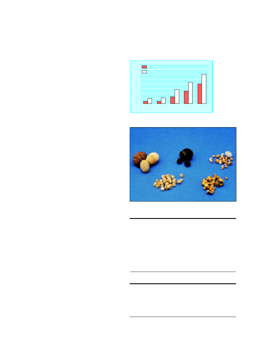
Age (years)
% of population
30
40
50
60
70
0
10
15
20
25
30
35
40
5
Men
Women
Figure 2.1 Prevalence of gall stones in United Kingdom
according to age
Figure 2.2 Gall stones vary from pure cholesterol (white), through mixed, to
bile salt predominant (black)
5
localised tenderness in the right upper quadrant. As well as gall
stones, ultrasonography may show a tender, thick walled,
oedematous gall bladder with an abnormal amount of adjacent
fluid. Liver enzyme activities are often mildly abnormal.
Initial management is with non-steroidal anti-inflammatory
drugs (intramuscular or per rectum) or opioid analgesic.
Although acute cholecystitis is initially a chemical inflammation,
secondary bacterial infection is common, and patients should
be given a broad spectrum parenteral antibiotic (such as a
second generation cephalosporin).
Progress is monitored by resolution of tachycardia, fever,
and tenderness. Ideally cholecystectomy should be performed
during the same admission as delayed cholecystectomy has a
15% failure rate (empyema, gangrene, or perforation) and a
15% readmission rate with further pain.
Jaundice
Jaundice occurs in patients with gall stones when a stone
migrates from the gall bladder into the common bile duct or,
less commonly, when fibrosis and impaction of a large stone in
Hartmann’s pouch compresses the common hepatic duct
(Mirrizi’s syndrome). Liver function tests show a cholestatic
pattern (raised conjugated bilirubin concentration and alkaline
phosphatase activity with normal or mildly raised aspartate
transaminase activity) and ultrasonography confirms dilatation
of the common bile duct ( > 7 mm diameter) usually without
distention of the gall bladder.
Acute cholangitis
When an obstructed common bile duct becomes contaminated
with bacteria, usually from the duodenum, cholangitis may
develop. Urgent treatment is required with broad spectrum
antibiotics together with early decompression of the biliary
system by endoscopic or radiological stenting or surgical
drainage if stenting is not available. Delay may result in
septicaemia or the development of liver abscesses, which are
associated with a high mortality.
Acute pancreatitis
Acute pancreatitis develops in 5% of all patients with gall stones
and is more common in patients with multiple small stones, a
wide cystic duct, and a common channel between the common
bile duct and pancreatic duct. Small stones passing down the
common bile duct and through the papilla may temporarily
obstruct the pancreatic duct or allow reflux of duodenal fluid or
bile into the pancreatic duct resulting in acute pancreatitis.
Patients should be given intravenous fluids and analgesia and
be monitored carefully for the development of organ failure
(see later article on acute pancreatitis).
Gallstone ileus
Acute cholecystitis may cause the gall bladder to adhere to the
adjacent jejunum or duodenum. Subsequent inflammation may
result in a fistula between these structures and the passage of a
gall stone into the bowel. Large stones may become impacted
and obstruct the small bowel. Abdominal radiography shows
obstruction of the small bowel and air in the biliary tree.
Treatment is by laparotomy and “milking” the obstructing stone
into the colon or by enterotomy and extraction.
Natural course of gallstone disease
Two thirds of gall stones are asymptomatic, and the yearly risk
of developing biliary pain is 1-4%. Patients with asymptomatic
gall stones seldom develop complications. Prophylactic
cholecystectomy is therefore not recommended when stones
Box 1.3 Charcot’s triad of symptoms in severe cholangitis
x
Pain in right upper quadrant
x
Jaundice
x
High swinging fever with rigors and chills
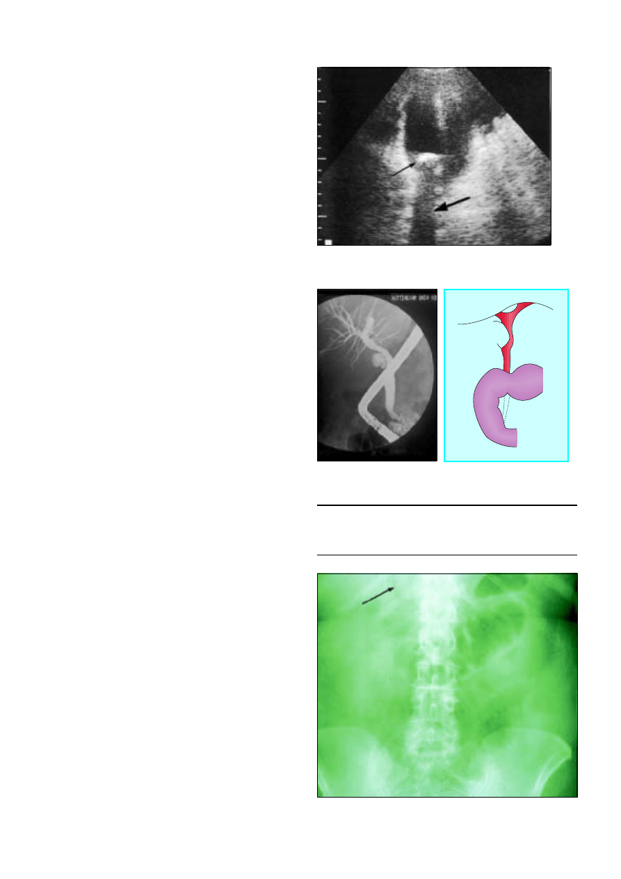
Figure 2.3 Ultrasonogram showing large gall stone (thin arrow)
casting acoustic shadow (thick arrow) in gall bladder
Figure 2.4 Type 1 Mirrizi’s syndrome: gallbladder stone in Hartmann’s
pouch compressing common bile duct and causing deranged liver function
Figure 2.5 Small bowel obstruction and gas in bile ducts in patient with
gallstone ileus
ABC of Liver, Pancreas, and Gall Bladder
6
are discovered incidentally by radiography or ultrasonography
during the investigation of other symptoms. Although gall
stones are associated with cancer of the gall bladder, the risk of
developing cancer in patients with asymptomatic gall stones is
< 0.01%—less than the mortality associated with
cholecystectomy.
Patients with symptomatic gall stones have an annual rate of
developing complications of 1-2% and a 50% chance of a
further episode of biliary colic. They should be offered
treatment.
Management of gallstone disease
Cholecystectomy
Cholecystectomy is the optimal management as it removes both
the gall stones and the gall bladder, preventing recurrent
disease. The only common consequence of removing the gall
bladder is an increase in stool frequency, which is clinically
important in less than 5% of patients and responds well to
standard antidiarrhoeal drugs when necessary.
Laparoscopic cholecystectomy has been adopted rapidly
since its introduction in 1987, and 80-90% of cholecystectomies
in the United Kingdom are now carried out in this way. The
only specific contraindications to laparoscopic cholecystectomy
are coagulopathy and the later stages of pregnancy. Acute
cholecystitis and previous gastroduodenal surgery are no
longer contraindications but are associated with a higher rate of
conversion to open cholecystectomy.
Laparoscopic cholecystectomy has a lower mortality than
the standard open procedure (0.1% v 0.5% for the open
procedure). This is mainly because of a lower incidence of
postoperative cardiac and respiratory complications. The
smaller incisions cause less pain, which reduces the requirement
for opioid analgesics. Patients usually stay in hospital for only
one night in most centres, and the procedure can be done as a
day case in selected patients. Most patients are able to return to
sedentary work after 7-10 days. This decrease in overall
morbidity and earlier recovery has led to a 25% increase in the
rate of cholecystectomy in some countries.
The main disadvantage of the laparoscopic technique has
been a higher incidence of injury to the common hepatic or
bile ducts (0.2-0.4% v 0.1% for open cholecystectomy). Higher
rates of injury are associated with inexperienced surgeons (the
“learning curve” phenomenon) and acute cholecystitis.
Furthermore, injuries to the common bile duct tend to be more
extensive with laparoscopic surgery. However, there is some
evidence suggesting that the rates of injury are now falling.
Box 1.4 Causes of pain after cholecystectomy
x
Retained or recurrent stone (dilatation of common bile duct seen in
only 30% of patients)
x
Iatrogenic biliary leak or stricture of common bile duct
x
Papillary stenosis or dysfunctional sphincter of Oddi
x
Incorrect preoperative diagnosis—for example, irritable bowel
syndrome, peptic ulcer, gastro-oesophageal reflux
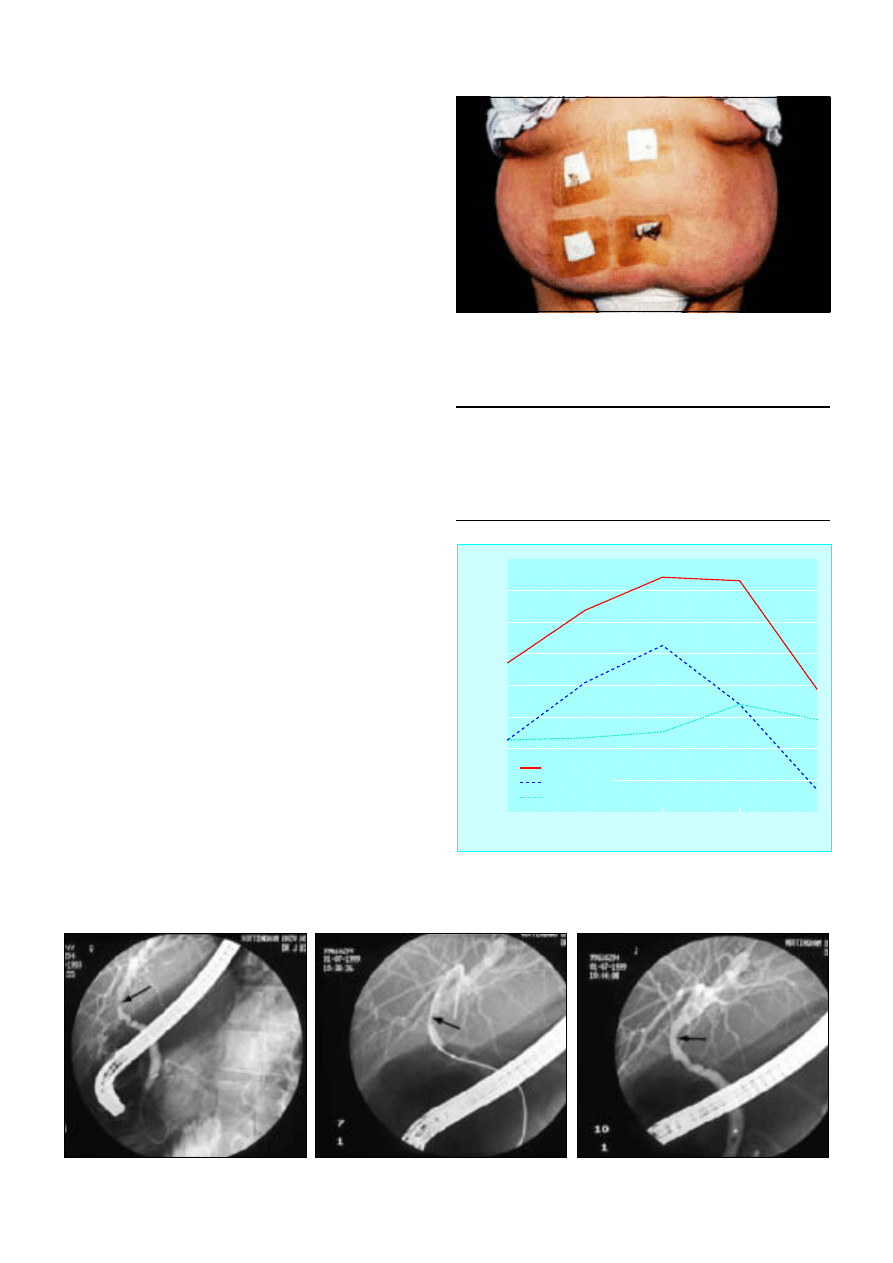
Figure 2.6 Laparoscopic cholecystectomy reduces the risk of surgery in
morbidly obese patients
Year of audit
Annual incidence of bile duct injury (%)
1991
1992
1993
1994
1995
0
0.2
0.3
0.4
0.5
0.6
0.7
0.8
0.1
Total
Major injury
Minor injury
Figure 2.7 Annual incidence of injury to bile duct during laparoscopic
cholescystectomy, United Kingdom,1991-5. Adapted from Br J Surg
1996;83:1356-60
Figure 2.8 Injury to common bile duct incurred during laparoscopic cholecystectomy before, during, and after repair by balloon dilatation
Gallstone disease
7
Alternative treatments
Several non-surgical techniques have been used to treat gall
stones including oral dissolution therapy (chenodeoxycholic
and ursodeoxycholic acid), contact dissolution (direct instillation
of methyltetrabutyl ether or mono-octanoin), and stone
shattering with extracorporeal shockwave lithotripsy.
Less than 10% of gall stones are suitable for non-surgical
treatment, and success rates vary widely. Stones are cleared in
around half of appropriately selected patients. In addition,
patients require expensive, lifelong treatment to counteract bile
acid in order to prevent stones from reforming. These
treatments should be used only in patients who refuse surgery.
Managing common bile duct stones
Around 10% of patients with stones in the gallbladder have
stones in the common bile duct. Patients may present with
jaundice or acute pancreatitis; the results of liver function tests
are characteristic of cholestasis and a dilated common bile duct
is visible on ultrasonography.
The optimal treatment is to remove the stones in both the
common bile duct and the gall bladder. This can be performed
in two stages by endocsopic retrograde
cholangiopancreatography followed by laparoscopic
cholecystectomy or as a single stage cholecystectomy with
exploration of the common bile duct by laparoscopic or open
surgery. The morbidity and mortality (2%) of open surgery is
higher than for the laparoscopic option. Two recent
randomised controlled trials have shown laparoscopic
exploration of the bile duct to be as effective as endoscopic
retrograde cholangiopancreatography in removing stones from
the common bile duct. Laparoscopic exploration has the
advantage that the gall bladder is removed in a single stage
procedure, thus reducing hospital stay. In practice, management
often depends on local availability and skills.
In elderly or frail patients endoscopic retrograde
cholangiopancreatography with division of the sphincter of
Oddi (sphincterotomy) and stone extraction alone (without
cholecsytectomy) may be appropriate as the risk of developing
further symptoms is only 10% in this population.
When stones in the common bile duct are suspected in
patients who have had a cholecystectomy, endoscopic
retrograde cholangiopancreatography can be used to diagnose
and remove the stones. Stones are removed with the aid of a
dormia basket or balloon. For multiple stones, a pigtail stent can
be inserted to drain the bile; this often allows subsequent
passage of the stones. Large or hard stones can be crushed with
a mechanical lithotripter. When cholangiopancreatography is
not technically possible the stones have to be removed
surgically.
Box 1.5 Criteria for non-surgical treatment of gall stones
x
Cholesterol stones < 20 mm in diameter
x
Fewer than 4 stones
x
Functioning gall bladder
x
Patent cystic duct
x
Mild symptoms
Summary points
x
Gall stones are the commonest cause for emergency hospital
admission with abdominal pain
x
Laparoscopic cholecystectomy has become the treatment of choice
for gallbladder stones
x
Risk of bile duct injury with laparoscopic cholecystectomy is around
0.2%
x
Asymptomatic gall stones do not require treatment
x
Cholangitis requires urgent treatment with antibiotics and biliary
decompression by endoscopic retrograde
cholangiopancreatography
Further reading
x
Beckingham IJ, Rowlands BJ. Post cholecystectomy problems. In
Blumgart H, ed. Surgery of the liver and biliary tract. 3rd ed. London:
WB Saunders, 2000
x
National Institutes of Health consensus development conference
statement on gallstones and laparoscopic cholecystectomy Am J
Surg
1993;165:390-8
x
Cuschieri A, Lezoche E, Morino M, Croce E, Lacy A, Toouli J, et al.
EAES multicenter prospective randomized trial comparing
two-stage vs single-stage management of patients with gallstone
disease and ductal calculi. Surg Endosc 1999;13:952-7
Figure 2.9 Magnetic resonance cholangiopancreatogram showing stone in
common bile duct
Figure 2.10p Large angular common bile duct stones. These are
difficult to remove endoscopically
ABC of Liver, Pancreas, and Gall Bladder
8

3 Acute hepatitis
S D Ryder, I J Beckingham
Acute hepatic injury is confirmed by a raised serum alanine
transaminase activity. The activity may be 100 times normal, and
no other biochemical test has been shown to be a better
indicator. Alkaline phosphatase and ã-glutamyltransferase
activities can also be raised in patients with an acute hepatic
injury, but their activites are usually proportionately lower than
that of alanine transaminase.
Acute viral hepatitis
Hepatitis can be caused by the hepatitis viruses A, B, C, D, or E.
The D and E forms are rare in the United Kingdom. A large
proportion of infections with hepatitis viruses of all types are
asymptomatic or result in anicteric illnesses that may not be
diagnosed as hepatitis. Hepatitis A virus causes a typically minor
illness in childhood, with more than 80% of cases being
asymptomatic. In adult life infection is more likely to produce
clinical symptoms, although only a third of patients with acute
hepatitis A infections are jaundiced. Infections with hepatitis B
and C viruses are also usually asymptomatic except in
intravenous drug users, in whom 30% of hepatitis B infections
are associated with jaundice.
In the preicteric phase, patients often have non-specific
systemic symptoms together with discomfort in the right upper
quadrant of the abdomen. An illness resembling serum sickness
occurs in about 10% of patients with acute hepatitis B infection
and 5-10% of patients with acute hepatitis C infection. This
presents with a maculopapular rash and arthralgia, typically
affecting the wrist, knees, elbows, and ankles. It is due to
formation of immune complexes, and patients often test
positive for rheumatoid factor. It is almost always self limiting,
and usually settles rapidly after the onset of jaundice.
Rarely, patients with acute hepatitis B infection present with
acute pancreatitis. Up to 30% of patients have raised amylase
activity, and postmortem examinations in patients with
fulminant hepatitis B show histological changes of pancreatitis
in up to 50%. Myocarditis, pericarditis, pleural effusion, aplastic
anaemia, encephalitis, and polyneuritis have all been reported
in patients with hepatitis.
Physical signs in viral hepatitis
Physical examination of patients before the development of
jaundice usually shows no abnormality, although hepatomegaly
(10% of patients), splenomegaly (5%), and lymphadenopathy
(5%) may be present. Patients with an acute illness should not
have signs of chronic liver disease. The presence of these signs
suggests that the illness is either the direct result of chronic liver
disease or that the patient has an acute event superimposed on
a background of chronic liver disease—for example, hepatitis D
virus superinfection in a carrier of hepatitis B virus.
A small proportion of patients with acute viral hepatitis
develop a profound cholestatic illness. This is most common
with hepatitis A and can be prolonged, with occasional patients
remaining jaundiced for up to eight months.
Table 3.1 Liver enzyme activity in liver disease
Hepatitis
Cholestasis or
obstruction
“Mixed”
Alkaline phosphatase
Normal
Raised
Raised
ã
-glutamyltransferase
Normal
Raised
Raised
Alanine transaminase
Raised
Normal
Raised
Box 3.1 Common symptoms of acute viral hepatitis
x
Myalgia
x
Nausea and vomiting
x
Fatigue and malaise
x
Change in sense of smell or taste
x
Right upper abdominal pain
x
Coryza, photophobia, headache
x
Diarrhoea (may have pale stools and dark urine)
Table 3.2 Types and modes of transmission of human
hepatitis viruses
A
B
C
D
E
Virus type
Picorna-
viridae
Hepadna-
viridae
Flavi-
viridae
Delta-
viridae
Calci-
viridae
Nucleic acid
RNA
DNA
RNA
RNA
RNA
Mean (range)
incubation
period (days)
30
(15-50)
80
(28-160)
50
(14-160)
Variable
40
(15-45)
Mode of transmission:
Orofaecal
Yes
Possible
No
No
Yes
Sexual
Yes
Yes
Rare
Yes
No
Blood
Rare
Yes
Yes
Yes
No
Chronic
infection
No
Yes
Yes
Yes
No
Box 3.2 Other biochemical or haematological abnormalities
seen in acute hepatitis
x
Leucopenia is common ( < 5
·
10
9
/l in 10% of patients)
x
Anaemia and thrombocytopenia
x
Immunoglobulin titres may be raised
Figure 3.1 Structure of hepatitis B virus
9
Acute liver failure (fulminant hepatitis)
Death from acute viral hepatitis is usually due to the
development of fulminant hepatitis. This is usually defined as
development of hepatic encephalopathy within eight weeks of
symptoms or within two weeks of onset of jaundice. The risk of
developing fulminant liver failure is generally low, but there are
groups with higher risks. Pregnant women with acute hepatitis
E infection have a risk of fulminant liver failure of around 15%
with a mortality of 5%. The risk of developing fulminant liver
failure in hepatitis A infection increases with age and with
pre-existing liver disease. Fulminant hepatitis B is seen in adult
infection and is relatively rare.
The primary clinical features of acute liver failure are
encephalopathy and jaundice. Jaundice almost always precedes
encephalopathy in acute liver failure The peak of alanine
transaminase activity does not correlate with the risk of
developing liver failure. Prolonged coagulation is the biochemical
hallmark of liver failure and is due to lack of synthesis of liver
derived factors. Prolongation of the prothrombin time in acute
hepatitis, even if the patient is clinically well without signs of
encephalopathy, should be regarded as sinister and the patient
monitored closely. Hypoglycaemia is seen only in fulminant liver
disease and can be severe.
Diagnosis of acute hepatitis
Hepatitis A
Hepatitis A infection can be reliably diagnosed by the presence
of antihepatitis A IgM. This test has high sensitivity and
specificity. Occasional false positive results occur in patients with
liver disease due to other causes if high titres of
immunoglobulin are present, but the clinical context usually
makes this obvious.
Hepatitis B
Hepatitis B infection is usually characterised by the presence of
hepatitis B surface antigen. Other markers are used to
determine if the virus is active and replicating, when it can
cause serious liver damage.
In acute hepatitis B infection the serology can be difficult to
interpret. Acute hepatitis develops because of immune
recognition of infected liver cells, which results in T cell
mediated killing of hepatocytes. Active regeneration of
hepatocytes then occurs. As well as a cell mediated immune
response, a humoral immune response develops; this is
probably important in removing viral particles from the blood
and thus preventing reinfection of hepatocytes. Because of the
immune response attempting to eradicate hepatitis B virus, viral
replication may already have ceased by the time a patient
presents with acute hepatitis B, and the patient may be positive
for hepatitis B surface antigen and negative for e antigen.
It is difficult in this situation to be certain that the patient
had acute hepatitis B and that the serology does not imply past
infection unrelated to the current episode. To enable a clear
diagnosis, most reference centres now report the titre of IgM
antibody to hepatitis B core antigen (IgM anticore). As core
antigen never appears in serum, its presence implies an
immune response against hepatitis B virus within liver cells and
is a sensitive and specific marker of acute hepatitis B infection.
Rarely, the immune response to hepatitis B infection is so
rapid that even hepatitis B surface antigen has been cleared
from the serum by the time of presentation with jaundice. This
may be more common in patients developing severe acute liver
disease and has been reported in up to 5% of patients with
fulminant hepatitis diagnosed by an appropriate pattern of
antibody response.
The onset of confusion or drowsiness in a patient with
acute viral hepatitis is always sinister
Replication of hepatitis B virus is assessed by measuring e
antigen (a truncated version of the hepatitis B core
antigen that contains the viral replication mechanism) and
hepatitis B DNA
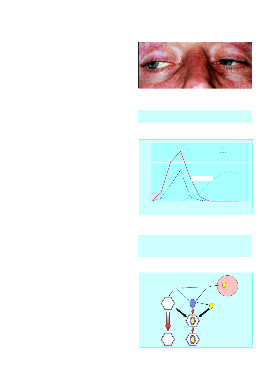
Figure 3.2 Disconjugate gaze due to cerebral oedema in jaundiced patient
with fulminant hepatitis
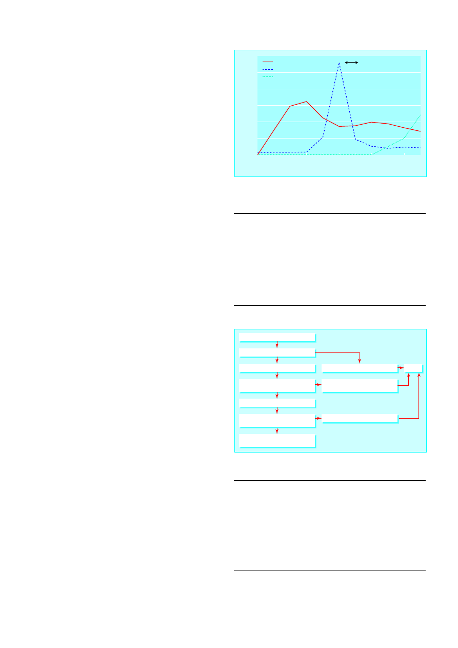
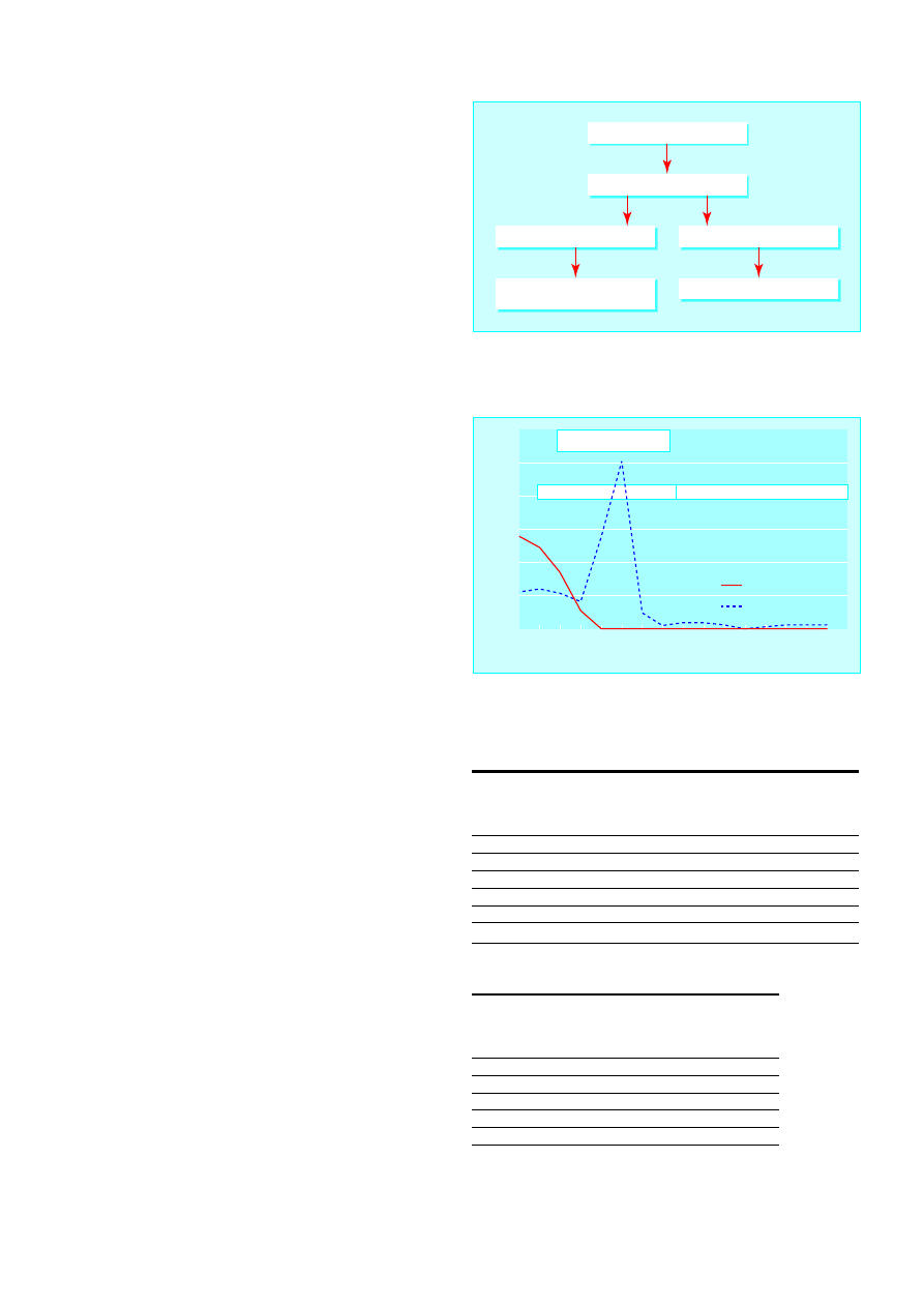
Time (days)
Titre
0
1
2
3
4
5
6
7
8
9
10
0
100
150
50
Viral DNA
e antigen
Anti-e antibody
Jaundice
Figure 3.3 Appearance of serological markers in acute self limiting hepatitis
B virus infection
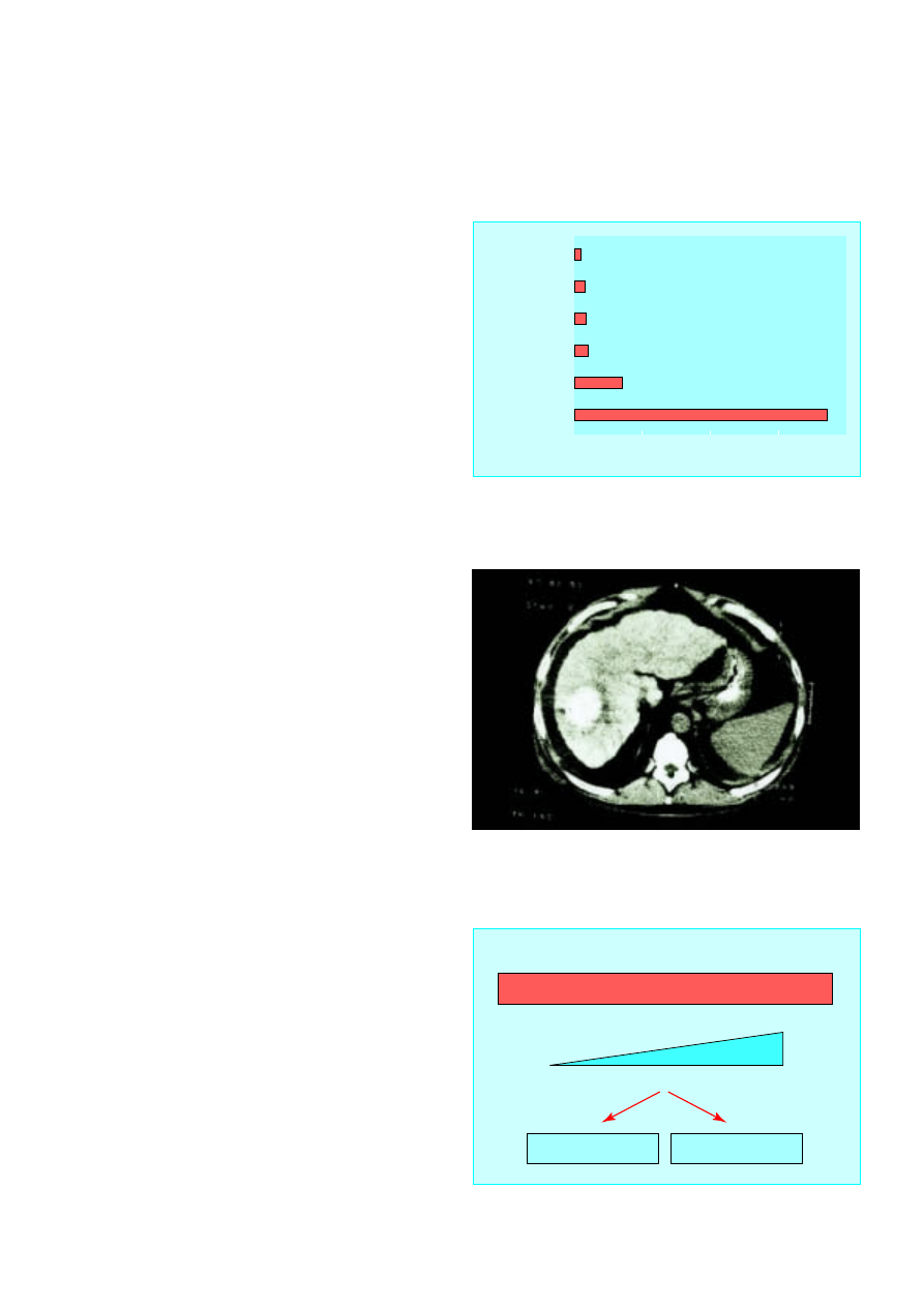
Surface
antigen
Surface
antigen
Virion assembled
Incomplete virus
exported
Core
antigen
RNA
Proteins
Hepatitis B virus
DNA
Hepatitis B virus
DNA
Complete virion
Figure 3.4 Mechanism of assembly and excretion of hepatitis B virus from
infected hepatocytes
ABC of Liver, Pancreas, and Gall Bladder
10
Hepatitis C
Screening tests for hepatitis C virus infection use enzyme linked
immunosorbent assays (ELISA) with recombinant viral antigens
on patients’ serum. Acute hepatitis C cannot be reliably
diagnosed by antibody tests as these often do not give positive
results for up to three months.
Hepatitis C virus was the cause of more than 90% of all
post-transfusion hepatitis in Europe and the United States.
Before 1991, the risk of infection in the United Kingdom was
0.2% per unit of blood transfused, but this has fallen to 1
infection per 10 000 units transfused since the introduction of
routine serological screening of blood donors. Acute hepatitis C
infection is therefore now seen commonly only in intravenous
drug users.
Antibodies to hepatitis C appear relatively late in the course
of the infection, and if clinical suspicion is high, the patient’s
serum should be tested for hepatitis C virus RNA to establish
the diagnosis.
Non-A-E viral hepatitis
Epstein Barr virus causes rises in liver enzyme activities in
almost all cases of acute infection, but it is uncommon for the
liver injury to be sufficiently severe to cause jaundice. When
jaundice does occur in patients with Epstein Barr virus
infection, it can be prolonged with a large cholestatic element.
Diagnosis is usually relatively easy because the typical symptoms
of Epstein Barr infection are almost always present and
serological testing usually gives positive results.
Cytomegalovirus can also cause acute hepatitis. This is unusual,
rarely severe, and runs a chronic course only in
immunosuppressed patients.
The cause of about 7% of all episodes of acute presumed
viral hepatitis remains unidentified. It seems certain that other
viral agents will be identified that cause acute liver injury.
Management of acute viral hepatitis
Hepatitis A
Most patients with hepatitis A infection have a self limiting
illness that will settle totally within a few weeks. Management is
conservative, with tests being aimed at identifying the small
group of patients at risk of developing fulminant liver failure.
Hepatitis B
Acute hepatitis B is also usually self limiting, and most patients
who contract the virus will clear it completely. All cases must be
notified and sexual and close household contacts screened and
vaccinated. Patients should be monitored to ensure fulminant
liver failure does not develop and have serological testing three
months after infection to check that the virus is cleared from
the blood. About 5-10% of patients will remain positive for
hepatitis B surface antigen at three months, and a smaller
proportion will have ongoing viral replication (e antigen
positive). All such patients require expert follow up (see article
on chronic viral hepatitis).
Hepatitis C
Early identification and referral of cases of acute hepatitis C
infection is important because strong evidence exists that early
treatment with interferon alpha reduces the risk of chronic
infection. The rate of chronicity in untreated patients is about
80%; treatment with interferon reduces this to below 50%.
Box 3.3 Hepatitis D and E infection
Hepatitis D
x
Incomplete RNA virus that requires hepatitis B surface antigen to
transmit its genome from cell to cell
x
Occurs only in patients positive for hepatitis B surface antigen
x
Usually confined to intravenous drug users in United Kingdom
Hepatitis E
x
Transmitted by orofaecal route
x
Produces an acute self limiting illness similar to hepatitis A
x
Common in developing world
x
High mortality in pregnant women
Summary points
x
Symptoms of hepatitis are non-specific and often occur without the
development of jaundice
x
Serum alanine transaminase is the most useful screening test for
hepatitis in general practice
x
Hepatitis A rarely causes fulminant liver failure or chronic liver
disease
x
In the developed world, new cases of hepatitis C are mainly seen in
intravenous drug users
x
Most adults who contract hepatitis B virus clear the virus, with
< 10% developing chronic liver infection
Time (months)
Alanine transaminase (u/l)
0
1
2
3
4
5
6
7
8
9
10
0
800
1200
1000
600
200
400
Hepatitis C virus
Alanine transaminase
Antibody to hepatitis C virus
Jaundice
Figure 3.5 Appearance of hepatitis C virus RNA, antibodies to hepatitis C
virus, and raised alanine transaminase activity in acute hepatitis C infection
Hepatitis A IgM positive
Check international normalised ratio
International normalised ratio <2
Better
Review with liver function tests
in 5-7 days
No improvement
(clinical or biochemical)
International normalised ratio >2
Abnormal
Refer
Repeat liver function
tests at 6 weeks
Normal
No follow up
Figure 3.6 Management of acute hepatitis A infection in general practice
Acute hepatitis
11
4 Chronic viral hepatitis
S D Ryder, I J Beckingham
Most cases of chronic viral hepatitis are caused by hepatitis B or
C virus. Hepatitis B virus is one of the commonest chronic viral
infections in the world, with about 170 million people
chronically infected worldwide. In developed countries it is
relatively uncommon, with a prevalence of 1 per 550
population in the United Kingdom and United States.
The main method of spread in areas of high endemicity is
vertical transmission from carrier mother to child, and this may
account for 40-50% of all hepatitis B infections in such areas.
Vertical transmission is highly efficient; more than 95% of
children born to infected mothers become infected and develop
chronic viral infection. In low endemicity countries, the virus is
mainly spread by sexual or blood contact among people at high
risk, including intravenous drug users, patients receiving
haemodialysis, homosexual men, and people living in
institutions, especially those with learning disabilities. These
high risk groups are much less likely to develop chronic viral
infection (5-10%). Men are more likely then women to develop
chronic infection, although the reasons for this are unclear.
Up to 300 million people have chronic hepatitis C infection
mainly worldwide. Unlike hepatitis B virus, hepatitis C infection
is not mainly confined to the developing world, with 0.3% to
0.7% of the United Kingdom population infected. The virus is
spread almost exclusively by blood contact. About 15% of
infected patients in Northern Europe have a history of blood
transfusion and about 70% have used intravenous drugs. Sexual
transmission does occur, but is unusual; less than 5% of long
term sexual partners become infected. Vertical transmission is
also unusual.
Presentation
Chronic viral liver disease may be detected as a result of finding
abnormal liver biochemistry during serological testing of
asymptomatic patients in high risk groups or as a result of the
complications of cirrhosis. Patients with chronic viral hepatitis
usually have a sustained increase in alanine transaminase
activity. The rise is lower than in acute infection, usually only
two or three times the upper limit of normal. In hepatitis C
infection, the ã-glutamyltransferase activity is also often raised.
The degree of the rise in transaminase activity has little
relevance to the extent of underlying hepatic inflammation.
This is particularly true of hepatitis C infection, when patients
often have normal transaminase activity despite active liver
inflammation.
Hepatitis B
Most patients with chronic hepatitis B infection will be positive
for hepatitis B surface antigen. Hepatitis B surface antigen is on
the viral coat, and its presence in blood implies that the patient
is infected. Measurement of viral DNA in blood has replaced e
antigen as the most sensitive measure of viral activity.
Chronic hepatitis B virus infection can be thought of as
occurring in phases dependent on the degree of immune
response to the virus. If a person is infected when the immune
response is “immature,” there is little or no response to the
hepatitis B virus. The concentrations of hepatitis B viral DNA in
serum are very high, the hepatocytes contain abundant viral
Lived in endemic area
1.6
Amateur tattoo
3.2
None known
Sexual
3.4
3.6
Blood products
14
0
20
40
60
80
% of patients
Intravenous drug user
74
Figure 4.1 Risk factors for hepatitis C virus infection among 1500 patients
in Trent region,1998. Note: professional tattooing does not carry a risk
Figure 4.2 Computed tomogram showing hepatocellular carcinoma, a
common complication of cirrhosis
Increasing fibrosis
Tolerant
Viral DNA high
minimal liver disease
Viral DNA concentrations fall
increasing inflammation
Immune recognition
Death from cirrhosis
Viral clearance
Figure 4.3 Phases of infection with hepatitis B virus
12
particles (surface antigen and core antigen) but little or no
ongoing hepatocyte death is seen on liver biopsy because of the
defective immune response. Over some years the degree of
immune recognition usually increases. At this stage the
concentration of viral DNA tends to fall and liver biopsy shows
increasing inflammation in the liver. Two outcomes are then
possible, either the immune response is adequate and the virus
is inactivated and removed from the system or the attempt at
removal results in extensive fibrosis, distortion of the normal
liver architecture, and eventually death from the complications
of cirrhosis.
Assessment of chronic hepatitis B infection
Patients positive for hepatitis B surface antigen with no
evidence of viral replication, normal liver enzyme activity, and
normal appearance on liver ultrasonography require no further
investigation. Such patients have a low risk of developing
symptomatic liver disease or hepatocellular carcinoma.
Reactivation of B virus replication can occur, and patients
should therefore have yearly serological and liver enzyme tests.
Patients with abnormal liver biochemistry, even without
detectable hepatitis B viral DNA or an abnormal liver texture
on ultrasonography, should have liver biopsy, as 5% of patients
with only surface antigen carriage at presentation will have
cirrhosis. Detection of cirrhosis is important as patients are at
risk of complications, including variceal bleeding and
hepatocellular carcinoma. Patients with repeatedly normal
alanine transaminase activity and high concentrations of viral
DNA are extremely unlikely to have developed advanced liver
disease, and biopsy is not always required at this stage.
Treatment
Interferon alfa was first shown to be effective for some patients
with hepatitis B infection in the 1980s, and it remains the
mainstay of treatment. The optimal dose and duration of
interferon for hepatitis B is somewhat contentious, but most
clinicians use 8-10 million units three times a week for four to
six months. Overall, the probability of response (that is,
stopping viral replication) to interferon therapy is around 40%.
Few patients lose all markers of infection with hepatitis B, and
surface antigen usually remains in the serum. Successful
treatment with interferon produces a sustained improvement in
liver histology and reduces the risk of developing end stage liver
disease. The risk of hepatocellular carcinoma is also probably
reduced but is not abolished in those who remain positive for
hepatitis B surface antigen.
In general, about 15% of patients receiving interferon have
no side effects, 15% cannot tolerate treatment, and the
remaining 70% experience side effects but are able to continue
treatment. Depression can be a serious problem, and both
suicide and admissions with acute psychosis are well described.
Viral clearance occurs through induction of immune mediated
killing of infected hepatocytes. Transient hepatitis can therefore
occasionally cause severe decompensation requiring liver
transplantation.
Lamivudine is a nucleoside analogue that is a potent inhibitor
of hepatitis B viral DNA replication. It has a good safety profile
and has been widely tested in patients with chronic hepatitis B
virus infection, mainly in the Far East. In long term trials almost
all treated patients showed prompt and sustained inhibition of
viral DNA replication, with about 17% becoming e antigen
negative when treatment was continued for 12 months. There was
an associated improvement of inflammation and a reduction in
progression of fibrosis on liver biopsy. Side effects are generally
mild. Combination therapy with interferon and lamivudine has
not been found to have additional benefit.
Table 4.1 Factors indicating likelihood of response to
interferon in chronic hepatitis B infection
High probability
Low probablility
Age (years)
< 50
> 50
Sex
Female
Male
Viral DNA
Low
High
Activity of liver inflammation
High
Low
Country of origin
Western world
Asia or Africa
Coinfection with HIV
Absent
Present
Table 4.2 Side effects of treatment with
interferon alpha
Symptoms
Frequency (%)
Fever or flu-like illness
80
Depression
25
Fatigue
50
Haematological abnormalities
10
No side effects
15
Hepatitis B surface antigen present
Viral DNA not detected
Liver function abnormal
Liver function normal
Liver biopsy
Yearly liver function tests and tests for
hepatitis B surface antigen and DNA
Figure 4.4 Investigation of patients positive for hepatitis B surface antigen
without viral replication
Time (months)
Alanine transaminase/viral DNA
0
1
2
3
4
5
11
12
13
14
15
6
16
7
8
9
10
0
800
1200
1000
600
200
400
HBV DNA
Alanine transaminase
e antigen positive
e antibody positive
Interferon
Figure 4.5 Timing of interferon treatment in the management of hepatitis B
Chronic viral hepatitis
13
Hepatitis C
Chronic hepatitis C virus infection has a long course, and most
patients are diagnosed in a presymptomatic stage. In the United
Kingdom, most patients are now discovered because of an
identifiable risk factor (intravenous drug use, family history, or
blood transfusion) or because of abnormal liver biochemistry.
Screening for hepatitis C virus infection is based on enzyme
linked immonosorbent assays (ELISA) using recombinant viral
antigens and patients’ serum. These have high sensitivity and
specificity. The diagnosis is confirmed by radioimmunoblot and
direct detection of viral RNA in peripheral blood by polymerase
chain reaction. Viral RNA is regarded as the best test to
determine infectivity and assess response to treatment.
Natural course of hepatitis C infection
In order to assess the need for treatment it is important to have
a clear understanding of the natural course of hepatitis C
infection and factors that may predispose to more severe
outcome. Our knowledge is limited because of the relatively
recent discovery of the virus. It is clear, however, that hepatitis C
is usually slowly progressive, with an average time from
infection to development of cirrhosis of around 30 years, albeit
with a high level of variability. The main factors associated with
increased risk of progressive liver disease are age > 40 at
infection, high alcohol consumption, and male sex.
Viraemic patients with abnormal alanine transaminase
activity need a liver biopsy to assess the stage of disease (amount
of fibrosis) and degree of necroinflammatory change (Knodell
score). Management is usually based on the degree of liver
damage, with patients with more severe disease being offered
treatment. Patients with mild changes are usually followed up
without treatment as their prognosis is good and future treatment
is likely to be more effective than present regimens.
Treatment of hepatitis C
Interferon alfa (3 million units three times a week) in
combination with tribavirin (1000 mg a day for patients under
75 kg and 1200 mg for patients >75 kg) has recently been
shown to be more effective than interferon alone. A large study
in Europe showed no advantage to continuing treatment
beyond six months in patients who had a good chance of
response, whereas those with a poorer outlook needed longer
treatment (12 months) to maximise the chance of clearing their
infection. About 30% of patients will obtain a “cure” (sustained
response). The main determinant of response is viral genotype,
with genotypes 1 and 4 having poor response rates.
Combination therapy has the same side effects as interferon
monotherapy with the additional risk of haemolytic anaemia.
Patients developing anaemia should have their dose of
tribavirin reduced. All patients should have a full blood count
and liver function tests weekly for the first four weeks of
treatment and monthly thereafter if haemoglobin concentration
and white cell count are stable. Many new treatments are
currently entering clinical trials, including long acting
interferons and alternative antiviral drugs.
Box 4.1 Investigations required in patients positive for
antibodies to hepatitis C virus
Assessing hepatitis C virus
x
Polymerase chain reaction
for viral RNA
x
Viral load
x
Genotype
Excluding other liver
diseases
x
Ferritin
x
Autoantibodies/
immunoglobulins
x
Hepatitis B serology
x
Liver ultrasonography
Further reading
Szmuness W. Hepatocellular carcinoma and the hepatitis B virus:
evidence for a causal association. Prog Med Virol 1978;24:40-8.
Stevens CE, Beasley RP, Tsui V, Lee WC. Vertical transmission of
hepatitis B antigen in Taiwan. N Engl J Med 1975;292:771-4.
Knodell RG, Ishak G, Black C, Chen TS, Craig R, Kaplowitz N, et al.
Formulation and application of numerical scoring system for
activity in asymptomatic chronic active hepatitis. Hepatology
1981;1:431-5.
Summary points
x
Viral hepatitis is relatively common in United Kingdom (mainly
hepatitis C)
x
Presentation is usually with abnormal alanine transaminase activity
x
Disease progression in hepatitis C is usually slow (median time to
development of cirrhosis around 30 years)
x
Liver biopsy is essential in managing chronic viral hepatitis
x
New treatments for hepatitis C (interferon and tribavirin) and
hepatitis B (lamivudine) have improved the chances of eliminating
these pathogens from chronically infected patients
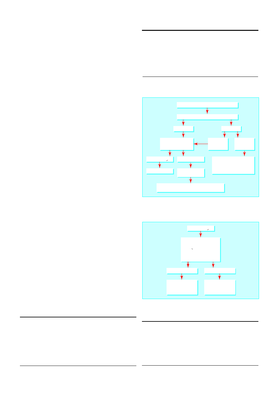
Exclude other liver diseases
Polymerase chain reaction for viral DNA
Repeat viral RNA every 3 months
Save serum six monthly for
polymerase chain reaction
Ensure liver function test results
remain normal
Positive
Irrespective
of liver
function
tests
Knodell score > 6
Negative
Abnormal
liver function
test results
Liver biopsy
Interferon tribavirin
Repeat liver function
tests every 3 months
Repeat liver biopsy at 2 years or if clinically indicated,
for example, alanine transaminase 2x initial value
Knodell score < 6
Normal
liver function
test results
Figure 4.6 Management of chronic hepatitis C virus infection
Knodell score > 6
0, 1 or 2
3, 4 or 5
Stratify for "response factors"
• Genotype 2 or 3
• RNA < 2x10
6
/l
• Age < 40 years
• Fibrosis score < 2
• Female
Interferon plus
tribavirin for 1 year
(sustained
response 30%)
Interferon plus
tribavirin for 6 months
(sustained
response 54%)
Figure 4.7 Combination therapy for hepatitis C
ABC of Liver, Pancreas, and Gall Bladder
14
5 Other causes of parenchymal liver disease
S D Ryder, I J Beckingham
Autoimmune hepatitis
Autoimmune hepatitis is a relatively uncommon disease that
mainly affects young women. The usual presentation is with
fatigue, pain in the right upper quadrant of the abdomen, and
polymyalgia or arthralgia associated with abnormal results of
liver function tests. Other autoimmune diseases are present in
17% of patients with classic autoimmune hepatitis,
predominantly thyroid disease, rheumatoid arthritis, and
ulcerative colitis.
Autoimmune hepatitis is an important diagnosis as
immunosuppressive drugs (prednisolone and azathioprine)
produce lasting remission and an excellent prognosis. Although
the condition can produce transient jaundice that seems to
resolve totally, the process can continue at a subclinical level
producing cirrhosis and irreversible liver failure. The diagnosis
is based on detection of autoantibodies (antinuclear antibodies
(60% positive), antismooth muscle antibodies (70%)) and high
titres of immunoglobulins (present in almost all patients, usually
IgG).
Metabolic causes of liver disease
Metabolic liver disease rarely presents as jaundice, and when it
does the patient probably has end stage chronic liver disease.
Haemochromatosis
Haemochromatosis is the commonest inherited liver disease in
the United Kingdom. It affects about 1 in 200 of the population
and is 10 times more common than cystic fibrosis.
Haemochromatosis produces iron overload, and patients
usually present with cirrhosis or diabetes due to excessive iron
deposits in the liver or pancreas. The genetic defect responsible is
a single base change at a locus of the HFE gene on chromosome
6, with this defect responsible for over 90% of cases in the United
Kingdom. Genetic analysis is now available both for confirming
the diagnosis and screening family members. The disease
typically affects middle aged men. Menstruation and pregnancy
probably account for the lower presentation in women.
Patients who are homozygous for the mutation should have
regular venesection to prevent further tissue damage.
Heterozygotes are asymptomatic and do not require treatment.
Cardiac function is often improved by venesection but diabetes,
arthritis, and hepatic fibrosis do not improve. This emphasises
the need for early recognition and treatment.
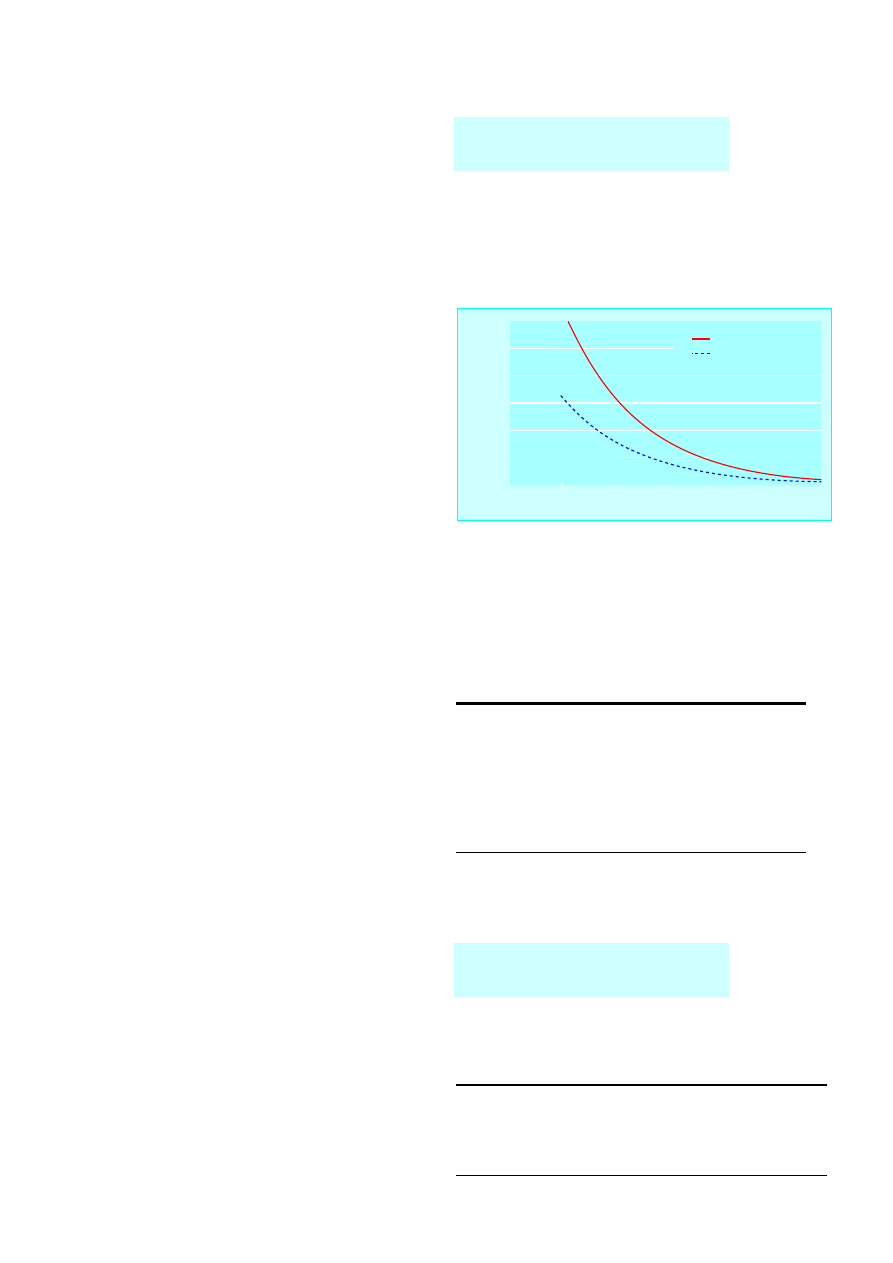
Wilson’s disease
Wilson’s disease is a rare autosomal recessive cause of liver
disease due to excessive deposition of copper within
hepatocytes. Abnormal copper deposition also occurs in the
basal ganglia and eyes. The defect lies in a decrease in
production of the copper carrying enzyme ferroxidase. Unlike
most other causes of liver disease, it is treatable and the
prognosis is excellent provided that it is diagnosed before
irreversible damage has occurred.
Patients may have a family history of liver or neurological
disease and a greenish-brown corneal deposit of copper (a
Kayser-Fleischer ring), which is often discernible only with a slit
lamp. Most patients have a low caeruloplasmin level and low
About 40% of patients with autoimmune
hepatitis present acutely with jaundice
Box 5.1 Presenting conditions in haemochromatosis
x
Cirrhosis (70%)
x
Diabetes (adult onset) (55%)
x
Cardiac failure (20%)
x
Arthropathy (45%)
x
Skin pigmentation (80%)
x
Sexual dysfunction (50%)
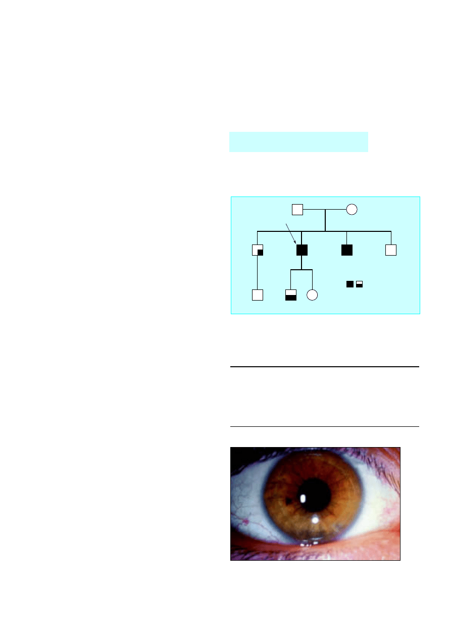
?
?
282 CY
Index
282 CC
282 CY
282 CY
282 YY
282 YY
The amount of shade in each box
represents the degree of iron excess
(liver biopsy or serum markers)
282 CC
Figure 5.1 Use of genetic analysis to screen family members for
haemochromatosis. The index case was a 45 year old man who presented
with cirrhosis. His brothers were asymptomatic and had no clinical
abnormalities. However, the brother who had inherited two abnormal genes
(282YY) was found to have extensive iron loading on liver biopsy
Figure 5.2 Kayser-Fleischer ring in patient with Wilson’s disease
15
serum copper and high urinary copper concentrations. Liver
biopsy confirms excessive deposition of copper.
Treatment is with penicillamine, which binds copper and
increases urinary excretion. Patients who are unable to tolerate
penicillamine are treated with trientene and oral zinc acetate.
Asymptomatic siblings should be screened and treated in the
same way.
Drug related hepatitis
Most drugs can cause liver injury. It is relatively uncommon for
drug reactions to present as acute jaundice, and only 2-7% of
hospital admissions for non-obstructive jaundice are drug related.
Different drugs cause liver injury by a variety of mechanisms and
with differing clinical patterns. In general terms, drug related
jaundice can be due to predictable direct hepatotoxicity, such as is
seen in paracetamol overdose, or idiosyncratic drug reactions.
Paracetamol poisoning
Paracetamol is usually metabolised by a saturable enzyme
pathway. When the drug is taken in overdose, another metabolic
system is used that produces a toxic metabolite that causes
acute liver injury. Hepatotoxicity is common in paracetamol
overdose, and prompt recognition and treatment is required.
The lowest recorded fatal dose of paracetamol is 11 g, but
genetic factors mean that most people would have to take
considerably higher doses to develop fulminant liver failure.
Overdose with paracetamol is treated by acetylcysteine,
which provides glutathione for detoxification of the toxic
metabolites of paracetamol. This is generally a preventive
measure, and decision to treat is based on the serum
concentrations of paracetamol. It is important to be certain of
the time that paracetamol was taken in order to interpret the
treatment nomogram accurately. If there is doubt over the
timing of ingestion treatment should be given.
Paracetamol poisoning is by far the commonest cause of
fulminant liver failure in the United Kingdom and is an
accepted indication for liver transplantation. As this is an acute
liver injury, patients who survive without the need for
transplantation will always regain normal liver function.
Idiosyncratic drug reactions
The idiosyncratic drug reactions are by their nature
unpredictable. They can occur at any time during treatment and
may still have an effect over a year after stopping the drug. The
management of acute drug reactions is primarily stopping the
potential causative agent, and if possible all drugs should be
withheld until the diagnosis is definite. Idiosyncratic drug
reactions can be severe, and they are an important cause of
fulminant liver failure, accounting for between 15% and 20% of
such cases. Any patient presenting with a severe drug reaction
will require careful monitoring as recovery can be considerably
delayed, particularly with drugs such as amiodarone, which has
a long half life in blood.
The drug history must also include non-prescribed
medications. Fulminant liver failure is well described in patients
who have taken Chinese herbal medicine.
Cholestatic non-obstructive jaundice
Initial investigation of patients with jaundice and a cholestatic
pattern on liver function tests is by ultrasonography. This will
detect dilatation of the bile duct in most cases of extrahepatic
biliary obstruction caused by tumour or stones and will also
detect most metastatic liver tumours, the other main cause of
Wilson’s disease should be suspected in
any patient presenting with chronic
hepatitis or cirrhosis under the age of 35
Box 5.2 Common drugs producing hepatic idiosyncratic
reactions
x
Sodium valproate
x
Non-steroidal anti-inflammatory drugs (diclofenac)
x
Amiodarone
x
Aspirin
x
Methyldopa
x
Isoniazid
x
Minocycline
Complementary medicines may account
for as much as 5% of all drug induced
liver disease
Box 5.3 Common drugs producing cholestatic reactions
x
Chlorpromazine
x
Oestrogens (hormone replacement therapy or contraceptive pill)
x
Co-amoxiclav or flucloxacillin
x
Chlorpropamide
Time (hours)
Plasma paracetamol concentration (mmol/l)
0
4
8
12
16
20
24
0
0.2
0.4
0.6
0.8
1.0
1.2
Normal treatment line
High risk treatment line
Figure 5.3 Thresholds for treatment of paracetamol poisoning in normal
and high risk patients. Adapted from British National Formulary
ABC of Liver, Pancreas, and Gall Bladder
16
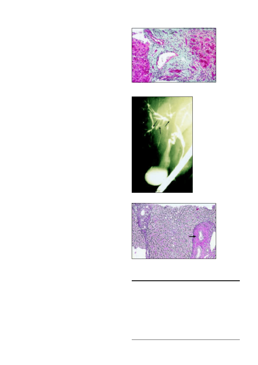
cholestatic malignant jaundice. Dilatation of the biliary tree may
not always be present in early biliary obstruction, and if doubt
exists, either repeat ultrasonography or endoscopic retrograde
cholangiopancreatography is advisable. Particular attention is
required in patients with no apparent drug cause for their
jaundice and in whom serological tests for other causes of
cholestasis give negative results.
Primary biliary cirrhosis
Primary biliary cirrhosis is relatively common and mainly affects
middle aged women. It typically presents as cholestatic jaundice,
but with more widespread use of liver enzyme tests it is
increasingly found at a presymptomatic stage because of raised
alkaline phosphatase and ã-glutamyltransferase activities during
investigation of associated symptoms such as pruritus. When
patients present with jaundice, it is usually associated with
cutaneous signs of chronic liver disease, xanthoma, and other
extrahepatic features such as Sjögren’s syndrome.
Primary biliary cirrhosis is immunologically mediated, and
the presence of M2 antimitochondrial antibodies is diagnostic.
Immunoglobulin titres, particularly IgM, are often raised. Liver
biopsy is used to stage the disease rather than to confirm the
diagnosis. Treatment with ursodeoxycholic acid has been shown
to slow disease progression. Patients with advanced liver disease
require liver transplantation.
Primary sclerosing cholangitis
Sclerosing cholangitis is characterised by progressive fibrosing
inflammation of the bile ducts. The changes are often diffuse, but
symptoms usually arise from dominant strictures at the hilum or
within the extrahepatic bile ducts. Primary sclerosing cholangitis
usually occurs in men younger than 50 years old and is associated
with inflammatory bowel disease in 70-80% of cases. The
incidence of primary sclerosing cholangitis in patients with
ulcerative colitis is 2-10%. Cholangiocarcinoma develops in 20%
to 30% of patients with primary sclerosing cholangitis and is an
important cause of death in patients with ulcerative colitis.
Sclerosing cholangitis may be asymptomatic but usually
presents with fluctuating jaundice, nausea, and pruritus. The
diagnosis is suggested by cholangiography (endoscopic
retrograde cholangiopancreatography, percutaneous
transhepatic cholangiography, or magnetic resonance
cholangiopancreatography). Multiple strictures with beading of
ducts, duct pruning (scanty ducts), irregularities of the duct wall,
and diverticula are typical features. Liver biopsy is a
supplementary investigation that shows characteristic
histological features in 30-40% of patients. Raised serum titres
of smooth muscle antibody (70% of patients) and perinuclear
antineutrophil cytoplasmic antibody (60%) may help diagnosis.
Raised concentrations of serum CA19-9 tumour marker are
highly suspicious of cholangiocarcinoma.
Treatment of primary sclerosing cholangitis is at present
limited to the management of recurrent cholangitis. Treatment
with ursodeoxycholic acid (7 mg/kg/day) may improve
symptoms and liver function, but no strong evidence exists for
its effectiveness. Dominant strictures may be improved with
endoscopic dilatation or surgical resection. Liver
transplantation is required for patients with deteriorating liver
function with progressive secondary biliary cirrhosis.
Summary points
x
Most drugs have potential to cause liver injury, and 2-7% of
admissions with non-obstructive jaundice are for drug related
hepatitis
x
Herbal remedies and illegal drugs can also cause jaundice and liver
damage
x
Primary biliary cirrhosis typically presents as cholestatic jaundice in
middle aged women
x
Primary sclerosing cholangitis is associated with ulcerative colitis in
75% of cases, although the two may develop at different times
x
Haemochromatosis is the commonest inherited liver disease in the
United Kingdom, and a gene probe for clinical testing is now
available
Figure 5.4 Broad fibrosis band in patient with primary
biliary cirrhosis
Figure 5.5 Endoscopic
retrograde
cholangiopancreatogram in
patient with primary sclerosing
cholangitis showing irregular
stricturing and dilatation of
intrahepatic bile ducts
Figure 5.6 Liver biopsy specimen of patient with primary
sclerosing cholangitis. Characteristic “onion skin” fibrosis is
visible round portal tracts
Other causes of parenchymal liver disease
17
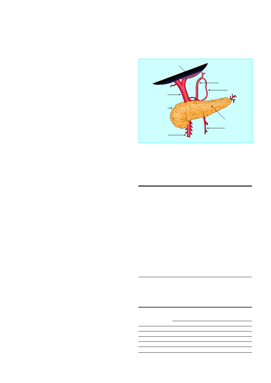
6 Portal hypertension—1: varices
J E J Krige, I J Beckingham
The portal vein carries about 1500 ml/min of blood from the
small and large bowel, spleen, and stomach to the liver at a
pressure of 5-10 mm Hg. Any obstruction or increased
resistance to flow or, rarely, pathological increases in portal
blood flow may lead to portal hypertension with portal
pressures over 12 mm Hg. Although the differential diagnosis is
extensive, alcoholic and viral cirrhosis are the leading causes of
portal hypertension in Western countries, whereas liver disease
due to schistosomiasis is the main cause in other areas of the
world. Portal vein thrombosis is the commonest cause in
children.
Increases in portal pressure cause development of a
portosystemic collateral circulation with resultant compensatory
portosystemic shunting and disturbed intrahepatic circulation.
These factors are partly responsible for the important
complications of chronic liver disease, including variceal
bleeding, hepatic encephalopathy, ascites, hepatorenal
syndrome, recurrent infection, and abnormalities in
coagulation. Variceal bleeding is the most serious complication
and is an important cause of death in patients with cirrhotic
liver disease.
Varices
In Western countries variceal bleeding accounts for about 7% of
episodes of gastrointestinal bleeding, although this varies
according to the prevalence of alcohol related liver disease
(11% in the United States, 5% in the United Kingdom). Patients
with varices have a 30% lifetime risk of bleeding, and a third of
those who bleed will die. Patients who have bled once from
oesophageal varices have a 70% chance of bleeding again, and
about a third of further bleeding episodes are fatal.
Several important considerations influence choice of
treatment and prognosis. These include the natural course of
the disease causing portal hypertension, location of the
bleeding varices, residual hepatic function, presence of
associated systemic disease, continuing drug or alcohol misuse,
and response to specific treatment. The modified Child-Pugh
classification identifies three risk categories that correlate well
with survival.
Initial measures
Prompt resuscitation and restoration of circulating blood
volume is vital and should precede any diagnostic studies. While
their blood is being cross matched, patients should receive a
rapid infusion of 5% dextrose and colloid solution until blood
pressure is restored and urine output is adequate. Saline
infusions may aggravate ascites and must be avoided. Patients
who are haemodynamically unstable, elderly, or have
concomitant cardiac or pulmonary disease should be
monitored by using a pulmonary artery catheter as injudicious
administration of crystalloids, combined with vasoactive drugs,
can lead to the rapid onset of oedema, ascites, and
hyponatraemia. Concentrations of clotting factors are often low,
and fresh blood, fresh frozen plasma, and vitamin K
1
(phytomenadione) should be given. Platelet transfusions may be
necessary. Sedatives should be avoided, although haloperidol is
useful in patients with symptoms of alcohol withdrawal.
Box 6.1 Causes of portal hypertension
Increased resistance to flow
Prehepatic (portal vein obstruction)
x
Congenital atresia or stenosis
x
Thrombosis of portal vein
x
Thrombosis of splenic vein
x
Extrinsic compression (for example, tumours)
Hepatic
x
Cirrhosis
x
Acute alcoholic liver disease
x
Congenital hepatic fibrosis
x
Idiopathic portal hypertension (hepatoportal sclerosis)
x
Schistosomiasis
Posthepatic
x
Budd-Chiari syndrome
x
Constrictive pericarditis
Increased portal blood flow
x
Arterial-portal venous fistula
x
Increased splenic flow
Table 6.1 Child-Pugh classification of liver failure
No of points
1
2
3
Bilirubin (ìmol/l)
< 34
34-51
> 51
Albumin (g/l)
> 35
28-35
< 28
Prothrombin time
< 3
3-10
> 10
Ascites
None
Slight
Moderate to severe
Encephalopathy
None
Slight
Moderate to severe
Grade A = 5-6 points, grade B = 7-9 points, grade C = 10-15 points.
Left gastric vein
Right gastric vein
Splenic vein
Inferior
mesenteric vein
Superior mesenteric
vein
Pancreas
Portal vein
Liver
Figure 6.1 Anatomical relations of portal vein and branches
18
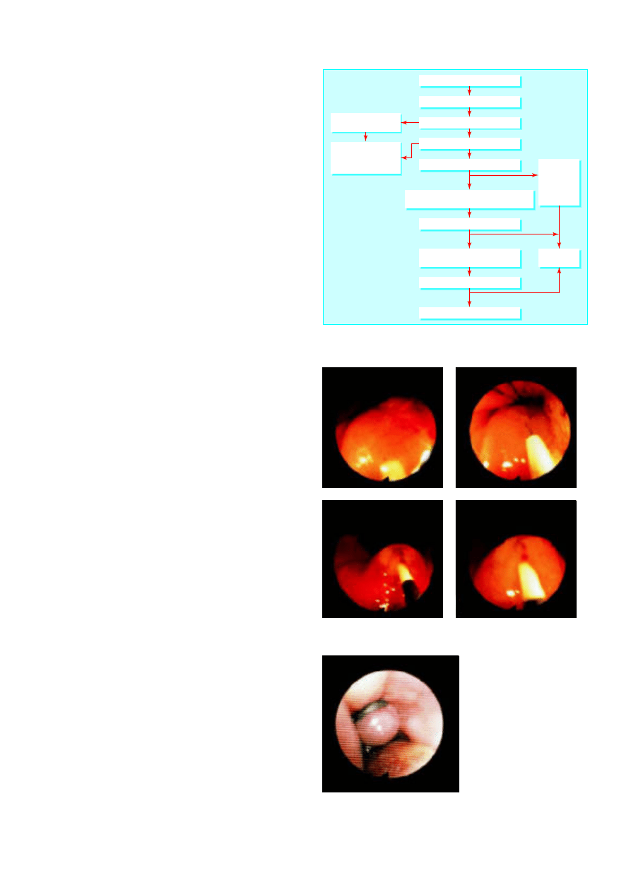
Pharmacological control
Drug treatment, aimed at controlling the acute bleed and
facilitating diagnostic endoscopy and emergency sclerotherapy,
may be useful when variceal bleeding is rapid. Octreotide, a
synthetic somatostatin analogue, reduces splanchnic blood flow
when given intravenously as a constant infusion (50 ìg/h) and
can be used before endoscopy in patients with active bleeding.
Vasopressin (0.4 units/min), or the long acting synthetic
analogue terlipressin, combined with glyceryl trinitrate
administered intravenously or transdermally through a skin
patch is also effective but has more side effects than octreotide.
Glyceryl trinitrate reduces the peripheral vasoconstriction
caused by vasopressin and has an additive effect in lowering
portal pressure.
Emergency endoscopy
Emergency diagnostic fibreoptic endoscopy is essential to
confirm that oesophageal varices are present and are the source
of bleeding. Most patients will have stopped bleeding
spontaneously before endoscopy (60% of bleeds) or after drug
treatment. Endotracheal intubation may be necessary during
endoscopy, especially in patients who are bleeding heavily,
encephalopathic, or unstable despite vigorous resuscitation. In
90% of patients variceal bleeding originates from oesophageal
varices. These are treated by injection with sclerosant or by
banding.
Sclerotherapy
In sclerotherapy a sclerosant solution (ethanolamine oleate or
sodium tetradecyl sulphate) is injected into the bleeding varix
or the overlying submucosa. Injection into the varix obliterates
the lumen by thrombosis whereas injection into the submucosa
produces inflammation followed by fibrosis. The first injection
controls bleeding in 80% of cases. If bleeding recurs, the
injection is repeated. Complications are related to toxicity of the
sclerosant and include transient fever, dysphagia and chest pain,
ulceration, stricture, and (rarely) perforation.
Band ligation
Band ligation is achieved by a banding device attached to the
tip of the endoscope. The varix is aspirated into the banding
chamber, and a trip wire dislodges a rubber band carried on the
banding chamber, ligating the entrapped varix. One to three
bands are applied to each varix, resulting in thrombosis. Band
ligation eradicates oesophageal varices with fewer treatment
sessions and complications than sclerotherapy.
Balloon tube tamponade
The balloon tube tamponade may be life saving in patients with
active variceal bleeding if emergency sclerotherapy or banding
is unavailable or not technically possible because visibility is
obscured. In patients with active bleeding, an endotracheal tube
should be inserted to protect the airway before attempting to
place the oesophageal balloon tube.
The Minnesota balloon tube has four lumens, one for
gastric aspiration, two to inflate the gastric and oesophageal
balloons, and one above the oesophageal balloon for suction of
secretions to prevent aspiration. The tube is inserted through
the mouth, and correct siting within the stomach is checked by
auscultation while injecting air through the gastric lumen. The
gastric balloon is then inflated with 200 ml of air. Once fully
inflated, the gastric balloon is pulled up against the
oesophagogastric junction, compressing the submucosal
varices. The tension is maintained by strapping a split tennis
ball to the tube at the patient’s mouth.
Suspected variceal bleed
Transjugular intrahepatic
portosystemic shunt
Resuscitate (with vasoactive drugs)
Repeat therapeutic endoscopy with or without
vasoactive drugs or balloon tamponade
Diagnosis endoscopy
Therapeutic endoscopic
expertise unavailable
Vasoactive drugs with
or without balloon
tamponade and transfer
to specialist unit
Band ligation
eradication
programme
(or long term
b
blocker
treatment)
Endoscopic
surveillance
Band ligation or sclerotherapy
Bleeding controlled?
Bleeding controlled?
Yes
No
Yes
No
Successful?
Shunt surgery or transection
Yes
No
Figure 6.2 Algorithm for management of acute variceal haemorrhage
Figure 6.3 Injection of varices with sclerosant
Figure 6.4 Band ligation of
oesophageal varix
Portal hypertension—1: varices
19
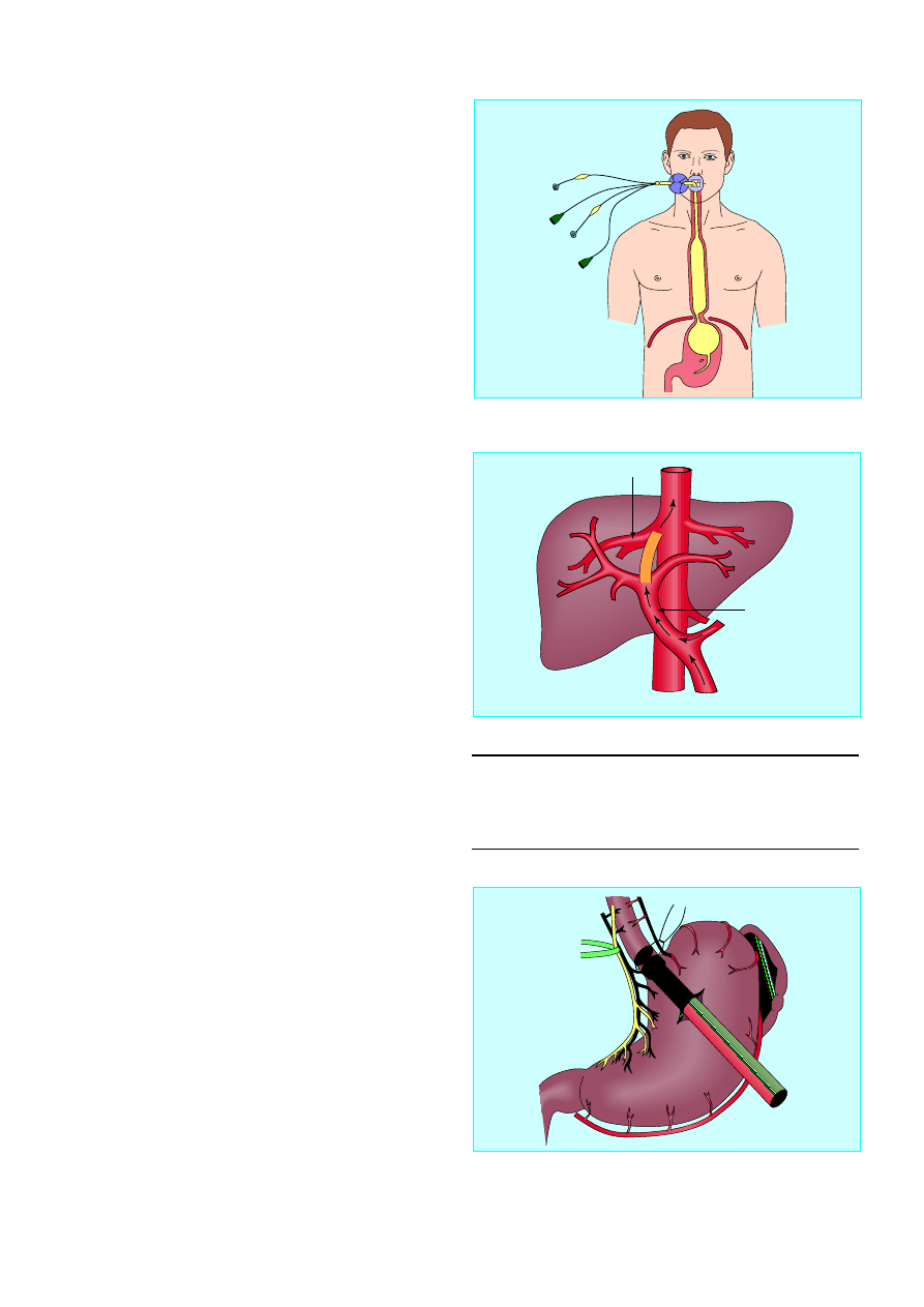
The oesophageal balloon is rarely required. The main
complications are gastric and oesophageal ulceration, aspiration
pneumonia, and oesophageal perforation. Continued bleeding
during balloon tamponade indicates an incorrectly positioned
tube or bleeding from another source. After resuscitation, and
within 12 hours, the tube is removed and endoscopic treatment
repeated.
Alternative management
Transjugular intrahepatic portosystemic shunt
Transjugular intrahepatic portosystemic shunt is the best
procedure for patients whose bleeding is not controlled by
endoscopy. It is effective only in portal hypertension of hepatic
origin. The procedure is performed via the internal jugular vein
under local anaesthesia with sedation. The hepatic vein is
cannulated and a tract created through the liver parenchyma
from the hepatic to the portal vein, with a needle under
ultrasonographic and fluoroscopic guidance. The tract is dilated
and an expandable metal stent inserted to create an
intrahepatic portosystemic shunt. The success rate is excellent.
Haemodynamic effects are similar to those found with surgical
shunts, with a lower procedural morbidity and mortality.
Transjugular intrahepatic portosystemic shunting is an
effective salvage procedure for stopping acute variceal
haemorrhage, controlling bleeding from gastric varices, and
congestive gastropathy after failure of medical and endoscopic
treatment. However, because encephalopathy occurs in up to
25% of cases and up to 50% of shunts may occlude by one year,
its primary role is to rescue failed endoscopy or as a bridge to
subsequent liver transplantation.
Long term management
After the acute variceal haemorrhage has been controlled,
treatment should be initiated to prevent rebleeding, which
occurs in most patients.
Repeated endoscopic treatment
Repeated endoscopic treatment eradicates oesophageal varices
in most patients, and provided that follow up is adequate
serious recurrent variceal bleeding is uncommon. Because the
underlying portal hypertension persists, patients remain at risk
of developing recurrent varices and therefore require lifelong
regular surveillance endoscopy.
Long term drug treatment
The use of â blockers after variceal bleeding has been shown to
reduce portal blood pressures and lower the risk of further
variceal bleeding. All patients should take â blockers unless they
have contraindications. Best results are obtained when portal
blood pressure is reduced by more than 20% of baseline or to
below 12 mm Hg.
Surgical procedures
Patients with good liver function in whom endoscopic
management fails or who live far from centres where
endoscopic sclerotherapy services are available are candidates
for surgical shunt procedures. A successful portosystemic shunt
prevents recurrent variceal bleeding but is a major operation
that may cause further impairment of liver function. Partial
portacaval shunts with 8 mm interposition grafts are equally
effective to other shunts in preventing rebleeding and have a
low rate of encephalopathy.
Oesophageal transection and gastric devascularisation are
now rarely performed but have a role in patients with portal
Box 6.2 Options for long term management
x
Repeated endoscopic treatment
x
Long term â blockers
x
Surgical shunt
x
Liver transplantation
Figure 6.5 Minnesota balloon for tamponade of oesophageal varices
Portal vein
Hepatic vein
Figure 6.6 Transjugular intrahepatic portosystemic shunt
Figure 6.7 Surgical management of varices
ABC of Liver, Pancreas, and Gall Bladder
20
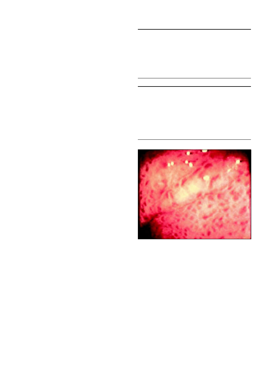
and splenic vein thrombosis who are unsuitable for shunt
procedures and continue to have serious variceal bleeding
despite endoscopic and drug treatment.
Liver transplantation is the treatment of choice in advanced
liver disease. Hepatic decompensation is the ultimate
decompressive shunt for portal hypertension and also restores
liver function. Transplantation treats other complications of
portal hypertension and has one year and five year survival
rates of 80% and 60% respectively.
Prophylactic management
Most patients with portal hypertension never bleed, and it is
difficult to predict who will. Attempts at identifying patients at
high risk of variceal haemorrhage by measuring the size or
appearance of varices have been largely unsuccessful. â blockers
have been shown to reduce the risk of bleeding, and all patients
with varices should take them unless contraindicated.
Gastric varices and portal
hypertensive gastropathy
Gastric varices are the source of bleeding in 5-10% of patients
with variceal haemorrhage. Higher rates are reported in
patients with left sided portal hypertension due to thrombosis
of the splenic vein. Endoscopic control of gastric varices is
difficult unless they are located on the proximal lesser curve in
continuation with oesophageal varices. Endoscopic
administration of cyanoacrylate monomer (superglue) is useful
for gastric varices. The transjugular intrahepatic portosystemic
shunt is increasingly used to control bleeding in this group.
Bleeding from portal hypertensive gastropathy accounts for
2-3% of bleeding episodes in cirrhosis. Although serious
bleeding from these sources is uncommon, when it occurs its
diffuse nature precludes the use of endoscopic treatment, and
optimal management is with a combination of terlipressin and
â
blockers.
Further reading
Krige JEJ, Terblanche J. Endoscopic therapy in the management of
oesophageal varices: injection sclerotherapy and variceal injection.
In: Blumgart LH, ed. Surgery of the liver and biliary tract. London:
Saunders, 2000:1885-1906
Sherlock S, Dooley J. Portal hypertension. In: Diseases of the liver and
biliary system
. Oxford: Blackwell Science, 1996
Sarin SK, Lamba GS, Kumar M, Misra A, Murthy NS. Comparison of
endoscopic ligation and propranolol for the primary prevention of
variceal bleeding. N Engl J Med 1999;340:988-93
Summary points
x
Variceal bleeding is an important cause of death in cirrhotic
patients
x
Acute management consists of resuscitation and control of bleeding
by sclerotherapy or balloon tamponade
x
After a bleed patients require treatment to eradicate varices and
lifelong surveillance to prevent further bleeds
x
All patients with varices should take â blockers to reduce the risk of
bleeding unless contraindicated by coexisting medical conditions
x
Surgery is now rarely required for acute or chronic control of
variceal bleeding
Figure 6.8 Hypertensive portal gastropathy
Portal hypertension—1: varices
21
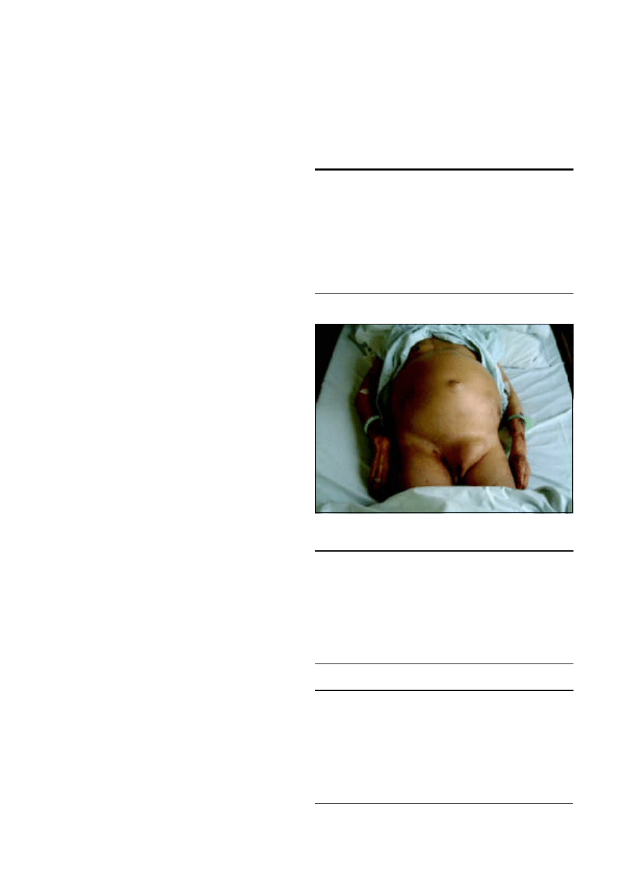
7 Portal hypertension—2. Ascites, encephalopathy, and
other conditions
J E J Krige, I J Beckingham
Ascites
Ascites is caused by cirrhosis in 75% of cases, malignancy in
10%, and cardiac failure in 5%; other causes account for the
remaining 10%. In most patients the history and examination
will give valuable clues to the cause of the ascites—for example,
signs of chronic liver disease, evidence of cardiac failure, or a
pelvic mass. The formation of ascites in cirrhosis is due to a
combination of abnormalities in both renal function and portal
and splanchnic circulation. The main pathogenic factor is
sodium retention. About half of patients with cirrhosis develop
ascites during 10 years of observation. The development of
ascites is an important event in chronic liver disease as half of
cirrhotic patients with ascites die within two years.
Diagnosis
Ascites may not be clinically detectable when present in small
volumes. In larger volumes, the classic findings of ascites are a
distended abdomen with a fluid thrill or shifting dullness.
Ascites must be differentiated from abdominal distension due to
other causes such as obesity, pregnancy, gaseous distension of
bowel, bladder distension, cysts, and tumours. Tense ascites may
cause appreciable discomfort, difficulty in breathing, eversion of
the umbilicus, herniae, and scrotal oedema. Rapid onset of
ascites in patients with cirrhosis may be due to gastrointestinal
haemorrhage, infection, portal venous thrombosis, or the
development of a hepatocellular carcinoma. Ascites can also
develop during a period of heavy alcohol misuse or excessive
sodium intake in food or drugs. Ultrasonography is used to
confirm the presence of minimal ascites and guide diagnostic
paracentesis.
Successful treatment depends on an accurate diagnosis of
the cause of ascites. Paracentesis with analysis of ascitic fluid is
the most rapid and cost effective method of diagnosis. It should
be done in patients with ascites of recent onset, cirrhotic
patients with ascites admitted to hospital, or those with clinical
deterioration. The most important analyses are quantitative cell
counts, fluid culture, and calculation of the serum:ascites
albumin gradient, which reflects differences in oncotic pressures
and correlates with portal venous pressure. Patients with
normal portal pressures have a serum:ascites albumin gradient
less than 11 g/l, whereas patients with ascites associated with
portal hypertension usually have a gradient above 11 g/l.
The traditional classification of transudative and exudative
ascites based on ascitic fluid protein concentrations below and
above 25 g/l is less useful than the serum:ascites albumin
gradient because diuresis can affect the total ascitic protein
concentration.
Treatment
The principal aim of treatment of symptomatic ascites in
cirrhotic patients is to improve general comfort and quality of
life. Most patients will respond to dietary sodium restriction and
diuretic induced excretion of sodium and water, but other
treatments are available for those who do not. Treatment does
not necessarily improve the prognosis for patients with cirrhosis
and may cause complications. Small amounts of ascites that are
asymptomatic should not be treated.
Box 7.1 Causes of ascites
Portal hypertension
x
Cirrhosis of liver
x
Congestive heart failure
x
Constrictive pericarditis
x
Budd-Chiari syndrome
x
Inferior vena cava obstruction
Hypoalbuminaemia
x
Nephrotic syndrome
x
Protein losing enteropathy
Neoplasms
x
Peritoneal carcinomatosis
x
Pseudomyxoma
Miscellaneous
x
Pancreatic ascites
x
Nephrogenic ascites
(associated with maintenance
haemodialysis)
x
Myxoedema
x
Meigs’s syndrome
Box 7.2 Analysis of ascitic fluid
x
Evaluate macroscopic appearance (straw coloured, turbid, bloody,
chylous)
x
Cell count and differential
x
Chemistry profile (protein, albumin, amylase)
x
Cytology
x
Gram stain and bacterial culture
Tests to consider ordering
x
Adenosine deaminase (if tuberculosis is suspected)
x
pH, lactate, lactate dehydrogenase (if bacterial peritonitis suspected)
Box 7.3 Classification of ascites by serum:ascites albumin
gradient
High gradient (>11 g/l)
x
Cirrhosis
x
Alcoholic hepatitis
x
Cardiac ascites
x
Fulminant hepatic failure
x
Budd-Chiari syndrome
x
Portal vein thrombosis
x
Veno-occlusive disease
Low gradient (<11g/l)
x
Peritoneal carcinomatosis
x
Tuberculous peritonitis
x
Pancreatic ascites
x
Biliary ascites
x
Nephrotic syndrome
x
Serositis of collagen, vascular
disease
Figure 7.1 Tense ascites with umbilical and left inguinal hernias
22
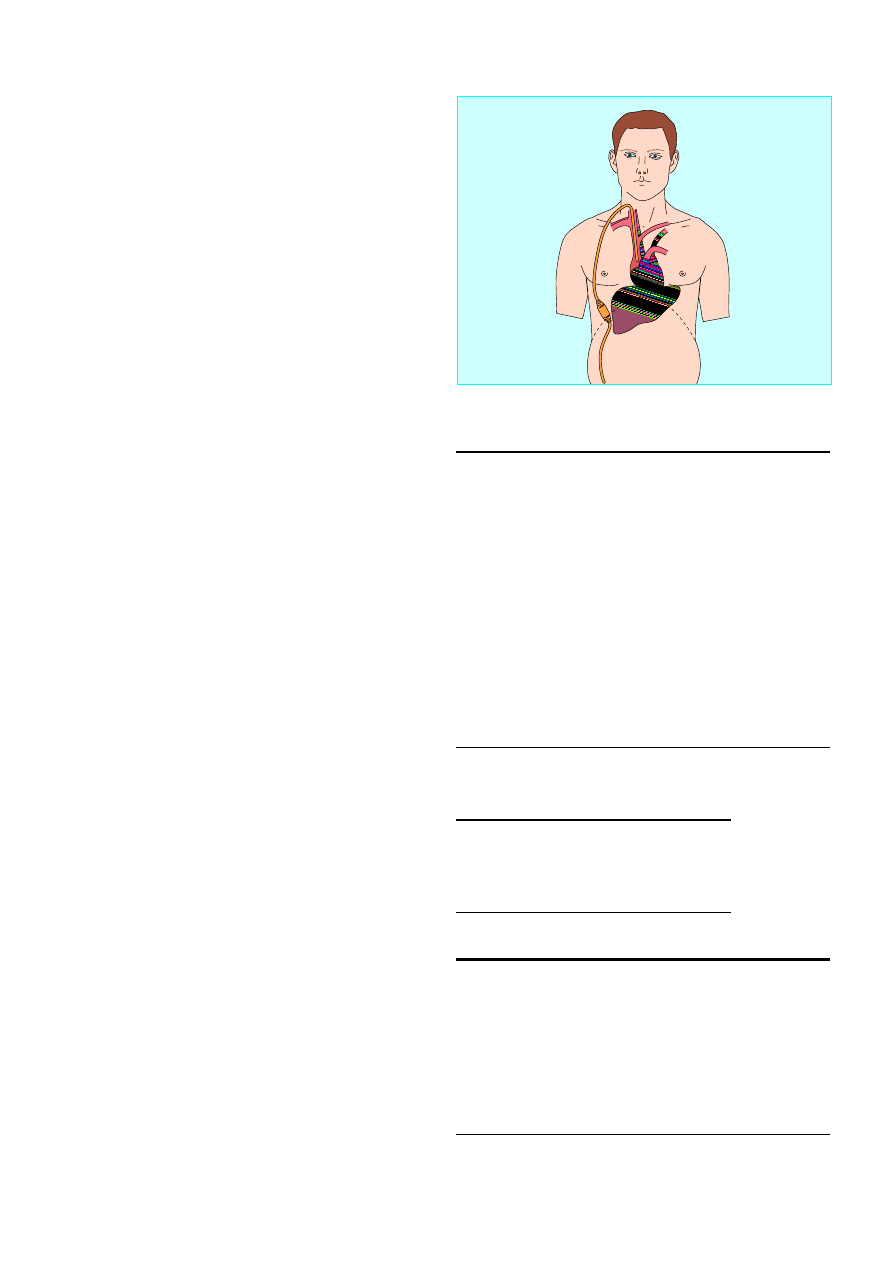
A crucial first step in treating ascites is to convince patients
with alcoholic cirrhosis to abstain from alcohol. Abstinence for
a few months can substantially improve the reversible
component of alcoholic liver disease. Dietary salt restriction is
the most important initial treatment. A low sodium diet of 1-1.5
g of salt (40-60 mmol/day) usually produces a net sodium loss,
which may be sufficient in patients with mild ascites but is
unpalatable and virtually impossible to adhere to in the long
term. In practical terms a “no added salt” diet with levels of 80
mmol/day is the lowest that is generally sustainable. Fluid
restriction is not needed for patients with cirrhotic ascites unless
they have severe hyponatraemia (serum sodium < 120 mmol/l).
Although conventional recommendations suggest bed rest, its
value is not supported by controlled trials.
Most patients need dietary restrictions combined with
diuretics. The usual diuretic regimen comprises single morning
doses of oral spironolactone (an aldosterone antagonist),
increasing the dose as necessary to a maximum of 400 mg/day.
Dietary sodium restriction and dual diuretic therapy is effective in
90% of patients. The patient’s weight, electrolyte concentrations,
and renal function should be carefully monitored. Treatment
should be cautious because of the dangers of iatrogenic
complications from aggressive treatment. Patients with ascites and
peripheral oedema may tolerate 1-2 kg loss per day, but loss of
0.5 kg should be the goal in patients without oedema. Potential
complications during diuresis are encephalopathy, hypokalaemia,
hyponatraemia, hypochloraemic alkalosis, and azotaemia.
Patients with tense ascites should have a total abdominal
paracentesis, followed by a sodium restricted diet and oral
diuretics. Options for patients who do not respond to routine
medical treatment include serial therapeutic paracentesis,
peritoneovenous shunt, transjugular intrahepatic portosystemic
shunt, and liver transplantation. Serial therapeutic paracentesis
should be performed as required, every two to three weeks.
Albumin infusion is unnecessary if < 5 litres of fluid is removed.
Peritoneovenous shunts are seldom used because of
problems with blockage and infection. They are reserved for
patients who are resistant to diuretics, are not transplant
candidates, and are unsuitable for paracentesis because of
abdominal scars.
Hepatic encephalopathy
Hepatic encephalopathy is a reversible state of impaired
cognitive function or altered consciousness that occurs in
patients with liver disease or portosystemic shunts. The typical
features of hepatic encephalopathy include impaired
consciousness (drowsiness), monotonous speech, flat affect,
metabolic tremor, muscular incoordination, impaired
handwriting, fetor hepaticus, upgoing plantar responses,
hypoactive or hyperactive reflexes, and decerebrate posturing.
Hepatic coma, especially in alcoholic patients, should be
diagnosed only after coma due to intracranial space occupying
and vascular lesions, trauma, infection, epilepsy, and metabolic,
endocrine, and drug induced causes has been excluded. Hepatic
encephalopathy is a hallmark of deteriorating liver function,
and patients should be assessed early for liver transplantation.
Hepatocellular insufficiency and portosystemic shunting
may act separately or in combination to cause encephalopathy.
Almost all cases of clinically apparent hepatic encephalopathy
occur in patients with cirrhosis. Less than 5% occur in patients
with non-cirrhotic forms of portal hypertension. However, a
disproportionately large proportion of patients with surgical
and radiological portosystemic shunts develop severe, often
intractable, hepatic encephalopathy. A combination of impaired
Box 7.4 Events precipitating hepatic encephalopathy in
cirrhotic patients
Electrolyte imbalance
x
Diuretics
x
Vomiting
x
Diarrhoea
Gastrointestinal bleeding
x
Oesophageal and gastric varices
x
Gastroduodenal erosions
Drugs
x
Alcohol withdrawal
x
Benzodiazepines
Infection
x
Spontaneous bacterial peritonitis
x
Urinary
x
Chest
Constipation
x
Dietary protein overload
Box 7.5 Drugs that can cause hepatic
encephalopathy
x
Barbiturates
x
Analgesics
x
Other sedatives
Box 7.6 Treatment of hepatic encephalopathy
x
Identify the precipitating factors
x
Stop diuretics
x
Check serum Na
+
, K
+
, and urea concentration
x
Empty bowels of nitrogen containing content
Control bleeding
Protein-free diet
x
Lactulose
x
Neomycin (1 g four times a day by mouth for 1 week)
x
Maintain energy, fluid, and electrolyte balance
x
Increase dietary protein slowly with recovery
Figure 7.2 Denver peritoneovenous shunt
Portal hypertension—2. Ascites, encephalopathy, and other conditions
23

hepatic and renal function is often associated with hepatic
encephalopathy. About half these patients have diuretic induced
renal impairment and half have functional renal failure.
Drugs are implicated in one quarter of patients with hepatic
encephalopathy. Another quarter of cases are precipitated by
haemorrhage in the gastrointestinal tract. This is often
associated with deep and prolonged coma. The combination of
gastrointestinal haemorrhage and hepatic encephalopathy
indicates a poor prognosis. A small proportion of cases are
precipitated by excess dietary protein, hypokalaemic alkalosis,
constipation, and deterioration of liver function secondary to
drugs, toxins, viruses, or hepatocellular carcinoma.
The treatment of hepatic encephalopathy is empirical and
relies largely on establishing the correct diagnosis, identifying
and treating precipitating factors, emptying the bowels of blood,
protein, and stool, attending to electrolyte and acid-base
imbalance, and the selective use of benzodiazepine antagonists.
Non-absorbable disaccharides, such as lactulose or lactitol, are
the mainstay of treatment. Antibiotics and protein restriction
(40 g/day) can be used if there is no response. In intractable
cases, closure of surgical shunts should be considered.
Hepatorenal syndrome
Hepatorenal syndrome is an acute oliguric renal failure
resulting from intense intrarenal vasoconstriction in otherwise
normal kidneys. It occurs in patients with chronic liver disease
(usually cirrhosis, portal hypertension, or ascites) or acute liver
failure; a clinical cause is often not found, treatment is often
ineffective, and prognosis is poor. Hepatorenal syndrome is
prevented by avoiding excessive diuresis and by early
recognition of electrolyte imbalance, bleeding, or infection.
Potentially nephrotoxic drugs such as aminoglycosides and
non-steroidal anti-inflammatories should be avoided.
Patients with hepatorenal syndrome should have blood
cultures taken and any bacteraemia treated. Most patients with
liver disease who develop azotaemia will have prerenal failure
or acute tubular necrosis. The diagnosis of hepatorenal
syndrome is one of exclusion, and it should not be diagnosed
until all potentially reversible causes of renal failure have been
excluded. The common potentially reversible causes are sepsis,
excessive diuresis or paracentesis, and nephrotoxic drugs. All
patients suspected to have hepatorenal syndrome should be
given an intravenous colloid infusion to exclude intravascular
hypovolaemia as a cause of prerenal azotaemia. Liver
transplantation, if otherwise appropriate and feasible, is the only
truly effective treatment, and patients have a poor prognosis.
Spontaneous bacterial peritonitis
Spontaneous bacterial peritonitis is usually the consequence of
bacteraemia due to defects in the hepatic reticuloendothelial
system and in the peripheral destruction of bacteria by
neutrophils. This allows secondary seeding of bacteria in the
ascitic fluid, which is deficient in antibacterial activity.
Clinical signs may be minimal, and a diagnostic paracentesis
should be performed in any cirrhotic patient who suddenly
deteriorates or presents with fever or abdominal pain. A
polymorphonuclear neutrophil count > 500
·
10
6
/l is indicative
of spontaneous bacterial peritonitis. Treatment with intravenous
broad spectrum antibiotics should be started while awaiting the
results of culture of ascitic fluid. Although the mortality
associated with acute spontaneous bacterial peritonitis
decreases with early treatment, it is still high (about 50%) and is
related to the severity of the underlying liver disease.
In patients with cirrhosis and ascites
spontaneous bacterial peritonitis is a
common cause of sudden deterioration
and may be present without any
abdominal symptoms or signs
Box 7.7 Characteristic findings associated with hepatorenal
syndrome
x
Ascites (but not necessarily jaundice) is usually present
x
Hyponatraemia is usual
x
Hepatic encephalopathy is commonly present
x
Blood pressure is reduced compared with previous pressures
recorded in patient
x
Pronounced oliguria
x
Low renal sodium concentration ( < 10mmol/l)
x
Urinary protein and casts are minimal or absent
Summary points
x
Cirrhosis is the commonest cause of ascites (90%)
x
Ninety per cent of cases can be managed by sodium restriction and
diuretics
x
Hepatic encephalopathy is most commonly precipitated by drugs
or gastrointestinal haemorrhage
x
Non-steroidal anti-inflammatory drugs should be avoided in
cirrhotic patients as they can cause renal failure
Further reading
Sherlock S, Dooley J. Diseases of the liver and biliary system. Oxford:
Blackwell Scientific, 1996
Riordan SM, Williams R. Management of liver failure. In: Blumgart
LH, ed. Surgery of the liver and biliary tract. London: W B Saunders,
2000:1825-38
Box 7.8 Spontaneous bacterial peritonitis
x
An infection of ascites that occurs in the absence of a local
infectious source
x
Mainly a complication of cirrhotic ascites
x
Prevalence is 15% to 20% (including culture negative cases)
x
Caused by Gram negative enteric bacteria in > 70% of cases
ABC of Liver, Pancreas, and Gall Bladder
24
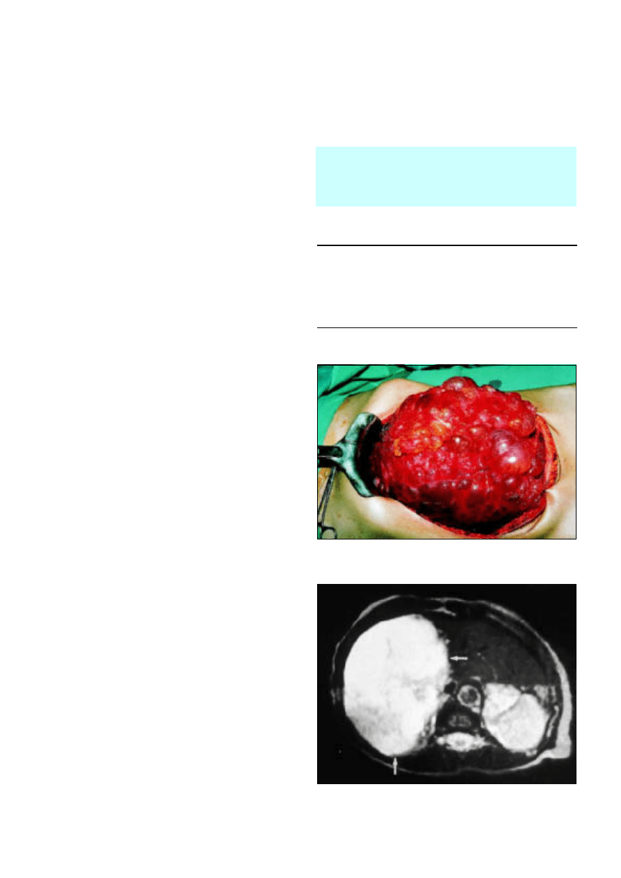
8 Liver tumours
I J Beckingham, J E J Krige
Tumours of the liver may be cystic or solid, benign or
malignant. Most are asymptomatic, with patients having normal
liver function, and they are increasingly discovered incidentally
during ultrasonography or computed tomography. Although
most tumours are benign and require no treatment, it is
important for non-specialists to be able to identify lesions that
require further investigation and thus avoid unnecessary biopsy.
Modern imaging combined with recent technical advances in
liver surgery can now offer many patients safe and potentially
curative resections for malignant, as well as benign, conditions
affecting the liver.
Cystic liver lesions
Cystic lesions of the liver are easily identified by
ultrasonography. Over 95% are simple cysts. Asymptomatic
cysts are regarded as congenital malformations and require no
further investigation or treatment as complications are rare.
Aspiration and injection of sclerosants should be avoided as it
may cause bleeding and infection and does not resolve the cyst.
Rarely, simple cysts can grow very large and produce
compressive symptoms. These are managed by limited surgical
excision of the cyst wall (cyst fenestration), which can usually be
done laparoscopically.
About half of patients with simple cysts have two or more
cysts. True polycystic liver disease is seen as part of adult
polycystic kidney disease, an uncommon autosomal dominant
disease that progresses to renal failure. Patients nearly always
have multiple renal cysts, which usually precede development of
liver cysts. Liver function is normal, and most patients have no
symptoms. Occasionally the cysts cause pain because of
distension of the liver capsule, and such patients may require
cyst fenestration or partial liver resection.
Thick walled cysts and those containing septa, nodules, or
echogenic fluid may be cystic tumours (cystadenoma,
cystadenocarcinoma) or infective cysts (hydatid cysts and
abscesses; see later article in this series), and patients should be
referred for specialist surgical opinion. Cystic dilatations of the
bile ducts (Caroli’s disease) are important as they may produce
cholangitis and are premalignant with the potential to develop
into cholangiocarcinoma.
Benign tumours
Benign liver tumours are common and are usually
asymptomatic. Although most need no treatment, it is
important to be able to differentiate them from malignant
lesions.
Haemangiomas
Haemangiomas are the commonest benign solid tumours of
the liver, with an incidence in the general population of around
3%. Those over 10 cm in diameter occasionally produce
non-specific symptoms of abdominal discomfort and fullness
and, rarely, fever, thrombocytopenia, and hypofibrinogenaemia
due to thrombosis in the cavernous cavities. Malignant
transformation and spontaneous rupture are rare. Contrast
enhanced computed tomography is usually sufficient to
diagnose most haemangiomas, and in equivocal cases magnetic
Liver biopsy of a tumour mass should be reserved for
patients with suspected malignancy who are not suitable
for surgery and in whom the diagnosis may have clinical
impact—for example, ovarian or neuroendocrine tumours,
carcinoid, or lymphoma
Box 8.1 Characteristics of simple cysts
x
Thin walled
x
Contain clear fluid
x
Contain no septa or debris
x
Surrounded by normal liver tissue
x
Usually asymptomatic
x
Present in 1% of population
Figure 8.1 Polycystic liver disease
Figure 8.2 T2 weighted magnetic resonance image of large benign
haemangioma showing light bulb sign
25
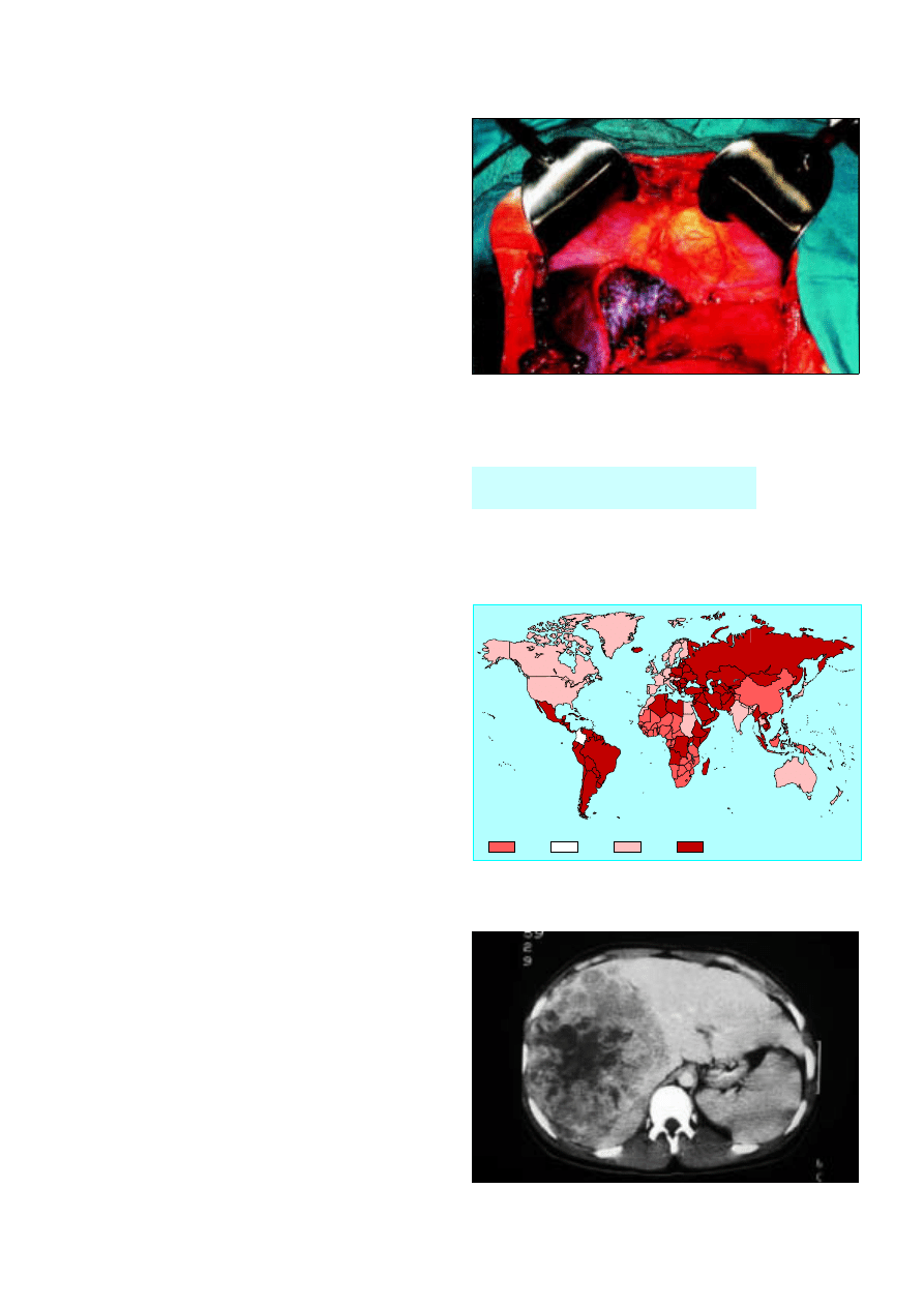
resonance imaging or technetium-99 labelled red blood cell
scintigraphy will confirm the diagnosis. Angiography and
biopsy are seldom required. Resection is indicated only for large
symptomatic tumours.
Liver cell adenoma and focal nodular hyperplasia
These uncommon tumours occur predominantly in women of
childbearing age. Liver cell adenoma became more prevalent
with the widespread use of oral contraceptives in the 1960s, but
the reduced oestrogen content of modern contraceptives has
made it less common. Most patients present with pain due to
rapid tumour growth, intratumour haemorrhage, or the
sensation of a mass. The risk of rupture is 10%, and malignant
transformation is found in 10% of resected specimens. Patients
should have liver resection to prevent these events.
Focal nodular hyperplasia is not related to use of oral
contraceptives, is usually asymptomatic, and is not
premalignant. Mass lesions usually contain a central stellate scar
on computed tomography and magnetic resonance imaging. It
does not require treatment unless symptomatic.
In a small proportion of patients a firm radiological
diagnosis cannot be reached and the distinction from a
malignant liver tumour is uncertain. Histological distinction
between focal nodular hyperplasia and cirrhosis and between
liver cell adenoma and well differentiated hepatocellular
carcinoma can be difficult with tru-cut biopsy or fine needle
aspiration samples, and biopsy has the added risk of bleeding
and tumour seeding. The histology should therefore be
determined by surgical resection, which in specialist centres has
a mortality of < 1%.
Malignant tumours
Hepatocellular carcinoma
Hepatocellular carcinoma is uncommon in the United
Kingdom and accounts for only 2% of all cancers. Worldwide
there are over one million new cases a year, with an annual
incidence of 100 per 100 000 men in parts of South Africa and
South East Asia. The incidence of hepatocellular carcinoma is
increased in areas with high carrier rates of hepatitis B and C
and in patients with haemochromatosis. More than 80% of
hepatocellular carcinomas occur in patients with cirrhotic livers.
Once viral infection is established it takes about 10 years for
patients to develop chronic hepatitis, 20 years to develop
cirrhosis, and 30 years to develop carcinoma. In African and
Asian countries aflatoxin, produced as a result of contamination
of imperfectly stored staple crops by Aspergillus flavus, seems to
be an independent risk factor for the development of
hepatocellular carcinoma, probably through mutation of the
p53 suppressor gene. Seasonal variation in incidence is seen in
these countries.
In patients with cirrhosis, the diagnosis should be suspected
when there is deterioration in liver function, an acute
complication (ascites, encephalopathy, variceal bleed, jaundice),
or development of upper abdominal pain and fever.
Ultrasonography will identify most tumours, and the presence
of a discrete mass within a cirrhotic liver, together with an
á
fetoprotein concentration above 500 ng/ml is diagnostic.
Biopsy is unnecessary and should be avoided to reduce the risk
of tumour seeding. Surgical resection is the only treatment that
can offer cure. However, owing to local spread of tumour and
severity of pre-existing cirrhosis, such treatment is feasible in
less than 20% of patients. Average operative mortality is 12% in
cirrhotic patients, and five year survival is around 15%.
Hepatocellular carcinoma is the
commonest malignant tumour worldwide
Figure 8.3 Intraoperative view after left hepatectomy—raw surfaces of liver
are coated with fibrin glue after resection to aid haemostasis and prevent
small bile leaks
10-15
Annual incidence (cases per 100 000)
3-10
1-3
Undefined
Figure 8.4 Distribution of hepatocellular carcinoma
Figure 8.5 Computed tomogram of large hepatocellular carcinoma
ABC of Liver, Pancreas, and Gall Bladder
26
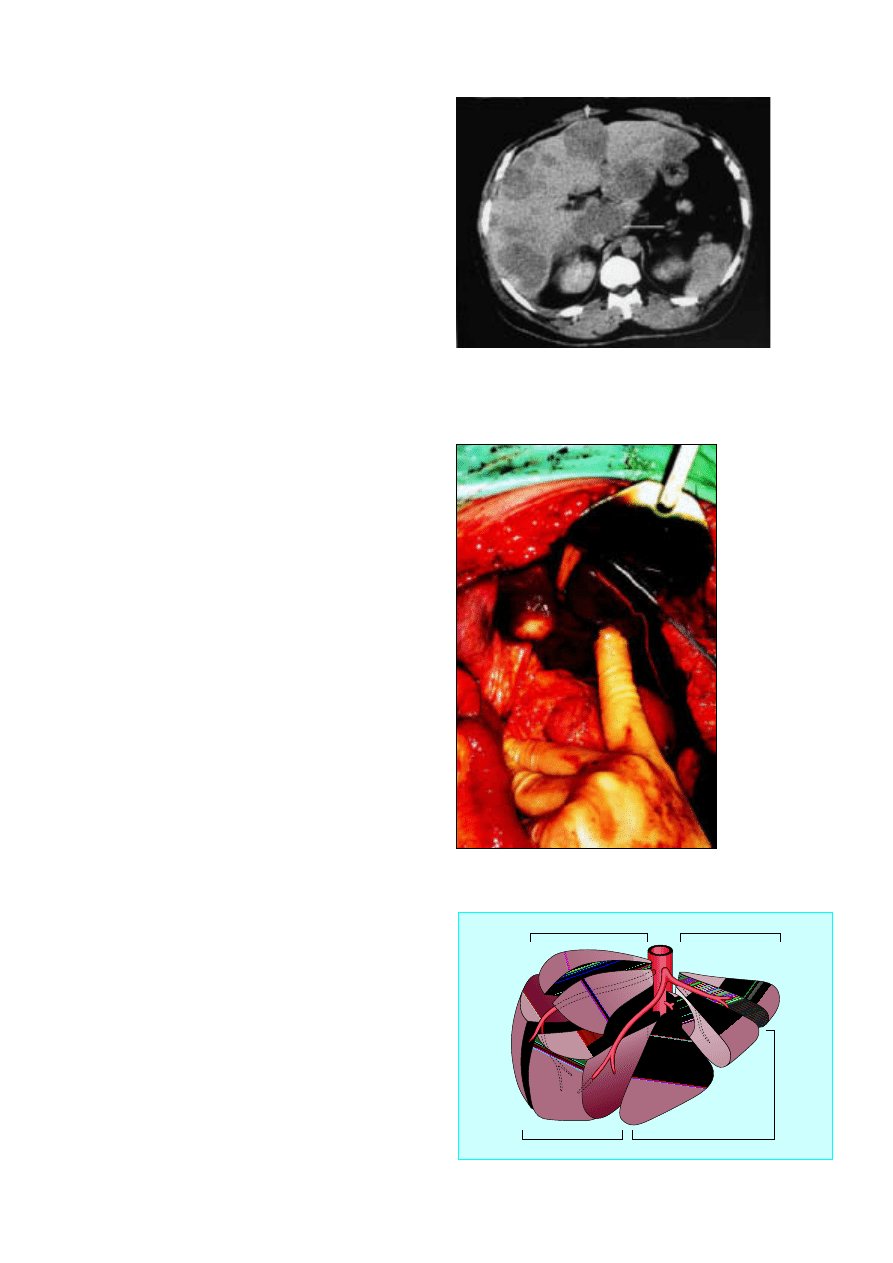
Patients with cirrhosis and small ( < 5 cm) tumours should
have liver transplantation. Injection of alcohol or
radiofrequency ablation can improve survival in patients with
small tumours who are unsuitable for transplantation. For
larger tumours, transarterial embolisation with lipiodol and
cytotoxic drugs (cisplatin or doxorubicin) may induce tumour
necrosis in some patients.
In patients without cirrhosis, hepatocellular carcinomas
usually present late with an abdominal mass and abnormal liver
function. Computed tomography has a greater sensitivity and
specificity than ultrasonography, particularly for tumours
smaller than 1 cm. á Fetoprotein concentrations are raised in
80% of patients but may also be raised in patients with testicular
or germ cell tumours.
Fibrolamellar carcinoma is an important subtype of
hepatocellular carcinoma. It occurs in patients without cirrhosis
or previous hepatitis infection. It accounts for 15% of
hepatocellular carcinoma in the Western hemisphere. The
prognosis is better than for other hepatocellular carcinomas,
with a five year survival of 40-50% after resection.
Metastatic tumours
Liver metastases are common and are found in 40% of all
patients dying from cancer. They are most frequently associated
with carcinomas of the gastrointestinal tract (colorectal,
pancreas, and stomach) but are nearly as common in
carcinomas of the bronchus, breast, ovary, and lymphoma. With
the exception of liver metastases of colorectal cancer, tumour
deposits are almost always multiple and seldom amenable to
resection.
Colorectal liver metastases
Around 8-10 % of patients undergoing curative resection of
colorectal tumours have isolated liver metastases suitable for
liver resection, equivalent to around 1000 patients in the United
Kingdom a year. Half will have metastases at the time of
diagnosis of the primary tumour (synchronous metastases) and
most of the rest will develop metastases within the next three
years (metachronous metastases).
Without surgical resection the five year survival rate for all
patients with liver metastases is zero, compared with an overall
five year survival after resection of 30%. Patients most suited for
resection are those with fewer than three or four metastases in
one lobe of the liver, but tumours need not be confined to one
lobe. The principle of complete tumour removal, however,
remains a prerequisite, and one limitation is the need to leave
enough liver to function. This depends both on the extent and
distribution of the tumour burden and the general fitness of the
patient and his or her liver. The liver has an enormous capacity
for regeneration. A fit patient with a healthy liver will regenerate
a 75% resection within three months. Age is only a relative
contraindication, and several series have reported low mortality
in septuagenarians.
Liver resection
Liver resection has advanced rapidly over the past two decades
because of several important developments. The segmental
anatomy of the liver, with each of the eight segments supplied
by its own branch of the hepatic artery, portal vein, and bile
duct, was first described by Couinaud in 1957. It is now possible
to remove each of these segments individually when required,
reducing the amount of normal liver unnecessarily removed.
Subsequently surgical techniques have been developed to
divide the liver parenchyma, either by crushing with a clamp or
Figure 8.6 Inoperable extensive liver metastases
Figure 8.7 Solitary metastasis in segment IV of liver
Right lateral (posterior) sector
Right medial (anterior) sector
Left medial
(anterior) sector
Left lateral (posterior) sector
VII
VIII
VI
V
IV
III
II
I
Figure 8.8 Couinaud’s segmental anatomy of liver
Liver tumours
27

by ultrasonic dissection, allowing the vascular and biliary
radicals to be individually ligated. Blood loss has been reduced
by occlusion of the vascular inflow (Pringle manoeuvre) and
where possible the appropriate hepatic vein, together with
lowering of the central venous pressure during resection, and
blood transfusion is now unnecessary in 60% of major liver
resections.
Improvements have also occurred in anaesthetic and
postoperative care, including epidural anaesthesia to reduce
postoperative pain and chest complications and the ability to
manage postoperative fluid or bile collections by radiological or
endoscopic drainage. These developments mean that the
median hospital stay for patients having liver resection is now
7-10 days and mortality is around 5%. Liver resection has
evolved from a hazardous bloody procedure into a routine
operation.
Summary points
x
Simple liver cysts are common, benign, and require no treatment
x
Patients with solitary liver masses should be referred to a
hepatobiliary surgeon and liver biopsy avoided
x
Liver resection is a safe procedure in non-cirrhotic patients, with a
mortality around 5%
x
10% of patients with colorectal cancer develop potentially curable
liver metastases and should have six monthly liver ultrasonography
or computed tomography
x
Five year survival after resection of colorectal metastases is > 30%
Further reading
x
Blumgart LH, Jarnogin W, Fang Y. Liver resection. In: Blumgart LH,
ed. Surgery of the liver and biliary tract. London: WB Saunders,
2000:1639-1714
x
Launois B, Jamieson GG. Modern operative techniques in liver surgery.
Edinburgh: Churchill Livingstone, 1993
x
Neeleman N, Andersson R. Repeated liver resection for recurrent
liver cancer. Br J Surg 1996;83:885-92
ABC of Liver, Pancreas, and Gall Bladder
28
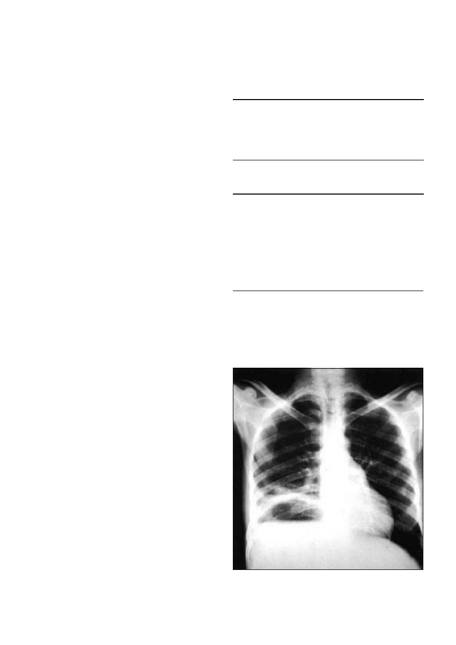
9 Liver abscesses and hydatid disease
J E J Krige, I J Beckingham
Liver abscesses are caused by bacterial, parasitic, or fungal
infection. Pyogenic abscesses account for three quarters of
hepatic abscess in developed countries. Elsewhere, amoebic
abscesses are more common, and, worldwide, amoebae are the
commonest cause.
Pyogenic liver abscesses
Aetiology
Most pyogenic liver abscesses are secondary to infection
originating in the abdomen. Cholangitis due to stones or
strictures is the commonest cause, followed by abdominal
infection due to diverticulitis or appendicitis. In 15% of cases no
cause can be found (cryptogenic abscesses). Compromised host
defences have been implicated in the development of
cryptogenic abscess and may have a role in the aetiology of
most hepatic abscesses. Diabetes mellitus has been noted in
15% of adults with liver abscesses.
Microbiology
Most patients presenting with pyogenic liver abscesses have a
polymicrobial infection usually with Gram negative aerobic and
anaerobic organisms. Most organisms are of bowel origin, with
Escherichia coli, Klebsiella pneumoniae,
bacteroides, enterococci,
anaerobic streptococci, and microaerophilic streptococci being
most common. Staphylococci, haemolytic streptococci, and
Streptococcus milleri
are usually present if the primary infection is
bacterial endocarditis or dental sepsis. Immunosuppression due
to AIDS, intensive chemotherapy, and transplantation has
increased the number of abscesses due to fungal or
opportunistic organisms.
Clinical features
The classic presentation is with abdominal pain, swinging fever,
and nocturnal sweating, vomiting, anorexia, malaise, and weight
loss. The onset may be insidious or occult in elderly people, and
patients may present with a primary infection (such as
diverticulitis or appendicitis) before developing symptoms from
their liver abscess. Single abscesses tend to be gradual in onset
and are often cryptogenic. Multiple abscesses are associated
with more acute systemic features and the cause is more often
identified.
Clinically, the liver is enlarged and tender, and percussion
over the lower ribs aggravates the pain. Clinical jaundice occurs
only in the late stage unless there is suppurative cholangitis.
Some patients do not have right upper quadrant pain or
hepatomegaly and present with fever of unknown origin.
Laboratory investigations
Two thirds of patients have appreciable leucocytosis, often
accompanied by anaemia of chronic infection and a raised
erythrocyte sedimentation rate. The alkaline phosphatase
activity is generally raised, hypoalbuminaemia is present, and
serum transaminase activity may be marginally abnormal.
Plain abdominal radiography may show hepatomegaly,
sometimes with an air fluid level in the abscess cavity. The right
diaphragm is often raised, with a pleural reaction or pneumonic
consolidation. Ultrasonography is the preferred initial method
of imaging as it is non-invasive, cost effective, and can be used to
Box 9.1 Typical features of pyogenic liver abscess
x
Right upper quadrant pain and tenderness
x
Nocturnal fevers and sweats
x
Anorexia and weight loss
x
Raised right hemidiaphragm in chest radiograph
x
Raised white cell count and erythrocyte sedimentation rate with
mild anaemia
Box 9.2 Origins and causes of pyogenic liver abscess
x
Biliary tract
Gall stones
Cholangiocarcinoma
Strictures
x
Portal vein
Appendicitis
Diverticulitis
Crohn’s disease
x
Hepatic artery
Dental infection
Bacterial endocarditis
x
Direct extension of:
Gall bladder empyema
Perforated peptic ulcer
Subphrenic abscess
x
Trauma
x
Iatrogenic
Liver biopsy
Blocked biliary stent
x
Cryptogenic
x
Secondary infection of liver cyst
Figure 9.1 Chest radiograph showing air-fluid level and raised right
hemidiaphragm in pyogenic liver abscess
29
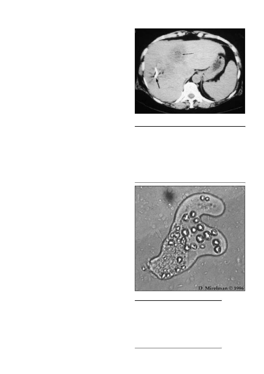
guide aspiration to identify the causative organism. Computed
tomography is useful to identify other intra-abdominal
abscesses. Endoscopic retrograde cholangiopancreatography is
used to define the site and cause of biliary obstruction and to
allow biliary stenting and drainage.
Treatment
Empirical broad spectrum parenteral antibiotic treatment should
be started as soon as an abscess is diagnosed. Antibiotics should
include penicillin, an aminoglycoside, and metronidazole, which
are effective against E coli, K pneumoniae, bacteroides,
enterococcus, and anaerobic streptococci. In elderly people and
those with impaired renal function a third generation
cephalosporin should be used instead of an aminoglycoside. The
regimen should be modified after culture has identified the
infective organism. Treatment is continued for two to four weeks
depending on the number of abscesses, the clinical response, and
the potential toxicity of the chosen regimen.
Antibiotics alone are effective in only a few patients, and
most patients will require percutaneous aspiration or catheter
drainage guided by ultrasonography or computed tomography.
In all cases the underlying cause should be sought and treated.
Early diagnosis, treatment with appropriate antibiotics, and
selective drainage have substantially reduced mortality. Factors
that increase the risk of death include shock, adult respiratory
distress syndrome, disseminated intravascular coagulation,
immunodeficiency states, severe hypoalbuminaemia, diabetes,
ineffective surgical drainage, and associated malignancy.
Amoebic liver abscess
About 10% of the world’s population is chronically infected with
Entamoeba histolytica
. Amoebiasis is the third commonest parasitic
cause of death, surpassed only by malaria and schistosomiasis.
The prevalence of infection varies widely, and it occurs most
commonly in tropical and subtropical climates. Overcrowding
and poor sanitation are the main predisposing factors.
Pathogenesis
The parasite is transmitted through the faeco-oral route with
the ingestion of viable protozoal cysts. The cyst wall
disintegrates in the small intestine, releasing motile
trophozoites. These migrate to the large bowel, where
pathogenic strains may cause invasive disease. Mucosal invasion
results in the formation of flask-shaped ulcers through which
amoebae gain access to the portal venous system. The abscess is
usually solitary and affects the right lobe in 80% of cases. The
abscess contains sterile pus and reddish-brown (“anchovy
paste”) liquefied necrotic liver tissue. Amoebae are occasionally
present at the periphery of the abscess.
Clinical presentation and diagnosis
Patients may have had symptoms from a few days to several
weeks before presentation. Pain is a prominent feature, and the
patient appears toxic, febrile, and chronically ill.
The diagnosis is based on clinical, serological, and
radiological features. The patient is usually resident in an
endemic area or has visited one recently, although there may be
no history of diarrhoea. Patients commonly have leucocytosis
with 70-80% polymorphs (eosinophilia is not a feature), a raised
erythrocyte sedimentation rate, and moderate anaemia In
patients with severe disease and multiple abscesses, alkaline
phosphatase activity and bilirubin concentration are raised.
Stools may contain cysts, or in the case of dysentery,
haematophagous trophozoites.
Box 9.3 Drainage requirements for liver abscesses
x
None—multiple small abscesses that respond to antibiotics
(Obstruction of bile duct must be excluded as a cause and
endoscopic retrograde cholangiopancreatography with stenting
performed if necessary)
x
Percutaneous aspiration—abscesses < 6 cm
x
Percutaneous catheter drainage—abscesses >6 cm
x
Open surgery
Failed percutaneous drainage
Very large or multilocular abscesses
Associated intra-abdominal infection requiring surgery such as bile
duct stones
Box 9.4 Symptoms of amoebic liver abscess
x
Pain
x
Enlarged liver with maximal tenderness over abscess
x
Intermittent fever (38-39°C)
x
Night sweats
x
Weight loss
x
Nausea
x
Vomiting
x
Cough
x
Dyspnoea
Figure 9.2 Computed tomogram showing multifocal liver abscess in segment
IV. Note drain and second abscess in segments VII and VIII
Figure 9.3 Amoebic trophozoite with large pseudopod
ABC of Liver, Pancreas, and Gall Bladder
30
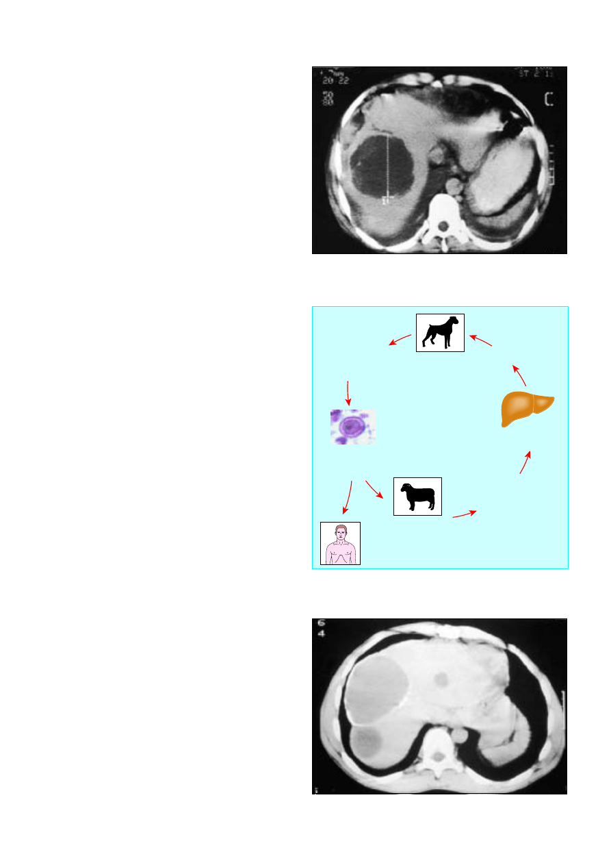
Chest radiography usually shows a raised right
hemidiaphragm with atelectasis or pleural effusion.
Ultrasonography shows the size and position of the abscess and
is useful when aspiration is necessary and to assess response to
treatment. Serological tests provide a rapid means of
confirming the diagnosis, but the results may be misleading in
endemic areas because of previous infection. Indirect
haemagglutination titres for entamoeba are raised in over 90%
of patients. In areas where amoebiasis is uncommon, failure to
consider the infection may delay diagnosis.
Serious complications occur as a result of secondary
infection or rupture into adjacent structures such as pleural,
pericardial, or peritoneal spaces. Two thirds of ruptures occur
intraperitoneally and one third intrathoracically.
Treatment
Ninety five per cent of uncomplicated amoebic abscesses
resolve with metronidazole alone (800 mg, three times a day for
five days). Supportive measures such as adequate nutrition and
pain relief are important. Clinical symptoms usually improve
greatly within 24 hours. Lower doses of metronidazole are often
effective in invasive disease but may fail to eliminate the
intraluminal infection, allowing clinical relapses to occur. After
the amoebic abscess has been treated, patients are prescribed
diloxanide furoate 500 mg, eight hourly for seven days, to
eliminate intestinal amoebae.
Patients should have ultrasonographically guided needle
aspiration if serology gives negative results or the abscess is
large ( > 10 cm), if they do not respond to treatment, or if there
is impending peritoneal, pleural, or pericardial rupture. Surgical
drainage is required only if the abscess has ruptured causing
amoebic peritonitis or if the patient has not responded to drugs
despite aspiration or catheter drainage.
Hydatid disease
Hydatid disease in humans is caused by the dog tapeworm,
Echinococcus granulosus
. Dogs are the definitive host. Ova are shed
in the faeces and then infect the natural intermediate hosts such
as sheep or cattle. Hydatid disease is endemic in many sheep
raising countries. Increasing migration and world travel have
made hydatidosis a global problem of increasing importance.
Human infection follows accidental ingestion of ova passed
in dog faeces. The ova penetrate the intestinal wall and pass
through the portal vein to the liver, lung, and other tissues.
Hydatid cysts can develop anywhere in the body, but two thirds
occur in the liver and one quarter in the lungs.
Presentation
Patients with a liver hydatid may present either with liver
enlargement and right upper quadrant pain due to pressure from
the cyst or acutely with a complication. Complications include
rupture of the cyst into the peritoneal cavity, which results in
urticaria, anaphylactic shock, eosinophilia, and implantation into
the omentum and other viscera. Cysts may compress or erode
into a bile duct causing pain, jaundice, or cholangitis, or the cyst
may become infected secondary to a bile leak.
Diagnsosis and treatment
Ultrasonography and computed tomography will show the size,
position, and number of liver cysts and any extrahepatic cysts.
Around 10% of patients with a liver cyst will also have a lung
hydatid on chest radiography. Eosinophilia is present in 40% of
patients. The diagnosis is confirmed by haemagglutination and
complement fixation tests. Endoscopic retrograde
Figure 9.4 Computed tomogram of amoebic liver abscess
The adult tapeworm is found
in the small intestine
of definitive host
Dog eats infected
sheep liver
(definitive host)
Eggs are passed
in the
host's faeces
Eggs are ingested by
intermediate host
Man (inadvertent
intermediate host)
Eggs hatch in small
intestine, penetrate intestinal
wall, and enter blood stream
Larvae distributed to liver
and other organs
Larva develops
into hydatid cyst
Figure 9.5 Lifecycle of Echinococcus granulosus
Figure 9.6 Hydatid cyst in right lobe of liver with calcifcation in the wall
Liver abscesses and hydatid disease
31
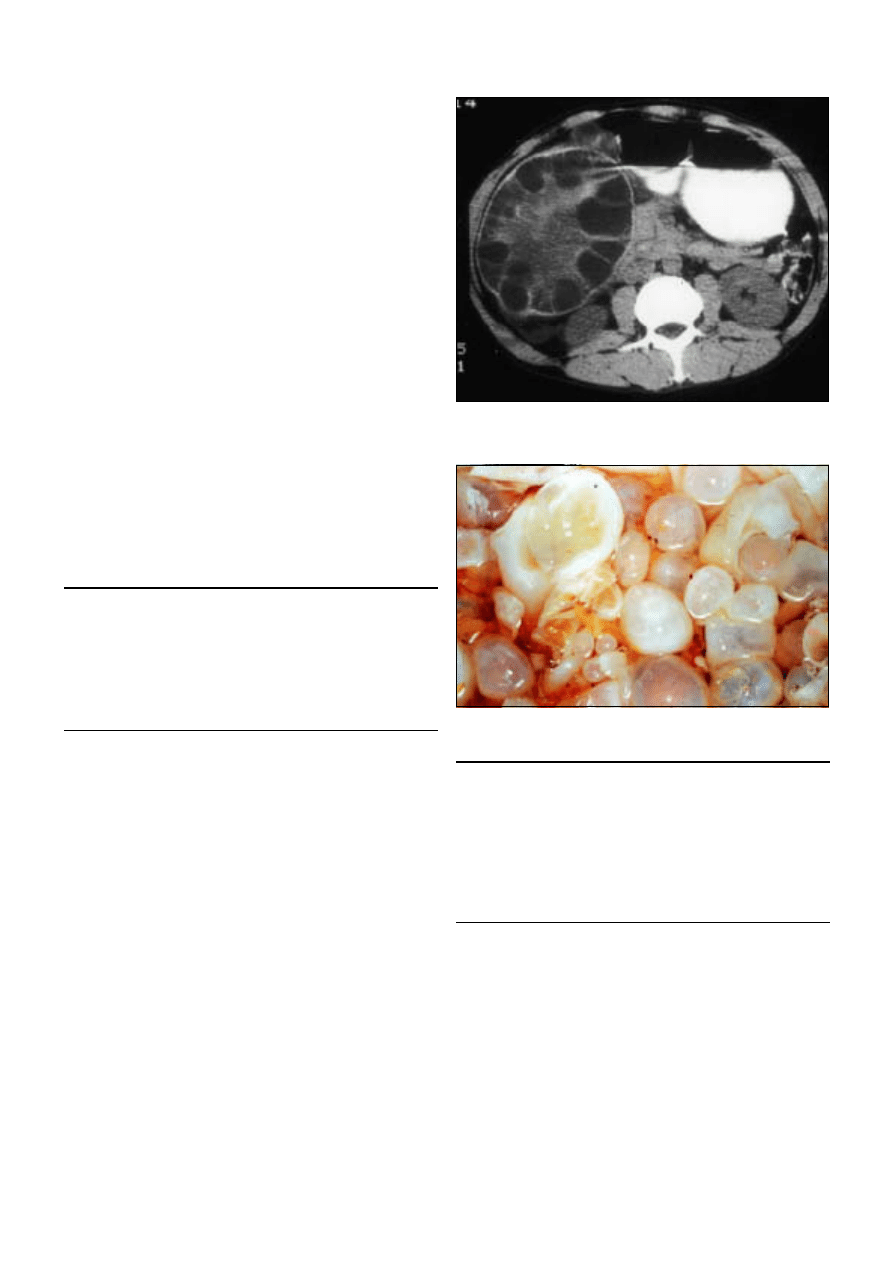
cholangiopancreatography will show communication between
the cyst and the bile ducts if patients are jaundiced, their serum
alkaline phosphatase or ã-glutamyltransferase activity is raised,
or their bilirubin concentration increased.
All symptomatic cysts require surgical removal to prevent
complications. Small densely calcified cysts (“golf ball”
appearance) signify death of the parasite and require no further
treatment. Careful isolation of the operative field by abdominal
swabs soaked in scolicidal fluid is essential to prevent spillage
and formation of intraperitoneal cysts. The cyst fluid is
aspirated and replaced by a scolicidal agent such as 0.5%
sodium hypochlorite or 0.5% silver nitrate. Scolicidal solutions
should not be injected if there is a bile leak because of possible
chemical injury to the biliary epithelium.
After decompression, the cyst and contents are carefully
shelled out by peeling the endocyst off the host ectocyst layer
along its cleavage plane. The fibrous host wall of the residual
cavity should be carefully examined for any bile leakage from
biliary-cyst communications, which are then sutured. The cavity
is drained and filled with omentum.
Conservative surgery is effective in most cysts, and liver
resection is seldom necessary. Albendazole, flubendazole, or
praziquantel are given for two weeks postoperatively to prevent
recurrence. Drug treatment can be used in patients unfit for
surgery and in those with disseminated, recurrent, or inoperable
disease and as an adjuvant in complex surgery. These drugs
must be used cautiously and patients monitored for side effects,
which include depression of bone marrow activity and liver and
renal toxicity.
The picture of the trophozoite was supplied by David Mirecman,
University of Utah.
Summary points
x
Most patients with pyogenic abscesses will require percutaneous
drainage and antibiotics
x
A cause can be identified in 85% of cases of liver abscess, most
commonly gall stones, diverticulitis, or appendicitis
x
Amoebic abscesses can be treated by metronidazole alone in 95%
of cases
x
Hydatid disease occurs throughout the world in sheep farming
areas
x
Symptomatic hydatid cysts should be surgically removed
Figure 9.7 Computed tomogram showing hydatid cyst: daughter cysts
containing hydatid larvae are visible within the main cyst
Figure 9.8 Operative specimen of opened hydatid cyst showing multiple
daughter cysts
Further reading
Krige JEJ. Pyogenic liver abscess. In: Kirsch R, Robson S, Trey C, eds.
Diagnosis and management of liver disease
. London: Chapman and
Hall, 1995:196-202
Krige JEJ, Adams S, Simjee A. Amoebic liver abscess. In: Kirsch R,
Robson S, Trey C, eds. Diagnosis and management of liver disease.
London: Chapman and Hall, 1995:186-95
Krige JEJ, Terblanche J. Hepatic echinococcosis. In: Cameron JL, ed.
Current surgical therapy
. 6th ed. Baltimore, MA: Mosby, 1998:326-30
ABC of Liver, Pancreas, and Gall Bladder
32
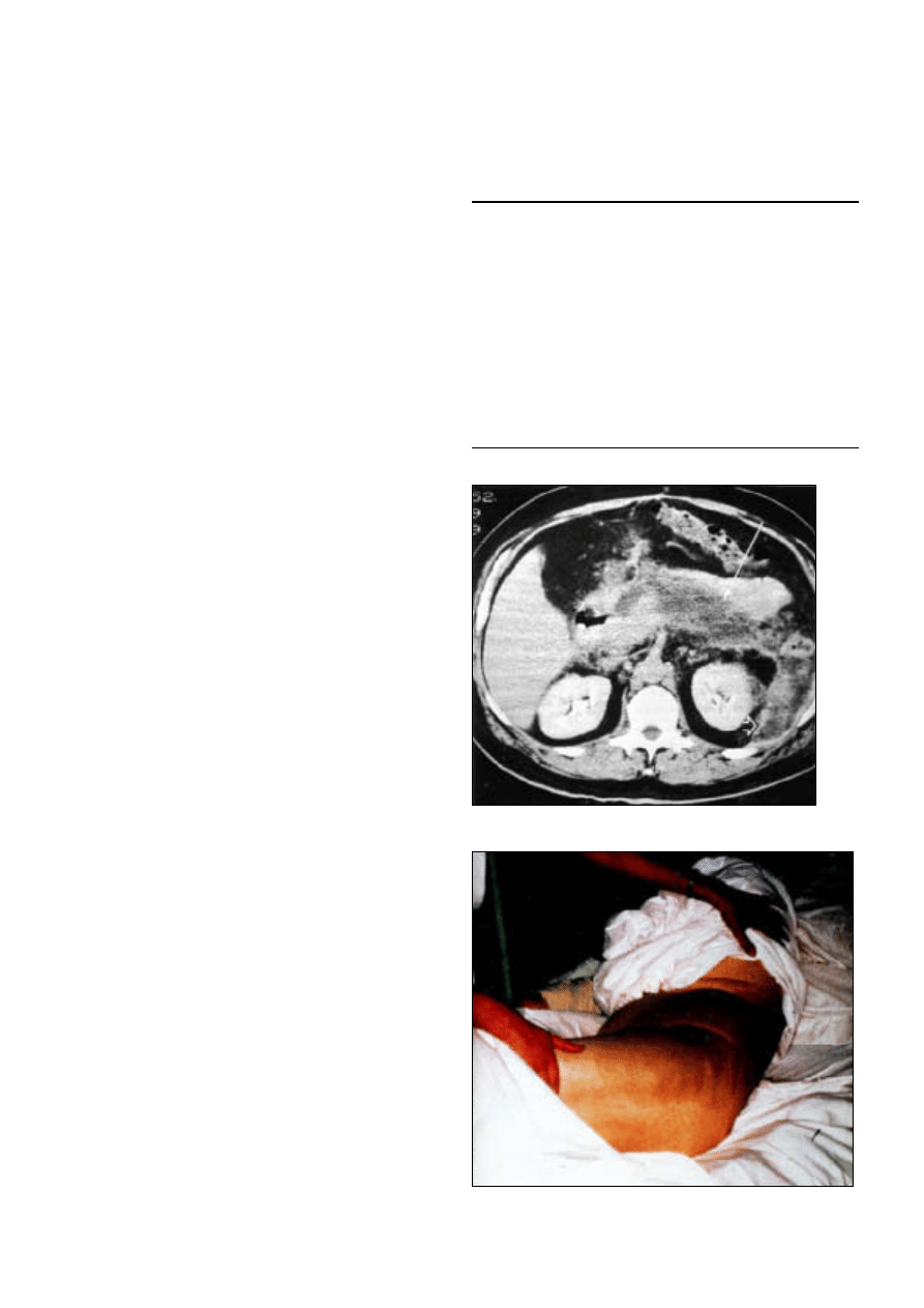
10 Acute pancreatitis
I J Beckingham, P C Bornman
Acute pancreatitis is relatively common, with an annual
incidence of 10-20/million population. In more than 80% of
patients the disease is associated with alcohol or gall stones,
although the ratio of these two causes has a wide geographical
variation. Gallstone disease predominates in most UK centres
by more than 2:1.
Pathogenesis and pathological
processes
Apart from mechanical factors such as the passage of gall
stones through the ampulla of Vater or cannulation at
endoscopic retrograde cholangiopancreatography, little is
known about how the disease process begins. What follows is
also unclear, but proteolytic enzymes are thought to be
activated while still within the pancreatic cells, setting off a local
and systemic inflammatory cell response.
The process is self limiting in most cases and pathologically
correlates with oedematous interstitial pancreatitis. In 15-20%
of cases the process runs a fulminating course, most commonly
within the first week. This is characterised by pancreatic necrosis
and associated cytokine activation resulting in multiple organ
dysfunction syndrome. The necrotic process mainly affects the
peripancreatic tissue (mostly fat) and may spread extensively
along the retroperitoneal space behind the colon and into the
small bowel mesentery. The necrotic tissue can become infected,
probably by translocation of bacteria from the gut.
Clinical presentation
Acute pancreatitis should always be considered in the
differential diagnosis of patients with acute abdomen. The
clinical presentation may vary considerably and is influenced by
the aetiological factor, age, other associated illnesses, the stage
of the disease, and the severity of the attack.
In alcohol induced pancreatitis symptoms usually begin
6-12 hours after an episode of binge drinking. Gall stones
should be suspected in patients over 50 years of age (especially
women), those who do not drink alcohol, and when the attack
begins after a large meal. In patients with an alcohol history and
proved gall stones it can be difficult to distinguish between the
two causes. A serum alanine transaminase activity greater than
three times above normal usually indicates that gall stones are
the cause.
Patients usually have pain in the epigastrium that typically
radiates through to the back. It is often associated with nausea
and vomiting. Severe attacks often mimic other abdominal
catastrophes such as perforated or ischaemic bowel and
ruptured aortic aneurysm. Abdominal distension with or
without a vague palpable epigastric mass is common in severe
attacks. More rarely, patients develop discoloration in the
lumbar regions and periumbilical area because of associated
bleeding in the retroperitoneal space.
Acute pancreatitis may develop after cardiac or abdominal
operations—for example, gastrectomy or biliary surgery—and
the onset may be insidious with unexplained cardiorespiratory
failure, fever, and ileus associated with minimal abdominal
signs.
Box 10.1 Causes of acute pancreatitis
x
Gallstones
}
80%
x
Alcohol
x
Idiopathic: 10%
x
Endoscopic retrograde cholangiopancreatography or
sphincterectomy: 5%
x
Miscellaneous: 5%
Hyperlipidaemia
Trauma
Hyperparathyroidism
Viral (mumps, Epstein-Barr virus, cytomegalovirus coxsackievirus)
Drug induced (thiazide diuretics, angiotensin converting enzyme
inhibitors, oestrogens, corticosteroids, azathioprine)
Anatomical (pancreas divisum, annular pancreas)
Parasites (Ascaris lumbricoides)
Figure 10.1 Computed tomogram showing extensive pancreatic
necrosis (arrow) spreading into perinephric fat (open arrow head)
Figure 10.2 Discoloration of flank in patient with acute pancreatitis
(Grey-Turner’s sign)
33
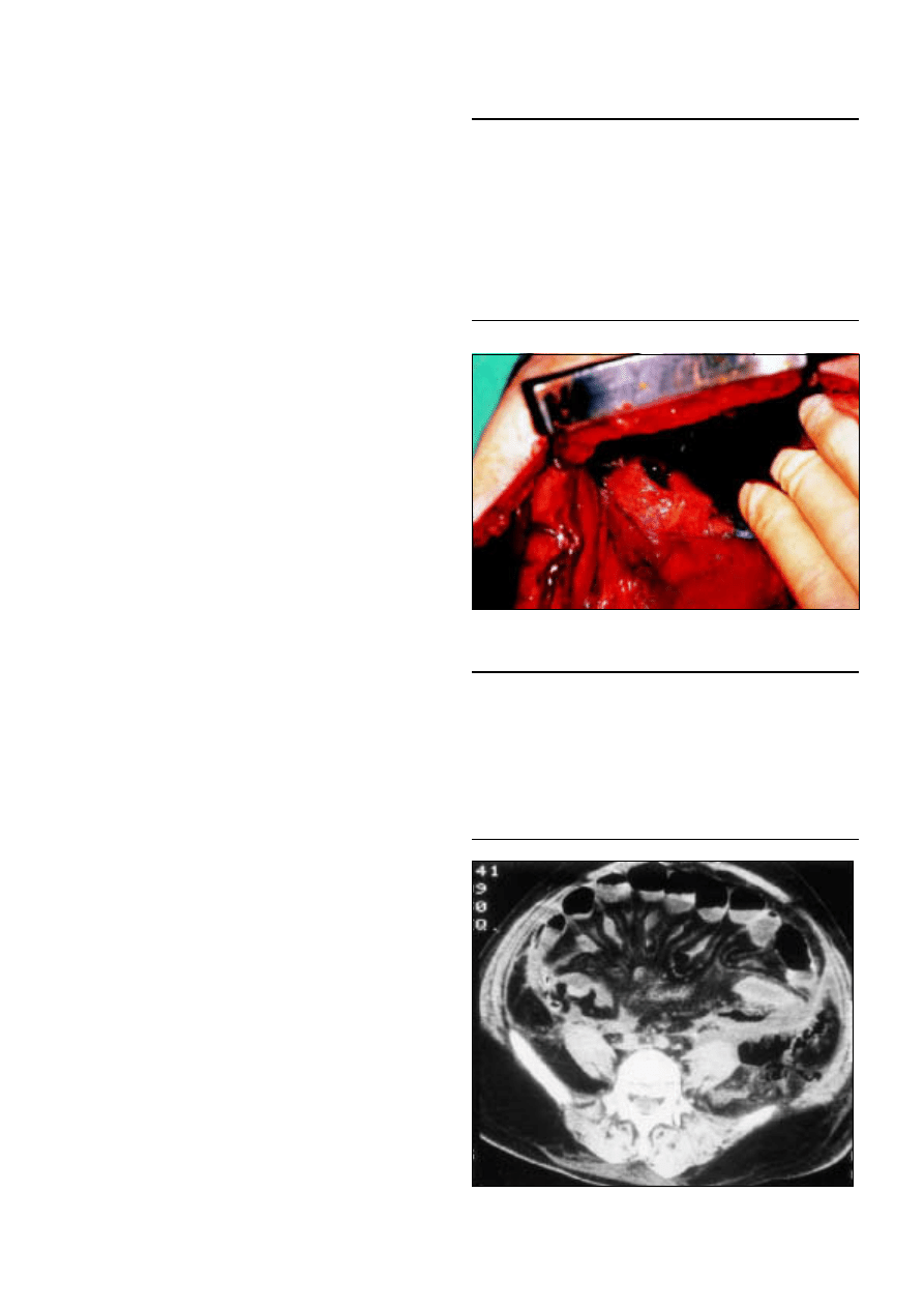
Diagnosis
Pancreatitis is diagnosed on the basis of a combination of
appropriate clinical features and a serum amylase activity over
three times above normal ( > 330 U/l.). Lower activities do not
rule out the diagnosis as serum amylase activity may reduce or
normalise within the first 24-48 hours. Measurement of urinary
amylase activity, which remains high for longer periods, may be
helpful in this situation.
Although amylase activity may be raised in several other
conditions with similar clinical signs (notably perforated peptic
ulcer and ischaemic bowel), the increase is rarely more than
three times above normal. Serum lipase measurement has a
higher sensitivity and specificity, and now that simpler methods
of measurement are available it is likely to become the
preferred diagnostic test.
Clinical course
Whatever the underlying cause of pancreatitis, the clinical
course is usually similar. The disease process is self limiting in
80% of cases, but in severe cases, there are usually three phases:
local inflammation and necrosis, a systemic inflammatory
response leading to multiple organ dysfunction syndrome
during the first two weeks, and, finally, local complications such
as the development of a pseudocyst or infection in the
pancreatic and peripancreatic necrotic tissue.
Assessment of severity
Early identification of patients with a severe attack is important
as they require urgent admission to a high dependency or
intensive care unit. Initial predictors of a severe attack include
first attack of alcohol induced pancreatitis, obesity,
haemodynamic instability, and severe abdominal signs (severe
tenderness and haemorrhage of the abdominal wall).
Several scoring systems have been developed to predict
patients with mild or severe pancreatitis. The most widely used
in the United Kingdom is the modified Glasgow system (Imrie),
which has a sensitivity of 68% and a specificity of 84%. Other
commonly used systems are Ranson’s and the acute
physiological and chronic health evaluation (APACHE II).
Changes in C reactive protein concentration and APACHE II
scores correlate well with the ongoing disease process.
Radiology
Chest and abdominal radiography are rarely diagnostic but are
useful to exclude other acute abdominal conditions such as a
perforated peptic ulcer. Abdominal ultrasonography is
indicated at an early stage to identify gall stones and exclude
biliary dilatation. The pancreas is visible in only 30-50% of
patients because of the presence of bowel gas and obesity.
When visible it appears oedematous and may be associated with
fluid collections. Small gall stones may be missed during an
acute episode, and if no cause is found patients should have
repeat ultrasonography six to eight weeks after the attack.
In patients in whom a diagnosis of pancreatitis is uncertain,
early computed tomography is useful to look for pancreatic and
peripancreatic oedema and fluid collections. This avoids
inappropriate diagnostic laparotomy. Patients who are thought
to have severe pancreatitis or in whom treatment is failing to
resolve symptoms should have contrast enhanced computed
tomography after 72 hours to look for pancreatic necrosis.
Box 10.2 Differential diagnosis of acute pancreatitis
Mild attack
x
Biliary colic or acute cholecystitis
x
Complicated peptic ulcer disease
x
Acute liver conditions
x
Incomplete bowel obstruction
x
Renal disease
x
Lung disease (for example, pneumonia or pleurisy)
Severe attack
x
Perforated or ischaemic bowel
x
Ruptured aortic aneurysm
x
Myocardial infarction
Box 10.3 Modified Glasgow criteria
x
Age > 55 years
x
White cell count > 15
·
10
9
/l
x
Blood glucose > 10 mmol/l
x
Urea > 16 mmol/l
x
Arterial oxygen partial pressure < 8.0 kPa
x
Albumin < 32 g/l
x
Calcium < 2.0 mmol/l
x
Lactate dehydrogenase > 600 U/l
Severe disease is present if >3 factors detected within 48 hours
Figure 10.3 Removal of amylase-rich pericardial fluid from patient with
acute pancreatitis
Figure 10.4 Computed tomogram showing extensive mesenteric oedema
caused by retroperitoneal fluid due to acute pancreatitis
ABC of Liver, Pancreas, and Gall Bladder
34
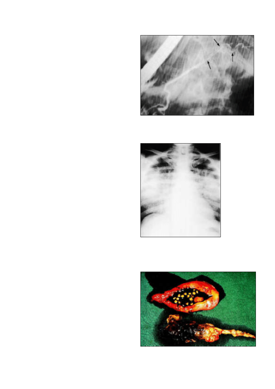
Treatment of acute attacks
Mild pancreatitis
Treatment of mild pancreatitis is supportive. Patients require
hospital admission, where they should receive intravenous
crystalloid fluids and appropriate analgesia and should stop all
oral intake. Most patients will require opiate analgesia, and
although this may cause spasm of the sphincter of Oddi, there
is no evidence that this affects the outcome of the disease. A
nasogastric tube may be helpful if vomiting is severe. Antibiotics
are of no benefit in the absence of coexisting infections.
Investigations are limited to the initial blood tests and
ultrasonography when gall stones are suspected. Most patients
will recover in 48-72 hours, and fluids can be restarted once
abdominal pain and tenderness are resolving.
Severe pancreatitis
Patients with severe pancreatitis should be admitted to a high
dependency or intensive care unit for close monitoring.
Adequate resuscitation of hypovolaemic shock (which is often
underestimated) remains the cornerstone of management, and
patients often require surprisingly large volumes of fluids over
the first 24-48 hours. Resuscitation is mainly with crystalloids,
but colloids may be required to restore circulating volume.
Progress is monitored by ensuring that urine output is adequate
( > 30 ml/h). Measurement of central venous or pulmonary
arterial pressure may be required, particularly in patients with
cardiorespiratory compromise. Patients who develop adult
respiratory distress syndrome and renal failure require
ventilation and dialysis.
The role of prophylactic antibiotics in severe pancreatitis
remains unclear, but recent randomised trials have shown a
marginal benefit with antibiotics that have good penetration
into pancreatic tissue (such as high dose cefuroxime and
imipenem).
Patients with severe gallstone pancreatitis and biliary sepsis
or obstruction benefit from endoscopic retrograde
cholangiopancreatography and removal of stones from the
common bile duct within the first 48 hours of admission.
However, the benefit of sphincterotomy is equivocal in patients
without biliary obstruction.
Despite intensive search, no effective drug has been
developed to prevent the development of severe pancreatitis.
Several new drugs including antagonists of platelet activating
factor (Lexipafant) and free radical scavengers that may limit
propagation of the cytokine cascade hold theoretical promise,
but initial clinical trials have been disappointing.
Patients who deteriorate despite maximum support pose a
difficult management problem. The possibility of infection in
the necrotic process should be considered, particularly when
deterioration occurs after the first week. Infection can usually be
confirmed by computed tomography guided fine needle
aspiration. Patients with infected pancreatic necrosis have a 70%
mortality and require surgical debridement (necrectomy). The
role of necrectomy in patients without infection is unclear .
Several new approaches are being investigated, including the
use of minimally invasive necrectomy and lavage and the use of
enteral rather than parenteral nutrition, which may reduce gut
permeability and bacterial translocation and limit infection in
the necrotic pancreas.
Prognosis
The overall mortality of patients with acute pancreatitis is
10-15% and has not changed in the past 20 years. The mortality
of mild pancreatitis is below 5% compared with 20-25% in
severe pancreatitis.
Figure 10.5 Ascaris lumbricoides in pancreatic duct: a rare cause of acute
pancreatitis
Figure 10.6 Chest radiograph of patient with adult
respiratory distress syndrome as a complication of
acute pancreatitis
Figure 10.7 Gall bladder and severe necrotic pancreas (necrectomy
specimen) removed from patient with acute pancreatitis induced by gall
stones
Acute pancreatitis
35

Long term management
Patients with gall stones are best treated by laparoscopic
cholecystectomy. This should ideally be done within the same
hospital admission after the acute episode has settled to prevent
recurrent attacks, which may be fatal. In high risk patients who
are considered unfit for surgery, an endoscopic sphincterotomy
will prevent most recurrent attacks.
Newer investigative techniques, including bile sampling and
analysis and endoscopic ultrasonography, are showing that
many patients with “idiopathic” pancreatitis have biliary
microlithiasis due to cholesterol crystals, biliary sludge, or small
stones that are missed by routine abdominal ultrasonography.
Early results confirm that laparoscopic cholecystectomy is
curative in most of these cases.
Box 10.4 Key points
x
Acute pancreatitis is a common cause of severe acute abdominal
pain and gall stones are the commonest cause in the United
Kingdom
x
Severity scoring should be used to identify patients at greatest risk
of complications
x
Treatment is mainly supportive
x
Patients with acute gallstone pancreatitis require early laparoscopic
cholecystectomy once the attack has settled
x
Biliary microlithiasis is increasingly recognised as a cause of
“idiopathic” pancreatitis
x
Mortality for acute pancreatitis is 10% overall but rises to 70% in
patients with infected severe pancreatitis
Further reading
Glazer G, Mann DV on behalf of working party of British Society of
Gastroenterology. United Kingdom guidelines for the management
of acute pancreatitis. Gut 1998;42(suppl 2)
A review of acute pancreatitis. Eur J Gastroenterol Hepatol
1997;9:1-120
Bradley EL. Complications of acute pancreatitis and their
management. In: Trede M, Carter DC, Longmire WP, eds. Surgery of
the pancreas.
Edinburgh: Churchill Livingstone, 1997:245-62
ABC of Liver, Pancreas, and Gall Bladder
36
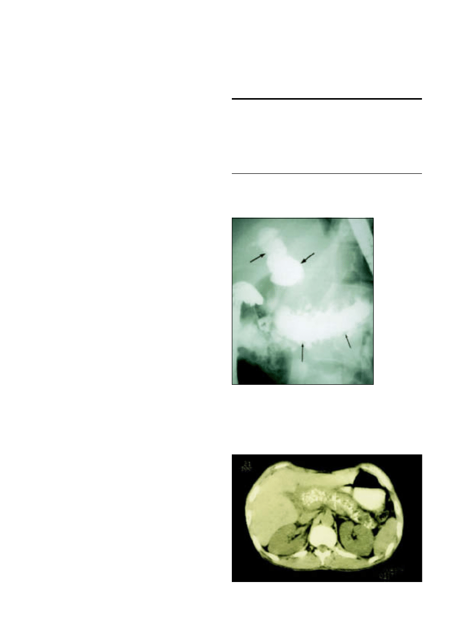
11 Chronic pancreatitis
P C Bornman, I J Beckingham
Chronic pancreatitis has an annual incidence of about one
person per 100 000 in the United Kingdom and a prevalence
of 3/100 000. In temperate areas alcohol misuse accounts for
most cases, and it mainly affects men aged 40-50 years. There is
no uniform threshold for alcohol toxicity, but the quantity and
duration of alcohol consumption correlates with the
development of chronic pancreatitis. Little evidence exists,
however, that either the type of alcohol or pattern of
consumption is important. Interestingly, despite the common
aetiology, concomitant cirrhosis and chronic pancreatitis is
rare.
In a few tropical areas, most notably Kerala in southern
India, malnutrition and ingestion of large quantities of cassava
root are implicated in the aetiology. The disease affects men
and women equally, with an incidence of up to 50/1000
population.
Natural course
Alcohol induced chronic pancreatitis usually follows a
predictable course. In most cases the patient has been drinking
heavily (150-200 mg alcohol/day) for over 10 years before
symptoms develop. The first acute attack usually follows an
episode of binge drinking, and with time these attacks may
become more frequent until the pain becomes more persistent
and severe. Pancreatic calcification occurs about 8-10 years after
the first clinical presentation. Endocrine and exocrine
dysfunction may also develop during this time, resulting in
diabetes and steatorrhoea. There is an appreciable morbidity
and mortality due to continued alcoholism and other diseases
that are associated with poor living standards (carcinoma of the
bronchus, tuberculosis, and suicide), and patients have an
increased risk of developing pancreatic carcinoma. Overall, the
life expectancy of patients with advanced disease is typically
shortened by 10-20 years.
Symptoms and signs
The predominant symptom is severe dull epigastric pain
radiating to the back, which may be partly relieved by leaning
forward. The pain is often associated with nausea and vomiting,
and epigastric tenderness is common. Patients often avoid
eating because it precipitates pain. This leads to severe weight
loss, particularly if patients have steatorrhoea.
Steatorrhoea presents as pale, loose, offensive stools that are
difficult to flush away and, when severe, may cause incontinence.
It occurs when over 90% of the functioning exocrine tissue is
destroyed, resulting in low pancreatic lipase activity,
malabsorption of fat, and excessive lipids in the stools.
One third of patients will develop overt diabetes mellitus,
which is usually mild. Ketoacidosis is rare, but the diabetes is
often “brittle,” with patients having a tendency to develop
hypoglycaemia due to a lack of glucagon. Hypoglycaemic coma
is a common cause of death in patients who continue to drink
or have had pancreatic resection.
Box 11.1 Aetiology of chronic pancreatitis
x
Alcohol (80-90%)
x
Nutritional (tropical Africa and Asia)
x
Pancreatic duct obstruction (obstructive pancreatitis)
Acute pancreatitis
Pancreas divisum
x
Cystic fibrosis
x
Hereditary
x
Idiopathic
Figure 11.1 Endoscopic retrograde
cholangiopancreatogram showing dilated common bile
duct (thick arrow) and main pancreatic ducts (thin
arrow) in patient with advanced chronic pancreatitis
Figure 11.2 Computed tomogram showing dilated pancreatic duct with
multiple calcified stones
37
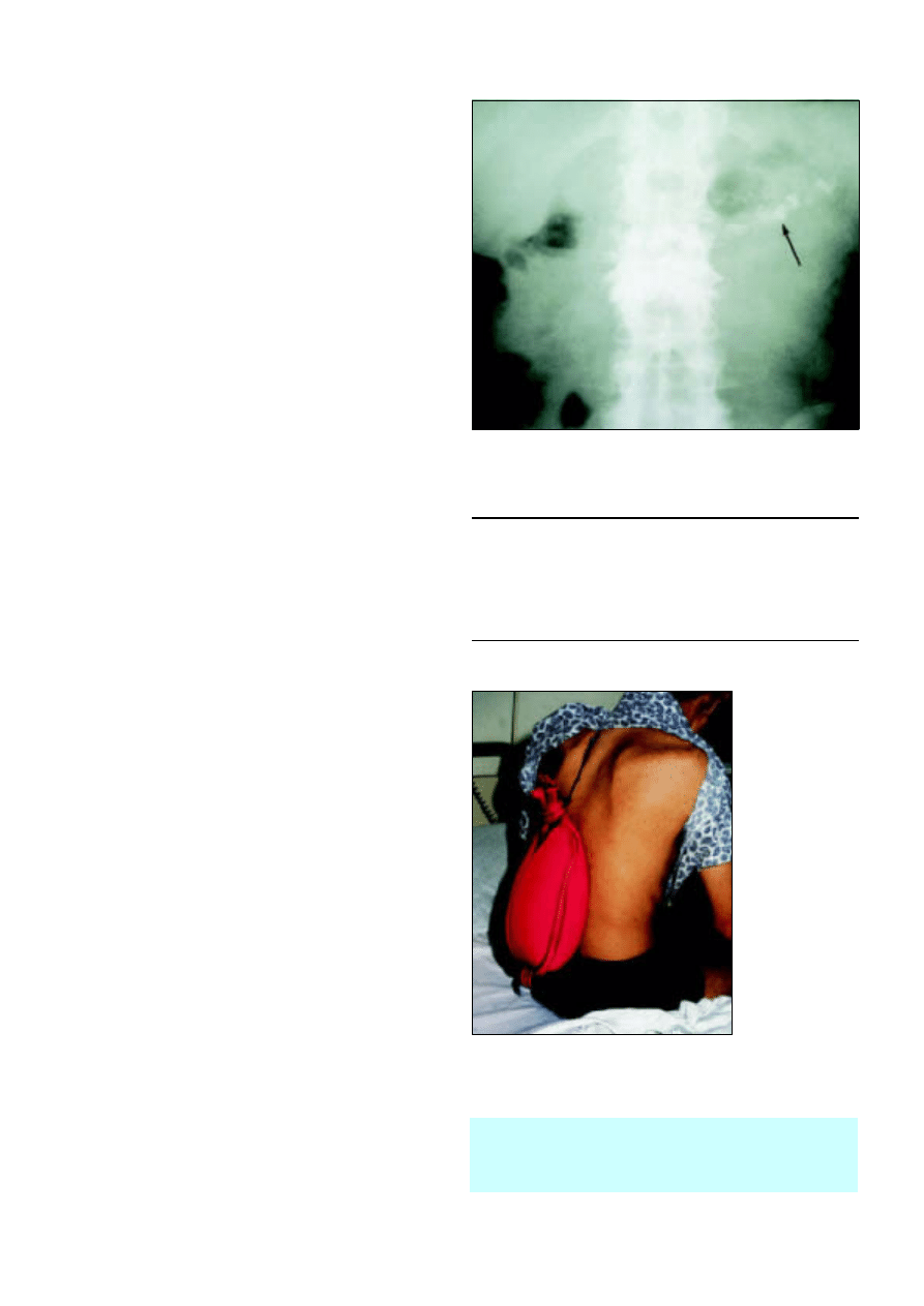
Diagnosis
Early diagnosis of chronic pancreatitis is usually difficult. There
are no reliable biochemical markers, and early parenchymal
and ductal morphological changes may be hard to detect. The
earliest signs (stubby changes of the side ducts) are usually seen
on endoscopic retrograde cholangiopancreatography, but a
normal appearance does not rule out the diagnosis. Tests of
pancreatic function are cumbersome and seldom used to
confirm the diagnosis. Thus, early diagnosis is often made by
exclusion based on typical symptoms and a history of alcohol
misuse.
In patients with more advanced disease, computed
tomography shows an enlarged and irregular pancreas, dilated
main pancreatic duct, intrapancreatic cysts, and calcification.
Calcification may also be visible in plain abdominal
radiographs. The classic changes seen on endoscopic
retrograde cholangiopancreatography are irregular dilatation of
the pancreatic duct with or without strictures, intrapancreatic
stones, filling of cysts, and smooth common bile duct stricture.
Treatment
Treatment is focused on the management of acute attacks of
pain and, in the long term, control of pain and the metabolic
complications of diabetes mellitus and fat malabsorption. It is
important to persuade the patient to abstain completely from
alcohol. A team approach is essential for the successful long
term management of complex cases.
Pain
Persistent or virtually permanent pain is the most difficult
aspect of management and is often intractable. The cause of the
pain is unknown. Free radical damage has been suggested as a
cause, and treatment with micronutrient antioxidants (selenium,
â
carotene, methionine, and vitamins C and E) produces
remission in some patients. However, further randomised trials
are required to confirm the efficacy of this approach. In the later
stages of disease pain may be caused by increased pancreatic
ductal pressure due to obstruction, or by fibrosis trapping or
damaging the nerves supplying the pancreas.
The mainstay of treatment remains abstinence from alcohol,
but this does not always guarantee relief for patients with
advanced disease. Analgesics should be prescribed with caution
to prevent narcotic dependency as many patients have addictive
personalities. Non-steroidal analgesics are the preferred
treatment, but most patients with ongoing and relentless pain
will ultimately require oral narcotic analgesics such as tilidine,
tramadol, morphine, or meperidine. Slow release opioid
patches (such as fentanyl) are increasingly used. Once this stage
is reached patients should be referred to a specialist pain clinic.
Use of large doses of pancreatic extract to inhibit pancreatic
secretion and reduce pain has unfortunately not lived up to
expectations. Likewise coeliac plexus blocks have been
disappointing, and it remains to be seen whether minimal
access transthoracic splanchnicectomy will be effective.
Steatorrhoea
Steatorrhoea is treated with pancreatic replacements with the
aim of controlling the loose stools and increasing the patient’s
weight. Pancreatic enzyme supplements are rapidly inactivated
below pH5, and the most useful supplements are high
concentration, enteric coated microspheres that prevent
deactivation in the stomach—for example, Creon or Pancrease.
A few patients also require H
2
receptor antagonists or dietary
fat restriction.
Box 11.2 Team for management of complex cases
x
General practitioner
x
Physician or surgeon with an interest in chronic pancreatitis
x
Dietician
x
Clinical psychologist
x
Chronic pain team
x
Diabetologist
Patients who do not gain weight despite adequate
pancreatic replacement therapy and control of diabetes
should be investigated for coexistent malignancy or
tuberculosis.
Figure 11.3 Plain abdominal radiograph showing multiple calcified stones
within the pancreatic duct
Figure 11.4 Patient using hot water bottle to relieve
back pain due to chronic pancreatitis
ABC of Liver, Pancreas, and Gall Bladder
38
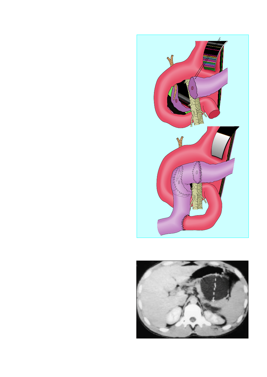
Diabetes mellitus
The treatment of diabetes is influenced by the relative rarity of
ketosis and angiopathy and by the hazards of potentially lethal
insulin induced hypoglycaemia in patients who continue to
drink alcohol or have had major pancreatic resection. It is thus
important to undertreat rather than overtreat diabetes in these
patients, and they should be referred to a diabetologist when
early symptoms develop. Oral hypoglycaemic drugs should be
used for as long as possible. Major pancreatic resection
invariably results in the development of insulin dependent
diabetes.
Endoscopic procedures
Endoscopic procedures to remove pancreatic duct stones, with
or without extracorporeal lithotripsy and stenting of strictures,
are useful both as a form of treatment and to help select
patients suitable for surgical drainage of the pancreatic duct.
However, few patients are suitable for these procedures, and
they are available only in highly specialised centres.
Surgery
Surgery should be considered only after all forms of
conservative treatment have been exhausted and when it is clear
that the patient is at risk of becoming addicted to narcotics.
Unless complications are present, the decision to operate is
rarely easy, especially in patients who have already become
dependent on narcotic analgesics.
The surgical strategy is largely governed by morphological
changes to parenchymal and pancreatic ductal tissue. As much
as possible of the normal upper gastrointestinal anatomy and
pancreatic parenchyma should be preserved to avoid problems
with diabetes mellitus and malabsorption of fat. The currently
favoured operations are duodenal preserving resection of the
pancreatic head (Beger procedure) and extended lateral
pancreaticojejunostomy (Frey’s procedure). More extensive
resections such as Whipple’s pancreatoduodenectomy and total
pancreatectomy are occasionally required. The results of
surgery are variable; most series report a beneficial outcome in
60-70% of cases at five years, but the benefits are often not
sustainable in the long term. It is often difficult to determine
whether failures are surgically related or due to narcotic
addiction.
Complications of chronic pancreatitis
Pseudocysts
Pancreatic pseudocysts are localised collections of pancreatic
fluid resulting from disruption of the duct or acinus. About 25%
of patients with chronic pancreatitis will develop a pseudocyst.
Pseudocysts in patients with chronic pancreatitis are less likely
to resolve spontaneously than those developing after an acute
attack, and patients will require some form of drainage
procedure. Simple aspiration guided by ultrasonography is
rarely successful in the long term, and most patients require
internal drainage. Thin walled pseudocysts bulging into the
stomach or duodenum can be drained endoscopically, with
surgical drainage reserved for thick walled cysts and those not
bulging into the bowel on endoscopy. Occasionally, rupture into
the peritoneal cavity causes severe gross ascites or, via
pleuroperitoneal connections, a pleural effusion.
Raised amylase activity in the ascitic or pleural fluid (usually
> 20 000 iu/l) confirms the diagnosis. Patients should be given
intravenous or jejunal enteral feeding to rest the bowel and
,,
,,
,,
Figure 11.5 Duodenal preserving resection of the pancreatic head (Beger
procedure). Top: pancreatic head resected. Bottom: reconstruction with
jejunal Roux loop
Figure 11.6 Large pseudocyst in patient with chronic pancreatitis. The cyst is
thin walled and bulging into the stomach and is ideal for endoscopic
drainage
Chronic pancreatitis
39
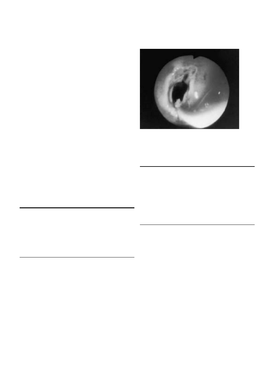
minimise pancreatic stimulation, somatostatin infusion, and
repeated aspiration. The cyst resolves in 70% of cases after two
to three weeks. Persistent leaks require endoscopic stenting of
the pancreatic duct or surgery to drain the site of leakage if it is
proximal or resection if distal.
Biliary stricture
Stenosis of the bile duct resulting in persistent jaundice (more
than a few weeks) is uncommon and usually secondary to
pancreatic fibrosis. The duct should be drained surgically, and
this is often done as part of surgery for associated pain or
duodenal obstruction. Endoscopic stenting is not a long term
solution, and is indicated only for relief of symptoms in high
risk cases.
Gastroduodenal obstruction
Gastroduodenal obstruction is rare (1%) and usually due to
pancreatic fibrosis in the second part of the duodenum. It is
best treated by gastrojejunostomy.
Splenic vein thrombosis
Venous obstruction due to splenic vein thrombosis (segmental
or sinistral hypertension) may cause splenomegaly and gastric
varices. Most thrombi are asymptomatic but pose a severe risk if
surgery is planned. Splenectomy is the best treatment for
symptomatic cases.
Gastrointestinal bleeding
Gastrointestinal bleeding may be due to gastric varices,
coexisting gastroduodenal disease, or pseudoaneurysms of the
splenic artery, which occur in association with pseudocysts.
Endoscopy is mandatory in these patients. Pseudoaneurysms
are best treated by arterial embolisation or surgical ligation.
Summary points
x
In most areas of the world alcohol is the main cause of chronic
pancreatitis
x
Early diagnosis is often difficult and relies on appropriate clinical
history and imaging
x
Stopping alcohol intake is essential to reduce attacks of pain,
preserve pancreatic function, and aid management of
complications
x
Patients often require opiate analgesics, and pain is best managed
in a multidisciplinary setting
x
Surgery should be reserved for patients with intractable pain or
with complications of chronic pancreatitis
Further reading
Beckingham IJ, Krige JEJ, Bornman PC, Terblanche J. Endoscopic
drainage of pancreatic pseudocysts. Br J Surg 1997;84:1638-45
Eckhauser FE, Colletti LM, Elta GH, Knol JA. Chronic pancreatitis. In:
Pitt HA, Carr-Locke DL, Ferrucci JT, eds. Hepatobiliary and pancreatic
disease. The team approach to management
. Boston: Little, Brown,
1995:395-412
Misiewicz JJ, Pounder RE, Venables CW, eds. Chronic pancreatitis. In:
Diseases of the gut and pancreas
. Oxford: Blackwell Science,
1994:441-54
Figure 11.7 Endoscopic drainage of pseudocyst: sphincterotome is
cutting a hole between stomach and pseudocyst wall
ABC of Liver, Pancreas, and Gall Bladder
40
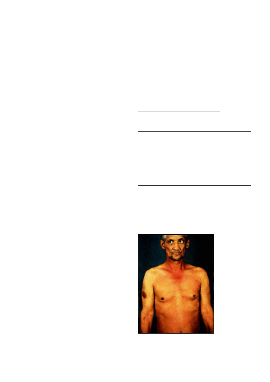
12 Pancreatic tumours
P C Bornman, I J Beckingham
Neoplasms of the pancreas may originate from both exocrine
and endocrine cells, and they vary from benign to highly
malignant. Clinically, 90% of pancreatic tumours are malignant
ductal adenocarcinomas, and most of this article concentrates
on this disease.
Ductal adenocarcinoma
Incidence and prognosis
Carcinoma of the pancreas has become more common in most
Western countries over the past three decades, and although
there is evidence of plateauing in some countries such as the
United States, it still ranks as the sixth commonest cause of
cancer death in the United Kingdom. Most patients are over the
age of 60 years (80%) and many will have concurrent medical
illnesses that complicate management decisions, particularly
because the median survival from diagnosis is less than six
months.
Clinical presentation
Two thirds of pancreatic cancers develop in the head of the
pancreas, and most patients present with progressive,
obstructive jaundice with dark urine and pale stools. Pruritus,
occurring as a result of biliary obstruction, is often troublesome
and rarely responds to antihistamines. Back pain is a poor
prognostic sign, often being associated with local invasion of
tumours. Severe cachexia, as a result of increased energy
expenditure mediated by the tumour, is also a poor prognostic
indicator. Cachexia is the usual presenting symptom in patients
with tumours of the body or tail of the pancreas.
Examination
The commonest sign is jaundice, with yellowing of the sclera
and, once the bilirubin concentration exceeds 35 ìmol/l, the
skin. Many patients with high bilirubin concentrations will have
skin scratches associated with pruritus. Patients with advanced
disease have severe weight loss accompanied by muscle wasting
and occasionally an enlarged supraclavicular lymph node. A
palpable gall bladder suggests pancreatic malignancy, but it can
be difficult to detect when displaced laterally or covered by an
enlarged liver. The presence of ascites or a palpable epigastric
mass usually indicates end stage disease. Full assessment of the
patient’s general fitness is essential to develop an individualised
management plan.
Investigation
Because of the poor prognosis, care should be taken not to
overinvestigate or embark on treatment strategies based on the
unrealistic expectations of patients, their families, or the
referring doctor. An increasing number of investigations are
available, and the aim is to select patients who will not benefit
from major resection by use of the fewest, least invasive, and
least expensive means. The choice of investigation will vary
according to local availability, particularly of newer
investigations such as laparoscopic and endoscopic
ultrasonography, and it remains to be seen if these techniques
offer major advantages over the latest generation of computed
tomography and magnetic resonance imaging scanners. Early
cooperation between a gastroenterologist, radiologist, and
Box 12.1 Types of pancreatic neoplasms
x
Benign exocrine
Serous cyst adenoma
Mucinous cyst adenoma
x
Malignant exocrine
Ductal adenocarcinoma
Mucinous cyst adenocarcinoma
x
Endocrine
Gastrinoma
Insulinoma
Other
Box 12.2 Factors predicting poor prognosis
x
Back pain
x
Rapid weight loss
x
Poor performance status—for example, World Health Organization
or Karnofsky scoring systems
x
Ascites and liver metastases
x
High C reactive protein and low albumin concentrations
Box 12.3 Rarer presentations of pancreatic carcinoma
x
Recurrent or atypical venous thromboses (thrombophlebitis
migrans)
x
Acute pancreatitis
x
Late onset diabetes mellitus
x
Upper gastrointestinal bleeding
Figure 12.1 Patient with jaundice, bruising, and
weight loss due to pancreatic carcinoma
41
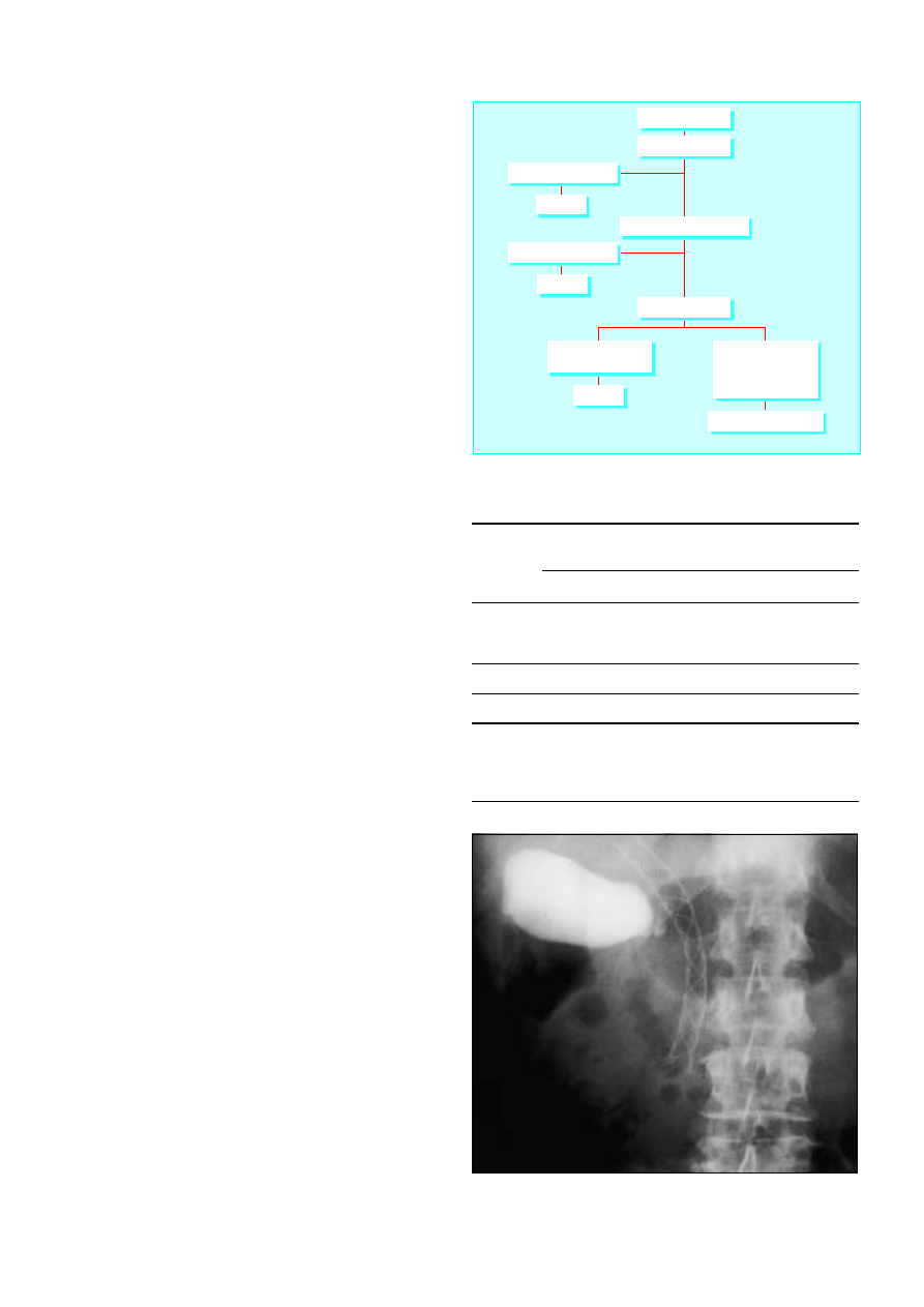
surgeon should avoid inappropriate investigations and
treatment that might interfere with patients’ quality of life.
Endoscopic retrograde cholangiopancreatography is an
important investigation in patients with obstructive jaundice. As
well as showing biliary and pancreatic strictures, the pathology
can be confirmed by taking brushings for cytology or biopsy
specimens of the duct for histology. The technique can also be
used to place a stent to relieve biliary obstruction. However, it is
important not to use this approach before patients are properly
selected for treatment.
The diagnosis can also be confirmed by fine needle
aspiration guided by ultrasonography or computed
tomography, but this investigation has a high rate of false
negative results and is rarely necessary. Fine needle aspiration
should be avoided in patients with potentially resectable
tumours as it can cause seeding and spread of the tumour.
Treatment
Surgical resection does not improve survival in patients with
locally advanced or metastatic disease. Tumour stage and the
patient’s fitness for major surgical resection are the main factors
in determining optimal treatment.
Resectable tumours
Surgical resection, usually a pancreaticoduodenectomy
(Whipple’s procedure), is the only hope for cure. Less than 15%
of tumours are suitable for resection. Very few tumours of the
body and tail are resectable (3%) as patients usually present late
with poorly defined symptoms.
The outcome of resection has been shown to be better in
specialised pancreatobiliary centres that perform the procedure
regularly than in small units. Mortality has fallen to 5-10% in
dedicated units. The overall five year survival rate of 10-15%
after resection remains disappointing, although survival is as
high as 20-30% in some subgroups such as patients with small
( < 2 cm), node negative tumours. Furthermore, the median
survival of patients who have resection is 18 months compared
with six months for patients without metastatic disease who do
not have resection.
Preoperative biliary drainage remains controversial. The
reduced complications from resolution of jaundice are offset by
more inflammatory tissue at surgery and higher rates of biliary
sepsis after stenting. Ideally patients with minimal jaundice
should be operated on without stenting whereas those with
higher bilirubin concentrations ( > 100 ìmol/l) probably benefit
from endoscopic stenting and reduction of bilirubin
concentrations before surgery.
Locally advanced disease
Several options are available for the 65% of patients who have
locally advanced disease. These depend on factors such as age,
disease stage, and the patient’s fitness. Endoscopic insertion of a
plastic or metal wall stent relieves jaundice in most patients.
Plastic stents are cheaper but have a median half life of three to
four months compared with six months for metal stents.
Blockage of a stent results in rigors and jaundice, and patients
should be given antibiotics and have the stent replaced.
Surgical exploration and bypass should be used in
patients who are predicted to survive longer than six months,
in whom it is not certain that the tumour cannot be resected,
in areas with limited access to endoscopic retrograde
cholangiopancreatography, or with recurrent stent blockage or
obstruction of the gastric outlet. Surgical bypass (open or
laparoscopic) now has a low mortality and has the advantage of
long term palliation of jaundice with a low risk of recurrence.
Table 12.1 Treatment of pancreatic ductal carcinoma
Fitness
of
patient
Tumour stage
Resectable
Locally
advanced
Metatstatic
Low risk
Pancreatoduodectomy
Surgical
bypass or
endoscopic
stent
Palliative
care + /
-
endoscopic
stent
High risk
Endoscopic stent
Endoscopic
stent
Palliative
care
Box 12.4 Tumours suitable for resection
x
< 4 cm in diameter
x
Confined to pancreas
x
No local invasion or metastases
Obstructive jaundice
Ultrasonography
Spiral computed tomography
Laparoscopy
Peritoneal metastases
liver metastases
Resectable tumour
no metastases
no local invasion
no vascular involvement
Pancreatoduodenectomy
Liver metastases, ascites
Liver metastases, ascites
Palliation
Palliation
Palliation
Palliation = endoscopic stent, surgical bypass, or medical
palliation alone depending on patient's general health and symptoms
Figure 12.2 Investigation and management of pancreatic ductal carcinoma
Figure 12.3 Metal wall stent in common bile duct of patient with pancreatic
carcinoma. (Note contrast in gall bladder from endoscopic retrograde
cholangiopancreatography)
ABC of Liver, Pancreas, and Gall Bladder
42
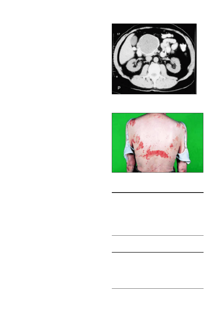
Metastatic disease
Patients with metastatic disease are often cachectic and rarely
survive more than a few weeks. Treatment should focus on
alleviation of pain and improving quality of life with input from
palliative care teams. Patients with less advanced metastatic
disease may require endoscopic stenting, especially if they have
intractable pruritus.
Radiotherapy and chemotherapy
Despite numerous trials, radiotherapy with or without
chemotherapy has not been shown to prolong survival. The
search for new chemotherapeutic and immunotherapeutic drugs
continues, but they currently have little role outside clinical trials.
Symptom control
A liberal policy of pain control with paracetamol, non-steroidal
anti-inflammatory drugs, and opiate analgesics should be
followed. In difficult cases and when increasingly large doses of
opiates are required, patients should be referred to a specialist
pain clinic for consideration of coeliac plexus block or
thoracoscopic splanchnicectomy. Early referral to the palliative
care team and Macmillan nurses, who can bridge the gap
between hospital and community care, is beneficial.
Cachexia is an important cause of disability in many
patients. Nutritional supplementation rarely combats weight
loss, and pancreatic replacement therapy is also of doubtful
benefit. Encouraging results have recently been reported with
polyunsaturated fatty acids (fish oil) and non-steroidal
anti-inflammatories, which seem to inhibit the inflammatory
response provoked by the tumour and reduce the speed of
weight loss with some survival benefit. Impaired gastric
emptying is generally underdiagnosed and may be functional or
mechanical in origin.
Cystic tumours
These rare tumours (1% of all pancreatic neoplasms) are mostly
benign, but the mucinous type (about 50%) is premalignant.
They are important because they occur predominantly in young
women and usually have a good prognosis when resected.
Cystic tumours may be mistaken for benign pseudocysts,
although they can usually be differentiated on the basis of
history and computed tomographic findings (tumours have
septa within the cyst and calcification of the rim of the cyst wall
without calcification in the rest of the pancreas). If the diagnosis
is in doubt, surgical resection with frozen section at the time of
definitive surgery is the optimal management.
Endocrine tumours
Tumours arising from the islets of Langerhans can produce
high concentrations of the hormones normally produced by the
islets (insulin, glucagon, somatostatin, etc) or non-pancreatic
hormones (such as gastrin or vasoactive peptide). Endocrine
tumours are rare, with an incidence of around 1-2 per million
population. The commonest forms are gastrinoma and
insulinoma. They may be sporadic or occur as part of the
multiple endocrine neoplasia syndrome, when they are
associated with tumours of the pituitary, parathyroid, thyroid,
and adrenal glands.
Patients usually present with a clinical syndrome produced
by hormonal excess, typically of a single peptide. With the
exception of insulinomas most endocrine tumours are
malignant. Treatment is by surgical excision, and survival is
generally good; 10 year survival rates for patients with
malignant lesions are around 50%.
Summary points
x
6000 people die from pancreatic cancer each year in the United
Kingdom
x
Presentation is usually with painless insidious jaundice
x
Median survival from diagnosis is less than six months
x
Less than 15% of all pancreatic tumours are resectable, and five
year survival after resection is 10-15%
x
Endoscopic retrograde cholangiopancreatography and surgical
biliary drainage offer good palliation of jaundice
x
Cystic and endocrine pancreatic tumours are uncommon but have
a better prognosis
Further reading
Trede M, Carter DC. Surgical options for pancreatic cancer. In: Surgery
of the pancreas
. Edinburgh: Churchill Livingstone, 1997:383-515
Cameron JL, Grochow LB, Milligan FD, Venbrux AC. Pancreatic
cancer. In: Pitt HA, Carr-Locke DL, Ferrucci JT, eds. Hepatobiliary and
pancreatic disease—the team approach to management
. Boston: Little
Brown, 1995:475-86
Cotton P, Williams C. ERCP and therapy. In: Practical gastrointestinal
endoscopy
. Oxford: Blackwell Science, 1996:167-86
Figure 12.4 Cystic tumour in head of pancreas with calcified rim
Figure 12.5 Necrolytic erythema migrans is pathognomic in patients with
glucagonoma
Pancreatic tumours
43
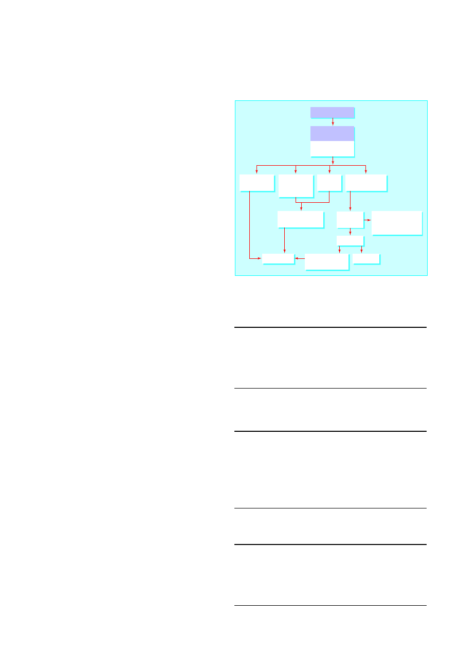
13 Liver and pancreatic trauma
I J Beckingham, J E J Krige
The liver is the most commonly injured solid intra-abdominal
organ, but injuries to the pancreas are fairly rare. The primary
goal in the treatment of severe abdominal injuries is to preserve
life, and management is divided into four sequential phases:
resuscitation, evaluation, initial management, and definitive
treatment.
Liver trauma
Liver trauma constitutes a broad spectrum of injuries. The
magnitude of the injury, the management requirements, and
the complexity of the surgical repair are determined by the
extent, anatomical location, and mechanism of injury. Blunt
liver trauma is usually due to road traffic accidents, assaults, or
falls from heights, and results in deceleration injuries with
lacerations of liver tissue from shearing stresses. High velocity
projectiles, close range shotgun injuries, and crushing blunt
trauma cause fragmentation of the hepatic parenchyma with
laceration of vessels and massive intraperitoneal haemorrhage.
Penetrating injuries such as stab or gunshot wounds cause
bleeding without much devitalisation of the liver parenchyma.
Resuscitation
Resuscitation follows standard advanced trauma life support
principles: maintenance of a clear airway, urgent fluid
resuscitation, ventilatory and circulatory support, and control of
bleeding. Effective venous access should be obtained and
volume replacement started immediately. The patient’s blood is
grouped and crossmatched, and blood samples should be sent
for urgent analysis of haemoglobin concentration, white cell
count, blood gas pressures, and urea, creatinine, and electrolyte
concentrations. Patients should also have a nasogastric tube and
urinary catheter inserted.
A liver injury should be suspected in patients with evidence
of blunt trauma or knife or gunshot wounds to the right upper
quadrant or epigastrium. Occasionally physical signs may be
minimal, and gunshot entry and exit wounds can be deceptively
distant from the liver. Diagnosis may be difficult in patients who
are not fully conscious or have head or spinal cord injuries. The
insidious onset of shock in a multiply injured or unconscious
patient can easily be missed.
Evaluation
The most important decision after initial resuscitation is
whether urgent surgery is needed. Patients who respond to fluid
resuscitation and remain stable can be observed closely,
investigated, and re-evaluated. Patients who remain shocked
after 3 litres of intravenous fluid usually have continued
bleeding and require urgent laparotomy. Surgery should not be
delayed to obtain the results of special investigations.
Computed tomography of the abdomen is useful in
haemodynamically stable patients suspected of having a major
liver injury. Patients with large intrahepatic haematomas or
limited capsular tears who have small volumes of blood in the
peritoneal cavity can be treated non-operatively. They must,
however, be observed carefully and have repeated physical
examination.
Box 13.1 Clinical features of serious liver injury
x
Hypovolaemic shock:
x
Hypotension
x
Tachycardia
x
Decreased urine output
x
Low central venous pressure
x
Abdominal distension
Box 13.2 Criteria for non-operative management of liver
injuries
x
Haemodynamically stable after resuscitation
x
No persistent or increasing abdominal pain or tenderness
x
No other peritoneal injuries that require laparotomy
x
< 4 units of blood transfusion required
x
Haemoperitoneum < 500 ml on computed tomography
x
Simple hepatic parenchymal laceration or intrahepatic haematoma
on computed tomography
Box 13.3 Indications for laparotomy
x
Stab or gunshot wounds that have penetrated the abdomen
x
Signs of peritonitis
x
Unexplained shock
x
Evisceration
x
Uncontrolled haemorrhage
x
Clinical deterioration during observation
Abdominal injury
Resuscitation
Evaluation
Investigation
Management
Rapid completion of
basic investigations
Laparotomy
Unstable, bleeding,
or peritonitis
Stable
Diagnostic
studies
Observe
Chest radiography
Ultrasonography
Computed tomography
Exsanguinating
haemorrhage
Hypovolaemia
and abdominal
distention
Signs of
peritonitis
Stable no bleeding
or peritonitis
Figure 13.1 Management of major abdominal trauma
44
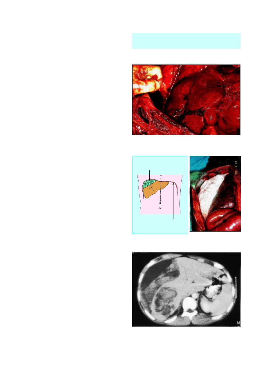
Surgical management
Most liver injuries are simple and can be treated relatively easily.
Complex lesions need to be diagnosed early and may require
major surgery by an experienced hepatobiliary surgeon.
Operative approach
Patients should be prepared from the sternal notch to the pubis
so that the incision can be extended into the chest if more
proximal control of the vena cava or aorta is needed. A midline
incision is used, and the first step is to remove blood and clots
and control active bleeding from liver lacerations by packing.
Care should be taken to avoid sustained periods of
hypotension, and it is important to restore the patient’s
circulating blood volume during surgery. Any perforations in
the bowel should be rapidly sutured to minimise contamination.
Most liver injuries have stopped bleeding spontaneously by
the time of surgery. These wounds do not require suturing but
should be drained to prevent bile collections. Liver bleeding can
usually be stopped by compressing the liver with abdominal
packs while experienced surgical and anaesthetic help is
summoned.
If visibility is obscured by continued bleeding, the hepatic
artery and portal vein should be temporarily clamped with a
vascular clamp to allow accurate identification of the site of
bleeding. If bleeding cannot be stopped the area should be
packed; absorbable gauze mesh can be wrapped around an
injured lobe and sutured to maintain pressure and tamponade
bleeding. The abdomen is then closed without drainage and the
packing removed under general anaesthesia two to three days
later. Packing is also used if a coagulopathy develops or to allow
the patient to be transferred to a tertiary referral unit for
definitive management.
Patients with blunt injuries associated with substantial
amounts of parenchymal destruction may require resectional
debridement. Rarely, a severe crushing injury necessitates a
formal hepatic lobectomy. The most difficult problems are
lacerations of the vena cava and major hepatic veins behind the
liver because temporary clamping of the inflow vessels does not
slow blood loss from hepatic veins. Advanced operative
techniques including total hepatic vascular isolation by
clamping the portal vein and the vena cava above and below the
liver or lobectomy may be required.
Postoperative complications
Rebleeding from the injury, bile leaks, ischaemic segments of
liver, and infected fluid collections are the main postoperative
complications associated with liver trauma. Angiography (with
selective embolisation) is useful if recurrent arterial bleeding or
haemobilia occur. Computed tomography is used to define
intra-abdominal collections, which are best drained by
ultrasound guided needle aspiration or a percutaneous catheter.
Subhepatic sepsis develops in about a fifth of cases and is
usually related to bile leaks, ischaemic tissue, undrained
collections, or bowel injury. The site of bile leaks is best
identified by endoscopic retrograde cholangiopancreatography
and treated by endoscopic sphincterotomy or stenting, or both.
Prognosis
The overall mortality after liver injury is 10-15% and depends
largely on the type of injury and the extent of injury to other
organs. Only 1% of penetrating civilian wounds are lethal,
whereas the mortality after blunt trauma exceeds 20%. The
mortality after blunt hepatic injury is 10% when only the liver is
injured. If three major organs are injured, however, mortality
approaches 70%. Bleeding causes more than half of the deaths.
The priorities of surgery are to stop haemorrhage, remove
dead or devitalised liver tissue, and ligate or repair
damaged blood vessels and bile ducts.
Figure 13.2 Stellate fracture of right lobe of liver
Diaphragm
Packing
Figure 13.3 Packing of bleeding liver
Figure 13.4 Large intrahepatic haematoma in patient with blunt trauma
Liver and pancreatic trauma
45
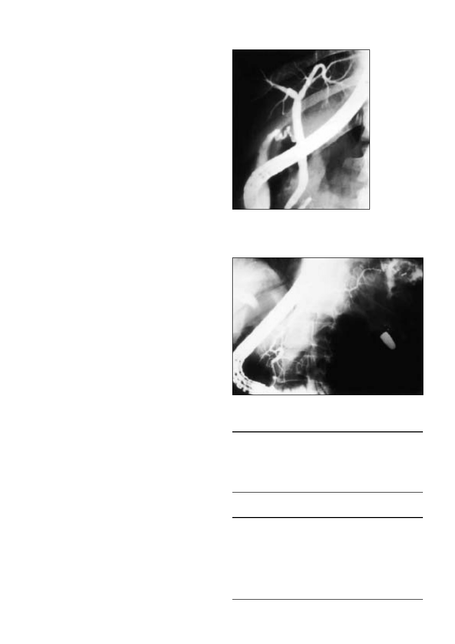
Pancreatic trauma
Injuries to the pancreas are uncommon and account for 1-4%
of severe abdominal injuries. Most pancreatic injuries occur in
young men. Blunt trauma to the pancreas and duodenum
usually results from road traffic accidents, when an unrestrained
driver is thrown on to the steering wheel. Handlebars may
inflict similar injuries to motorcyclists or children on bicycles.
Deceleration injury during blunt trauma to the epigastrium may
transect the neck of the pancreas over the vertebral bodies. The
deep location of the pancreas means that considerable force is
needed to cause an injury, and patients often have damage to
surrounding organs, including the liver, spleen, stomach,
duodenum, and colon. Penetrating injuries may also damage
the portal vein or inferior vena cava.
Diagnosis
Blunt trauma to the pancreas may be clinically occult and
parenchymal and duct injury may not be recognised during
initial evaluation or even surgery. A high index of suspicion is
therefore necessary in patients with severe upper abdominal
injuries. Abdominal radiographs may show retroperitoneal air
and a ruptured duodenum. Serum amylase activity is a poor
indicator. It can be normal in patients with severe pancreatic
damage and increased in patients with no demonstrable injury
to the pancreas. Contrast enhanced computed tomography is
the best investigation. Features of pancreatic injury include
pancreatic oedema or swelling and fluid collections within or
behind the peritoneum or in the lesser peritoneal sac.
Endoscopic retrograde cholangiopancreatography can be use
to assess the integrity of the main ducts in stable patients.
Intraoperative evaluation
Pancreatic injury is usually diagnosed at laparotomy. The injury
may be obvious in patients with a major fracture of the body or
neck of the pancreas or associated duodenal injury. Other clues
to pancreatic injury are retroperitoneal bile staining, fat
necrosis, peripancreatic oedema, or subcapsular haematoma.
The pancreas should be fully mobilised and exposed to rule out
a duct injury. Failure to detect a major injury to the pancreatic
duct is an important cause of postoperative morbidity.
Haemodynamically stable patients with minor injuries of the
body or tail of the pancreas who have no visible damage to the
duct can be managed by external drainage of the injury site
with soft silastic drains. Major injuries of the body and tail of the
pancreas that affect the duct should be treated by distal
pancreatectomy. Patients with injuries to the head of the
pancreas without devitalisation of pancreatic tissue can be
managed by external drainage provided that any associated
duodenal injury is amenable to simple repair.
Pancreatoduodenectomy as a primary procedure is
restricted to stabilised patients with disruption of the ampulla of
Vater or major devitalising injuries of the pancreatic head and
duodenum. Injuries of this severity occur after blunt trauma or
gunshot wounds but are uncommon with stab wounds.
Complications
Pancreatic fistulas are the most common complication and
generally close spontaneously. However, if a serious duct injury
is present, a chronic fistula may develop and require surgical
intervention. Pseudocysts that follow pancreatic injury are
usually the result of inadequate drainage of pancreatic injury or
failure to recognise a major ductal injury. They can be treated
by percutaneous drainage guided by ultrasonography or
computed tomography if the pancreatic injury was minor.
However, patients who have major duct injury may require
internal drainage or pancreatic resection.
Further reading
Feliciano DV, Lewis CA. Hepatic trauma. In: Pitt H, Carr-Locke DL,
Ferrucci R, eds. Hepatobiliary and pancreatic disease. Boston: Little,
Brown, 1994:107-24
Pachter HL, Hofsetter SR. The current status of nonoperative
management of adult blunt hepatic injuries. Am J Surg
1995;169:442-54.
Krige JEJ, Bornman PC, Beningfield SJ, Funnell I. Pancreatic trauma.
In: Pitt H, Carr-Locke DL, Ferrucci R, eds. Hepatobiliary and
pancreatic disease
. Boston: Little, Brown, 1994:421-36
Box 13.4 Complications associated with pancreatic trauma
x
Intra-abdominal abscess
x
Wound infection
x
Pancreatic fistula
x
Pseudocyst
x
Pancreatic abscess or ascites
x
Acute or chronic pancreatitis
Figure 13.5 Endoscopic retrograde
cholangiopancreatograph showing injury to pancreatic
duct with obstruction in the pancreatic neck
Figure 13.6 Pancreatic leak caused by a gunshot wound. The bullet is also
visible
ABC of Liver, Pancreas, and Gall Bladder
46
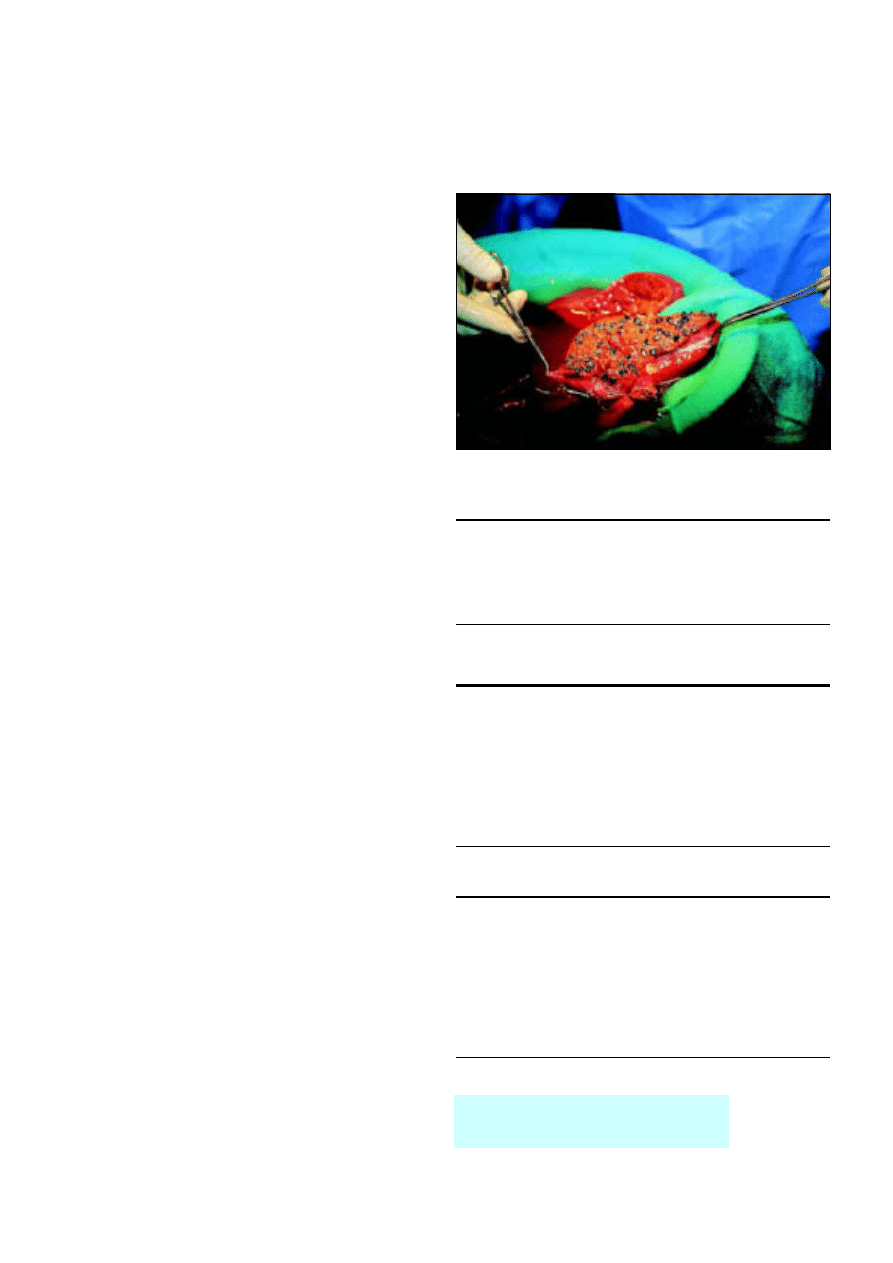
14 Transplantation of the liver and pancreas
K R Prasad, J P A Lodge
Liver transplantation is carried out for many chronic liver
diseases and for fulminant hepatic failure. The United Kingdom
has seven liver transplantation units, which perform 600-700
transplantations a year. Activity is limited by availability of donor
organs, and there are around 200 patients waiting for a liver
transplant at any one time. Transplantation of the pancreas is less
well established. The pancreas is usually transplanted together
with a kidney in patients with end stage diabetes mellitus and
renal failure. Worldwide, around 1000 patients (mainly in the
United States) receive a pancreatic transplant each year. Only
20-30 a year are transplanted in the United Kingdom.
Liver transplantation
Indications and contraindications
Hepatocellular carcinoma complicates many chronic liver
diseases, and a small tumour is not a contraindication to
transplantaton because tumour recurrence is uncommon in
these patients. However, most patients with large ( > 5 cm) or
multiple hepatomas or most other types of cancer are not
considered for transplantation as tumours recur rapidly.
Patients with certain rare tumours, such as liver metastases from
neuroendocrine disease and sarcomas, can do well for several
years. Contraindications to liver transplantation include
extrahepatic malignancy, severe cardiopulmonary disease,
systemic sepsis, and an inability to comply with regular drug
treatment.
Timing and selection of patients for transplantation
The preoperative status of the patient is one of the most
important factors predicting the outcome after transplantation.
Patients with chronic liver disease and signs of decompensation
should be assessed for transplantation before they become
critically ill. In certain diseases, such as primary biliary cirrhosis,
quality of life issues may form the basis for indication for
transplantation. For example, chronic lack of energy can be
debilitating in patients with biliary cirrhosis.
Acute liver failure and timing of transplantation
Liver transplantation greatly improves the prognosis of patients
with fulminant liver failure. In the United Kingdom paracetamol
overdose is now the commonest cause of acute liver failure,
followed by seronegative (non-A, non-B, non-C) hepatitis.
The mortality from fulminant liver failure can be as high as
90%, whereas one year survival after urgent transplantation is
often above 70%. In the United Kingdom, criteria developed at
King’s College Hospital are used for listing patients for “super
urgent” transplantation. This scheme relies on cooperation
between the liver transplantation centres to allow
transplantation within 48 hours of listing whenever possible.
Surgical procedure
Before organs are removed an exploratory laparotomy is done
on the donor to rule out any disease process (such as
unexpected carcinoma) that may preclude organ donation. The
major vessels are then dissected and blood flow controlled in
preparation for hypothermic perfusion with a cold preservation
solution. University of Wisconsin preservation solution is used
most widely. It can preserve the liver adequately for about 13
hours, with acceptable results up to 24 hours.
Box 14.1 Common indications for liver transplantation
x
Primary biliary cirrhosis
x
Primary sclerosing cholangitis
x
Cryptogenic cirrhosis
x
Chronic active hepatitis (usually secondary to hepatitis B and C)
x
Alcoholic liver disease (after a period of abstinence)
Box 14.2 Signs of decompensation in chronic liver disease
x
Tiredness
x
Ascites
x
Encephalopathy
x
Peripheral oedema
x
Jaundice (not always a feature)
x
Spontaneous bacterial peritonitis—abdominal pain (a late sign)
x
Bleeding oesophageal or gastric varices
x
Low albumin concentration
x
Raised prothrombin time
Box 14.3 Paracetamol overdose
x
Causes death by acute liver failure
x
Renal failure develops as a hepatorenal syndrome and by acute
tubular necrosis but is usually recoverable
x
Early deaths usually result from raised intracranial pressure, and
comatose patients require monitoring in an intensive care unit
x
Death in later stages can occur from multiorgan failure and
systemic sepsis
x
If the patient survives without transplantation, the liver will recover
without the development of cirrhosis
The donor organ is usually procured as
part of a multiorgan retrieval from a
heart beating, brain dead patient
Figure 14.1 Donor liver from adult cut down for insertion into child
recipient
47
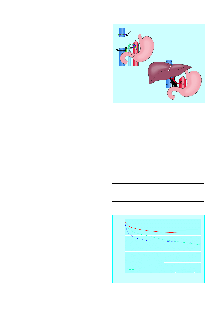
Hepatectomy in the organ recipient is the most difficult part
of the operation as the patient is at risk of developing a serious
haemorrhage due to a combination of portal hypertension,
defective clotting, and fibrinolysis. Improvements in surgical
technique and anaesthesia have resulted in large reductions in
blood loss, and the average requirement for transfusion is now
four units of blood. At reimplantation, the suprahepatic and
infrahepatic inferior vena cava and the portal vein are
anastomosed and the organ is reperfused with blood. This is
followed by reconstruction of the hepatic artery and bile duct.
Postoperative management
Patients are usually managed in an intensive care unit for the first
12-24 hours after surgery. Enteral feeding is restarted as early as
possible, and liver function tests are done daily.
Immunosuppressive protocols usually include a combination of
cyclosporin or tacrolimus together with azathioprine or
mycophenolate mofetil and prednisolone. The dose of steroids is
rapidly tapered off, and they can often be stopped after two to
three months. The doses of cyclosporin or tacrolimus are
reduced gradually during the first year (during which pregnancy
should be avoided) and continued at much lower levels for life.
Acute rejection occurs in about half of patients, but this is
easily treated in most cases with extra steroids or by altering the
drug regimen. Despite routine use of prophylactic treatment
against bacterial, viral, and fungal pathogens, infections remain
a major cause of morbidity. The side effects of the drugs are
usually well controlled before the patient leaves hospital about
two weeks after surgery.
At discharge, patients need to be familiarised with the drug
regimen and side effects and educated about the warning signs
of rejection and infection. Patients are usually followed up
weekly for the first three months and then at gradually
increasing intervals thereafter.
Results
The five year survival is 60-90%, depending on the primary
disease and the clinical state of the patient before
transplantation. The newer antiviral drugs plus the preoperative
and postoperative adjuvant therapies for malignancies should
lead to further improvements in survival. Although alcoholic
liver disease remains a controversial indication for
transplantation, carefully selected patients do well.
After successful transplantation patients have a greatly
improved lifestyle and are often able to return to work and
normal social activities. However, some patients experience
medical and social problems. Drug compliance is one of the
biggest problems after all types of organ transplantation. Poor
compliance leads to chronic rejection and loss of the graft.
An extensive network of support services is available to help
liver transplant patients. These include the transplant team,
referring physician, general practitioner, social services, and
local liver patient support groups. Shared care protocols
operate in most regions, with most patients cared for primarily
by their general practitioner and a gastroenterologist at their
local hospital. The mainstay of follow up is regular liver
function tests to detect any dysfunction of the transplant.
Regular discussion of concerns with the transplant team is
essential, and many problems can be sorted out by telephone.
Paediatric liver transplantation
In children, the most common indication for liver
transplantation is biliary atresia, often after failure to respond to
a portoenterostomy. Most children who need a liver transplant
are young (under 3 years) and small ( < 20 kg). Size matched
donors are in short supply, and reduced size (“cut down”) and
Table 14.1 Side effects of immunosuppresive drugs
Drug
Side effect
Monitoring
Cyclosporin
Neurotoxicity, nephrotoxicity,
hypertension, hirsutism, gum
hyperplasia, diabetes
Drug
concentrations
Tacrolimus
Nephrotoxicity, neurotoxicity,
hair loss, hypertension,
diabetes
Drug
concentrations
Azathioprine
Leucopenia, hair loss
White blood cell
count
Mycophenolate
mofetil
Gastrointestinal upset,
leucopenia
White blood cell
count and
gastrointestinal
symptoms
Steroids
Osteoporosis, diabetes,
cushingoid face, hypertension
Symptoms
General
Infections, malignancy
Liver and renal
function tests,
regular follow up,
and high index of
suspicion
Clamp on inferior vena cava
After recipient hepatectomy
After implantation of donor liver
Figure 14.2 Implantation of liver transplant after hepatectomy
100
90
80
70
60
50
40
30
20
10
0
0
1
2
Chronic liver failure (1673)
Fulminant hepatic failure (383)
Other (745)
Recipient primary disease (No at risk at day 0)
3
4
5
6
7
8
9
10
11
12
% sur
vival
No of months since transplant
Figure 14.3 One year survival after first liver transplant according to
primary disease, United Kingdom 1985-94
ABC of Liver, Pancreas, and Gall Bladder
48
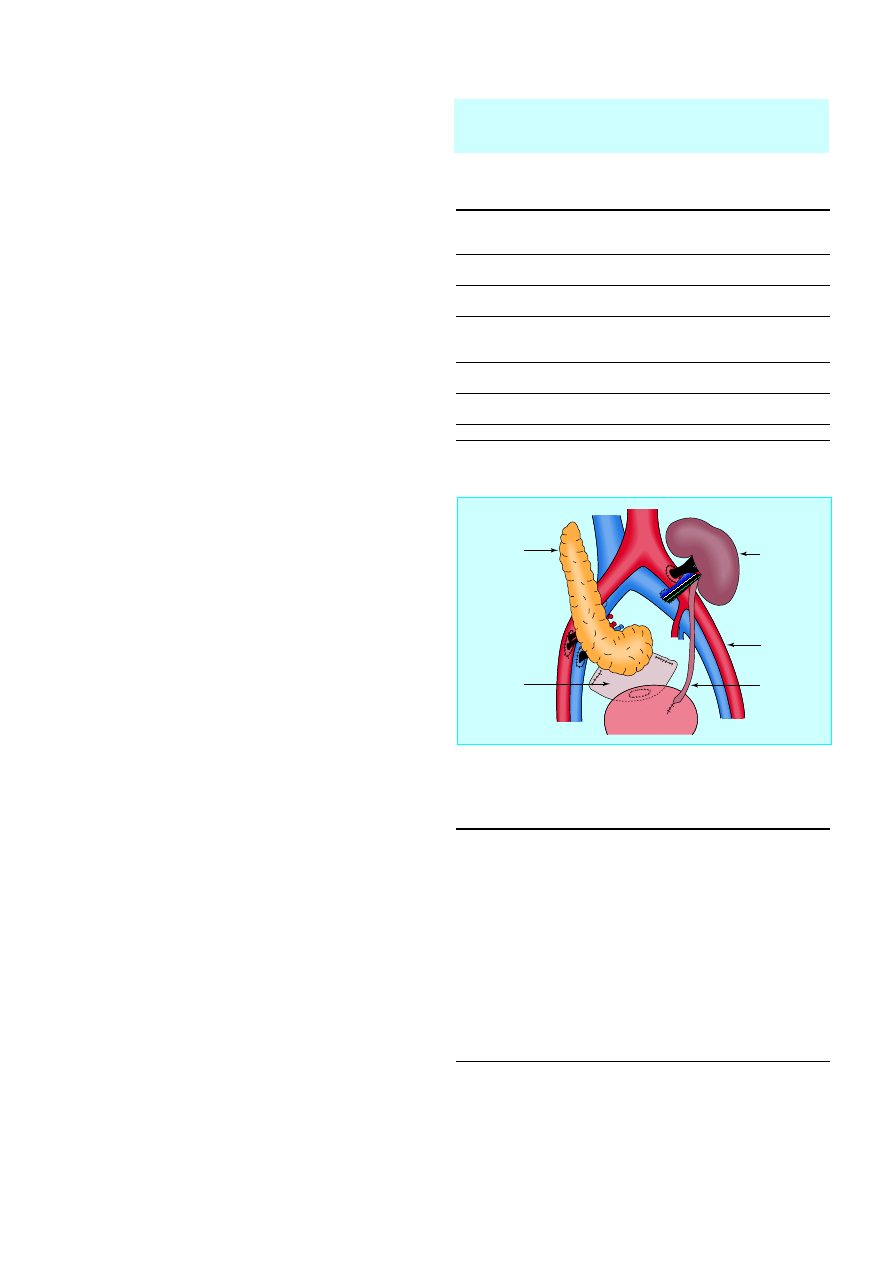
split (where one liver is split between two recipients) liver
techniques have been used to overcome this problem. Donation
of the left lobe of the liver by a living relative is also possible.
Pancreatic transplantation
The goals of transplantation of the pancreas are to eliminate the
morbidity associated with labile blood glucose concentrations,
stabilise or improve secondary diabetic complications, and
improve the quality of life of patients with diabetes mellitus by
restoring normal glucose metabolism. The stabilisation of
diabetic control, the avoidance of exogenous insulin, and the
ability to return to a normal diet for the first time since childhood
are indisputable benefits of this procedure.
The selection of recipients for pancreatic transplantation is
crucial. The magnitude of the surgery and need for long term
immunosuppression means that whole organ transplantation is
currently reserved for patients with end stage disease.
Recipients are typically young ( < 50 years) with type 1 diabetes
and end stage renal disease but without untreatable peripheral
vascular or coronary artery disease. Simultaneous
transplantation of the pancreas and kidney is the commonest
procedure. Separate transplantation of the pancreas after
kidney transplantation increases the chances of getting a good
HLA matching for the kidney and allows a kidney to be
donated by a living relative. The presence of immunosuppression
at the time of implantation of the pancreas is also advantageous.
The transplanted pancreas is usually placed in the pelvis and
anastamosed to the iliac vessels, with the pancreatic duct
anastomosed to the bladder or a loop of small bowel.
First year mortality is 3-10% in large units, with most deaths
due to overwhelming sepsis. Transplant survival is 86% for the
kidney and 70% for the pancreas. Successful transplantation
greatly improves quality of life, and most patients are fully
rehabilitated. Glucose homoeostasis seems to be excellent after
pancreatic transplantation. Patients can stop exogenous insulin
treatment and have normal glycated haemoglobin
concentrations and glucose tolerance test results within three
months of transplantation.
The long term effect on diabetic complications will not be
known for several years, but recent results are encouraging.
Evidence that diabetic nephropathy does not recur in the
kidney transplant is accumulating, but there is no evidence for
amelioration of established glomerular lesions in native kidneys.
Improvements in autonomic and peripheral neuropathy have
been documented. Further studies are needed to examine the
potential for reducing the rate of progression of retinopathy
and macrovascular disease.
Isolated pancreatic islet transplantation
A more logical approach is to attempt to prevent the
development of the irreversible complications of diabetes by
improving blood glucose metabolism at an early stage.
Transplantation of pancreatic islet cells has been studied
extensively as an alternative to whole organ grafting and has
several theoretical and practical advantages. Pancreatic islets can
be isolated by using collagenase digestion to separate the
endocrine from the exocrine tissues and purified by density
gradient separation. Some difficulties remain, particularly with
the purification stage. The islets are injected into the recipient
liver via the portal vein or by subcapsular injection into the
kidney or spleen. Rejection of the islets remains a problem, and
the success rates of this type of transplantation have been poor
in the clinical setting.
The shortage of child liver donors has been partly
resolved by using smaller sections of adult livers, usually
the left lobe
Table 14.2 Types of pancreatic transplantation
Type
Indication
Simultaneous pancreas and kidney
transplant (SPK)
Diabetic renal failure
Pancreas after kidney transplant (PAK)
After successful kidney
transplant
Pancreas transplant alone (PTA)
Prerenal failure, unstable
diabetic control, severe
neuropathy
Segmental (transplantation of
pancreatic tail)
Applicable to live donation
Multivisceral (pancreas transplanted
with liver and sometimes small bowel)
For example, extensive
abdominal tumour
Isolated pancreatic islets
The future solution?
Summary points
x
Hepatitis C cirrhosis is the commonest worldwide indication for
liver transplantation
x
Alcoholic liver disease remains a controversial indication for liver
transplantation but carefully selected patients do well
x
Patients with chronic liver disease and signs of decompensation
should be assessed for transplantation before they become critically
ill
x
Drug compliance is an important problem, with poor compliance
leading to chronic rejection and graft loss
x
Paracetamol overdose is the commonest cause of acute liver failure
in the United Kingdom and accounts for 5% of all liver transplants
in Britain
x
Pancreas transplantation is most commonly performed for patients
with end stage diabetes mellitus and renal failure
The photo of donor liver was obtained from J L Martha/Science Photo
Library.
Donor
pancreas
Donor
duodenum
Iliac artery and
vein
Donor kidney
Ureter
Bladder
Figure 14.4 Simultaneous transplantation of pancreas and kidney with
bladder drainage
Transplantation of the liver and pancreas
49

This Page Intentionally Left Blank

abscesses see liver abscesses
acetylcysteine 16
acute physiological and chronic health
evaluation (APACHE) score 34
acute tubular necrosis 24
adenocarcinomas 41–3
adenomas, liver cell 4, 26
adrenal gland tumours 43
adult respiratory distress syndrome 30, 35
advanced trauma life support 44
aflatoxin 26
alanine transaminase 2
acute hepatitis, 9, 10
acute pancreatitis 33
chronic hepatitis 12
liver abscesses 29
albendazole 32
albumin 2
see also hypoalbuminaemia
alcohol consumption
acute pancreatitis 33, 34
ascites 22, 23
chronic pancreatitis 37, 38
cirrhosis 1, 18
hepatitis 1
liver transplantation 48
alkaline phosphatase 2, 9
hydatid disease 32
liver abscesses 29, 30
primary biliary cirrhosis 17
alkalosis, hypochloraemic 23
alpha
1
-antitrypsin deficiency 3
alpha-fetoprotein 26, 27
aminoglycosides 24, 30
amiodarone 16
amoebiasis 30–31
ampulla of Vater trauma 46
amylase
acute pancreatitis 34
pancreatic pseudocyst 39
pancreatic trauma 46
anaemia
hepatitis 9
liver abscesses 29, 30
prehepatic jaundice 1
tribavirin therapy 14
anaphylactic shock 4, 31
angiography 45
antimitochondrial antibodies 3, 17
antinuclear antibodies 15
antismooth muscle antibodies 15, 17
APACHE score 34
appendicitis 29
arthralgia 15
Ascaris lumbricoides 5, 35
ascites
cirrhosis 22
diuretics 23
hepatocellular carcinoma 22, 26
hepatorenal syndrome 24
pancreatic carcinoma 41
portal hypertension 18, 22–3
ruptured pancreatic pseudocyst 39
spontaneous bacterial peritonitis 24
Aspergillus flavus 26
atelectasis 31
autoantibodies 3, 15
azathioprine 15, 48
azotaemia 23, 24
back pain 41
Bacteroides spp. 29, 30
Beger procedure 39
benzodiazepine antagonists 24
beta blockers 20, 21
beta-carotene 38
beta-glucuronidase 5
bile ducts
see also common bile ducts
cystic dilatation (Caroli’s disease) 25
obstruction 3, 16–17, 41, 42
parasites 1
strictures 1, 3, 40
bile leaks 45
bile salts 5
biliary atresia 48–9
biliary colic 5
bilirubin 1–2, 5
see also hyperbilirubinaemia
bilirubinuria 2
blood transfusion
hepatitis C 11, 12
liver transplantation 48
bone disease 2
C reactive protein 34
CA19-9 tumour marker 17
cachexia 41, 43
caeruloplasmin 15
calcium 2
calcium bilirubinate 5
cardiac failure 22
Caroli’s disease 25
cefuroxime 35
cephalosporins 6, 30
cerebral oedema 10
ceruloplasmin 3
Charcot’s triad 6
chemotherapy 43
chenodeoxycholic acid 5, 8
Child-Pugh classification 18
Chinese herbal medicine 16
cholangiocarcinoma 3, 17
cholangiography, percutaneous transhepatic
(PTC) 3, 17
cholangitis 2
acute 6
Charcot’s triad 6
gall stones 6
hydatid disease 31
liver abscesses 29
primary sclerosing 3, 17
sclerosing 1
cholecystectomy 7
acute cholecystitis 6
common bile duct stones 8
gallstone pancreatitis 36
laparoscopic 7, 36
postoperative pain 7
cholecystitis
acute 5–6
chronic 5
emphysematous 3
intrahepatic 1–2
viral hepatitis 9
cholesterol 5
cholic acid 5
chronic liver disease
decompensation 47
hepatorenal syndrome 24
cirrhosis 3
alcoholic 1, 18
ascites 22
autoimmune hepatitis 15
gall stones 5
hepatic encephalopathy 23
hepatitis B infection 13
hepatocellular carcinoma 26–7
hepatorenal syndrome 24
iron overload 15
liver biopsy 4
portal hypertension 18
portal hypertensive gastropathy 21
primary biliary 1, 3, 17, 37
spontaneous bacterial peritonitis 24
variceal bleeding 18
viral 18
cisplatin 27
coagulation factors 2–3, 18
coeliac block 43
colorectal carcinoma 27
common bile duct
laparoscopic injuries 7
stones 1, 2, 8
strictures 38
computed tomography 3
acute pancreatitis 33, 34
chronic pancreatitis 37, 38
hydatid disease 31
intra-abdominal collections 45
liver abscesses 30
liver trauma 44
liver tumours 25, 26, 27
pancreatic carcinoma 41
pancreatic trauma 46
copper deposition 3, 15–16
Couinaud’s segmental anatomy of liver 27
51
Index

Creon 38
crush injuries 44
cyanoacrylate monomer 21
cyclosporin 48
cystadenocarcinomas 25
cystadenomas 25
cysts
bile ducts 25
hydatid 4, 25, 31–2
infective 25
liver 3, 25
pancreatic 38
cytomegalovirus 11
deceleration injuries 44, 46
Denver peritoneovenous shunt 23
diabetes mellitus
see also hypoglycaemia
chronic pancreatitis 37, 38, 39
iron overload 15
liver abscesses 29, 30
nephropathy 49
pancreatic transplantation 47, 49
diaphragm 29, 31
diloxanide furoate 31
disseminated intravascular coagulation 30
diuretics 23
diverticulitis 29
doxorubicin 27
drug-related conditions
hepatic encephalopathy 24
hepatitis 16
jaundice 1, 2
liver failure 16
dysentery 30
Echinococcus granulosus 31
embolisation 27, 45
emphysema 3
encephalitis 9
encephalopathy, hepatic 23–4
fulminant hepatitis 10
hepatocellular carcinoma 24, 26
varices 18, 20
endocarditis, bacterial 29
endoscopic retrograde
cholangiopancreatography (ERCP) 3
bile leaks 45
cholestatic non-obstructive jaundice 17
chronic pancreatitis 37, 38
common bile duct stones 8
gallstone pancreatitis 35
hydatid disease 31–2
liver abscesses 30
pancreatic carcinoma 42
pancreatic trauma 46
primary sclerosing cholangitis 17
endoscopy
emergency 19–20
gastric varices 21
oesophageal varices 19–20
pancreatic duct stones 39
pseudocysts 39, 40
Entamoeba histolytica 30–31
enterococci 29, 30
eosinophilia 31
epigastric pain
acute pancreatitis 33
chronic pancreatitis 37
differential diagnosis 5
Epstein-Barr virus 11
erythema migrans, necrolytic 43
Escherichia coli 5, 29, 30
ethanolamine oleate 19
extracorporeal shockwave lithotripsy 8
fat malabsorption 37, 38, 39
fat necrosis 46
fentanyl 38
ferroxidase 3, 15
fibrolamellar carcinoma 27
fine needle aspiration 42
fish oil 43
fistulae 46
flubendazole 32
focal nodular hyperplasia 26
Frey’s procedure 39
gall stones 5–8
acute pancreatitis 6, 33, 34, 35, 36
biliary colic 5
black pigment 5
brown pigment 5
calcified 3
cholangitis 6
cholesterol 5
cirrhosis 5
common bile duct 1, 2, 8
disease course 6–7
extrahepatic biliary obstruction 16–17
gallbladder cancer 7
gallstone ileus 6
management 7–8
oral dissolution therapy 8
pancreas 38
pancreatic ducts 39
ultrasound 3, 5, 6
gallbladder cancer 7
gamma-glutamyl transferase 2
hepatitis 9, 12
hydatid disease 32
primary biliary cirrhosis 17
gastrin 43
gastrinomas 43
gastroduodenal obstruction 40
gastrointestinal haemorrhage
hepatic encephalopathy 24
splenic vein thrombosis 40
gastrojejunostomy 40
gastropathy, portal hypertensive 21
Gilbert’s syndrome 1
Glasgow scoring system 34
glucagon 43
glyceryl trinitrate 19
Grey-Turner’s sign 33
gunshot wounds 44, 46
haemangiomas 25–6
haematomas
intrahepatic 44, 45
subcapsular 4, 46
haemobilia 45
haemochromatosis 3, 15
hepatocellular carcinoma 26
haemorrhage
gastrointestinal see gastrointestinal
haemorrhage; varices/variceal bleeding
intraperitoneal 44
haemostasis 45
haloperidol 18
hepatic coma 23
hepatic vein lacerations 45
hepatitis
acute 2, 9–11
acute viral, 9–11
alcoholic 1
anaemia 9
autoimmune 3, 15
chronic active 3
chronic viral 12–14
cytomegalovirus 11
diagnosis, 10–11
drug-related 16
Epstein-Barr virus 11
fulminant (acute liver failure) 10
liver biopsy 4, 13, 14
management 11
seronegative (non-A, non-B, non-C) 47
vertical transmission 12
viral 1, 9–11, 12–14
hepatitis A 9, 10, 11
hepatitis B 9, 10, 11
chronic 12–13
cirrhosis 13
hepatocellular carcinoma 13, 26
surface antigen 10, 12–13
hepatitis C 9, 11
chronic 12, 14
hepatocellular carcinoma 26
hepatitis D 9, 11
hepatitis E 9, 10, 11
hepatocellular carcinoma 26–7
ascites 22, 26
cirrhosis 26–7
hepatic encephalopathy 24, 26
hepatitis B 13, 26
hepatitis C 26
liver transplantation 27, 47
seeding 4
hepatomegaly 3, 9
hepatorenal syndrome 18, 24
herniae 22
HIV/AIDS 29
hydatid disease 4, 25, 31–2
hyperbilirubinaemia 1
amoebic liver abscesses 30
hydatid disease 32
pancreatic carcinoma 41
hypoalbuminaemia
ascites 22
liver abscesses 29, 30
hypochloraemic alkalosis 23
hypofibrinogenaemia 25
hypoglycaemia
see also diabetes mellitus
acute hepatitis 10
chronic pancreatitis 37, 39
hypokalaemia
Index
52

diuresis 23
hepatic encephalopathy 24
hyponatraemia 23
hypovolaemic shock 35
IgA 3
IgG 3, 15
IgM 3, 10, 17
ileus, gallstone 6
imipenem 35
immunodeficiency states 30
immunoglobulins 3, 15
immunosuppression
liver abscesses 29
liver transplantation 48
pancreatic transplantation 49
inflammatory bowel disease 15, 17
insulin 43, 49
insulinomas 43
interferon alpha
hepatitis B 11, 13
hepatitis C 14
international normalised ratio (INR) 2
islets of Langerhans 43
transplantation 49
jaundice 1
autoimmune hepatitis 15
bile duct strictures 40
cholestatic non-obstructive 16–17
common bile duct stones 8
drug-related 16
gall stones 6
hepatic (parenchymal) 1–2
hepatocellular carcinoma 26
hydatid disease 31
liver abscesses 29
pancreatic carcinoma 1, 41, 42
posthepatic (obstructive) 1–2, 41, 42
prehepatic 1
primary biliary cirrhosis 17
primary sclerosing cholangitis 17
viral hepatitis 9, 10
Kayser-Fleischer ring 15
kidney transplantation 49
Klebsiella spp. 5, 29, 30
knife wounds 44
Knodell score 14
lactitol 24
lactulose 24
lamivudine 13
laparoscopic cholecystectomy 7, 36
laparotomy 44, 46
lecithin 5
Lexipafant 35
lipase 34
lipiodol 27
liver
abscesses see liver abscesses
biopsy see liver biopsy
cysts 3, 25
(see also cysts)
enzymes 2
failure see liver failure
function tests 2
lobectomy 45
packing 45
polycystic 25
regeneration 27
resection 27–8
segmental anatomy 27
stellate lobar fracture 45
transplantation see liver transplantation
trauma 44–5
liver abscesses 3, 6, 25, 29–31
amoebic 20–31
cryptogenic 29
drainage 30, 31
pyogenic 29–30
rupture 31
liver biopsy 4
cirrhosis 4
focal nodular hyperplasia 26
hepatitis B 13
hepatitis C 14
liver cell adenomas 26
percutaneous 4
primary biliary cirrhosis 17
primary sclerosing cholangitis 17
transjugular 4
tumour seeding 26
liver cell adenomas 4, 26
liver failure
autoimmune hepatitis 15
Child-Pugh classification 18
drug-related 16
fulminant 16, 47
fulminant hepatitis 10
hepatorenal syndrome 24
liver function tests 2
liver transplantation 47–9
acute rejection 48
alcoholic liver disease 48
ascites 23
children 48–9
chronic liver disease 47
contraindications 47
fulminant liver failure 47
hepatic encephalopathy 23
hepatocellular carcinoma 27, 47
hepatorenal syndrome 24
infection 48
liver metastases 47
living donor left lobe 49
paracetamol overdose 16, 47
portal hypertension 21
primary biliary cirrhosis 17, 37
primary sclerosing cholangitis 17
results 48
seronegative hepatitis 47
split liver 48–9
super urgent 47
surgical procedure 47–8
varices 21
lobar fracture, stellate 45
lobectomy 45
lymphadenopathy 9
magnetic resonance
cholangiopancreatography 3–4, 8, 17
magnetic resonance imaging 3–4, 25–6, 41
meperidine 38
metastatic tumours 4, 16–17, 27, 43, 47
methionine 38
methyltetrabutyl ether 8
metronidazole 30, 31
Minnesota balloon tube 19–20
Mirrizi’s syndrome 6
mono-octanoin 8
morphine 38
multiple endocrine neoplasm syndrome 43
multiple organ dysfunction syndrome 33, 34
mycophenolate mofetil 48
myocarditis 9
necrectomy 35
nephropathy, diabetic 49
nephrotoxic drugs 24
non-steroidal anti-inflammatory drugs
chronic pancreatitis 38
gall stones 6
hepatorenal syndrome 24
pancreatic carcinoma 43
5`-nucleotidase 2
octreotide 19
oedema
cerebral 10
pancreatic 34, 46
peripheral 23
oesophageal transection 20–21
Opisthorchis senensis 5
oral contraceptives 26
pain
back pain 41
epigastric see epigastric pain
pancreatitis 38
postoperative 7
pancreas
carcinoma 41–43
cystic tumours 43
cysts 38
endocrine tumours 43
fistulae 46
gall stones 38
imaging 3
islet cells see islets of Langerhans
necrosis 33, 34
oedema 34, 46
pseudocysts see pseudocysts, pancreatic
resection 42
transplantation 47, 49
trauma 46
Pancrease 38
pancreatectomy
distal 44
total 39
pancreatic carcinoma
ascites 41
back pain 41
biliary obstruction 41, 42
chronic pancreatitis 37
metastic disease 43
posthepatic jaundice 1, 41, 42
pancreatic ducts
Index
53

adenocarcinomas 41–3
chronic pancreatitis 38
stones 39
trauma 36
pancreatic enzyme supplements 38
pancreatic head, duodenal preserving
resection (Beger procedure) 39
pancreaticoduodenectomy 39, 42, 46
pancreaticojejunostomy 39
pancreatitis, acute 33–6
alcohol-induced 33, 34
biliary microlithiasis 36
biliary sepsis 35
common bile duct stones 8
gallstones 6, 33, 34, 35, 36
hepatitis B infection 9
necrectomy 35
scoring systems 34
pancreatitis, chronic 1, 37–40
alcohol misuse 37, 38
calcification 3, 37, 38
pain 38
pancreatic carcinoma 37
pseudocysts 39–40
splenic vein thrombosis 40
steatorrhoea 37, 38
pancreatitis, oedematous interstitial 33
paracentesis 23
diagnostic 22, 24
paracetamol poisoning 16, 43, 47
parathyroid tumours 43
penicillamine 16
penicillin 30
percutaneous transhepatic cholangiography
(PTC) 3, 17
pericarditis 9
perinuclear antineutrophil cytoplasmic
antibody 17
peripancreatic tissue
necrosis 33, 34
oedema 46
peritoneovenous shunts 23
peritonitis
amoebic 31
spontaneous bacterial 24
phosphorus 2
pituitary tumours 43
plasma proteins 2–3
pleural effusion 9, 31, 39
polycystic liver disease 25
polymyalgia 15
polyneuritis 9
polyunsaturated fatty acids 43
portal hypertension 18–24
ascites 18, 22–3
cirrhosis 18
hepatic encephalopathy 23–4
hepatorenal syndrome 24
varices 18–21
portal hypertensive gastropathy 21
portal vein thrombosis 18, 20–21
portal venous gas 3
portocaval shunts 20
portoenterostomy 48
portosystemic collateral circulation 18
portosystemic shunts 20, 23
praziquantel 32
prednisolone 15, 48
prerenal failure 24
primary biliary cirrhosis 1, 3, 17, 37
primary sclerosing cholangitis 3, 17
Pringle manoeuvre 28
proteolytic enzymes 33
prothrombin time 10
pruritis 17, 41, 43
pseudoaneurysms 40
pseudocysts, pancreatic 1, 34, 39–40
pancreatic trauma 46
ruptured 39
radiofrequency ablation 27
radiography 3
acute pancreatitis 34
chronic pancreatitis 38
liver abscesses 29, 31
pancreatic trauma 46
radiotherapy 43
Ranson’s scoring system 34
renal failure
acute pancreatitis 35
hepatic encephalopathy 24
pancreatic transplantation 47, 49
resuscitation 44
retinopathy, diabetic 49
retroperitoneal space
bile staining 46
necrosis 33
rheumatoid arthritis 15
rheumatoid factor 9
road traffic accidents 44, 46
schistosomiasis 18
scintigraphy 26
sclerotherapy 19
selenium 38
serum:ascites albumin gradient 22
shotgun injuries 44
sickle cell disease 1, 5
Sjögren’s syndrome 17
sodium tetradecyl sulphate 19
sodium, dietary 22, 23
somatostatin 40, 43
somatostatin analogues 19
spherocytosis 1, 5
sphincter of Oddi division (sphincterotomy)
8
bile leaks 45
gallstone pancreatitis 35, 36
spironolactone 23
splanchnicectomy, thoracoscopic 43
splenectomy 40
splenic artery pseudoaneurysms 40
splenic vein thrombosis 20–21, 40
splenomegaly 1, 9, 40
stab wounds 44
staphylococci 29
steatorrhoea 37, 38
stents 3
acute cholangitis 6
bile leaks 45
common bile duct stones 8
pancreatic carcinoma 42, 43
streptococci 29, 30
tacrolimus 48
terlipressin 19, 21
thalassaemia 1, 5
thrombocytopenia 25
thyroid disease 15
thyroid tumours 43
tilidine 38
total hepatic vascular isolation 45
tramadol 38
transjugular intrahepatic portosystemic shunt
20, 21, 23
tribavirin 14
trientene 16
tubular necrosis, acute 24
tumour embolisation 27
tumour seeding 4, 26
ulcerative colitis 15, 17
ultrasound 3
acute pancreatitis 34, 35
ascites 22
cholestatic non-obstructive jaundice
16–17
common bile duct stones 8
cystic liver lesions 3, 25
gall stones 3, 5, 6
hepatocellular carcinoma 26
hepatomegaly 3
liver abscesses 29–30, 31
liver biopsy 4
pancreatic carcinoma 41
ursodeoxycholic acid 8, 17
urticaria 31
varices/variceal bleeding 18–21
balloon tube tamponade 19–20
band ligation 19
cirrhosis 18
endoscopy 19–20, 21
gastric 21, 40
hepatic encephalopathy 18, 20
hepatitis B infection 13
hepatocellular carcinoma 26
liver transplantation 21
oesophageal 18–21
portal hypertension 18–21
sclerotherapy 19
splenic vein thrombosis 40
vasoactive peptide 43
vasopressin 19
vena cava lacerations 45
vitamin C 38
vitamin E 38
vitamin K 2
vitamin K
1
(phytomenadione) 18
Whipple’s pancreaticoduodenectomy 39, 42,
46
Wilson’s disease 3, 15–16
xanthoma 17
zinc acetate 16
Index
54
Document Outline
- Cover
- Contents
- Preface
- 1. Investigation of liver and biliary disease
- 2. Gallstone disease
- 3. Acute hepatitis
- 4. Chronic viral hepatitis
- 5. Other causes of parenchymal liver disease
- 6. Portal hypertension - 1. Varices
- 7. Portal hypertension - 2. Ascites, encephalopathy, and
- 8. Liver tumours
- 9. Liver abscesses and hydatid disease
- 10 Acute pancreatitis
- 11. Chronic pancreatitis
- 12. Pancreatic tumours
- 13. Liver and pancreatic trauma
- 14. Transplantation of the liver and pancreas
- Index
Wyszukiwarka
Podobne podstrony:
effect of high fiber vegetable fruit diet on the activity of liver damage and serum iron level in po
ABC Investigation of liver and biliary disease
ABC Of Arterial and Venous Disease
ABC Of Occupational and Environmental Medicine
ABC Of Conflict and Disaster
ABC Of Arterial and Venous Disease
ABC Of Occupational and Environmental Medicine
ABC Investigation of liver correct
ABC Liver abscesses and hydaid disease
Insensitive Semantics~ A Defense of Semantic Minimalism and Speech Act Pluralism
Estimation of Dietary Pb and Cd Intake from Pb and Cd in blood and urine
Development of Carbon Nanotubes and Polymer Composites Therefrom
ABC of Spinal Cord Injury 4th Ed
ABC Of Antenatal Care
ABC Of Sexually Transmitted Infections
więcej podobnych podstron