www.wjpps.com
Vol 4, Issue 2, 2014.
465
Kumar et al. World Journal of Pharmacy and Pharmaceutical Sciences
PHYTOCHEMICAL INVESTIGATION AND EVALUATION OF
ANTIMICROBIAL AND ANTITUBERCULAR ACTIVITY OF
KUNSTLERIA KERALENSIS
Kumar M.D
*1
, Sathishkumar shetty A
1
, Sathyanarayan N.D
2
, Vijay kumar M.L
3
,
Kuppast I.J
4
and Vasanthkumar Pai K
5
1
Department of Pharmaceutical Chemistry, NES Academy of Research and Development,
NES Campus, Shivamogga-577201, Karnataka, India.
2
Department of Pharmaceutical chemistry, Kuvempu university, P.G-centre, Kadur,
Chikkamangalore district, Karnataka, India.
3
Department of Microbiology, National College of Pharmacy, Balaraj Urs Road,
Shivamogga-577201, Karnataka, India.
4
Department of Pharmacology, National College of Pharmacy, Balaraj Urs Road,
Shivamogga-577201, Karnataka, India.
5
Department of Industrial chemistry, Kuvempu university, Shankarghatta, Shvamogga-
577201, Karnataka, India.
ABSTRACT
The hexane, chloroform and methanolic extract of bark and leaf of the
plant“Kunstleria keralensis” belonging to the family Fabaceae were
screened for antimicrobial and antitubercular activity. The
antimicrobial activity was carried against five target bacteria and five
target fungi by disc diffusion method. The hexane extract (HB),
chloroform extract (CB) of bark and hexane extract of leaf (HL)
showed
significant
antibacterial
activity
against
Klebsiella
pneumoniae, Escherichia coli and Proteus vulgaris. The further
evaluation of test samples by minimum inhibitory concentration (MIC)
showed that test sample HB had significant activity by inhibiting the
target bacteria K. pneumoniae at 50µg/ml, E. coli and P. vulgaris at a
concentration of 100µg/ml. The test samples CB and HL inhibited the
target organisms at 100µg/ml and 200µg/ml on different target organisms. The test sample
HL exhibited significant antitubercular activity by inhibiting the growth of Mycobacterium
tuberculae at 25μg/ml. The other test samples exhibited antitubercular activity at 50μg/ml.
However, none of the test samples exhibited antifungal activity.
W
W
O
O
R
R
L
L
D
D
J
J
O
O
U
U
R
R
N
N
A
A
L
L
O
O
F
F
P
P
H
H
A
A
R
R
M
M
A
A
C
C
Y
Y
A
A
N
N
D
D
P
P
H
H
A
A
R
R
M
M
A
A
C
C
E
E
U
U
T
T
I
I
C
C
A
A
L
L
S
S
C
C
I
I
E
E
N
N
C
C
E
E
S
S
S
S
J
J
I
I
F
F
I
I
m
m
p
p
a
a
c
c
t
t
F
F
a
a
c
c
t
t
o
o
r
r
2
2
.
.
7
7
8
8
6
6
V
V
o
o
l
l
u
u
m
m
e
e
4
4
,
,
I
I
s
s
s
s
u
u
e
e
2
2
,
,
4
4
6
6
5
5
-
-
4
4
7
7
9
9
.
.
R
R
e
e
s
s
e
e
a
a
r
r
c
c
h
h
A
A
r
r
t
t
i
i
c
c
l
l
e
e
I
I
S
S
S
S
N
N
2278 – 4357
Article Received on
19 Nov 2014,
Revised on 14 Dec 2014,
Accepted on 08 Jan 2015
Nov 2014
*Correspondence for
Author
Kumar M.D
Department of
Pharmaceutical Chemistry,
NES Academy of Research
and Development, NES
Campus, Shivamogga-
577201, Karnataka, India.

www.wjpps.com
Vol 4, Issue 2, 2014.
466
Kumar et al. World Journal of Pharmacy and Pharmaceutical Sciences
KEYWORDS: Antimicrobial activity, antitubercular, Kunstleria keralensis, solvent
extraction, human pathogens.
1. INTRODUCTION
Medicinal plants have been in use in human society to combat diseases since the dawn of
human civilization.
[1]
Plants used as traditional medicine contain a wide range of substances
that can be used to treat chronic non-infectious as well as infectious diseases.
[2]
Medicinal
plants are used by 80% of the world population as the only available medicines especially in
developing countries.
[3]
Plants are rich in wide variety of metabolites such as tannins,
terpenoids, alkaloids, flavonoids steroids and many other secondary metabolites and found to
exhibit varieties of therapeutic properties. Many plant metabolites are reported to have
antimicrobial activity against human pathogens.
[4,5]
The emerging menace of resistance to
existing antibiotics among microorganisms worldwide in the last three decades has forced the
scientific community to lookout for new antimicrobial drugs.
[4,6]
Kunstleria keralensis is a flowering plant belongs to the family Fabaceae, found in evergreen
and semi evergreen forest in the Southern Western Ghats of India. It is distributed mainly in
the districts of Kerala such as, Kannur, Thiruvananthapuram, Thissur, Pallakad, Mallapuram
Kasaragod and parts of Karnataka.
[7]
It is reported that the bark of the plant Kunstleria
keralensis is used as a medicine to heal the body pain and also had antifertility activity.
[8,9]
In
view of its medicinal properties, in the present study the solvent extracts of bark and leaf
materials of Kunstleria keralensis were screened for antimicrobial activity against human
pathogens.
2. MATERIALS AND METHODS
2.1 Collection and authentication of plant
The bark and leaves of Kunstleria keralensis were collected in the month of January 2012 in
Agumbe forest region. The materials wereshade dried, powdered and stored in air tight
containers. The plant was identified and authenticated by botanist Dr.K.G. Bhat, Professor in
botany, Poornapragna first grade college, Udupi, Karnataka. The herbarium of the identified
plant was prepared and submitted to the Department of Pharmacognosy, National College of
Pharmacy, Shivamogga, Karnataka, India. The specimen number of the herbarium is NCP-
14-2012-13 dated 22-11-12.
www.wjpps.com
Vol 4, Issue 2, 2014.
467
Kumar et al. World Journal of Pharmacy and Pharmaceutical Sciences
2.2 Preparation of plant extract
2.21 Hot extraction of bark
The dried bark powder of about one kilogram (Kg) was subjected to hot solvent extraction in
a soxhlet extractor by packing the bark material in a whatman filter paper. The packing was
made wet with nonpolar solvent hexane and temperature was set below the boiling point of
hexane. The hexane extract collected in a round bottom flask was evaporated in a rotary
evaporator. The residual extract was weighed, labeled as HB (Hexane bark) and percentage
yield was calculated.
Percentage yield (extract)
Where,
W
1
= Net weight of crude extract in grams after extraction.
W
2
= Total weight of wood powder in grams taken for extraction.
Similarly, the mark material was subjected to further successive solvent extraction with
chloroform and methanol after drying. The weight of chloroform extract and methanol extract
were measured and percentage yield was calculated. The chloroform extract was labeled as
CB (Chloroform bark) and methanol extract was labelled as MB (Methanol bark). All the
three extracts were stored in refrigerator at 4
o
C.
[10,11]
2.22 Cold extraction and fractionation of leaf extract
The cold extract was initiated with high polar solvent methanol. The dried leaf powder of
about one Kg was subjected to cold extraction in a three liter round bottom flask and
sufficient solvent was added slowly. The flask was shaken well for five minutes at regular
intervals of every one hour for five days. After one week the leaf material subjected to cold
extraction was squeezed by using thin cotton cloth. The total methanolic extract was air dried,
weighed and percentage yield was calculated. The methanolic extract was subjected to
fractionation in a separating funnel with water and hexane in a ratio 1:2. The flask was
shaken well for 15 minutes and allowed to separate hexane layer. Hexane extract was air
dried, weighed, percentage yield was calculated and labeled as test sample HL (Hexane leaf).
Similarly, the chloroform and methanol fractions obtained by maceration with chloroform
and methanol were labeled as test sample CL (Chloroform leaf) and ML (Methanol leaf)
respectively.
[12,13]

www.wjpps.com
Vol 4, Issue 2, 2014.
468
Kumar et al. World Journal of Pharmacy and Pharmaceutical Sciences
2.23 Phytochemical screening
The extracts of bark and leaves were screened for the presence of Carbohydrates, Proteins,
Amino acids, Alkaloids, Steroids, Triterpenoids, Tannins, Glycosides, Flavonoids and
Saponins.
[14,15]
2.24 Preparation of Discs
The test samples of bark labeled as HB, CB, MB and test samples of leaf labeled as HL, CL
and ML were dissolved in their respective solvents. Discs were prepared by loading 30μg of
test sample per disc and dried. Ciprofloxacin was used as a standard drug for evaluation of
antibacterial activity and clotrimazole was used as a standard drug for evaluation of
antifungal activity. The standard drugs ciprofloxacin and clotrimazole discs were also
prepared by loading 30μg of drug per disc by dissolving in sterilized water and were stored in
refrigerator at 4
o
C.
[16]
2.25 Antibacterial screening by disc diffusion method
The test samples prepared from bark extract (HB, CB and MB) and leafextract (HL, CL and
ML) were tested for their antibacterial activity against five target bacteria by disc diffusion
method. Among them two were gram positive bacteria Staphylococcus aureus (MTCC 3160),
Streptococcus pyogens (MTCC 7028) and three were gram negative bacteria Escherichia coli
(MTCC 723), Pseudomonas aeruginosa (MTCC 3541) and P. vulgaris (MTCC 1771). The
discs prepared by loading test samples and standard drug ciprofloxacin were placed on the
solidified Mueller Hinton agar and incubated at 37
o
C for 24 hours. After incubation the zone
of inhibition was measured.
[17]
2.26 Antifungal screening by disc diffusion method
The test samples were screened for antifungal activity against five human pathogens by disc
diffusion method. The human pathogenic fungal strains Candida albicans (MTCC 1637),
Chrysosporium indicum (MTCC 4395), Chrysosporium keratinophilum (MTCC 1367),
Trichophyton rubrum (MTCC 3272) and Microsporum gypsium (MTCC 2819) were used as
target organisms. The standard drug used for comparison of antifungal activity was
clotrimazole. The discs prepared by loading test samples and standard drug clotrimazole were
placed on the solidified sabouraud dextrose agar and incubated at 28
o
C for 72 hours. After
incubation the zone of inhibition was measured.
[18]

www.wjpps.com
Vol 4, Issue 2, 2014.
469
Kumar et al. World Journal of Pharmacy and Pharmaceutical Sciences
2.27 Determination of minimum inhibitory concentration (MIC)
The Minimum inhibitory concentration (MIC) was determined to the test samples that
showed significant antibacterial activity during disc diffusion method. The MIC study was
performed to the test samples HB, CB and HL. The antibiotic ciprofloxacin was used as a
standard drug for comparison. The test organisms used were S. aureus (MTCC 3160), S.
pyogens (MTCC 7028), K. pneumoniae (MTCC 7028), E. coli (MTCC 723) and P. vulgaris
(MTCC 1771).
[19]
2.28 Determination of antitubercular activity by using alamar blue dye
The antitubercular activity was performed for all the test samples. The antitubercular activity
of compounds was assessed against Mycobacterium tuberculae using Microplate Alamar
Blue Assay (MABA). The method used was BACTEC radiometric method. The Middlebrook
7H9 broth was used for the assay. The antitubercular drugs pyrazinamide, streptomycin and
ciprofloxacin were used as standard drugs for comparison with test samples. The final drug
concentrations tested was 100 to 0.2 µg/ml. Plates were covered and sealed with parafilm and
incubated at 37
o
C for five days. A blue color in the well was interpreted as no bacterial
growth and pink color was scored as growth. The MIC was defined as lowest drug
concentration which prevents the color change from blue to pink.
[20]
2.29 Statistical analysis
All the data were presented as Mean ± SEM. The statistical analysis was carried out by one
way analysis of variance (ANOVA).
3. RESULTS
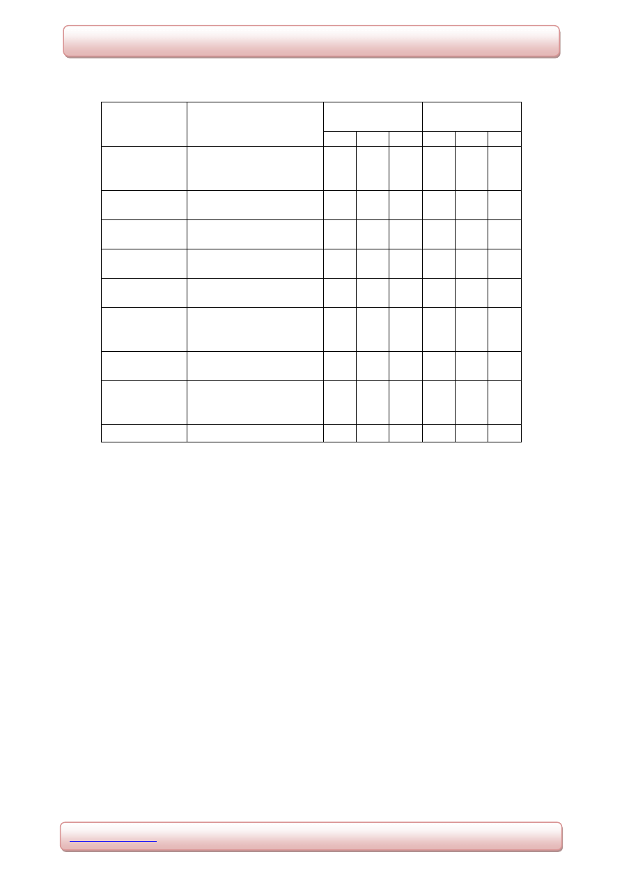
3.1 Phytochemical screening of test samples
The qualitative analysis of test samples of bark showed the presence of different secondary
metabolites. The HB sample showed the presence of alkaloids, the CB sample showed the
presence of steroids and MB sample showed the presence of carbohydrates, flavonoids and
saponins. Among the test samples of leaf extract HL sample showed the presence of
carbohydrates, triterpenoids. CL sample showed steroids, triterpenoids and ML sample
showed the presence of carbohydrates, flavonoids and saponins. The results of phytochemical
screening of bark and leaf are shown in the table 1.
www.wjpps.com
Vol 4, Issue 2, 2014.
470
Kumar et al. World Journal of Pharmacy and Pharmaceutical Sciences
Table 1.Phytochemical screening of test samples for biochemical constituents
(+) indicates presence of compound, (
_
) indicates absence of Compound.
Note: HB (Hexane extract of bark), CB (Chloroform extract of bark), MB (Methanolic
extract of bark), HL (Hexane extract of leaf), CL (Chloroform extract of leaf), ML
(Methanolic extract of leaf).
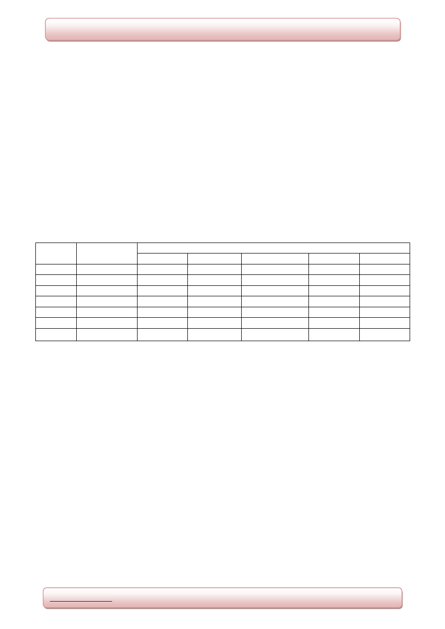
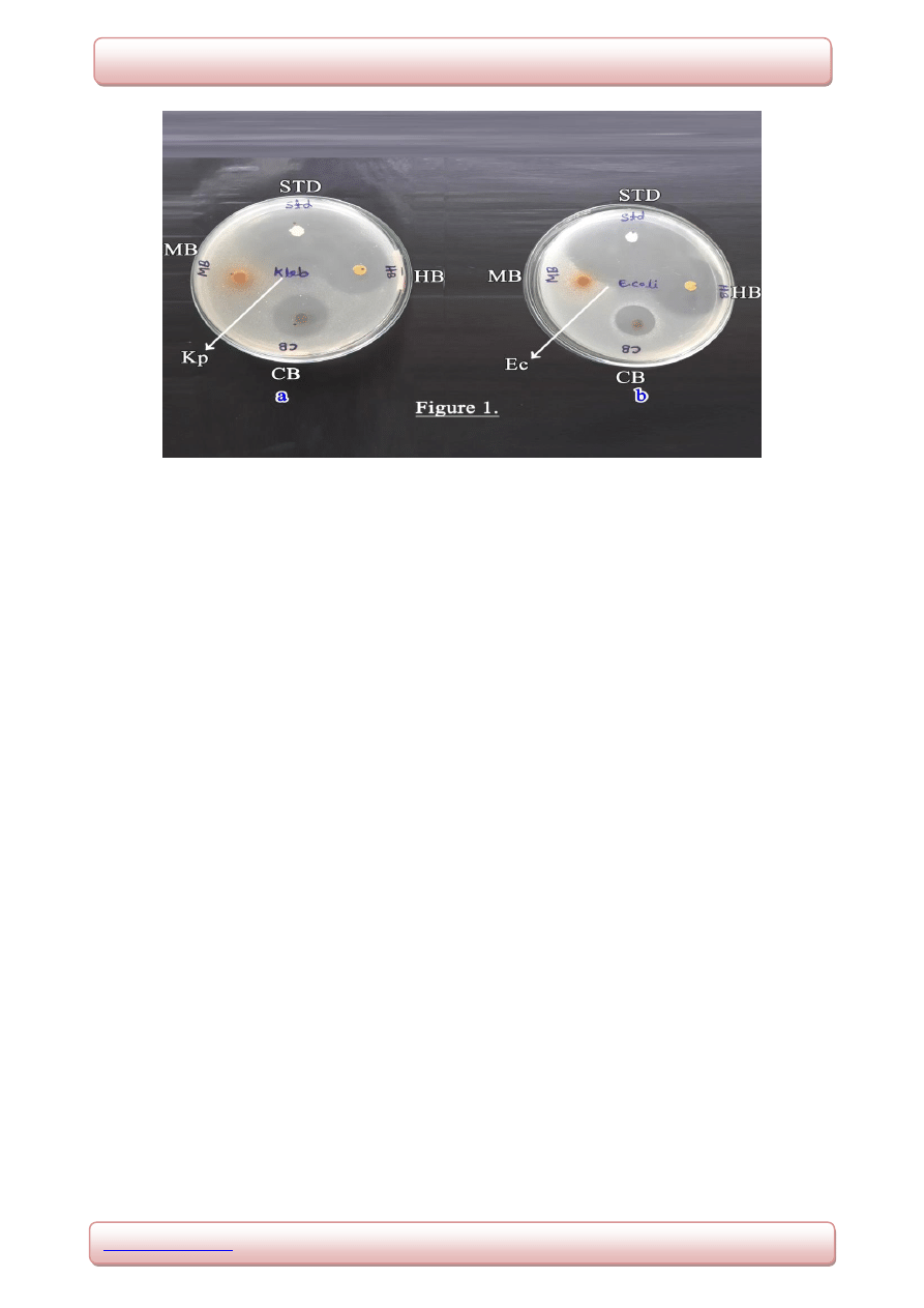
3.2 Antibacterial activity by disc diffusion method
The test samples evaluated for their antibacterial activity by disc diffusion showed significant
but varying degree of activity on target bacteria. The test sample HB showed maximum zone
of inhibition against E. coli (35mm) followed by K. pneumoniae (32mm), S. pyogens
(25mm), P. vulgaris (25mm) and S. aureus (23mm) respectively. Test sample CB showed
maximum zone of inhibition against K. pneumonia (24mm) followed by P. vulgaris (19mm),
E. coli (18mm), S. pyogens (15mm) and S. aureus (14mm) respectively. The test sample MB
showed less significant or no activity on different target bacteria with a maximum zone of
inhibition of 6mm against P. vulgaris and no activity against S. aureus, S. pyogens, K.
pneumonia and E. coli. The test sample HL exhibited maximum zone of inhibition against P.
vulgaris (17mm) followed by E. coli (16mm) and no activity against S. aureus, S. pyogens
Chemical
Constituents
Tests
Test samples
of Bark
Test samples
of Leaf
HB
CB
MB HL
CL
ML
Carbohydrates
Molisch’s test
Benedict’s test
Fehling’s test
_
_
_
_
_
_
+
+
+
+
+
+
_
_
_
+
+
+
Proteins and
Amino acids
Biuret test
Ninhydrin test
_
_
_
_
_
_
_
_
_
_
_
_
Alkaloids
Mayer’s test
Wagner’s test
+
+
_
_
_
_
_
_
_
_
_
_
Steroids
Libermann Buchard test
Salkowski Reaction
_
_
+
+
_
_
_
_
+
+
_
_
Triterpenoids
Libermann Buchard test
Salkowski Reaction
_
_
_
_
_
_
+
+
+
+
_
_
Tannins
Ferric chloride test
Lead acetate test
Vanillin-Hcl test
_
_
_
_
_
_
_
_
_
_
_
_
_
_
_
_
_
_
Glycosides
Borntrager’s test
Keller killiani test
_
_
_
_
_
_
_
_
_
_
_
_
Flavonoids
Shinoda test
Ferric chloride test
Mineral acid test
_
_
_
_
_
_
+
+
+
_
_
_
_
_
_
+
+
+
Saponins
Foam test
_
_
+
_
_
+
www.wjpps.com
Vol 4, Issue 2, 2014.
471
Kumar et al. World Journal of Pharmacy and Pharmaceutical Sciences
and K. pneumoniae. The test sample CL showed maximum zone of inhibition against E. coli
(15mm), P. vulgaris (10mm) and no activity against S. aureus, S. pyogens and K.
pneumoniae. The test sample ML showed zone of inhibition against E. coli (13mm), P.
vulgaris (6mm) and no activity against S. aureus, S. pyogens and K. pneumoniae. To the
standard drug ciprofloxacin the zone of inhibition obtained against different target organisms
were, E. coli (48mm), S. pyogens (42mm), P. vulgaris (40mm), K. pneumoniae (38mm) and
S. aureus (33mm). The results obtained from disc diffusion method against all the test
samples are shown in table 2. The photograph showing the zone of inhibition for test samples
and standard are shown in figure 1.
Table 2. Zone of inhibition exhibited by bark and leaf test samples against the target
bacteria by disc diffusion method (30μg/disc)
Values are expressed as mean ± standard deviation of the triplicates.
Concentration of Standard = 30µg/disc, Concentration of test Samples = 30µg/disc.
Note: HB (Hexane extract of bark), CB (Chloroform extract of bark), MB (Methanolic
extract of bark), HL (Hexane extract of Leaf), CL (Chloroform extract of Leaf), ML
(Methanolic extract of Leaf), CF (Ciprofloxacin), NA (No activity).
SL. NO.
Test
Samples
Zone of inhibition obtained against target organisms (in mm)
S. aureus
S. pyogens
K. pneumoniae
E. coli
P. vulgaris
01.
HB
18.67±1.15
27.33±1.15
31.67±1.53
33.67±1.53 31.67±1.53
02.
CB
26.00±1.00
15.33±0.58
26.67±0.58
18.33±0.58 28.00±1.00
03.
MB
NA
NA
NA
NA
6.33±0.58
04.
HL
NA
NA
NA
20.00±1.00 19.67±0.58
05.
CL
NA
NA
NA
12.67±0.58
7.33±0.58
06.
ML
NA
NA
NA
6.00±1.00
7.33±0.58
07.
CF (standard)
32.33±0.58
41.33±1.15
37.67±0.58
48.00±1.00 43.00±1.00
www.wjpps.com
Vol 4, Issue 2, 2014.
472
Kumar et al. World Journal of Pharmacy and Pharmaceutical Sciences
Fig 1. Zone of inhibition exhibited by test samples and standard drug by disc diffusion
method against K. pneumonia and E. coli.
3.3 Antifungal activity by disc diffusion method
The test samples screened for their antifungal activity by disc diffusion showed less
significant activity by exhibiting very low zone of inhibition on target fungi. The test sample
HB showed a maximum of 6mm zone of inhibition against C. albicans followed by C.
indicum (3mm), C. keratinophilum (4mm), T. rubrum (5mm) and M. gypsium (8mm). The
zone of inhibition exhibited by test sample CB was 3mm against C. albicans followed by C.
indicum (3mm), C. keratinophilum (7mm) and no activity against T. rubrum and M. gypsium.
The test sample MB showed maximum zone of inhibition against C. keratinophilum (2mm)
and no activity against C. albicans, C. indicum, T. rubrum and M. gypsium. The test sample
HL exhibited maximum zone of inhibition against C. keratinophilum (4mm), T. rubrum
(3mm), and no activity against C. albicans, C. indicum and M. gypsium. The test sample CL
showed maximum zone of inhibition against C. keratinophilum (4mm), T. rubrum (3mm),
and no activity against C. albicans, C. indicum and M. gypsium. The test sample ML showed
maximum zone of inhibition against C. albicans (2mm), C. keratinophilum (3mm) and no
activity against C. indicum, T. rubrum and M. gypsium. The standard drug clotrimazole
showed maximum zone of inhibition against C. albicans (38mm), C. indicum (36mm), C.
keratinophilum (24mm), T. rubrum (25mm) and M. gypsium (30mm). The results obtained
from disc diffusion method against all the test samples are shown in table 3.
www.wjpps.com
Vol 4, Issue 2, 2014.
473
Kumar et al. World Journal of Pharmacy and Pharmaceutical Sciences
Table 3. Zone of inhibition exhibited by bark and leaf test samples against thetarget
fungi by disc diffusion method (30μg/disc)
Sl.
No.
Test
Samples
Zone of inhibition obtained against target organisms (in mm)
C. albicans
C. indicum
C. keratinophilum
T. rubrum
M. gypsium
01. HB
5.67±0.58
2.33±0.58
4.00±0.00
4.33±0.58
7.6±0.58
02. CB
2.67±0.58
2.33±0.58
6.67±0.58
NA
NA
03.
MB
NA
NA
2.00±0.00
NA
NA
04.
HL
NA
NA
3.67±0.58
3.33±0.58
NA
05.
CL
NA
NA
2.67±0.58
3.33±0.58
NA
06.
ML
2.00±0.00
NA
2.67±0.58
NA
NA
07.
CI (standard)
38.00±0.58
36.33±0.33
23.67±0.33
25.33±0.33
30.33±0.33
Values are expressed as mean ± standard deviation of the triplicates
Concentration of standard = 30µg/disc, Concentration of test samples = 30µg/disc.
Note: HB (Hexane extract of bark), CB (Chloroform extract of bark), MB (Methanolic
extract of bark), HL (Hexane extract of Leaf), CL (Chloroform extract of Leaf), ML
(Methanolic extract of Leaf), CI (Clotrimazole), NA (No activity).
3.4 Determination of minimum inhibitory concentration (MIC)
During MIC studies the test sample HB showed growth inhibition at 50µg/ml against E. coli
and K. pneumoniae whereas the growth inhibition against S. pyogens and S. aureus was at
100µg/ml and 200µg/ml respectively. The test sample CB showed growth inhibition at
50µg/ml against S. aureus, at 100µg/ml against E. coli and K. pneumonia at 200µg/ml against
S. pyogens. The test sample HL exhibited MIC at 200µg/ml against all the target organisms.
The standard drug ciprofloxacin exhibited MIC at 15.6µg/ml against all the target organisms.
The results of MIC obtained against all the target bacteria are shown in table 4.
Table 4. The MIC values of test samples and standard drug against target bacteria
(μg/ml)
Sl. No.
Test
Organisms
Ciprofloxacin
Standard (µg/ml)
MIC values (µg/ml)
HB
CB
HL
01.
S. aureus
15.6
200
50
200
02.
S. pyogens
15.6
100
200
200
03.
E. coli
15.6
50
100
200
04.
K. pneumoniae
15.6
50
100
200
Note: HB (Hexane extract of bark), CB (Chloroform extract of bark), MB (Methanolic
extract of bark), HL (Hexane extract of Leaf), CL (Chloroform extract of Leaf), ML
(Methanolic extract of Leaf).
www.wjpps.com
Vol 4, Issue 2, 2014.
474
Kumar et al. World Journal of Pharmacy and Pharmaceutical Sciences
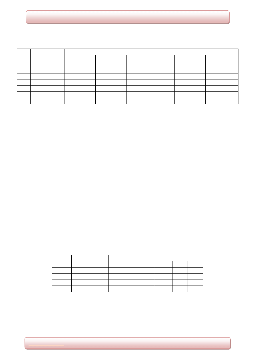
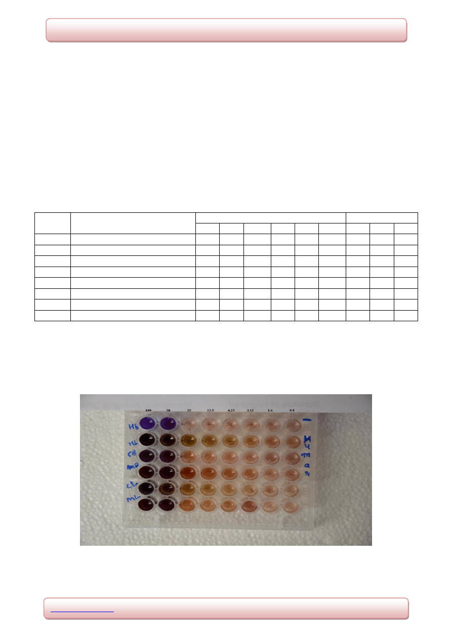
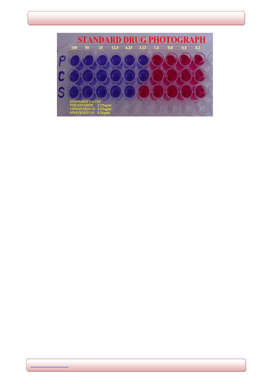
3.5 Antitubercular activity
The test organism M. tuberculae showed sensitivity to the test sample HL at 25μg/ml. The
other samples HB, CB, MB, CL and ML showed growth inhibition at 50μg/ml. The
standard antitubercular drugs pyrazinamide and ciprofloxacin showed growth inhibition at
3.125μg/ml and streptomycin at 6.25μg/ml. The results obtained for the antitubercular
activity is shown in the table 5. The photograph showing growth inhibition of test organism
to the test samples and standards are shown in fig 2 and 3 respectively.
Table 5. The MIC values of the test samples and standards against
Mycobacterium tuberculae
Sl. No.
Concentration of test samples
and standard (µg/ml)
Test Samples
Standards
HB
CB
MB
HL
CL
ML
PA
SM CF
01.
100
S
S
S
S
S
S
S
S
S
02.
50
S
S
S
S
S
S
S
S
S
03.
25
R
R
R
S
R
R
S
S
S
04.
12.5
R
R
R
R
R
R
S
S
S
05.
6.25
R
R
R
R
R
R
S
S
S
06.
3.12
R
R
R
R
R
R
S
R
S
07.
1.6
R
R
R
R
R
R
R
R
R
08.
0.8
R
R
R
R
R
R
R
R
R
Note: HB (Hexane extract of bark), CB (Chloroform extract of bark), MB (Methanolic
extract of bark), HL (Hexane extract of Leaf), CL (Chloroform extract of Leaf), ML
(Methanolic extract of Leaf), PA (Pyrazinamide), SM (Streptomycin), CF (Ciprofloxacin), R
(Resistant), S (Sensitive).
Fig 2.Microplate showing growth inhibition of Mycobacterium tuberculae for test
Samples by alamar blue dye method.
www.wjpps.com
Vol 4, Issue 2, 2014.
475
Kumar et al. World Journal of Pharmacy and Pharmaceutical Sciences
Fig 3.Microplate showing growth inhibition of Mycobacterium tuberculae for standards
byalamar blue dye method.
4. DISCUSSION
Plants are used as a source of medicine in curing diseases since centuries. Medicinal plants
are used to treat diseases like diabetes, ulcers, liver cirrhosis, body pain, hepatitis, cancer and
AIDS. Plants also represent a rich source of antimicrobial agents. The presence of secondary
metabolites such as alkaloids, glycosides, flavonoids, phenols, triterpenoids, steroids
andtannins have been reported in the past to possess antimicrobial activity.
[21]
In the present study the bark and leaf extracts of plant Kunsleria keralensis which belongs to
the family Fabaceae were tested for antimicrobial activities. Several reports are available on
many plant species belonging to the presently studied family Fabaceae with antimicrobial
activities. The antibacterial activity has been reported for bark and leaf extracts of many
plants like Delonix regia, Bauhinia purpurea, Ficus bengalensis, Diplotropis ferruginea of
Fabaceae against many human pathogens like S. aureus, S. pyogens, K. pneumoniae, E. coli,
P. vulgaris, B. amyloliquefaciens, B. pumilus, S. typhi, M.luteus and many more
bacteria.
[2,21,22,23,24]
Antibacterial as well as antifungal activity against human pathogens for
the extracts of Diplotropis ferruginea and Senna alata has been reported.
[25,26]
In the present study antibacterial activity was evaluated by disc diffusion method. The test
sample HB exhibited significant antibacterial activity against all the test organisms with a
maximum of 74% inhibition against P. vulgaris in comparison with standard drug
ciprofloxacin followed by 56%, 62% and 67% inhibition against S. aureus, S. pyogens and E.
coli respectively. The test sample CB also exhibited significant antibacterial activity against

www.wjpps.com
Vol 4, Issue 2, 2014.
476
Kumar et al. World Journal of Pharmacy and Pharmaceutical Sciences
all the test organisms showing a maximum of 67% inhibition against K. pneumoniae in
comparison with standard drug ciprofloxacin followed by 60% and 63% inhibition against S.
aureus and P. vulgaris. The test samples HL, CL and ML exhibited moderately significant
activity against two bacteria E. coli, and P. vulgaris and no activity against other target
bacteria.
Though both the bark and leaf extracts showed antibacterial activity, the bark extracts
exhibited comparatively more antibacterial activity than leaf extracts. The antibacterial
activity of HB test sample may be due to the presence of alkaloids. Whereas, in CB it may be
due to the presence of steroids and triterpenoids and in the sample MB antibacterial activity
may be due to the presence of flavonoids. These constituents were found during
phytochemical investigation and proved to have antimicrobial activities. The leaf extracts
exhibited comparatively less antibacterial activity against the target bacterial strains. This
may be due to the less presence of secondary metabolites in the leaf samples than bark.
The MIC study was performed on HB, CB and HL test samples since they exhibited
significant antibacterial activity during disc diffusion studies. Although the test samples
exhibited different MIC values, all the values were found significant. Among them the test
sample HB showing growth inhibition at 50µg/ml against E. coli and K. pneumoniae was
found significant. The test sample CB was more active against S. aureus. It exhibited growth
inhibition at 50µg/ml against S. aureus and found to be significant.
The evaluation of test samples for antitubercular activity showed that the hexane extract of
leaf (HL) had significant antitubercular activity exhibiting growth inhibition at 25μg/ml in
comparison with other test samples. All other samples HB, CB, MB, CL and ML showed
growth inhibition at 50μg/ml. The standard antitubercular drugs pyrazinamide and
ciprofloxacin showed growth inhibition at concentrations of 3.125μg/ml and streptomycin at
6.25μg/ml. The contrast in antitubercular activity by leaf extract may be due to the presence
of different constituents in leaf or due to extraction of particular antitubercular components
by hexane.
All the test samples evaluated for antifungal activity against human pathogens exhibited
insignificant activity.

www.wjpps.com
Vol 4, Issue 2, 2014.
477
Kumar et al. World Journal of Pharmacy and Pharmaceutical Sciences
5. CONCLUSION
The test samples of bark extracts HB and CB exhibited significant antibacterial activity
against the target bacteria. The test sample of leaf extract HL proved to have antitubercular
activity. The MIC studies confirmed their antibacterial and antitubercular potency. The
further isolation, purification and the spectral analysis of pure compounds may provide a
potential antibacterial lead molecule. The test samples can be evaluated further for
pharmacological properties which can be useful in designing of new drugs.
6. ACKNOWLEDGEMENT
We would sincerely thank Vision Group of Science and Technology, Government of
Karnataka for granting fund under CISE to procure the analytical instruments to carry out the
proposed research work.
7. REFERENCES
1. Saini and Sharma S. (2012). Dalbergia sissoo – an overview. International Journal of
Pharma Professional’s research, 2012; 3: 548-555.
2. Patil U.H and Gaikwad DK. (2011). Phytochemical screening and microbicidal activity of
stem bark of Pterocarpus marsupium. International Journal of Pharma Sciences and
Research, 2011; 2: 36-40.
3. Pavithra, Karsha V and Lakshmi OB. (2010). Antibacterial activity of black pepper
(Piper nigrum Linn) with special reference to its mode of action. Indian Journal of
Natural Products and Resources, 2010; 1: 213-215.
4. Nasciomentho G.F, Juliana L, Paulo CF and Giuliana LS. (2000). Antibacterial activity of
plant extracts and phytochemcicals on antibiotic resistant bacteria. Brazilian
Journal of Microbial researche’s, 2000; 31: 247-256.
5. Sharma R.A, Sharma P and Yadav A. (2013). Antimicrobial screening of sequential
extracts of Datura stramonium Linn. International Journal of Pharmacy and
Pharmaceutical Sciences, 2013; 5: 401-404.
6. Kumar V M.L, Tippeswamy B and Shivakumar CK. (2013). Evaluation of antimicrobial
activity of Bacillus cereus and Bacillus pumillus metabolites against human pathogens.
International Journal of Current Pharmaceutical review and Research, 4:47-60.
7. Mohanan CN and Nair NC. (1981). Kunstleria prain- A new genus record for India and a
new species in the genus. Proceeding of the Indian Academy of Science, 1981; 90: 207-
209.

www.wjpps.com
Vol 4, Issue 2, 2014.
478
Kumar et al. World Journal of Pharmacy and Pharmaceutical Sciences
8. Binu S. (2011). Medicinal plants used for treating body pain by the tribals in
Pathanamthitta district, Kerala, India. Indian Journal of Traditional knowledge, 2011; 10:
547-549.
9. Goel KA. (2010). Development and poverty alleviation. National conference on
biodiversity, 2010; 1: 100-101.
10. Harwood, Laurence M, Moody and Christopher J. (1989). Experimental organic
chemistry Principles and Practice (Illustrated edition), p.122–125.
11. Jensen and William B. (2007). The origin of the soxhlet extractor. Journal of Chemical
Education, 2007; 84: 1913–1914.
12. Bandar H.A, Rammal H, Hachem A, Saad Z and Badran B. (2013). Techniques for the
extraction of bioactive compounds from Lebanese urticadioica. American Journal of
Phytomedicine and Clinical Therapeutics, 2013; 6: 507-513.
13. Patil A.G, Koli S.P, Patil DA and Phatak AV. (2012). Evaluation of extraction techniques
with various solvents to determine extraction efficiency of selected medicinal plants.
International Journal of Pharmaceutical Sciences and Research, 2012; 3: 2607-2612.
14. CK Kokate, P Purohit and B Gokhale. (1999). Procedure of phytochemical tests. Text
Book of Pharmacognosy; Vallabh Prakash publishers, 1999; 12: 172-173.
15. Khandelwal KR. (2008). Test procedures of phytoconstituents, Text Book of
Pharmacognosy; Nirali Prakash publishers, 2008; 19: 149-156.
16. Espinel I.A, Shadomy S and White S. (1991). Agar disk diffusion susceptibility tests with
cilofungin. Pub Med Journal, 1991; 29: 93-98.
17. National Committee for Clinical Laboratory Standards. (1999). Performance standards for
antimicrobial
susceptibility
testing,
Ninth
informational
supplement.
Wayne,
Pennsylvania, nccls, p.1526-1543.
18. Rusenova N and Parvanov P. (2009). Antimicrobial activities of twelve essential oils
against microorganisms of veterinary importance. Journal of Sciences, 2009; 7: 37-43.
19. Daouk K.D, Dagher MS and Sattont JE. (1995). Antifungal activity of the essential oil of
Organum syriacum. Food prot, 1995; 58: 1147-1149.
20. Maria C.S, Lourenco, Marcus V.N, Alessandra CP, Marcelle L. Ferreira, Rasnisb B,
Goncalves, Thais Cristina MN and Monica AP. (2007). Evaluation of antitubercular
activity of nicotinic and isoniazid analogues. ARKIVOC, 2007; 15: 181-191.
21. Sama K, Raja VA and Yadav HR. (2012). Antibacterial and pharmacognostical
evaluation of Delonix regia root bark. International Journal of Pharmacy and Life
Sciences, 2012; 3: 1628-1630.

www.wjpps.com
Vol 4, Issue 2, 2014.
479
Kumar et al. World Journal of Pharmacy and Pharmaceutical Sciences
22. Chaudhari G.M, Bhoomi B, Mistry J.K.N, Dabhi B and Lal S. (2013). In Vitro
antimicrobial activity of stem bark of Bauhinia purpurea. International Science Press
(India), 2013; 4: 29-35.
23. Gayathri M and Kannabiran K. (2009). Antimicrobial activity of Hemidesmus indicus,
Ficus bengalensis and Pterocarpus marsupium roxb. Indian Journal of Pharmaceutical
Sciences, 2009; 5: 578-581.
24. Dhale DA, Chamle DR and Panchal VH. (2010). Evaluations of phytochemical
constituents and antimicrobial activity of Butea monosperma (Fabaceae). Journal of
phytology, 2010; 2: 17-21.
25. Cerqueria G.S, Rocha N.F.M, Almedia J.G.S, Freitas de A.F, Filho J.M.B, Freitas RM
and Diniz MF. (2011). Antimicrobial activity of the extract of stem bark of Diplotropis
ferruginea benth. Journal of Young pharmacists, 2011; 3: 284-286.
26. Ehinowemwenguan G, Inetianbor JE and Yakubu JM. (2014). Antimicrobial qualities of
Senn alata. Journal of Pharmacy and Biological Sciences, 2014; 2: 47-52.
Wyszukiwarka
Podobne podstrony:
Danuta Kunstler1
Anna Keraleigh [Fairy 01] Fair Flavor [Evernight] (pdf)
Kunstler Piotr Wpływ asan jogi na organizm człowieka cz I III
więcej podobnych podstron