
Pdr1 regulates multidrug resistance in Candida
glabrata: gene disruption and genome-wide expression
studies
John-Paul Vermitsky,
1
Kelly D. Earhart,
2
W. Lamar Smith,
1
Ramin Homayouni,
3
Thomas D. Edlind
1
* and P. David Rogers
2
1
Department of Microbiology and Immunology, Drexel
University College of Medicine, Philadelphia, PA, USA.
2
Department of Pharmacy and Pharmaceutical
Sciences, College of Pharmacy, and Department of
Pediatrics, College of Medicine, University of Tennessee
Health Science Center, Children’s Foundation Research
Center at Le Bonheur Children’s Medical Center,
Memphis, TN, USA.
3
Department of Neurology, College of Medicine and
Center for Genomics and Bioinformatics, University of
Tennessee Health Science Center, Memphis, TN, USA.
Summary
Candida glabrata emerged in the last decade as a
common cause of mucosal and invasive fungal infec-
tion, in large part due to its intrinsic or acquired resis-
tance to azole antifungals such as fluconazole. In
C. glabrata clinical isolates, the predominant mecha-
nism behind azole resistance is upregulated expres-
sion of multidrug transporter genes CDR1 and PDH1.
We previously reported that azole-resistant mutants
(MIC
ⱖ 64 mg ml
-1
) of strain 66032 (MIC
= 16 mg ml
-1
)
similarly show coordinate CDR1-PDH1 upregulation,
and in one of these (F15) a putative gain-of-function
mutation was identified in the single homologue
of Saccharomyces cerevisiae transcription factors
Pdr1–Pdr3.
Here
we
show
that
disruption
of
C. glabrata PDR1 conferred equivalent fluconazole
hypersensitivity (MIC
= 2 mg ml
-1
) to both F15 and
66032
and
eliminated
both
constitutive
and
fluconazole-induced CDR1-PDH1 expression. Rein-
troduction of wild-type or F15 PDR1 fully reversed
these effects; together these results demonstrate a
role for this gene in both acquired and intrinsic azole
resistance. CDR1 disruption had a partial effect,
reducing fluconazole trailing in both strains while
restoring wild-type susceptibility (MIC
= 16 mg ml
-1
) to
F15. In an azole-resistant clinical isolate, PDR1 dis-
ruption reduced azole MICs eight- to 64-fold with no
effect on sensitivity to other antifungals. To extend
this analysis, C. glabrata microarrays were generated
and used to analyse genome-wide expression in F15
relative to its parent. Homologues of 10 S. cerevisiae
genes previously shown to be Pdr1–Pdr3 targets were
upregulated (YOR1, RTA1, RSB1, RPN4, YLR346c and
YMR102c along with CDR1, PDH1 and PDR1 itself)
or downregulated (PDR12); roles for these genes
include small molecule transport and transcriptional
regulation. However, expression of 99 additional
genes was specifically altered in C. glabrata F15; their
roles include transport (e.g. QDR2, YBT1), lipid
metabolism (ATF2, ARE1), cell stress (HSP12, CTA1),
DNA repair (YIM1, MEC3) and cell wall function
(MKC7, MNT3). These azole resistance-associated
changes could affect C. glabrata tissue-specific viru-
lence; in support of this, we detected differences in
F15 oxidant, alcohol and weak acid sensitivities.
C. glabrata provides a promising model for studying
the genetic basis of multidrug resistance and its
impact on virulence.
Introduction
Increasing numbers of individuals are immunocompro-
mised in association with AIDS, organ and tissue trans-
plantation, aggressive treatments for cancer and immune-
related diseases, diabetes, premature birth and advanced
age. These individuals are at high risk for opportunistic
fungal infection, in particular mucosal or systemic candidi-
asis. In the previous decade, Candida glabrata emerged as
a common cause of these infections (10–30% of yeast
isolates), trailing only Candida albicans (Pfaller et al.,
1999; Safdar et al., 2001; Ostrosky-Zeichner et al., 2003;
Richter et al., 2005). In some populations such as diabetics
and the elderly, C. glabrata may be the dominant pathogen
(Diekema et al., 2002; Kontoyiannis et al., 2002; Grimoud
et al., 2005; Goswami et al., 2006). In C. glabrata can-
didaemia, mortality rates of 38–53% have been reported
(Viscoli et al., 1999; Safdar and Armstrong, 2002; Klingspor
et al., 2004). Nevertheless, the basis for C. glabrata patho-
genicity is not yet clear, because it is deficient in the
Accepted 10 May, 2006. *For correspondence. E-mail tedlind@
drexelmed.edu; Tel. (
+1) 215 9918377; Fax (+1) 215 8482271.
Molecular Microbiology (2006) 61(3), 704–722
doi:10.1111/j.1365-2958.2006.05235.x
First published online 27 June 2006
© 2006 The Authors
Journal compilation © 2006 Blackwell Publishing Ltd

virulence factors implicated in C. albicans infection: dimor-
phism, strong adhesion, secreted hydrolases and biofilm
formation (Douglas, 2003; Nikawa et al., 2003; Kaur et al.,
2005; Schaller et al., 2005). On the other hand, C. glabrata
demonstrates relative resistance to azoles, the most
widely used antifungal group which includes topical imida-
zoles such as miconazole and oral/parenteral triazoles
such as fluconazole. Specifically, the fluconazole MIC
inhibiting 50% of clinical isolates is 8
mg ml
-1
, compared
with 0.25
mg ml
-1
for C. albicans (Ostrosky-Zeichner et al.,
2003; Pfaller et al., 2004). Azoles inhibit lanosterol dem-
ethylase, product of the ERG11 gene (CYP51 in moulds),
which results in depletion of the membrane component
ergosterol and accumulation of toxic sterol products (for
review, see Akins, 2005). The emergence of C. glabrata
(from
ⱕ 5% of yeast isolates in the 1980s) parallels the
introduction in the early 1990s of fluconazole and over-the-
counter imidazoles, along with widespread application of
agricultural azole fungicides. Indeed, its intrinsic low-level
azole resistance, the molecular basis for which remains
undefined, may represent a C. glabrata ‘virulence factor’.
Candida glabrata also demonstrates a high capacity for
acquired high-level azole resistance, with 8–27% of iso-
lates demonstrating a fluconazole MIC
ⱖ 64 mg ml
-1
(Safdar et al., 2002; Ostrosky-Zeichner et al., 2003;
Pfaller et al., 2004). RNA analysis of these clinical isolates
suggests that the predominant basis for acquired azole
resistance is the constitutively upregulated expression of
multidrug transporter genes CDR1 and, to a lesser extent,
PDH1 (Miyazaki et al., 1998; Sanglard et al., 1999; 2001;
Redding et al., 2003; Bennett et al., 2004; Vermitsky and
Edlind, 2004; Sanguinetti et al., 2005). In support of this,
CDR1 or CDR1-PDH1 disruption was shown to confer
azole hypersensitivity (Sanglard et al., 2001; Izumikawa
et al., 2003). In this respect, C. glabrata resembles
C. albicans and other fungi in which azole resistance has
been attributed to upregulated expression of multidrug
transporters (Akins, 2005). Initial laboratory studies of
C. glabrata acquired azole resistance using standard
glucose-supplemented
medium
yielded
avirulent
respiratory-deficient mitochondrial mutants (Sanglard
et al., 2001; Brun et al., 2005). Using glycerol-
supplemented
medium,
we
isolated
respiratory-
competent mutants with coordinately upregulated CDR1-
PDH1 analogous to that observed in azole-resistant
clinical isolates (Vermitsky and Edlind, 2004). Coordinate
upregulation of these genes was also observed following
brief exposure of susceptible cells to azoles, representing
a potential basis for intrinsic low-level resistance.
Coordinate
CDR1-PDH1
upregulation
implies
a
common transcription factor. Although very distinct in
terms of niche and human pathogenicity, C. glabrata is an
evolutionary close relative of Saccharomyces cerevisiae
(Barns et al., 1991; Dujon et al., 2004). In the latter, the
coordinate upregulation of multidrug transporter genes
PDR5 and SNQ2 is mediated by the paralogous Pdr1 and
Pdr3 zinc cluster transcription factors (Kolaczkowska and
Goffeau, 1999). Many gain-of-function mutations within
Pdr1–Pdr3 have been identified that result in constitutive
upregulation of PDR5-SNQ2 along with a diverse group of
additional genes (Carvajal et al., 1997; DeRisi et al., 2000;
Devaux et al., 2001). Our analysis of the recently released
C. glabrata genome sequence (Dujon et al., 2004)
revealed a single PDR1–PDR3 homologue, and a puta-
tive gain-of-function mutation in this gene was identified in
azole-resistant laboratory mutant F15 (Vermitsky and
Edlind, 2004). Here we demonstrate the central role of
C. glabrata PDR1 in acquired azole resistance, and iden-
tify a likely role in intrinsic resistance, by characterizing
Dpdr1 derivatives of laboratory strains and clinical
isolates. Furthermore, we report the first application of
microarrays to this organism, which identified multiple
genes coregulated with CDR1-PDH1 that are likely to
impact C. glabrata virulence.
Results and discussion
PDR1 disruption in F15 and parent
The laboratory selection of spontaneous fluconazole-
resistant mutants of C. glabrata ATCC strain 66032 was
previously described (Vermitsky and Edlind, 2004). One
of these mutants, F15, exhibited strong upregulation of
CDR1 and PDH1, modest upregulation of PDR1, and a
single base change predicted to alter the Pdr1 amino acid
sequence. We reasoned that disruption of PDR1 in F15
and parent 66032 would provide an initial test of the
hypothesis that this single base change is responsible for
the fluconazole resistance. To accomplish this, ura3
derivatives of F15 and 66032 were isolated by selection
on 5-fluoroorotic acid (5FOA) and screening for comple-
mentation by a URA3-encoding plasmid. Homologous
recombination is relatively non-specific in C. glabrata,
especially with short homology regions, but can be
enhanced by promoter-dependent disruption of genes
(PRODIGE) as previously described (Edlind et al., 2005).
This method was used to disrupt PDR1 (Fig. 1A). Trans-
formants were screened by polymerase chain reaction
(PCR); loss of the PDR1uF-PDR1iR product and genera-
tion of the PDR1uF-URA3iR product confirmed PDR1
disruption (Fig. 1B).
Broth microdilution assays were used to examine flu-
conazole susceptibility of F15
Dpdr1, 66032Dpdr1 and
their parents (Fig. 1C). Similar to previous results with
their parents (Vermitsky and Edlind, 2004), the 66032 and
F15 ura3 derivatives generated 24 h fluconazole MICs of
8–16 and
⬎ 64 mg ml
-1
respectively. In contrast, their
Dpdr1 derivatives were fluconazole hypersusceptible, with
Pdr1 regulates multidrug resistance in Candida glabrata
705
© 2006 The Authors
Journal compilation © 2006 Blackwell Publishing Ltd, Molecular Microbiology, 61, 704–722
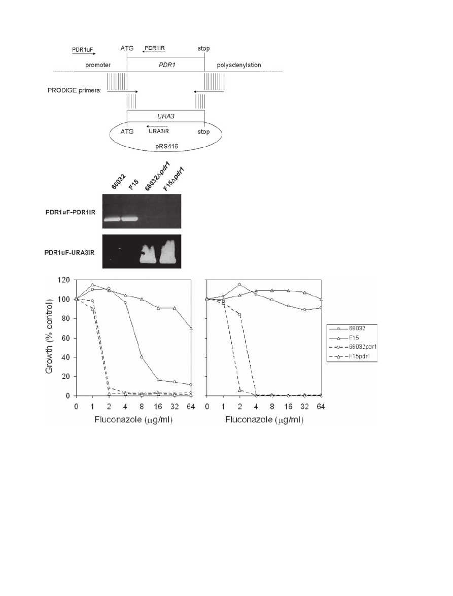
equivalent MICs of 2
mg ml
-1
. Although susceptible, 66032
exhibited trailing growth typical of many Candida species
(Rex et al., 1998), and by 48 h was fully grown at all
fluconazole concentrations tested (Fig. 1C). Trailing
growth was absent in the PDR1 disruptants. These results
support the role of Pdr1 in F15 fluconazole resistance.
Furthermore, the reduced MIC and trailing growth asso-
ciated with PDR1 disruption in 66032 suggests that Pdr1
is an important contributor to the intrinsic low-level resis-
tance that is characteristic of this species.
As hypothesized, RNA analysis showed that PDR1 dis-
ruption reversed the constitutive upregulation of CDR1
and PDH1 in untreated mutant F15 (Fig. 2). Moreover,
expression of these genes was reduced relative to their
expression in untreated parent 66032. This can explain
the greater susceptibility of the
Dpdr1 derivatives relative
to 66032. As previously described (Vermitsky and Edlind,
2004), fluconazole treatment induced the expression of
CDR1 and PDH1, most clearly in strains 66032 and
F15 respectively (Fig. 2A). PDR1 disruption completely
Fig. 1. Disruption of PDR1 and effects on
azole sensitivity.
A. Diagram illustrating PRODIGE primer
design for disruption of PDR1 with URA3
coding sequence amplified from pRS416. Also
shown are the upstream forward and two
internal reverse primers used to screen
transformants.
B. PCR screen of representative
Dpdr1
transformants selected on DOB-URA and their
parents 66032 and F15. DNA was purified
from isolated colonies, amplified with the
indicated primers pairs, and analysed by gel
electrophoresis; loss of the PDR1uF-PDR1iR
product and formation of the
PDR1uF-URA3iR product identified
Dpdr1
clones.
C. Broth microdilution assays examining
fluconazole sensitivities of parent 66032,
azole-resistant mutant F15, and their
respective
Dpdr1 disruptants. Absorbance at
630 nm was recorded after 24 or 48 h
incubation as indicated; growth was plotted as
percentage of drug-free control.
A
B
C
24
48
706
J.-P. Vermitsky et al.
© 2006 The Authors
Journal compilation © 2006 Blackwell Publishing Ltd, Molecular Microbiology, 61, 704–722

blocked this treatment-dependent upregulation. Finally,
we note that PDR1 itself, which is upregulated in F15
(Vermitsky and Edlind, 2004), is also induced by flucona-
zole treatment in 66032 and F15 (Fig. 2A).
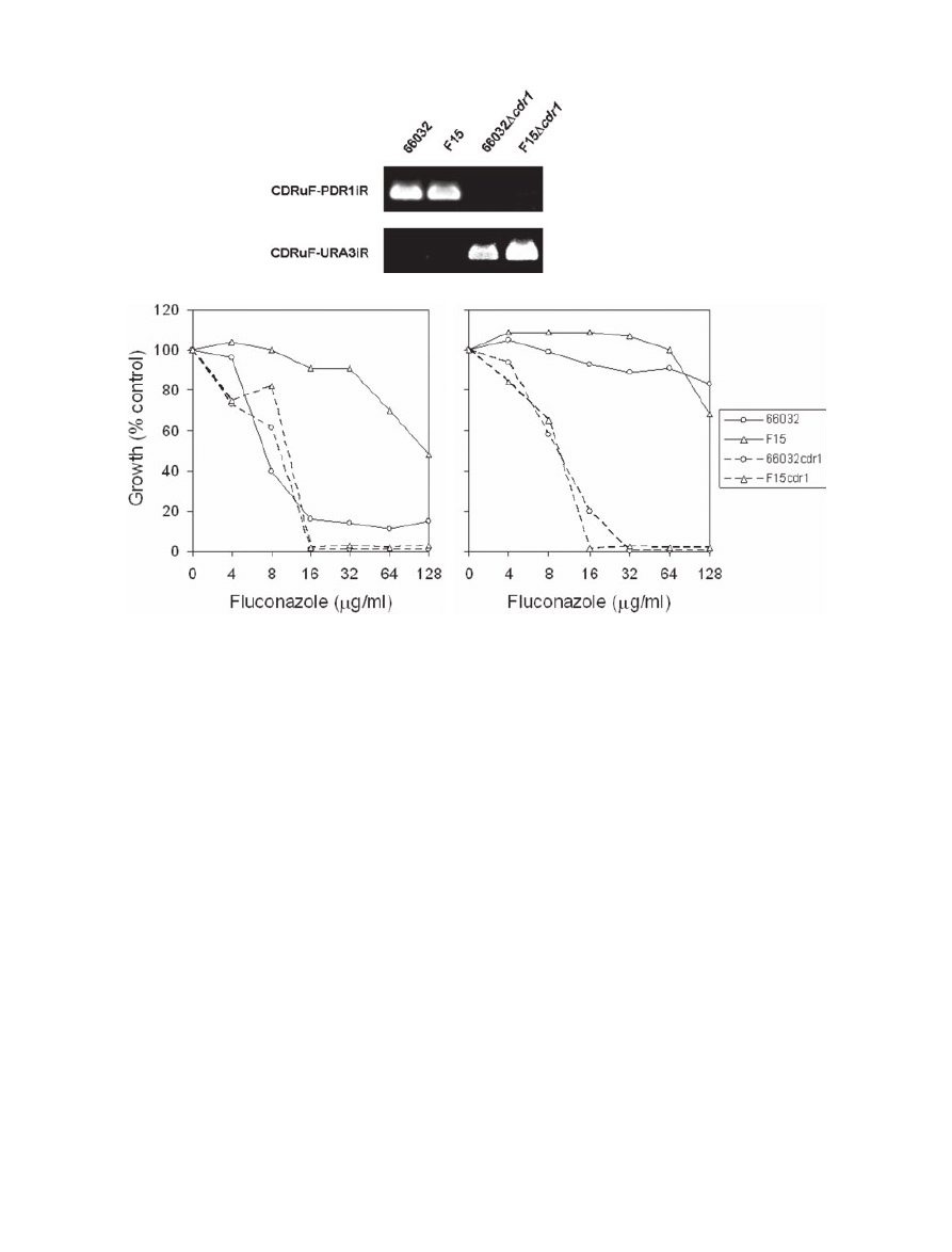
CDR1 disruption
To more directly assess the role in acquired or intrinsic
azole resistance of multidrug transporter gene CDR1, it
was similarly disrupted in the ura3 derivatives of 66032
and F15 (Fig. 3A). This reversed the fluconazole resis-
tance of F15 (Fig. 3B), although the MIC (16
mg ml
-1
)
remained eightfold above that observed with PDR1 dis-
ruption (Fig. 1C). With respect to 66032, CDR1 disruption
had minimal effect on fluconazole MIC at 24 h, but trailing
growth most apparent at 48 h was eliminated as it was
with PDR1 disruption. These results are consistent with
CDR1 being a major but not exclusive contributor to F15
azole resistance.
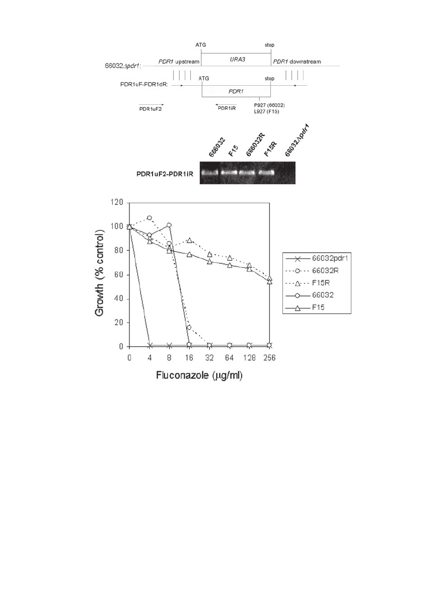
PDR1 replacement
To rigorously test the role of F15 PDR1 in azole resis-
tance, we employed gene replacement. The 66032
Dpdr1
strain (see above) was transformed with PCR products
representing wild-type and F15 PDR1, including 5
′ and 3′
flanking sequences which should direct PDR1 to its native
locus (Fig. 4A). We initially attempted to select homolo-
gous recombinants on fluconazole-containing medium.
However, this was precluded by a background of sponta-
neous fluconazole-resistant mutants in control (no added
DNA) transformations (see below for further characteriza-
tion of these mutants). As an alternative, the protein syn-
thesis inhibitor cycloheximide is a known substrate for
Cdr1-like multidrug transporters, and indeed C. glabrata
Dpdr1 strains are cycloheximide-hypersensitive (Edlind
et al., 2005). In contrast to fluconazole, cycloheximide-
containing plates yielded no spontaneous mutants while
five or six transformants were obtained with addition of
wild-type or F15 PDR1 respectively. PCR screening of
these transformants confirmed homologous recombina-
tion into the native locus (Fig. 4B). All F15 PDR1 replace-
ments demonstrated fluconazole resistance comparable
to F15 itself, while all but one of the wild-type PDR1
replacements demonstrated wild-type sensitivity (Fig.
4C). Sequencing of a representative F15 PDR1 replace-
ment confirmed there were no mutations other than the
previously described P927L (Vermitsky and Edlind, 2004).
Characterization of Pdr1-independent azole resistance
As noted above, a background of resistant mutants arose
on fluconazole-containing YP-glycerol medium in control
transformations of strain 66032
Dpdr1, which involved
plating c. 2
¥ 10
7
cells. To more rigorously examine this
Pdr1-independent
resistance,
equivalent
numbers
Fig. 2. Expression of multidrug transporter
genes CDR1 and PDH1 and transcriptional
activator gene PDR1 in parent 66032, mutant
F15 and their respective
Dpdr1 disruptants.
A. RNA was isolated from log phase cultures,
slot-blotted to membranes, and hybridized to
the indicated gene probes; ACT1 served as
loading control. U, untreated cultures; T,
treated with 256
mg ml
-1
fluconazole for 2.5 h.
B. Quantitative real-time RT-PCR analysis of
relative CDR1 and PDH1 expression in F15
versus 66032, F15
Dpdr1 versus 66032, and
66032
Dpdr1 versus 66032. Data are shown
as mean
± SD.
A
B
Pdr1 regulates multidrug resistance in Candida glabrata
707
© 2006 The Authors
Journal compilation © 2006 Blackwell Publishing Ltd, Molecular Microbiology, 61, 704–722
(3
¥ 10
5
) of 66032 and 66032
Dpdr1 cells were plated on
YP-glycerol medium with fluconazole ranging from 0 to
256
mg ml
-1
(Vermitsky and Edlind, 2004). After 4 days
incubation, the MIC was 32
mg ml
-1
for 66032, and about
30 mutant colonies (frequency
= 1 ¥ 10
-4
) were observed
on each of the four plates at or above this concentration.
With 66032
Dpdr1, the MIC was 4 mg ml
-1
, one or two
colonies were observed at 4 and 8
mg ml
-1
, and no colonies
at 16–256
mg ml
-1
(frequency
⬍ 3 ¥ 10
-6
). Thus, Pdr1-
independent azole resistance occurs at significantly
reduced frequency.
PDR1 disruption in azole-resistant clinical isolates
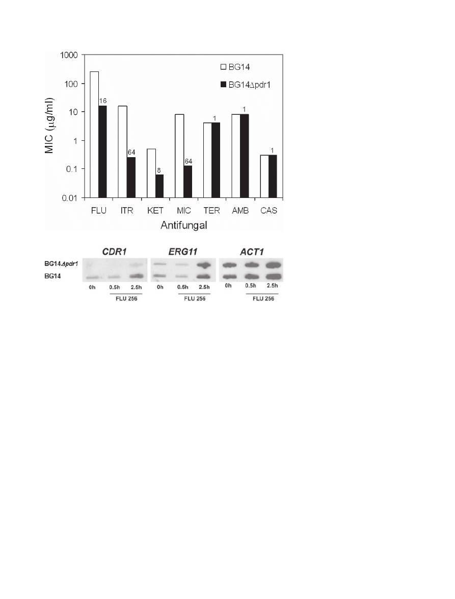
Strain BG14, a model for C. glabrata pathogenesis (e.g.
Domergue et al., 2005), is a ura3 derivative of a clinical
isolate from a patient who failed fluconazole therapy
(Cormack and Falkow, 1999). Consistent with this, BG14
is fluconazole-resistant (MIC
= 256 mg ml
-1
), the molecu-
lar basis for which is unknown. PDR1 disruption in BG14,
conferring cycloheximide hypersensitivity, was previously
reported (Edlind et al., 2005). Here we show that this
disruption also largely reversed BG14 azole resistance.
The fluconazole MIC decreased 16-fold to 16
mg ml
-1
(Fig. 5A); i.e. comparable to the typical clinical isolate
but eightfold above that observed for 66032
Dpdr1
(above). Ketoconazole, itraconazole and miconazole
MICs were similarly reduced in BG14
Dpdr1, but suscep-
tibilities to unrelated antifungals terbinafine, caspofungin
and amphotericin B were unchanged. Expression of
CDR1 and ERG11 was examined by RNA hybridization
(Fig. 5B). In BG14, constitutive expression of CDR1
appeared to be modestly upregulated, but remained
responsive to fluconazole-dependent upregulation. Both
of these were strongly reduced in the
Dpdr1 derivative,
while no effects on ERG11 expression were observed.
Strain 8512 represents a second azole-resistant clinical
isolate with high constitutive CDR1-PDH1 expression (Ver-
A
B
24
48
Fig. 3. Disruption of CDR1 and effects on azole sensitivity.
A. PCR screen of representative
Dcdr1 transformants selected on DOB-URA and their parents 66032 and F15. DNA was purified from isolated
colonies, amplified with the indicated primers pairs, and analysed by gel electrophoresis; loss of the CDR1uF-CDR1iR product and formation
of the CDR1uF-URA3iR product identified
Dcdr1 clones.
B. Broth microdilution assays examining fluconazole sensitivities of parent 66032, azole-resistant mutant F15, and their respective
Dcdr1
disruptants. Absorbance at 630 nm was recorded after 24 or 48 h incubation as indicated; growth was plotted as percentage of drug-free
control.
708
J.-P. Vermitsky et al.
© 2006 The Authors
Journal compilation © 2006 Blackwell Publishing Ltd, Molecular Microbiology, 61, 704–722
mitsky and Edlind, 2004). Following 5FOA-mediated con-
version to ura3, PDR1 was disrupted in strain 8512 (not
shown). Broth microdilution assays demonstrated reduc-
tion of fluconazole MIC from
⬎ 256 to 32 mg ml
-1
. Taken
together, these data suggest that PDR1 is a major deter-
minant of azole sensitivity in C. glabrata, although addi-
tional gene mutations may contribute to clinical resistance.
Microarray analysis: upregulated genes
In light of the major role played by transcription activator
gene PDR1 in C. glabrata azole sensitivity, an examina-
tion of genome-wide changes in gene expression in
mutant F15 was warranted. We first attempted this with
S. cerevisiae microarrays, because these two yeast are
A
B
C
Fig. 4. PDR1 replacement confirms role in azole resistance.
A. Diagram illustrating replacement and PCR screening strategies.
B. PCR screen of representative PDR1 replacements 66032R and F15R (wild-type and F15-derived PDR1, respectively) selected on
cycoheximide-containing plates, and their parent 66032
Dpdr1; strains 66032 and F15 were included as positive controls. DNA was purified
from isolated colonies, amplified with the PDR1uF2-PDR1iR primer pair, and analysed by gel electrophoresis; formation of product confirmed
replacement of PDR1 into its native locus in 66032
Dpdr1.
C. Broth microdilution assay showing that replacement into 66032
Dpdr1 of 66032-derived (66032R) or F15-derived (F15R) PDR1 conferred
the expected low or high-level fluconazole resistance associated with 66032 and F15. Absorbance at 630 nm was recorded after 24 h
incubation; growth was plotted as percentage of drug-free control.
Pdr1 regulates multidrug resistance in Candida glabrata
709
© 2006 The Authors
Journal compilation © 2006 Blackwell Publishing Ltd, Molecular Microbiology, 61, 704–722
closely related. However, the only confirmable change
was upregulation of the PDR5 (18-fold) and PDR15
(ninefold) homologues (data not shown); both of these
genes share 73% nucleotide identity with CDR1.
Therefore, C. glabrata microarrays were developed for
the Affymetrix platform (see Experimental procedures)
and used to examine changes in F15 relative to 66032. In
F15, 78 genes were upregulated
ⱖ twofold relative to
66032. These genes are listed in Table 1 , grouped by
probable function and ordered by expression level.
Among the upregulated are homologues of nine genes
previously identified in microarray studies of S. cerevisiae
Pdr1–Pdr3 gain-of-function mutants (DeRisi et al., 2000;
Devaux et al., 2001). Five of these nine genes encode
putative membrane proteins with roles in small molecule
transport or lipid metabolism. These include, in addition to
CDR1 and PDH1, the upregulated genes YOR1 involved
in oligomycin efflux, RSB1 involved in sphingoid base-
resistance, and RTA1 involved in 7-aminocholesterol
resistance (see SGD website (http://www.yeastgenome.
org)
for
further
information
on
these
genes
and
references).
The
four
remaining
genes
upregulated
in
both
S. cerevisiae and C. glabrata gain-of-function mutants
include PDR1 itself (as previously reported; Vermitsky
and Edlind, 2004), the stress-induced RPN4 encoding a
proteasome gene transcription factor, and the uncharac-
terized open reading frames (ORFs) YLR346C and
YMR102C. The latter encodes a relatively large and
evolutionarily conserved protein with a WD40 domain
commonly found in signalling proteins, and its disruption
has been associated with fluconazole resistance in
S. cerevisiae (Anderson et al., 2003). The YLR346C
product, in contrast, is not conserved; indeed, the
C. glabrata and S. cerevisiae genes are not detectably
homologous in terms of sequence but rather in terms of
chromosomal synteny, flanked in both yeast by unambigu-
ous YLR345W and YLR347C homologues. Also, both
YLR346C products are short (101 and 112 amino acids)
and highly charged in their C-terminal regions. In
S. cerevisiae, Ylr346c is mitochondria-localized and forms
a two-hybrid interaction with MAP kinase Slt2, suggesting
a possible role in mitochondria-nucleus retrograde
signalling.
Among the 69 genes whose upregulation appears to be
C. glabrata F15-specific (i.e. not similarly upregulated in
S. cerevisiae) are three additional homologues encoding
small molecule transporters including quinidine and bile
Fig. 5. Antifungal sensitivities and
CDR1-ERG11 expression in a
Dpdr1
derivative of azole-resistant clinical isolate
BG14.
A. MIC values (at 24 h) determined by broth
microdilution for BG14 and BG14
Dpdr1. A log
scale was used to facilitate comparison of
MICs over a wide range. Numbers above the
BG14
Dpdr1 bars indicate the fold-change
relative to BG14. FLU, fluconazole; ITR,
itraconazole; KET, ketoconazole; MIC,
miconazole; TER, terbinafine; AMB,
amphotericin B; and CAS, caspofungin.
B. RNA was isolated from log-phase BG14
and BG14
Dpdr1 cultures exposed to
256
mg ml
-1
fluconazole for 0–2.5 h,
slot-blotted to membranes, and hybridized to
the indicated gene probes.
A
B
710
J.-P. Vermitsky et al.
© 2006 The Authors
Journal compilation © 2006 Blackwell Publishing Ltd, Molecular Microbiology, 61, 704–722

T
able
1.
C.
glabrata
gene
s
upregulated
ⱖ
twofold
in
fluconazole-resistant
mutant
F15.
Group
Systematic
name
S.
cerevisiae
homologue
a
C.
glabrata
designation
Description
Expression
b
PDRE
c
F15
F15/66032
Small
molecule
transport
IPF6352
IPF9719
d
PDR5
(CDR1
)
d
PDR15
(PDH1
)
CAGL0M01760g
CAGL0F02717g
ABC
transporter
involved
in
azole/multidrug
resistance
ABC
transporter
involved
in
azole/multidrug
efflux
41
29
2.5
9.6
134,298,388,516
521,557
IPF1620
QDR2
CAGL0G08624g
MFS
transporter
involved
in
quinidine/multidrug
efflux
23
4.5
848
IPF8922
d
YOR1
CAGL0G00242g
ABC
transporter
involved
in
multidrug
efflux
16
1
1
648
IPF982
YBT1
CAGL0C03289g
ABC
transporter
involved
in
bile
acid
transport
13
7.7
450
IPF3303
OAC1
CAGL0K1
1616g
Mitochondrial
inner
membrane
transporter
4.7
2.5
839
Lipid,
fatty
acid,
and
sterol
metabolism
IPF5152
IPF2180
IPF4136
d
RT
A
1
HFD1
d
RSB1
CAGL0K00715g
CAGL0K03509g
CAGL0L10142g
Overexpression
confers
7-aminocholesterol
resistance
Putative
mitochondrial
fatty
aldehyde
dehydrogenase
Sphingolipid
flippase
22
13
12
7.0
5.6
2.8
300,379
218
641,881
IPF8678
LCB5
CAGL0K05995g
Minor
sphingoid
long-chain
base
kinase
12
2.4
804
IPF8367
LAC1
CAGL0M10219g
Ceramide
synthase
component
5.7
2.5
531
IPF1002
ARE1
CAGL0C02981g
acyl-CoA:sterol
acyltransferase;
sterol
esterification
4.6
4.6
1
1
4
IPF4884
A
TF2
CAGL0D05918g
Alcohol
acetyltransferase;
steroid
detoxification
3.1
9.6
30,195,560,772
IPF2739
SPO14
CAGL0L03135g
Phospholipase
D
0.8
2.6
–
IPF2620
CSR1
CAGL0D00946g
Phosphatidylinositol
transfer
protein
0.4
3.8
239
Cell
stress
IPF6847
HSP12
CAGL0J04202g
Stress-induced
membrane
protein
29
4.0
849
IPF3173
YNL134c
CAGL0K09702g
Alcohol
dehydrogenase
motif;
stress-induced
14
9.5
541
IPF4605
YML131W
CAGL0K12958g
Alcohol
dehydrogenase
motif,
stress-induced
7.3
9.1
594
IPF6629
HSP31
CAGL0C00275g
Possible
chaperone
and
cysteine
protease
3.5
2.0
–
IPF4140
YOR052C
CAGL0L10186g
Uncharacterized;
stress-induced
2.3
3.2
–
IPF8736
TPS3
CAGL0H02387g
T
rehalose-6-phosphate
synthase/phosphatase
subunit
1.5
4.4
–
IPF5558
HSP42
CAGL0E00803g
Small
cytosolic
stress-induced
chaperone
0.6
4.9
–
T
ranscription
IPF5076
d
RPN4
CAGL0K01727g
T
ranscription
factor
for
proteasomegenes
16
3.9
378,394,552
IPF5932
SUT1
CAGL0I04246g
T
ranscription
factor
involved
in
sterol
uptake
15
2.4
–
IPF3325
d
PDR1
CAGL0A00451g
T
ranscription
factor
involved
in
multidrug
resistance
9.6
2.3
557,701
IPF7202
T
AF9
CAGL0M05005g
Subunit
of
TFIID
and
SAGA
complexes
1.0
6.3
–
IPF6366
YPR013C
CAGL0M01870g
Uncharacterized;
potential
zinc
finger
0.8
2.9
–
IPF21
13
SIP3
CAGL0I01980g
Activates
transcription
through
DNA-bound
Snf1
0.4
2.2
–
IPF1
18
HOT1
CAGL0H08866g
T
ranscription
factor
involved
in
osmostress
response
0.1
4.9
228
DNA
replication
and
damage
response
IPF9036
IPF9035
IPF2521
YIM1
MEC3
DBF4
CAGL0M09713g
CAGL0M09735g
CAGL0E04576g
Implicated
in
DNA
damage
response
DNA
damage
checkpoint
Regulatory
subunit
of
Cdc7p-Dbf4p
kinase
complex
40
1.6
0.7
12
15
2.4
127,179
1
10,162
–
IPF785
DPB3
CAGL0B03355g
DNA
polymerase
II
subunit
0.2
2.7
–
Protein
synthesis,
modification,
or
degradation
IPF3014
IPF6742
IPF3846
OCH1
UFD1
NCE3
CAGL0A01738g
CAGL0J08096g
CAGL0G01540g
Mannosyltransferase
of
cis
-Golgi
apparatus
Recognition
of
polyubiquitinated
proteins
Carbonic
anhydrase-like;
non-classical
protein
export
5.2
4.4
3.1
2.5
2.2
2.9
–
–
–
IPF3072
RPN8
CAGL0K08866g
Non-A
TPase
regulatory
subunit
of
26S
proteasome
2.9
2.9
–
IPF8484
PCI8
CAGL0M12749g
Possible
shared
subunit
of
Cop9
signalosome
and
eIF3
0.2
2.4
–
V
esicular
and
protein
transport
IPF7414
IPF8257
GSF2
YPT52
CAGL0L01485g
CAGL0G07689g
ER
membrane,
hexose
transporter
secretion
GTPase
required
for
vacuolar
protein
sorting
10
1.6
2.4
2.8
208
–
IPF8439
MEH1
CAGL0L0221
1g
Component
of
the
EGO
complex;
microautophagy
0.5
5.7
–
IPF4445
VPS28
CAGL0H05181g
Component
of
ESCR
T
-I
complex;
protein
trafficking
0.5
2.2
–
IPF4173
VTI1
CAGL0L10604g
Involved
in
cis
-Golgi
membrane
traffic
0.3
6.7
741
IPF3260
GYL1
CAGL0K10934g
putativegAP
for
Y
pt1
involved
in
polarized
exocystosis
0.3
4.0
–
IPF271
VPS51
CAGL0H06809g
Golgi-associated
retrograde
protein
complex
0.2
6.1
–
Pdr1 regulates multidrug resistance in Candida glabrata
711
© 2006 The Authors
Journal compilation © 2006 Blackwell Publishing Ltd, Molecular Microbiology, 61, 704–722

T
able
1.
cont.
Group
Systematic
name
S.
cerevisiae
homologue
a
C.
glabrata
designation
Description
Expression
b
PDRE
c
F15
F15/66032
Signal
transduction
IPF1489
IPF8227
BAG7
CDC25
CAGL0I07249g
CAGL0D06512g
GAP
for
Rho1;
cell
wall
and
cytoskeleton
homeostasis
Membrane
bound
GEF
for
Ras1-Ras2
1.6
1.2
2.9
5.8
–
–
IPF351
GAC1
CAGL0F04917g
Regulatory
subunit
forglc7
protein
phosphatase
0.9
4.7
–
IPF2382
YNL234W
CAGL0J07502g
Similar
toglobins
with
haem-binding
domain
0.5
3.1
–
IPF512
GPG1
CAGL0F071
17g
Subunit
of
heterotrimericg
protein,
interacts
withgrp1
0.4
2.6
–
IPF5914
KIN3
CAGL0I04422g
Protein
kinase
0.2
5.6
–
Mitochondrial
IPF2122
FMP48
CAGL0K04301g
Ser/Thr
protein
kinase;
mitochondrial
1
1
2.8
–
IPF7121
YGR046W
CAGL0G03861g
Essential
protein
involved
in
mitochondria
transport
0.4
4.4
–
Cell
wall
IPF9549
FLO1
CAGL0E00209g
Flo1-like
family
of
cell
wall
proteins
2.6
3.0
284,419
Amino
acid
and
carbohydrate
metabolism
IPF496
IPF4499
IPF5315
PYC1
STR3
MET8
CAGL0F06941g
CAGL0L06094g
CAGL0K06677g
Pyruvate
carboxylase
isoform
Cystathionine
beta-lyase
Bifunctional
dehydrogenase
and
ferrochelatase
4.6
0.7
0.2
2.3
5.0
7.8
–
–
–
Chromatin/
chromosome
structure
IPF8319
IPF390
IPF8077
SMD3
SPC19
SPC97
CAGL0M04631g
CAGL0F05467g
CAGL0I02464g
Core
Sm
spliceosome
protein
Sm
D3
Component
of
Dam1
spindle
pole
complex
Component
of
microtubule-nucleating
T
ub4
complex
0.9
0.5
0.5
3.7
13.1
2.8
–
–
–
IPF2730
SPC34
CAGL0L03223g
Component
of
Dam1
spindle
pole
complex
0.4
2.5
–
Other
metabolism
IPF6032
CAGL0M14091g
Putative
quinone
reductase/NADPH
dehydrogenase
3.8
9.8
244,532
IPF6034
ADH6
CAGL0M14047g
NADPH-dependent
cinnamyl
alcohol
dehydrogenase
2.3
2.7
–
IPF1279
YPR1
CAGL0A02816g
2-methylbutyraldehyde
reductase
0.6
5.6
–
IPF4182
INP1
CAGL0L10736g
Peripheral
membrane
protein
of
peroxisomes
0.2
5.5
–
Uncharacterized
IPF61
16
YIL077C
CAGL0M12947g
Uncharacterized
20
38
472,502
IPF8009
YJL163C
CAGL0M08426g
Uncharacterized;
ARS
in
promoter
9.1
10
450
IPF2520
CAGL0E04554g
Uncharacterized;
no
similarities
8.8
6.4
–
IPF2196
d
YMR102C
CAGL0K03377g
T
ranscribed
along
with
MDRgenes
by
Yrr1/Yrm1
8.1
4.4
898
IPF3019
CAGL0A01650g
Uncharacterized;
no
similarities
7.2
4.4
564
IPF3875
d
YLR346C
CAGL0G01
122g
Uncharacterized;
syntenic
but
minimal
similarities
3.3
22
793,803
IPF3655
YLR177W
CAGL0B01078g
Uncharacterized
2.7
2.5
–
IPF1546
CAGL0G09603g
Uncharacterized;
very
weak
similarity
to
Y
or186w
2.1
6.1
–
IPF2382
YNL234W
CAGL0J07502g
Similar
toglobins
with
haem-binding
domain
0.5
3.1
–
IPF4149
YOR059C
CAGL0L10318g
Uncharacterized
0.5
2.4
–
IPF6420
YGR126W
CAGL0I10604g
Uncharacterized
0.1
19
–
IPF2249
YHL010C
CAGL0K02563g
Uncharacterized;
mammalian
BRAP2
homologue
0.1
5.0
–
IPF9234
CAGL0M07766g
Uncharacterized;
no
similarities
0.1
4.0
–
a.
Parentheses
indicate
a
previously
named
C.
glabrata
gene
.
b.
Expression
in
C.
glabrata
66032
is
represented
in
arbitrary
microarray
units
(for
comparison,
actin
and
b
-tubulin
gene
homologues
ACT1
and
TUB2
had
average
expression
levels
of
66
and
8.6
respectively).
F15/66032
represents
the
ratio
of
expression
in
the
fluconazole-resistant
mutant
vs.
its
parent
(for
comparison,
ACT1
and
TUB2
had
ratios
of
0.6
and
0.8
respectively).
c.
Promoter
regions
(900
bp)
were
searched
for
matches
to
the
S.
cerevisiae
PDRE
consensus
TCCR
YGSR.
Numbers
indicate
the
distance
(in
bp)
upstream
of
the
A
T
G
start
codon
of
the
PDRE;
hyphens
(–)
indicate
the
absence
of
a
PDRE.
d.
Genes
similarly
upregulated
in
S.
cerevisiae
Pdr1–Pdr3
gain-of-function
mutants
(DeRisi
et
al
.,
2000;
Devaux
et
al
.,
2001).
712
J.-P. Vermitsky et al.
© 2006 The Authors
Journal compilation © 2006 Blackwell Publishing Ltd, Molecular Microbiology, 61, 704–722

acid efflux protein genes QDR2 and YBT1. Additional lipid
metabolism genes include ARE1 whose disruption in
S. cerevisiae confers azole hypersensitivity (T. Edlind,
unpubl. data) and ATF2 involved in fatty acid and steroid
detoxification. A third group of well-represented genes are
involved in the cell stress response, including membrane
protein gene HSP12, and YML131W-YNL134C; the latter
two are unrelated by
BLAST
but share an ADH_zinc_N
domain (identified by CD-search; Marchler-Bauer and
Bryant, 2004) characteristic of zinc-dependent alcohol
dehydrogenases-oxidoreductases. YML131W-YNL134C
are also coordinately upregulated in S. cerevisiae in
response to diverse stresses including heat, oxidizing
agents, ethanol, nitrogen depletion and stationary phase
(Gasch et al., 2000; see Expression Connection at
http://www.yeastgenome.org).
A
similarly
regulated
S. cerevisiae gene is GRE2, also encoding an oxi-
doreductase and among the genes upregulated in Pdr1–
Pdr3 gain-of-function mutants (DeRisi et al., 2000;
Devaux et al., 2001). This provides an example of analo-
gous
but
non-homologous
genes
upregulated
in
C. glabrata F15 and S. cerevisiae Pdr1–Pdr3 mutants.
Notable among the remaining upregulated genes with
significant expression levels are: SUT1 encoding a tran-
scription factor involved in sterol uptake and hypoxic gene
expression, YIM1 implicated in DNA damage response,
OCH1 and GSF2 involved in Golgi-ER functions,
proteasome-related genes UFD1 and RPN8, putative
mitochondrial protein kinase gene FMP48, a quinone
reductase-like gene curiously lacking in other fungal
genomes but present in many bacteria and vertebrates,
and the uncharacterized YIL077C whose product has
been mitochondria-localized but interacts with a nuclear
transcriptional complex.
Microarray analysis: downregulated genes
There were 31 genes downregulated
ⱖ twofold in F15
relative to parent 66032 (Table 2). Only one of these was
also downregulated in S. cerevisiae Pdr1–Pdr3 gain-of-
function mutants: membrane transporter gene PDR12
involved in efflux of weak organic acids such as sorbate.
Additional genes with significantly downregulated expres-
sion include zinc transporter gene ZRT1, major facilitator
genes including FLR1 implicated in fluconazole efflux, and
homologues of cell surface protein genes MUC1-EPA2 and
MKC7 implicated in adhesion and aspartic protease activ-
ity respectively. Finally, a gene was downregulated whose
product has clear homology to the WRY family of putative
membrane-anchored proteins previously identified only in
C. albicans (unpublished annotation in NCBI database).
This family has nine paralogues in C. albicans and seven in
C. glabrata but none, surprisingly, in S. cerevisiae, sug-
gesting a possible role in mammalian colonization.
Confirmation of microarray results
As our studies represent the first application of these
C. glabrata microarrays, it was important to validate the
results by independent methods. RNA blots or real-time
reverse transcription (RT)-PCR were used to examine the
expression of selected genes identified as upregulated in
the microarray (other than already confirmed CDR1,
PDH1 and PDR1). For five of seven genes tested
(YLR346C, YOR1, YNL134C, YML131W and RTA1),
RNA blots confirmed F15 upregulation relative to the
parent 66032 strain (Fig. 6A). The two exceptions (MEC3
and YJL163C) represent genes whose expression in both
parent and F15 were below the level of detection by RNA
blot (not shown).
For all nine genes tested by real-time RT-PCR (YOR1,
RTA1, RPN4, QDR2, MET8, BAG7, CSR1, PDR1 and
YBT1), the upregulation observed by microarray was
confirmed (Fig. 5B). For most of these, the results were
quantitatively
similar;
e.g.
YOR1
was
upregulated
16-fold by microarray and 17-fold by RT-PCR and CSR1
was upregulated 3.8-fold by microarray and 2.9-fold by
RT-PCR. Expression of ERG11 encoding the azole
target lanosterol demethylase was essentially unaltered
by RT-PCR (not shown), in agreement with microarray
analysis (F15/66032
= 0.6) and RNA hybridization (Ver-
mitsky and Edlind, 2004). One anomaly in the microar-
ray analysis was the relatively low upregulation of CDR1
(2.5-fold) compared with its high upregulation (c. 20-fold)
in both RNA blots and RT-PCR (Fig. 2). Furthermore,
Cdr1 was strongly upregulated on the protein level, as
shown by SDS-PAGE of membrane preparations fol-
lowed by mass spectrometric identification of eluted
bands (Rogers et al., submitted for publication). Poten-
tial explanations for this anomaly include degradation or
masking of the CDR1 mRNA region targeted by the
microarray, or a saturation effect due to the relatively
high CDR1 basal expression.
Promoter sequence analysis
To identify a candidate C. glabrata Pdr1 response
element (PDRE), we took advantage of the F15 microar-
ray data, the available genome sequence, and the
evolutionary relatedness of this yeast to S. cerevisiae.
The promoter regions (900 bp upstream of the start
codon) for all genes listed in Tables 1 and 2 were
searched for a match to the consensus S. cerevisiae
PDRE (DeRisi et al., 2000; Devaux et al., 2001):
TCC(GA)(CT)G(GC)(AG). At least one match to this
sequence was identified in 31 of the 78 genes (40%)
upregulated
ⱖ twofold. Moreover, one or more PDRE
were identified in all nine genes upregulated in both
C. glabrata and S. cerevisiae gain-of-function mutants, in
Pdr1 regulates multidrug resistance in Candida glabrata
713
© 2006 The Authors
Journal compilation © 2006 Blackwell Publishing Ltd, Molecular Microbiology, 61, 704–722

T
able
2.
C.
glabrata
genes
downregulated
ⱖ
twofold
in
fluconazole-resistant
mutant
F15
compared
with
parent
66032.
Group
Systematic
name
S.
cerevisiae
homologue
a
C.
glabrata
designation
Description
Expression
b
PDRE
c
66032
F15/66032
Small
molecule
transport
IPF5505
IPF4392
ZR
T1
FLR1
CAGL0E01353g
CAGL0H06017g
High-affinity
zinc
transporter
Multidrug
efflux
pump
of
major
facilitator
superfamily
10
6.1
0.5
0.4
–
–
IPF6218
d
PDR12
CAGL0M07293g
ABC
transporter
of
weak
organic
acids
4.5
0.2
–
IPF7808
YHR048W
CAGL0J00363g
Uncharacterized;
major
facilitator
superfamily
2.2
0.4
–
IPF867
A
TR1
CAGL0B02343g
Multidrug
efflux
pump
of
major
facilitator
superfamily
1.8
0.4
–
IPF1568
SNG1
CAGL0G09273g
Nitrosoguanidine
resistance;
putative
transporter
1.3
0.1
–
Cell
stress
IPF3249
CT
A1
CAGL0K10868g
Catalase
A
8.8
0.4
–
IPF1
1021
CRS5
CAGL0H04257g
Copper-binding
metallothionein-like
protein
3.4
0.4
–
IPF9018
L
TV1
CAGL0J00891g
Required
for
growth
at
low
temperature
1.0
0.4
656
IPF9641
SLG1
CAGL0F01507g
Sensor
of
stress-activated
Pkc1-Slt2
pathway
0.8
0.5
–
IPF5937
CUP2
(AMT1
)
CAGL0I04180g
Metal-activated
transcriptional
factor
0.9
0.2
–
Carbohydrate
metabolism
IPF986
IPF61
17
SDH2
DOG2
CAGL0C03223g
CAGL0M12925g
Succinate
dehydrogenase
iron-sulphur
protein
subunit
2-deoxyglucose-6-phosphate
phosphatase
4.9
1.6
0.4
0.3
–
–
IPF290
PCK1
CAGL0H06633g
Phosphoenolpyruvate
carboxykinase
1.2
0.3
–
Cell
cycle
control
IPF2555
SDS22
CAGL0D00264g
Nuclear
regulatory
subunit
ofglc7
phosphatase
0.3
0.2
–
RNA
processing
IPF8796
PNO1
CAGL0K09460g
Nucleolar
protein
required
for
pre-rRNA
processing
3.0
0.4
–
IPF1
130
AAR2
CAGL0A04543g
Component
of
the
U5
snRNP
1.0
0.4
–
IPF9324
DIM1
CAGL0L07678g
Essential
18S
rRNA
dimethylase
1.0
0.5
–
IPF5272
NOP8
CAGL0B01397g
Nucleolar
protein
required
for
ribosome
biogenesis
0.7
0.4
–
Cell
surface
and
cytoskeleton
IPF9500
IPF8398
MKC7
CAGL0J01793g
CAGL0E01771g
Muc1/Epa2-like
putative
cell
surface
protein
GPI-anchored
aspartyl
protease
4.2
3.6
0.3
0.3
–
–
IPF807
MNT3
CAGL0B03003g
a
-1,3-mannosyltransferase
involved
in
O-glycosyltation
3.1
0.4
–
IPF3539
ECM4
CAGL0G02101g
Promoter
insertion
mutant
is
calcofluor-hypersensitive
1.6
0.3
–
IPF810
MNT3
CAGL0B02992g
a
-1,3-mannosyltransferase
involved
in
O-glycosyltation
1.4
0.3
–
IPF821
CAGL0B02882g
C.
albicans
WR
Y
family;
transmembrane
domain
0.5
0.1
–
IPF5228
ECM18
CAGL0B01969g
Insertion
mutant
is
calcofluor
hypersensitive
0.3
0.3
–
IPF7581
TPM2
CAGL0L08338g
Minor
isoform
of
tropomyosin
0.3
0.4
–
Uncharacterized
IPF493
YIR035C
CAGL0F06897g
Uncharacterized;
alcohol
dehydrogenase
domain
3.3
0.4
–
IPF3428
YNL095C
CAGL0G06468g
Uncharacterized;
related
to
ECM3
2.7
0.4
–
IPF7665
SIP5
CAGL0L06864g
Uncharacterized;
interacts
withglc7
and
Snf1
0.4
0.3
–
IPF5688
FMP16
CAGL0G05269g
Uncharacterized;
mitochondrial
0.3
0.3
–
a.
Parentheses
indicate
a
previously
named
C.
glabrata
gene
.
b.
Expression
in
C.
glabrata
66032
is
represented
in
arbitrary
microarray
units
(for
comparison,
actin
and
b
-tubulin
gene
homologues
ACT1
and
TUB2
had
average
expression
levels
of
66
and
8.6
respectively).
F15/66032
represents
the
ratio
of
expression
in
the
fluconazole-resistant
mutant
vs.
its
parent
(for
comparison,
ACT1
and
TUB2
had
ratios
of
0.6
and
0.8
respectively).
c.
Promoter
regions
(900
bp)
were
searched
for
matches
to
the
S.
cerevisiae
PDRE
consensus
TCCR
YGSR.
Numbers
indicate
the
distance
(in
bp)
upstream
of
the
A
T
G
start
codon
of
the
PDRE;
hyphens
(–)
indicate
the
absence
of
a
PDRE.
d.
Genes
similarly
downregulated
in
S.
cerevisiae
Pdr1–Pdr3
gain-of-function
mutants
(DeRisi
et
al
.,
2000;
Devaux
et
al
.,
2001).
714
J.-P. Vermitsky et al.
© 2006 The Authors
Journal compilation © 2006 Blackwell Publishing Ltd, Molecular Microbiology, 61, 704–722

14 of 15 genes in the small molecule transport and lipid
metabolism groups, and in 26 of 34 genes with expression
level
⬎ 3 (arbitrary microarray units). Conversely, only
one PDRE was identified among the 31 downregulated
gene promoters (Table 2); similarly, none was identified in
the promoters of representative housekeeping genes
(ACT1, TEF1, TDH3) or azole target gene ERG11. This
analysis therefore identifies TCC(AG)(TC)G(GC)(AG) as
a strong candidate for the C. glabrata PDRE. More spe-
cifically, we note a clear preference for G as the penulti-
mate base (95% of PDREs) and A as the final base (84%),
although two of the four PDREs within the CDR1 promoter
have G as the final base.
Two exceptions warrant discussion. The promoters of
C. glabrata upregulated genes RPN8 and YOR052C lack
a PDRE but include perfect matches to the S. cerevisiae
Rpn4 transcription factor-binding site GGTGGCAAA
(Mannhaupt et al., 1999); perfect or near-perfect matches
are also found in the promoters of their S. cerevisiae
homologues. As noted above, Rpn4 is upregulated in
both C. glabrata Pdr1 and S. cerevisiae Pdr1–Pdr3
gain-of-function mutants. Thus, RPN8-YOR052C upregul-
ation is likely Rpn4-mediated and only indirectly Pdr1-
mediated.
Candida glabrata F15 exhibits additional phenotypes
predicted by microarray analysis which may alter
virulence
As coordinate CDR1-PDH1 upregulation is commonly
observed in C. glabrata azole-resistant clinical isolates
(Bennett et al., 2004; Vermitsky and Edlind, 2004; San-
guinetti et al., 2005), the responsible mutations must
have minimal effects on fitness. On the other hand,
these mutations could alter C. glabrata in subtle ways
that affect, for example, its relative virulence in the
bloodstream versus mucosa. The F15 microarray results
provided us with an opportunity to begin to test this
general hypothesis. Specifically, we looked for pheno-
types other than azole susceptibility predicted to be
associated with altered expression of genes coregulated
with CDR1-PDH1.
Upregulation of the YOR1 transporter 11-fold (Table 1)
predicts that azole-resistant F15 should be cross-
resistant to oligomycin, an inhibitor of mitochondrial
F
1
F
0
ATPase and known S. cerevisiae Yor1 substrate.
This
was
confirmed
by
broth
microdilution
assay
(MIC
= 0.5 mg ml
-1
for F15 vs. 0.125
mg ml
-1
for parent
66032), using medium with glycerol as respiratory
carbon source. Yor1 also confers tolerance in S. cerevi-
siae to a wide range of organic anions such as lepto-
mycin B and acetic acid, along with cadmium (Cui et al.,
1996). PDR12, downregulated fivefold in F15, similarly
encodes an efflux pump with specificity for organic
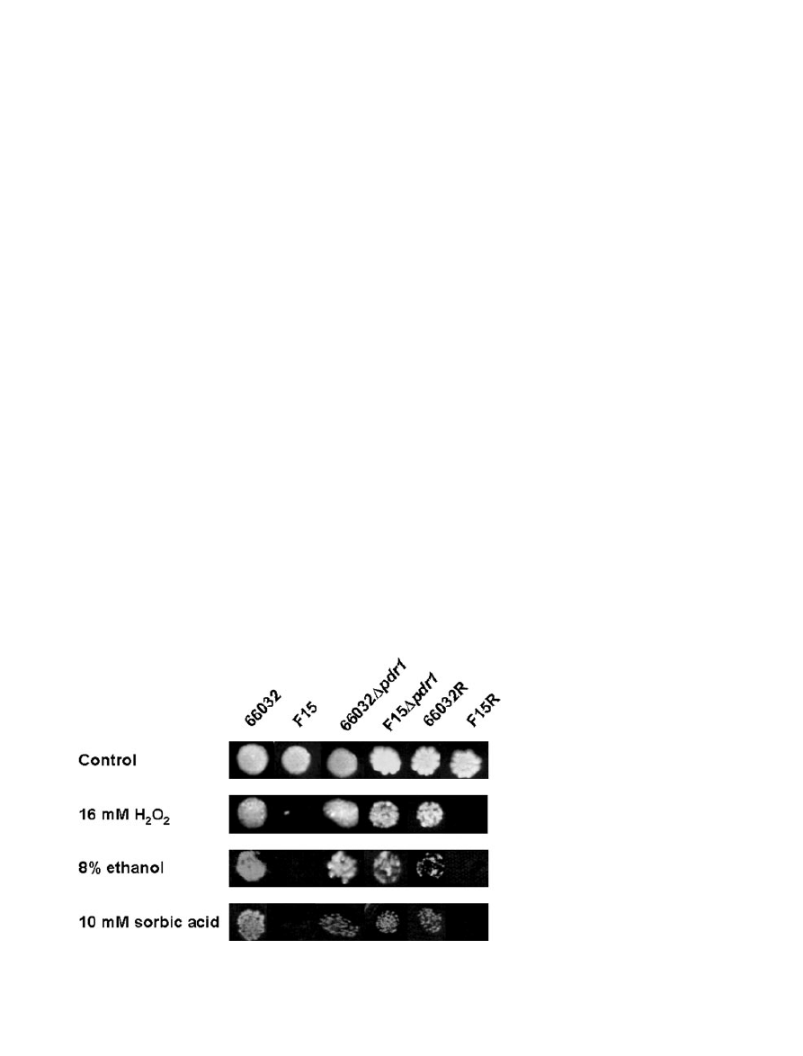
acids, in particular sorbic acid (Piper et al., 1998). Spot
assays (Fig. 7) confirmed sorbate hypersensitivity of
F15, although the effects on MIC were modest (4 mM for
F15 vs. 8 mM for 66032). With respect to organic acid
sensitivity, PDR12 downregulation may be largely offset
by YOR1 upregulation. There was no detectable change
in sensitivity to acetic, boric, or lactic acids (MICs
= 64,
16 and 250 mM respectively).
Fig. 6. Confirmation of F15/66032 microarray
results for selected genes by RNA
hybridization and real-time RT-PCR.
A. RNA was isolated from log phase cultures,
slot-blotted to membranes, and hybridized to
the indicated gene probes; ACT1 served as
loading control.
B. Quantitative real-time RT-PCR analysis of
relative gene expression in F15 versus 66032.
Data are shown as mean
± SD.
A
B
Pdr1 regulates multidrug resistance in Candida glabrata
715
© 2006 The Authors
Journal compilation © 2006 Blackwell Publishing Ltd, Molecular Microbiology, 61, 704–722
YML131W and YNL134C homologues were similarly
upregulated c. ninefold in F15. As noted above, the
products of these uncharacterized genes share a
domain
characteristic
of
alcohol
dehydrogenases/
oxidoreductases; furthermore, they are coregulated in
response to environmental stresses including heat shock
and treatment with reactive oxygen species or ethanol
(Expression Connection, SGD website). Conversely, cata-
lase gene CTA1 was downregulated 2.5-fold. Consistent
with this, F15 demonstrated hypersensitivity to hydrogen
peroxide by spot assay (Fig. 7) and broth microdilution
(MIC
= 16 mM vs. 32 mM for 66032). Similarly, F15 dem-
onstrated hypersensitivity to ethanol (Fig. 7; MIC
= 2% vs.
4% for 66032). Equivalent results were obtained with F15
PDR1 replacement clone F15R as compared with wild-
type PDR1 replacement clone 66032R (Fig. 7), confirm-
ing that these altered phenotypes resulted from the PDR1
gain-of-function mutation. (Note that the ura3 phenotype
of 66032R can account for its variable growth relative to
66032, an effect also observed with the 66032 ura3 strain;
not shown.) Finally, we examined sensitivity to heat shock
by exposing mid-log or early stationary phase cultures to
50°C for 10 min, following by plating on YPD with incuba-
tion at 35°C for 3 days to obtain colony counts. For F15
versus 66032, viability was 6 versus 0.3% and 16 versus
5% in log and stationary phase cultures respectively.
Taken together, these data suggest that regulatory muta-
tions conferring azole resistance in C. glabrata may have
both positive and negative effects on fitness and
virulence.
Conclusions
An important ‘virulence factor’ for the emerging opportunist
C. glabrata appears to be its capacity for intrinsic low-level
and acquired high-level azole resistance. The studies com-
pleted here with laboratory mutant F15, and initial studies
with representative clinical isolates, identify the zinc cluster
transcriptional activator Pdr1 as a key regulator of azole/
multidrug transporter genes CDR1 and PDH1. Constitutive
upregulation of these genes is observed in most azole-
resistant clinical isolates; furthermore, they are transiently
upregulated in sensitive isolates following azole exposure.
Consistent with this, in PDR1 disruptants acquired resis-
tance was reversed and intrinsic resistance was reduced.
We have shown that F15 Pdr1 has a gain-of-function
mutation analogous to those previously characterized in
S. cerevisiae Pdr1–Pdr3, and this mutation is sufficient to
confer azole resistance. Pdr1 mutation is not, however,
necessary for resistance, because at least one resistant
strain analysed had unchanged PDR1 (Vermitsky and
Edlind, 2004). Azole resistance may potentially arise from
mutations in upstream signalling proteins or transcription
cofactors, both of which remain to be defined (although
histone modifying enzymes represent likely cofactors).
Moreover, we observed here that PDR1 disruptants,
although azole hypersensitive, continued to yield sponta-
neous azole-resistant mutants at reduced frequency.
These Pdr1-independent resistance mechanisms, and
their clinical relevance, warrant further study.
Microarray analysis of genome-wide gene expression
has become a central tool in molecular genetics, and the
arrays developed and tested here should be particularly
useful in studies of C. glabrata in large part because of
its close evolutionary relatedness to S. cerevisiae.
Most genes with altered expression in F15 had well-
characterized S. cerevisiae homologues. This allowed
us to predict F15 phenotypes, a number of which were
tested including sensitivity to organic acids, alcohols
and oxidants. Ultimately, these data should help us to
Fig. 7. Spot assays examining sensitivity of
66032 and its azole-resistant mutant F15 to
hydrogen peroxide, ethanol and sorbic acid.
Approximately 300 cells were spotted on YPD
agar with the indicated inhibitor. Plates were
incubated for 2–4 days at 35°C. For
comparison, 66032
Dpdr1, F15Dpdr1 and the
66032
Dpdr1-PDR1 replacement strains
66032R and F15R were examined in parallel.
716
J.-P. Vermitsky et al.
© 2006 The Authors
Journal compilation © 2006 Blackwell Publishing Ltd, Molecular Microbiology, 61, 704–722

understand and possibly exploit the consequences for
C. glabrata of regulatory mutations leading to azole
resistance. F15 hypersensitivity to hydrogen peroxide is
of particular interest, because this implies hypersensitiv-
ity to immune cells such as neutrophils and environ-
ments such as the lactobacillus-colonized vaginal tract in
which hydrogen peroxide plays an important role.
Although the relatedness of C. glabrata and S. cerevi-
siae is invaluable in terms of predicting gene function,
microarray analysis indicated that the Pdr1 and Pdr1-
Pdr3 gain-of-function mutants of these yeast are more
different than similar. This no doubt reflects the very dif-
ferent pressures placed on these organisms by their
very different niches; e.g. the skin of a grape versus the
human mucosa.
Following submission of this manuscript, Tsai et al.
(2006) reported results that parallel and complement
those described here. Specifically, a C. glabrata labora-
tory strain with transposon-disrupted PDR1 exhibited flu-
conazole hypersensitivity and diminished CDR1-PDH1
expression. Importantly, two fluconazole-resistant clinical
isolates with increased CDR1-PDH1 expression were
shown to harbour PDR1 mutations, and integrative
transformation of these alleles conferred fluconazole
resistance and upregulated CDR1-PDH1 expression on
the pdr1::Tn strain. These results confirm the relevance
of laboratory mutant F15 as a model for clinical
resistance.
Experimental procedures
Media, inhibitors and strains
For most experiments, the medium employed was YPD (1%
yeast extract, 2% peptone, 2% dextrose). Gene disruptants
and ura3 mutants were selected on DOB (synthetic defined
medium with dextrose) with complete supplement mixture
(CSM) or CSM lacking uracil/uridine (-URA) (Qbiogene/BIO
101). Drugs were obtained from the following sources: flu-
conazole (Pfizer), itraconazole (Janssen), terbinafine (Novar-
tis); caspofungin (Merck), amphotericin B, miconazole and
cyloheximide (Sigma-Aldrich). They were dissolved in dim-
ethyl sulphoxide (DMSO); the final DMSO concentration was
ⱕ 0.5% in all experiments which had no detectable effect on
growth. Sorbic acid, lactic acid, acetic acid and hydrogen
peroxide (Sigma) were diluted as necessary in water. Strains
were previously described (Vermitsky and Edlind, 2004) or
constructed as described below.
Isolation of ura3 strains
Wild-type URA3 yeast strains are sensitive to 5FOA. To
isolate 5FOA-resistant mutants, a single colony from a fresh
YPD plate was streaked on DOB
+ CSM agar containing
0.1% 5FOA (Research Products International) and incu-
bated at 35°C for 3 days. Colonies were streaked for isola-
tion on YPD and DOB-URA; those that failed to grow on the
latter were then tested for URA3 complementation by trans-
formation with pRS416 (shuttle vector with S. cerevisiae
URA3) and selection on DOB-URA plates. Yeast transfor-
mations employed the Frozen-EZ Yeast Transformation II
Kit (Zymo Research) as previously described (Edlind et al.,
2005).
Gene disruption and replacement
The PRODIGE method for PCR product-mediated gene dis-
ruption was employed (Fig. 1A; Edlind et al., 2005). Briefly,
primers (80 mers; Table 3) were designed to precisely
replace, after homologous recombination, a C. glabrata
coding sequence (CDS) with the selection marker CDS.
These primers consisted of c. 60 nucleotides at the 5
′ end
complementary to C. glabrata sequences directly upstream
and downstream of the targeted CDS and c. 20 nucleotides
at the 3
′ end complementary to the S. cerevisiae URA3
CDS contained in plasmid template pRS416. PCR products
generated with these primers were used to transform
C. glabrata ura3 strains. Following selection on DOB-URA
medium, transformants were screened by PCR with specific
primer pairs (Table 3; Fig. 1A) to confirm replacement of the
targeted CDS with URA3 CDS. DNA was generally pre-
pared by phenol extraction of glass bead-disrupted cells
(Edlind et al., 2005); some screens employed colony PCR
in which a small volume of cells was added directly to the
PCR mix.
For PDR1 replacement, a PCR product representing the
PDR1 CDS plus 430–680 bp upstream and downstream
sequence was amplified with primers PDR1uF-PDR1dR
(Table 3) from 66032 or F15 genomic DNA. These products
were used to transform 66032
Dpdr1 strain with selection on
1
mg ml
-1
cycloheximide-containing YPD plates. Colonies
were screened as above with primer pair PDR1uF2-PDR1iR
(Table 3; Fig. 4A).
Broth microdilution assay
Fresh overnight cultures from a single colony were diluted
1 : 100 in YPD, incubated for 3 h with aeration, and then
counted
in
a
haemocytometer
and
diluted
again
to
1
¥ 10
4
cells ml
-1
. Aliquots of 100
ml were distributed to wells
of a 96-well flat-bottomed plate, except for row A which
received 200
ml. Inhibitor was added to row A to the desired
concentration and then serially twofold diluted to rows B
through G; row H served as inhibitor-free control. Plates were
incubated at 35°C for the indicated times. Absorbance at
630 nm was read with a microplate reader; background due
to medium was subtracted from all readings. The MIC
(minimum inhibitory concentration) was defined as the lowest
concentration inhibiting growth at least 80% relative to the
drug-free control.
RNA hybridization
Log phase cultures in YPD at 35°C were adjusted to 3
¥ 10
6
cells ml
-1
and incubated for an additional 3 h. In some
studies, cultures were divided into equal portions to which
Pdr1 regulates multidrug resistance in Candida glabrata
717
© 2006 The Authors
Journal compilation © 2006 Blackwell Publishing Ltd, Molecular Microbiology, 61, 704–722

Table 3. Primers used in this study (grouped by application).
PRODIGE-based gene disruption
PDR1-URA3F
5
′-GCCTTTTTTTTTAGAATATATTGGTAAAGTCATTCTTTAGC
TACGTTATTGAGAGAATATGTCGAAAGCTACATATAAGG-3
′
PDR1-URA3R
5
′-TGATTTTTCAGATTAAATATAAAATTATACAGGCTATGCACA
CTGTCTAAATTAATAGCATTAGTTTTGCTGGCCGCATC-3
′
CDR1-URA3F
5
′-TACTTACAGGAAAAAGAATTTACAACTCTTGATATATACAA
AGTAAAGAAAAGTAACAATGTCGAAAGCTACATATAAGG-3
′
CDR1-URA3R
5
′-TTTTCCGAATGCAATATGTATTAATACCAGAGCCAGATTATG
AGCGCAGGCTAAATAAATTAGTTTTGCTGGCCGCATC-3
′
PCR screening and PDR1 replacement
PDR1uF
5
′-GGCGTATTCATAGAATCCGAA-3′
PDR1uF2
5
′-GGTCCTTCTAATAGTCATCTTT-3′
PDR1iR
5
′-CCATAGTATTCGTCGAGAGCA-3′
PDR1dR
5
′-GACCTCTGTGAAAAGCTACTG-3′
URA3iR
5
′-CAGCAACAGGACTAGGATGAG-3′
CDR1uF
5
′-GCAGCTATGAGTTGAGGAAG-3′
CDR1iR
5
′-ACGCCACATCGGCATCCTT-3′
DNA Probes for RNA hybridization
ACT1F
5
′-TTGACAACGGTTCCGGTATG-3′
ACT1R
5
′-CCGCATTCCGTAGTTCTAAG-3′
CDR1F
5
′-ACAATGTCTCTTGCAAGTGAC-3′
CDR1R
5
′-AAGTGTTTTCTGATGTGCTTT-3′
PDH1F
5
′-GTGATGAACCCCGATGA-3′
PDH1R
5
′-TTCTTGATCTCGTTGGGCGT-3′
PDR1F
5
′-AGTGCCACCACTAAGTCACT-3′
PDR1R
5
′-CCATAGTATTGCTGCAGAGCA
YLR346F
5
′-GGAACTGAAACGCAGAACCA-3′
YLR346R
5
′-ATCCTTCCATGTGTCGGCAT-3′
YOR1F
5
′-GAACAAGCCACAGACGTATC-3′
YOR1R
5
′-CAAATTGCCAAGATGGCTGG-3′
YNL134F
5
′-CCACCATGAAAGCTGCTGTA-3′
YNL134R
5
′-AACTTAGGATCAGCTGGCAG-3′
YML131F
5
′-AATGAACCCACACCGGGTTA-3′
YML131R
5
′-TTCACCAGTTGCATCAACCAT-3′
RTA1F
5
′-CGTTCGCGGTGTTGTTTCTT-3′
RTA1R
5
′-CATCTTCAATATCGGCTTCGA-3′
MEC3F
5
′-TAGCGTCATTACGGAGCCTT-3′
MEC3R
5
′-TATCGGGACCGCTTTCTTGT-3′
YJL163F
5
′-TAGGTGCCTCGCATTCTGAT-3′
YJL163R
5
′-ATCTTGCCAGCTAATCCAGG-3′
Real-time RT-PCR
18SrtF
5
′-TCGGCACCTTACGAGAAATCA-3′
18SrtR
5
′-CGACCATACTCCCCCCAGA-3′
CDR1rtF
5
′-CATACAAGAAACACCAAAGTCGGT-3′
CDR1rtR
5
′-GAGACACGCTTACGTTCACCAC-3′
PDH1rtF
5
′-ACGAGGAGGAAGACGACTACGA-3′
PDH1rtR
5
′-CTTTACTGGAGAACTCATCGCTGTT-3′
CSR1rtF
5
′-TGGATTTTTTCTCCCATCTGGA-3′
CSR1rtR
5
′-ACCACAGGGTCAAGCCATTTT-3′
PDR1rtF
5
′-TTTGACTCTGTTATGAGCGATTAC-3′
PDR1rtR
5
′-TTCGGATTTTTCTGTGACAATGG-3′
KAD2rtF
5
′-AACCCGCAGTCATCGTGG-3′
KAD2rtR
5
′-CCTGTCTCTCAGTTCTTGGAAACC-3′
YOR1rtF
5
′-CCATCGGTGCTTGTGTAATGTTA-3′
YOR1rtR
5
′-TTGAGAGGCGTGGAAAAAATG-3′
RTA1rtF
5
′-TCCTGTTTGTCATTAGGGTTAGGG-3′
RTA1rtR
5
′-TGGCAATTTTGTTCTTATTCCTCAG-3′
QDR2rtF
5
′-GACGAATGAGGACGAGGCTG-3′
QDR2rtR
5
′-GGTTGGACCTGGTTCTGTAAATAGG-3′
SUT1rtF
5
′-ACGAGAGCCAGAAGTTGATGG-3′
SUT1rtR
5
′-TGGAGGCGATAGGAATTGGT-3′
RPN4rtF
5
′-AGCCAGTATGCTGACCCGAG-3′
RPN4rtR
5
′-ACACGCCACATCGCCC-3′
SAC7rtF
5
′-CGCTGGAGACGCCTGG-3′
SAC7rtR
5
′-TCGTATCCGCTTGCTGTTCC-3′
YBT1rtF
5
′-AAGTGCTTCTTCCGCCTCATT-3′
YBT1rtR
5
′-AACAGGAGCTGGTGTAGTACCCA-3′
MET8rtF
5
′-TCCACCGCTATGCGATTTCT-3′
MET8rtR
5
′-GGAGATGACCCATTGGATGAA-3′
718
J.-P. Vermitsky et al.
© 2006 The Authors
Journal compilation © 2006 Blackwell Publishing Ltd, Molecular Microbiology, 61, 704–722

either fluconazole or a comparable volume of DMSO was
added, followed by incubation for the indicated times. In all
studies, culture volumes corresponding to 3
¥ 10
7
cells were
removed and centrifuged to pellet cells. RNA preparation and
hybridization analysis were as previously described (Smith
and Edlind, 2002). Briefly, cell pellets were suspended in
sodium acetate-EDTA buffer and stored frozen. After thawing,
RNA was extracted by vortexing in the presence of glass
beads, SDS and buffer-saturated phenol alternating with
incubation at 65°C for 10–15 min. Samples were cooled on
ice and centrifuged, and RNA was ethanol precipitated from
the aqueous phase. RNAs were dissolved in water and dena-
tured in formaldehyde-SSPE with incubation for 15 min at
65°C. Either 40
ml (for ACT1 probing) or 200 ml (for other
probes) of denatured RNA (approximately 4 or 20
mg, respec-
tively) was applied to nylon membrane by using a slot blot
apparatus. Membranes were rinsed in SSPE, UV cross-
linked, hybridized to purified PCR products (see Table 3 for
primers) labelled with
32
P by random priming (Takara), and
exposed to film.
Construction of C. glabrata microarrays
The nucleotide sequences corresponding to 5272 C. gla-
brata ORFs were downloaded from the Génolevures Con-
sortium (http://cbi.labri.fr/Genolevures/about.php, Build 2).
Following the Affymetrix Design Guide, two separate probe
sets for each ORF were designed, each consisting of 13
perfect match and 13 mismatch overlapping 25 base oligo-
nucleotides targeted to the 3
′ 600 bp region. For ORFs
⬍ 600 bp the sequence was divided in two equal segments
for subsequent design procedures. For quality control and
normalization purposes, we designed two to three additional
probe sets spanning the C. glabrata 18 s rRNA, TDH1 and
ACT1 genes in addition to standard Affymetrix controls
(BioB, C, D, cre, DAP, PHE, LYS, THR). The probe selec-
tion was performed by the Chip Design group at Affymetrix,
using their proprietary algorithm to calculate probe set
scores, which includes a probe quality metric, cross-
hybridization penalty, and gap penalty. The probe sets were
then examined for cross-hybridization against all other
sequences in the C. glabrata genome as well as a number
of constitutively expressed genes and rRNA from other
common organisms. Duplicate probesets are made to dis-
tinct regions of the ORF, thereby allowing two independent
measurements of the mRNA level for that particular gene.
C. glabrata custom Affymetrix NimbleExpress Arrays were
manufactured by NimbleGen Systems (Albert et al., 2003)
per our specification.
RNA preparation for microarrays
Total RNA was isolated using the hot SDS-phenol method
(Schmitt et al., 1990). Frozen cells were suspended in 12 ml
of 50 mM sodium acetate (pH 5.2), 10 mM EDTA at room
temperature, after which 800
ml of 25% sodium dodecyl sul-
phate and 12 ml of acid phenol (Fisher Scientific) were
added. This mixture was incubated 10 min at 65°C with vor-
texing each minute, cooled on ice for 5 min, and centrifuged
for 15 min at 12 000 g. Supernatants were transferred to new
tubes containing 15 ml of chloroform, mixed and centrifuged
at 200
¥ g for 10 min. The aqueous layer was removed to
new tubes, RNA was precipitated with 1 vol isopropanol and
0.1 vol 2 M sodium acetate (pH 5.0), and then collected by
centrifugation at 17 000 g for 35 min at 4°C. The RNA pellet
was suspended in 10 ml of 70% ethanol, collected again by
centrifugation, and suspended in diethyl pyrocarbonate-
treated water.
cRNA synthesis and labelling
Immediately prior to cDNA synthesis, the purity and concen-
tration of RNA samples were determined from A
260
/A
280
readings and RNA integrity was determined by capillary
electrophoresis using the RNA 6000 Nano Laboratory-on-a-
Chip kit and Bioanalyzer 2100 (Agilent Technologies) as per
the manufacturer’s instructions. First and second strand
cDNA was synthesized from 15
mg total RNA using the
SuperScript Double-Stranded cDNA Synthesis Kit (Invitro-
gen) and oligo-dT24-T7 primer (PrOligo) according to the
manufacturer’s instructions. cRNA was synthesized and
labelled with biotinylated UTP and CTP by in vitro transcrip-
tion using the T7 promoter-coupled double stranded cDNA
as template and the Bioarray HighYield RNA Transcript
Labelling Kit (ENZO Diagnostics). Double stranded cDNA
synthesized from the previous steps was washed twice with
70% ethanol and suspended in 22
ml of Rnase-free water.
The cDNA was incubated as recommended with reaction
buffer, biotin-labelled ribonucleotides, dithtiothreitol, Rnase
inhibitor mix and T7 RNA polymerase for 5 h at 37°C. The
labelled cRNA was separated from unincorporated ribo-
nucleotides by passing through a CHROMA SPIN-100
column (Clontech) and ethanol precipitated at
-20°C
overnight.
Oligonucleotide array hybridization and analysis
The cRNA pellet was suspended in 10
ml of Rnase-free water
and 10
mg was fragmented by ion-mediated hydrolysis at
95°C for 35 min in 200 mM Tris-acetate (pH 8.1), 500 mM
potassium acetate, 150 mM magnesium acetate. The frag-
mented cRNA was hybridized for 16 h at 45°C to the
C. glabrata NimbleExpress GeneChip arrays. Arrays were
washed at 25°C with 6
¥ SSPE, 0.01% Tween 20 followed by
a stringent wash at 50°C with 100 mM MES, 0.1 M NaCl,
0.01% Tween 20. Hybridizations and washes employed the
Affymetrix Fluidics Station 450 using their standard EukGE-
WS2v5 protocol. The arrays were then stained with
phycoerythrein-conjugated streptavidin (Molecular Probes)
and the fluorescence intensities were determined using the
GCS
3000
high-resolution
confocal
laser
scanner
(Affymetrix). The scanned images were analysed using soft-
ware resident in GeneChip Operating System v2.0 (GCOS;
Affymetrix). Sample loading and variations in staining were
standardized by scaling the average of the fluorescent
intensities of all genes on an array to a constant target
intensity (250). The signal intensity for each gene was
calculated as the average intensity difference, represented
by [
S(PM – MM)/(number of probe pairs)], where PM and
MM denote perfect-match and mismatch probes.
Pdr1 regulates multidrug resistance in Candida glabrata
719
© 2006 The Authors
Journal compilation © 2006 Blackwell Publishing Ltd, Molecular Microbiology, 61, 704–722

Microarray data analysis
The scaled gene expression values from GCOS software
were imported into GeneSpring 7.2 software (Agilent Tech-
nologies) for preprocessing and data analysis. Probesets
were deleted from subsequent analysis if they were called
absent by the Affymetrix criterion and displayed an absolute
value below 20 in all experiments. The expression value of
each gene was normalized to the median expression of all
genes in each chip as well as the median expression for
that gene across all chips in the study. Pairwise comparison
of gene expression was performed for each matched
experiment (F15 vs. 66032). Genes were included in the
final data set if their expression changed by at least twofold
between strain F15 and strain 66032 in two independent
experiments.
Quantitative real-time RT-PCR
First strand cDNAs were synthesized from 2
mg total RNA in
a 21
ml reaction volume using the SuperScript First-Strand
Synthesis System for RT-PCR (Invitrogen) as per the manu-
facturer’s instructions. Quantitative real-time PCR was per-
formed in triplicate using the 7000 Sequence Detection
System (Applied Biosystems). Independent amplifications
were performed using the same cDNA for both the gene of
interest and 18S rRNA, using the SYBR Green PCR Master
Mix (Applied Biosystems). Gene-specific primers were
designed for the gene of interest and 18S rRNA using
Primer Express software (Applied Biosystems) and the
Oligo Analysis and Plotting Tool (Qiagen). The PCR condi-
tions consisted of AmpliTaq Gold activation at 95°C for
10 min, followed by 40 cycles of denaturation at 95°C for
15 s and annealing/extension at 60°C for 1 min. A dissocia-
tion curve was generated at the end of each cycle to verify
that a single product was amplified using software provided
with the 7000 Sequence Detection System. The change in
fluorescence of SYBR Green I dye in every cycle was moni-
tored by the system software, and the threshold cycle (C
T
)
above background for each reaction was calculated. The C
T
value of 18S rRNA was subtracted from that of the gene of
interest to obtain a
DC
T
value. The
DC
T
value of an arbitrary
calibrator (e.g. untreated sample) was subtracted from the
DC
T
value of each sample to obtain a
DDC
T
value. The gene
expression level relative to the calibrator was expressed as
2
–
DDCT
.
Acknowledgements
We thank V. Pirrone for assistance with the heat shock assay,
and J. Rex and B. Cormack for providing strains. Support was
provided by NIH Grant AI047718 (to T.D.E.) and AI058145 (to
P.D.R.).
References
Akins, R.A. (2005) An update on antifungal targets and
mechanisms of resistance in Candida albicans. Med Mycol
43: 285–318.
Albert, T.J., Norton, J., Ott, M., Richmond, T., Nuwaysir, K.,
Nuwaysir, E.F., et al. (2003) Light-directed 5
′ → 3′ synthe-
sis of complex oligonucleotide microarrays. Nucleic Acids
Res 31: e35.
Anderson, J.B., Sirjusingh, C., Parsons, A.B., Boone, C.,
Wickens, C., Cowen, L.E., and Kohn, L.M. (2003) Mode of
selection and experimental evolution of antifungal drug
resistance in Saccharomyces cerevisiae. Genetics 163:
1287–1298.
Barns, S.M., Lane, D.J., Sogin, M.L., Bibeau, C., and Weis-
burg, B.G. (1991) Evolutionary relationships among patho-
genic Candida species and relatives. J Bacteriol 173:
2250–2255.
Bennett, J.E., Izumikawa, K., and Marr, K.A. (2004) Mecha-
nism of increased fluconazole resistance in Candida gla-
brata during prophylaxis. Antimicrob Agents Chemother
48: 1773–1777.
Brun, S., Dalle, F., Saulnier, P., Renier, G., Bonnin, A.,
Chabasse, D., and Bouchara, J.P. (2005) Biological
consequences of petite mutations in Candida glabrata. J
Antimicrob Chemother 56: 307–314.
Carvajal, E., van den Hazel, H.B., Cybularz-Kolaczkowska,
A., Balzi, A., and Goffeau, A. (1997) Molecular and pheno-
typic characterization of yeast PDR1 mutants that show
hyperactive transcription of various ABC multidrug trans-
porter genes. Mol Gen Genet 256: 406–415.
Cormack, B.P., and Falkow, S. (1999) Efficient homologous
and
illegitimate
recombination
in
the
opportunistic
yeast pathogen Candida glabrata. Genetics 151: 979–987.
Cui, Z., Hirata, D., Tsuchiya, E., Osada, H., and Miyakawa, T.
(1996) The multidrup resistance-associated protein (MRP)
subfamily (Yrs1/Yor1) of Saccharomyces cerevisiae is
important for the tolerance to a broad range of organic
anions. J Biol Chem 271: 14712–14716.
DeRisi, J., van den Hazel, B., Marc, P., Balzi, E., Brown, P.,
Jacq, C., and Goffeau, A. (2000) Genome microarray
analysis of transcriptional activation in multidrug resistance
yeast mutants. FEBS Lett 470: 156–160.
Devaux, F., Marc, P., Bouchoux, C., Delaveau, T., Hikkel, I.,
Potier, M.-C., and Jacq, C. (2001) An artificial transcription
activator mimics the genome-wide properties of the yeast
Pdrl transcription factor. EMBO Rep 2: 493–498.
Diekema, D.J., Messer, S.A., Brueggemann, A.B., Coffman,
S.L., Doern, G.V., Herwaldt, L.A., and Pfaller, M.A. (2002)
Epidemiology of candidemia: 3-year results from the
emerging infections and the epidemiology of Iowa organ-
isms study. J Clin Microbioil 40: 1298–1302.
Domergue, R., Castano, I., De Las Penas, A., Zupancic, M.,
Lockatell, V., Hebel, J.R., et al. (2005) Nicotinic acid limi-
tation regulates silencing of Candida adhesins during UTI.
Science 308: 866–870.
Douglas, L.J. (2003) Candida biofilms and their role in
infection. Trends Microbiol 11: 30–36.
Dujon, B., Sherman, D., Fischer, G., Durrens, P., Casar-
egola, S., Lafontaine, I., et al. (2004) Genome evolution in
yeasts. Nature 430: 35–44.
Edlind, T.D., Henry, K.W., Vermitsky, J.-P., Edlind, M.P., Raj,
S., and Katiyar, S.K. (2005) Promoter-dependent disrup-
tion of genes: simple, rapid, and specific PCR-based
method with application to three different yeast. Curr Gen
48: 117–125.
720
J.-P. Vermitsky et al.
© 2006 The Authors
Journal compilation © 2006 Blackwell Publishing Ltd, Molecular Microbiology, 61, 704–722

Gasch, A.P., Spellman, P.T., Kao, C.M., Carmel-Harel, O.,
Eisen, M.B., Storz, G., et al. (2000) Genomic expression
programs in the response of yeast cells to environmental
changes. Mol Biol Cell 11: 4241–4257.
Goswami, D., Goswami, R., Banerjee, U., Dadhwal, V.,
Miglani, S., Lattif, A.A., and Kochupillai, N. (2006) Pattern
of Candida species isolated from patients with diabetes
mellitus and vulvovaginal candidiasis and their response to
single dose oral fluconazole therapy. J Infect 52: 111–117.
Grimoud, A.M., Lodter, J.P., Marty, N., Andrieu, S., Bocquet,
H., Linas, M.D., et al. (2005) Improved oral hygiene and
Candida species colonization level in geriatric patients.
Oral Dis 11: 163–169.
Izumikawa, K., Kakeya, J., Tsai, J.-F., Grimberg, B., and
Bennett, J.E. (2003) Function of Candida glabrata ABC
transporter gene, PDH1. Yeast 20: 249–261.
Kaur, R., Domergue, R., Zupancic, M.L., and Cormack, B.P.
(2005) A yeast by any other name: Candida glabrata and its
interaction with the host. Curr Opin Microbiol 8: 378–384.
Klingspor, L., Torngvist, E., Johansson, A., Petrini, B.,
Forsum, U., and Hedin, G. (2004) A prospective epidemio-
logical survey of candidemia in Sweden. Scand J Infect Dis
36: 52–55.
Kolaczkowska, A., and Goffeau, A. (1999) Regulation of
pleiotropic drug resistance in yeast. Drug Resist Updates
2: 403–414.
Kontoyiannis, D.P., Reddy, B.T., Hanna, H., Bodey, G.P.,
Tarrand, J., and Raad, I.I. (2002) Breakthrough candidemia
in patients with cancer differs from de novo candidemia in
host factors and Candida species but not intensity. Infect
Control Hosp Epidemiol 23: 542–545.
Mannhaupt, G., Schnall, R., Karpov, V., Vetter, I., and Feld-
mann, H. (1999) Rpn4p acts as a transcription factor
binding to PACE, a nonamer box found upstream of 26S
proteasomal and other genes in yeast. FEBS Lett 450:
27–34.
Marchler-Bauer, A., and Bryant, S.H. (2004) CD-Search:
protein domain annotations on the fly. Nucleic Acids Res
32: W327–W331.
Miyazaki, H., Miyazaki, Y., Geber, A., Parkinson, T., Hitch-
cock, C., Falconer, D.J., et al. (1998) Fluconazole
resistance associated with drug efflux and increased tran-
scription of a drug transporter gene, PDH1, in Candida
glabrata. Antimicrob Agents Chemother 42: 1695–1701.
Nikawa, H., Egusa, H., Makihira, S., Nishimura, M., Ishida,
K., Furukawa, M., and Hamada, T. (2003) A novel tech-
nique to evaluate the adhesion of Candida species to gin-
gival epithelial cells. Mycoses 46: 384–389.
Ostrosky-Zeichner, L., Rex, J.H., Pappa, P.G., Hamill, R.J.,
Larsen, R.A., Horowitz, H.W., et al. (2003) Antifungal sus-
ceptibility survey of 2,000 bloodstream Candida isolates in
the United States. Antimicrob Agents Chemother 47:
3149–3154.
Pfaller, M.A., Messer, S.A., Hollis, R.J., Jones, R.N., Doern,
G.V., Brandt, M.E., and Hajjeh, R.A. (1999) Trends in
species distribution and susceptibility to fluconazole
among blood stream isolates of Candida species in
the United States. Diagn Microbiol Infect Dis 33: 217–222.
Pfaller, M.A., Messer, S.A., Boyken, L., Hollis, R.J., Rice, C.,
Tendolkar, S., and Diekema, D.J. (2004) In vitro activities of
voriconazole, posaconazole, and fluconazole against
4,169 clinical isolates of Candida spp. & Cryptococcus
neoformans collected during 2001 and 2002 in the
ARTEMIS global antifungal surveillance program. Diagn
Microbiol Infect 48: 201–205.
Piper, P., Mahe, Y., Thompson, S., Pandjaitan, R., Holyack,
C., Egner, R., et al. (1998) The Pdr12 ABC transporter is
required for the development of weak organic acid resis-
tance in yeast. Embo J 17: 4257–4265.
Redding, S.W., Kirkpatrick, W.R., Saville, S., Coco, B.J.,
White, W., Fothergill, A., et al. (2003) Multiple patterns of
resistance to fluconazole in Candida glabrata isolates from
a patient with oropharyngeal candidiasis receiving head
and neck radiation. J Clin Microbiol 41: 619–622.
Rex, J.H., Nelson, P.W., Paetznick, V.L., Lozano-Chiu, M.,
Espinel-Ingroff, A., and Anaissie, E.J. (1998) Optimizing
the correlation between results of testing in vitro and thera-
peutic outcome in vivo for fluconazole by testing critical
isolates in a murine model of invasive candidiasis. Antimi-
crob Agents Chemother 42: 129–134.
Richter, S.S., Galask, R.P., Messer, S.A., Hollis, R.J.,
Diekema, D.J., and Pfaller, M.A. (2005) Antifungal suscep-
tibilities of Candida species causing vulvovaginitis and epi-
demiology of recurrent cases. J Clin Microbiol 30: 2155–
2162.
Safdar, A., and Armstrong, D. (2002) Prospective evaluation
of Candida species colonization in hospitalized cancer
patients: impact on short-term survival in recipients of
marrow transplantation and patients with hematological
malignancies. Bone Marrow Transplant 30: 931–935.
Safdar, A., Chaturved, V., Cross, E.W., Park, S., Bernard,
E.M., Armstrong, D., and Perlin, D.S. (2001) Prospective
study of Candida species in patients at a comprehensive
cancer center. Antimicrob Agents Chemother 45: 2129–
2133.
Safdar, A., Armstrong, D., Cross, E.W., and Perlin, D.S.
(2002) Prospective epidemiologic analysis of triazole-
resistant nosocomial Candida glabrata isolated from
patients at a comprehensive cancer center. Int J Infect Dis
6: 198–201.
Sanglard, D., Ischer, F., Calabrese, D., Majcherczyk, P.A.,
and Bille, J. (1999) The ATP binding cassette transporter
gene CgCDR1 from Candida glabrata is involved in the
resistance of clinical isolates to azole antifungal agents.
Antimicrob Agents Chemother 43: 2753–2765.
Sanglard, D., Ischer, F., and Bille, J. (2001) Role of
ATP-binding-cassette
transporter
genes
in
high-
frequency acquisition of resistance to azole antifungals
in Candida glabrate. Antimicrob Agents Chemother 45:
1174–1183.
Sanguinetti, M., Posteraro, B., Fiori, B., Ranno, S., Torelli, R.,
and Fadda, G. (2005) Mechanisms of azole resistance in
clinical isolates of Candida glabrata collected during a hos-
pital survey of antifungal resistance. Antimicrob Agents
Chemother 49: 668–679.
Schaller, M., Borelli, C., Korting, H.C., and Hube, B. (2005)
Hydrolytic enzymes as virulence factors of Candida
albicans. Mycoses 48: 365–377.
Schmitt, M.E., Brown, T.A., and Trumpower, B.L. (1990) A
rapid and simple method for preparation of RNA from
Saccharomyces cerevisiae. Nucleic Acids Res 18: 3091–
3092.
Pdr1 regulates multidrug resistance in Candida glabrata
721
© 2006 The Authors
Journal compilation © 2006 Blackwell Publishing Ltd, Molecular Microbiology, 61, 704–722

Smith, W.L., and Edlind, T.D. (2002) Histone deacetylase
inhibitors enhance Candida albicans sensitivity to azoles
and related antifungals: correlation with reduction in CDR
and ERG upregulation. Antimicrob Agents Chemother 46:
3532–3539.
Tsai, H.-F., Krol, A.A., Sarti, K.E., and Bennett, J.E. (2006)
Candida glabrata PDR1, a transcriptional regulator of a
pleiotropic drug resistance network, mediates azole resis-
tance in clinical isolates and petites mutants. Antimicrob
Agents Chemother 50: 1384–1392.
Vermitsky, J.-P., and Edlind, T.D. (2004) Azole resistance in
Candida glabrata: coordinate upregulation of multidrug
transporters and evidence for a Pdr1-like transcription
factor. Antimicrob Agents Chemother 48: 3773–3781.
Viscoli, C., Girmenia, C., Marinus, A., Collette, L., Martino, P.,
Vandercam, B., et al. (1999) Candidemia in cancer
patients: a prospective, multicenter surveillance study by
the Invasive Fungal infection Group (IFIG) of the European
Organization for Research and Treatment of Cancer
(EORTC). Clin Infect Dis 28: 1071–1079.
722
J.-P. Vermitsky et al.
© 2006 The Authors
Journal compilation © 2006 Blackwell Publishing Ltd, Molecular Microbiology, 61, 704–722
Wyszukiwarka
Podobne podstrony:
Regulation of multidrug resistance in pathogenic fungi
Proteomics of drug resistance in C glabrata
Azole resistance in C glabrata
Proteomics of drug resistance in C glabrata
Screening for effectors that modify multidrug resistance in yeast
Regulation of pleiotropic drug resistance in yeast
Multidrug resistance as a dominant molecular marker in yeast
Formation of a new chromosomes as a virulence mechanism in C glabrata
Antifungal drug resistance in S cerevisiae
Regulations for pupils in schools
Antifungal resistance of C glabrata
Pathogenesis and antifungal drug resistance of C glabrata
Bearden Tech papers Bedini s Method for Forming Negative Resistors in Batteries (www cheniere org)
Regulating Aversion Tolerance in the Ag Wendy Brown
Multiple drug resistance in Sacch cerevisiae
więcej podobnych podstron