Introduction
Recent advances in combinatorial synthesis chemistries have
led to both larger and more focused libraries of new chemical
entities. The challenge continues to be testing these
compounds for biological effects while maintaining cost
economy. Although increasing throughput has helped
efficiency, a number of factors shape the quality of the data
derived from a screen. Ultimately, this quality dictates the
probability of identifying active compounds worthy of further
characterization.
Cell-based assay applications are being adopted with
increasing frequency by drug discovery programs because
cell systems are often inherently predictive of in vivo
responses. For example, simple cell-based systems can
be used to address potential compound toxicity, metabolic
degradation or impaired permeability. Engineered or
phenotype-specific lines also can be exploited to screen
for compounds that modulate specific signaling cascades or
regulatory elements. These data are not available from
biochemical systems. However, cell-based systems also have
clear limitations with respect to biological variability. This
variability arises from various sources including unexpected
toxicity or lack of uniformity in cell number. This variability
can complicate data analysis and quality. Here we outline
measures that improve screening data, leading to greater
operator confidence.
Assay Principle and Chemistry
The MultiTox-Fluor Cytotoxicity Assay
(a)
simultaneously
measures the relative number of live and dead cells in
culture by detecting changes in cell membrane integrity.
The assay reagent consists of two distinct fluorogenic
peptide substrates that are introduced into the culture well
via a physiologically balanced buffer (Figure 1). Viable cells
are detected when the cell-permeant substrate (GF-AFC)
enters the cell and is cleaved by a conserved and
constitutive proteolytic activity resulting in liberated
fluorophore. This live-cell protease activity is proportional to
cell number, is restricted to viable cells and cannot be
measured in dead-cell populations. The cytotoxic population
is measured by the presence of a dead-cell proteolytic
activity that results from cells leaking their cytoplasmic
contents into cell culture medium after membrane damage.
The dead-cell substrate (bis-AAF-R110) is not cell permeant,
so no appreciable signal is generated by intact viable cells.
MULTIPLEXED VIABILITY
,
CYTOTOXICITY AND APOPTOSIS ASSAYS
FOR CELL
-
BASED SCREENING
ANDREW NILES
,
M
.
S
.
1
,
TRACY WORZELLA
,
M
.
S
.
1
,
MICHAEL SCURRIA
,
B
.
S
.
2
,
WILLIAM DAILY
,
PH
.
D
.
2
,
LAURENT BERNAD
,
PH
.
D
.
2
,
PAM GUTHMILLER
,
B
.
S
.
1
,
BRIAN MCNAMARA
,
PH
.
D
.
1
,
KAY RASHKA
,
B
.
S
.
1
,
DEBORAH LANGE
,
B
.
S
.
1
,
AND TERRY L
.
RISS
,
PH
.
D
.
1
1
PROMEGA CORPORATION
,
2
PROMEGA BIOSCIENCES
,
INC
.
Previously, we introduced the MultiTox-Fluor Multiplex Cytotoxicity Assay technology (1) as a novel means for determining the
relative number of live and dead cells in culture. In this article, we demonstrate that the assay technology is sufficiently scalable,
sensitive and robust to be multiplexed with other downstream assays to increase the quality of screening data.
CELL NOTES ISSUE 16 2006
12
www.promega.com
HTS
CYTOTO
XICITY
5814MA
Assay Buffer
MultiTox-Fluor Multiplex
Cytotoxicity Assay Reagent
GF-AFC
(live-cell substrate)
bis-AAF-R110
(dead-cell substrate)
Add GF-AFC and
bis-AAF-R110 Substrates
to Assay Buffer to create
the MultiTox-Fluor Multiplex
Cytotoxicity Assay Reagent.
Add reagent to plate in
proportional volumes, mix
and incubate.
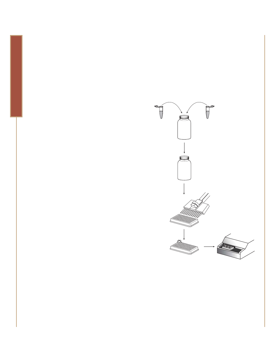
Figure 1. Schematic diagram of the MultiTox-Fluor Multiplex
Cytotoxicity Assay. The MultiTox-Fluor Multiplex Cytotoxicity Reagent
is created by adding the fluorogenic peptide substrates to the assay
buffer. This reagent can then be added to a multiwell plate. After at
least 30 minutes of incubation at 37˚C, the resulting fluorescent
signal may be measured.
www.promega.com
13
CELL NOTES ISSUE 16 2006
High-Throughput Cytotoxicity and Cell-Based Screening
HTS
CYTOTO
XICITY
The liberated fluorophores generated from these respective
activities can then be measured using conventional multiwell
fluorometers. This is possible because the optimal excitation
and emission spectra for the liberated fluors are sufficiently
separated to allow multiplexed live- and dead-cell
measurements.
Sensitive, Scalable and Fast
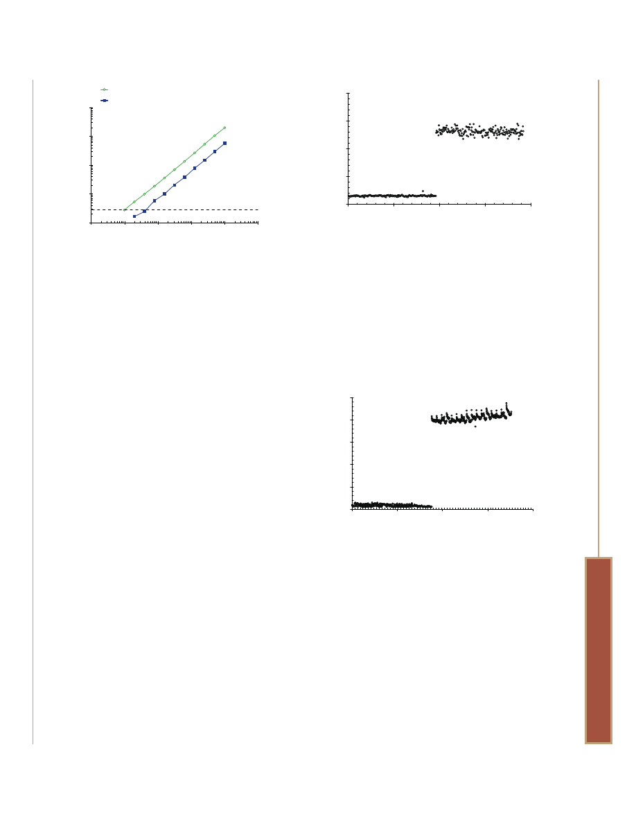
High-density, cell-based formats require high assay sensitivity.
In most cases, the MultiTox-Fluor Assay offers statistical
sensitivities approaching the level of the CellTiter-Glo
®
Assay
(which measures ATP-levels) after only 30 minutes of
incubation at 37ºC (Figure 2). Unlike ATP-based assays,
which require cell lysis to liberate ATP, the MultiTox-Fluor
Cytotoxicity Assay sensitivity is achieved without affecting
viability. This high sensitivity allows the reagent to be
employed in 384- and 1536-well plate formats where both
cell number and well volumes are significantly reduced
compared to 96-well formats (Figures 3 and 4).
The final reagent concentration required for the MultiTox-Fluor
Cytotoxicity Assay is also flexible to accommodate well volume
restrictions when conducting other sequentially multiplexed
assays within the same well. For instance, a concentrated
reagent can be prepared and delivered in a 1/10th volume
when multiplexing with other assays.
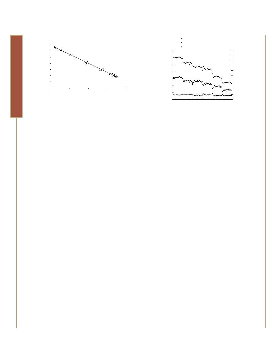
The Ratiometric Response
The MultiTox-Fluor Cytotoxicity Assay measures two
independent but inversely correlated protease activities as
markers of cellular viability and cytotoxicity. When used in
combination, these measures can complement each other
and provide a ratiometric value that is useful for counter-
5930MA
1
10
100
1,000
10,000
Cells or Cell Equivalents/Well
Signal-to-Noise Ratio
Dead-Cell Signal (bis-AAF-R110 Substrate)
Live-Cell Signal (GF-AFC Substrate)
1
10
100
1,000
10,000 100,000
Figure 2. Statistical sensitivity of the MultiTox-Fluor Assay. Jurkat cells
were twofold serially diluted from 10,000 to 10 cells/well in an
opaque-walled, 96-well plate. RPMI 1640 with 10% fetal bovine
serum served as the background control. Medium or medium
containing detergent was added to matched wells to simulate
cytotoxicity. MultiTox-Fluor Assay Reagent was added in an equal
volume, and data were collected on a BMG POLARstar plate reader
after 30 minutes of incubation at 37°C. Signal-to-noise determinations
were calculated by dividing the net RFU (raw values minus
background) by the standard deviation of the background at each
fluorescence wavelength. The dotted line represents the statistical
level of sensitivity (2). The best-fit lines for the dead- and live-cell
measurements have r
2
values of 0.9998 and 0.9991, respectively.
Figure 3. Demonstration of performance in a 384-well primary necrosis
model. Jurkat cells were plated at a density of 5,000 cells per well in
10µl volumes using a CyBio CyBi
®
Well 384/1536 automated
dispenser. Ionomycin was diluted in RPMI 1640 with 10% fetal
bovine serum and added in an additional 10µl volume to half of the
plate to a final concentration of 50µM. Complete medium was added
to the other half of the plate, and the plate was incubated for 5 hours
at 37°C. MultiTox-Fluor Assay Reagent was added in 20µl volumes
and incubated for 30 minutes prior to fluorescence measurement
using a Tecan Safire
2
™ Multichannel Monochromator reader. Z´-factor
values were determined by using the ratio of fluorometric signals for
each well rather than comparing average signals for control
populations (2).
Figure 4. Demonstration of performance in a 1536-well format. Jurkat
cells were adjusted to 625,000 cells/ml in RPMI 1640 with 10% fetal
bovine serum. The pool was divided, and one fraction gently
sonicated to simulate 100% cytotoxicity. Cells or lysate were delivered
in 4µl volumes (2,500 cell equivalents) using a Deerac Fluidics
Equator™ automated dispenser. MultiTox-Fluor Assay Reagent was
added in an additional 4µl volume and the plate incubated at 37°C
for 2 hours prior to measuring fluorescence using a Tecan Safire
2
™
Multichanel Mononchromator reader. Z´-factor values were calculated
by using the ratio of fluorometric signals for each well rather than
comparing average signals for control populations (2).
5931MA
0
1
2
3
4
Well Number
Ratio R110/AFC Fluorescence
Z´ factor = 0.8325
Untreated
Ionomycin Treated
0
100
200
300
400
5932MA
0
1
2
3
4
5
Well Number
Ratio R110/AFC Fluorescence
Viable
Cytotoxic
Z´ factor = 0.8569
0
400
800
1,200
1,600
CELL NOTES ISSUE 16 2006
14
www.promega.com
High-Throughput Cytotoxicity and Cell-Based Screening
HTS
CYTOTO
XICITY
confirmation of the result (Figure 5). In other words, if the
viability measurement is low compared to an untreated
control, the cytotoxicity measurement should be high
compared to the untreated control (and vice versa).
Divergence from the ratiometric relationship with any single
compound or treatment may occur in three experimental
situations: a proliferative event in the absence of cytotoxicity,
compound interference with one of the fluorometric
measures, or dead-cell enzyme activity decay over long
exposure periods. Proliferation will demonstrate an increase
in only the live-cell signal with respect to the control.
Fluorescence interference will demonstrate either a
disproportional increase or decrease in the signal. However,
because of the spectral distance between the respective
fluorophores’ excitation and emission spectra, the likelihood of
both markers being affected by an autofluorescent compound
would be statistically rare (3). Finally, the dead-cell response
may be underestimated in cases of primary necrosis or rapid
apoptosis induction, because the marker demonstrates an
enzymatic half-life of about nine hours after cell death.
Nevertheless, the duality of the measures allows “flagging” of
problematic data points, whereas single-parameter cytotoxicity
assay measures may lead to potentially false-positive or
-negative conclusions.
The ratiometric response is also useful for improving the
precision of data resulting from variability in cell number due
to cellular clumping or pipetting errors as well as differential
growth patterns or edge effects in assay plates. This variability
is particularly troublesome for single-point cytotoxicity assays
and single-parameter assays that measure responses such as
caspase induction potential or genetic reporter activity. The
risk is that the data set from these screens may indicate
statistically significant increases or decreases in activity when
they are in fact false-negative or -positive. Because of the
ratiometric proportionality of MultiTox-Fluor Cytotoxicity Assay,
response variability arising from cell number differences in
cytotoxicity assays can be resolved simply by using the
quotient of dead-cell and live-cell values for each well (Figure
6). In some instances, data from primary activity assays can
be normalized by first measuring the relative number of
remaining live or dead cells in culture prior to adding the
second reagent (Figure 7). Care should be exercised with such
normalization, as differences in pharmacokinetic induction
rates (and activity) may precede changes in cellular viability.
Increasing Content
The true cost of a screening effort is typically more than time,
assay reagents and other consumable expenses. Compound
usage and the informational value of compound
characterization contribute to screening costs. Multiplex
assays extract the valuable data and reduce library
consumption by eliminating parallel assays. By multiplexing
the MultiTox-Fluor Cytotoxicity Assay with specific cell-
response assays, you not only reduce false-negative and
-positive determinations, but also gain information about
5929MA
r
2
= 0.9956
0
200
400
600
800
1,000
1,200
1,400
1,600
bis-AAF-R110 Fluorescence (RFU)
GF-AFC Fluorescence (RFU)
0
5,000
10,000
15,000
20,000
Figure 5. The inverse proportionality of the ratiometric response.
Jurkat cells were adjusted to 100,000 cells/ml in RPMI 1640 with
10% fetal bovine serum, then divided into two pools. One fraction
was treated by sonication to simulate cytotoxicity, the other left
untreated. The fractions were blended in various ratios to create
viabilities from 100% to 0%. 100µl of each blend was added to wells
of a 96-well plate. Medium-only served as background control. After
30 minutes of incubation at 37°C, fluorescence was measured using
a BMG POLARstar plate reader. Net fluorescence values (minus
cell-free background) from both wavelengths were plotted against
each other for each well.
5928MA
0
1,000
2,000
3,000
4,000
5,000
6,000
7,000
Well Number
GF-AFC or bis-AAF-R110
Fluorescence (RFU)
0
2
4
6
8
10
12
14
16
18
20
Ratiometric V
alue (R110/AFC)
GF-AFC CV = 26%
bis-AAF-R110 CV = 28%
Ratiometric CV = 7%
1
12
23
34
45
56
67
Figure 6. Improving assay precision by ratiometric means. A pool of
Jurkat cells was divided, and one fraction was subjected to sonication
to simulate cytotoxicity. The two fractions were combined to make a
50% viable pool. To simulate the effects of cellular clumping, the pool
was diluted with additional RPMI 1640 with fetal bovine serum and
delivered to wells of a 96-well plate at calculated densities of 12,500
(wells 1–12), 11,000 (wells 13–24), 10,000 (wells 25–36), 9,000
(wells 37–48), 7,500 (wells 49–60) and 5,000 cells/well (wells
61–72) in 100µl volumes. MultiTox-Fluor Assay Reagent was added
in an equal volume to each well, and data were collected after 30
minutes of incubation at 37°C. The raw data collected at the AFC
and R110 wavelengths were plotted against well number. The
quotient of values derived from the same data is also represented.
The coefficient of variation percentage is derived from the standard
deviation of each data set divided by the average signal x 100.
www.promega.com
15
CELL NOTES ISSUE 16 2006
High-Throughput Cytotoxicity and Cell-Based Screening
HTS
CYTOTO
XICITY
inherently flawed or problematic compounds. For instance,
compounds that induce a cytotoxic response in the absence
of caspase activation are therapeutically unattractive (Figure
8). These “frequent hitters” then can be culled from the
collection to reduce the throughput burden and strengthen
the quality of future data.
Summary
The MultiTox-Fluor Multiplex Cytotoxicity Assay has many
attributes that make it useful for cell-based assay screening.
Use of the assay not only indicates compound effects on
cellular viability but can also improve data quality by
improving assay precision, detecting false-positives and
-negatives, and by increasing content on a per well basis by
multiplexes with specific response assays. This homogeneous,
“add-mix-measure” assay is readily scalable for HTS
applications but can be used at any stage throughout potency
and lead selection testing.
■
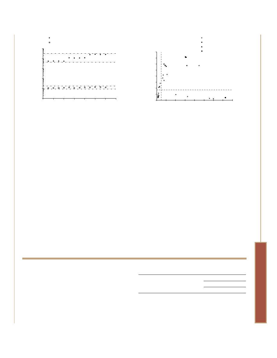
Figure 7. Primary caspase activity response data can be normalized by
viability values. U266 cells were seeded at 5,000 (wells 1–4), 10,000
(wells 5–8), and 20,000 cells/well (wells 9–12) in 50µl of RPMI 1640
with 10% fetal bovine serum. Staurosporine was diluted in medium
and added to 2µM final concentration with an additional 50µl
volume. The plate was incubated for a period of 6 hours at 37°C to
allow caspase activation by apoptosis induction. MultiTox-Fluor Assay
Reagent was made by adding 10µl of each substrate to 1.0ml of
assay buffer. The reagent was then added in 10µl volumes to the
wells, and data were collected after 30 minutes of incubation at
37°C. Caspase-Glo
®
3/7 Reagent was added in 100µl volumes, and
luminescence was measured after 30 minutes. Raw Caspase-Glo
®
3/7 response data from the varying cell number wells are plotted
together with the same data normalized by live cell response values.
The coefficient of variation for each was calculated by dividing the
standard deviation of the data set by the average signal x 100 to
arrive at a percentage.
5927MA
1
10
100
1,000
10,000
100,000
Well Number
Raw or Normalized Luminescence (RLU)
Raw Caspase-3/7 CV = 62.7%
Normalized Caspase-3/7 CV = 8.7%
0
2
6
8
10
12
4
14
Figure 8. Creating a cytotoxicity index using MultiTox-Fluor Assay and
a luminescent caspase-3/7 assay. Jurkat cells at a density of 5,000
cells/well in 50µl volumes were exposed to 80 compounds from the
LOPAC library (plate 4) in additional 50µl volumes for a final
concentration of 10µM. Known apoptotic inducers, anti-FAS mAb at
100ng/ml (black triangles) and staurosporine at 2µM (black
diamonds) served as caspase-activation controls. Detergent (black
square) served as a primary necrosis control. The plate was
incubated at 37°C for an 8-hour exposure period. MultiTox-Fluor
Reagent was prepared and added as described in Figure 7, and
fluorescence was measured after 30 minutes at 37°C. Caspase-Glo
®
3/7 Reagent was added in 100µl volumes and luminescence
measured after 30 minutes. A “cytotoxicity index” was established for
each compound by dividing the dead-cell value by the live-cell value.
The resulting data were plotted v. raw luminescence and partitioned
into quadrants based on thresholds established from untreated cell
values. Compounds that kill cells but do not induce caspase
activation are clearly distinguished from compounds that induce
caspase activation and are cytotoxic. Compounds that induce
caspase activation but show no apparent cytotoxicity reflect early
stage apoptosis.
5926MA
0
500
1,000
1,500
2,000
2,500
3,000
3,500
4,000
Cytotoxicity Index (Dead/Live)
Caspase-3/7 Luminescence (RFU)
Cytotoxic with no caspase activation
Cytotoxic with caspase activation
0
5
10
15
20
25
30
35
40
LOPAC compounds
Staurosporine
Anti-FAS mAb
Detergent
References
1. Niles, A.L.
et al. (2006) Cell Notes 15, 11–15.
2. Zhang, J.
et al. (1999) J. Biomol. Screen. 4, 67–73.
3. Grant, S.
et al. (2002) J. Biomol. Screen. 7, 531–40.
Protocol
MultiTox-Fluor Multiplex Cytotoxicity Assay Technical
Bulletin #TB348
(www.promega.com/tbs/tb348/tb348.html)
Ordering Information
Product
Size
Cat.#
MultiTox-Fluor Multiplex Cytotoxicity Assay
10ml
G9200
5 × 10ml
G9201
2 × 50ml
G9202
For Laboratory Use.
(a)
Patent Pending.
Products may be covered by pending or issued patents or may have certain limitations. Please
visit our Web site for more information.
Caspase-Glo and CellTiter-Glo are registered trademarks of Promega Corporation. CyBi is a
registered trademark of CyBio AG. Safire
2
is a trademark of Tecan AG. Equator is a trademark of
Allegro Technologies, Ltd.
Wyszukiwarka
Podobne podstrony:
Apoptosis, Cytotoxicity and Cell Proliferation
Antioxidant, anticancer, and apoptosis inducing effects in HELA cells
Cytotoxicity and Modes of Action of the Methanol Extracts
Cytolytic Effects and Apoptosis Induction
Cytotoxicity of Aqueous and Ethanolic Extracts
Dungeons and Dragons 3 5 Accessory Color Character Sheets with Multiple Spell Lists
Capote In Cold Blood A True?count of a Multiple Murder and Its Consequences
Apoptosis Induction, Cell Cycle Arrest and in Vitro Anticancer Activity
[17]Chromosomal DNA fragmentation in apoptosis and necrosis induced by oxidative stress
Gilles Deleuze Dualism, Monism And Multiplicities
Year 5 Block 4 Multiplication and Division Dec 2017
więcej podobnych podstron