
Osteoarthritis
OA
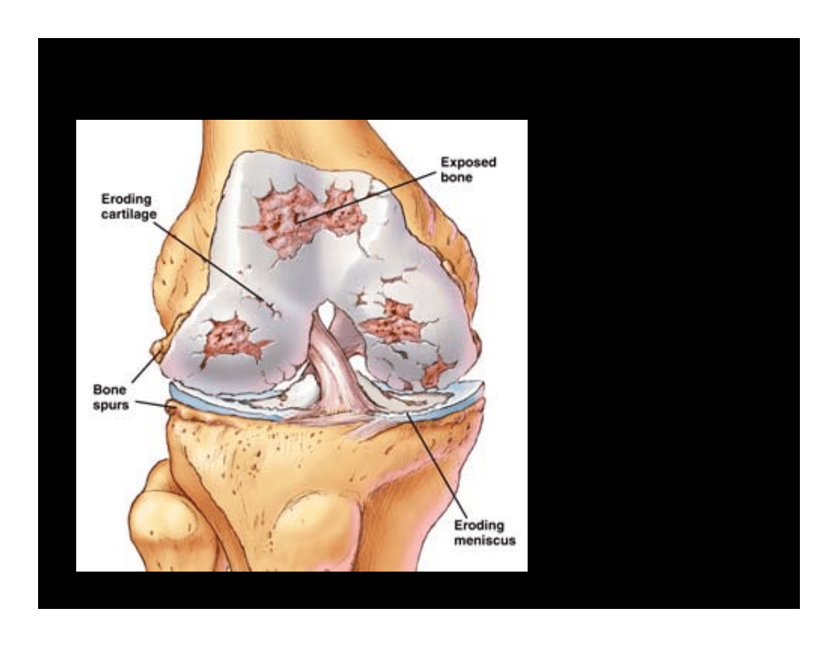
OA is a degenerative
disease of joints that
affects all of the
weight-bearing
components of the
joint:
•Articular
cartilage
•Menisci
•Bone
Osteoarthritis (OA)
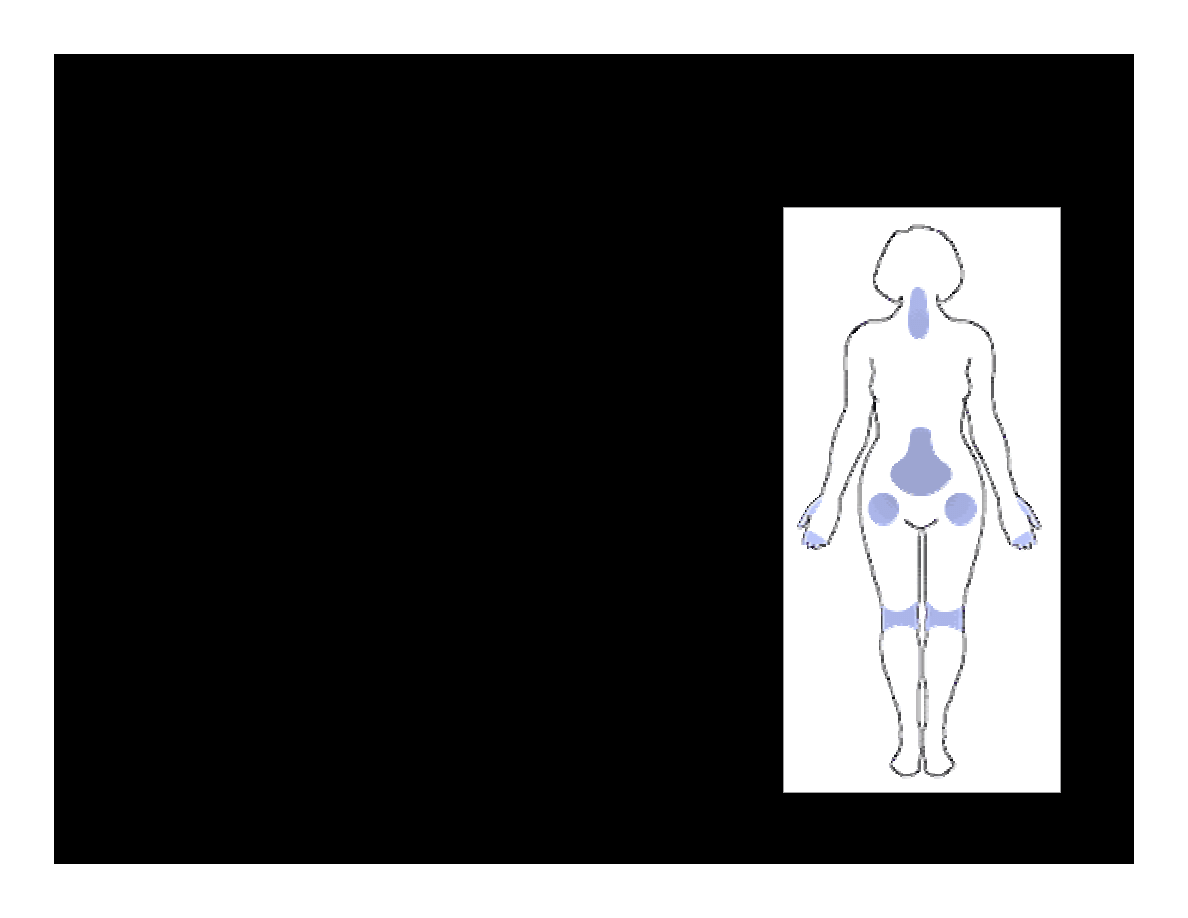
• Most common form of arthritis
• Most common joint disease
• Over 10 million Americans
suffer from OA of the knee alone
• Most OA patients > age 45
• Women > men.
• Most often appears at the ends of
the fingers, thumbs, neck, lower
back, knees, and hips.
OA
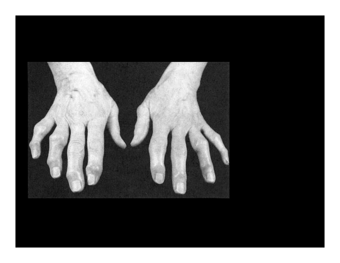
Nodal osteoarthritis
Bony enlargement of
distal and proximal
interphalangeal
joints (Heberden's,
Bouchard's nodes,
respectively).

OA- Risk Factors
Age
– Strongest risk factor
– OA can start in young adulthood but risk increases with age
Female Gender
– Arthritis in general affects more women than men
– OA more common in men before age 45, women after age 45
– OA of the hand particularly common in women
Joint Alignment
– Abnormal alignment or motion predisposes joint to OA
• Bow legs, dislocations, double-jointed

OA- Risk Factors
Hereditary gene defect
– Collagen component of cartilage is damaged
– Increased deterioration of cartilage
Joint injury/Overuse from physical labor or sports
– Trauma to any joint increases risk of OA
– Ligament or meniscus tears
– Repeated movements in certain jobs increase risk
Obesity
– Joint overload is among strongest risks for knee OA
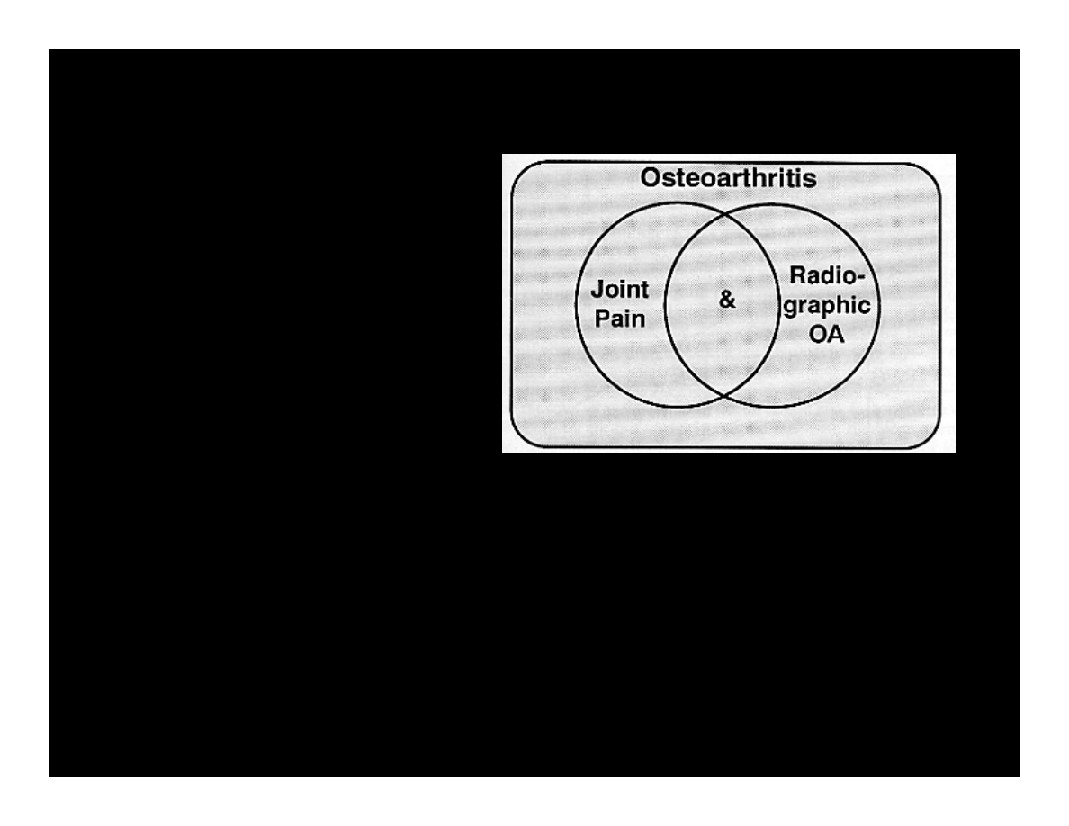
OA – Symptoms
• Gradual onset - It takes
many years before the
damage to the joint
becomes noticeable
• Only a third of whose X-
rays show OA report pain
or other symptoms:
– Steady or intermittent
pain
in a joint
– Stiffness
that tends to follow periods of inactivity, such as sleep
or sitting
– Swelling or tenderness
in one or more joints
– Crunching feeling or sound of bone rubbing on bone (called
crepitus
) when the joint is used
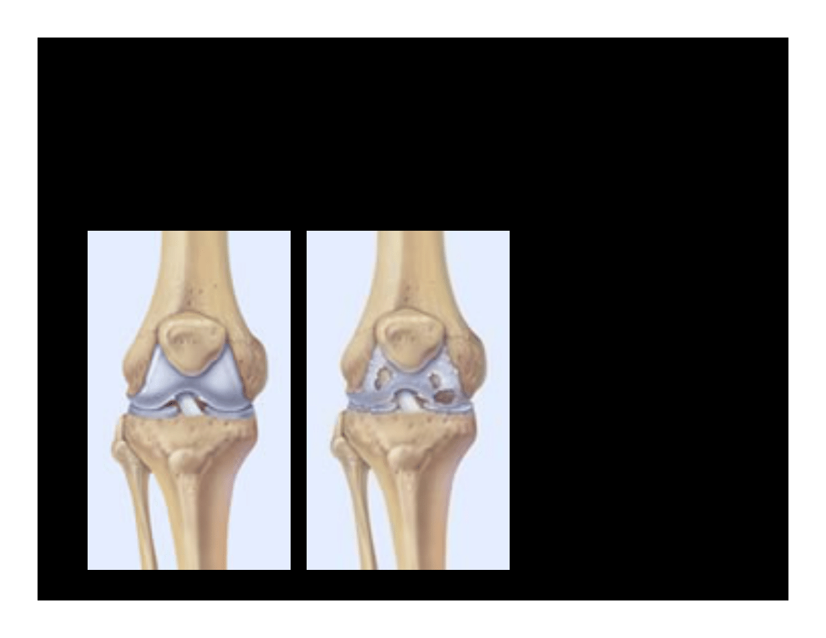
Osteoarthritis (OA) - Definition
Osteoarthritis may result from wear and tear
on the joint
•The normal
cartilage lining
is gradually
worn away and
the underlying
bone is
exposed.
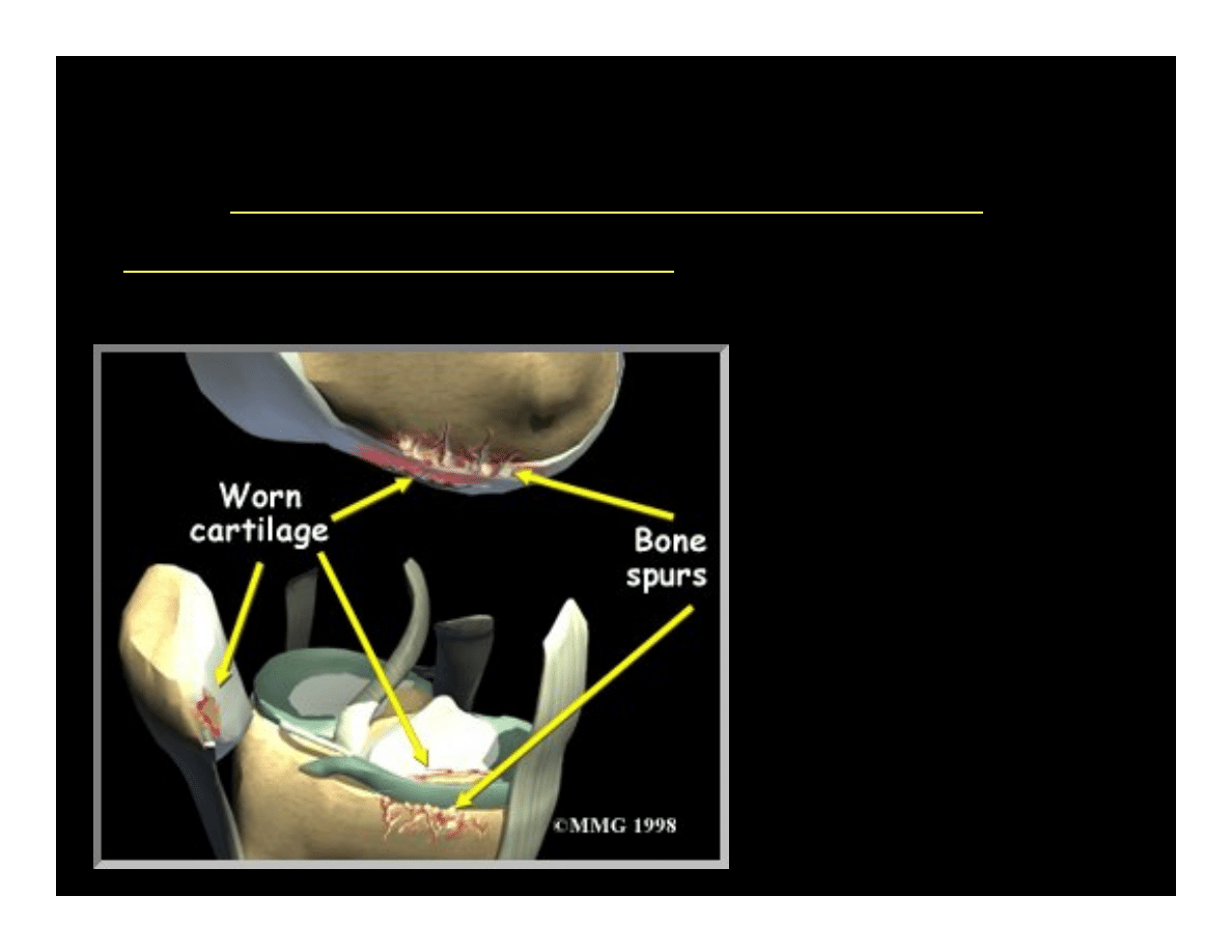
Osteoarthritis (OA) - Definition
•The repair mechanisms of rebsorption and
synthesis get out of balance and result in
osteophyte formation (bone spurs) and bone cysts
Osteophyte (spur) is
formed when Osteoblast
formation increases while
resorption decreases
OA – Articular Cartilage
Articular cartilage is the main tissue affected
•Increased swelling
•Change in color
•Cartilage fibrillation
•Cartilage erosion down to subchondral bone
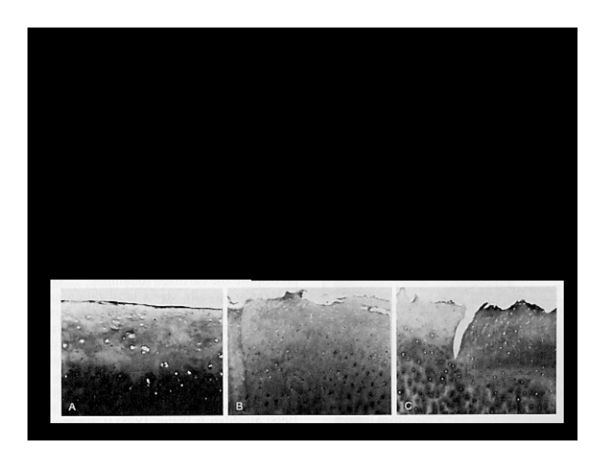
OA – Articular Cartilage
Micrograph
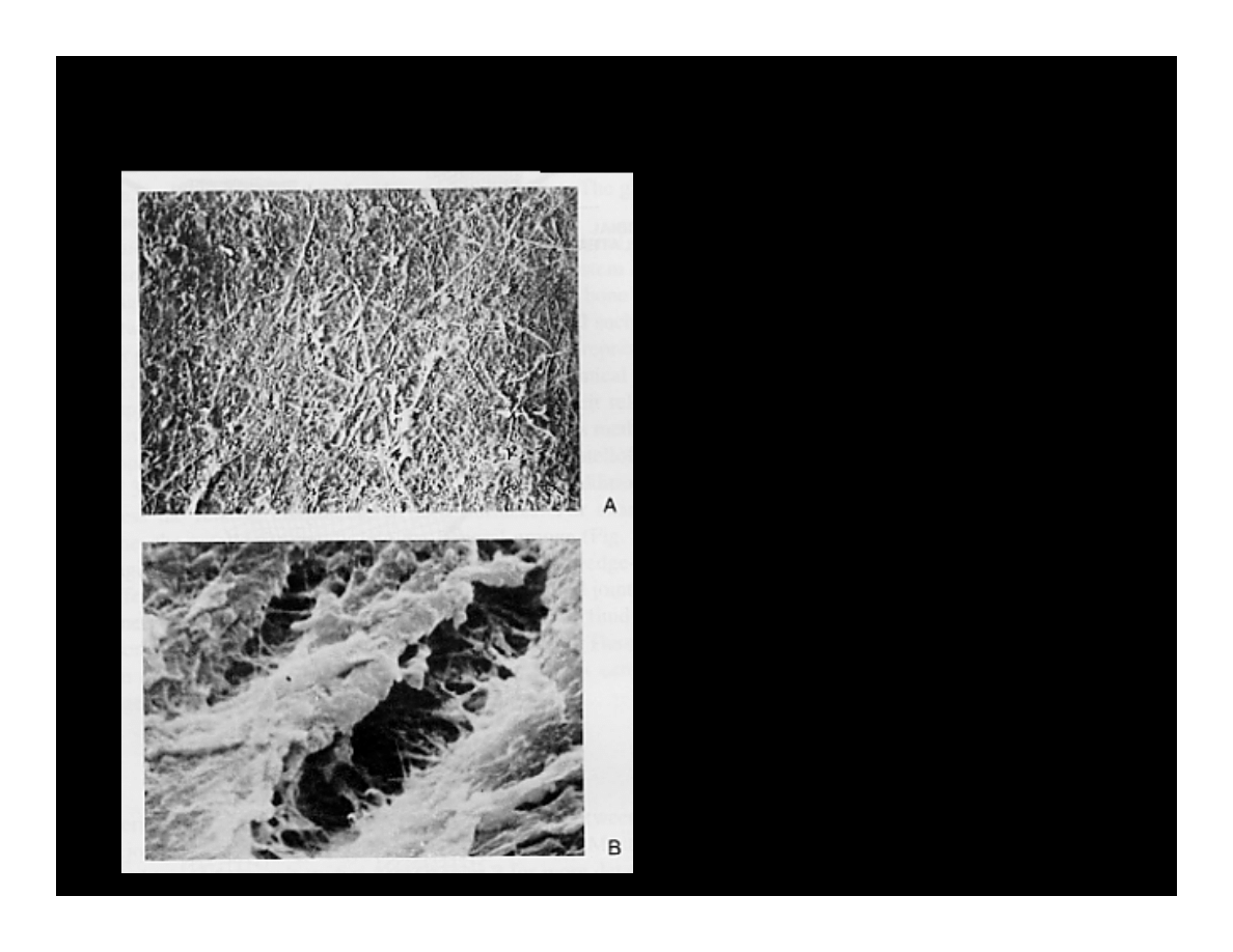
OA – Articular Cartilage
A) Normal articular cartilage
from 21-year old adult
(3000X)
B) Osteoarthritic cartilage
(3000X)
• Surface changes alter
the distribution of
biomechanical forces
• This triggers active
changes by the tissue
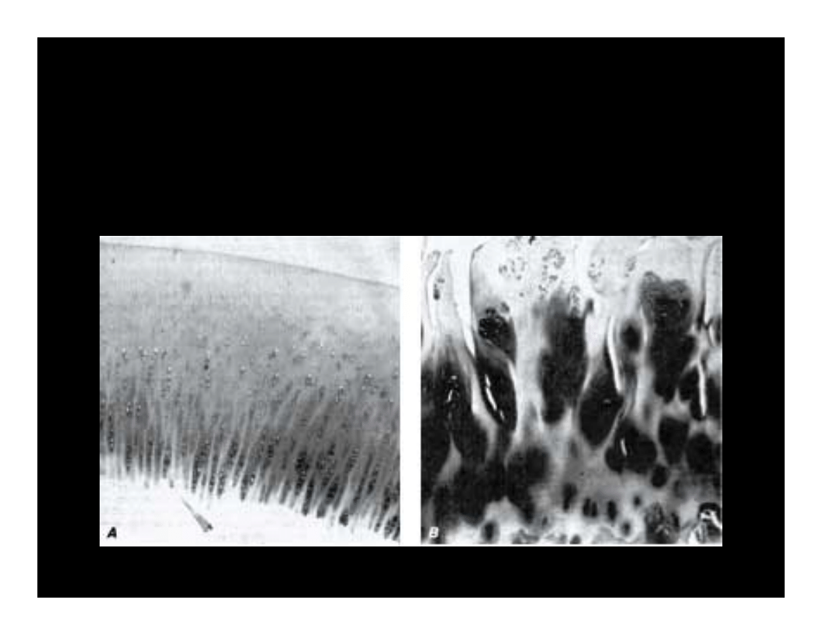
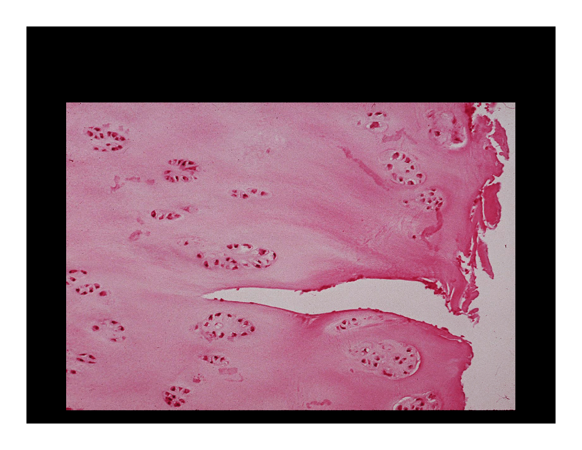
OA – Articular Cartilage
Chondrocyte cloning in an attempt to restore articular
surface
(Normal adult chondrocytes are fully differentiated and
do not proliferate)
(A) Normal articular cartilage (B) Osteoarthritic cartilage
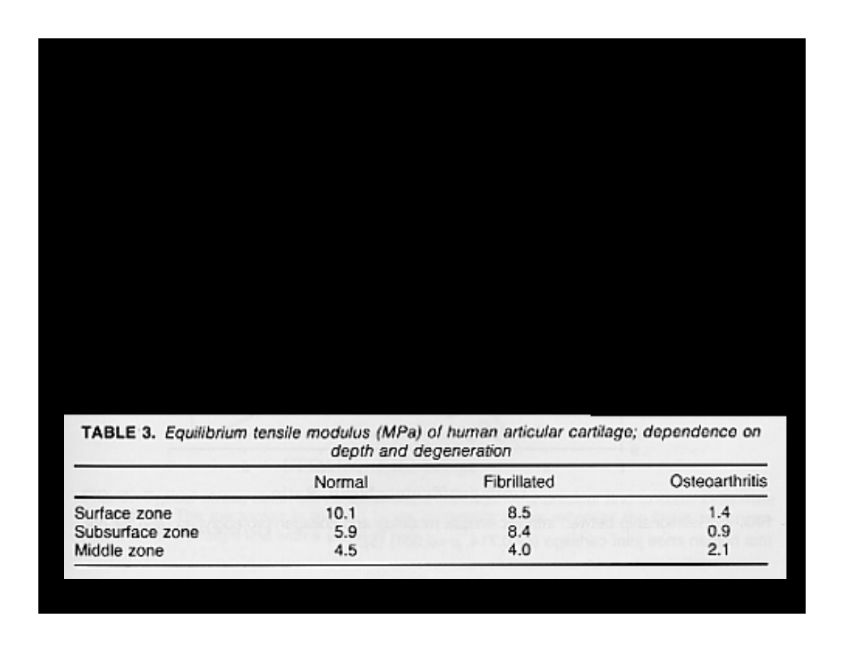
OA – Articular Cartilage
• Newly dividing cells do not differentiate fully
• Cannot effectively synthesize the elements needed for
matrix maintenance
• Results in net loss of matrix components
• Collagen content stays constant but fibrils are thinner
and more disorganized
-
Decreased tensile strength
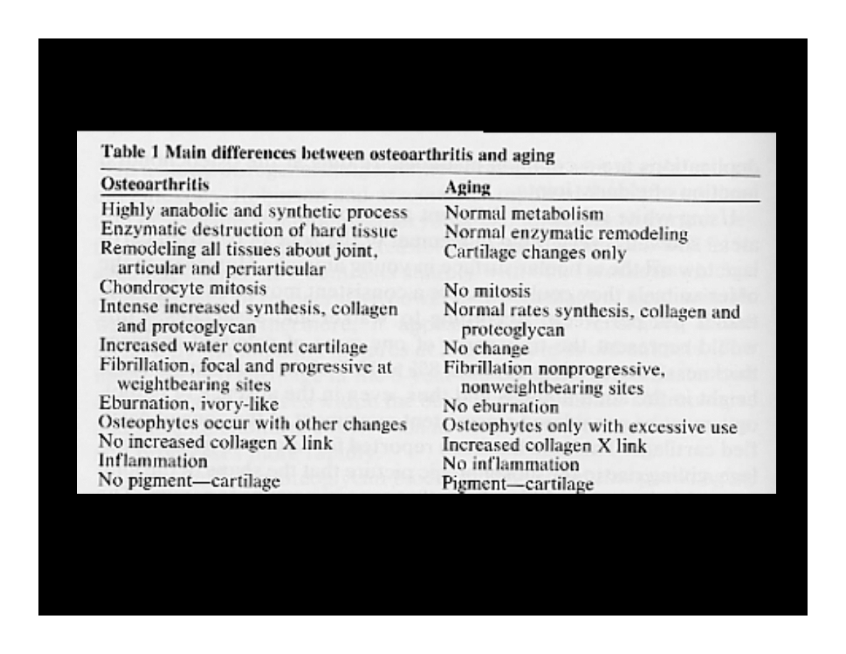
OA vs. Aging
Unlike aging, OA is
progressive
and a significantly
more
active process
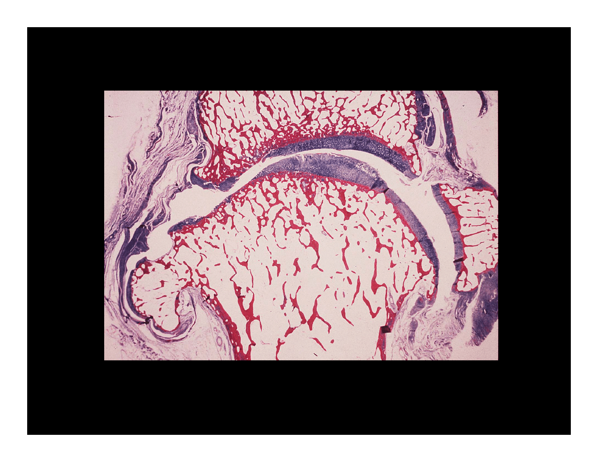
OA – Overall Changes
Osteoarthritis with osteophyte, loss of articular cartilage and some
subchondral bony sclerosis.
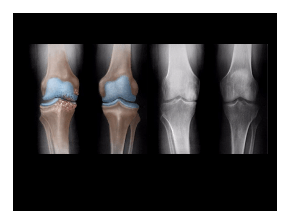
OA – Radiographic Diagnosis
Asymmetrical joint space narrowing
from loss of
articular cartilage
•
Medial (inside) part of knee most commonly affected by OA.
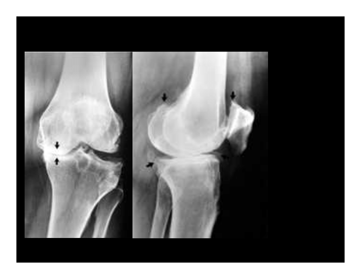
OA – Radiographic Diagnosis
•Asymmetrical
joint space
narrowing
•Subchondral
sclerosis and
cysts
•Osteophytes
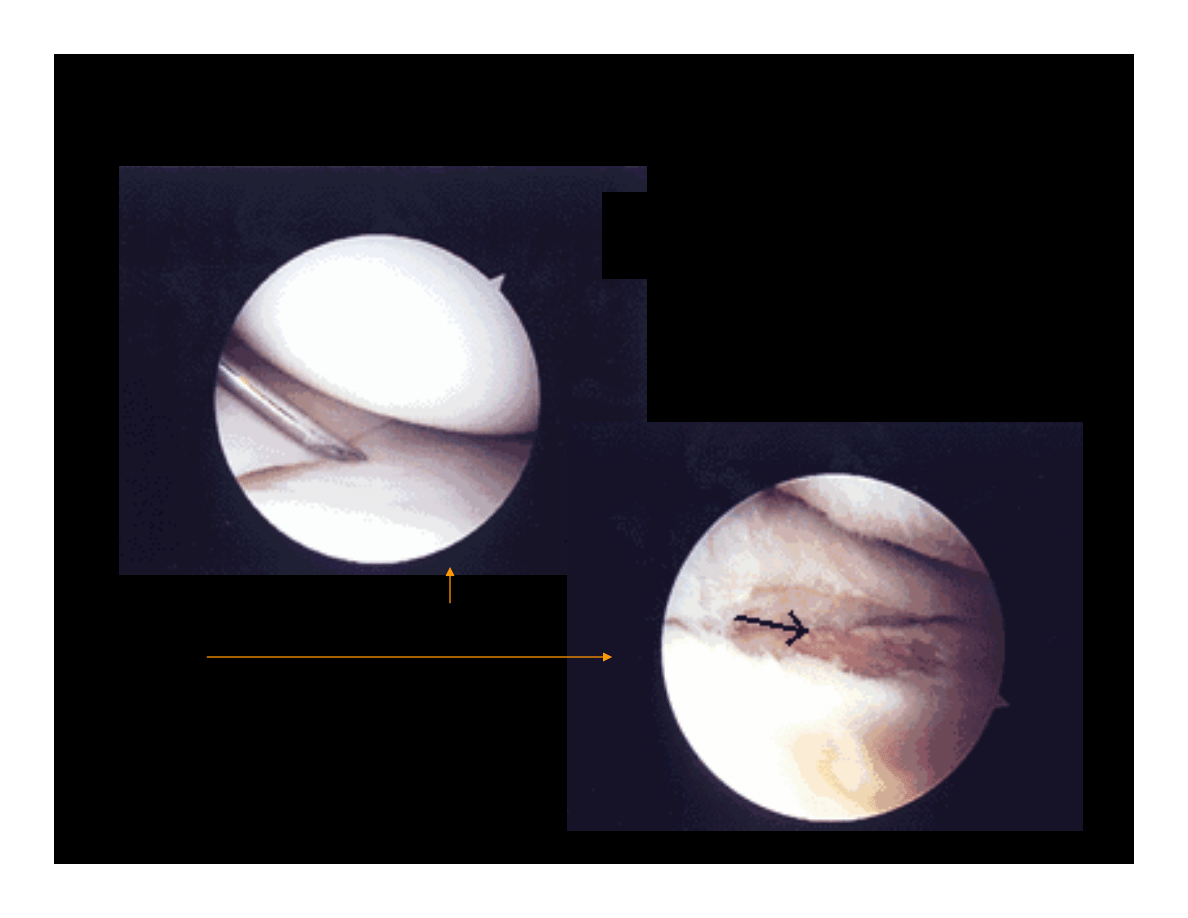
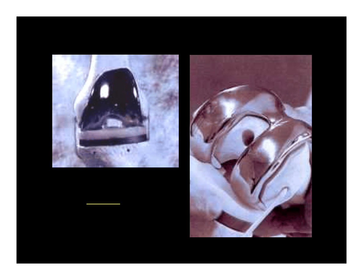
OA – Arthroscopic Diagnosis
Normal Articular Cartilage
Ostearthritic cartilage with exposed
subchondral bone
Arthroscopy allows earlier
diagnosis by demonstrating the
more subtle cartilage changes
that are not visible on x-ray
OA – Arthroscopic Treatment
• Most accurate way of determining stage of OA
• Debridement of the knee joint:
– Cleaning out the joint of all debris and loose bodies.
– Loose bodies of cartilage removed
– Saline solution.
–
Micro-fracture techniques
• Badly worn areas may be treated with sub-chondral holes (fracture) to
promote growth of new cartilage
– Fibro-cartilage that is scar tissue.
– Usually offer temporary relief of symptoms
• 6 months to 2 years.
• Graft-transplantation

OA – Management
•
Slow progression over many years
- Cannot be cured
• Treatment directed at symptoms and slowing
progress of the condition
• Goals:
•
Decreasing pain
•Increase range of motion
•Increase muscle strength
OA – Non-operative Treatments
• Pain medications
• Physical therapy
• Walking aids
-
Unloading
• Re-alignment
- Orthotics and/or
surgery

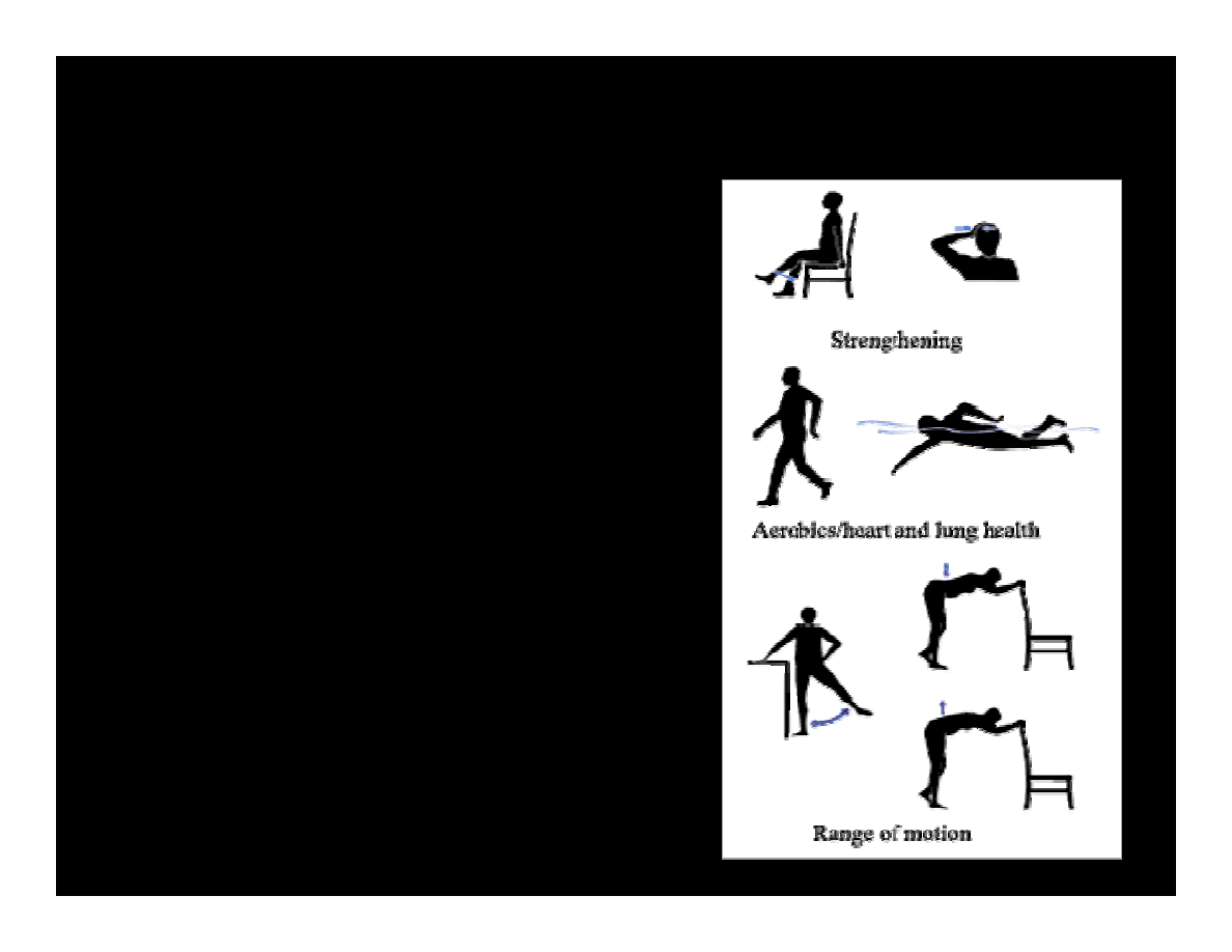
Physical Therapy
• Accomplishes all 3 goals : reduce pain,
increase range of motion and strength
– Heat, electrical stimulation, & ultrasound
decrease pain
– Manipulate muscles & tendons surrounding
joint
• Better strength means better weight support
– Low impact (especially aquatic) exercises is
both safe & effective
• Improves balance and coordination of bones &
muscles

Physical Therapy 2
• Increased activity decreases overall body
weight
– Decreases load & pain on joints
• Improves physical function due to increased
strength
– Also lowers forces and stress on joints
• Improves quality of life due to pain relief &
wider range of movement
• Slows progression of OA

Pain Management
• Non-Steroidal Anti-Inflammatory Drugs
(NSAID)
– Drugs that reduce pain, inflammation and fever
• Inhibit prostaglandins which play role in inflammation
– Are not made from steroids or narcotics
• No sedation, depression, addiction/dependence
• Examples:
– Ibuprofen (Motrin/Aleve), Naproxen, Diclofenac (Voltaren)
– Asprin
→ *Note: Acetamenophen (Tylenol) is NOT an NSAID because has no anti
inflammatory use

COX-2 Inhibitors
• Some NSAIDs Inhibit COX-1 enzyme
which acts as messenger molecule during
inflammation
– Results in gastrointestinal side effects
• COX-2 is secondary enzyme that selectively
inhibit without disrupting GI system
– Examples: Meloxicam (Mobic), Celecoxib
(Celebrex), Rofecoxib (Vioxx)

Pain Management
• Steroid Injections: 2 types
– Cortisone/Corticosteroid
• Reduce inflammation response around joints
• Tend to have more rapid effect than NSAIDs
– Viscous supplement
• Replace modified synovial fluid in joints
• Increase viscosity & elasticity of fluid

Pain Management
• Various Corticosteroids
– Cortone
– Depo-Medrol
• Visco-Supplements
– Hyalgan
– Euflexxa
– Orthovisc
– Synvisc
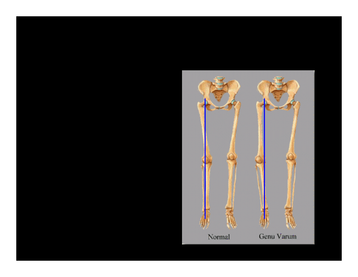
•Osteoarthritis usually
affects the inside half
(medial compartment) of
the knee more often than
the outside (lateral
compartment).
•This can lead to the lower
extremity becoming
slightly bowlegged or a
genu varum deformity
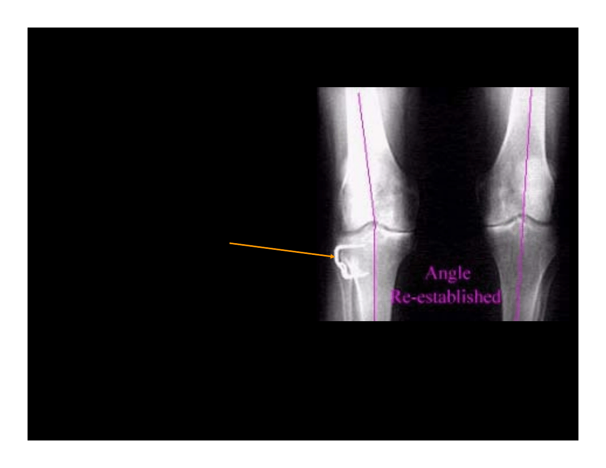
Realignment Surgery:
Proximal Tibial Osteotomy

Proximal Tibial Osteotomy
• The problem:
•The weight bearing line passes more medially (towards
the medial compartment of the knee).
• Increased pressures are transferred through the medial
joint surfaces, which leads to more pain and deformity.
• The aim:
• re-aligning the angles in the lower extremity by shifting
the weight-bearing line towards the midline or lateral
compartment of the knee. This places more of the weight-
bearing force into a healthier compartment.
• The result is pain reduction and delay in the progression of
the degeneration of the medial compartment.
Proximal Tibial Osteotomy
•In the procedure to realign the
leg, a wedge of bone is
removed or added to the upper
tibia.
•A staple or plate and screws
are used to hold the bone in
place until it heals.
•The Proximal Tibial Osteotomy buys some time before needing to
perform a total knee replacement. Pain relief usually lasts for 5-7
years.
Total Knee Replacement
Click HERE for link to
TKA Lecture

…The End
Document Outline
- Osteoarthritis
- OA
- Osteoarthritis (OA)
- OA
- OA- Risk Factors
- OA- Risk Factors
- OA – Symptoms
- Osteoarthritis (OA) - Definition
- Osteoarthritis (OA) - Definition
- OA – Articular Cartilage
- OA – Articular Cartilage Micrograph
- OA – Articular Cartilage
- OA – Articular Cartilage
- OA – Articular Cartilage
- OA vs. Aging
- Physical Therapy
- Physical Therapy 2
- Pain Management
- COX-2 Inhibitors
- Pain Management
- Pain Management
- …The End
Wyszukiwarka
Podobne podstrony:
Wstrzasy ang ppt
Wstrzasy ang ppt
08 BIOCHEMIA mechanizmy adaptac mikroor ANG 2id 7389 ppt
Choroba zwyrodnieniowa stawów (Osteoartroza) ppt
08 BIOCHEMIA mechanizmy adaptac mikroor ANG 2id 7389 ppt
03 Sejsmika04 plytkieid 4624 ppt
Choroby układu nerwowego ppt
10 Metody otrzymywania zwierzat transgenicznychid 10950 ppt
10 dźwigniaid 10541 ppt
03 Odświeżanie pamięci DRAMid 4244 ppt
Hydrocephalus(ang)
Prelekcja2 ppt
więcej podobnych podstron