
©FUNPEC-RP www.funpecrp.com.br
Genetics and Molecular Research 13 (2): 3981-3990 (2014)
In vitro cytotoxicity screening of wild plant
extracts from Saudi Arabia on human breast
adenocarcinoma cells
M.A. Ali
1
, M. Abul Farah
2
, F.M. Al-Hemaid
1
and F.M. Abou-Tarboush
2
1
Department of Botany and Microbiology, College of Science,
King Saud University, Riyadh, Saudi Arabia
2
Department of Zoology, College of Science, King Saud University,
Riyadh, Saudi Arabia
Corresponding author: M.A. Ali
E-mail: ajmalpdrc@gmail.com
Genet. Mol. Res. 13 (2): 3981-3990 (2014)
Received June 3, 2013
Accepted October 2, 2013
Published May 23, 2014
DOI http://dx.doi.org/10.4238/2014.May.23.9
ABSTRACT. This study investigated the in vitro anticancer activities
of a total of 14 wild angiosperms collected in Saudi Arabia. The
cytotoxic activity of each extract was assessed against human breast
adenocarcinoma (MCF-7) cell lines by using the MTT assay. Among the
plants screened, the potential cytotoxic activity exhibited by the extract
of Lavandula dentata (Lamiaceae) was identified, and we analyzed its
anticancer potential by testing antiproliferative and apoptotic activity.
Our results clearly show that ethanolic extract of L. dentata exhibits
promising cytotoxic activity with an IC
50
value of 39 µg/mL. Analysis
of cell morphological changes, DNA fragmentation and apoptosis
(using an Annexin V assay) also confirmed the apoptotic effect of
L. dentata extract, and thus, our data call for further investigations to
determine the active chemical constituent(s) and their mechanisms of
inducing apoptosis.
Key words: Cytotoxicity; Apoptosis; Cancer; MCF-7; Saudi Arabia

3982
©FUNPEC-RP www.funpecrp.com.br
Genetics and Molecular Research 13 (2): 3981-3990 (2014)
M.A. Ali et al.
INTRODUCTION
Breast cancer is a leading cause of death in women worldwide (American Cancer
Society, 2012). There has long been standing interest in the identification of natural products
for the treatment of various diseases for thousands of years. Natural products possess immense
pharmacological significance in the development of drugs (Dixon et al., 2007; Baker et al.,
2007; Harvey, 2008) including cancer (Graham et al., 2000; Figueroa-Hernández et al., 2005;
Madhuri and Pandey, 2009; Tan et al., 2011; Newman and Cragg, 2012; Kuno et al., 2012),
and were discovered through plant bioprospecting (Mann, 2002). The majority of drug candi-
dates, such as paclitaxel, etoposide, camptothecin, vinca alkaloids, indole alkaloids, podophyl-
lotoxin derivatives, etoposide and teniposide, currently used in clinical cancer chemotherapy,
were originally derived from plants. The efficacy of chemotherapy, radiotherapy, hormonal
therapy, or surgery, which are mainly used for the treatment of cancer, are well-known for
side effects (Stopeck and Thompson, 2012); hence, the identification of novel natural prod-
ucts that possess better effectiveness against cancer, but less harmful effects have become
desirable (Lachenmayer et al., 2010), and therefore, natural products are continuously being
explored worldwide. The floral elements of unique arid plant biodiversity of Saudi Arabia are
being practiced in folk medicine since ancient times (Rahman et al., 2004). Plants that grow
under harsh desert stress conditions produced a high concentration of secondary metabolites
that impart a wide range of pharmacological effects including anticancer activities (Harlev et
al., 2012). As part of our efforts to study wild plants from desert regions for pharmacological
activities, our study provides data on the cytotoxic potential from a total number of 14 extracts
from wild flowering plants of Saudi Arabia.
MATERIAL AND METHODS
Plant materials and preparation of crude extracts
A total of 14 flowering plants growing wildly in nature were collected along with
voucher specimens (Table 1) from different geographical regions of Saudi Arabia. The plants
were identified through consultation of the flora of Saudi Arabia (Chaudhary, 2001), and spec-
imens were housed at the Herbarium of the King Saud University (KSUH) in Riyadh, Saudi
Arabia. The collected plant materials were rinsed thoroughly with tap water to remove extra-
neous contaminants and then cut into small pieces, oven-dried at 50°C until the dry weight
stabilized, and grounded into a powder with an electric-grinder. A crude extract was prepared
by macerating the powdered plant materials (1000 g) in 95% ethanol at room temperature for
1 week. Extracts were filtered and concentrated using a rotary evaporator at low temperature
and pressure. The crude extracts were weighed and stored at -20°C until use.
Dilution of test materials and reference drugs
The crude extract from each plant was initially dissolved in 50% ethanol. Concentrated
stock solution (100 mg/mL) of each extract was prepared, diluted to 1.0 mg/mL by adding
complete cell culture media, and then serially diluted with the same media to obtain working
solutions of 6 concentrations: 1.0, 0.50, 0.25, 0.12, 0.06 and 0.03 mg/mL. Doxorubicin

3983
©FUNPEC-RP www.funpecrp.com.br
Genetics and Molecular Research 13 (2): 3981-3990 (2014)
Anticancer activities of extracts from Saudi Arabian plants
(Sigma Aldrich, St. Louis, MO, USA) was dissolved in complete cell culture medium at a
concentration of 100 µM for use as a positive control.
Cell culture methods
The human breast adenocarcinoma cell line (MCF-7) was procured from ATCC
(Rockville, MD, USA). The cells were cultured in a humid environment at 37°C and 5% CO
2
in minimum essential medium (Invitrogen, Carlsbad, CA, USA) supplemented with 15% fetal
bovine serum and 1% penicillin/streptomycin (Invitrogen). At 85-90% confluence, cells were
harvested using 0.25% trypsin/ EDTA solution and sub-cultured onto 6-well or 96-well plates
according to the experimental requirements.
Cytotoxicity assay
The 3-(4,5-dimethylthiazol-2-yl)-2,5-diphenyl tetrazolium bromide (MTT) colori-
metric assay developed by Mosmann (1983) with modification was used to screen the cy-
totoxic activity of plant extracts. Briefly, the MCF-7 cells (1 x 10
4
cells/well) were grown
overnight on 96-well flat bottom cell culture plates, and were then exposed to 6 different con-
centrations (1.00, 0.5, 0.25, 0.12, 0.06, and 0.03 mg/mL) of each ethanolic extract of plants
for 24 h. In addition, negative/vehicle controls, and a positive control (Doxorubicin) were
also used for comparison. After the completion of desired treatment, 10 µL MTT reagent
(Invtrogen) prepared in 5.0 mg/mL phosphate buffered saline (PBS) was added to each well
and further incubated for 3 h at 37°C. Finally, the medium with MTT solution was removed,
and 200 µL of DMSO (Sigma Aldrich) were added to each well and further incubated for
20 min. The optical density (OD) of each well was measured at 550 nm by using a Synergy
microplate reader (BioTek, Winooski, VA, USA). Results were generated from 3 independent
experiments and each experiment was performed in triplicate. The percentage of cytotoxicity
compared to the untreated cells was determined. The potential cytotoxic activity exhibited by
the extract of Lavandula dentata was chosen, and further MTT assay was performed with 6
Taxon
Family
Voucher specimen
Percentage survival
1.0 0.5 0.25 0.12 0.06 0.03
Aizoon canariense
Aizoaceae
FMA22
(KSUH) 75 78 80 85 91 98
Alhagi maurorum
Fabaceae
FMA34
(KSUH) 60 64 70 73 78 88
Anastatica hierochuntica
Brassicaceae
FMA20
(KSUH) 70 72 76 84 88 90
Capparis spinosa
Capparaceae
FMA27
(KSUH) 72 76 81 86 91 95
Centaurothamnus maximus
Asteraceae
FMA29
(KSUH) 62 68 71 76 80 89
Clutia lanceolata
Euphorbiaceae
FMA12
(KSUH) 52 58 65 72 81 84
Echinops sheilae
Asteraceae
FMA28
(KSUH) 60 65 72 75 82 87
Fagonia indica
Zygophyllaceae
FMA14
(KSUH) 50 68 62 68 75 78
Hyphaene thebaica
Arecaceae
FMA26
(KSUH) 72 80 84 91 96 98
Lavandula dentata
Lamiaceae
FMA8
(KSUH) 25 31 37 42 46 59
Malva parviflora
Malvaceae
FMA19
(KSUH) 72 75 81 85 93 97
Neurada procumbens
Neuradaceae
FMA9
(KSUH) 56 65 83 88 90 96
Teucrium oliverianum
Lamiaceae
FMA3
(KSUH) 66 69 73 76 89 91
Tribulus macropterus
Zygophyllaceae
FMA30
(KSUH) 56 64 79 83 89 96
Table 1. Plant extracts screened in this study.
The extracts were subjected to cytotoxicity testing against MCF-7 cells by MTT assay using two-fold dilutions of
six concentrations (1.0 to 0.03 mg/mL).

3984
©FUNPEC-RP www.funpecrp.com.br
Genetics and Molecular Research 13 (2): 3981-3990 (2014)
M.A. Ali et al.
lower ranges of concentrations (300, 150, 75, 37.5, 18.75, and 9.37 µg/mL) to determine the
IC
50
value (the concentration at which 50% cell proliferation is inhibited).
Morphological changes analysis
MCF-7 cells were treated with IC
50
concentration of crude ethanolic extract of L.
dentata for 24 h. After the end of incubation period, cells were observed under phase contrast
inverted microscope equipped with a digital camera (Olympus IX51, Tokyo, Japan) at 400X
magnification.
Apoptosis induction assay
Apoptosis was measured using flow cytometry to quantify the levels of detectable
phosphatidylserine on the outer membrane of apoptotic cells (Evens et al., 2004). An annexin-
V FITC apoptosis detection Kit (BD Biosciences, San Jose, CA, USA) was used for the dif-
ferentiation of apoptotic and necrotic cells. Briefly, MCF-7 cells at a density of 1 x 10
5
cells/mL
were incubated with extract (IC
50
concentration) of L. dentata for 24 h. All adhering cells were
harvested using trypsin/EDTA solution and washed twice with PBS before being transferred
to a sterile centrifuge tube (1 x 10
6
cells/mL). Samples were prepared following manufacturer
instructions. Annexin-V/propidium iodide (PI) fluorescence was analyzed for each sample us-
ing a FACSCalibur flow cytometer (BD Biosciences). A total of 10,000 events were acquired
for each sample and data were analyzed using the Cell Quest Pro software (BD Biosciences).
Apoptotic DNA ladder assay
MCF-7 cells were treated with various concentrations of crude ethanolic extract of L.
dentata (30, 20 and 10 μg/mL) for 24 h. After incubation, DNA from treated and untreated cells
was extracted using an apoptotic DNA ladder kit (Roche, Mannheim, Germany), following man-
ufacturer instructions. The quantity and purity of extracted DNA was estimated by measuring
OD at A
260
nm and A
280
nm using a GeneQuant UV spectrophotometer (Amersham Biosciences,
Amersham, UK). DNA samples were separated using 1% agarose gel electrophoresis and stained
with 10 μg/mL ethidium bromide. Finally, the DNA was visualized under UV light and images
were captured using a gel documentation system (Bio-Rad, Hercules, CA, USA).
Statistical analysis
All experiments were carried out with 3 replicates and values are reported as means
± standard error (SE). Microsoft Office Excel was used for calculations and plotting the esti-
mated means and standard deviations in the graphs. Data were statistically analyzed using the
Student t-test and applying a significance level of P < 0.05.
RESULTS
In this study, we carried out an initial screen of ethanolic extracts from 14 species
(belonging to twelve families) of flowering plants that grow wildly in Saudi Arabia: Aizoon
3985
©FUNPEC-RP www.funpecrp.com.br
Genetics and Molecular Research 13 (2): 3981-3990 (2014)
Anticancer activities of extracts from Saudi Arabian plants
canariense L. (Aizoaceae), Alhagi maurorum Medik. (Fabaceae), Anastatica hierochuntica
L. (Brassicaceae), Capparis spinosa L. (Capparaceae), Centaurothamnus maximus (Forssk.)
Wagenitz & Dittrich (Asteraceae), Clutia lanceolata Forssk. (Euphorbiaceae), Echinops
sheilae Kit Tan (Asteraceae), Fagonia indica Burm. f. (Zygophyllaceae), Hyphaene thebaica
(L.) Mart. (Arecaceae), L. dentata L. (Lamiaceae), Malva parviflora L. (Malvaceae), Neurada
procumbens L. (Neuradaceae), Teucrium oliverianum Ging. Ex Benth. (Lamiaceae), and
Tribulus macropterus Boiss. (Zygophyllaceae). Screening was performed against human breast
adenocarcinoma cells. To date, the identification of candidates having anticancer potential from
the arid floristic biodiversity of the Arabian gulf region in general is concerned; an exhaustive
literature survey reveals that there are such limited documentation (Amin and Mousa, 2007;
Mothana et al., 2009), and wild plants of Saudi Arabia in particular have been previously poorly
explored. Apart from some recent reports (Almehdar et al., 2012; Elkady, 2013) on medicinal
plants, a perusal of literature also reveals that the plants screened in this this study have not been
previously included in a plant bioprospecting program for anticancer activity. The percentage
viability of cells after treatment of the extract indicates that out of the above-mentioned 14 plants
extracts, only L. dentata induced over 40% cell death at a minimum treatment concentration 0.3
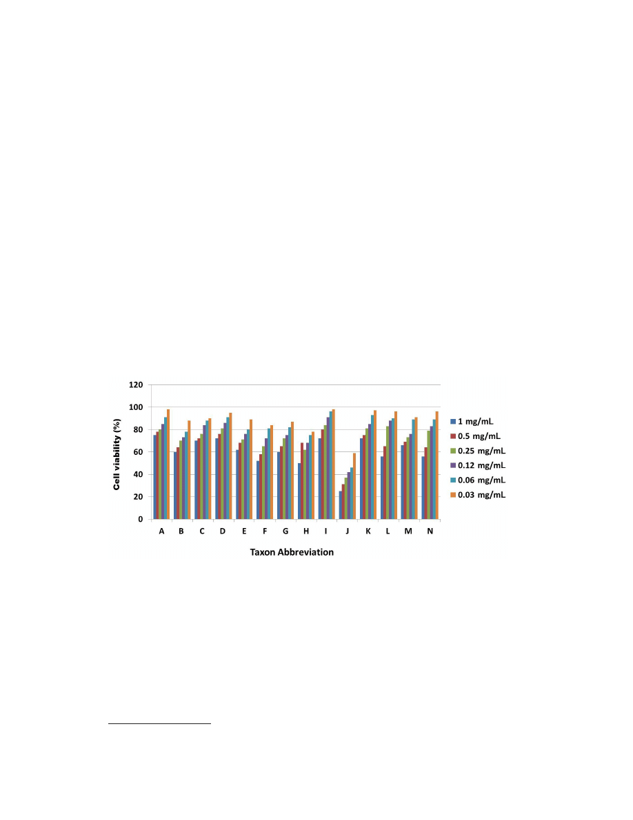
mg/mL (Table 1). Dose response studies of all the extracts are summarized in Figure 1.
Figure 1. A total of 14 plant extracts were screened for their ability to inhibit MCF-7 cell proliferation using MTT
assay after 24 h of exposure to the extracts. Most of the extract showed mild cytotoxicity at higher concentrations.
However, ethanolic extract of Lavandula dentata showed high toxicity at lower concentration. Data are reported as
mean values of three independent experiments. Taxon abbreviations: A. Aizoon canariense; B. Alhagi maurorum; C.
Anastatica hierochuntica; D. Capparis spinosa; E. Centaurothamnus maximus; F. Clutia lanceolata; G. Echinops
sheilae; H. Fagonia indica; I. Hyphaene thebaica; J. L. dentata; K. Malva parviflora; L. Neurada procumbens; M.
Teucrium oliverianum; N. Tribulus macropterus.
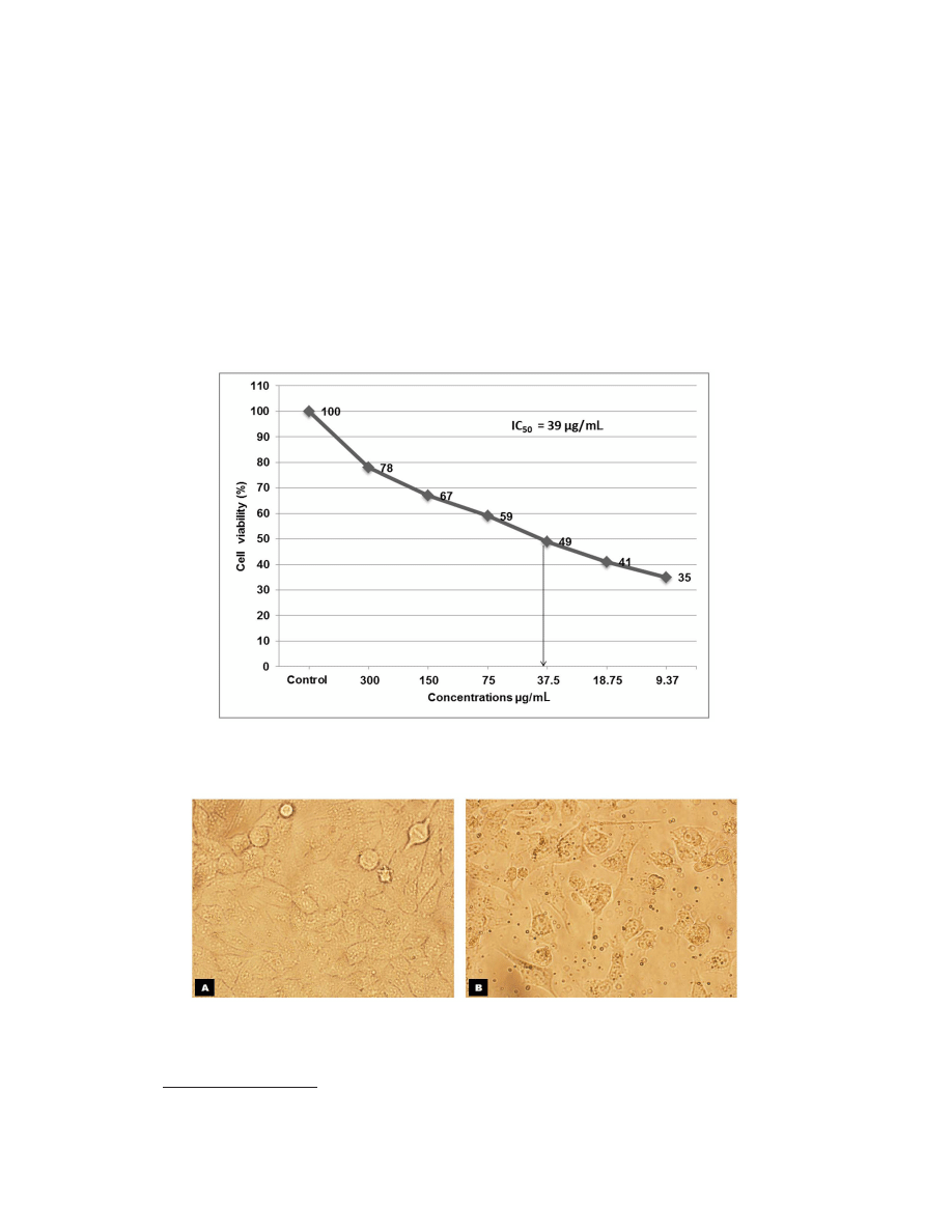
Based on these screening results (Table 1), the extract of L. dentata was selected, and
subjected to IC
50
determination (Figure 2). The relative number of viable cells as a percentage
of control was calculated, defining the absorbance at 550 nm for the control as 100%. The
IC
50
value was graphically obtained by plotting the percentage growth inhibition against the
corresponding different concentrations of the test compound used. The extracts of L. dentata
3986
©FUNPEC-RP www.funpecrp.com.br
Genetics and Molecular Research 13 (2): 3981-3990 (2014)
M.A. Ali et al.
showed cytotoxicity at an IC
50
value of approximately 39 µg/mL, while doxorubicin showed
63% growth inhibition at 100 μM (data not shown). The anticancer potential was analyzed ac-
cording to proliferation inhibition and apoptotic activity. No significant change in morphology
was observed in control cells. The cells appeared to have a normal shape, were attached to the
surface, and reached about 95-100% confluence (Figure 3A). Characteristics of apoptotic cell
death were evident in L. dentata treated samples. Cell shrinkage, loss of cell adhesion, reduced
cell density, and cell debris were clearly observed (Figure 3B).
Figure 2. Inhibition of MCF-7 cell proliferation by crude ethanolic extract of Lavandula dentata. Cells were treated
with indicated concentrations of extract for 24 h, and cell viability was determined by the MTT assay. The IC
50
value was estimated at 39 μg/mL (indicated by arrow).
Figure 3. Morphological changes in MCF-7 cells after the cells were exposed to IC
50
concentration of ethanolic
extract of Lavandula dentata for 24 h. A. Untreated cells appeared in normal shape with about 95-100% confluence.
B. Cell shrinkage, loss of cell adhesion, reduced cell density along with cell debris were observed in treated cells.
Images were captured in inverted microscope (Olympus, Japan) at 400X magnification.
3987
©FUNPEC-RP www.funpecrp.com.br
Genetics and Molecular Research 13 (2): 3981-3990 (2014)
Anticancer activities of extracts from Saudi Arabian plants
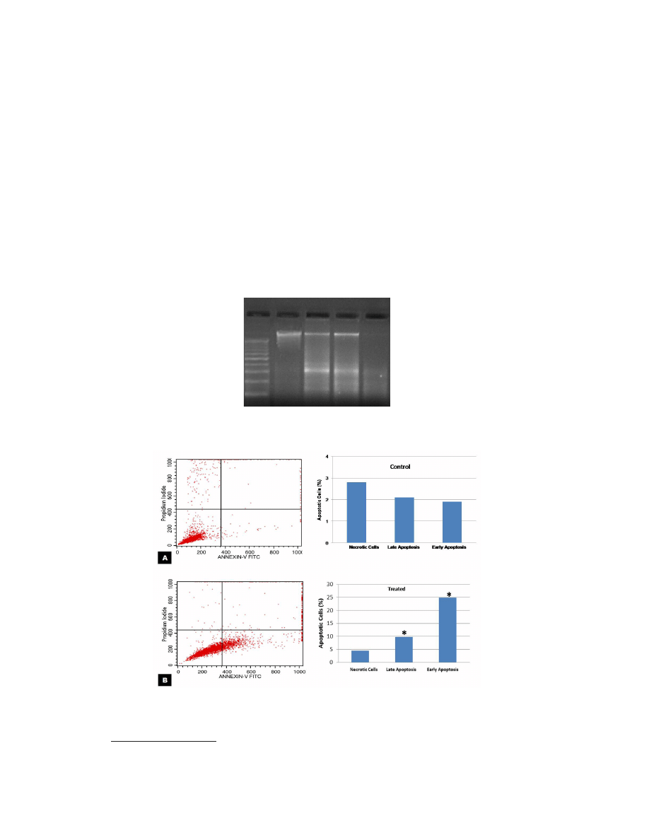
DNA fragmentation resulting from significant DNA damage was observed as a lad-
der pattern by agarose gel electrophoresis (Figure 4). The DNA from all 3 treated-group DNA
showed 180-200 base pair internucleosomal DNA fragments, whereas untreated DNA appeared
as single band. Furthermore, we quantified the extent of apoptosis in cells labeled with Annex-
in-V/PI staining using flow cytometry. For control MCF-7 cells, no significant difference in the
proportion of normal cells and those in early or late apoptosis were observed (Figure 5A). Only
2-3% cells were dead or undergoing apoptosis which is a normal event for cells growing in
cultures. However, after exposure to crude ethanolic extract of L. dentata (30 µg/mL for 24 h),
the proportion of early and late apoptotic cells increased significantly (P < 0.05) as compared
with control cells with values of about 25 and 10%, respectively (Figure 5B).
Figure 5. Apoptosis analysis of MCF-7 cells treated with IC
50
concentration of Lavandula dentata extract for 24
h using annexin-V FITC and propidium iodide staining. A. Flow cytometric scans of untreated cells showed only
2-3% of cells in early or late apoptosis stage. B. In treated cells, significant increase (P < 0.05) marked by asterisks
in percentage distribution of cells in early (25%) and late (10%) apoptosis were observed.
Figure 4. Analysis of genomic DNA fragmentation in MCF-7 cells after treatment for 24 h with crude ethanolic extract
of Lavandula dentata (30, 20, 10 μg/mL). DNA Fragmentation was assessed on agarose gel electrophoresis. Lane 1
= 100 bp DNA ladder used as marker; lane 2 = control; lane 3 = 30 μg/mL; lane 4 = 20 μg/mL; lane 5 = 10 μg/mL.
1 2 3 4 5

3988
©FUNPEC-RP www.funpecrp.com.br
Genetics and Molecular Research 13 (2): 3981-3990 (2014)
M.A. Ali et al.
DISCUSSION
The genus Lavandula (commonly known as lavender) belongs to the family Lamia-
ceae and comprises 28 species. Many members of the family Lamiaceae are well-known for
their pharmacological effects such as anticonvulsant, sedative, antispasmodic, analgesic, an-
tioxidant, or local anaesthetic activity (Ghelardini et al., 1999; Lis-Balchin and Hart, 1999;
Hosseinzadeh et al., 2000; Kovatcheva et al., 2001). Phytochemical analyses of lavender have
demonstrated the presence of many monoterpenes (especially linalyl acetate, linalool), which
are responsible for its pharmacologic activity (Lis-Balchin and Hart, 1999; Gilani et al., 2000).
Owing to its delightful odor, lavender has been widely used in perfumes and cosmetics (Gi-
lani et al., 2000), and oils from a variety of lavender species have been demonstrated to have
neurological, antimicrobial, pesticidal, and dermatological activities (Cavanagh and Wilkin-
son, 2002). The oil of L. angustifolia is chiefly composed of linalyl acetate and linalool and
is considered to be one of the mildest of known plant essential oils with known effects on
wound healing, cytotoxicity to human skin cells (Prashar et al., 2004), and inflammation and
analgesia (Hajhashemi et al., 2003). An aqueous extract of L. angustifolia protected the neu-
rons against glutamate toxicity (Buyukokuroglu et al., 2003). Moon et al. (2006) demonstrated
that low (≤1%) concentrations of Lavandula angustifolia and Lavandula intermedia oil can
completely eliminate Trichomonas vaginalis, Giardia duodenalis and Hexamita inflate. An
aqueous extract of Lavandula stoechas possess cytotoxic and genotoxic effects (Çelik and
Aslantürk, 2007). Berrington and Lall (2012) evaluated L. spica for anticancer activity on the
cervical epithelial carcinoma (HeLa) cell line. L. dentata has previously been reported to have
range of biological activities, such as anticonvulsant, sedative, antispasmodic (Gilani et al.,
2000), wound healing, rheumatic, urine retention, kidney stones, antiseptic (Rahman et al.,
2004), and antiprotozoal (Al-Musayeib et al., 2012).
By screening 14 ethanolic extracts of wild plants from Saudi Arabia for their anti-
proliferative properties against human adenocarcinoma breast cancer (MCF-7) cell lines, we
found that most extracts showed mild or no toxicity, whereas the crude ethanolic extract of
L. dentata exhibited promising cytotoxic activity. The identification of novel bioactive com-
pounds with anti-cancer properties, and the elucidation of the mechanisms by which the anti-
cancer properties derived from the natural products are of immense importance. The results of
this study provide the basis for further investigation of L. dentata for potential identification
of novel bioactive compounds with therapeutic and anti-cancer properties.
ACKNOWLEDGMENTS
The authors would like to extend their sincere appreciation to the Deanship of Scientific
Research at King Saud University for its funding of this research through the research group
project #RGP-VPP-195.
REFERENCES
Al-Musayeib NM, Mothana RA, Matheeussen A, Cos P, et al. (2012). In vitro antiplasmodial, antileishmanial and
antitrypanosomal activities of selected medicinal plants used in the traditional Arabian Peninsular region. BMC
Complement. Alternat. Med. 12: 49.
Almehdar H, Abdallah HM, Osman AM and Abdel-Sattar EA (2012). In vitro cytotoxic screening of selected Saudi
medicinal plants. J. Nat. Med. 66: 406-412.

3989
©FUNPEC-RP www.funpecrp.com.br
Genetics and Molecular Research 13 (2): 3981-3990 (2014)
Anticancer activities of extracts from Saudi Arabian plants
American Cancer Society (2012). Breast Cancer Facts & Figures 2011-2012. American Cancer Society, Inc., Atlanta.
Amin A and Mousa M (2007). Merits of anti-cancer plants from the Arabian Gulf Region. Cancer Ther. 5: 55-66.
Baker DD, Chu M, Oza U and Rajgarhia V (2007). The value of natural products to future pharmaceutical discovery. Nat.
Prod. Rep. 24: 1225-1244.
Berrington D and Lall N (2012). Anticancer activity of certain herbs and spices on the cervical epithelial carcinoma (HeLa)
cell line. Evid. Based Complement. Alternat. Med. 2012: 564927.
Buyukokuroglu ME, Gepdiremen A, Hacimuftuoglu A and Oktay M (2003). The effects of aqueous extract of
Lavandula angustifolia flowers in glutamate-induced neurotoxicity of cerebellar granular cell culture of rat pups. J.
Ethnopharmacol. 84: 91-94.
Cavanagh HM and Wilkinson JM (2002). Biological activities of lavender essential oil. Phytother. Res. 16: 301-308.
Çelik TA and Aslantürk OS (2007). Cytotoxic and genotoxic effects of Lavandula stoechas aqueous extracts. Biol.
Bratislava 62: 292-296.
Chaudhary S (2001). Flora of the Kingdom of Saudi Arabia. Ministry of Agriculture and Water, Kingdom of Saudi Arabia,
Riyadh.
Dixon N, Wong LS, Geerlings TH and Micklefield J (2007). Cellular targets of natural products. Nat. Prod. Rep. 24:
1288-1310.
Elkady AI (2013). Crude alkaloid extract of Rhazya stricta inhibits cell growth and sensitizes human lung cancer cells to
cisplatin through induction of apoptosis. Genet. Mol. Biol. 36: 12-21.
Evens AM, Prachand S, Shi B, Paniaqua M, et al. (2004). Imexon-induced apoptosis in multiple myeloma tumor cells is
caspase-8 dependent. Clin. Cancer Res. 10: 1481-1491.
Figueroa-Hernández JL, Sandoval GG, Ascencio VJ, Figueroa-Espitia JL, et al. (2005). Plant products with anti-cancer
properties employed in the treatment of bowel cancer: literature review 1985 and 2004. Proc. West Pharmacol. Soc.
48: 77-83.
Ghelardini C, Galeotti N, Salvatore G and Mazzanti G (1999). Local anaesthetic activity of the essential oil of Lavandula
angustifolia. Planta Med. 65: 700-703.
Gilani AH, Aziz N, Khan MA, Shaheen F, et al. (2000). Ethnopharmacological evaluation of the anticonvulsant, sedative
and antispasmodic activities of Lavandula stoechas L. J. Ethnopharmacol. 71: 161-167.
Graham JG, Quinn ML, Fabricant DS and Farnsworth NR (2000). Plants used against cancer - an extension of the work of
Jonathan Hartwell. J. Ethnopharmacol. 73: 347-377.
Hajhashemi V, Ghannadi A and Sharif B (2003). Anti-inflammatory and analgesic properties of the leaf extracts and
essential oil of Lavandula angustifolia Mill. J. Ethnopharmacol. 89: 67-71.
Harlev E, Nevo E, Lansky EP, Lansky S, et al. (2012). Anticancer attributes of desert plants: a review. Anticancer Drugs
23: 255-271.
Harvey AL (2008). Natural products in drug discovery. Drug Discov. Today 13: 894-901.
Hosseinzadeh H, Ramezani M and Salmani G (2000). Antinociceptive, anti-inflammatory and acute toxicity effects of
Zataria multiflora Boiss extracts in mice and rats. J. Ethnopharmacol. 73: 379-385.
Kovatcheva EG, Koleva II, Ilieva M, Pavlov A, et al. (2001). Antioxidant activity of extract from Lavandula vera MM
cell cultures. Food Chem. 72: 295-300.
Kuno T, Testuya T, Akira H and Takuji T (2012). Cancer chemoprevention through the induction of apoptosis by natural
product. J. Biophysical. chem. 3: 156-173.
Lachenmayer A, Alsinet C, Chang CY and Llovet JM (2010). Molecular approaches to treatment of hepatocellular
carcinoma. Dig. Liver Dis. 42 (Suppl 3): S264-S272.
Lis-Balchin M and Hart S (1999). Studies on the mode of action of the essential oil of lavender (Lavandula angustifolia
P. Miller). Phytother. Res. 13: 540-542.
Madhuri S and Pandey G (2009). Some anticancer medicinal plants of foreign origin. Curr. Sci. 96: 779-783.
Mann J (2002). Natural products in cancer chemotherapy: past, present and future. Nat. Rev. Cancer 2: 143-148.
Moon T, Wilkinson JM and Cavanagh HM (2006). Antiparasitic activity of two Lavandula essential oils against Giardia
duodenalis, Trichomonas vaginalis and Hexamita inflata. Parasitol. Res. 99: 722-728.
Mosmann T (1983). Rapid colorimetric assay for cellular growth and survival: application to proliferation and cytotoxicity
assays. J. Immunol. Methods 65: 55-63.
Mothana RA, Lindequist U, Gruenert R and Bednarski PJ (2009). Studies of the in vitro anticancer, antimicrobial and
antioxidant potentials of selected Yemeni medicinal plants from the island Soqotra. BMC Complement. Alternat.
Med. 9: 7.
Newman DJ and Cragg GM (2012). Natural products as sources of new drugs over the 30 years from 1981 to 2010. J. Nat.
Prod. 75: 311-335.

3990
©FUNPEC-RP www.funpecrp.com.br
Genetics and Molecular Research 13 (2): 3981-3990 (2014)
M.A. Ali et al.
Prashar A, Locke IC and Evans CS (2004). Cytotoxicity of lavender oil and its major components to human skin cells.
Cell Prolif. 37: 221-229.
Rahman MA, Mossa JS, Al-Said MS and Al-Yahya MA (2004). Medicinal plant diversity in the flora of Saudi Arabia 1:
a report on seven plant families. Fitoterapia 75: 149-161.
Stopeck AT and Thompson PA (2012). Breast Cancer Treatment and Management. (WebMD Health Professional
Network) Retrieved. Available at [http://emedicine.medscape.com/article/1947145-treatment]. Accessed September
29, 2012.
Tan W, Lu J, Huang M, Li Y, et al. (2011). Anti-cancer natural products isolated from Chinese medicinal herbs. Chin.
Med. 6: 27.
Wyszukiwarka
Podobne podstrony:
In vitro antitumor actions of extracts
In Vitro Anticancer Activity of Ethanolic Extract
In vitro antitumor actions of extracts
In vitro cytotoxicity activity
In vitro corrosion resistance of titanium made using differe
In vitro biological effects of titanium rough surface obtain
2001 In vitro fermentation characteristics of native and processed cereal grains and potato
Cytotoxic Properties of Some Medicinal Plant Extracts
Evaluation of in vitro anticancer activities
więcej podobnych podstron