
Brain (1999), 122, 1317–1325
Semantic integration in reading: engagement of the
right hemisphere during discourse processing
M. St George, M. Kutas, A. Martinez and M. I. Sereno
University of California, San Diego, USA
Correspondence to: Marie St George, Center for Research
in Language, 0526, University of California, San Diego,
9500 Gilman Drive, La Jolla, CA 92093-0526, USA
E-mail: stgeorge
@crl.ucsd.edu
Summary
We examined the brain areas involved in discourse
processing by using functional MRI in 10 individuals as
they read paragraphs, with or without a title, word by
word for comprehension. Functional data were collected
from 20 adjacent 5 mm axial slices. Discourse processing
was associated with activation in inferior frontal and
temporal regions of both cerebral hemispheres in the titled
and untitled conditions. Moreover, there was substantially
more right hemisphere activation for untitled than for the
titled paragraphs. More specifically we found: (i) greater
Keywords: functional MRI; language; semantic integration; right hemisphere
Abbreviations: BA
5 Brodmann area; ERP 5 event-related brain potential; fMRI 5 functional MRI
Introduction
Language comprehension involves processes at multiple
levels of analysis including lexical, syntactic, semantic,
pragmatic and discourse. Research on individuals with brain
damage has led to the realization that both cerebral
hemispheres are involved in language comprehension, albeit
to varying degrees with regard to these different levels of
linguistic analysis (Caplan, 1992). The present study is aimed
at identifying regions of the right and left hemispheres that
show systematic changes in blood flow [as measured via
functional MRI (fMRI)] during processing of written text.
We focus on discourse processes as they are an integral part
of our daily communications, subsuming, but also going
beyond, the processes engaged in recognizing words, syntactic
parsing
and
comprehending
isolated
sentences
(e.g.
Gernsbacher, 1994).
The classic model of language organization, based on
a century of studying aphasic patients, situates language
comprehension and production squarely in the perisylvian
regions of the left hemisphere (e.g. Wernicke, 1874;
Geschwind, 1970). However, individuals with damage to
various parts of the right hemisphere (e.g. following
cerebrovascular accidents) also experience subtle language
problems (Joanette et al., 1990). For example, patients with
© Oxford University Press 1999
activation in the inferior temporal sulcus of both
hemispheres for untitled than titled paragraphs; (ii)
greater average volume of activation in response to
untitled than titled paragraphs in the middle temporal
sulcus of the right hemisphere and the reverse pattern in
the left middle temporal sulcus. Consistent with previous
studies of individuals with right hemisphere damage, we
suggest that the right middle temporal regions may be
especially important for integrative processes needed to
achieve global coherence during discourse processing.
right hemisphere damage are often described as experiencing
difficulties at the level of discourse. They tend not to elaborate
on details of a discourse, producing fewer propositions and
fewer complex propositions, although their basic knowledge
of scripts or event schema appears to be intact. Right
hemisphere damage patients are frequently unable to maintain
the theme of a conversation, missing the main point altogether
(Brownell and Martino, 1998). In laboratory experiments,
right hemisphere damage patients have been found to have
difficulties drawing certain types of inferences (Beeman,
1993) or revising them when new information comes up in
a discourse (Birhle et al., 1986). Right hemisphere damage
patients often fail to understand jokes (failing to connect the
premise to the punch line) (Brownell et al., 1983), and are
reported to experience difficulties in appreciating metaphors,
idioms and indirect requests (Weylman et al., 1989).
Altogether, the data suggest right hemisphere damage patients
can recognize individual words and comprehend sentences
(presumably with their intact left hemisphere), but have
trouble connecting and integrating semantically and/or
temporally distant concepts. By contrast, aphasic patients can
produce relatively normal discourse structures using devices
such as pronominalization and explicit connectives that

1318
M. St George et al.
create discourse coherence despite significant difficulties in
producing words and sentences (Bates et al., 1983). Of
course, as language impairments become especially severe,
even aphasic patients lose their ability to produce coherent,
elaborate discourse. It thus seems that both the left and the
right hemisphere must be intact for proper understanding of
discourse. Moreover, an intact right hemisphere seems to be
especially important for higher level integrative processes
that lead to functional coherence in discourse.
We chose to examine this hypothesis further by contrasting
fMRI data from individuals as they read a series of paragraphs
(see below) that were identical in all structural respects but
one: namely, whether they were preceded by a title or were
untitled (Bransford and Johnson,1972; see also Dooling and
Lachman, 1971).
‘This is very rewarding but tends to be quite expensive
even if you own all that you need. The outfit does not
really matter. One can get seriously injured without proper
instruction even if it comes more naturally to some people
than others. Some don’t like the smell or the lack of
control. So some people are scared to try it even if they’ve
dreamed of it since they were a kid reading about it in
books and watching it on television. A running start is
uncommon, although there are some who do it. Typically,
success requires that you start with your left leg, and
make sure that it is securely in place. Then swing your
body high into the air. The direction matters. Once you
are settled, your thumbs should be pointing up. Sometimes
there is no security but the animal’s hair. Other times you
can hang off to the side. In any case you will be sore if
this is your first time.’
Bransford and Johnson (Bransford and Johnson, 1972) found
that without a title (in this case, Horse-back riding) paragraphs
like this proved to be quite difficult for readers to understand
and to remember. This was the case despite the fact that all
the sentences within the paragraphs were grammatically well
formed and meaningful. It was as if without a title, these
paragraphs were meaningless as a discourse, nothing more
than a series of disconnected and semantically vague
propositions, which were thus difficult to recall. The mere
presence of a title, however, seemed to render these
paragraphs more comprehensible and effectively doubled the
number of words that readers could recall from them.
Comprehending
the
sentences
that
comprise
such
paragraphs requires a whole host of operations for word
recognition and syntactic analysis that have been shown to
activate predominantly, if not exclusively, left hemisphere
brain areas (e.g. Binder et al., 1997; Price et al., 1997). Thus,
we would expect these regions to be active during the
paragraph reading, with or without a title. In addition,
discourse comprehension requires that mental connections be
made between various parts of a text in order to understand
it. Any given sentence can be connected to information in
the previous sentence and/or within working memory to
achieve ‘local coherence’ but needs to be connected to a text
macrostructure or to information earlier in the text to achieve
‘global coherence’. We believe it is this process that is faulty
in right hemisphere damage patients and is typically the
province of an intact right hemisphere. Accordingly, we also
expect some increased activation in regions of the right
hemisphere in both paragraph conditions as the readers
attempt to achieve understanding at a discourse level.
However, since the processes required to achieve global
coherence are more taxed in the untitled condition, we predict
relatively greater activation in the right hemisphere for the
untitled than titled paragraphs.
Method
Subjects
The participants were 10 healthy, native English speaking
volunteers (23–45 years of age; half of them were male). All
were right-handed with the exception of one who considered
herself ambidextrous. The subjects gave informed consent and
the study was approved by the Human Subjects Committee of
the University of California, San Diego.
Materials
Sixteen paragraphs (eight titled, eight untitled) were presented
visuocentrically, one word (duration
5 200 ms) every 300
ms. An additional half second delay followed the last word
of each sentence. Paragraphs ranged between 8 and 14
sentences (mean 9.7) in length; sentences varied between 3
and 21 words (mean 9.6) in length. In the control task, words
were replaced by strings of Xs of variable length. Whether
a given paragraph appeared as untitled or titled was
counterbalanced across subjects; no individual read the same
paragraph twice. The materials were presented in four runs
lasting 4.5 min each; each paragraph lasted 30 s and each
run began and ended with 30 s of flashing Xs. In other
words, each run consisted of 4.5 cycles (five half-cycles of
Xs; four half-cycles of paragraphs), where a ‘cycle’ is the
total time consumed by one experimental and one control task.
Imaging
Imaging was performed on a 1.5T GE Signa scanner fitted
with a high performance local head gradient and an RF coil
which is a quadrature transmit-receive elliptical endcapped
birdcage that is optimized for brain imaging (Wong et al.,
1992).
Axial images were acquired for 20 adjacent 5 mm slices
[TR (repetition time)
5 3 s] for eight subjects and 13 adjacent
5 mm slices with a TR of 2.5 for the other two subjects,
using an echo planar single shot pulse sequence with a matrix
size of 64
3 64, and in-plane resolution of 3.75 3 3.75 mm.
The start point was at the bottom of the temporal lobes for
all 10 subjects. The first axial image or slice began at
approximately –28 mm in Talairach space (Talairach and
Semantic integration in right hemisphere
1319
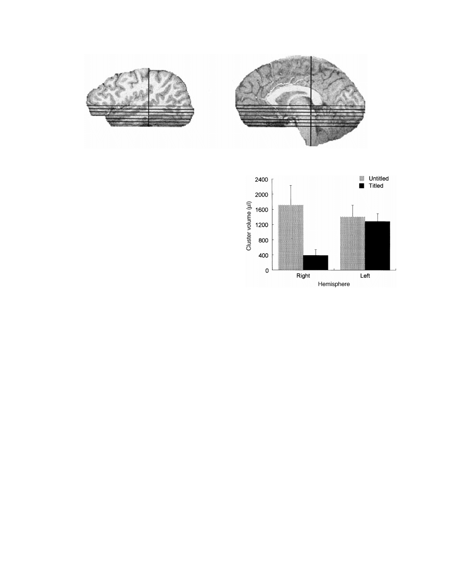
Fig. 1 Sagittal views of location of six functional slices selected for region of interest analyses.
Tournoux, 1988). Ninety-two images were acquired for each
slice, while the participant alternated between control and
task (30 s each) with a total of 4.5 cycles during each of the
four trials. For anatomical localization, we acquired a T
1
-
weighted 3D MPRAGE sequence [TR
5 30 ms, TE (echo
time)
5 5 ms, flip angle 5 45°, 256 3 192 3 60 mm
matrix]; functional maps were subsequently overlaid on the
corresponding structural images.
Procedure
Participants lay flat inside the magnet and viewed the stimuli
(projected on to a screen at their feet) via a mirror located
above their eyes. Each participant saw any given paragraph
only once; paragraphs were counterbalanced (titled versus
untitled) across participants. Participants saw all four
paragraphs of each run as either titled or all untitled (i.e.
presentation of conditions was blocked). Half of the
participants saw an untitled run first, while the other half
saw a titled run first; thereafter the conditions alternated.
Data analysis and results
Individual subject analysis
Before any analysis, the images were spatially registered so
as to check and correct for motion artefacts. Timeseries data
for runs of similar conditions (titled/untitled) were then
averaged together. All of the subsequent analyses were
performed using AFNI (Analysis of Functional Neuro Images)
software, available through the Medical College of Wisconsin
(Cox, 1996). The averaged titled and untitled runs were
analysed by correlating the time course for each voxel with
an ideal 4.5 cycle trapezoidal reference waveform as shown
in Fig. 5 (Bandettini et al., 1993). Voxels meeting or exceeding
a correlation coefficient of 0.50 (P
, 0.000001) were
considered reliably associated with the task. Clusters were
defined as groups of activated voxels at least 400
µ
l in
volume with a connectivity radius of 6 mm. Four regions of
interest spanning six inferior slices were defined (see Fig. 1
for slice locations). All six horizontal slices imaged primarily
temporal areas, with some frontal and occipital cortex as well,
ranging from Talairach coordinates –20 to
110 (Talairach and
Fig. 2 Mean volume of activated clusters for the titled and
untitled paragraph runs as a function of cerebral hemisphere.
Tournoux, 1988). Clusters of activity were found in frontal
cortex [Brodmann areas (BA) 44, 45 and 47], inferior
temporal sulcus (BA 19, 20 and 37), middle temporal sulcus
(BA 21 and 38) and superior temporal sulcus (BA 22 and
42) in the six slices. The volumes of all clusters falling
within an region of interest, across all six slices, were
summed.
Across-subject analysis
These cluster volumes were then subjected to a (2
3 2 3 4)
repeated measures ANOVA with three within-participants
factors: Context (titled, untitled), Hemisphere (left, right) and
Region (frontal, inferior temporal sulcus, middle temporal
sulcus, superior temporal sulcus).
Neither the main effect of Context (titled
5 834; untitled 5
1553) nor Hemisphere (left
5 1343; right 5 1045) reached
significance [F(1,9)
5 1.81, P 5 0.21; F(1,9) 5 1.34, P 5
0.28, respectively]. The only reliable interaction was of
Context by Hemisphere [F(1,9)
5 7.29, P 5 0.02]. As can
be seen in Fig. 2, this interaction reflects the greater activation
in the untitled compared with the titled condition [F(1,9)
5
8.9, P
5 0.015] in the right hemisphere but comparable
activations in the left hemisphere [F(1,9)
5 0.32, P 5 0.59].
The raw data from each participant (timecourse for each
voxel) were averaged across the eight for whom 20 slices at
1320
M. St George et al.
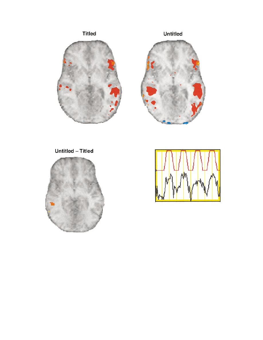
Fig. 3 Structural and functional data averaged across eight participants. Colour shading indicates percentage signal change (warm colours
represent positive correlation; cool colours represent negative correlation), with thresholds set at r
5 0.60 with the reference waveform.
Note that the hemispheres are presented according to radiological convention, with the right hemisphere on the left side.
Fig. 4 Axial view of the untitled minus titled difference averaged
across participants, with the threshold for right hemisphere
activation (on the left of the image) set at r
5 0.60 with the
reference waveform.
repetition time of 3 s were acquired; the other two could not
be included in the average as their data were acquired with
a slightly different protocol. The structural and functional
datasets for each participant were resampled at 2.5 mm cubed
and subsequently averaged. The averaged time-series dataset
was then correlated with the ideal 4.5 cycle trapezoidal
reference waveform mentioned previously. These data are
plotted in Figs 3 and 4. Figure 3 shows the axial slices
for untitled and titled conditions averaged across eight
participants. Figure 4 shows the average difference between
the two conditions (untitled minus titled) across eight
participants. To do this, the raw data for the titled condition
Fig. 5 Time course waveform in MRI units for a voxel in the
middle temporal gyrus of the right hemisphere during reading of
untitled paragraphs. The red waveform is the reference with
which the data (time courses from every voxel) were correlated.
were subtracted from the untitled raw data, and then the
difference waveform was correlated with the ideal reference
waveform, mentioned above. The slice shown in Figs 3 and
4 corresponds to Talairach coordinate Z
5 –1 (Talairach and
Tournoux, 1988). Figure 5 shows the timecourse of a voxel
from the eight subject average that was highly correlated
with the ideal 4.5 cycle reference waveform.
Planned comparisons were conducted to contrast the
average volume for the titled versus untitled conditions in
each region of interest in each hemisphere, separately. As
can be seen in Table 1, the pattern of effects varied with
region of interest. There were no reliable effects in the
superior temporal sulcus and only a marginal effect in the
right frontal region with slightly greater volume for the
untitled than titled condition [F(1,9)
5 3.75, P 5 0.063]. As
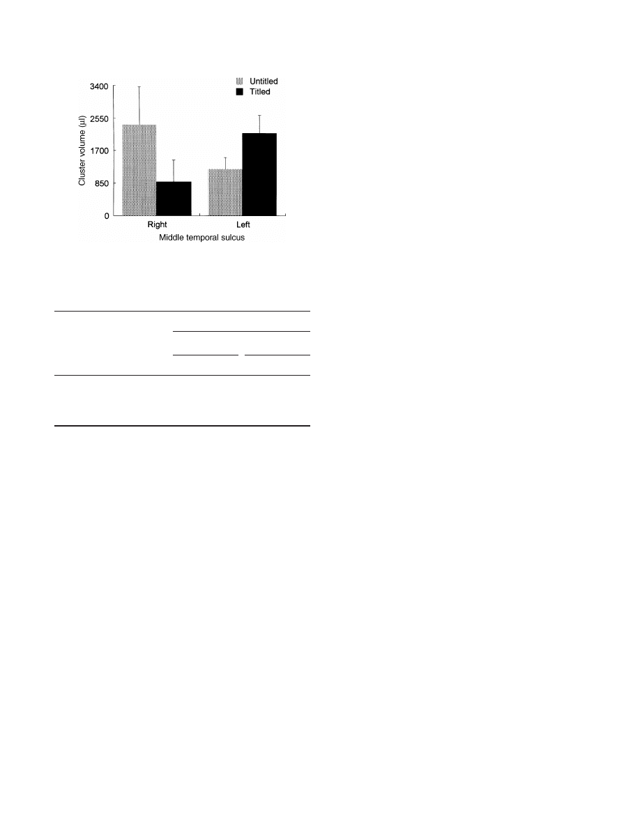
shown in Fig. 6, in the middle temporal sulcus, the pattern
went in opposite directions in the two hemispheres: in the
right hemisphere the untitled condition was associated with
Semantic integration in right hemisphere
1321
Fig. 6 Mean volume of activated clusters for the titled and
untitled paragraph runs in the middle temporal regions of the right
and left hemispheres.
Table 1 Pattern of context effects by region of interest
Region of
BA
Average volume (
µ
l)
interest
Left
Right
Titled
Untitled
Titled
Untitled
Fr
44, 45, 47
425
1125
207
1062
ITS
19, 20, 37
1259
2362
98
2306
MTS
21, 38
2158
1212
886
2367
STS
22, 42
1294
907
347
1085
Fr
5 frontal cortex; ITS 5 inferior temporal sulcus; MTS 5
middle temporal sulcus; STS
5 superior temporal sulcus.
larger volume [F(1,9)
5 10.37, P 5 0.005], whereas in the
left hemisphere the titled condition was associated with the
larger volume [F(1,9)
5 4.24, P 5 0.05]. In inferior temporal
sulcus, the average volume was larger in response to the
untitled than titled paragraphs in both hemispheres [right
hemisphere: F(1,9)
5 25.03, P 5 0.0001; left hemisphere:
F(1,9)
5 6.26, P 5 0.02].
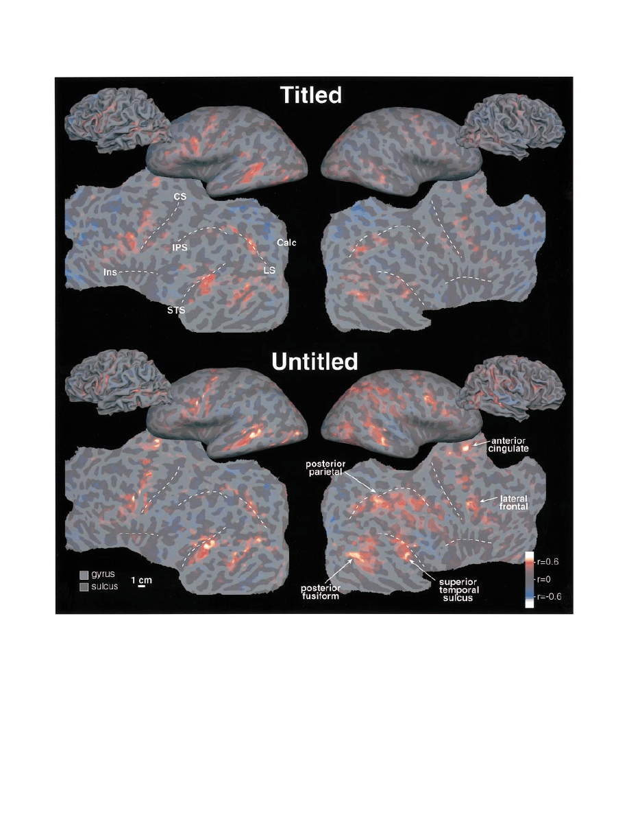
The response to titled and untitled paragraphs (see Fig. 7,
top and bottom, respectively) is shown for the left and right
hemispheres in folded, unfolded and flattened views for a
single female participant. The main effect described above
is visible in the pattern of positive responses (red-orange)
shown in the figure. Untitled paragraphs elicited an increased
response in both hemispheres but the increase was more
prominent in the right hemisphere; left is greater than right
for titled while right is greater than left for untitled. Five
major regions of activity are present in both hemispheres in
both conditions, and are labelled at the lower right. Several
of the foci that were significantly activated in this participant
did not reach significance in the overall Talairach average
(Talairach and Tournoux, 1988). This partly reflects the
effects of cross-subject anatomical variability. It may also
reflect differences in strategy and more prosaically, head
stability, across subjects.
The reconstructed cortical surface passes through the deep
layers of the cortex, which is why it is possible to see into
the sulci in the folded views. The unfolded view was made
from the folded one by unfolding without stretching. The
flattened view was made by making five medial cuts in each
hemisphere’s surface (along the calcarine sulcus, at the
temporal pole, at the anterior pole, and in the anterior and
posterior cingulate regions) to allow the cortex to lie flat
without undue distortion. Light and dark grey indicate the
local curvature of the folded cortex (convex and concave)
which approximately correspond to gyrus and sulcus.
Discussion
As expected, we observed increased blood flow (fMRI signal)
in both cerebral hemsipheres as individuals read short
paragraphs word by word for comprehension. In addition, we
found that the extent and degree of right hemisphere activation
was modulated by the presence or absence of a title for the
paragraph: a greater volume of the right inferior temporal
sulcus and right middle temporal sulcus were activated for
the untitled than the titled paragraphs. Our results are thus
consistent with a handful of functional imaging studies using
primarily cerebral blood flow measures reporting bilateral
activation to spoken or read narratives compared with rest
(Mazziotta et al., 1982; Huettner et al., 1989; Lechevalier et al.,
1989), reading or listening to word lists (Mazoyer et al., 1993;
Binder, et al., 1994 using fMRI) and paragraphs of random
sentences (Fletcher, et al., 1995). The most common finding
across these studies has been increased activation of the
temporal poles and middle temporal gyri, often with more
diffuse activation in the right than left hemisphere.
The relatively greater involvement of the right hemisphere
during the processing of written and spoken narratives contrasts
with numerous neuroimaging studies concluding that ‘. . .
cortical activation associated with language processing is
strongly lateralized to the left cerebral hemisphere’ (e.g. Binder
et al., 1997). Most of them have found either no or substantially
little right hemisphere activation for language processing at
the level of the letter, word or literal comprehension of single
sentences (e.g. Bavelier et al., 1997; Helenius et al., 1998) (for
bilateral activation to single words, see Wise et al., 1991; Pugh
et al., 1996).
We can look to our data together with that of previous studies
to suggest at least part of right hemisphere’s role in natural
language processing. Bilateral activations have also been
obtained for stimulus materials that patients with right
hemisphere damage find especially taxing and thus it has been
suggested that the right hemisphere areas are invoked primarily
to deal with special types of language materials such as jokes,
metaphors and fables. For example, Bottini et al. (Bottini et al.,
1994) found bilateral PET activation of the middle temporal
gyrus for the processing of metaphors compared with literal
sentences in a task that required participants to indicate whether
the sentence was or was not plausible, where approximately
50% were not. They attributed the increased activity in the right
hemisphere to its specific role in the processing of figurative
1322
M. St George et al.
Fig. 7 The response to titled and untitled paragraphs (top and bottom) is shown for the left and right hemispheres (left and right) in
folded, unfolded and flattened views (all to the same scale) for a single female subject. The main effect described in the text is visible in
the pattern of positive responses (red-orange). Dotted lines on the flattened representation indicate the general trend of several major
sulci to help in identifying corresponding points on the folded and unfolded representations. These include the central sulcus (CS), lateral
sulcus and the insula (Ins), the intraparietal sulcus (IPS), the superior temporal sulcus (STS), the calcarine sulcus (Calc) and the lunate
sulcus (LS—also sometimes called the transverse occipital sulcus or even the intraparietal sulcus).
aspects of language. Likewise, Nichelli et al. (Nichelli et al.,
1995) observed greater activation in BA 21, middle temporal
gyrus, as well as the inferior frontal gyrus (BA 47) of the right
hemisphere when participants monitored Aesop’s fables for
their moral rather than for their semantic features of a main
character (e.g. is it an animal with scales?) and interpreted the
increase in terms of the right hemisphere’s specific role in
thematic processing (for fable morals). Our results, however,
clearly show that right hemisphere engagement during sentence
comprehension is not specific to the processing of figurative

Semantic integration in right hemisphere
1323
aspects of language. Rather, right hemisphere engagement
appears to be a more general phenomenon that occurs routinely
as readers attempt to construct a unitary coherent model of a
discourse and discover the producer’s intent.
Our stimulus materials are relatively ordinary paragraphs,
in some cases much like the instruction manual one might
find for assembly of a newly purchased product. Each of our
sentences is semantically congruent and internally cohesive;
there are no outright violations or denotative violations as
Bottini et al. (Bottini et al., 1994) called metaphors or idioms.
Our paragraphs do not include indirect requests and can readily
be interpreted literally. Moreover, our participants were not
asked to render any extraneous decisions (e.g. lexical decision,
plausibility, etc.) about any aspect of the stimulus materials,
but merely to read them for comprehension so as to be able to
answer occasional comprehension queries. In fact, we used
exactly the same texts and the same task in both our
experimental conditions, which differed only in the presence or
absence of a title. Specifically, our titled and untitled paragraphs
are orthographically (the size and shape of the letters and
words), lexically (the frequency, length and meaning of the
words) and syntactically (level of complexity of the sentence
structure) identical. Readers saw the same words at the same
position on the screen, in the same orders, within the same
sentences in both conditions. The only difference between the
conditions is the presence or absence of a title and what this
entails about the paragraphs’ processing (discussed below).
Sentences within a coherent discourse differ from a string of
isolated sentences in being formally connected (via referential
links or sentence connectors), with each sentence being
logically consistent with the previous one, as well as
pragmatically relevant to the underlying discourse theme or
topic. Thus, in processing text, readers need to infer the nature
of the relations among ideas, events and states described therein
and make use of referential cohesion, causal cohesion,
relational cohesion, etc. to create a coherent representation of
the discourse. Based on our experience as readers, we expect
sentences in a discourse to sustain a sense of continuity in time,
place, participants and episodes. Accordingly, we routinely use
various integrative processes to find the connectedness between
adjacent or nearby sentences as well as the overall topic. We
believe that a good title serves to cue our semantic knowledge
in such a way as to ease and/or expedite these processes of
global coherence, i.e. in the generation of a meaningful
message-level representation, which are, nonetheless, part and
parcel of comprehending any titled or untitled discourse.
Our paragraphs, however, were designed such that without
a title constructing a coherent message level representation
would require more time and more effort. Accordingly, one
might argue that the increased activity in the right hemisphere
merely reflects greater arousal or effort in the more difficult
untitled condition. Greater difficulty might be reflected in a
general increase in effort or arousal or in a specific increase in
effort at one or more linguistic levels. A general increase in
effort is likely to lead to increased activation of exactly the
same areas, or perhaps activation in an additional area. For
example, many researchers have found that manipulations of
task difficulty do modulate activity in the anterior cingulate
(e.g. D’Esposito et al., 1995); thus, it has been suggested that
the anterior cingulate may be involved in the mediation of
motivational and/or affective responses to task difficulty
(Barch et al., 1997). While some of our participants did show
activity in the anterior cingulate, neither of these patterns was
what we observed. Instead, we found that increased activation
in the right middle temporal suclus during the untitled condition
was coupled with decreased activation in the left middle
temporal sulcus. Although we do not yet have a good
hypothesis as to the function of the decreased activity in the
left middle temporal sulcus, we cannot reconcile this particular
pattern of activations with any explanation based solely on
increase in non-specific arousal. Given the highly controlled
nature of our materials, insofar as there is an increase in
difficulty from the titled to the untitled paragraphs, it must
be linked to higher level processes that cut across individual
sentence boundaries. Thus, we think that the middle temporal
gyrus and inferior frontal gyrus contribute to language
processing at the level of semantic integrative processes at the
level of discourse. Perhaps the decreased activity in the left
middle temporal sulcus and increased activity in the right
middle temporal sulcus reflects a trade-off in processing
resources. That is, the decreased activity in the left middle
temporal sulcus in the untitled condition reflects reduced effort
devoted to matching individual words with their meanings, i.e.
lexical integration (responsibility of the left middle temporal
sulcus) in the absence of top-down constraints and greater
effort devoted to figuring out what the discourse is about (which
is the responsibility of the right middle temporal sulcus, and
may feed back to the left).
Electrophysiological recordings have shown that within the
temporal domain this same title manipulation has its effects on
the processing of any word within the text between 250 and
550 ms after its appearance. Specifically, St George et al.
(St George et al., 1994) recorded event-related brain potentials
(ERPs) from the scalp of individuals as they read these texts
one word at a time either with or without a preceding title. The
significant difference between the ERPs elicited by words in
the two experimental conditions was in the amplitude of a
negativity (N400) starting around 200 ms and peaking around
400 ms after each word; this N400 was reliably larger for words
from paragraphs without than with a title. Much research has
linked N400 amplitude to semantic processing, especially
semantic or contextual integrative processes (for review see
Osterhout and Holcomb, 1995). Such research has shown that
N400 amplitude is sensitive to (i) semantic relatedness in word
pairs, being smaller for related than unrelated words; (ii)
semantic congruity at a sentence level, being smaller for
congruous than incongruous endings; and (iii) extra-sentential
semantic constraints within discourse, being smaller for more
than less predictable words.
The N400 has a broad distribution across the scalp. In
response to words within sentences, the N400 is often slightly
laterally asymmetric, being larger over posterior regions of the

1324
M. St George et al.
right than left hemisphere. Some researchers have suggested
that it may be a composite of multiple negative components
(e.g. Pritchard et al., 1991). Given the inverse problem, it is
impossible unequivocally to infer neural generators from scalp
recordings. Intracranial recordings from one or both
hemispheres of patients with intractable epilepsy, however,
have led to the hypothesis that at least some of the N400 is
generated bilaterally in the neocortex near the collateral sulcus
and the anterior fusiform gyrus (McCarthy et al., 1995). This
conclusion is based on the intracranial pattern of field potentials
and their sensitivity to the same task manipulations (e.g.
semantic congruity, word class) that modulate the scalp N400.
In the present study we obtained reliable bilateral increases in
fMRI activations in the inferior temporal sulcus and fusiform
gyrus as the untitled paragraphs were being read. These may
eventually prove to be related to the N400 amplitude
modulations elicited by these same stimulus texts.
The mechanisms by which the right hemisphere achieves
global coherence or integrates information across sentences
within a discourse remain unknown as there is not even a
consensus on just how discourse coherence should be defined
(Hellman, 1995). Integration requires that there be multiple
pieces of information to be integrated; clearly, both temporal
and spatial summation can aid integrative processes. Thus,
we can look to see whether stimulation of the right and left
hemisphere lead to different types or amounts of information
being activated, different time courses of information
activation, or both. Both behavioural and ERP data suggest
greater possibility for integration in the right than the left
hemisphere. For example, Beeman et al. (1994) found that in
visual
half-field
semantic
priming
experiments
target
processing in both hemispheres benefited from ‘direct’ primes
(one strongly related word and two unrelated words) but only
the right hemisphere benefited from viewing ‘summation’
primes (a series of words weakly related to target). In a similar
vein, Swaab and colleagues (Hagoort, et al., 1996; Swaab,
et al., 1998) found that, whereas elderly controls showed N400
priming effects for both the strongly (e.g. cottage-cheese) and
weakly (e.g. skirt-shoe) related word pairs, right hemisphere
damage patients showed priming effects only for the strongly-
related word pairs. Beeman (Beeman et al., 1994; Beeman,
1998) accounts for these sorts of effects in terms of the concept
of ‘coarse coding’ borrowed from vision research. Specifically,
Beeman hypothesized that each word is associated with a large,
diffuse ‘semantic’ field in the right hemisphere and a smaller,
more focal ‘semantic’ field in the left hemisphere. In other
words, in the right hemisphere, many concepts give rise to
weak activation for some time, whereas in the left hemisphere
that activation is limited to the target and its most closely linked
associates. The presence of large semantic fields in the right
hemisphere thus leads to a greater potential for overlap (and
integration through spatial and temporal summation) of many
different, but related concepts. In this way, semantically distant
words needed to understand metaphors, draw inferences and
appreciate the many nuances of discourse, can be accessed and
integrated. Such an account may explain the bilateral cerebral
activation for discourse materials as well as for the processing
of metaphors and fables. In addition, we suggest that the size
of the semantic fields for any given word might be even larger
in our untitled paragraphs as readers search for sense with no
title to help constrain the semantic activation. To summarize,
we suggest that our pattern of fMRI activations is consistent
with the view that the right hemisphere serves to maintain the
activation of distantly related concepts, perhaps via ‘coarse’
coding. In so doing, the right hemisphere (especially inferior
temporal regions and middle temporal sulcus) contributes to
the establishment of global coherence for effective discourse
processing. It remains to be seen whether this search for
coherence is specific to language processing or may also be
invoked as we attempt to make sense of input from other
domains.
Acknowledgements
The authors wish to thank Kara Federmeier and Mark Beeman
for useful comments on an earlier draft. This work was
supported by the following grants: MH52893, AG08813 and
HD22614. Marie St George was supported by a post-doctoral
fellowship from the Center for Research in Language. In
addition, we would like to thank the local fMRI committee for
their award of six scanning sessions at no charge.
References
Bandettini PA, Jesmanowicz A, Wong EC, Hyde JS. Processing
strategies for time-course data sets in functional MRI of the human
brain. Magn Reson Med 1993; 30: 161–73.
Barch DM, Braver TS, Nystrom LE, Forman SD, Noll DC, Cohen
JD. Dissociating working memory from task difficulty in human
prefrontal cortex. Neuropsychologia 1997; 35: 1373–80.
Bates E, Hamby S, Zurif E. The effects of focal brain damage on
pragmatic expression. Can J Psychol 1983; 37: 59–84.
Bavelier D, Corina D, Jessard P, Padmanabhan S, Clark VP, Karni A,
et al. Sentence reading: a functional MRI study at 4 tesla. J Cogn
Neurosci 1997; 9: 664–86.
Beeman M. Semantic processing in the right hemisphere may
contribute to drawing inferences from discourse. [Review]. Brain
Lang 1993; 44: 80–120.
Beeman M. Coarse semantic coding and discourse comprehension.
In: Beeman M, Chiarello C, editors. Right hemisphere language
comprehension: perspectives from cognitive neuroscience. Mahwah
(NJ): Lawrence Erlbaum; 1998. p. 255–84.
Beeman M, Friedman RB, Grafman J, Perez E, Diamond S, Lindsay
MB. Summation priming and coarse semantic coding in the right
hemisphere. J Cogn Neurosci 1994; 6: 26–45.
Bihrle AM, Brownell HH, Powelson JA, Gardner H. Comprehension
of humorous and nonhumorous materials by left and right brain-
damaged patients. Brain Cogn 1986; 5: 399–411.
Binder JR, Rao SM, Hammeke TA, Yetkin FZ, Jesmanowicz A,
Bandettini PA, et al. Functional magnetic resonance imaging of human

Semantic integration in right hemisphere
1325
auditory cortex [see comments]. Ann Neurol 1994; 35: 662–72.
Comment in: Ann Neurol 1994; 35: 637–8.
Binder JR, Frost JA, Hammeke TA, Cox RW, Rao SM, Prieto T.
Human brain language areas identified by functional magnetic
resonance imaging. J Neurosci 1997; 17: 353–62.
Bottini G, Corcoran R, Sterzi R, Paulesu E, Schenone P, Scarpa P,
et al. The role of the right hemisphere in the interpretation of figurative
aspects of language. A positron emission tomography activation study.
Brain 1994; 117: 1241–53.
Bransford
JD,
Johnson
MK.
Contextual
prerequisites
for
understanding: some investigations of comprehension and recall. J
Verb Learn Verb Behav 1972; 11: 717–26.
Brownell H, Martino G. Deficits in inference and social cognition:
the effects of right hemisphere brain damage on discourse. In: Beeman
M, Chiarello C, editors. Right hemisphere language comprehension:
perspectives from cognitive neuroscience. Mahwah (NJ): Lawrence
Erlbaum; 1998. p. 309–28.
Brownell HH, Michel D, Powelson J, Gardner H. Surprise but not
coherence: sensitivity to verbal humor in right-hemisphere patients.
Brain Lang 1983; 18: 20–7.
Caplan D. Language: structure, processing, and disorders. Cambridge
(MA): MIT Press; 1992.
Cox RW. AFNI: software for analysis and visualization of functional
magnetic resonance neuroimages. Comput and Biomed Res 1996; 29:
162–73.
D’Esposito M, Detre JA, Alsop DC, Shin RK, Atlas S, Grossman M.
The neural basis of the central executive system of working memory.
Nature 1995; 378: 279–81.
Dooling DJ, Lachman R. Effects of comprehension on retention of
prose. J Exp Psychol 1971; 88: 216–22.
Fletcher PC, Happe F, Frith U, Baker SC, Dolan RJ, Frackowiak RS,
et al. Other minds in the brain: a functional imaging study of ‘theory
of mind’ in story comprehension. Cognition 1995; 57: 109–28.
Gernsbacher MA, editor. Handbook of psycholinguistics. San Diego
(CA): Academic Press; 1994.
Geschwind N. The organization of language and the brain. Science
1970; 170: 940–4.
Hagoort P, Brown CM, Swaab TY. Lexical-semantic event-related
potential effects in patients with left hemisphere lesions and aphasia,
and patients with right hemisphere lesions without aphasia. Brain
1996; 119: 627–49.
Helenius P, Salmelin R, Service E, Connolly JF. Distinct time courses
of word and context comprehension in the left temporal cortex. Brain
1998; 121: 1133–42.
Hellman C. The notion of coherence in discourse. In: Rickheit G,
Habel C, editors. Focus and coherence in discourse processing. Berlin:
Walter de Gruyter; 1995. p. 190–202.
Huettner MIS, Rosenthal BL, Hynd GW. Regional cerebral blood
flow (rCBF) in normal readers: bilateral activation with narrative text.
Arch Clin Neuropsychol 1989; 4: 71–8.
Joanette Y, Goulet P, Hannequin D. Right hemisphere and verbal
communication. New York: Springer-Verlag; 1990.
Lechevalier B, Petit MC, Eustache F, Lambert J, Chapon F, Viader F.
Regional cerebral blood flow during comprehension and speech (in
cerebrally healthy subjects). Brain Lang 1989; 37: 1–11.
Mazoyer BM, Tzourio N, Frak V, Syrota A, Murayama N, Levrier O,
et al. The cortical representation of speech. J Cogn Neurosci 1993; 5:
467–79.
Mazziotta JC, Phelps ME, Carson RE, Kuhl DE. Tomographic
mapping of the human cerebral metabolism: auditory stimulation.
Neurology 1982; 32: 921–37.
McCarthy G, Nobre AC, Bentin S, Spencer DD. Language-related
field potentials in the anterior-medial temporal lobe: I. Intracranial
distribution and neural generators. J Neurosci 1995; 15: 1080–9.
Nichelli P, Grafman J, Pietrini P, Clark K, Lee KY, Miletich R. Where
the brain appreciates the moral of a story. Neuroreport 1995; 6:
2309–13.
Osterhout L, Holcomb PJ. Event-related potentials and language
comprehension.
In:
Rugg
MD,
Coles
MGH,
editors.
Electrophysiology of mind: event-related brain potentials and
cognition. Oxford psychology series, No. 25. Oxford: Oxford
University Press; 1995. p. 171–215.
Price CJ, Moore CJ, Humphreys GW, Wise RJS. Segregating semantic
from phonological processes during reading. J Cogn Neurosci 1997;
9: 727–33.
Pritchard WS, Shappell SA, Brandt ME. Psychophysiology of N200/
N400: a review and classification scheme. In: Jennings JR, Ackles
PK Coles MGH, editors. Advances in psychophysiology: a research
annual, Vol. 4. London: Jessica Kingsley Publishers; 1991. p. 43–106.
Pugh KR, Shaywitz BA, Shaywitz SE, Constable RT, Skudlarski P,
Fulbright RK, et al. Cerebral organization of component processes in
reading. Brain 1996; 119: 1221–38.
St George M, Mannes S, Hoffman JE. Global semantic expectancy
and language comprehension. J Cogn Neurosci 1994; 6: 70–83.
Swaab T, Baynes K, Knight RT. Coarse semantic coding in the right
hemisphere: an ERP study. In 5th Annual Meeting of the Cognitive
Neuroscience Society. Supplement. 1998. p. 34.
Talairach J, Tournoux P. Co-planar stereotaxic atlas of the human
brain. Stuttgart: Thieme; 1988.
Weylman ST, Brownell HH, Roman M, Gardner H. Appreciation of
indirect requests by left- and right-brain-damaged patients: the effects
of verbal context and conventionality of wording. Brain Lang 1989;
36: 580–91.
Wernicke
C.
Der
aphasische
Symptomencomplex.
Eine
psychologische Studie auf anatomicher Basis. Breslau: Cohn und
Weigert: 1874.
Wise R, Chollet F, Hadar U, Friston K, Hoffner E, Frackowiak R.
Distribution of cortical neural networks involved in word
comprehension and word retrieval. Brain 1991; 114: 1803–17.
Wong EC, Bandettini PA, Hyde JS. Echo-planar imaging of the human
brain using a three axis local gradient coil. In: Book of Abstracts,
11th Annual Meeting, Society for Magnetic Resonance in Medicine.
Berlin: 1992. p. 105.
Received July 21, 1998. Revised December 15, 1998
Accepted February 15, 1999
Wyszukiwarka
Podobne podstrony:
AREK ST, Wy˙sza Szko˙a In˙ynierska w Koszalinie
81 Everything In Its Right Place
Integration in Psychotherapy
81 Everything In Its Right Place
George Griffith A Honeymoon in Space
Seahra S Path integrals in quantum field theory (web draft, 2002)(36s) PQft
Feynman s Path Integral in Quantum Theory
George Gershwin Rhapsody In Blue
George Winston Living in the country
come join our family discipline and integration in corporate organizational culture
49 6 minute St George s Day
The Gossip Move In the Right Direction
Margaret May Business Process Management Integration in a Web Enabled Environment 2003 (By Laxxu
George Winston Canon In D
DIMENSIONS OF INTEGRATION MIGRANT YOUTH IN POLAND
Easy reading in spanish la chica del tren
reading list in
How?n the?stitution of Soul in Modern Times? Overcome
więcej podobnych podstron