
NADPH Oxidase Deficient Mice Develop Colitis and
Bacteremia upon Infection with Normally Avirulent,
TTSS-1- and TTSS-2-Deficient
Salmonella
Typhimurium
Boas Felmy
1.
, Pascal Songhet
1.
, Emma Marie Caroline Slack
1
, Andreas J. Mu¨ller
1¤
, Marcus Kremer
2
,
Laurye Van Maele
3
, Delphine Cayet
3
, Mathias Heikenwalder
4
, Jean-Claude Sirard
3
, Wolf-Dietrich Hardt
1
*
1 Institute of Microbiology, D-BIOL, ETH Zu¨rich, Zurich, Switzerland, 2 Institut fu¨r Allgemeine Pathologie und Pathologische Anatomie, Technische Universita¨t Mu¨nchen,
Munich, Germany,
3 Institut Pasteur de Lille, Center for Infection and Immunity of Lille; Institut National de la Sante´ et de la Recherche Me´dicale; CNRS, UMR 8204;
University Lille Nord de France, Lille, France,
4 Institute for Virology, Technical University Munich/Helmholtz Center Munich, Munich, Germany
Abstract
Infections, microbe sampling and occasional leakage of commensal microbiota and their products across the intestinal
epithelial cell layer represent a permanent challenge to the intestinal immune system. The production of reactive oxygen
species by NADPH oxidase is thought to be a key element of defense. Patients suffering from chronic granulomatous
disease are deficient in one of the subunits of NADPH oxidase. They display a high incidence of Crohn’s disease-like
intestinal inflammation and are hyper-susceptible to infection with fungi and bacteria, including a 10-fold increased risk of
Salmonellosis. It is not completely understood which steps of the infection process are affected by the NADPH oxidase
deficiency. We employed a mouse model for Salmonella diarrhea to study how NADPH oxidase deficiency (Cybb
2
/2
) affects
microbe handling by the large intestinal mucosa. In this animal model, wild type S. Typhimurium causes pronounced
enteropathy in wild type mice. In contrast, an avirulent S. Typhimurium mutant (S.Tm
avir
; invGsseD), which lacks virulence
factors boosting trans-epithelial penetration and growth in the lamina propria, cannot cause enteropathy in wild type mice.
We found that Cybb
2
/2
mice are efficiently infected by S.Tm
avir
and develop enteropathy by day 4 post infection. Cell
depletion experiments and infections in Cybb
2
/2
Myd88
2
/2
mice indicated that the S.Tm
avir
-inflicted disease in Cybb
2
/2
mice hinges on CD11c
+
CX
3
CR1
+
monocytic phagocytes mediating colonization of the cecal lamina propria and on Myd88-
dependent proinflammatory immune responses. Interestingly, in mixed bone marrow chimeras a partial reconstitution of
Cybb-proficiency in the bone marrow derived compartment was sufficient to ameliorate disease severity. Our data indicate
that NADPH oxidase expression is of key importance for restricting the growth of S.Tm
avir
in the mucosal lamina propria.
This provides important insights into microbe handling by the large intestinal mucosa and the role of NADPH oxidase in
maintaining microbe-host mutualism at this exposed body surface.
Citation: Felmy B, Songhet P, Slack EMC, Mu¨ller AJ, Kremer M, et al. (2013) NADPH Oxidase Deficient Mice Develop Colitis and Bacteremia upon Infection with
Normally Avirulent, TTSS-1- and TTSS-2-Deficient Salmonella Typhimurium. PLoS ONE 8(10): e77204. doi:10.1371/journal.pone.0077204
Editor: Mrutyunjay Suar, KIIT University, India
Received July 25, 2013; Accepted September 8, 2013; Published October 15, 2013
Copyright: ß 2013 Felmy et al. This is an open-access article distributed under the terms of the Creative Commons Attribution License, which permits
unrestricted use, distribution, and reproduction in any medium, provided the original author and source are credited.
Funding: This work was supported by grants to WDH from the Swiss National Science Foundation (310000-113623/1 and 310030-132997/1) and from the
European Union (SavinMucoPath INCO-CT-2006-032296). The funders had no role in study design, data collection and analysis, decision to publish, or preparation
of the manuscript.
Competing Interests: The authors have declared that no competing interests exist.
* E-mail: hardt@micro.biol.ethz.ch
.
These authors contributed equally to this work.
¤ Current address: Dynamics of Immune Responses, Institute Pasteur, Inserm U688, Paris, France
Introduction
The intestinal immune system is capable of handling occasional
breaches by the microbiota and by mucosal-invading pathogens.
This is facilitated by efficient secondary barriers, such as the large
number of specialized lymphoid and myeloid cells of the gut-
associated immune system (e.g. Peyer’s patches and isolated
lymphoid follicles) and the lamina propria (LP) of the absorptive
mucosa. Normally, commensals and pathogens which breach the
epithelial layer are taken up, killed, processed and presented by
diverse phagocytes, in particular by diverse mononuclear phago-
cyte populations and polymorphonuclear leukocytes/granulocytes
(PMN). Therefore, these populations are thought to play an
important role in limiting bacterial loads in the LP and preventing
disease.
In the infected mucosa, a mixture of different phagocytes is
found. This includes the PMN and at least three different
monocytic phagocyte populations, i.e. dendritic cells performing
functions
in
antigen
transport
and
presentation
(e.g.
CD11b
+
CD11c
+
CD103
+
CX
3
CR1
2
cells), macrophages contrib-
uting
to
microbe
phagocytosis
and
elimination
(e.g.
CD11b
+
CD11c
2
CD103
2
CX
3
CR1
2
cells) and CX
3
CR1
+
mono-
nuclear phagocytes (e.g. CD11b
+
CD11c
+/2
CD103
2
CX
3
CR1
+
cells) which are thought to facilitate luminal antigen sampling,
eliciting T
H
1 and T
H
17 differentiation, and to control pro- and
anti-inflammatory responses [1].
PLOS ONE | www.plosone.org
1
October 2013 | Volume 8 | Issue 10 | e77204

The antimicrobial repertoire of PMN includes proteases and
reactive oxygen species (ROS) produced by the NADPH oxidase
complex, containing CYBB [2]. Interestingly, NADPH oxidase
deficiency leads to a pronounced susceptibility to bacterial
infection and inflammatory disease [3,4]. This condition is termed
chronic granulomatous disease (CGD) and is traceable to genetic
disruptions of NADPH oxidase, i.e. in approximately 65% of cases
to mutations of the Cybb gene encoding the cytochrome b-245 H
chain catalytic subunit [5]. CGD patients are highly susceptible to
systemic infection and/or granuloma formation by Staphylococcus
spp., Mycobacterium spp., Salmonella spp., Aspergillus spp., Pseudomonas
spp. and Burkholderia cepacia and chronic gut inflammation
resembling inflammatory bowel diseases [5–7]. The latter indicates
that NADPH oxidase is of significant importance for limiting
microbe growth and/or access to the LP and/or regulation of
inflammation in the intestine [8,9].
To analyze NADPH oxidase mediated defense in the intestinal
mucosa, we have employed a mouse model for Salmonella enterica
subspecies 1 serovar Typhimurium (S. Typhimurium) diarrhea.
CGD patients display an approximately 10-fold increased rate of
infection with Salmonella spp. than the normal population and
Salmonella spp. have been isolated from stools of CGD patients with
intestinal inflammation [6,7,10]. Similarly, S. Typhimurium grows
in systemic sites in NADPH oxidase deficient and in PMN-
depleted mice [3,11–17]. However, the importance of Cybb
expression by PMN in preventing mucosal infection has not been
fully understood.
Two different versions of the streptomycin pretreated mouse
model for S. Typhimurium diarrhea [18] were of particular
interest for probing NADPH oxidase function in the gut. In the
standard model [19], mice are infected with wild type S.
Typhimurium and develop a pronounced gut inflammation in
the cecum. In contrast, isogenic S. Typhimurium mutants lacking
type three secretion system (TTSS)-1 and TTSS-2, responsible for
the secretion of virulence factors boosting epithelial cell invasion
and pathogen growth within LP phagocytes, do not cause disease.
In a second version of this model, which employs S. Typhimurium
mutants lacking a functional TTSS-1 (e.g. SL1344 DinvG, S.
Tm
invG
), the pathogen relies on CD11c
+
CX
3
CR1
+
monocytic
phagocytes to traverse the epithelial barrier, grows within
CD11c
2
CX
3
CR1
2
monocytes of the LP and causes overt
mucosal inflammation 3 days post infection (p.i.) [20,21]. This
model allows analysis of pathogen virulence factors (e.g. TTSS-2;
[20]) as well as the mechanisms used by the host to restrict
pathogen growth within mucosal monocytic phagocytes [18].
Using these well-established mouse models for S. Typhimurium
colitis, we have analyzed the role of NADPH oxidase in the
infected mucosa. Our findings might be of general importance for
understanding pathogen and commensal handling by the mucosal
immune system and might help to understand the effects of a
partial restoration of Cybb-functionality in CGD patients by gene
therapy or bone marrow transfer.
Results
Cybb
2
/2
Mice Fail to Control Infection with a Normally
Avirulent S. Typhimurium Mutant
To analyze the role of NADPH oxidase in mucosal defense, we
have worked in the genetic background of S. Tm
invG
(lacking
TTSS-1). S. Tm
invG
requires CD11c
+
CX
3
CR1
+
monocytic
phagocytes for traversing the epithelial barrier, grows within the
LP and elicits enteropathy in a Myd88-dependent fashion by day
3 p.i. in wild type mice. This has been termed the ‘‘alternative
pathway’’ [18,21]. We speculated that this pathway might be
particularly sensitive for NADPH oxidase deficiency, as Cybb
might help restricting bacterial growth in the LP.
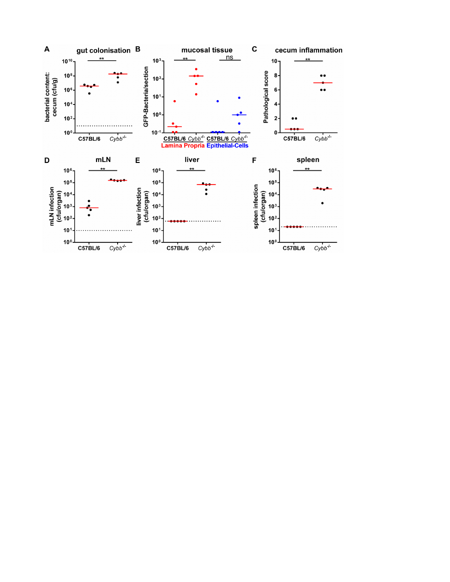
We pretreated wild type and Cybb
2/2
mice with streptomycin
and infected them for 4 days with S.Tm
avir
(5610
7
cfu by
gavage) to analyze the role of Cybb in restricting the growth of
S.Tm
avir
in the mucosal tissue. High S.Tm
avir
loads were
detected in the gut lumen of wild type and Cybb
2/2
mice
(Fig. 1A). Bacterial loads in the LP were significantly lower in
the wild type than in the Cybb
2/2
animals (Fig. 1B) and only
the latter developed pronounced mucosal inflammation by day
4 p.i. (Fig. 1C). Furthermore, the Cybb
2/2
mice displayed
significantly increased loads of S.Tm
avir
in the mesenteric lymph
nodes (mLNs, Fig. 1D), the livers (Fig. 1E) and the spleens
(Fig. 1F) compared to C57BL/6 mice. This high susceptibility to
systemic spread was expected as NADPH oxidase is known to
be of key importance for limiting systemic infections [3,4]. Our
data extended these findings by showing that NADPH oxidase
is essential for restricting the growth of S.Tm
avir
not only at
systemic sites, but also in the cecal LP.
iNOS does not Contribute Significantly to Mucosal
Defense against S.Tm
avir
The inducible NO synthase (iNOS) is an important defense
mechanism of monocytic macrophages [2] and can help restricting
pathogen growth in various models [22–27]. In order to assess the
role of iNOS in our infection model, we included iNOS-deficient
(Nos2
2/2
) animals and Cybb
2/2
Nos2
2/2
double KO mice into the
infection experiments with S.Tm
avir
shown in Fig. 1. Neither
cecum pathology (Fig. S1C) nor tissue loads (Fig. S1B, D–F) in the
Nos2
2/2
mice differed from wild type C57BL/6 animals.
Similarly, the cecum pathology (Fig. S1C) and the tissue loads in
the cecal mucosa (Fig. S1B) and the livers (Fig. S1E) did not differ
significantly between the Cybb
2/2
and the Cybb
2/2
Nos2
2/2
mice.
The Cybb
2/2
Nos2
2/2
animals displayed slightly but significantly
elevated S.Tm
avir
tissue loads only in the mLNs (Fig. S1D) and the
spleens (Fig. S1F). However, even in these organs, Cybb-deficiency
had a more pronounced effect than Nos2-deficiency and significant
contributions of Nos2 were only detectable in the presence of Cybb,
suggesting a possible synergistic role for Nos2 [16]. In conclusion,
restriction of S.Tm
avir
in the cecal mucosa and the protection from
enteropathy seems to hinge on NADPH oxidase while iNOS
seems to contribute little (maximally in a synergistic manner) to
mucosal defense, at least during the first 4 days of infection.
Increased Mucosal NADPH Oxidase Expression in
Response to Infection of Wild Type Mice with Wild Type
S. Typhimurium
The standard streptomycin model for murine S. Typhimurium
diarrhea was used to assess Cybb expression in the infected mucosa
of wild type C57BL/6 mice. Streptomycin pretreated animals
were infected with wild type S. Typhimurium (S.Tm
wt
; 5610
7
cfu
by gavage) for 12 or 24 h. Samples of the cecum tissue (the site of
the initial and most pronounced enteropathy [18,19]) were
recovered to analyze Cybb expression by reverse transcription
quantitative real-time PCR (RT-qPCR). In line with earlier data
[28], the abundance of Cybb mRNA in the cecum increased by
about 3-fold after 12 h and about 8-fold after 24 h of infection
compared to streptomycin-treated animals (Fig. S2A). This went
along with mucosal inflammation and infiltration of neutrophils
and monocytic phagocytes into the cecal mucosa as observed by
histopathology and flow cytometry analysis (Fig. S2B, C, D).
NADPH Oxidase in Mucosal Defense
PLOS ONE | www.plosone.org
2
October 2013 | Volume 8 | Issue 10 | e77204
S.Tm
avir
Colitis in Cybb
2
/2
Mice is Similarly Dependent on
Myd88 and Mucosal CD11c
+
Monocytic Phagocytes as
S.Tm
invG
–induced Colitis in Wild Type C57BL/6 Mice
The role of Cybb and or PMN in the acute infection is not
completely understood. Thus, we performed a number of control
experiments to analyze the pathogenetic mechanism of S.Tm
avir
colitis in Cybb
2/2
mice. First, we analyzed uninfected and infected
gut tissues with immuno-histopathological stainings for markers
characteristic for a set of immune cells. The S.Tm
avir
infected
mucosa of Cybb
2/2
mice at day 4 p.i. displayed a patchy
pathology characterized by non-inflamed regions interspaced with
pronounced inflammatory foci (Fig. S3). Such patchy pathology is
characteristic for S.Tm
invG
(lacking only TTSS-1) infections at day
3 p.i. in wild type C57BL/6 mice (Fig. S4; [20]). This provided
first hints suggesting that Cybb-deficiency allows S.Tm
avir
to elicit
enteropathy via the ‘‘alternative pathway’’. In line with this, RT-
qPCR analysis of a panel of 27 genes for cytokines or antimicrobial
defenses revealed similar mucosal gene expression profiles (Fig.
S4G) in Cybb
2/2
mice (4 days p.i. with S.Tm
avir
) and wild type
C57BL/6 mice (3 days p.i. with S.Tm
invG
) at their first day of overt
enteropathy (Fig. S4). In this experiment, the counts in the mLNs
(Fig. S4D), livers (Fig. S4E) and spleens (Fig. S4F) were lower in
the C57BL/6 mice compared to Cybb
2/2
mice. However, most
importantly and in line with the RT-qPCR data, the degree of
cecum inflammation was alike (Fig. S4B, C).
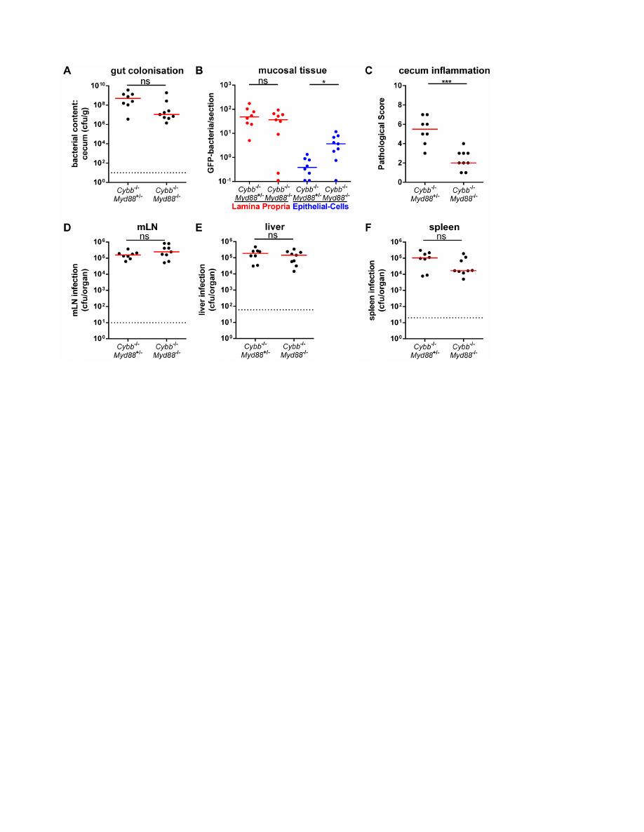
We then assessed the Myd88-dependency of the inflammatory
response as a typical feature of the ‘‘alternative pathway’’. For
this purpose we infected Cybb
2/2
Myd88
2/2
mice or Cybb
2/
2
Myd88
+/2
littermate controls for 4 days with S.Tm
avir
(5610
7
cfu by gavage). High loads of S.Tm
avir
were observed in the gut
lumen (Fig. 2A), LP (Fig. 2B), mLNs (Fig. 2D), livers (Fig. 2E)
and spleens (Fig. 2F) of both groups. However, only the Myd88-
proficient mice developed mucosal inflammation, while the
Cybb
2/2
Myd88
2/2
animals did not (Fig. 2C). It is interesting to
note that the Cybb
2/2
Myd88
2/2
animals displayed slightly but
significantly elevated S.Tm
avir
loads in the cecal epithelium
(Fig. 2B; blue symbols). It is unclear whether this might be
explained by reduced epithelial turnover rates of non-infected
tissue in Myd88
2/2
animals [29]. Alternatively, this might be
indicative of a Myd88-dependent, but Cybb-independent defense
mechanism which may contribute to limiting bacterial growth in
the enterocytes. Such mechanisms could be an interesting topic
for future research. To this end, the data verified the Myd88-
dependency of enteropathy in S.Tm
avir
infected Cybb
2/2
mice.
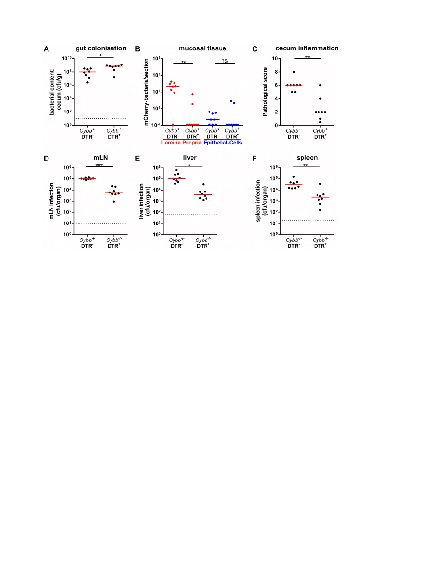
Finally, we have assessed the dependency on mucosal monocytic
phagocytes. Using transgenic mice expressing the diphtheria-toxin
receptor under control of the CD11c promoter (DTR
+/2
; [30])
and diphtheria toxin-mediated (DTX) cell depletion, it has been
previously established that S.Tm
invG
relies on mucosal CD11c
+
monocytic phagocytes for traversing the gut epithelium and
colonizing the cecal LP [21]. Thus, Cybb
2/2
DTR
+/2
mice or
Cybb
2/2
littermates were treated with DTX and infected for 4
days with S.Tm
avir
. High loads of S.Tm
avir
were detected in the gut
lumen of both groups (Fig. 3A). In contrast, the DTX-mediated
cell depletion abolished mucosa tissue infection (Fig. 3B) and the
elicitation of mucosal inflammation (Fig. 3C). Furthermore, it
significantly reduced the infection of mLNs (Fig. 3D), livers
(Fig. 3E) and spleens (Fig. 3F). These data were all in line with the
notion that S.Tm
avir
infection of Cybb
2/2
mice follows a similar
pathogenetic mechanism as described earlier for the S.Tm
invG
Figure 1.
S
.Tm
avir
infection of
Cybb
2
/2
mice leads to pathogen growth in the LP and enteropathy. Cybb
2
/2
mice (C57BL/6 background)
and C57BL/6 control mice were pretreated with streptomycin and infected for 4 days with S.Tm
avir
. The bacterial loads in the gut lumen (A), the LP
(red (B)) or the epithelial cells of the cecum (blue (B)), the degree of mucosal inflammation (C) and bacterial loads in the mLNs (D), livers (E) and
spleens (F) were analyzed. *: p,0.05; **: p,0.01; ns: not significant; red line: median; dashed line: minimal detectable value.
doi:10.1371/journal.pone.0077204.g001
NADPH Oxidase in Mucosal Defense
PLOS ONE | www.plosone.org
3
October 2013 | Volume 8 | Issue 10 | e77204
infection in wild type mice. The key difference between both
infections seems to reside in the failure of the Cybb
2/2
mice to
control S.Tm
avir
infection/growth in the infected mucosa. This
would suffice to explain the susceptibility of Cybb
2/2
(but not wild
type) mice to S.Tm
avir
-triggered enteropathy and suggests that the
model might be of interest for studying the role of Cybb in
restricting bacterial growth in the cecal LP.
S.Tm
avir
Induces Intermediate Levels of Enteropathy in
Mice Reconstituted with a Mix of Cybb
2
/2
and Wild Type
Bone Marrow
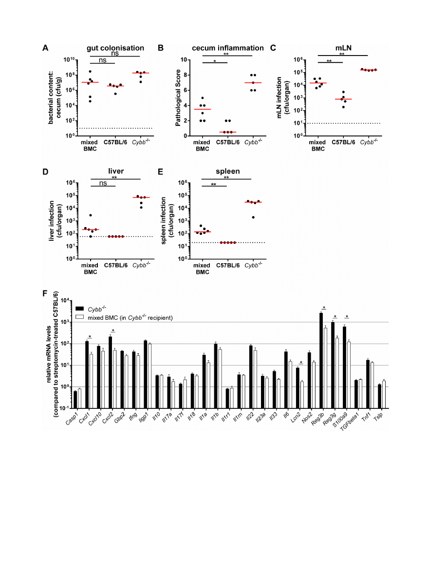
To further analyze how Cybb restricts S.Tm
avir
infection of the
cecal mucosa, we performed an experiment with mixed bone
marrow chimeric mice. The chimeras were generated by
reconstituting irradiated Cybb
2/2
mice (congenic marker Ly5.2)
with a mix of 50% Cybb
2/2
(congenic marker Ly5.2) and 50%
C57BL/6 (congenic marker Ly5.1) bone marrow. After 8 weeks,
these chimeras displayed 69% Cybb
2/2
(congenic marker Ly5.2)
and 31% C57BL/6 (congenic marker Ly5.1) cells in the cecal LP
as tested by flow cytometry at the end of the experiment (data not
shown). In these mice, the stromal cells and all CD45.2
+
cells (i.e.
phagocytes, B-cells, T-cells, etc.) were Cybb-deficient, while the
CD45.1
+
cells were Cybb-proficient. Four days p.i. with S.Tm
avir
all
chimeric mice (mixed BMC, Fig. 4) displayed high pathogen loads
in the gut lumen (Fig. 4A) and significant amounts of bacteria in
the mLNs (Fig. 4C) and spleens (Fig. 4E). The levels of mLN and
spleen colonization in the mixed chimeras were lower than in the
Cybb
2/2
mice, but higher than in the wild type C57BL/6 animals
(Fig. 4C, E). The same intermediate phenotype was observed for
the cecum pathology (Fig. 4B), whereas liver colonization was not
distinguishable from wild type C57BL/6 animals (Fig. 4D).
Interestingly, approximately one third of Cybb-proficient cells in
a Cybb-knock out background lead to a reduction of bacterial
counts in spleens and livers by 217- and 333-fold (ratio between
the medians), respectively, if compared with mice completely
deficient for Cybb. Additionally, RT-qPCR analysis of proinflam-
matory cytokines confirms the alleviated inflammatory phenotype
(Fig. 4F). These data indicate that Cybb-proficiency in only 31% of
the bone-marrow-derived compartment is sufficient to achieve a
significant restriction of S.Tm
avir
colonization of the host tissue and
enteropathy.
Discussion
Here, we have analyzed NADPH oxidase defenses of the
intestinal mucosa. We established that NADPH oxidase deficient
mice were not able to limit gut mucosa colonization and
enteropathy by a normally avirulent S. Typhimurium strain. This
demonstrated that disease via the ‘‘alternative’’ pathway hinges on
a fine balance between microbe entry into the LP, microbe growth
at this site and pathogen killing in the LP. Our data confirmed that
LP access is controlled at least in part by dendritic cells (monocytic
phagocytes), and demonstrated that microbe growth/killing is
controlled by bacterial virulence factors (e.g. TTSS-2) and host
defenses (e.g. NADPH oxidase-mediated killing in PMN).
While the central role of NADPH oxidase, i.e. CYBB, is well
established in antimicrobial defense, the nature of the cell types
facilitating the NADPH oxidase dependent defenses had remained
less clear. The role of NADPH oxidase in the anti-microbial
activity of neutrophils is well established [21], the same holds true
for its role in dendritic cell-mediated antigen presentation and T-
Figure 2.
Myd88
-dependency of enteropathy in
S
.Tm
avir
infected
Cybb
2
/2
mice. Cybb
2
/2
Myd88
+/2
mice or Cybb
2
/2
Myd88
2
/2
littermates
mice were pretreated with streptomycin and infected with S.Tm
avir
for 4 days. The bacterial loads in the gut lumen (A), the LP (red (B)) or the epithelial
cells of the cecum (blue (B)), the degree of mucosal inflammation (C) and bacterial loads in the mLNs (D), livers (E) and spleens (F) were analyzed. *:
p,0.05; **: p,0.01; ns: not significant; red line: median; dashed line: minimal detectable value.
doi:10.1371/journal.pone.0077204.g002
NADPH Oxidase in Mucosal Defense
PLOS ONE | www.plosone.org
4
October 2013 | Volume 8 | Issue 10 | e77204
cell priming [31–33]. However, what is the mechanism activating
NADPH oxidase in the mucosal phagocytes? Besides NADPH
oxidase-deficiency/CGD, other primary immune deficiencies
enhancing susceptibility to bacterial infection are deficiencies in
Toll-like receptor- and IFNc-R-signalling [34,35]. In mouse
models for systemic and intestinal Salmonella infection, Toll-like
receptor - and IFNc-R-signalling were indeed found to restrict
pathogen growth [20,36–42]. NADPH oxidase (and iNOS) are
known to be activated via both MyD88- and IFNc-signalling.
However, S.Tm
avir
did not colonize the LP of MyD88
2/2
or
IFNc-R
2/2
mice and did not cause enteropathy (this work and
data not shown). This indicated that NADPH oxidase is activated
by several redundant signalling pathways in LP cells. Deciphering
such signalling pathways and the cell type mainly responsible for
NADPH oxidase expression will be an interesting topic for future
work. The S.Tm
avir
infection model would be well suited for such
studies, because S. Tm
avir
offers well defined genetics and virulence
factors. The removal of the latter from S. Tm
wt
still leads to disease
in a mouse model of CGD. This indicates that even very low
virulence is sufficient to cause enteropathy in mice deficient in a
subunit of the NADPH oxidase, broadening our understanding of
how commensals might induce enteropathy in CGD patients.
The diffusible nature of some of the ROS (i.e. hydrogen
peroxide) has raised some interest, as neighboring cells might be
affected, even if they are not by themself capable of expressing
NADPH oxidase. This has in fact been demonstrated in vitro
[43,44] and has thus complicated the interpretation of data from
mouse experiments with cell-type specific NADPH oxidase
deficiencies [45].
Our data demonstrate that the augmentation by neighboring
cells (by ROS-signalling or by wild type cell mediated decreases of
the pathogen loads) might be indeed of importance, at least in the
S.Tm
avir
infected intestinal mucosa. The mixed Cybb-proficient
and -deficient bone marrow chimeras displayed .2006 lower
systemic S.Tm
avir
loads than the Cybb
2/2
controls. Apparently,
31% of Cybb-proficient CD45
+
cells are sufficient for this. This is in
line with other publications focusing on A. fumigatus infections. In
vitro, A. fumigatus hyphae could be damaged by a mixture of normal
and ‘‘CGD neutrophils’’ [46]. Furthermore, Cybb
2/2
mice with
.92% Cybb-deficient and 4–8% Cybb-proficient cells were fully
protected [46,47] to challenge with a dose of A. fumigatus sufficient
to cause disease in Cybb
2/2
mice. Furthermore, the reported
amount of Cybb-proficient cells necessary to respond similarly to an
infection (i.e. survive) as wild type mice is 21–35% or 32–41% for
challenge with S. aureus or B. cepacia, respectively [47]. Similarly,
survival of CGD patients after entering adulthood was strongly
associated with residual reactive oxygen intermediates production
[48]. In extension, our data and the evidence from the other
infection models discussed above indicate that even a partial
therapy of CGD patients might be sufficient to significantly
decrease their disease susceptibility far beyond the degree of
achieved reconstitution. The need for less than 100% reconstitu-
tion (as typically observed in gene therapy [49]) might be of
relevance for preclinical testing and the design of gene therapy
approaches for treating CGD.
Up to date, it is unclear why a partial restoration of Cybb
expression is sufficient to ameliorate the phenotype drastically.
There are three possible explanations.
Figure 3. Cell depletion demonstrating the monocytic phagocyte dependency of
S
.Tm
avir
infection in
Cybb
2
/2/
DTR
+
mice. Cybb
2
/
2
/
DTR
+
mice and Cybb
2
/2/
DTR
2
littermates were pretreated with streptomycin, injected with DTX 18 h prior to and 30 h post infection and infected
for 4 days with S.Tm
avir
. The bacterial loads in the gut lumen (A), the LP (red (B)) or the epithelial cells of the cecum (blue (B)), the degree of mucosal
inflammation (C) and bacterial loads in the mLNs (D), livers (E) and spleens (F) were analyzed. *: p,0.05; **: p,0.01; ns: not significant; red line:
median; dashed line: minimal detectable value.
doi:10.1371/journal.pone.0077204.g003
NADPH Oxidase in Mucosal Defense
PLOS ONE | www.plosone.org
5
October 2013 | Volume 8 | Issue 10 | e77204
Firstly, we showed recently that S. Tm
invG
is found exclusively in
CD11c
+
cells at 1 day p.i. in our infection mouse model and only
from 2 days p.i. on also in CD11c
2
cells [21,50]. This mechanism
might also apply for the S. Tm
avir
infection mouse model, since the
S. Tm
invG
infection in C57BL/6 mice seems to be phenotypically
similar to S. Tm
avir
infection in Cybb
2/2
mice. The transition
between CD11c
+
and CD11c
2
cells can possibly be the reason for
an incidental exposure (and killing) of some S. Tm
avir
bacteria to
Cybb-proficient phagocytes. Killing in the Cybb-proficient phago-
cyte populations could explain the reduced tissue loads and disease
Figure 4. 31% wild type cells in
Cybb
2
/2
mice are sufficient to reduce systemic loads of
S
.Tm
avir
. Cybb
2
/2
mice were irradiated and
reconstituted with a mix of C57BL/6 Ly5.1 and Cybb
2
/2
Ly5.2 bone marrow (mixed BMC). Mice were pretreated with streptomycin and infected for 4
days with S.Tm
avir
. The bacterial loads in the gut lumen (A), the degree of mucosal inflammation (B) and bacterial loads in the mLNs (C), livers (D) and
spleens (E) were analyzed. The data for the C57BL/6 and Cybb
2
/2
mice were replotted from Fig. 2 for a better comparison. Relative mRNA expression
levels were compared between bone marrow chimeras and similarly treated Cybb
2
/2
mice, data partly replotted from Fig. S4G for a better
comparison (F). Data is displayed as mean
+ SEM (F); *: p,0.05; **: p,0.01; ns: not significant; red line: median; dashed line: minimal detectable value.
doi:10.1371/journal.pone.0077204.g004
NADPH Oxidase in Mucosal Defense
PLOS ONE | www.plosone.org
6
October 2013 | Volume 8 | Issue 10 | e77204

pathology of the mixed bone marrow chimeras. Secondly, ROS
produced by Cybb-proficient cells play an important role in
controlling signalling pathways. Here, the reversible oxidation and
inactivation of protein tyrosine phosphatases and MAP kinase
phosphatases by ROS are interesting examples [51]. As a measure
for proinflammatory signalling levels, we quantified mRNA levels
of 27 genes related to inflammation and defense against S.
Typhimurium infection. However, although 6 out of 27 genes
were expressed less in the S.Tm
avir
infections of mixed bone
marrow chimeras compared to Cybb
2/2
deficient mice, 21 out of
27 gene expression levels were similarly induced, if induced at all.
This might indicate that only a part of the signalling pathways are
affected by Cybb-deficiency.
Thirdly, we cannot exclude that the, already discussed, diffusion
of some of the ROS (i.e. hydrogen peroxide) from Cybb-proficient
cells into neighboring Cybb-deficient cells described in vitro [43,44]
may also occur in vivo. The current data is insufficient to tease
apart these three mechanistic explanations. Nonetheless, the
reduced disease severity of S. Tm
avir
infections in the mixed bone
marrow chimeras provides a useful basis for addressing this issue.
In CGD patients, Crohn’s disease-like inflammation of the
intestinal mucosa is frequently observed [7,52]. Salmonella spp. can
be isolated from the stools of some, but clearly not from all of these
patients [7]. This indicates that, on the one hand, growth
restriction of normally avirulent Salmonella by NADPH oxidase
may be of relevance for CGD patients, but on the other hand that
other microbial stimuli can also trigger enteropathy. In the ‘‘non-
Salmonella-related’’ cases, inflammation might be attributable to
insufficient restriction of commensal microbiota species which
would not cause disease in NADPH oxidase proficient hosts. In
this case, NADPH oxidase-mediated growth restriction by LP cells
may function as an immunological barrier of general importance
for maintaining homeostasis in the intestinal mucosa. Our findings
might be of importance for understanding microbe handling by
the intestinal immune system and for elucidating strategies
employed by pathogens to overcome this defense.
Our findings may also contribute to our understanding of the
evolution of S. Typhimurium as a successful enteropathogen.
During its divergence from a commensal E. coli lineage, this
pathogen has acquired two novel genetic loci of importance for
enteropathogensis which encode the two TTSSs [53–55]. In wild
type hosts, TTSS-2 was shown to enhance pathogen survival in LP
phagocytes
and
thereby
enhance
mucosal
inflammation
[20,21,56–58]. Tissue culture experiments suggested that this is
attributable to TTSS-2 dependent interference with NADPH
oxidase (or iNOS-) delivery to the Salmonella containing phagosome
[59,60]. This is supported by our finding that S.Tm
avir
, which lacks
a functional TTSS-2, is only capable of colonizing the LP and
cause enteropathy in Cybb-deficient, but not in wild type mice. In
conclusion, TTSS-2 may represent a pathogen-specific adaptation
to overcome and subvert the NADPH oxidase mediated mucosal
defense. This would explain how wild type S. Typhimurium
colonizes these cells of the intestinal mucosa in wild type hosts
[21,53].
Apparently, greatly reduced virulence of S. Typhimurium is
sufficient to cause enteropathy in Cybb mice. This is clearly not due
to deficiency in immune regulation, because bacterial species not
recognized as pathogenic are capable of triggering enteropathy in
CGD. S. Tm
avir
are a highly genetically amenable tool to study the
mechanisms why. Direct and indirect susceptibility to ROS may
be a determining feature of host microbiota species that permits
their close relationship with the host.
Materials and Methods
Ethics Statement
All animal experiments and generation of new mouse-lines were
approved by the legal authorities (licenses 201/2007 and 223/
2010; Kantonales Veterina¨ramt Zu¨rich, Switzerland) and carried
out in the legally required manner.
Mice
C57BL/6ptprc
b
(congenic marker Ly5.2
+
; originally from
Charles River), C57BL/6ptprc
a
(congenic marker Ly5.1
+
; [61]),
Cybb
2/2
(B6.129S-Cybb
tm1Din
/J; C57BL/6 background; [62])
and Nos2
2/2
(B6.129P2-Nos2
tm1Lau
/J; C57BL/6 background;
[63]) were kept and bred under specific pathogen free (SPF)
conditions. Cybb
2/2/
Nos2
2/2
mice have been described before
and were generated by crossing Cybb
2/2
and Nos2
2/2
mice
[13]. Cybb
2/2/
DTR
+
were generated by crossing Cybb
2/2
with
DTR
+
(B6.FVB-Tg[Itgax-DTR/EGFP]57Lan/J; [64]). Cybb
2/
2/
Myd88
2/2
mice were generated by crossing Cybb
2/2
with
Myd88
2/2
mice
(C57BL/6
background;
[65]).
All
newly
generated double knockout mice and transgenic Cybb
2/2
mice
bred and developed in a similar manner as Cybb
2/2
mice. All
animals were kept under SPF conditions at the RCHCI of the
ETH Zurich. For experiments mice were age (8–12 weeks old)
matched and treated as described previously [19,21]. In brief,
mice were pretreated with streptomycin (1 dose, 25 mg/animal,
by gavage). 24 h later mice were infected with 5610
7
cfu by
gavage. Infections were performed for 12 h, 24 h, 72 h (3 days
p.i.) and 96 h (4 days p.i.). Bacterial loads of gut lumen content,
mLNs, livers and spleens were determined by plating [21].
Generation of Mixed Bone Marrow Chimeras
The generation of bone marrow chimeras has been described
before [21,66]. Shortly, from euthanatized donor mice bone
marrow from femur, tibia, brachium and pelvis was extracted.
Recipient mice (Cybb
2/2
) were c-irradiated (1000 rad) and
reconstituted with 2.5610
6
Cybb
2/2
(congenic marker Ly5.2)
and 2.5610
6
C57BL/6ptprc
a
(congenic marker Ly5.1) bone
marrow cells intravenously. Animals were checked regularly and
received drinking water containing Borgalß (Intervet) for 2 weeks.
After 8 weeks, reconstitution efficiency was controlled after
infection by flow cytometry (Ly5.1/CD45.1, Ly5.2/CD45.2) on
LP cells. The reconstitution lead to a proportion of 6963%
Cybb
2/2
(Ly5.2) and 3163% C57BL/6ptprc
a
(Ly5.1) cells
(analyzed: percentage of CD45.2 vs CD45.1 in the cecal LP,
mean 6 standard deviation).
Bacterial Strains
S.Tm
avir
(DinvG; sseD::aphT; M557; [20]) and S.Tm
invG
(DinvG;
SB161; [67]) are isogenic derivatives of the wild type Salmonella
SL1344 (S.Tm
wt
; [68]). For infection, bacteria were cultured in
0.3 M NaCl LB for 12 h at 37
uC and subcultivated for 4 h as
described before [69]. For detection of bacteria within mucosal
tissue, bacteria harbored the reporter plasmid pM973 (ssaH
promoter fused to gfp; [20]) or pM2121 (ssaH promoter fused to
mcherry; this study).
Mucosal Tissue Colonization and Cell-type Localization
Bacteria harboured a reporter plasmid expressing either gfp
(pM973; [20]) or mcherry under the control of the ssaH promoter
(pM2121; this study). For the evaluation of cecum-tissue invaded
bacteria, the cecum tissue was fixed in 4% PFA and stored as
described before [21]. 20
m
m cryosections were stained with
Armenian hamster anti-ICAM-I/CD54 (clone 3E2, 1:100; Becton
NADPH Oxidase in Mucosal Defense
PLOS ONE | www.plosone.org
7
October 2013 | Volume 8 | Issue 10 | e77204

Dickson), DAPI (1:1000, Sigma-Aldrich), Cy3-conjugated or Cy5-
conjugated or FITC-conjugated goat anti-Armenian hamster IgG
(1:100, Jackson ImmunoResearch Laboratories) and Alexa-
Fluor647
conjugated
phalloidin
(1:100,
Molecular
Probes)
[21,66]. The average number of invaded bacteria in the
epithelium and LP was evaluated by analyzing 3–9 tissue sections
per mouse.
Flow Cytometry
Cecum and mLNs were chopped and digested in RPMI
(Invitrogen) and Liberase TL (Roche) for 45 min at 37
uC under
vigorous shaking. The resulting cell suspension was filtered
through a 100
m
m nylon cell-strainer (Milian) and stained in
buffer containing PBS, 5 mM EDTA, 10% FCS and 50
m
g/ml
streptomycin. All fluorophore-labeled monoclonal antibodies were
purchased from BD Biosciences or Biolegend. The LP cells were
analyzed on a LSR II cytometer (Becton Dickinson) and graphs
were produced with FlowJo software (Tree Star, Inc.).
In vivo Dendritic Cell Depletion
DTX was injected i.p. (100 ng/25 g body weight; [64]) at 18 h
before and 30 h after the infection. The depletion efficiency
(.80%) and its negligible effect on other mucosal cell populations
have been described before [21].
Histopathological Evaluation
Tissues were embedded in OCT (Sakura, Torrance, CA) and
snap-frozen in liquid nitrogen. Five
m
m cryosections were stained
with hematoxylin and eosin (H&E). The degree of cecal mucosal
tissue inflammation, i.e. edema, PMN infiltration, reduced
numbers of goblet cells containing visible mucus-filled vacuoles
and epithelium disruption, was judged by a pathologist yielding to
a score of inflammation between 0–13 points as described before
[19,66].
RT-qPCR
The excised cecum tissue was washed in cold PBS, placed in
600
m
l RNAlater (Qiagen) and subsequently frozen at 280
uC.
Total RNA extraction was done using the RNeasy mini kit
(Qiagen) with RNase-free DNase digest (Qiagen). For reverse-
transcription of 1
m
g mRNA aliquots, the RT
2
HT First Strand
cDNA Kit (Qiagen) was used. Custom RT
2
Profiler PCR Arrays
(Qiagen) were run with RT
2
SYBR Green ROX FAST
(QIAGEN) on an Applied Biosystems 7900 HT Fast Real-Time
PCR System to amplify the resulting cDNA. Relative mRNA
levels (2
2DCq
) were determined by comparing the PCR quantifi-
cation cycle (Cq, determined with the Software SDS 2.2.1) for 27
genes related to inflammation and defense against S. Typhimur-
ium infection (the selection is based on Songhet et al., 2010) with
the reference gene Actb. The differences in their Cq cycles were
calculated (DCq). In all experiments, the upper limit of Cq was
fixed to 35 cycles. Then, the fold-increase over streptomycin-
treated C57BL/6 mice was calculated and plotted. Each sample
was controlled for mouse genomic DNA contamination. All DNA-
positive data were excluded from further analysis. Lastly, RNA
quality was monitored with the Agilent RNA 6000 Nano Kit
(Agilent Technologies) on a 2100 Bioanalyzer (Agilent Technol-
ogies) and only samples with a RNA integrity number (RIN)
.9.90 were included.
Statistical Analysis
Statistical analysis was performed using the exact Mann-
Whitney U test with the software GraphPad Prism 6. Values of
p,0.05 (two tailed) were considered as significantly different
between two groups. The minimal detectable bacterial coloniza-
tion levels were set to 10 cfu/mLNs, 20 cfu/spleen, 60 cfu/liver
(Fig. 1–4) or 30 cfu/liver (Fig. S4) or 10 cfu/g cecum content in
cases where no bacteria were detected by plating. Messenger RNA
levels of two groups were compared using Mann-Whitney U tests
with Hochberg corrections for multiple comparisons using R x64
3.0.1 (Fig. 4F, S4G).
Supporting Information
Figure S1
NADPH oxidase is expressed in the infected
mucosa and PMNs increase in number by infection.
C57BL/6 mice were pretreated with streptomycin and infected
with S.Tm
wt
for 12 h or 24 h, as indicated. RT-qPCR for Cybb
expression in cecal tissues (A). Representative H&E sections
(contrast and brightness were adjusted, color was enhanced, scale
bar: 50
m
m, arrow indicates a PMN) (B). Quantity of PMNs/high-
power field (C). FC of cecal LP (pregated on CD45
+
cells) (D). *:
p,0.05; ns: not significant; red line: median; dashed line:
detection limit.
(TIF)
Figure S2
Cybb (but not iNOS) is important in mucosal
defense against
S.Tm
avir
infection. C57BL/6 mice (data
replotted from Fig. 1), Nos2
2/2
mice (C57BL/6 background),
Cybb
2/2
Nos2
2/2
mice (C57BL/6 background) or Cybb
2/2
mice
(C57BL/6 background; data replotted from Fig. 1) were
pretreated with streptomycin and infected for 4 days with
S.Tm
avir
. The bacterial loads in the gut lumen (A), the LP (red
(B)) or the epithelial cells of the cecum (blue (B)), the degree of
mucosal inflammation (C) and bacterial loads in the mLNs (D),
livers (E) and spleens (F) were analyzed. *: p,0.05; **: p,0.01; ns:
not significant; red line: median; dashed line: minimal detectable
value.
(TIF)
Figure S3
Immunohistology of S.TminvG infected wild
type C57BL/6 mice and S.Tmavir infected Cybb
2/2
mice is similar. Cryo-sections of the cecal tissue from
streptomycin pretreated wild type and Cybb
2/2
mice infected for
3 days with S.Tm
invG
or for 4 days with S.Tm
avir
, were stained with
antibodies against CD11c (A), CD11b (B), CD68 (C), Gr-1 (D),
CD3 (E) and CD8 (F) and imaged by bright field microscopy. The
different times of infection are explained by the different disease
kinetics of S.Tm
invG
and S.Tm
avir
. The former requires 3 days (in
C57BL/6 mice) and the latter 4 days (in Cybb
2/2
mice) before
overt inflammation of the cecal tissue is observed. The left panel
shows representative pictures. The right panel shows the
quantification. *: p,0.05; **: p,0.01; ns: not significant. Data is
displayed as mean + SEM. S.Tm
invG
was able to elicit gut
inflammation in wild type C57BL/6 and in Cybb
2/2
mice. In
contrast, S.Tm
avir
triggered enteropathy only in the Cybb
2/2
mice,
but not in wild type C57BL/6 animals. Please note that the
inflammatory lesions in the S.Tm
avir
infected Cybb
2/2
mice
displayed localized inflammatory lesions of equivalent immuno-
histopathology as the lesion triggered by S.Tm
invG
in C57BL/6
mice.
(TIF)
Figure S4
S.Tm
invG
infection in wild type C57BL/6 mice
and
S.Tm
avir
infection in
Cybb
2/2
mice are similar.
C57BL/6 mice were pretreated with streptomycin and infected
with S.Tm
invG
for 3 days. Cybb
2/2
mice were pretreated with
streptomycin and infected with S.Tm
avir
for 4 days. The bacterial
loads in the gut lumen (A), the degree of mucosal inflammation (B),
NADPH Oxidase in Mucosal Defense
PLOS ONE | www.plosone.org
8
October 2013 | Volume 8 | Issue 10 | e77204

representative H&E pictures (contrast and brightness were
adjusted and color was enhanced, scale bar: 200
m
m, C) and
bacterial loads in the mLNs (D), livers (E) and spleens (F) were
analyzed. *: p,0.05; **: p,0.01; ns: not significant; red line:
median; dashed line: minimal detectable value. Relative mRNA
expression levels were compared between S.Tm
invG
infected
C57BL/6 mice and S.Tm
avir
infected Cybb
2/2
mice, data
replotted partly in Figure 4 (G). Data is displayed as mean +
SEM, differences were not significant (G).
(TIF)
Acknowledgments
We are grateful to Hardt lab members for discussions, to Manja Barthel for
technical support, to Balamurugan Periaswamy and Anja Bu¨rkli for
statistical advice, to Lisa Maier and Carmen Dolores Cordova for critical
reading of the manuscript, to Michael Detmar for access to the RT-qPCR
machine and to the RCHCI team, in particular K. Holzinger, S. Egger, J.
Fehr, S. Freedrich and T.C. Weber for excellent support.
Author Contributions
Conceived and designed the experiments: BF PS ES WDH. Performed the
experiments: BF PS ES MH. Analyzed the data: BF PS ES AJM MK.
Contributed reagents/materials/analysis tools: MH LVM DC JCS. Wrote
the paper: BF PS WDH.
References
1. Varol C, Zigmond E, Jung S (2010) Securing the immune tightrope:
mononuclear phagocytes in the intestinal lamina propria. Nat Rev Immunol
10: 415–426.
2. Nathan C (2006) Neutrophils and immunity: challenges and opportunities. Nat
Rev Immunol 6: 173–182.
3. Fang FC (2004) Antimicrobial reactive oxygen and nitrogen species: concepts
and controversies. Nat Rev Microbiol 2: 820–832.
4. Nathan C, Shiloh MU (2000) Reactive oxygen and nitrogen intermediates in the
relationship between mammalian hosts and microbial pathogens. Proc Natl
Acad Sci U S A 97: 8841–8848.
5. Holland SM (2010) Chronic granulomatous disease. Clin Rev Allergy Immunol
38: 3–10.
6. Soler-Palacin P, Margareto C, Llobet P, Asensio O, Hernandez M, et al. (2007)
Chronic granulomatous disease in pediatric patients: 25 years of experience.
Allergol Immunopathol (Madr) 35: 83–89.
7. van den Berg JM, van Koppen E, Ahlin A, Belohradsky BH, Bernatowska E, et
al. (2009) Chronic granulomatous disease: the European experience. PLoS One
4: e5234.
8. Kraaij MD, Savage ND, van der Kooij SW, Koekkoek K, Wang J, et al. (2010)
Induction of regulatory T cells by macrophages is dependent on production of
reactive oxygen species. Proc Natl Acad Sci U S A 107: 17686–17691.
9. Lee K, Won HY, Bae MA, Hong JH, Hwang ES (2011) Spontaneous and aging-
dependent development of arthritis in NADPH oxidase 2 deficiency through
altered differentiation of CD11b+ and Th/Treg cells. Proc Natl Acad Sci U S A
108: 9548–9553.
10. Simonsen J, Molbak K, Falkenhorst G, Krogfelt KA, Linneberg A, et al. (2009)
Estimation of incidences of infectious diseases based on antibody measurements.
Stat Med 28: 1882–1895.
11. Conlan JW (1997) Critical roles of neutrophils in host defense against
experimental systemic infections of mice by Listeria monocytogenes, Salmonella
typhimurium, and Yersinia enterocolitica. Infect Immun 65: 630–635.
12. Conlan JW (1996) Neutrophils prevent extracellular colonization of the liver
microvasculature by Salmonella typhimurium. Infect Immun 64: 1043–1047.
13. Mastroeni P, Vazquez-Torres A, Fang FC, Xu Y, Khan S, et al. (2000)
Antimicrobial actions of the NADPH phagocyte oxidase and inducible nitric
oxide synthase in experimental salmonellosis. II. Effects on microbial
proliferation and host survival in vivo. J Exp Med 192: 237–248.
14. Ackermann M, Stecher B, Freed NE, Songhet P, Hardt WD, et al. (2008) Self-
destructive cooperation mediated by phenotypic noise. Nature 454: 987–990.
15. White JK, Mastroeni P, Popoff JF, Evans CA, Blackwell JM (2005) Slc11a1-
mediated resistance to Salmonella enterica serovar Typhimurium and
Leishmania donovani infections does not require functional inducible nitric
oxide synthase or phagocyte oxidase activity. J Leukoc Biol 77: 311–320.
16. Shiloh MU, MacMicking JD, Nicholson S, Brause JE, Potter S, et al. (1999)
Phenotype of mice and macrophages deficient in both phagocyte oxidase and
inducible nitric oxide synthase. Immunity 10: 29–38.
17. Mutunga M, Graham S, De Hormaeche RD, Musson JA, Robinson JH, et al.
(2004) Attenuated Salmonella typhimurium htrA mutants cause fatal infections
in mice deficient in NADPH oxidase and destroy NADPH oxidase-deficient
macrophage monolayers. Vaccine 22: 4124–4131.
18. Kaiser P, Diard M, Stecher B, Hardt WD (2012) The streptomycin mouse
model for Salmonella diarrhea: functional analysis of the microbiota, the
pathogen’s virulence factors, and the host’s mucosal immune response. Immunol
Rev 245: 56–83.
19. Barthel M, Hapfelmeier S, Quintanilla-Martinez L, Kremer M, Rohde M, et al.
(2003) Pretreatment of mice with streptomycin provides a Salmonella enterica
serovar Typhimurium colitis model that allows analysis of both pathogen and
host. Infect Immun 71: 2839–2858.
20. Hapfelmeier S, Stecher B, Barthel M, Kremer M, Mu¨ller A, et al. (2005) The
Salmonella Pathogenicity Island (SPI)-1 and SPI-2 Type III Secretion Systems
Allow Salmonella Serovar Typhimurium to trigger Colitis via MyD88-
Dependent and MyD88-Independent Mechanisms. J Immunol 174: 1675–1685.
21. Hapfelmeier S, Muller AJ, Stecher B, Kaiser P, Barthel M, et al. (2008) Microbe
sampling by mucosal dendritic cells is a discrete, MyD88-independent step in
DeltainvG S. Typhimurium colitis. J Exp Med 205: 437–450.
22. Chakravortty D, Hensel M (2003) Inducible nitric oxide synthase and control of
intracellular bacterial pathogens. Microbes Infect 5: 621–627.
23. Khan IA, Schwartzman JD, Matsuura T, Kasper LH (1997) A dichotomous role
for nitric oxide during acute Toxoplasma gondii infection in mice. Proc Natl
Acad Sci U S A 94: 13955–13960.
24. MacMicking JD, Nathan C, Hom G, Chartrain N, Fletcher DS, et al. (1995)
Altered responses to bacterial infection and endotoxic shock in mice lacking
inducible nitric oxide synthase. Cell 81: 641–650.
25. Vallance BA, Deng W, De Grado M, Chan C, Jacobson K, et al. (2002)
Modulation of inducible nitric oxide synthase expression by the attaching and
effacing bacterial pathogen citrobacter rodentium in infected mice. Infect
Immun 70: 6424–6435.
26. Alam MS, Akaike T, Okamoto S, Kubota T, Yoshitake J, et al. (2002) Role of
nitric oxide in host defense in murine salmonellosis as a function of its
antibacterial and antiapoptotic activities. Infect Immun 70: 3130–3142.
27. Alam MS, Zaki MH, Sawa T, Islam S, Ahmed KA, et al. (2008) Nitric oxide
produced in Peyer’s patches exhibits antiapoptotic activity contributing to an
antimicrobial effect in murine salmonellosis. Microbiol Immunol 52: 197–208.
28. Songhet P, Barthel M, Rohn TA, Van Maele L, Cayet D, et al. (2010) IL-17A/
F-signaling does not contribute to the initial phase of mucosal inflammation
triggered by S. Typhimurium. PLoS One 5: e13804.
29. Rakoff-Nahoum S, Paglino J, Eslami-Varzaneh F, Edberg S, Medzhitov R
(2004) Recognition of commensal microflora by toll-like receptors is required for
intestinal homeostasis. Cell 118: 229–241.
30. Jung S, Aliberti J, Graemmel P, Sunshine MJ, Kreutzberg GW, et al. (2000)
Analysis of fractalkine receptor CX(3)CR1 function by targeted deletion and
green fluorescent protein reporter gene insertion. Mol Cell Biol 20: 4106–4114.
31. Savina A, Jancic C, Hugues S, Guermonprez P, Vargas P, et al. (2006) NOX2
controls phagosomal pH to regulate antigen processing during crosspresentation
by dendritic cells. Cell 126: 205–218.
32. Elsen S, Doussiere J, Villiers CL, Faure M, Berthier R, et al. (2004) Cryptic O2-
-generating NADPH oxidase in dendritic cells. J Cell Sci 117: 2215–2226.
33. Mantegazza AR, Savina A, Vermeulen M, Perez L, Geffner J, et al. (2008)
NADPH oxidase controls phagosomal pH and antigen cross-presentation in
human dendritic cells. Blood 112: 4712–4722.
34. Gordon MA (2008) Salmonella infections in immunocompromised adults.
J Infect 56: 413–422.
35. van de Vosse E, van Dissel JT, Ottenhoff TH (2009) Genetic deficiencies of
innate immune signalling in human infectious disease. Lancet Infect Dis 9: 688–
698.
36. Suar M, Periaswamy B, Songhet P, Misselwitz B, Muller A, et al. (2009)
Accelerated type III secretion system 2-dependent enteropathogenesis by a
Salmonella enterica serovar enteritidis PT4/6 strain. Infect Immun 77: 3569–
3577.
37. Talbot S, Totemeyer S, Yamamoto M, Akira S, Hughes K, et al. (2009) Toll-like
receptor 4 signalling through MyD88 is essential to control Salmonella enterica
serovar typhimurium infection, but not for the initiation of bacterial clearance.
Immunology 128: 472–483.
38. Weiss DS, Raupach B, Takeda K, Akira S, Zychlinsky A (2004) Toll-like
receptors are temporally involved in host defense. J Immunol 172: 4463–4469.
39. Rhee SJ, Walker WA, Cherayil BJ (2005) Developmentally regulated intestinal
expression of IFN-gamma and its target genes and the age-specific response to
enteric Salmonella infection. J Immunol 175: 1127–1136.
40. Silva-Herzog E, Detweiler CS (2008) Intracellular microbes and haemophago-
cytosis. Cell Microbiol 10: 2151–2158.
41. Santos RL, Raffatellu M, Bevins CL, Adams LG, Tukel C, et al. (2009) Life in
the inflamed intestine, Salmonella style. Trends Microbiol 17: 498–506.
42. Harrington L, Srikanth CV, Antony R, Shi HN, Cherayil BJ (2007) A role for
natural killer cells in intestinal inflammation caused by infection with Salmonella
enterica serovar Typhimurium. FEMS Immunol Med Microbiol 51: 372–380.
NADPH Oxidase in Mucosal Defense
PLOS ONE | www.plosone.org
9
October 2013 | Volume 8 | Issue 10 | e77204

43. Ohno Y, Gallin JI (1985) Diffusion of extracellular hydrogen peroxide into
intracellular compartments of human neutrophils. Studies utilizing the
inactivation of myeloperoxidase by hydrogen peroxide and azide. J Biol Chem
260: 8438–8446.
44. Rex JH, Bennett JE, Gallin JI, Malech HL, Melnick DA (1990) Normal and
deficient neutrophils can cooperate to damage Aspergillus fumigatus hyphae.
J Infect Dis 162: 523–528.
45. Pizzolla A, Hultqvist M, Nilson B, Grimm MJ, Eneljung T, et al. (2012) Reactive
oxygen species produced by the NADPH oxidase 2 complex in monocytes
protect mice from bacterial infections. J Immunol 188: 5003–5011.
46. Bjorgvinsdottir H, Ding C, Pech N, Gifford MA, Li LL, et al. (1997) Retroviral-
mediated gene transfer of gp91phox into bone marrow cells rescues defect in
host defense against Aspergillus fumigatus in murine X-linked chronic
granulomatous disease. Blood 89: 41–48.
47. Dinauer MC, Gifford MA, Pech N, Li LL, Emshwiller P (2001) Variable
correction of host defense following gene transfer and bone marrow
transplantation in murine X-linked chronic granulomatous disease. Blood 97:
3738–3745.
48. Kuhns DB, Alvord WG, Heller T, Feld JJ, Pike KM, et al. (2010) Residual
NADPH oxidase and survival in chronic granulomatous disease. N Engl J Med
363: 2600–2610.
49. Grez M, Reichenbach J, Schwable J, Seger R, Dinauer MC, et al. (2011) Gene
therapy of chronic granulomatous disease: the engraftment dilemma. Mol Ther
19: 28–35.
50. Kappeli R, Kaiser P, Stecher B, Hardt WD (2011) Roles of spvB and spvC in S.
Typhimurium colitis via the alternative pathway. Int J Med Microbiol 301: 117–
124.
51. Tonks NK (2005) Redox redux: revisiting PTPs and the control of cell signaling.
Cell 121: 667–670.
52. Marciano BE, Rosenzweig SD, Kleiner DE, Anderson VL, Darnell DN, et al.
(2004) Gastrointestinal involvement in chronic granulomatous disease. Pediatrics
114: 462–468.
53. Muller AJ, Kaiser P, Dittmar KE, Weber TC, Haueter S, et al. (2012)
Salmonella gut invasion involves TTSS-2-dependent epithelial traversal,
basolateral exit, and uptake by epithelium-sampling lamina propria phagocytes.
Cell Host Microbe 11: 19–32.
54. Hensel M, Shea JE, Gleeson C, Jones MD, Dalton E, et al. (1995) Simultaneous
identification of bacterial virulence genes by negative selection. Science 269:
400–403.
55. Galan JE, Curtiss R, (1989) Cloning and molecular characterization of genes
whose products allow Salmonella typhimurium to penetrate tissue culture cells.
Proc Natl Acad Sci U S A 86: 6383–6387.
56. Bispham J, Tripathi BN, Watson PR, Wallis TS (2001) Salmonella pathogenicity
island 2 influences both systemic salmonellosis and Salmonella-induced enteritis
in calves. Infect Immun 69: 367–377.
57. Coombes BK, Coburn BA, Potter AA, Gomis S, Mirakhur K, et al. (2005)
Analysis of the contribution of Salmonella pathogenicity islands 1 and 2 to
enteric disease progression using a novel bovine ileal loop model and a murine
model of infectious enterocolitis. Infect Immun 73: 7161–7169.
58. Coburn B, Li Y, Owen D, Vallance BA, Finlay BB (2005) Salmonella enterica
serovar Typhimurium pathogenicity island 2 is necessary for complete virulence
in a mouse model of infectious enterocolitis. Infect Immun 73: 3219–3227.
59. Chakravortty D, Hansen-Wester I, Hensel M (2002) Salmonella pathogenicity
island 2 mediates protection of intracellular Salmonella from reactive nitrogen
intermediates. J Exp Med 195: 1155–1166.
60. Vazquez-Torres A, Xu Y, Jones-Carson J, Holden DW, Lucia SM, et al. (2000)
Salmonella pathogenicity island 2-dependent evasion of the phagocyte NADPH
oxidase. Science 287: 1655–1658.
61. Charbonneau H, Tonks NK, Walsh KA, Fischer EH (1988) The leukocyte
common antigen (CD45): a putative receptor-linked protein tyrosine phospha-
tase. Proc Natl Acad Sci U S A 85: 7182–7186.
62. Pollock JD, Williams DA, Gifford MA, Li LL, Du X, et al. (1995) Mouse model
of X-linked chronic granulomatous disease, an inherited defect in phagocyte
superoxide production. Nat Genet 9: 202–209.
63. Laubach VE, Shesely EG, Smithies O, Sherman PA (1995) Mice lacking
inducible nitric oxide synthase are not resistant to lipopolysaccharide-induced
death. Proc Natl Acad Sci U S A 92: 10688–10692.
64. Jung S, Unutmaz D, Wong P, Sano G, De los Santos K, et al. (2002) In vivo
depletion of CD11c(+) dendritic cells abrogates priming of CD8(+) T cells by
exogenous cell-associated antigens. Immunity 17: 211–220.
65. Adachi O, Kawai T, Takeda K, Matsumoto M, Tsutsui H, et al. (1998)
Targeted disruption of the MyD88 gene results in loss of IL-1- and IL-18-
mediated function. Immunity 9: 143–150.
66. Muller AJ, Hoffmann C, Galle M, Van Den Broeke A, Heikenwalder M, et al.
(2009) The S. Typhimurium effector SopE induces caspase-1 activation in
stromal cells to initiate gut inflammation. Cell Host Microbe 6: 125–136.
67. Kaniga K, Bossio JC, Galan JE (1994) The Salmonella typhimurium invasion
genes invF and invG encode homologues of the AraC and PulD family of
proteins. Mol Microbiol 13: 555–568.
68. Hoiseth SK, Stocker BA (1981) Aromatic-dependent Salmonella typhimurium
are non-virulent and effective as live vaccines. Nature 291: 238–239.
69. Hapfelmeier S, Ehrbar K, Stecher B, Barthel M, Kremer M, et al. (2004) Role of
the Salmonella pathogenicity island 1 effector proteins SipA, SopB, SopE, and
SopE2 in Salmonella enterica subspecies 1 serovar Typhimurium colitis in
streptomycin-pretreated mice. Infect Immun 72: 795–809.
NADPH Oxidase in Mucosal Defense
PLOS ONE | www.plosone.org
10
October 2013 | Volume 8 | Issue 10 | e77204
Wyszukiwarka
Podobne podstrony:
Ileal lymphoid nodular hyperplasia, non specific colitis, and pervasive developmental disorder in ch
Effect of caffeine on fecundity egg laying capacity development time and longevity in Drosophila
Tourism Human resource development, employment and globalization in the hotel, catering and tourism
Developmental protective and risk factors in bpd (using aai)
Effect of caffeine on fecundity egg laying capacity development time and longevity in Drosophila
Wójcik, Marcin; Suliborski, Andrzej The Origin And Development Of Social Geography In Poland, With
Develop a Strong and Interesting Lifestyle
Drugs Used in Bacterial & Viral Infections 2
Transient TLR Activation Restores Inflammatory Response and Ability To Control Pulmonary Bacterial I
4 Plant Structure, Growth and Development, before ppt
Development of Carbon Nanotubes and Polymer Composites Therefrom
JOINT CAPABILITIES INTEGRATION AND DEVELOPMENT SYSTEM
Biogas Situation and Developmen Nieznany
Inequality of Opportunity and Economic Development
Gender and Child Development
więcej podobnych podstron