A maximal isokinetic pedalling exercise for EMG
normalization in cycling
Eneko Ferna´ndez-Pen˜a
a,b
, Francesco Lucertini
a
, Massimiliano Ditroilo
a,b,*
a
Istituto di Ricerca sull’Attivita` Motoria, Universita` degli Studi di Urbino ‘‘Carlo Bo”, Via I Maggetti, 26/2, 61029 Urbino, Italy
b
Scuola Regionale dello Sport – Coni, Comitato Regionale Marchigiano, Ancona, Italy
Received 26 April 2007; received in revised form 27 November 2007; accepted 27 November 2007
Abstract
An isometric maximal voluntary contraction (iMVC) is mostly used for the purpose of EMG normalization, a procedure described in
the scientific literature in order to compare muscle activity among different muscles and subjects. However, the use of iMVC has certain
limitations. The aims of the present study were therefore to propose a new method for the purpose of EMG amplitude normalization in
cycling and assess its reliability. Twenty-three cyclists performed 10 trials of a maximal isokinetic protocol (MIP) on a cycle ergometer,
then another four sub-maximal trials, whilst the EMG activity of four lower limbs muscles was registered. During the 10 trials power
output (CV = 2.19) and EMG activity (CV between 4.46 and 8.70) were quite steady. Furthermore, their maximal values were reached
within the 4th trial. In sub-maximal protocol EMG activity exhibited an increase as a function of exercise intensity.
MIP entails a maximal dynamic contraction of the muscles involved in the pedalling action and the normalization session is
performed under the same biomechanical conditions as the following test session. Thus, it is highly cycling-specific.
MIP has good logical validity and within-subject reproducibility. Three trials are enough for the purpose of EMG normalization in
cycling.
Ó 2007 Elsevier Ltd. All rights reserved.
Keywords: Surface electromyography; Maximal dynamic exercise; Sub-maximal dynamic exercise; Reproducibility; SRM ergometer
1. Introduction
Surface electromyography (EMG) is a non-invasive
method used to obtain information on muscle activity.
Absolute EMG amplitude level is of interest, for instance,
in clinical studies, since patients usually can not perform
maximum contractions (
), or to make
differences in EMG activity between a pain and a non-pain
group come to light (
). However, absolute
EMG values depend on many factors unrelated to the level
of muscle activation (e.g.
). It is
widely accepted that a procedure of EMG amplitude nor-
malization is required in order to: (i) make a between-
and within-subject comparison of activation level in work-
ing muscles (
Bolgla and Uhl, 2007; Lehman and McGill,
), (ii) facilitate comparison between
two different muscles, or right and left side muscles of the
same subject (
), (iii) allow for
comparisons between different joint angles, namely differ-
ent specific positions throughout the range of motion of
a joint (
), (iv) compare results with similar data
from other studies (
).
Most published studies have used an isometric maximal
voluntary contraction (iMVC) for the purpose of EMG
normalization (
Arokoski et al., 1999; Lobbezoo et al.,
). Although this method has been
demonstrated to be reliable (
Dankaerts et al., 2004; Kollm-
), it is strongly dependent on the specific
joint angles used during the iMVC. In fact, an EMG signal
1050-6411/$ - see front matter
Ó 2007 Elsevier Ltd. All rights reserved.
doi:10.1016/j.jelekin.2007.11.013
*
Corresponding author. Address: Istituto di Ricerca sull’Attivita`
Motoria, Universita` degli Studi di Urbino ‘‘Carlo Bo”, Via I Maggetti,
26/2, 61029 Urbino, Italy. Tel.: +39 0722 303413; fax: +39 0722 303401.
E-mail address:
(M. Ditroilo).
Available online at www.sciencedirect.com
Journal of Electromyography and Kinesiology xxx (2008) xxx–xxx
www.elsevier.com/locate/jelekin
ARTICLE IN PRESS
Please cite this article in press as: Ferna´ndez-Pen˜a E et al., A maximal isokinetic pedalling exercise for EMG ..., J Electromyogr Ki-
nesiol (2008), doi:10.1016/j.jelekin.2007.11.013

collected during an iMVC performed at a reference joint
angle should be used only for normalization of muscle
activity recorded at the same specific joint angle, otherwise
a considerable error can occur (
). A second potential limitation is the
assumption that subjects can actually perform an effort
involving maximal force generation, especially if they are
not trained and well motivated.
The use of normalization to sub-maximal isometric con-
traction is present in studies conducted with the patient
population and when assessing low level of muscle activity
(
Dankaerts et al., 2004; Hunt et al., 2003
). This method
was found to be even more reliable, compared to iMVC,
in between-days repeated measures, although the correct
determination of relative sub-maximal loads for every mus-
cle is difficult (
). Moreover, the EMG
associated with a dynamic activity has also been proposed
as reference value (e.g.
The problem of a correct selection of an EMG normal-
ization procedure is essential. A recent paper (
) has widely addressed this issue. The authors
underlined that while executing a specific task, physiologi-
cal modifications in the neural drive should be reflected in
the EMG signal. Other authors pointed out that when deal-
ing with sports movements the electromyogram should be
the expression of the dynamic involvement of specific mus-
cles (
). In cycling, EMG is often per-
formed in order to assess the muscular intervention during
the pedalling action. For the normalization purpose it is
therefore pivotal to choose a meaningful reference contrac-
tion so that its activation is regulated by the same neuro-
muscular pattern as the pedalling action. This means that
the task parameters of the reference contraction (e.g. move-
ment amplitude, joint position, speed, etc.) should repro-
duce, as much as possible, the pedalling action (
).
Despite the above considerations, several studies exam-
ining cycling have improperly implemented EMG normal-
ization using an iMVC as a reference contraction, and then
expressing the dynamic EMG activity as a percentage of it
(
Ericson, 1986; Ericson et al., 1985; Hautier et al., 2000;
Marsh and Martin, 1995; Neptune et al., 1997
). In
published a paper comparing four normaliza-
tion protocols: three of them involved an iMVC, the fourth
a dynamic pedalling action against a constant load, which
was repeatedly increased until the subject could no longer
complete a full revolution of the pedal. The authors found
that the iMVC test performed on an isometric leg extension
dynamometer yielded the highest iEMG amplitude values
and concluded suggesting that, for this reason, the use of
iMVC as a normalization procedure for dynamic cycling
activity would be better. This assumption, however, has
been recently questioned since the reference EMG signals
collected during iMVC can hardly represent the maximal
neural drive obtained during cycling (
). Furthermore, other authors compared the EMG
amplitude signal during iMVC and maximal dynamic
cycling contractions (
Hautier et al., 2000; Rouffet and Hau-
) and found that the electrical activity of some of
the analysed muscles were not significantly different
between the two methods, or even higher when the
dynamic contraction was used.
More recently, alternative dynamic methods for the
EMG normalization in cycling have been proposed.
set the integrated EMG corresponding to
the lowest cadence (45 rpm) as reference value, while
normalized the vastus lateralis EMG activity
with a 40 W intensity exercise. However, it could be argued
that due to the low intensity chosen, the muscular recruit-
ment pattern could be quite different from a pedalling
action at higher intensity; furthermore, the vastus lateralis
activity at 40 W intensity is probably not different from
baseline.
, assessing the adaptation of
muscle coordination when traditional and elliptical chain-
rings were adopted, used the highest EMG value observed
across all trials for normalization purposes. Since the
experimental design entailed a variation of pedalling bio-
mechanical conditions (e.g. instantaneous crank angular
velocity), the normalization procedure chosen could not
represent all the different tests performed.
Hug et al. (2004a) and Laplaud et al. (2006)
normalized
the EMG of a graded pedalling exercise as a percentage of
the highest intensity step. Interestingly,
showed that the reference EMG amplitude value (a
maximal ‘‘unfatigued” EMG value obtained by rapidly
increasing the resistance until the subject could no longer
maintain the fixed cadence) was about twice than the one
reached during the last step of the graded exercise. It could
be maintained therefore that when the EMG reference
value is the latter, a normalization to a sub-maximal
dynamic contraction is performed and the limit of this
procedure, as previously reported, is the determination of
equivalent sub-maximal efforts for different muscles
(
Dankaerts et al., 2004; Marras and Davis, 2001
) and
subjects.
Several methods have been proposed for the purpose of
EMG amplitude normalization in cycling but, based on the
above evidence, the best reference contraction to use is still
controversial. Methods grounded on iMVC or sub-maxi-
mal dynamic contractions have evidenced limitations.
Accordingly, the main aim of this paper was to present a
maximal isokinetic protocol (MIP) as a new method for
the purpose of EMG normalization in cycling. Briefly,
this protocol should produce a maximal dynamic contrac-
tion of the muscles involved in the pedalling action.
Furthermore, the normalization session is performed under
the same biomechanical conditions as the following test
session, thus making the protocol highly specific. It is
therefore hypothesized that the cyclists do not need to
learn the required task as it is inherent in their pedalling
patterns.
The second aim of this investigation was to detect the
intra-individual variability of the method proposed.
2
E. Ferna´ndez-Pen˜a et al. / Journal of Electromyography and Kinesiology xxx (2008) xxx–xxx
ARTICLE IN PRESS
Please cite this article in press as: Ferna´ndez-Pen˜a E et al., A maximal isokinetic pedalling exercise for EMG ..., J Electromyogr Ki-
nesiol (2008), doi:10.1016/j.jelekin.2007.11.013
2. Methods
2.1. Subjects
Twenty-three recreational and competitive healthy male
cyclists
(age
29.3 ± 9.0 yr,
height
177.5 ± 7.4 cm,
weight
71.6 ± 9.8 kg) volunteered and gave their written informed con-
sent to participate in the study, which was previously approved by
the Human Ethics Committee of the University of Urbino (Italy).
All the cyclists used to train about 10 h per week with a quite
homogeneous training programme. Competitive cyclists, unlike
recreational ones, competed during weekends at Masters level.
They all had covered an average of 9000 km during the last sea-
son. None of them had previous experience in riding an isokinetic
cycle ergometer, and they were asked to refrain from exhausting
exercise 24 h before testing.
2.2. Exercise protocol
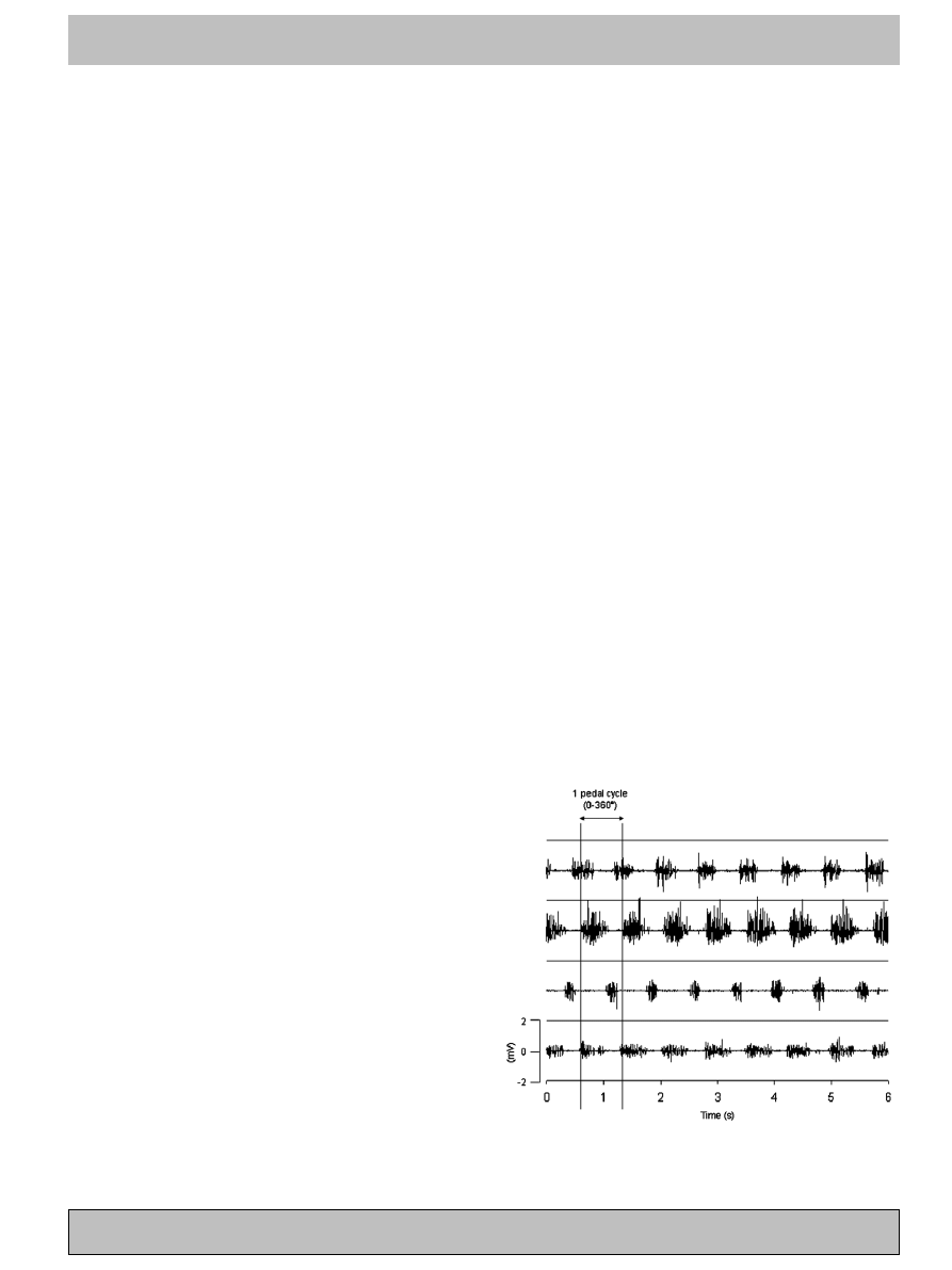
An SRM ergometer (Schoberer Rad Meßtechnik SRM
GmbH, Ju¨lich, Germany) was used for all tests, mounted with the
two flywheels and in the ninth gear. The SRM crankset, equipped
with strain gauges, directly measured the torque produced by the
force applied to the pedals perpendicularly to the crank length.
The ergometer was customized with subject’s own bicycle’s mea-
sures and clipless pedals. A display was available close to the
handlebars of the ergometer to let the cyclists check their pedal-
ling cadence.
2.2.1. Warm up
Cyclists performed a 10 min warm up at a recommended
cadence of 80 rpm. The power constantly increased from 75 to
200 W during the first 6 min (25 W min
1
), the intensity was then
set to 125 W for the next 2 min and increased to 200 and 250 W
for the last 2 min. Depending on the performance level of the
subjects, the warm up intensity could be increased by no more
than 25 W per step.
2.2.2. Maximal isokinetic protocol (MIP)
MIP was performed in the isokinetic mode of the ergometer, at
a fixed pedalling frequency of 80 rpm. This mode allows the
subject to pedal without resistance up to the fixed cadence, while
resistance is automatically and proportionally increased when the
subject tries to overcome it. Prior to the maximal effort, cyclists
pedalled at 80 rpm and low intensity (50–100 W) and at the signal
they started to pedal as forcefully as possible for 6 s, while a
vigorous verbal encouragement was given. They were instructed
to remain seated and to hold the hands on the low part of the
handlebars during the trial. Every cyclist completed a total of ten
6 s all-out sprints. A full recovery was ensured by a 3 min rest
period between sprints, in which they were allowed to drink water
and pedal at a low intensity.
2.2.3. Sub-maximal protocol (SMP)
After the MIP, cyclists rested for 10 min, pedalling at 50 W at
a freely chosen cadence. They were then asked to perform four
sub-maximal exercises at 0%, 20%, 40% and 60% of the maximum
power output obtained during the MIP. In order to perform the
0% exercise, the brake was turned off. It is however useful to know
that, due to the friction of the moving parts, the workload was
actually about 30–35 W. Concerning the other sub-maximal
exercises, while the subject was pedalling at 50 W, the load was
increased and as soon as a steady pedalling cadence of 80 rpm was
reached, data were collected for 20 s. Trials were separated by a
2 min active rest period. The 80% intensity was too demanding to
be maintained for at least 20 s, hence it was not included in the
SMP.
2.3. Recording of EMG and angular crank position
Following the recommendations of the SENIAM project
(
), EMG of four muscles of the right leg was
recorded during the MIP and the SMP. The selected muscles were
vastus lateralis (VL), biceps femoris (BF), tibialis anterior (TA)
and gastrocnemius lateralis (GL). Skin was shaved, slightly
abraded with sandpaper and cleaned with alcohol. Ag/AgCl
bipolar electrodes (Blue Sensor N-00-S, Ambu Medicotest A/S,
Ølstykke, Denmark) were placed over the muscle belly of selected
muscles at an interelectrode distance of 20 mm. To avoid artefacts
from lower limb movements, the wires connecting electrodes were
well secured with tape.
Signal was amplified at a gain of 600. Common mode rejection
rate and input impedance were respectively 95 dB and 10 GX.
Raw electromyographic data were band-pass filtered using a
fourth order Butterworth filter, with cut-off frequencies of 10 and
350 Hz.
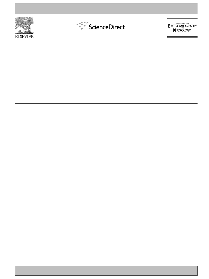
depicts an individual example of the raw EMG
signals as a function of time, related to the four analysed muscles.
In order to measure the instantaneous angular position of the
crank, a rotational encoder (EL40B, Eltra, Sarego (VI), Italy)
with a resolution of 2000 pulses per turn was coupled to the left
crank of the ergometer by a chain drive. Since the gear ratio
between the gear wheel of the left crank and the sprocket of the
encoder was 53/15, the total resolution of the system was 7066.7
pulses per pedal cycle (
The EMG and angular position of the crank signals were
synchronized, sampled at 1000 Hz and stored on a PC using a
16 bit A/D converter data acquisition system (APLabDAQ,
APLab, Rome, Italy).
Fig. 1. Raw EMG signals from a single, representative trial recorded
during the maximal isokinetic protocol. The window corresponding to a
pedal cycle is also shown. VL = vastus lateralis; BF = biceps femoris;
TA = tibalis anterior; GL = gastrocnemius lateralis.
E. Ferna´ndez-Pen˜a et al. / Journal of Electromyography and Kinesiology xxx (2008) xxx–xxx
3
ARTICLE IN PRESS
Please cite this article in press as: Ferna´ndez-Pen˜a E et al., A maximal isokinetic pedalling exercise for EMG ..., J Electromyogr Ki-
nesiol (2008), doi:10.1016/j.jelekin.2007.11.013
2.4. Data processing
Torque applied to the crankset during the MIP was recorded
at 200 Hz for power output calculation purposes. Average power
output of each maximal trial (PO) was calculated as the product
of the average torque over the 6 s (in Nm) and the actual average
cadence (in rad/s). For each subject, the best PO (PO
B
) was set to
represent 100% and the other trials were calculated as a per-
centage of PO
B
.
Raw EMG data were processed by root mean square (RMS)
determination for each complete cycle, defined as a full revolution
of the right crank from top dead center (TDC at 0
°) to the next
TDC. The mean RMS was calculated averaging the RMS values
of the eight pedal cycles completed in every MIP trial, and aver-
aging the complete pedal cycles of the last 10 s (about 13 pedal
cycles) in every SMP trial.
For MIP assessment, the highest EMG activity achieved for
each muscle was set to 100% and the other trials were calculated
as a percentage of the highest. In contrast, for SMP assessment,
the EMG activity of all muscles corresponding to PO
B
was set to
100% and the values obtained during the submaximal exercises
(0%, 20%, 40% and 60% of PO
B
) were expressed as a percentage
Picture 1. A rotational encoder was coupled to the left crank of the SRM ergometer in order to measure the instantaneous angular position of the crank
and synchronize it with the EMG activity.
Fig. 2. Example of individual muscle activity, as a function of crank angle, obtained at five different intensities for the vastus lateralis (A), biceps femoris
(B), tibialis anterior (C) and gastrocnemius lateralis (D).
4
E. Ferna´ndez-Pen˜a et al. / Journal of Electromyography and Kinesiology xxx (2008) xxx–xxx
ARTICLE IN PRESS
Please cite this article in press as: Ferna´ndez-Pen˜a E et al., A maximal isokinetic pedalling exercise for EMG ..., J Electromyogr Ki-
nesiol (2008), doi:10.1016/j.jelekin.2007.11.013
of the former. An example of individual EMG patterns obtained
for the four analysed muscles is represented in
. The raw
data were root mean squared with a moving window length of
100 ms.
2.5. Statistical analysis
In order to evaluate the within-subject reproducibility of PO
and EMG for the four muscles analysed, the variables were
checked for normality and homoscedasticity and were log-trans-
formed when these assumptions were violated. Thereafter the
intra-subject standard error of measurement (SEM), the coeffi-
cient of variation (CV) and the intra-class correlation coefficient
(ICC) were calculated as proposed by
with the
assistance of a reliability spreadsheet (
). The CV is
defined as 100
ðe
SD=
ffiffi
2
p
1Þ, where SD is the standard deviation
of the change scores of natural log of the measure. The ICC is
defined as (V
v)/V, where V is the between-subject variance
averaged over the two trials analysed, and v is the square of the
standard error of measurement.
3. Results
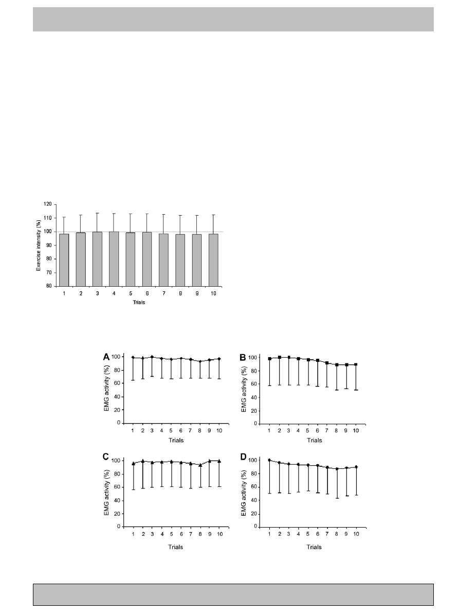
shows PO (mean ± SD) reached during the 10 tri-
als. The PO
B
(100%) was achieved during the 4th trial. The
PO values were, however, very close to each other, ranging
from 98.0 to 100.0, thus indicating a quite high reproduc-
ibility of the variable, with no observable learning or fati-
gue effect. The PO
B
obtained ranged from 664.6 to
1013.9 W (data not shown).
EMG activity (mean ± SD) is shown for VL (
A),
BF (
C) and GL (
D), registered
during the 10 trials. For each of the muscles included in
the analysis, the 100% activity was achieved within the
3rd trial, although BF (
D) tend
to decrease thereafter.
Reliability measures from consecutive pairs of trials are
summarized in
. SEM and CV are presented as a
mean value, whilst for ICC maximal and minimal values
are shown. PO has the lowest CV (2.19), indicating very
good consistency between repeated measures. Among the
EMG activities, VL and TA show, respectively, the lowest
(CV = 4.46) and the highest variability (CV = 8.70). ICCs
in all the variables analysed, ranging from 0.922 to 0.994,
are considerably high.
Fig. 3. Power output (mean ± SD) reached during the 10 trials of the
maximal isokinetic protocol. The best power output was set equal to 100
and the other trials’ were calculated as a percentage of the best one.
Fig. 4. EMG activity (mean ± SD) for vastus lateralis (A), biceps femoris (B), tibialis anterior (C) and gastrocnemius lateralis (D) registered during the 10
trials of the maximal isokinetic protocol. The highest EMG activity was set equal to 100 and the other trials’ were calculated as a percentage of the best
one.
E. Ferna´ndez-Pen˜a et al. / Journal of Electromyography and Kinesiology xxx (2008) xxx–xxx
5
ARTICLE IN PRESS
Please cite this article in press as: Ferna´ndez-Pen˜a E et al., A maximal isokinetic pedalling exercise for EMG ..., J Electromyogr Ki-
nesiol (2008), doi:10.1016/j.jelekin.2007.11.013
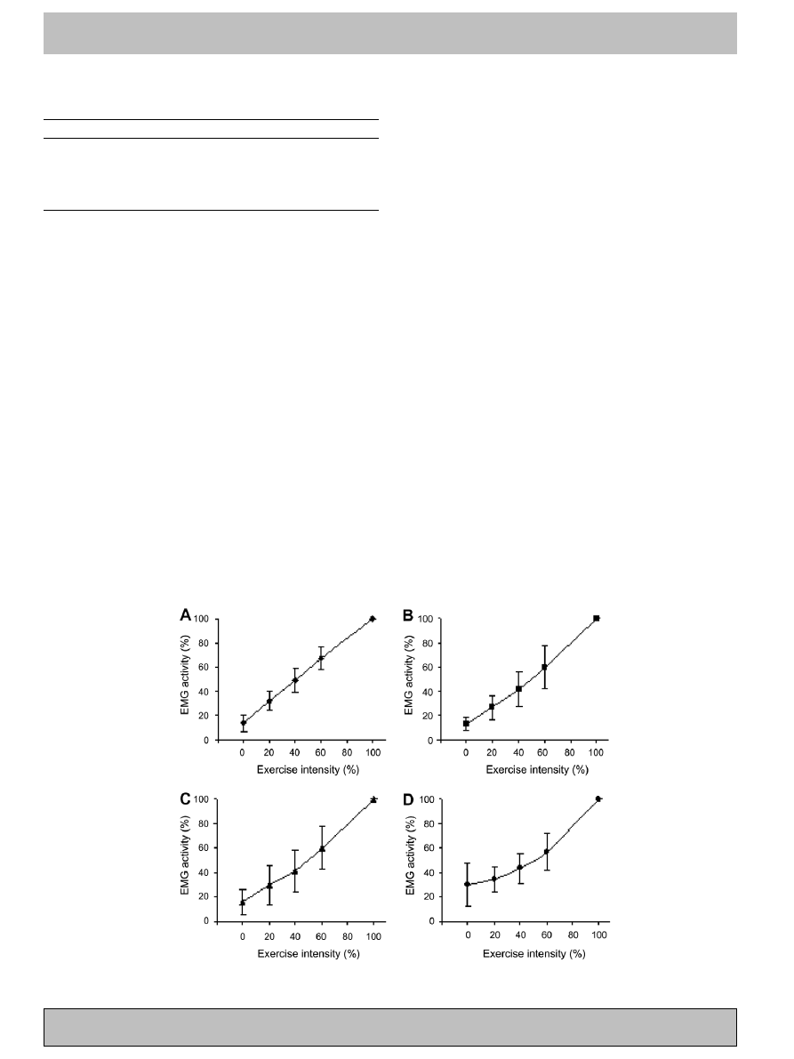
The EMG activity (mean ± SD) corresponding to 0%,
20%, 40% and 60% intensities of PO
B
is presented for VL
A), BF (
B), TA (
D). A
visual inspection showed a linear EMG activity increase
when moving from 0% to 100% of exercise intensity, for
VL (
A), BF (
B) and TA (
C). GL
D) instead exhibited a curvilinear trend.
4. Discussion
The aim of this study was to propose a new method for
EMG amplitude normalization in cycling and assess its
reliability. Different investigations suggesting isometric or
sub-maximal dynamic contractions for EMG normaliza-
tion in cycling have been gone over, and the limitations
were highlighted.
A MIP was introduced as a reference contraction for the
EMG normalization procedure. The task required, a max-
imal 6-s pedalling action, and the submaximal cycling are
comparable for at least three reasons: (a) the contribution
of the muscles of the lower limb is similar for maximal
sprint and submaximal bicycling conditions, as discussed
by
; (b) mechanical conditions
of the MIP and the following test session match com-
pletely: pedalling frequency, posture and joint angle ranges
of the cyclist (hip, knee, ankle), and type of muscular con-
traction; (c) the muscles are activated at the same part of
the pedalling cycle, as shown in the EMG profiles (
).
The EMG activity registered during SMP supports the
validity of the method proposed. Pedalling at 0%, 20%,
40% and 60% intensities of PO
B
resulted in a coherent level
of muscular activity, which altogether exhibited an increase
as a function of exercise intensity.
Three of the muscles analysed (VL, BF and TA) showed
a linear trend, whilst the GL had a curvilinear shape. This
latter result was deemed to be due to the somehow unusual
pedalling pattern when cycling at very low intensities. In
fact, it seems that when riding at 0 W the cyclist has to
avoid pushing down the pedal during the downstroke in
order to maintain the predetermined cadence and a rela-
tively smooth pedal stroke, thus making the knee extensor
muscles to remain inactive. GL instead is often overactive
to counterbalance the knee extensor muscle inactivity and
produce the little amount of power needed for keeping
the flywheel rotation. Eight of the 23 subjects showed a
similar or even higher GL activity at 0% compared to
20%. These data suggest therefore that due to the high
Table 1
Statistical measures of reliability from consecutive pairs of trials
SEM (mean)
CV (mean)
ICC (range)
Power output (PO, Watt)
17.11
2.19
0.962–0.986
VL EMG activity
0.34
4.46
0.969–0.987
BF EMG activity
0.47
7.32
0.934–0.983
GL EMG activity
0.37
6.85
0.922–0.985
TA EMG activity
0.43
8.70
0.948–0.994
SEM and CV are expressed as a mean, whilst ICC as a range value.
VL = vastus
lateralis;
BF = biceps
femoris;
TA = tibialis
anterior;
GL = gastrocnemius lateralis.
The EMG activity (AU) is the root mean square of the raw EMG data for
each complete pedalling cycle.
SEM = standard error of measurement; CV = coefficient of variation;
ICC = intraclass coefficient of correlation.
Fig. 5. EMG activity (mean ± SD) corresponding to 0%, 20%, 40% and 60% intensities of the best power output for vastus lateralis (A), biceps femoris
(B), tibialis anterior (C) and gastrocnemius lateralis (D).
6
E. Ferna´ndez-Pen˜a et al. / Journal of Electromyography and Kinesiology xxx (2008) xxx–xxx
ARTICLE IN PRESS
Please cite this article in press as: Ferna´ndez-Pen˜a E et al., A maximal isokinetic pedalling exercise for EMG ..., J Electromyogr Ki-
nesiol (2008), doi:10.1016/j.jelekin.2007.11.013

inter-subject variability in recruitment pattern of some
muscles (e.g. GL) normalization methods based on low
intensity reference contractions in cycling are questionable.
The activity of each muscle during submaximal contrac-
tions is calibrated in reference to its maximal activity, the
two enabling similar neuromuscular responses. On the
other hand, it was evidenced that iMVC and pedalling
movement differ significantly in biomechanical function
and regulation of the lower limb muscles activation (
Another interesting similarity between MIP and the
cycling action is the angular velocity profile of the crank.
It has been previously demonstrated that the instantaneous
crank angular velocity is not constant throughout the ped-
alling action, rather, it is dependent on the angles of the
lower limb relative to the crank position. In fact, the high-
est and lowest angular velocities occur when the cranks are
near vertical and horizontal, respectively (
). This pattern was evident during MIP, despite the
isokinetic modality. This is due to the fact that the accom-
modating braking power exerted by the ergometer is
reduced to near zero when the cranks are in vertical posi-
tion and increased when the cranks are in horizontal posi-
tion, in order to maintain the desired rotational speed
(
). Actually, this mechanism does not seem
to be able to keep completely steady the instantaneous
angular velocity of the crank, at least when the cyclist is
asked to pedal at 80 rpm and maximal intensity.
From the issues above discussed it could be maintained
that the procedure employed in the current setting is highly
specific to the cycling gesture. Furthermore, using the cur-
rent procedures, each muscle may be assessed at the same
time, this being a time- and energy-saving process. In con-
trast, an isometric contraction would entail a more compli-
cated procedure, especially when several muscles require to
be analysed, namely an iMVC has to be produced sepa-
rately for every single muscle involved in the action (
et al., 2006; Rouffet and Hautier, 2007
).
A common problem of iMVC is that a certain degree of
familiarization is required during the normalization session.
A useful implication from the data presented is that cyclists
do not need to get skilled with the MIP, since no learning
effect was demonstrated during the trials (
). Indeed,
both PO and EMG activity reached their maximal values
respectively, on average, on the 4th and within the 3rd trial.
Although the PO and EMG maximal values are not
attained at the same trial, it is important to underline that
the variability among trials is negligible. Indeed, the average
difference between the best and the worst value in the first
five trials is 2% for PO and from 3% (BF) to 7% (GL) for
the EMG of the four analysed muscles. As a result, based
on the data collected, three trials of the MIP proposed
appear to be sufficient for the purpose of EMG normaliza-
tion in cycling. Thus, the important goal of time efficiency
during testing sessions is also achieved.
As recently pointed out, the repeatability of EMG
recorded during dynamic exercise, especially the muscles
involved in a cycling action, has not been fully established
(
). A strong point of the method pro-
posed is the high degree of reliability. EMG signal of the
VL, compared to the other muscles analysed, showed the
lowest variability and this is in agreement with previous
studies which found the EMG activity of VL during cycling
to have a high reproducibility (
). When comparing the activity
of the same muscles, the CVs are considerably lower than
those presented by
who used
a maximal torque–velocity test, although it is important
to mention that the subjects of their study were not cyclists.
4.1. Limitations
The protocols proposed were performed at 80 rpm. This
cadence is widely used in cycling related researches (
and Li, 2003; Hull and Jorge, 1985
); furthermore, previous
studies found that freely chosen cadence in cyclists ranges
between 78 and 91 rpm (
Foss and Halle´n, 2005; Hagberg
et al., 1981; Nielsen et al., 2004
). Notwithstanding, a
cadence of 100–115 rpm, rather than 80 rpm, makes the
cyclist attain the maximal power output (
Baron et al., 1999; Sargeant et al., 1981
). Consequently,
the pedalling frequency chosen in the present study is situ-
ated in the ascending part of the power–cadence curve.
demonstrated that the EMG/cadence
curve at constant power and pedalling frequency ranging
from 45 to 105 rpm showed a quadratic trend. The mini-
mum EMG value was registered at 60 rpm, afterward it
exhibited an increase as a function of cadence. It is there-
fore expected that maximal isokinetic pedalling at higher
cadences would lead to higher EMG values, although this
issue needs to be investigated further on.
As a consequence, the reference contraction here pro-
posed at 80 rpm should be used only for submaximal bicy-
cling exercises performed at the same cadence. In general
terms it could be argued that, whatever the pedalling rate
chosen for SMP, the MIP should be performed at the same
cadence.
5. Conclusion
The new protocol proposed for the purpose of EMG
normalization in cycling, which consists of three 6-s maxi-
mal isokinentic pedalling sprints, has very good logical
validity and within-subject reproducibility. Further, the
test is also highly specific to the actions associated with
cycling. The chosen cadence of the normalization protocol
should be the same as the sub-maximal exercises.
Acknowledgements
The authors would like to thank APLab engineers, espe-
cially Nunzio Lanotte, who have designed the data acqui-
sition
system,
for
their
technical
assistance;
Mark
Watsford, Ph.D., lecturer in Exercise and Sports Science
E. Ferna´ndez-Pen˜a et al. / Journal of Electromyography and Kinesiology xxx (2008) xxx–xxx
7
ARTICLE IN PRESS
Please cite this article in press as: Ferna´ndez-Pen˜a E et al., A maximal isokinetic pedalling exercise for EMG ..., J Electromyogr Ki-
nesiol (2008), doi:10.1016/j.jelekin.2007.11.013

School of Leisure, Sport and Tourism University of Tech-
nology (Sydney) and Francesco Felici, M.D., professor of
exercise physiology, Istituto Universitario di Scienze Moto-
rie (Rome) for their assistance and suggestions in revising
the paper.
References
Arokoski JP, Kankaanpaa M, Valta T, Juvonen I, Partanen J, Taimela S,
et al. Back and hip extensor muscle function during therapeutic
exercises. Arch Phys Med Rehabil 1999;80:842–50.
Baron R. Aerobic and anaerobic power characteristics of off-road cyclists.
Med Sci Sports Exerc 2001;33:1387–93.
Baron R, Bachl N, Petschnig R, Tschan H, Smekal G, Pokan R.
Measurement of maximal power output in isokinetic and non-
isokinetic cycling. A comparison of two methods. Int J Sports Med
1999;20:532–7.
Baum BS, Li L. Lower extremity muscle activities during cycling are
influenced
by
load
and
frequency.
J
Electromyogr
Kinesiol
2003;13:181–90.
Bolgla LA, Uhl TL. Reliability of electromyographic normalization
methods for evaluating the hip musculature. J Electromyogr Kinesiol
2007;17:102–11.
Clarys JP, Cabri J. Electromyography and the study of sports movements:
a review. J Sports Sci 1993;11:379–448.
Dankaerts W, O’Sullivan PB, Burnett AF, Straker LM, Danneels LA.
Reliability of EMG measurements for trunk muscles during maximal
and sub-maximal voluntary isometric contractions in healthy controls
and CLBP patients. J Electromyogr Kinesiol 2004;14:333–42.
Danoff JV. EMG normalization. Phys Ther 1986;66:270–2.
Enoka RM, Fuglevand AJ. Neuromuscular basis of the maximum
voluntary force capacity of muscle. In: Grabiner MD, editor. Current
issues in biomechanics. Champaign, IL: Human Kinetics; 1993. p.
215–35.
Ericson M. On the biomechanics of cycling. A study of joint and muscle
load during exercise on the bicycle ergometer. Scand J Rehabil Med
Suppl 1986;16:1–43.
Ericson MO, Nisell R, Arborelius UP, Ekholm J. Muscular activity during
ergometer cycling. Scand J Rehabil Med 1985;17:53–61.
Foss Ø, Halle´n J. Cadence and performance in elite cyclists. Eur J Appl
Physiol 2005;93:453–62.
Freriks B, Hermens H, Disselhorst-Klug C, Rau G. The recommendations
for sensors and sensor placement procedures for surface electromyog-
raphy. In: Hermens H, Freriks B, Merletti R, Stegeman D, Blok J, Rau
G, et al., editors. European recommendations for surface electromy-
ography. Enschede (The Netherlands): Roessingh Research and
Development BV; 1999. p. 15–54.
Hagberg JM, Mullin JP, Giese MD, Spitznagel E. Effect of pedaling rate
on submaximal exercise responses of competitive cyclists. J Appl
Physiol 1981;51:447–51.
Hautier CA, Arsac LM, Deghdegh K, Souquet J, Belli A, Lacour JR.
Influence of fatigue on EMG/force ratio and cocontraction in cycling.
Med Sci Sports Exerc 2000;32:839–43.
Hopkins WG. A new view of statistics. Available from:
[Accessed 2007 Feb 27].
Hopkins WG. Measures of reliability in sports medicine and science. Sport
Med 2000;30:1–15.
Hsu WL, Krishnamoorthy V, Scholz JP. An alternative test of electro-
myographic normalization in patients. Muscle Nerve 2006;33:232–41.
Hug F, Bendahan D, Le Fur Y, Cozzone PJ, Grelot L. Heterogeneity of
muscle recruitment pattern during pedaling in professional road
cyclists: a magnetic resonance imaging and electromyography study.
Eur J Appl Physiol 2004a;92:334–42.
Hug F, Decherchi P, Marqueste T, Jammes Y. EMG versus oxygen
uptake during cycling exercise in trained and untrained subjects. J
Electromyogr Kinesiol 2004b;14:187–95.
Hull ML, Jorge M. A method for biomechanical analysis of bicycle
pedalling. J Biomech 1985;18:631–44.
Hunt MA, Sanderson DJ, Moffet H, Inglis JT. Biomechanical changes
elicited by an anterior cruciate ligament deficiency during steady rate
cycling. Clin Biomech 2003;18:393–400.
Hunter AM, St Clair Gibson A, Lambert M, Noakes TD. Electromyo-
graphic (EMG) normalization method for cycle fatigue protocols. Med
Sci Sports Exerc 2002;34:857–61.
Kollmitzer J, Ebenbichler GR, Kopf A. Reliability of surface electro-
myographic measurements. Clin Neurophysiol 1999;110:725–34.
Laplaud D, Hug F, Grelot L. Reproducibility of eight lower limb muscles
activity level in the course of an incremental pedaling exercise. J
Electromyogr Kinesiol 2006;16:158–66.
Latash ML. Neurophysiological basis of movement. 1st ed. Champaign,
IL: Human Kinetics; 1998.
Lehman GJ, McGill SM. The importance of normalization in the
interpretation of surface electromyography: a proof of principle. J
Manipulative Physiol Ther 1999;22:444–6.
Lobbezoo F, van der Glas HW, Buchner R, van der Bilt A, Bosman F.
Gain and threshold of the jaw-jerk reflex in man during isometric
contraction. Exp Brain Res 1993;93:129–38.
Marras WS, Davis KG. A non-MVC EMG normalization technique for
the trunk musculature: Part 1. Method development. J Electromyogr
Kinesiol 2001;11:1–9.
Marsh AP, Martin PE. The relationship between cadence and lower
extremity EMG in cyclists and noncyclists. Med Sci Sports Exerc
1995;27:217–25.
Mirka GA. The quantification of EMG normalization error. Ergonomics
1991;34:343–52.
Moussay S, Bessot N, Gauthier A, Larue J, Sesboue B, Davenne D.
Diurnal
variations
in
cycling
kinematics.
Chronobiol
Int
2003;20:879–92.
Neptune RR, Herzog W. Adaptation of muscle coordination to altered
task mechanics during steady-state cycling. J Biomech 2000;33:
165–72.
Neptune RR, Kautz SA, Hull ML. The effect of pedaling rate on
coordination in cycling. J Biomech 1997;30:1051–8.
Nielsen JS, Hansen EA, Sjogaard G. Pedalling rate affects endurance
performance during high-intensity cycling. Eur J Appl Physiol
2004;92:114–20.
Prilutsky BI, Gregor RJ, Ryan MM. Coordination of two-joint rectus
femoris and hamstrings during the swing phase of human walking and
running. Exp Brain Res 1998;120:479–86.
Rouffet DM, Hautier CA. EMG normalization to study muscle activa-
tion
in
cycling.
J
Electromyogr
Kinesiol
2007.
doi:
Ryan MM, Gregor RJ. EMG profiles of lower extremity muscles during
cycling at constant workload and cadence. J. Electromyogr. Kinesiol.
1992;2:69–80.
Sargeant AJ, Hoinville E, Young A. Maximum leg force and power
output
during
short-term
dynamic
exercise.
J
Appl
Physiol
1981;51:1175–82.
Smith J, Padgett DJ, Dahm DL, Kaufman KR, Harrington SP, Morrow
DA, et al. Electromyographic activity in the immobilized shoulder
girdle musculature during contralateral upper limb movements. J
Shoulder Elbow Surg 2004;13:583–8.
Soderberg GL, Knutson LM. A guide for use and interpretation of
kinesiologic electromyographic data. Phys Ther 2000;80:485–98.
Takaishi T, Yamamoto T, Ono T, Ito T, Moritani T. Neuromuscular,
metabolic, and kinetic adaptations for skilled pedaling performance in
cyclists. Med Sci Sports Exerc 1998;30:442–9.
Taylor AD, Bronks R. Reproducibility and validity of the quadriceps
muscle integrated electromyogram threshold during incremental cycle
ergometry. Eur J Appl Physiol Occup Physiol 1995;70:252–7.
van Diee¨n JH, Selen LP, Cholewicki J. Trunk muscle activation in low-
back pain patients, an analysis of the literature. J Electromyogr
Kinesiol 2003;13:333–51.
Wooles A. SRM user manual. 4th ed. Schoberer Rad Meßtechnik; 2006.
8
E. Ferna´ndez-Pen˜a et al. / Journal of Electromyography and Kinesiology xxx (2008) xxx–xxx
ARTICLE IN PRESS
Please cite this article in press as: Ferna´ndez-Pen˜a E et al., A maximal isokinetic pedalling exercise for EMG ..., J Electromyogr Ki-
nesiol (2008), doi:10.1016/j.jelekin.2007.11.013
Eneko Ferna´ndez Pen˜a received his degree in
Physical Education from the Basque Institute
of Physical Education (SHEE/IVEF, Vitoria-
Gasteiz, Spain) in July 2003, and his Ph.D.
degree from the University of Urbino ‘‘Carlo
Bo” (Italy) in March 2007. He is currently a
post-doctoral fellow at the Institute of Health
and Physical Exercise (Urbino, Italy), and his
research interest focuses on biomechanics of
cycling.
Francesco Lucertini received his diploma in
Physical Education (1998) and his degree in
Exercise Sciences (2001) from University of
Urbino ‘‘Carlo Bo” (Italy). He received the
Ph.D. degree in February 2006 from the Fac-
ulty of Health and Sport Sciences of the same
University and he is currently a post-doctoral
fellow at the Institute of Health and Physical
Exercise (Urbino, Italy). His research interest
focuses on performance assessment in both
sport- and health-related topics.
Massimiliano Ditroilo has a diploma in Physical
Education (1992), a degree in Biological Sci-
ences (1999) and a master degree in Methods of
Training (2001). He is currently working within
the Institute of Health and Physical Exercise at
Urbino University (Italy). His research focuses
on biomechanics and performance assessment
of cycling, athletics, swimming and team
sports.
E. Ferna´ndez-Pen˜a et al. / Journal of Electromyography and Kinesiology xxx (2008) xxx–xxx
9
ARTICLE IN PRESS
Please cite this article in press as: Ferna´ndez-Pen˜a E et al., A maximal isokinetic pedalling exercise for EMG ..., J Electromyogr Ki-
nesiol (2008), doi:10.1016/j.jelekin.2007.11.013
Document Outline
Wyszukiwarka
Podobne podstrony:
Facial Exercise for Look Younger
Academic Studies English Support Materials and Exercises for Grammar Part 1 Parts of Speech
Guitar Exercises for speed and accuracy
EXERCISES FOR GAP FILLING 01
Exercises for TRX
Exercises for Stretching the Fingers
101 Greatest Exercises For Size
Arpeggio Exercise For Piano
22 Practice Charts for Eye Exercises
Exercise%20Problems%20for%20students%20(CH%204)[1]
Exercise Standards for Testing and Training
Exercise Programs for Children with Cerebral Palsy
22 Practice Charts for Eye Exercises
Oxford Vocabulary Exercises Film Suitable for Printing
Word Order for Adjectives Exercise at Auto
Reading for pleasure Judy Steiner (Extensive reading exercises and activities)
Exercices Hanon for jazz(1)
więcej podobnych podstron