
The Legionella pneumophila chaperonin – an unusual
multifunctional protein in unusual locations
1,2
*, Audrey Chong
and David S. Allan
1
Department of Microbiology and Immunology, Dalhousie University, Halifax, NS, Canada
2
Division of Infectious Diseases, Department of Medicine, Dalhousie University, Halifax, NS, Canada
3
Laboratory of Intracellular Parasites, National Institute of Allergy and Infectious Diseases, National Institute of Health Rocky Mountain Laboratories,
Hamilton, MT, USA
Edited by:
Carmen Buchrieser, Pasteur Institute,
France
Reviewed by:
Philippe Mazodier, Pasteur Institute,
France
Helene Bierne, French National
Institute for Agricultural Research,
France
*Correspondence:
Rafael A. Garduño, Department of
Microbiology and Immunology, Sir
Charles Tupper Medical Building, 7th
floor, 5850 College Street, Halifax,
NS, Canada B3H-1X5.
e-mail: rafael.garduno@dal.ca
The Legionella pneumophila chaperonin, high temperature protein B (HtpB), was discovered
as a highly immunogenic antigen, only a few years after the identification of L. pneumophila
as the causative agent of Legionnaires’ disease. As its counterparts in other bacterial
pathogens, HtpB did not initially receive further attention, particularly because research
was focused on a few model chaperonins that were used to demonstrate that chaperonins
are essential stress proteins, present in all cellular forms of life and involved in helping
other proteins to fold. However, chaperonins have recently attracted increasing interest,
particularly after several reports confirmed their multifunctional nature and the presence of
multiple chaperonin genes in numerous bacterial species. It is now accepted that bacterial
chaperonins are capable of playing a variety of protein folding-independent roles. HtpB is
clearly a multifunctional chaperonin that according to its location in the bacterial cell, or in
the L. pneumophila-infected cell, plays different roles. HtpB exposed on the bacterial cell
surface can act as an invasion factor for non-phagocytic cells, whereas the HtpB released in
the host cell can act as an effector capable of altering organelle trafficking, the organization
of actin microfilaments and cell signaling pathways.The road to discover the multifunctional
nature of HtpB has been exciting and here we provide a historical perspective of the key
findings linked to such discovery, as well as a summary of the experimental work (old and
new) performed in our laboratory. Our current understanding has led us to propose that
HtpB is an ancient protein that L. pneumophila uses as a key molecular tool important to
the intracellular establishment of this fascinating pathogen.
Keywords: HtpB, Hsp60, GroEL, pathogenesis, mitochondria, microfilaments, polyamines
BACKGROUND
CHAPERONINS AND THEIR ESSENTIAL PROTEIN FOLDING FUNCTION
Chaperonins are a family of structurally and functionally con-
served, essential proteins, present in virtually all prokaryotic and
eukaryotic forms of life. Intuitively, then, contemporary chaper-
onins must be related to one of the first proteins present in the
common ancestor of all organisms currently known. The striking
amino acid sequence and structural conservation of the chap-
eronin groups clearly suggests that these proteins must be very
important. The primary function of chaperonins, recognized to be
important enough to explain their essential nature, is in helping
other proteins to fold properly and reach their native (functional)
state.
Because this review is focused on the protein folding-
independent functions of the Legionella chaperonin, a discussion
on the protein folding ability of chaperonins is not forthcoming.
Therefore, we provide the following key references for the benefit
of those with further interests in this topic (
;
;
). In
particular, recent comprehensive reviews that cover various aspects
of the fascinating structure, biochemistry, and physiology of these
formidable protein folding molecular machines (or nanoboxes
in which proteins can fold) are those of
,
,
, and
.
CLASSIFICATION OF CHAPERONINS
It seems that
were the first to coin
the term “chaperonins” to describe a small group of related
proteins involved in “post-translational assembly of oligomeric
protein structures.” Since then, investigators have recognized the
existence of different chaperonin types, which are currently clas-
sified into two groups based on their structure and evolution-
ary origin. Group I chaperonins are found in bacteria and in
endosymbiotic organelles of eukaryotes (e.g., mitochondria and
chloroplasts), have a mass of
∼60-kDa and are typically induced
under stress, e.g., heat shock. Therefore, group I chaperonins
are also known as heat shock proteins 60 (Hsp60s;
Zeilstra-
Ryalls et al., 1991
). These proteins form homo-oligomeric rings
that consist of seven chaperonin subunits (
).
Two of these 7-mer rings come together to form the 14-mer
barrel complex that mediates protein folding in association with
a third homo-oligomeric ring, comprised of seven subunits of

Garduño et al.
HtpB, the L. pneumophila chaperonin
co-chaperonin, a protein of
∼10-kDa also known as Hsp10. Asso-
ciation with the 10-kDa co-chaperonins is an exclusive feature
of Group I chaperonins. Other designations for Hsp10/Hsp60,
are GroES/GroEL, Cpn10/Cpn60, and HtpA/HtpB. The inten-
sively investigated Escherichia coli GroEL chaperonin constitutes
the paradigm of Group I chaperonins.
Group II chaperonins, also known as TriC (TCP-1 ring com-
plex) or CCT (chaperonin-containing TCP-1), are found in
archaea, and the cytoplasm of eukaryotes (
). Group
II chaperonins form eight- or nine-membered hetero-oligomeric
rings with subunits that may have different masses (
;
). CCTs mediate the special-
ized folding of proteins (many of which are linked to the cytoskele-
ton), but do not team with 10 kDa co-chaperonins, although the
protein prefoldin (
) has been identified as a co-
chaperone for CCTs. Group II chaperonins have an extended apical
domain thought to cap the central cavity of the double-ringed
complex, which replaces the need for the 7-mer co-chaperonin
ring of Group I chaperonins (
;
). Group II chaperonins are het-
erogeneous and are thought to have evolved by gene duplication
and subsequent mutation (
). While conserved
within their respective groups, Group I and Group II chaperonins
are only distantly related, but thought to share a common protein
ancestor (
).
A third chaperonin group has been recently reported in bacte-
ria (
). Its representative chaperonin is
that of the bacterium Carboxydothermus hydrogenoformans, which
forms a 16-mer structure capable of refolding denatured proteins
in an ATP-dependent manner. Group III chaperonins are distantly
related to both Group I and Group II chaperonins, and thus they
might represent an ancient horizontal transfer event from archaea
to bacteria.
PROTEIN FOLDING-INDEPENDENT FUNCTIONS OF GROUP I
CHAPERONINS
The Hsp60 of the bacterial endosymbiont Buchnera aphidicola
(also called symbionin) acts as a histidine kinase (
), whereas the GroEL of symbiotic Enterobacter aerogenes is
a potent insect toxin (
), and the chaperonin of
Mycobacterium leprae, is a protease (
). Two views
could be advanced to explain this functional diversity. In the first
view, functional diversity is a preserved characteristic of chaper-
onins. That is, Group I chaperonins started as jacks-of-all-trades
and gradually evolved toward specialization in protein folding.
Thus, the contemporary examples of diversity mentioned above,
represent evolutionary remnants of original functions preserved
after specialization. In the second view, functional diversity is a
newly emerged characteristic. That is, ancient chaperonins started
as specialized proteins that gradually evolved toward functional
diversity.
Two cases of functional chaperonin diversity resulting from
few amino acid changes seem to favor the second view of “newly
emerged functions.” Only 11 amino acids are different between the
toxic chaperonin from endosymbiotic E. aerogenes, and the non-
toxic chaperonin of E. coli, of which four amino acid positions
are critical for toxicity. When the non-toxic E. coli chaperonin was
engineered at the four critical residues to resemble the E. aerogenes
chaperonin, it too became a potent insect toxin (
). In the case of the Hsp65 chaperonin of M. leprae, only
three amino acids (Thr-375, Lys-409, and Ser-502) comprise the
threonine catalytic group responsible for protease activity (
Portaro
et al., 2002
).
In a recent article based on the analysis of 669 complete bacterial
genomes, Lund proposed that one of the mechanisms responsi-
ble for functional diversity in Group I chaperonins relies on gene
duplication followed by unconstrained mutation of the duplicated
gene sequences (
). The analysis showed that 467/669
genomes contained a single chaperonin gene, 183/669 genomes
contained multiple chaperonin genes (from 2 to a maximum of
7), and 13 Mycoplasma genomes contained no discernable chap-
eronin genes.
thus argued that the essential protein
folding needs of a bacterial cell are met by a single chaperonin
(whose gene would be constrained for change), while the other
chaperonins would be free to mutate and acquire functional spe-
cializations. At least in the case of Mycobacterium tuberculosis,
this notion has been experimentally substantiated. M. tubercu-
losis has two chaperonin genes encoding the chaperonins Cpn60.1
and Cpn60.2, where cpn60.2 is essential whereas cpn60.1 can be
deleted from the genome (
). These two chaperonins
are functionally different (
) supporting the
idea of functional diversity afforded by gene duplication.
However, there are other cases in which functional diversity
rests on a single chaperonin. As it will be discussed below in
detail, one of these cases is the chaperonin of Legionella pneu-
mophila. Other examples include those bacterial pathogens that
typically use their chaperonins as adherence factors, or immune-
modulators. In this capacity, chaperonins have been recently added
to the list of “moonlighting” proteins (
). The term
moonlighting is defined in the Webster’s Dictionary of the English
Language as “working at a job in addition to one’s regular one,” and
was introduced in the biochemical field to describe those proteins
that perform a well-recognized function by day (regular job in a
given environment or cellular location), and a not so obvious yet
important function by night (other jobs in a different environment
or cellular location).
Actinobacillus actinomycetemcomitans (
), Borrelia burgdorferi (
Chlamydia spp. (
), Clostridium difficile (
Hennequin
et al., 2001
), Helicobacter pylori (
), Haemophilus
ducreyi (
), Listeria monocytogenes (
), and Salmonella enterica sv. Typhimurium (
Ensgraber and
Loos, 1992
), are but some examples of bacterial pathogens that
display their chaperonin in extracytoplasmic locations, and where
the surface-associated, periplasmic, or released/secreted chaper-
onin seems to play alternate functional roles. For instance, the
chaperonin of some of the aforementioned pathogens acts as an
adhesion factor, but there are many that interact with mammalian
cell surface receptors to initiate signaling events that result in
cytokine production (reviewed by
), phospho-
rylation of signaling molecules (
), or other
physiological outputs (
Group I chaperonins of endosymbiotic organelles are also func-
tionally diverse, but given the nature of our review and its focus
Frontiers in Microbiology | Cellular and Infection Microbiology
Garduño et al.
HtpB, the L. pneumophila chaperonin
on a bacterial pathogen, we will not discuss here organellar chap-
eronins. Therefore, readers interested in the prominent role of
chaperonins in immunity and autoimmunity are referred to a
recent scholar review (
) that includes details on
the immune-modulatory ability of these proteins. In summary,
chaperonins are ancient proteins, essential for the life of eukary-
otic and prokaryotic cells. Their essential nature seemingly rests
on their protein folding ability, but in several cases chaperonins
appear to be multifunctional.
THE CHAPERONIN OF LEGIONELLA PNEUMOPHILA, HtpB
The remaining portion of this review will be devoted to a dis-
cussion of the L. pneumophila chaperonin as a multifunctional
(“moonlighting”) protein (
Figure 1), including a presentation
of our recent experimental findings. To facilitate the distinc-
tion between the chaperonins that we will be discussing, and
to respect current nomenclature, the L. pneumophila chaperonin
will be subsequently referred to as high temperature protein B
(for HtpB). The designation HtpA is used for the L. pneumophila
co-chaperonin, which is encoded by the first gene in the L. pneu-
mophila htpAB operon. The chaperonin/co-chaperonin system of
E. coli will be referred to as GroEL/GroES. Other chaperonins will
be referred to as Hsp60 or Cpn60.
HISTORICAL PERSPECTIVE OF HtpB RESEARCH BEFORE 1998
Discovery and initial characterization
Between the mid-1980s and early 1990s, a number of publications
reported the existence of a common antigen of about 60-kDa in
many bacterial species.
referred to it
as the “common antigen,” and
used the
term “cross-reacting protein antigen.” These antigens were even-
tually identified as chaperonins. Similarly, HtpB was first spotted
as a 58-kDa common antigen cross-reactive with 60-kDa anti-
gens from several Legionella species and other bacteria (
Sampson
et al., 1986
;
). This antigen prominently reacted
with sera from patients diagnosed with Legionnaires’ disease (LD)
and was used to confirm, by serology, culture-positive cases of
LD (
). This study also showed that when
a rabbit serum raised against L. pneumophila serogroup 1 was
pre-absorbed with whole L. pneumophila Philadelphia-1 cells, the
58-kDa antigen was no longer recognized by immunoblot. This
is an interesting result because implies that the common antigen
was surface exposed on the whole L. pneumophila cells used for
cross-absorption.
were the first to purify
HtpB and raise a rabbit hyperimmune serum against the purified
protein, and shortly after,
reported an opti-
mized method for the purification of HtpB. A modification of this
optimized method, which involves a combination of ammonium
sulfate precipitation, size-exclusion, and ion-exchange chromatog-
raphy, is the one used in the Garduño lab for the purification
of HtpB.
We will close this section by mentioning that
Gabay and Hor-
witz (1985)
characterized HtpB as the major cytoplasmic mem-
brane protein of L. pneumophila. Their studies are important
because they established the ability of HtpB to interact with the
bacterial cytoplasmic membrane, a trait that we believe is impor-
tant in both the translocation of HtpB into the L. pneumophila
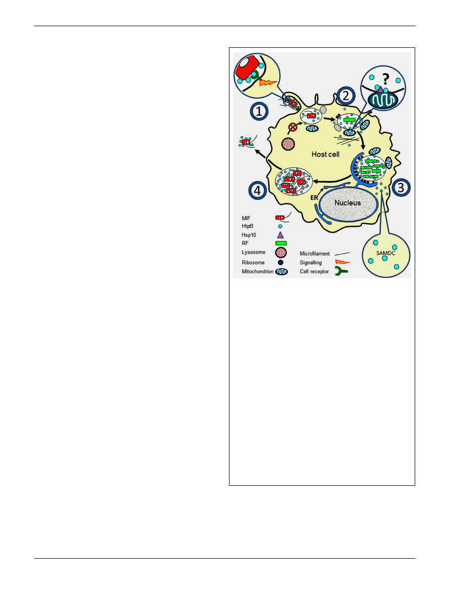
FIGURE 1 | Surface exposed or released HtpB accompanies
L. pneumophila along its growth cycle in host cells. (1) Extracellular
L. pneumophila upregulates expression of HtpB in the presence of host
cells (see Links Between HtpB and L. pneumophila Virulence), and the
interaction of surface-exposed HtpB with cell receptors (Inset 1) triggers a
signal leading to internalization (see Surface-Exposed HtpB Acts as an
Invasion Factor). (2) Internalized legionellae associate with ER-derived
vesicles, attracts mitochondria, and inhibit fusion with lysosomes. HtpB
bound to beads is sufficient to mimic the last two events (see
Surface-Exposed HtpB Alters Organelle Traffic). HtpB reaches the cytoplasm
of the host cell where it could alter the actin cytoskeleton (Inset 2). The
mechanism by which HtpB attracts mitochondria is unknown, but alteration
of actin fibers and tethering via mitochondrial Hsp10 could be involved (see
HtpB in the Eukaryotic Cytoplasm has Several Protein Targets). (3) During
replication, released HtpB accumulates in the LCV from which it could
reach the host cell cytoplasm (see Links Between HtpB and L. pneumophila
Virulence and HtpB is Found in Extracytoplasmic Locations). Inset 3: HtpB in
the cytoplasm of host cells (mammalian and amebal) interacts with SAMDC
to potentially increase the intracellular pool of polyamines (see HtpB in the
eukaryotic cytoplasm has several protein targets). (4) As L. pneumophila
differentiates into MIFs, the amount of HtpB associated with the cell
envelope and bacterial cell surface increases (see HtpB is Found in
Extracytoplasmic Locations). As the LCV ruptures, large amounts of HtpB
are likely released together with MIFs. Immunomodulatory effects (see
Immunological Studies with HtpB) can be triggered by HtpB at any stage of
the cycle. Key: ER, endoplasmic reticulum; RF, replicative form; MIF, mature
infectious form; SAMDC, S-adenosyl methionine decarboxylase.
periplasm (see HtpB is Found in Extracytoplasmic Locations
below), and across the Legionella-containing vacuole (LCV)
membrane into the host cell cytosol (see HtpB is Found in Extracy-
toplasmic Locations and Intracellularly Released HtpB Alters the
Actin Cytoskeleton of Host Cells).

Garduño et al.
HtpB, the L. pneumophila chaperonin
Monoclonal antibodies and the unique epitopes of HtpB
Several monoclonal antibodies were raised against HtpB once it
was available as a purified protein (
Sampson
et al., 1991
;
). These early monoclonal anti-
bodies demonstrated that HtpB possesses epitopes cross-reactive
with many other Group I chaperonins, as well as HtpB-specific epi-
topes. Monoclonal antibody GW2X4B8B2H6 (
does not cross-react with many Group I chaperonins (except for
a few, including the Bordetella Cpn60), and recognizes the C-
terminus of HtpB (
). We have widely used
this antibody to monitor expression of recombinant HtpB. Mon-
oclonal antibody 2125 (
) is highly specific
for HtpB and does not cross-react with any other bacterial chap-
eronin tested. Therefore, 2125 has been used as a tool for the rapid
identification of Legionella spp. (
). But our
interest here is focused on the screening method used by
Stein-
metz et al. (1991)
to identify their monoclonal antibodies, because
they used whole, live, non-permeabilized, non-fixed cells attached
to wells of 96-well ELISA plates, which, again, implied that HtpB
was surface exposed in (or easily released by) Legionella. How-
ever, these investigators could not detect Legionella whole cells by
immunofluorescence microscopy. Another interesting finding of
is that not all the L. pneumophila strains
tested had surface-exposed HtpB, in spite of showing abundant
HtpB after sonication. In conclusion, experimentation with mon-
oclonal antibodies against HtpB has clearly shown that HtpB has
unique structural regions not found in other Group I chaperonins,
and also suggested that HtpB is surface exposed in some strains of
L. pneumophila.
Early molecular biology experiments with HtpB
Paul S. Hoffman’s lab was the first to clone and express the L. pneu-
mophila htpAB operon in E. coli (
) and a year
later, the nucleotide sequence of htpB was published almost simul-
taneously by
and
.
There was good agreement between the two published DNA
sequences of htpB, but only
reported the
sequence and gene organization of the htpAB operon. The expres-
sion of ectopic HtpB in E. coli also allowed
to determine that HtpB could not complement a temperature-
sensitive GroEL defect in E. coli strain CG218 [groEL100(Ts)].
This is an important experimental result because it indicated, at
the molecular level, that GroEL and HtpB are not functionally
equivalent.
Links between HtpB and L. pneumophila virulence
Hoffman et al. (1990)
showed by immunofluorescence microscopy
that HtpB is detectable on the surface of the virulent L. pneu-
mophila Philadelphia-1 strain SVir suspended in Dulbecco-
modified Eagle’s medium (DMEM). In contrast, surface-exposed
HtpB was only weakly detectable on the salt-tolerant aviru-
lent derivative AVir suspended in DMEM. Clearly, only virulent
legionellae suspended in DMEM had the ability to display HtpB
on their cell surface, an observation that provided the first link
between HtpB and virulence. These investigators also showed that
HtpB is abundantly expressed (and released) in L. pneumophila-
infected HeLa cells, which were immuno-labeled with an intense
diffuse pattern (rather than a particulate one), suggesting that
HtpB was free in the LCV where this bacterium replicates (
Hoff-
man et al., 1990
). Its abundant release in the LCV also suggested
that HtpB might play a role in the intracellular establishment of
L. pneumophila.
An early response of L. pneumophila strain 2064 to the presence
of host cells involves de novo synthesis of increasing amounts of
HtpB (
; see Induction of HtpB Expression
by Heat Shock and Presence of Host Cells below). However, an
isogenic, salt-tolerant, avirulent derivative of 2064 was unable to
respond, and showed no de novo synthesis of HtpB in the same
experimental conditions used for 2064 (
).
This observation provided an additional link between HtpB and L.
pneumophila virulence, and suggested that HtpB might be required
at an early stage of the infection process, even before L. pneu-
mophila is internalized. In conclusion, the abilities to produce new
HtpB in response to host mammalian cells, and display HtpB on
the bacterial cell surface, are lost in avirulent legionellae.
Induction of HtpB expression by heat shock and presence of host
cells
High temperature protein B is induced by heat shock. Increased
levels of HtpB were detected in L. pneumophila (
and in L. pneumophila and E. coli (
) upon
temperature increases. However, the maximum increase in HtpB
expression upon heat shock was
∼twofold, and at all temperatures
tested HtpB remained as one of the most abundant proteins in
L. pneumophila. This constitutes a pattern of heat shock that is
different from the pattern typically seen in other bacteria (e.g., E.
coli as shown in
), where the basal levels of
chaperonin are low and a sharp increase is observed at high tem-
peratures. Clearly, HtpB is not a typical Hsp in L. pneumophila. In
addition, HtpB seems to be induced in virulent L. pneumophila by
the presence of mammalian host cells (monocytes and L929 cells),
as demonstrated by
using pulse radio-
labeling in cycloheximide-treated, Legionella-infected cells. The
induced synthesis of new HtpB did not require bacterial internal-
ization (inhibited with cytochalasin D), suggesting that contact
with host cells was sufficient to trigger the response. Finally,
determined by immunoelectron
microscopy that HtpB epitopes were present on the phagosomal
membrane and the cytoplasm of the infected cell.
Immunological studies with HtpB
From its very discovery, HtpB was regarded as strongly antigenic.
Thus, investigators focused on establishing whether HtpB was a
protective antigen, potentially applicable for vaccination against
LD. Immunization with HtpB protected guinea pigs from a lethal
aerosol challenge with L. pneumophila, and the protection was
mediated by a strong cellular response (
). These authors wondered how HtpB is released intracellu-
larly to elicit a cellular response, and performed immunoelectron
microscopy localization studies (reported as unpublished data)
indicating that HtpB was abundantly released into phagosomes of
infected human monocytes. Finally these authors also mentioned
that HtpB is released into the supernatant of liquid L. pneumophila
cultures, suggesting it could be a secreted protein.
Frontiers in Microbiology | Cellular and Infection Microbiology

HtpB, the L. pneumophila chaperonin
Weeratna et al. (1994)
also immunized guinea pigs with HtpB,
but contrary to the results of
, they
did not record a strong protective effect. The response to HtpB
immunization was mainly humoral. However, guinea pigs that
recovered from a L. pneumophila infection showed strong cuta-
neous delayed-type hypersensitivity, as well as strong lymphocyte
proliferative responses to HtpB, suggesting that the presentation
of HtpB during infection differs from the presentation of soluble
HtpB during vaccination. To date, the experimental differences
observed in the protective abilities of HtpB between these two
immunization studies have not been resolved.
Purified bacterial chaperonins, including HtpB, are capable of
triggering the secretion of interleukin (IL)-1 and the transcription
of several cytokine genes in antigen presenting cells (
), an effect demonstrated to be LPS-independent. In partic-
ular, HtpB was shown to interact with macrophage cell receptors
and trigger a signaling cascade that involved PKC (
). The IL-1
β response was greatly reduced by heat inactiva-
tion of HtpB, a treatment that would not affect LPS-induced effects
(
).
In summary, HtpB is highly immunogenic, capable of inter-
acting with cell surface receptors on macrophages, and able to
elicit immunological responses via activation of signaling cas-
cades. These early studies with HtpB resonate with those that
recognized chaperonins as an important danger signal easily rec-
ognized by antigen presenting cells (
), as part of
an immune surveillance mechanism (
).
Are there multiple copies of HtpB in L. pneumophila?
We would like to end this historical perspective with a brief dis-
cussion of the puzzling notion advanced by
that L. pneumophila has two HtpB chaperonins, encoded
by two copies of the htpB gene. By SDS-PAGE, these authors
showed that L. pneumophila has two HtpB species of different mass
and protease-digestion patterns. Southern blot analysis of DNA
hybridized with an htpAB probe showed two distinct bands. These
results are in sharp conflict with those of
,
who by Southern blot showed only one htpAB locus. In addition,
the completed genome sequences of five different L. pneumophila
strains (
), indicate that there is only one copy of
the htpAB operon in the common lab strains of L. pneumophila.
Our own results (see HtpB Exists in Different Forms and HtpB
is Essential for L. pneumophila Viability below) also confirm the
presence of only one htpAB locus in two L. pneumophila strains.
HtpB RESEARCH – 1998 TO DATE
The evidence presented above, reveals HtpB as an intriguing
L. pneumophila protein that potentially moonlights as a virulence
factor. There is only one copy of the htpAB operon in the L. pneu-
mophila chromosome, which shows the typical gene organization
of Group 1 chaperonins (Figure 2), where a single regulatory
region with one
σ
32
stress promoter (recognized by RpoH) and
a housekeeping
σ
70
promoter, is present upstream of the co-
chaperonin gene htpA. The putative htpAB transcripts produced
from each of the promoters are bicistronic. Dr. K. Brassinga (cur-
rently at the University of Manitoba, Canada) mapped three inte-
gration host factor (IHF) binding sites in the regulatory region of
the htpAB operon. One of these IHF binding sites overlaps an UP
element immediately upstream of the
σ
32
stress promoter, and has
been hypothesized to be responsible for the high basal level of HtpB
expression in L. pneumophila (unpublished results). Interestingly,
the expression of L. pneumophila IHF is developmentally regu-
lated (
), with the highest levels being present in
the differentiated mature infectious forms that emerge from host
cells. What follows is an account of the HtpB research performed
in our lab, which has confirmed the virulence functions of this
intriguing chaperonin.
HtpB is found in extracytoplasmic locations
To substantiate previous (mostly anecdotal) suggestions that HtpB
is found on the cell surface of L. pneumophila (see Discovery and
Initial Characterization, Monoclonal Antibodies and the Unique
Epitopes of HtpB, and Links Between HtpB and L. pneumophila
Virulence),
undertook a detailed ultra-
structural study based on immunoelectron microscopy, to define
the localization of HtpB in L. pneumophila. Using a polyclonal
antibody raised against the purified ectopic HtpB expressed in E.
coli, and the monoclonal antibody GW2X4B8B2H6 (
), it was found that
∼58% of the HtpB epitopes detected
by immunoelectron microscopy were extracytoplasmic. An addi-
tional
∼16% of the epitopes were found in the cytoplasmic mem-
brane. Among the extracytoplasmic HtpB epitopes,
∼30–40%
were associated with the outer membrane or on the bacterial cell
surface. In addition, the polyclonal antibody labeled the surface of
whole, unfixed L. pneumophila cells, confirming the presence of
surface-exposed HtpB. To date, similar results have been obtained
with the Philadelphia-1 strains Svir, Lp02, and JR32, and the Olda
clinical isolate 2064.
also demonstrated
that L. pneumophila abundantly releases HtpB in the LCV while
replicating in HeLa cells, confirming the previous suggestion of
that HtpB accumulates in phago-
somes, and explaining the diffuse labeling pattern observed in L.
pneumophila-infected HeLa cells by
.
This immunolocalization study also showed that in E. coli
the GroEL and HtpB chaperonins largely reside in the cyto-
plasm. Thus, we hypothesized that L. pneumophila must have
FIGURE 2 | Upstream regulatory region and gene organization of the
L. pneumophila htpAB operon. Diagram (not at scale) showing the known
regulatory elements in the promoter region and the putative bicistronic
transcripts (thin, right angle arrows) produced from the
σ
32
stress promoter
and the housekeeping
σ
70
promoter. The dotted thick line represents an UP
element, and the solid thick lines represent integration host factor binding
sites. SD, Shine–Dalgarno sequence. The regulatory mechanism that controls
the expression of the htpAB operon is not well understood.

Garduño et al.
HtpB, the L. pneumophila chaperonin
a translocation mechanism, not present in E. coli, which allows
the mobilization of HtpB to extracytoplasmic locations, includ-
ing the bacterial cell surface. Using a combined experimen-
tal approach involving immunoelectron microscopy, protease-
sensitivity, osmotic shock, and immunoblotting we have deter-
mined that
∼1% of the total cell-associated HtpB is present in
the periplasm of L. pneumophila, and that a functional Dot/Icm
type IV secretion system is required for the surface localization of
HtpB (
). That is, loss-of-function dot mutations
led to absence of surface-exposed HtpB and its accumulation in
the periplasm of L. pneumophila. In particular, an Lp02
dotB
mutant accumulated up to fourfold more HtpB in the periplasmic
space than the parent strain Lp02 (
). We still
do not know how HtpB reaches the periplasm of L. pneumophila,
but from the periplasm it reaches the bacterial cell surface in a
Dot/Icm-dependent manner (unpublished results). It is possible
that the strong association of HtpB with the inner membrane of
L. pneumophila (
) results in its passage
to the periplasm, by a mechanism similar to that described for
the cell-penetrating peptides (
). A similar
mechanism could be invoked for the passage of HtpB across the
LCV membrane (see Intracellularly Released HtpB Alters the Actin
Cytoskeleton of Host Cells).
Structural changes of the bacterial cell envelope during the
morphological differentiation of L. pneumophila, correlate with
an increased level of periplasmic HtpB and its association with the
outer membrane, as detected by immunogold electron microscopy
(
) and cell fractionation (
). Finally,
found small amounts of HtpB
among the secreted proteins of L. pneumophila, and a larger
amount in outer membrane vesicles (OMVs). We were also able
to detect HtpB in purified OMVs by immunoblot, but detection
had to rely on our polyclonal HtpB-specific antibody, because
monoclonal antibody GW2X4B8B2H6 was not reactive with this
material, suggesting that in OMVs the C-terminus of HtpB is
hidden.
Collectively, the experimental results presented in this section
suggest that HtpB is clearly present in extracytoplasmic loca-
tions, and that extracytoplasmic HtpB appears to be impor-
tant for L. pneumophila biology, including its morphological
differentiation.
HtpB exists in different forms
The notion advanced by
that L. pneu-
mophila has two HtpB chaperonins is appealing, not at the gene
level, but at the protein level, mainly because in our own investi-
gations we have often seen in SDS-PAGE gels two distinct protein
bands clearly labeled with HtpB-specific antibodies. In addition,
under non-reducing conditions, an additional species of HtpB
with an apparent mass of 80-kDa is shown (unpublished data).
This 80-kDa band is only labeled with polyclonal antibody and
is not recognized by monoclonal antibody GW2X4B8B2H6, sug-
gesting that the C-terminus of this form of HtpB is not accessible.
However, when this band is excised from the non-reducing gel
and then re-run in a reducing SDS-PAGE gel, a single 60-kDa
HtpB band is observed, which can now be labeled with mon-
oclonal antibody GW2X4B8B2H6. Additional evidence for the
existence of post-translational modifications in HtpB, comes from
the analysis of our various preparations of purified HtpB. When
HtpB is purified as a recombinant protein from E. coli, it runs in
2-D protein gels as a series of clustered spots of slightly differ-
ent isoelectric points (pI). This pattern is common in bacterial
chaperonins, particularly GroEL, where the differences in pI are
likely due to different levels of phosphorylation (
Sherman and
Goldberg, 1992
). It should be considered here that, inevitably, this
preparation of recombinant HtpB is mixed with GroEL, which
would increase the heterogeneity of the sample. However, 2-D pro-
tein gels of the highly purified HtpB from L. pneumophila show a
series of scattered spots of different mass and pI, all of which yield
identity to HtpB by mass spectrometry (unpublished data). Thus,
it is clear that HtpB experiences post-translational modifications
in L. pneumophila, which might involve crosslinking via disulfide
bonds, phosphorylation, cleavage, and(or) altered binding abili-
ties. Some of these modifications have been documented in other
bacterial chaperonins. For instance, the phosphorylated chaper-
onins of E. coli (
), M. tuberculosis
), and Streptomyces granaticolor (
) have altered binding properties, and the secreted chaper-
onin of M. tuberculosis Cpn60.2 is cleaved by the surface anchored
protease Rv2224c (
). We currently do not
know whether the differentially processed HtpB forms are meant
to have different locations or perform particular functions, but
homologs of Rv2224c are not found in L. pneumophila.
We discovered that overexpression of HtpB in L. pneumophila
correlates with filamentation (unpublished results). That L. pneu-
mophila forms long filaments is a widely known fact, and fila-
mentation has been previously linked to the ability of L. pneu-
mophila to survive in the environment and form biofilms (
Piao
et al., 2006
). Thus, we have identified htpB as the first L. pneu-
mophila gene implicated in filamentation. Furthermore, HtpB
expressed alone from an IPTG-induced promoter, or in combi-
nation with HtpA from its own promoter, is sufficient to induce
filamentation in E. coli. Expression of HtpA alone from its own
promoter does not induce filamentation in E. coli (unpublished
results) The molecular mechanism that links HtpB and filamen-
tation remains to be elucidated, but we hypothesize that it is
mediated by one of the HtpB forms present in the bacterial
cytoplasm (simply because in E. coli HtpB is confined to the cyto-
plasm). That is, excess HtpB could result in either sequestration
or misfolding of a protein involved in cell division (
), or stabilization/activation of a cell division inhibitor, e.g.,
MinD (
). Alternatively, excess htpB transcript
could interact with other transcripts or with RNA-binding fac-
tors, modifying the expression of components of the cell division
machinery. Interestingly, impairment of the E. coli GroEL func-
tion by temperature-sensitive mutations (
and severe heat shock in some bacterial species, e.g., Aeromonas
salmonicida (
) results in filamentation, but
the mechanism involved is unknown. Since HtpB is upregu-
lated during the interaction of L. pneumophila with mammalian
cells (refer to Links Between HtpB and L. pneumophila Virulence
and Induction of HtpB Expression by Heat Shock and Presence
of Host Cells above, and
) it would be
expected that the interacting legionellae would become filamen-
tous, a phenomenon that we have observed in human macrophage
lines.
Frontiers in Microbiology | Cellular and Infection Microbiology

Garduño et al.
HtpB, the L. pneumophila chaperonin
HtpB is essential for L. pneumophila viability
Attempts to replace htpB with a kanamycin- or a gentamicin-
resistance cassette repeatedly yielded negative results (
). We recovered numerous putative post-allelic replacement
clones with the correct antibiotic selection phenotype, but in all
clones tested we still detected HtpB by immunoblot and htpAB
by PCR. This is not surprising because chaperonins are essential
and bacteria harboring a single chaperonin gene cannot afford
to lose it. However, in bacteria with multiple chaperonin genes,
usually one of the genes can be deleted (refer to Protein Folding-
Independent Functions of Group I Chaperonins above, and
Hu
et al., 2008
). Therefore, we attempted to delete htpAB in a L.
pneumophila mutant carrying the groELS operon of E. coli in its
chromosome. Immunoblot confirmed that recombinant GroEL
was expressed in the mutant at levels comparable to those of
HtpB. Nonetheless, allelic exchange of htpAB with a gentamicin-
resistance cassette was still unsuccessful, suggesting that groELS
could not genetically complement the htpAB operon. Interest-
ingly, Southern blot analysis of putative post-allelic replacement
clones showing the correct antibiotic-resistance phenotype (from
L. pneumophila carrying or not a chromosomal groELS operon)
indicated the presence of two htpAB loci, one apparently intact
and another with the integrated gentamicin-resistance cassette.
In summary, the htpAB locus is essential for the viability
of L. pneumophila, cannot be genetically complemented by the
groELS operon of E. coli, and attempts to delete it result in genetic
rearrangements that seem to involve gene duplication. Not being
able to obtain a
htpB mutant, and being convinced that the use
of temperature-sensitive htpB mutants is not useful to study the
protein folding-independent functions of HtpB (mainly because
chaperonins fold so many important proteins in bacterial cells
(
) and would thus be impossible to ascribe
phenotypes to either HtpB or its obligate folding substrates), we
have relied on functional tests, which involve purified or recombi-
nant HtpB, to determine whether HtpB is a bona fide moonlighting
protein.
HtpB meets the defining characteristics of a moonlighting protein
As explained in Section “Protein Folding-Independent Functions
of Group I Chaperonins” above, a moonlighting protein performs
two different roles when it is in different cellular locations or in
different molecular environments. If HtpB is found in cytoplas-
mic and extracytoplasmic locations, as well as associated with the
cytoplasmic membrane of L. pneumophila, we wondered whether
it would play different functional roles according to its location.
In the following subsections we will describe HtpB as a multi-
functional protein that according to its location and molecular
environment plays different roles.
Surface-exposed HtpB acts as an invasion factor. The HtpB
found on the legionellae surface (as confirmed by its suscep-
tibility to trypsin and neutralization by antibodies) turned out
to play the role of an invasion factor, mediating the internaliza-
tion of L. pneumophila by HeLa cells (
).
Five different lines of experimental evidence collectively indi-
cated that surface-exposed HtpB interacts with specific receptors
on HeLa cells promoting both attachment and internalization
of L. pneumophila (or inert HtpB-coated latex microbeads). We
attempted to identify the HeLa cell receptor for HtpB, and focused
upon an
∼70-kDa HeLa cell membrane protein pulled down by
HtpB-coated beads. In addition, a protein band of the same mol-
ecular size was labeled in an overlay membrane assay where HeLa
cell membrane proteins separated by SDS-PAGE were transferred
to nitrocellulose, incubated with purified HtpB, and subsequently
washed and labeled with an HtpB-specific antibody (unpublished
data). Although we were not able to unequivocally identify this
protein, others have reported a number of receptors for Group
I chaperonins, which include Toll-like receptor (TLR)-4 (
Ohashi
et al., 2000
;
the
β2 integrin CD18 (
), and cellular prion pro-
tein (
). Regardless of the identity of the HeLa
cell receptor for HtpB, a signaling event was clearly involved in
the phagocytosis of HtpB-coated beads into a tight phagosome
(
Surface-exposed HtpB alters organelle traffic. In HeLa cells, the
internalized HtpB-coated beads appeared to traffic differently than
bovine serum albumin (BSA)-coated beads, so we engaged in the
characterization of trafficking events that followed the internal-
ization of HtpB-coated beads. It took several years to complete
a series of experiments that substantiated the notion that HtpB-
coated beads indeed have a unique trafficking in relation to beads
coated with GroEL or BSA. These experiments showed that inter-
nalized HtpB-coated beads attract mitochondria in CHO cells
and macrophages, delay the fusion of phagosomes with Texas
red-ovalbumin-labeled lysosomes in CHO cells and bone marrow-
derived mouse macrophages, and induce a transient disappearance
of stress fibers in CHO cells (
). Therefore, the
purified HtpB attached to inert microbeads is capable of mimick-
ing 3 post-internalization events that typify the early trafficking of
L. pneumophila, and constitutes the first L. pneumophila protein
that alone is sufficient to recruit mitochondria.
Outer membrane vesicles purified from L. pneumophila cul-
tures and attached to microbeads via antibodies that recognize
the L. pneumophila lipopolysaccharide, were able to transiently
inhibit phagosome–lysosome fusion (
). Since HtpB is present in OMVs in a unique form (see HtpB
is Found in Extracytoplasmic Locations above), and HtpB-coated
beads also transiently inhibit phagosome–lysosome fusion, we are
tempted to speculate here that the HtpB present in OMVs might
moonlight as a factor that delays fusion with lysosomes.
Intracellularly released HtpB alters the actin cytoskeleton of host
cells. Since our intention was to conduct a direct comparison
between the effects of HtpB from without (as it would be pre-
sented by extracellular L. pneumophila) and its effects from within
(as it would be presented by intracellular L. pneumophila during
infection), we needed a host cell type that would interact well with,
and internalize, exogenously added protein-coated beads while
being also amenable for genetic manipulation to express ectopic
HtpB in their cytoplasm. CHO cells met these requirements, and
therefore our experiments were focused on the stably transfected
CHO-AA8 Tet-Off cells (Clontech-BD, Palo Alto, CA, USA) car-
rying an integrated vector (pTRE2hyg ) containing the htpB gene.

Garduño et al.
HtpB, the L. pneumophila chaperonin
These cells are subsequently referred to as CHO-htpB cells (
Chong
et al., 2009
). The aforementioned HtpB effects from without (see
Surface-Exposed HtpB Alters Organelle Traffic above), were inves-
tigated in CHO-htpB cells not expressing ectopic HtpB to which
we added beads coated with HtpB, or the control proteins BSA
and GroEL.
The first experiment conducted with CHO-htpB cells to address
effects from within, was to determine whether or not HtpB is
indeed presented from within as a protein that reaches the infected
cell’s cytoplasm. Using fusions with the translocation reporter gene
cyaA (encoding the calmodulin-dependent Bordetella pertussis
adenylate cyclase subunit) we were able to determine that dur-
ing infection of CHO-htpB cells with L. pneumophila strains Lp02
and JR-32, HtpB reaches the cytoplasm of the infected cell. These
results were confirmed in U937-derived macrophages (unpub-
lished data). Therefore, we confidently proceeded to investigate the
effects of HtpB from within, which required induction of ectopic
HtpB in CHO-htpB cells in the absence of doxycycline.
The ectopically expressed HtpB in CHO-htpB cells (presented
from within as the HtpB released from the LCV during infection)
induced the disappearance of stress fibers and the relocalization
of polymerized actin at the periphery of the cell. The same effect
(but transiently) was produced by HtpB presented from without
(see Surface-Exposed HtpB Alters Organelle Traffic), indicating
the ability of HtpB to trigger the same effect from opposite sides
of a membrane. The most convincing explanation for this obser-
vation is that HtpB is capable of triggering a signaling pathway
by interacting with membrane receptors, and that this interaction
involves the integration of HtpB in the membrane. Alternatively,
it is possible that the HtpB present in the eukaryotic cytoplasm
acts as a foreign protein folding machine that could trigger con-
formational changes in specific host factors and initiate signaling
cascades. In this respect, it should be recalled that (i) several chap-
eronin receptors do exist (see Surface-Exposed HtpB Acts as an
Invasion Factor), (ii) chaperonins, in general, have demonstrated
their ability to act as signaling molecules (
), (iii)
chaperonins can integrate into membranes (
and (iv) chaperonins can interact with small GTP-binding pro-
teins like Ras (
). We have hypothesized
that the alteration of actin microfilaments could be involved in
the altered trafficking of mitochondria in L. pneumophila-infected
cells, and in cells with internalized HtpB-coated beads (
Chong
et al., 2009
).
HtpB in the eukaryotic cytoplasm has several protein targets.
To search for eukaryotic proteins that could potentially interact
with the intracellularly released HtpB, we expressed HtpB in the
genetically tractable eukaryote Saccharomyces cerevisiae, and also
conducted a series of yeast two-hybrid assays.
In S. cerevisiae, HtpB (but not GroEL nor the yeast Hsp60)
induced pseudohyphal growth, a yeast phenotype assumed during
sexual reproduction that is tightly regulated by a Ras2-controlled
signaling cascade (
). That HtpB uses this signal-
ing cascade was demonstrated by showing that a S. cerevisiae
ras2
mutant does not filament upon expression of ectopic HtpB. These
observations were followed by a series of yeast 2-hybrid assays
against a yeast genomic library and a HeLa cell cDNA library, where
HtpB (bait) was shown to interact with yeast S-adenosyl methion-
ine decarboxylase (SAMDC), mammalian merlin-associated pro-
tein, and mitochondrial Hsp10 (
, and unpub-
lished results). The hit with SAMDC was particularly meaningful
in relation to pseudohyphal growth, mainly because alterations
in intracellular levels of polyamines had been previously corre-
lated with fungal filamentation (
). We cloned
SPE.2, the yeast gene that encodes SAMDC, and determined that its
overexpression in S. cerevisiae also induced pseudohyphal growth,
a result that validated SAMDC as a target of HtpB, and linked
polyamines to HtpB and pseudohyphal growth signaling in S. cere-
visiae. It was puzzling, however, that SAMDC was not identified
in the yeast 2-hybrid screening of the HeLa cDNA library, but
we have recently obtained evidence for the interaction of HtpB
with mammalian and amebal SAMDC, by far western and dot
blot (unpublished results). The fact that SAMDC is part of the
mechanism by which HtpB effects intracellular signaling and fil-
amentation in yeast, clearly established a link between HtpB and
polyamines. Therefore, we wondered whether polyamines have a
physiological impact on L. pneumophila.
It turns out that polyamines enhance the intracellular growth of
L. pneumophila, whereas the inhibition of their synthesis impairs
such growth. In addition, according to our bioinformatics analysis
of the L. pneumophila genomes, L. pneumophila lacks 10 of the
12 enzymes described so far that are involved in the biosynthesis
of polyamines in bacteria. This was a striking finding suggesting
that L. pneumophila is incapable of synthesizing all polyamines,
and that it might acquire them directly from its hosts. There-
fore, we have hypothesized that one of the functions performed
by the HtpB released into the cytoplasm of host cells could be to
(through its interaction with SAMDC) increase the intracellular
pool of polyamines, which L. pneumophila subsequently takes up.
We are currently testing this hypothesis by: (i) measuring the levels
of polyamines in CHO-htpB cells expressing and not expressing
HtpB, as well as in L. pneumophila-infected cells, and (ii) determin-
ing whether HtpB extends the half-life of mammalian or amebal
SAMDC, protecting it from early natural degradation. For now the
role of polyamines on the physiology of L. pneumophila, and the
hypothetical role of HtpB in the process constitutes an unfolding
story.
As for the interactions with merlin-associated protein and
Hsp10, future investigation awaits to elucidate their meaning.
However, both interactions could have potential implications for
the already identified effects of HtpB in mammalian cells. That is,
merlin-associated protein is a member of the band 4.1 superfam-
ily (
) considered microfilament reorganizers.
The protein Merlin itself is closely related to ezrin, radixin, and
moesin, which are involved in the organization of cortical actin
(
). An HtpB interaction with these
proteins is certainly relevant to the redistribution of actin filaments
in CHO cells exposed to HtpB-coated beads and in CHO-htpB cells
expressing HtpB (see Intracellularly Released HtpB Alters the Actin
Cytoskeleton of Host Cells above). However, any specific involve-
ment is yet to be demonstrated. On the other hand, an interaction
with mitochondrial Hsp10 could be relevant to the recruitment of
mitochondria by HtpB-coated beads (see Surface-Exposed HtpB
Alters Organelle Traffic above) simply because Hsp10 has been
Frontiers in Microbiology | Cellular and Infection Microbiology

Garduño et al.
HtpB, the L. pneumophila chaperonin
Table 1 | Identified functions of the L. pneumophila chaperonin, HtpB, according to its location in the bacterial cell and in the host cell.
HtpB location
Identified functions (confirmed or hypothetical)
Reference(s)
Bacterial cytoplasm
Protein folding (hypothetical based on essentiality)
Chong et al. (2009)
/UR
Filamentation factor (confirmed)
Bacterial inner membrane
Lipochaperonin (hypothetical)
Török et al. (1997)
Bacterial outer membrane and bacterial surface
Invasion factor (confirmed)
Chong et al. (2009)
/
Garduño et al. (1998b)
/
Signaling molecule (confirmed)
Retzlaff et al. (1994)
Immunomodulator (confirmed)
Bacterial OMVs
Inhibition of phagosome-lysosome fusion (hypothetical)
UR
Microbead surface (as a purified protein)
Recruitment of mitochondria (confirmed)
Chong et al. (2009)
Alteration of actin cytoskeleton (confirmed)
LCV membrane
Recruitment of mitochondria (hypothetical)
Chong et al. (2009)
Alteration of actin cytoskeleton (hypothetical)
Host cell cytoplasm
Alteration of actin cytoskeleton (confirmed)
Chong et al. (2009)
/UR
Modulation of polyamine levels (hypothetical)
Intracellular signaling (hypothetical)
UR, unpublished results.
detected on the surface of mitochondria, as well as in other extra-
mitochondrial locations where Hsp10 moonlights as the early
pregnancy factor (
). This is not entirely
surprising since Hsp10 is a mitochondrial protein whose encod-
ing gene resides in the cell nucleus, and it is synthesized in the
eukaryotic cytosol, from where Hsp10 needs to be imported into
the mitochondria (
). While mitochondrial pro-
tein import is mostly co-translational, it is entirely possible that
some Hsp10 molecules could stay on the mitochondrial surface
(bound to the import apparatus) after translation, and therefore
be available to interact with HtpB.
AN INTEGRATED FUNCTIONAL MODEL FOR HtpB
The identified functions of HtpB (both confirmed and hypothet-
ical) are summarized in
Table 1. Based on these functions we
have envisioned the following model to explain how HtpB moon-
lighting activities might impact the biology and pathogenesis of L.
pneumophila (Figure 1): HtpB in the bacterial cytoplasm meets the
essential protein folding needs of L. pneumophila helping in adap-
tation to stress and mounting responses to potential hosts. At the
same time, elevated levels of HtpB in the bacterial cytoplasm cor-
relate with filamentation, a phenotype that seems to favor the sur-
vival of L. pneumophila in the aquatic environment. As the major
cytoplasmic membrane protein of L. pneumophila, HtpB could ful-
fill a lipochaperonin function (
). Surface-exposed
HtpB, which increases in the presence of mammalian host cells, as
well as during the morphological differentiation of L. pneumophila
into mature infectious forms, interacts with eukaryotic cell recep-
tors and mediates attachment to and invasion of host cells. The
abundantly released HtpB in the lumen of early phagosomes and
LCV has no identified functions, as yet, but possibly it is from this
compartment that HtpB reaches the cytoplasm of host cells, either
via OMVs (see HtpB is Found in Extracytoplasmic Locations), or
by direct passage through the LCV membrane (
). It is in the cytoplasm of host cells (either free in the cytosol,
or bound to the LCV membrane) that HtpB mediates recruitment
of mitochondria, alters the actin cytoskeleton of the host cell, and
putatively increases the intracellular pool of polyamines.
The study of HtpB functions, which are not seemingly shared
by other Group 1 chaperonins, promises to increase our general
understanding of chaperonin biology and the evolution of intra-
cellular pathogens that have adapted to the human host by using
an ancient protein tool.
ACKNOWLEDGMENTS
The work performed in the Garduño lab, has been funded by
the Canadian Natural Sciences and Engineering Research Coun-
cil (NSERC). Rafael A. Garduño holds a Canada Research Chair,
Tier II, in Foodborne and Waterborne Bacterial Pathogens. We
acknowledge the valuable suggestions received from the anony-
mous reviewers of our original manuscript, which resulted in a
much improved revised version.
REFERENCES
Archibald, J. M., Logsdon, J. M., and
Doolittle, W. F. (2000). Origin
and evolution of eukaryotic chap-
eronins: phylogenetic evidence for
ancient duplications in CCT genes.
Mol. Biol. Evol. 17, 1456–1466.
Bethke, K., Staib, F., Distler, M., Schmitt,
U., Jonuleit, H., Enk, A. H., Galle,
P. R., and Heike, M. (2002). Differ-
ent efficiency of heat shock proteins
(HSP) to activate human mono-
cytes and dendritic cells: superi-
ority of Hsp60. J. Immunol. 169,
6141–6148.
Blander, S. J., and Horwitz, M. A. (1993).
Major cytoplasmic membrane pro-
tein of Legionella pneumophila, a
genus common antigen and mem-
ber of the hsp 60 family of heat
shock proteins, induces protective
immunity in a guinea pig model of
Legionnaires’ disease. J. Clin. Invest.
91, 717–723.
Bobek, J., Halada, P., Angelis, J., Vohrad-
ský, J., and Mikulík, K. (2004). Acti-
vation and expression of proteins
during synchronous germination of
aerial spores of Streptomyces granati-
color. Proteomics 4, 3864–3880.
Braig, K., Otwinowski, Z., Hegde, R.,
Boisvert, D. C., Joachimiak, A., Hor-
wich, A. L., and Sigler, P. B. (1994).
The crystal structure of the bacterial
chaperonin GroEL at 2.8 Å. Nature
371, 578–586.
Cehovin, A., Coates, A. R. M., Hu,
Y., Riffo-Vasquez, Y., Tormay, P.,
Botanch, C., Altare, F., and Hen-
derson, B. (2010). Comparison of
the moonlighting actions of the two
highly homologous chaperonin 60
proteins of Mycobacterium tubercu-
losis. Infect. Immun. 78, 3196–3206.

Garduño et al.
HtpB, the L. pneumophila chaperonin
Chong, A., Lima, C. A., Allan, D. S., Nas-
rallah, G. K., and Garduño, R. A.
(2009). The purified and recombi-
nant Legionella pneumophila chap-
eronin alters mitochondrial traffick-
ing and microfilament organization.
Infect. Immun. 77, 4724–4739.
Chong, A., Riveroll, A., Allan, D. S.,
Garduño, E., and Garduño, R. A.
(2006). “TheHsp60 chaperonin of
Legionella pneumophila: an intrigu-
ing player in infection of host cells,”
in Legionella: State of the Art 30 Years
after Its Recognition, eds N. P. Cian-
ciotto, Y. Abu Kwaik, P. H. Edel-
stein, B. S. Fields, D. F. Geary, T. G.
Harrison, C. A. Joseph, R. M. Rat-
cliff, J. E. Stout, and M. S. Swan-
son (Washington, DC: ASM Press),
255–260.
D’Auria,
G.,
Jimenez-Hernandez,
N., Peris-Bondia, F., Moya, A.,
and Latorre, A. (2010). Legionella
pneumophila
pangenome reveals
strain-specific
virulence
factors.
BMC
Genomics
11,
181.
doi:
10.1186/1471-2164-11-181
England, J., Lucent, D., and Pande,
V. (2008). Rattling the cage: com-
putational models of chaperonin-
mediated protein folding. Curr.
Opin. Struct. Biol. 18, 163–169.
Ensgraber, M., and Loos, M. (1992). A
66-kilodalton heat shock protein of
Salmonella typhimurium is respon-
sible for binding of the bacterium to
intestinal mucus. Infect. Immun. 60,
3072–3078.
Fenton, W. A., Weissman, J. S., and Hor-
wich, A. L. (1996). Putting a lid on
protein folding: structure and func-
tion of the co-chaperonin, GroES.
Chem. Biol. 3, 157–161.
Fernandez, R. C., Logan, S. M., Lee,
S. H., and Hoffman, P. S. (1996).
Elevated levels of Legionella pneu-
mophila stress protein Hsp60 early
in infection of human mono-
cytes and L929 cells correlate
with virulence. Infect. Immun. 64,
1968–1976.
Fernandez-Moreira, E., Helbig, J. H.,
and Swanson, M. S. (2006). Mem-
brane vesicles shed by Legionella
pneumophila
inhibit
fusion
of
phagosomes with lysosomes. Infect.
Immun. 74, 3285–3295.
Frisk, A., Ison, C. A., and Lagergärd,
T. (1998). GroEL heat shock protein
of Haemophilus ducreyi: association
with cell surface and capacity to bind
to eukaryotic cells. Infect. Immun. 66,
1252–1257.
Fujiwara, K., Ishihama, Y., Nakahigashi,
K., Soga, T., and Taguchi, H. (2010).
A systematic survey of in vivo
obligate chaperonin-dependent sub-
strates. EMBO J. 29, 1552–1564.
Gabay, J. E., and Horwitz, M. A.
(1985). Isolation and character-
ization of the cytoplasmic and
outer membranes of the Legion-
naires’ disease bacterium (Legionella
pneumophila). J. Exp. Med. 161,
409–422.
Galdiero, M., de l’Ero, G. C., and
Marcatili, A. (1997). Cytokine and
adhesion molecule expression in
human monocytes and endothelial
cells stimulated with bacterial heat
shock proteins. Infect. Immun. 65,
699–707.
Galka, F., Wai, S. N., Kusch, H., Engel-
mann, S., Hecker, M., Schmeck, B.,
Hippenstiel, S., Uhlin, B. E., and
Steinert, M. (2008). Proteomic char-
acterization of the whole secre-
tome of Legionella pneumophila and
functional analysis of outer mem-
brane vesicles. Infect. Immun. 76,
1825–1836.
Garduño, R. A., Faulkner, G., Trevors,
M. A., Vats, N., and Hoffman, P.
S. (1998a). Immunolocalization of
Hsp60 in Legionella pneumophila. J.
Bacteriol. 180, 505–513.
Garduño, R. A., Garduño, E., and
Hoffman, P. S. (1998b). Surface-
associated Hsp60 chaperonin of
Legionella pneumophila mediates
invasion in a HeLa cell model. Infect.
Immun. 66, 4602–4610.
Garduño, R. A., Garduño, E., Hiltz, M.,
and Hoffman, P. S. (2002). Intra-
cellular growth of Legionella pneu-
mophila gives rise to a differen-
tiated form dissimilar to station-
ary phase forms. Infect. Immun. 70,
6273–6283.
Garduño, R. A., Lee, E. J. Y., and Kay,
W. W. (1992). S-layer mediated asso-
ciation of Aeromonas salmonicida
with murine macrophages. Infect.
Immun. 60, 4373–4382.
Goulhen, F., Hafezi, A., Uitto, V.-J.,
Hinode, D., Nakamura, R., Grenier,
D., and Mayrand, D. (1998). Sub-
cellular localization and cytotoxic
activity of the GroEL-like protein
isolated from Actinobacillus actino-
mycetemcomitans. Infect. Immun. 66,
5307–5313.
Gupta, R. S. (1995). Evolution of the
chaperonin families (Hsp60, Hsp10,
and Tcp-1) of proteins and the ori-
gin of eukaryotic cells. Mol. Micro-
biol. 15, 1–11.
Gutsche, I., Essen, L. O., and Baumeis-
ter, W. (1999). Group II chaperonins:
new TRiC(k)s and turns of a protein
folding machine. J. Mol. Biol. 293,
295–312.
Helsel, L. O., Bibb, W. F., Butler, C. A.,
Hoffman, P. S., and McKinney, R.
M. (1988). Recognition of a genus-
wide antigen of Legionella by a
monoclonal-antibody. Curr. Micro-
biol. 16, 201–208.
Hemmingsen, S. M., Woolford, C., van
der Vies, S. M., Tilly, K., Dennis, D. T.,
Georgopoulos, C. P., Hendrix, R. W.,
and Ellis, R. J. (1988). Homologous
plant and bacterial proteins chap-
erone oligomeric protein assembly.
Nature 333, 330–334.
Henderson, B. (2010). Integrating the
cell stress response: a new view of
molecular chaperones as immuno-
logical and physiological homeosta-
tic regulators. Cell Biochem. Funct.
28, 1–14.
Hennequin, C., Porcheray, F., Waligora-
Dupriet, A.-J., Collignon, A., Barc,
M.-C., Bourlioux, P., and Kar-
jalainen, T. (2001). GroEL (Hsp60)
of Clostridium difficile is involved
in cell adherence. Microbiology 147,
87–96.
Herrero, A. B., Lopez, M. C., Gar-
cia, S., Schmidt, A., Spaltmann, F.,
Ruiz-Herrera, J., and Dominguez,
A. (1999). Control of filament for-
mation in Candida albicans by
polyamine levels. Infect. Immun. 67,
4870–4878.
Hoffman, P. S., Butler, C. A., and
Quinn, F. D. (1989). Cloning and
temperature-dependent expression
in Escherichia coli of a Legionella
pneumophila gene coding for a
genus-common
60-kDa
antigen.
Infect. Immun. 57, 1731–1739.
Hoffman, P. S., Houston, L., and But-
ler, C. A. (1990). Legionella pneu-
mophila htpAB heat shock operon:
nucleotide sequence and expression
of the 60 kilodalton antigen in
L. pneumophila-infected HeLa cells.
Infect. Immun. 58, 3380–3387.
Horwich, A. L., Fenton, W. A., Chap-
man, E., and Farr, G. W. (2007). Two
families of chaperonin: physiology
and mechanism. Annu. Rev. Cell Dev.
Biol. 23, 115–145.
Horwich, A. L., Low, K. B., Fenton, W.
A., Hirshfield, I. N., and Furtak, K.
(1993). Folding in vivo of bacterial
cytoplasmic proteins: role of GroEL.
Cell 74, 909–917.
Horwich, A. L., and Saibil, H. R. (1998).
The thermosome: chaperonin with
a built-in lid. Nat. Struct. Biol. 5,
333–336.
Houry, W. A., Frishman, D., Eckerskorn,
C., Lottspeich, F., and Hartl, F. U.
(1999). Identification of in vivo sub-
strates of the chaperonin GroEL.
Nature 402, 147–154.
Hu, Y., Henderson, B., Lund, P. A., Tor-
may, P., Ahmed, M. T., Gurcha, S.
S., Besra, G. S., and Coates, A. R.
(2008). A Mycobacterium tuberculo-
sis mutant lacking the groEL homo-
logue cpn60.1 is viable but fails to
induce an inflammatory response in
animal models of infection. Infect.
Immun. 76, 1535–1546.
Huesca, M., Borgia, S., Hoffman, P.
S., and Lingwood, C. A. (1996).
Acidic pH changes receptor bind-
ing specificity of Helicobacter pylori:
a binary adhesion model in which
surface heat shock (stress) proteins
mediate sulfatide recognition in gas-
tric colonization. Infect. Immun. 64,
2643–2648.
Ikawa, S., and Weinberg, R. A. (1992).
An interaction between p21ras and
heat shock protein hsp60, a chaper-
onin. Proc. Natl. Acad. Sci. U.S.A. 89,
2012–2016.
Jeffery, C. J. (2009). Moonlighting pro-
teins – an update. Mol. Biosyst. 5,
345–350.
Kerner, M. J., Naylor, D. J., Ishihama,
Y., Maier, T., Chang, H.-C., Stines,
A. P., Georgopoulos, C., Frishman,
D., Hayer-Hartl, M., Mann, M., and
Hartl, F. U. (2005). Proteome-wide
analysis of chaperonin-dependent
protein folding in Escherichia coli.
Cell 122, 209–220.
Kim, S., Willison, K. R., and Horwich,
A. L. (1994). Cystosolic chaperonin
subunits have a conserved ATPase
domain but diverged polypeptide-
binding domains. Trends Biochem.
Sci. 19, 543–548.
Klumpp, M., and Baumeister, W.
(1998). The thermosome: archetype
of group II chaperonins. FEBS Lett.
430, 73–77.
Kumar, C. M. S., Khare, G., Srikanth,
C. V., Tyagi, A. K., Sardesai, A.
A., and Mande, S. C. (2009).
Facilitated
oligomerization
of
mycobacterial
GroEL:
evidence
for
phosphorylation-mediated
oligomerization. J. Bacteriol. 191,
6525–6538.
Lema, M. W., Brown, A., Butler, C.
A., and Hoffman, P. S. (1988).
Heat shock response in Legionella
pneumophila. Can. J. Microbiol. 34,
1148–1153.
Lema, M. W., and Brown, A. (1995).
Legionella pneumophila has two
60-kilodalton heat-shock proteins.
Curr. Microbiol. 31, 332–335.
Lin, Z., and Rye, H. S. (2006).
GroEL-mediated protein folding:
making the impossible, possible.
Crit. Rev. Biochem. Mol. Biol. 41,
211–239.
Long, K. H., Gomez, F. J., Morris,
R. E., and Newman, S. L. (2003).
Identification of heat shock pro-
tein 60 as the ligand on Histoplasma
capsulatum that mediates bind-
ing to CD18 receptors on human
macrophages. J. Immunol. 170,
487–494.
Frontiers in Microbiology | Cellular and Infection Microbiology

Garduño et al.
HtpB, the L. pneumophila chaperonin
Lund, P. (2011). Insights into chap-
eronin function from studies on
archaeal thermosomes. Biochem.
Soc. Trans. 39, 94–98.
Lund, P. A. (2009). Multiple chaper-
onins in bacteria – why so many?
FEMS Microbiol. Rev. 33, 785–800.
Lund, P. A. (1995). The roles of mol-
ecular chaperones in vivo. Essays
Biochem. 29, 113–123.
McClatchey, A. I., and Fehon, R. G.
(2009). Merlin and the ERM pro-
teins – regulators of receptor distrib-
ution and signaling at the cell cortex.
Trends Cell Biol. 19, 198–206.
Morash, M. G., Brassinga, A. K. C.,
Warthan, M., Gourabathini, P., Gar-
duño, R. A., Goodman, S. D., and
Hoffman, P. S. (2009). Reciprocal
expression of integration host factor
and HU in the developmental cycle
and infectivity of Legionella pneu-
mophila. Appl. Environ. Microbiol.
75, 1826–1837.
Morioka, M., Muraoka, H., Yamamoto,
K., and Ishikawa, H. (1994). An
endosymbiont chaperonin is a novel
type of histidine protein kinase. J.
Biochem. 116, 1075–1081.
Nussbaum, G., Zanin-Zhorov, A., Quin-
tana, F., Lider, O., and Cohen, I. R.
(2006). Peptide p277 of HSP60 sig-
nals T cells: inhibition of inflamma-
tory chemotaxis. Int. Immunol. 18,
1413–1419.
Ohashi, K., Burkhart, V., Flohe, S.,
and Kolb, H. (2000). Cutting edge:
heat shock protein 60 is a puta-
tive endogenous ligand of the Toll-
like receptor-4 complex. J. Immunol.
164, 558–561.
Ohtaki, A., Noguchi, K., and Yohda, M.
(2010). Structure and function of
archaeal prefoldin, a co-chaperone
of group II chaperonin. Front. Biosci.
15, 708–717.
Paju, S., Goulhen, F., Asikainen, S., Gre-
nier, D., Mayrand, D., and Uitto, V.-J.
(2000). Localization of heat shock
proteins in clinical Actinobacillus
actinomycetemcomitans strains and
their effects on epithelial cell prolif-
eration. FEMS Microbiol. Lett. 182,
231–235.
Pau, C.-P., Plikaytis, B. B., Carlone, G.
M., and Warner, I. M. (1988). Purifi-
cation, partial characterization, and
seroreactivity of a genuswide 60-
kilodalton Legionella protein anti-
gen. J. Clin. Microbiol. 26, 67–71.
Piao, Z., Sze, C. C., Barysheva, O.,
Iida,
K.-I.,
and
Yoshida,
S.-I.
(2006).
Temperature-regulated
formation
of
mycelial
mat-like
biofilms by Legionella pneumophila.
Appl.
Environ.
Microbiol.
72,
1613–1622.
Plikaytis, B. B., Carlone, G. M., Pau,
C.-P., and Wilkinson, H. W. (1987).
Purified 60-kilodalton Legionella
protein antigen with Legionella-
specific and non-specific epitopes. J.
Clin. Microbiol. 25, 2080–2084.
Portaro, F. C., Hayashi, M. A., De Arauz,
L. J., Palma, M. S., Assakura, M. T.,
Silva, C. L., and de Camargo, A. C.
(2002). The Mycobacterium leprae
hsp65 displays proteolytic activity.
Mutagenesis studies indicate that the
M. leprae hsp65 proteolytic activ-
ity is catalytically related to the
HslVU protease. Biochemistry 41,
7400–7406.
Ranford, J. C., Coates, A. R., and Hen-
derson, B. (2000). Chaperonins are
cell-signalling proteins: the unfold-
ing biology of molecular chaper-
ones. Expert Rev. Mol. Med. 2,
1–17.
Rengarajan, J., Murphy, E., Park, A.,
Krone, C. L., Hett, E. C., Bloom,
B. R., Glimcher, L. H., and Rubin,
E. J. (2008). Mycobacterium tuber-
culosis Rv2224c modulates innate
immune responses. Proc. Natl. Acad.
Sci. U.S.A. 105, 264–269.
Retzlaff, C., Yamamoto, Y., Hoffman,
P. S., Friedman, H., and Klein,
T. W. (1994). Bacterial heat shock
proteins directly induce cytokine
mRNA and interleukin-1 secre-
tion in macrophage cultures. Infect.
Immun. 62, 5689–5693.
Retzlaff, C., Yamamoto, Y., Okubo,
S., Hoffman, P. S., Friedman, H.,
and Klein, T. W. (1996). Legionella
pneumophila heat-shock protein-
induced increase of interleukin-1
β
mRNA involves protein kinase C sig-
nalling in macrophages. Immunol-
ogy 89, 281–288.
Ryan, M. T., Naylor, D. J., Høj, P. B.,
Clark, M. S., and Hoogenraad, N.
J. (1997). The role of molecular
chaperones in mitochondrial pro-
tein import and folding. Int. Rev.
Cytol. 174, 127–193.
Sadacharan, S. K., Cavanagh, A. C., and
Gupta, R. S. (2001). Immunoelec-
tron microscopy provides evidence
for the presence of mitochondrial
heat shock 10-kDa protein (chaper-
onin 10) in red blood cells and a vari-
ety of secretory granules. Histochem.
Cell Biol. 116, 507–517.
Sampson, J. S., O’Connor, S. P., Hol-
loway, B. P., Plikaytis, B. B., Carlone,
G. M., and Mayer, L. W. (1990).
Nucleotide sequence of htpB, the
Legionella pneumophila gene encod-
ing the 58-kilodalton (kDa) com-
mon antigen, formerly designated
the 60-kDa common antigen. Infect.
Immun. 58, 3154–3157.
Sampson, J. S., Plikaytis, B. B., Aloisio,
C. H., Carlone, G. M., Pau, C.-P.,
and Stinson, A. R. (1991). Immuno-
logic characterization and speci-
ficity of three monoclonal antibod-
ies against the 58-kilodalton protein
of Legionella pneumophila. J. Clin.
Microbiol. 29, 836–841.
Sampson, J. S., Plikaytis, B. B., and
Wilkinson, H. W. (1986). Immuno-
logic response of patients with
legionellosis against major protein-
containing antigens of Legionella
pneumophila serogroup 1 as shown
by immunoblot analysis. J. Clin.
Microbiol. 23, 92–99.
Scorpio, A., Johnson, P., Laquerre, A.,
and Nelson, D. R. (1994). Sub-
cellular localization and chaper-
one activities of Borrelia burgdorferi
Hsp60 and Hsp70. J. Bacteriol. 176,
6449–6456.
Sherman, M. Y., and Goldberg, A. L.
(1992). Heat shock in Escherichia coli
alters the protein-binding properties
of the chaperonin groEL by induc-
ing its phosphorylation. Nature 357,
167–169.
Sherman, M., and Goldberg, A. L.
(1994). Heat shock-induced phos-
phorylation of GroEL alters its
binding
and
dissociation
from
unfolded proteins. J. Biol. Chem.
269, 31479–31483.
Sigler, P. B., Xu, Z., Rye, H. S., Burston,
S. G., Fenton, W. A., and Hor-
wich, A. L. (1998). Structure and
function in GroEL-mediated pro-
tein folding. Annu. Rev. Biochem. 67,
581–608.
Sompolinsky, D., Hertz, J. B., Hoiby, N.,
Jensen, K., Mansa, B., Pedersen, V. B.,
and Samra, Z. (1980a). An antigen
common to a wide range of bacteria.
2. A biochemical study of a “com-
mon antigen” from Pseudomonas
aeruginosa. Acta Pathol. Microbiol.
Scand. B 88, 253–260.
Sompolinsky, D., Hertz, J. B., Hoiby, N.,
Jensen, K., Mansa, B., and Samra,
Z. (1980b). An antigen common
to a wide range of bacteria. 1.
The isolation of a “common anti-
gen” from Pseudomonas aeruginosa.
Acta Pathol. Microbiol. Scand. B 88,
143–149.
Steinmetz, I., Rheinheimer, C., and
Bitter-Suermann, D. (1992). Rapid
identification of legionellae by a
colony blot assay based on a genus-
specific monoclonal antibody. J.
Clin. Microbiol. 30, 1016–1018.
Steinmetz, I., Rheinheimer, C., Hüb-
ner, I., and Suermann, D. B. (1991).
Genus-specific epitope on the 60-
kilodalton Legionella heat shock
protein recognized by a monoclonal
antibody. J. Clin. Microbiol. 29,
346–354.
Takeuchi, K., Kawashima, A., Nagafuchi,
A., and Tsukita, S. (1994). Structural
diversity of band 4.1 superfamily
members. J. Cell Sci. 107, 1921–1928.
Techtmann, S. M., and Robb, F. T.
(2010). Archaeal-like chaperonins in
bacteria. Proc. Natl. Acad. Sci. U.S.A.
107, 20269–20274.
Török, Z., Horváth, I., Goloubinoff, P.,
Kovács, E., Glatz, A., Balogh, G.,
and Vigh, L. (1997). Evidence for a
lipochaperonin: association of active
protein-folding GroESL oligomers
with lipids can stabilize membranes
under heat shock. Proc. Natl. Acad.
Sci. U.S.A. 94, 2192–2197.
Trost, M., Wehmhöner, D., Kärst, U.,
Dieterich, G., Wehland, J., and Jän-
sch, L. (2005). Comparative pro-
teome analysis of secretory pro-
teins from pathogenic and non-
pathogenic Listeria species. Pro-
teomics 5, 1544–1557.
Vabulas, R. M., Ahmad-Nejad, P., da
Costa, C., Miethke, T., Kirschn-
ing, C. J., Häcker, H., and Wag-
ner, H. (2001). Endocytosed HSP60s
use Toll-like receptor 2 (TLR2) and
TLR4 to activate the toll/interleukin-
1 receptor signaling pathway in
innate immune cells. J. Biol. Chem.
276, 31332–31339.
Watarai, M., Kim, S., Erdenebaatar, J.,
Makino, S.-I., Horiuchi, M., Shi-
rahata, T., Sakeguchi, S., and Kat-
amine, S. (2003). Cellular prion
protein promotes Brucella infection
into macrophages. J. Exp. Med. 198,
5–17.
Weeratna, R., Stamler, D. A., Edelstein, P.
H., Ripley, M., Marrie, T., Hoskin, D.,
and Hoffman, P. S. (1994). Human
and guinea pig immune responses
to Legionella pneumophila protein
antigens OmpS and Hsp60. Infect.
Immun. 62, 3454–3462.
Yamaguchi, H., Taguchi, H., Katura,
T., Kumada, J., Uekusa, T., and
Ogata, S. (1989). Purification of
cross-reacting
protein
antigen
shared by Yersinia enterocolitica and
other Gram-negative bacteria with
monoclonal antibody. Microbiol.
Immunol. 33, 683–688.
Yoshida, N., Oeda, K., Watanabe, E.,
Mikami, T., Fukita, Y., Nishimura,
K., Komai, K., and Matsuda, K.
(2001). Protein function. Chaper-
onin turned insect toxin. Nature
411, 44.
Zeilstra-Ryalls, J., Fayet, O., and Geor-
gopoulos, C. (1991). The univer-
sally conserved GroE (Hsp60) chap-
eronins. Annu. Rev. Microbiol. 45,
301–325.

Garduño et al.
HtpB, the L. pneumophila chaperonin
Zhang, L., Pelech, S. L., Mayrand, D.,
Grenier, D., Heino, J., and Uitto, V. J.
(2001). Bacterial heat shock protein-
60 increases epithelial cell prolif-
eration through the ERK1/2 MAP
kinases. Exp. Cell Res. 266, 11–20.
Zhang, L., Pelech, S., and Uitto, V.
J. (2004). Long-term effect of heat
shock protein 60 from Actinobacillus
actinomycetemcomitans on epithelial
cell viability and mitogen-activated
protein kinases. Infect. Immun. 72,
38–45.
Zorko, M., and Langel, Ü. (2005).
Cell penetrating peptides: mecha-
nism and kinetics of cargo delivery.
Adv. Drug Deliv. Rev. 57, 529–545.
Zügel, U., and Kaufmann, S. H. E.
(1999). Role of heat shock proteins
in protection from and pathogenesis
of infectious diseases. Clin. Micro-
biol. Rev. 12, 19–39.
Conflict of Interest Statement: The
authors declare that the research was
conducted in the absence of any
commercial or financial relationships
that could be construed as a potential
conflict of interest.
Received: 29 January 2011; paper pending
published: 22 February 2011; accepted:
17 May 2011; published online:
June
2011.
Citation: Garduño RA, Chong A, Nas-
rallah GK and Allan DS (2011) The
Legionella pneumophila chaperonin –
an unusual multifunctional protein
in unusual locations. Front. Microbio.
2:122. doi: 10.3389/fmicb.2011.00122
This article was submitted to Frontiers
in Cellular and Infection Microbiology, a
specialty of Frontiers in Microbiology.
Copyright © 2011 Garduño, Chong, Nas-
rallah and Allan. This is an open-access
article subject to a non-exclusive license
between the authors and Frontiers Media
SA, which permits use, distribution and
reproduction in other forums, provided
the original authors and source are cred-
ited and other Frontiers conditions are
complied with.
Frontiers in Microbiology | Cellular and Infection Microbiology
June 2011 | Volume 2 | Article 122 | 12
10
Document Outline
- The Legionella pneumophila chaperonin – an unusual multifunctional protein in unusual locations
- BACKGROUND
- THE CHAPERONINOF LEGIONELLA PNEUMOPHILA, HtpB
- HISTORICAL PERSPECTIVEOFHtpBRESEARCHBEFORE1998
- Discovery andinitialcharacterization
- Monoclonal antibodiesandtheuniqueepitopesofHtpB
- Early molecularbiologyexperimentswithHtpB
- Links betweenHtpBandL.pneumophilavirulence
- Induction ofHtpBexpressionbyheatshockandpresenceofhostcells
- Immunological studieswithHtpB
- Are theremultiplecopiesofHtpBinL.pneumophila?
- HtpB RESEARCH–1998TODATE
- HISTORICAL PERSPECTIVEOFHtpBRESEARCHBEFORE1998
- AN INTEGRATEDFUNCTIONALMODELFORHtpB
- ACKNOWLEDGMENTS
- REFERENCES
Wyszukiwarka
Podobne podstrony:
więcej podobnych podstron