
THE EVOLVING BRAIN
The Mind and the Neural Control
of Behavior

THE EVOLVING BRAIN
The Mind and the Neural Control
of Behavior
by
C. H. Vanderwolf
University of Western Ontario
London, Ontario, Canada

Library of Congress Control Number: 2006925095
ISBN-10: 0-387-34229-X
e-ISBN-10: 0-387-34230-3
ISBN-13: 978-0-387-34229-0
Printed on acid-free paper.
© 2007 Springer Science + Business Media, LLC
All rights reserved. This work may not be translated or copied in whole or in part without the
written permission of the publisher (Springer Science + Business Media, LLC, 233 Spring Street,
New York, NY 10013, USA), except for brief excerpts in connection with reviews or scholarly
analysis. Use in connection with any form of information storage and retrieval, electronic
adaptation, computer software, or by similar or dissimilar methodology now known or hereafter
developed is forbidden.
The use in this publication of trade names, trademarks, service marks and similar terms, even if
they are not identified as such, is not to be taken as an expression of opinion as to whether or not
they are subject to proprietary rights.
9 8 7 6 5 4 3 2 1
springer.com

Contents
Preface
vii
Acknowledgements
ix
I. The mind and the explanation of behavior
1
II. An introduction to behavior for neuroscientists
13
III. Brain organization and behavior: The big picture
19
IV. Human origins and adaptations
33
V. Human instinctive behavior
55
VI. Memory and experience-dependent behavior
67
VII. Neural mechanisms of locomotion in humans
75
VIII. The neural control of voluntary movement in humans
81
IX. About hunting
91
Index
99

Preface
The study of the higher level neural control of behavior has been dom-
inated by the theory that many aspects of cerebral activity are functionally
organized in accordance with psychological concepts such as perception,
attention, motivation, memory, emotion or cognition. I believe that this entire
approach is misguided because it is based on false assumptions derived from
the speculations of the ancient Greek philosophers. The series of essays in this
book discusses the implications of a mentalistic approach to the study of brain
function and points out the absence of significant progress associated with it.
The alternative that is proposed is that we abandon attempts to discover the
neural basis of mind as classically conceived and turn instead to an analysis
of the neural mechanisms that control behavior. This broad topic touches on
a variety of traditional fields. Therefore, the material discussed in this book
may be of interest, not only to neuroscientists and psychologists, but also
to animal behaviorists, anthropologists, evolutionary biologists, neurologists,
philosophers, psychiatrists, and others interested in the general field of the
brain, behavior and the mind.

Acknowledgements
I am indebted to the University of Western Ontario which provided financial
support for the preparation and publication of this book; to Daniella Chirila for
her patience in typing the manuscript; and to Francis Boon who prepared the
figures. I am also indebted to: Dr. Lee Foote (University of Alberta, Edmonton,
Alberta) for helpful comments on Chapter IV; and to Dr. Martin Kavaliers
(University of Western Ontario, London, Ontario) and Dr. T.E. Robinson
(University of Michigan, Ann Arbor, Michigan), who pointed out some useful
references to me.

I. The mind and the explanation of behavior
It is conventional to explain human behavior in terms of mental activity. We
are said to act as we do because of desires, wishes, opinions, beliefs, motives,
etc. This common sense approach to the mind and behavior has been very
influential in the broad field of brain research and neuroscience. In the past half
century an enormous research effort has been devoted to the study of the neural
basis of cognition (cognitive science, cognitive neuroscience), of memory, and
also of attention, motivation and emotion. It appears to be widely assumed
that we are in possession of a valid taxonomy of mental processes, a fund of
well-established knowledge about the organization of high level neural activity
that is obvious to everyone. What is the nature of this taxonomy, how was it
established and agreed on, and lastly, can we be certain of its validity?
Present day ideas about the mind do not appear to have departed very far
from the classic summary of psychological knowledge provided by William
James in 1890.
1
Chapter headings listed by James include: “The stream of
thought, The consciousness of self, Attention, Conception, Discrimination
and comparison, Association, The perception of time, Memory, Sensation,
Imagination, The perception of things, The perception of space, The perception
of reality, Reasoning, Instinct, The emotions, and Will”.
David Hume, writing in the 18
th
century
2
provided a similar though more
extensive list of mental faculties, processes, or states including the following:
“impressions, ideas, pride, humility, pleasure, pain, vice, virtue, vanity, wit,
humour, love of fame, sentiments, passions, love, hatred, esteem for the
rich and powerful, sympathy, benevolence, anger, compassion, pity, malice,
envy, respect, contempt, amorous passion, desire, aversion, grief, joy, hope,
fear, will, imagination, curiosity, reason, understanding, moral sense, feelings,
selfishness, generosity, a sense of justice, beliefs, respect, vanity, prejudice,
gratitude, zeal, disinterestedness, fidelity, esteem, industry, perseverance, pa-
tience, vigilance, application, constancy, temperance, frugality, irresolution,
uncertainty, reveries, thoughts.”
In addition to all the foregoing, one cannot ignore such concepts as the con-
scious mind, the preconscious, the unconscious, the ego, the id, the superego,
repression, and sublimation. All these concepts, and more, were introduced by
Sigmund Freud in the 20
th
century.
3

2
C. H. Vanderwolf
If one seeks the source of this long mentalistic tradition in the history of
Western thought, one comes, at last, to Aristotle, a Greek philosopher living
from 384–322 BC,
4
and his teacher Plato (428–348 BC). Aristotle proposed
that living things differ from non-living things because they possess a non-
corporeal psyche. The presence of the psyche, he thought, keeps the body
together throughout a long life but at death, when the psyche has departed,
the body speedily rots and disintegrates (especially in a hot Greek summer!).
All living things, said Aristotle, possess a vegetative psyche responsible for
nutrition, growth and reproduction. Plants, he said, have no further psychic
powers but animals have both a vegetative psyche and a sensitive psyche,
permitting reactivity to touch and other sensory stimuli. Only humans possess
the highest type of psyche which confers a capacity for rational thought. In
addition to these major subdivisions, the Aristotelian psyche also possessed
numerous faculties such as desire, opinion, memory, imagination, belief, judg-
ment, conviction, thinking, etc. Aristotle’s theories of the psyche and of many
other topics in what we now regard as physics, chemistry and biology were
adopted by the Christian Church and disseminated throughout the Western
world over a period of many centuries.
5
As a result his ideas were widely
accepted. However, the discovery by William Harvey (1578–1657) that the
circulation of the blood is a mechanical process and later work such as the
discovery by Antoine Lavoisier (1743–1794) that animal heat and life depend
on chemical processes gradually led to a general acceptance of the idea that life
processes are dependent on physical and chemical processes. The Aristotelian
theory of a psyche that was responsible for the phenomena of life became
unnecessary.
It appears that the French philosopher Rene Descartes (1596–1650) played
a major role in establishing the mechanistic point of view in biology.
6, 7
Descartes assumed that the bodies of humans and all aspects of the functioning
of non-human animals depended on mechanical principles. Animal behavior
was attributed to reflexes, simple sensori-motor reactions involving the nervous
system, but human behavior, although partly reflex, was held to be mainly
dependent on the activity of a rational soul. These ideas had two important
effects: (a) the study of the function of the body, up to and including the level of
reflexes, could be studied freely by physical and chemical methods, giving rise
to modern physiological science; and (b) human behavior was placed outside
the field of materialistic science, effectively separating psychology from the
rest of biological science and permitting Aristotelian ideas about the higher
levels of the psyche to persist into modern times.
To a modern scientifically literate reader, most of Aristotle’s ideas seem
bizarre and primitive. He tells us that circular motion is the fundamental type
but Galileo and Newton taught us to think that linear motion is fundamental.
Aristotle thought that falling objects move at a constant velocity; having

The Evolving Brain
3
no understanding whatever of gravity, he did not realize that falling bodies
accelerate. Knowing nothing about chemistry, Aristotle accepted the theory that
all material objects are made up of four elements: fire, water, earth and air. We
recognise a periodic table listing up to 107 elements that have no resemblance
to Aristotle’s elements.
In contrast, Aristotle’s discussion of psychological topics sounds rather
modern. Reason is said to be distinct from emotion, and is often opposed to
it. Thought always involves mental images and thought proceeds by a process
of association of ideas. Memory is compared to a physical information storage
device (a signet ring pressed into wax) in a manner that has many parallels
with modern comparisons of human memory to computer memory. There
can be little doubt that although Aristotelian ideas have been supplanted in
physics, chemistry and biology they have persisted to the present in philosophy,
psychology, psychiatry and common popular opinion.
As an example of the process by which mentalistic concepts were devel-
oped, let us consider the origin of the concept of cognition which forms the
intellectual basis of present-day cognitive science and cognitive neuroscience.
In the Republic, Plato
8
concludes that the ideal state should consist of three
social classes: a) rulers; b) soldiers; and c) farmers and workers of all kinds.
Further, Plato thought, what is true of the state must also be true of individuals.
Therefore, the psyche will also consist of three parts: a) reason, intellect or
cognition (corresponding to the rulers); b) feelings, spirit, will or conation
(corresponding to the soldiers); and c) desires, emotions or appetites (corre-
sponding to the farmers and workers). As evidence favouring this tripartite
division of the psyche, Plato pointed to the common observation that people
often seem to experience internal conflicts. For example, a man might be thirsty
yet unwilling to drink.
Although conation is rather neglected nowadays, cognition and emotion
figure prominently in cognitive neuroscience and the philosophy of mind. It
is, for example, widely believed that there is a separate entity, the limbic
system of the brain, which is the basis for emotion while the neocortex and
its connections provide the basis for intellect or cognition. However, one
may legitimately ask whether Plato and his followers really got it right. Are
reason, cognition, etc., really different in principle from desires, emotions,
appetites, etc.? When making decisions in everyday life, people often seem
to have difficulty distinguishing among self-interest, prejudice, and a logical
consideration of the available evidence. If such things were truly different there
should be no such difficulty. Self-deception would be less common than it is
now. If a thirsty man does not drink, perhaps because he thinks the available
water may be contaminated, one need not assume a conflict between desire
and intellect, as Plato thought. Perhaps there is a conflict between two desires
(thirst versus a desire to avoid illness). Perhaps there is a conflict between two

4
C. H. Vanderwolf
equally rational ideas: a) this water will do me good; and b) this water will do
me harm. Are arguments and evidence of the type presented by Plato really
sufficient to decide the question of the overall organization of the mind or the
brain? Is it reasonable to lump together such diverse things as hunger, thirst,
fear, rage, hatred and sexual lust into a single category? Why should Plato’s
idea of a tripartite psyche be taken seriously?
It is widely believed that the conventional theory of the mind or psyche can
be verified by simple introspective examination of one’s own thoughts, feelings,
motives, etc. Rene Descartes wrote: “I see clearly that there is nothing which
is easier for me to know than my mind.”
7
However, a systematic attempt to
analyze the mind in detail by introspection in the period between approximately
1880–1910 lead to failure and the conclusion that introspection is not a
valid method of study.
9
What one might call “mentation” or “cerebration” is
generally not available to introspection. There is a good deal of evidence that
what people are really aware of when they “introspect” is sensory input from
muscles, joints, viscera, etc.
10
There appears to be no capacity for the mind to
examine itself directly. The conventional sensory channels (visual, auditory,
gustatory, olfactory, tactile, thermoceptive, proprioceptive, nociceptive, and
interoceptive inputs) provide information about the state of the body and the
outside world, not the mind or the brain. Therefore, the conventional taxonomy
of mental processes cannot be verified by “introspection”.
The conclusion that introspection is impossible, that one cannot directly
observe one’s own mental activity, is intuitively implausible. As William James
put it (1, p. 185) “The word introspection need hardly be defined – it means, of
course, the looking into our own minds and reporting what we there discover”.
If we live a life of comfortable routine, we know very well our own likes and
dislikes and we feel confident that we know what we will do in the future.
Surely, a critical reader may suggest, this is due to introspection. Doubts about
this may appear if the settled routine of everyday life is suddenly overthrown
and one finds one’s self unexpectedly in great physical danger or in any
situation that elicits a strong reaction, violent sexual jealousy, for example.
One reacts to such situations in ways that may, on later sober reflection, appear
admirable or shameful, but in all such cases it seems to be common to be rather
startled by one’s own behavior. One asks: “How could I have done that?”
It may be that we are familiar with our own behavior, not through any direct
insight into the mechanisms that cause that behavior, but merely because we
have, many times over, experienced the sensory consequences of that behavior
in the past. Formal evidence that this is indeed the case is provided by a famous
series of experiments on obedience to authority by Stanley Milgram of Yale
University.
11
Under the guise of an experiment on the effect of punishment on
human learning, naïve subjects were instructed to deliver electric shocks to a
man strapped in a chair (the victim) whenever the victim made an error in a

The Evolving Brain
5
learning task. Although severe shocks were never, in fact, applied, the naïve
subject was lead to believe that he was administering shocks of increasing
intensity up to a level that might be dangerous (450 volts). Under the various
conditions of the experiment, 30–65% of the naïve subjects were willing to
administer shocks at the maximum voltage even though the victim, apparently
a talented actor, was struggling and screaming, and even though, under one
condition, the naïve subjects had to hold the victim’s hand forcibly on the
shock plate. Thus, a high proportion of normal adult men will obey an authority
(the experimenter) who orders them to do cruel and dangerous things to other
people.
These results, in addition to their relevance to the question of how despotic
regimes can induce ordinary people to perform acts of torture and murder, have
relevance to the question of how well people know their own mind. Milgram
asked groups of people (college students, middle-class adults) who had not
actually taken part in these experiments but had the methods used described to
them, how they themselves would have reacted if they had played the role of
naïve subjects. Not one of a group of 110 people believed themselves willing to
deliver high intensity shocks to the victim. A group of 39 psychiatrists thought
that perhaps one person in a thousand (0.1%) would be willing to do it, not
the 30–65% that actually will do it. We can conclude that people have no
introspective access to the behavioral control mechanisms that are activated
by the commands of someone in authority.
There is also reason to doubt that humans have conscious access to the
mechanisms that control purposive behavior in a general sense. It is conven-
tional to believe that people do things that result in a feeling of pleasure and
avoid doing things that result in pain. A clear demonstration that this may
not be entirely correct is provided by an experiment on the reinforcing and
subjective effects of morphine administration in men with a past history of
intravenous morphine use (post-addicts).
12
The term “reinforcing effect” refers
here to the ability of morphine injections to increase the rate of pressing a lever
above the rate obtainable with control (placebo) injections if, and only if, the
morphine injections are dependent on pressing the lever. The term “subjective
effects” refers here to the ability of the post-addicts to demonstrate that they
could detect the morphine injection by correctly stating, on a questionnaire,
that they had received the morphine and not the placebo. A dose of morphine
of 3.75 mg maintained lever pressing above control levels in four of the five
post-addicts, and doses of 7.5, 15 and 30 mg maintained lever pressing in all
five cases. However, according to the questionnaire results, the post-addicts
were aware only of the 30 mg dose. These results show that the reinforcing
effect of morphine is not dependent on a pleasurable effect that can be reported
verbally (on a questionnaire). This is consistent with the general conclusion that
behavior control mechanisms are not open to introspective examination. We

6
C. H. Vanderwolf
behave as we do as a result of the properties of the neural circuitry controlling
behavior and not as a result of any subjective feelings we may have.
In addition to the apparent non-existence of genuine introspection, there
is another reason for doubting the validity of the mentalistic concepts be-
queathed to us by the philosophers of ancient Greece. Western and non-Western
civilizations have devised different psychological systems. This point was
demonstrated quite clearly in a book by K. Danziger, a Canadian psychologist
who spent two years teaching at a university in Indonesia.
13
When Danziger
discovered that the host university already had a type of psychologist whose
teachings were based on Hindu philosophy, he suggested that the two of
them organize a joint seminar in which Eastern and Western approaches to
psychological problems could be compared. However, when he suggested
potential seminar topics such as learning, motivation or intelligence, the
Indonesian objected that the findings Danziger wished to include under each of
these headings were heterogenous collections of phenomena that had nothing
interesting in common. Conversely, the topics suggested by the Indonesian
appeared incomprehensible to Danziger. Since it proved to be impossible to
agree on suitable topics for discussion, the proposed joint seminar never took
place.
The difficulties experienced by K. Danziger and his Indonesian colleague
suggest that the familiar concepts of conventional psychology are purely verbal
constructs, useful in human discourse but having no real biological validity. In
much the same way we can speak of “learning by heart,” “affairs of the heart”,
having a “hard heart”, a “soft heart,” or a “broken heart” without implying
any relation to the hollow muscular organ that contracts rhythmically in every
human thorax. Expressions of this type, persisting in everyday speech, are
another indication of the persisting influence of Aristotelian ideas: Aristotle
thought that the various components of the psyche were associated particularly
with the heart.
A final reason for doubting the validity of the conventional theory of the
mind is that it has not been very successful in stimulating new discoveries.
Numerous authors have pointed out that no major advances have been made in
psychology in a long period despite a prodigious amount of research activity.
14
A similar situation prevails in much of behavioral or cognitive neuroscience.
For example, the theory that there is a localized region of the brain, the
hippocampus, which is responsible for the conventional faculty of memory
enjoyed almost universal support for over 40 years but is now no longer
regarded as valid by a growing number of investigators.
15
There is serious
doubt that “memory” is a meaningful functional category of brain activity
in the same sense that “visual activity” or “auditory activity” are meaningful
categories. The functions of the different sensory systems are anatomically
localized by the existence of specialized receptors and their connections to

The Evolving Brain
7
the nervous system but there is no good reason to think that conventional
psychological functions are localized in the same way.
As a further example of the failure of mentalistic approaches to neuro-
science, studies of the neural basis of attention have led to no definite advance
but only to a jumble of proposals relating this presumed mental entity or
process to: (a) the thalamic intralaminar nuclei and the brain stem reticular
formation; (b) the hippocampus; (c) the cingulate cortex; (d) the frontal cortex;
(e) the parietal cortex; (f) the cholinergic projections from the basal forebrain
to the neocortex; (g) noradrenergic projections from the locus coeruleus to the
cerebral cortex; (h) long-term potentiation in the entorhinal projections to the
dentate gyrus and Ammon’s horn; (i) the pyriform cortex; (j) peripheral filtering
of non-attended inputs; and (k) a miscellaneous group of structures including
the amygdala, globus pallidus, and superior colliculus.
16
There is no scientific
advance in any of this: it remains merely a mass of conflicting speculative
proposals which have been neither refuted nor strongly supported.
Cognitive neuroscience is currently in the midst of a grand program of
applying the new brain imaging technologies to the study of mental processes
as classically conceived. If the arguments advanced here are valid, we can
expect that this program will result in: (a) a modest amount of new knowledge
about the location of various sensori-motor processes in the human brain; and
(b) a mass of contradictory and inconclusive data, leading to disillusionment
and abandonment of the original program.
A major problem in attempts to study the conscious mind is that no one has
been able to devise a certain method for determining the presence or absence of
subjective experience in other living (or non-living) things. If we cannot decide
when subjective experience is present and when it is not it is impossible to
determine what its physical basis might be. Descartes proposed that subjective
experience is present only in living things that possess: (a) intelligent speech;
and (b) reason, i.e. genuine understanding. Therefore, according to him,
humans have subjective experiences but animals do not. Some of Descartes’
followers put this doctrine into practice, as shown in the following quotation.
“They administered beatings to dogs with perfect indifference, and made fun
of those who pitied the creatures as if they had felt pain. They said that the
animals were clocks; that the cries they emitted when struck were only the
noise of a little spring which had been touched, but that the whole body was
without feeling. They nailed poor animals up on boards by their four paws to
vivisect them and see the circulation of the blood which was a great subject of
conversation.”
17
Subsequent opinion has rejected Descartes’ proposal that animals, espe-
cially non-human mammals, can be regarded as automata completely devoid
of subjective experience. There are probably very few people alive today who
believe that dogs cannot feel pain even though they cannot speak as humans

8
C. H. Vanderwolf
do and may be rather deficient in reasoning abilities. However, an essentially
Cartesian distinction between sentient and non-sentient neural structures is very
widely accepted. Thus, modern discussions of reflex responses in the isolated
mammalian spinal cord are restricted to the physico-chemical processes oc-
curring in neurons; no one ever discusses the possible presence of subjective
experiences in the spinal cord. Yet this was not always the case. Edward
Pflüger, an eminent 19
th
century physiologist, proposed the existence of a
spinal consciousness.
18
Various other authors have proposed the existence of
subjective experience in insects and in micro-organisms (protozoa, bacteria).
19
A recent scholarly paper, reviving the ancient hypothesis of pan-psychism,
has proposed that subjectivity is a property of virtual photons, leading to the
conclusion “that the whole universe must be imbued with subjectivity”.
20
Such questions are not merely arcane academic matters. A knowledge of the
extent to which various species can experience pain and suffering would con-
tribute greatly to the humane treatment of animals. The problem of determining
the presence of subjective experience assumes great practical importance in
medicine in the condition known as the “locked-in syndrome”. After recovery
from anesthesia, surgical patients sometimes complain of having suffered
intense pain during the procedure even though they could not speak or move at
the time and appeared to the anesthetist to be fully anesthetized. Such reports
are often accompanied by accurate descriptions of events occurring during
the surgical procedure, such as a detailed account of conversations among the
surgical team members. Therefore, claims of preserved consciousness during a
state of what outwardly appears to be surgical anesthesia cannot be dismissed
as due to false memories or hallucinations.
21
In one case
22
a woman who had been judged to have totally lost the capacity
for consciousness after a severe head injury eventually recovered and informed
the world that she had been fully conscious even while plans were underfoot to
remove her life support systems and allow her to die. Such cases demonstrate
that conscientious trained professionals have sometimes failed to detect the
presence of subjectivity when it seems to have been present.
Related problems can occur after localized brain damage. After section of
the forebrain commissures (mainly the anterior commissure and the corpus
callosum) a neurosurgical patient may be able to name common household
objects concealed in a bag after feeling them with the right hand. This is
possible because somatosensory information from the right hand can reach
the left cerebral hemisphere in which the speech areas are usually located.
However if the objects are felt with the left hand, the patient cannot name the
objects because somatosensory information cannot reach the left hemisphere,
but may be able to reveal a knowledge of their uses by demonstrations with
the left hand which is controlled by the right hemisphere. According to
R.W. Sperry
23
the main discoverer of these intriguing phenomena, both the

The Evolving Brain
9
right and left hemispheres in such patients are fully conscious even though
only one hemisphere is capable of speech. However, according to J.C. Eccles,
24
evidently an implicit believer in the Cartesian criterion of speech as an infallible
indicator of consciousness, subjective experience is entirely confined to the
speaking hemisphere. Right hemisphere activity, Eccles tells us, can become
conscious only after transmission to the left (speaking) hemisphere via intact
forebrain commissures.
An implicit acceptance of speech as the highroad to conscious experience is
also apparent in discussions of “blindsight”, a condition associated with striate
cortex lesions, in which patients may continue to demonstrate some visually
guided behaviors (such as accurate reaching for objects) but verbally deny that
they can see those same objects.
25
This phenomenon demonstrates that the
striate cortex is the site of visual consciousness only if one assumes that an
absence of relevant speech is a certain indicator of a lack of consciousness.
A conceptually related phenomenon occurred in the case of a young woman
(D.F.) who suffered a localized bilateral occipital brain lesion as a result of
carbon monoxide poisoning.
26
This injury had no effect on many visuomotor
abilities such as the ability to step over sticks or rocks while walking or ability
to orient the position of the hand and adjust the size of the grasp appropriately
when picking up objects. Despite this, the patient could not verbally describe
the orientation or size of objects and could not indicate this information by
gestures.
Anatomical data provide a possible interpretation of these phenomena. It
appears that there are two cortico-cortical output pathways from the visual
cortex in the occipital lobe. A dorsal pathway to the parietal lobe, intact
in D.F., appears to be responsible for a variety of visuomotor abilities. A
ventral pathway to the temporal lobe, severely damaged in D.F., appears to
be responsible for visual activation of human communication abilities. The
speech areas of the dominant hemisphere in humans are involved in both vocal
speaking and in gestures such as the manual sign language of the deaf.
27
In the
patient D.F. the brain circuits involved in communication cannot be activated
by visual stimuli, but brain circuits involved in controlling locomotion and
manipulation can still be activated in this way.
The foregoing interpretation of the symptoms present in D.F., however, is
not the one offered by Goodale and Milner who discussed the case in detail in
a recent book.
26
Goodale and Milner instead propose that the dorsal occipito-
parietal pathway activates unconscious actions while the ventral occipito-
temporal pathway activates conscious perceptions. The grounds for denying
consciousness to the dorsal pathway are similar to the grounds used by Eccles
for denying consciousness to the minor hemisphere in patients with transection
of the forebrain commissures; i.e., an absence of control of speech. This is the
type of argument used by Descartes and his followers to deny consciousness

10
C. H. Vanderwolf
to dogs and other non-human animals. Why should we accept a Cartesian
argument in the one case and not in the other?
Conclusions. I suggest that the following conclusions can be drawn from
these various facts and arguments. (1) Most of the conventional beliefs about
the mind are based, not on factual evidence, but on ancient speculative
philosophical theories. (2) There is, at present, no clear objective means of
establishing the existence of subjective experience outside of one’s self. This
poses an enormous problem for any attempt to investigate the physical basis of
such experience. (3) The mechanisms that control behavior are, in general, not
open to introspective analysis. (4) Since there is very little reliable evidence
concerning the nature of mind or the conditions necessary for its existence,
conventional beliefs about the mind are not a valid basis for any program of
investigation of the functional organization of the brain.
Notes
1. James, W. (1950). The principles of psychology, New York: Dover Publications (first
published 1890).
2. Hume, D. (1978). A treatise of human nature, Oxford: Clarendon Press (first published
1739–40).
3. Freud, S. (1933). New introductory lectures on psycho-analysis. New York: W.W. Norton
and Company.
4. Barnes, J. (1984). The complete works of Aristotle, volumes 1 and 2. Princeton, N.J.,
Princeton University Press.
5. A discussion of Aristotle’s influence on Christian thought can be found in: Russell, B.
(1961). History of western philosophy. London: Allen and Unwin. Also see: Magoun, H.W.
(1958). Early development of ideas relating the mind with the brain. In: Wolstenholme,
G.E.W., and O’Connor, C.M. (eds.) Neurolgical basis of behavior, Ciba Foundation Sym-
posium, London: Churchill, pp. 4–27.
6. Huxley, T.H. (1970). Collected essays (1893–1894) volume 1, Method and results.
Hildesheim: Georg Olms.
Smith, H.W. (1959). The biology of consciousness. In: C.M. Brooks and P.F. Cranefield
(eds). The historical development of physiological thought. New York: Hafner, pp. 110–136.
7. Haldane, E.S. and Ross, G.R.T. (1955). The philosophical works of Descartes: Volume
1. New York: Dover publications (First published by Cambridge University Press, 1911).
Reprinted with corrections in 1931.
8. Demos, R. (1939). The philosophy of Plato, New York: Charles Scribner’s Sons, p. 92.
Grube, G.M.A. (1935). Plato’s thought, London: Methuen and Co., pp. 120–149.
9. Boring, E.G. (1953). A history of introspection. Psychological Bulletin, 50: 169–189.
Hebb, D.O. (1980). Essay on mind. Hillsdale, N.J. Lawrence Erlbaum.
Hebb, D.O. (1977). To know your own mind. In: J.M. Nicholas (ed.) Images perception and
knowledge. Dordrecht: Reidel; pp. 213–219.
Humphrey, G. (1951). Thinking: an introduction to its experimental psychology. New York:
Wiley.
Lyons, W. (1986). The disappearance of introspection. Cambridge, Mass: MIT Press.

The Evolving Brain
11
Nisbett, R.E., and Wilson, T.D. (1977). Telling more than we can know: verbal reports on
mental processes. Psychological Review, 84: 231–259.
10. Vanderwolf, C.H. (1998). Brain, behavior, and mind: What do we know and what can we
know? Neuroscience and Biobehavioral Reviews, 22: 125–142.
11. Milgram, S. (1974). Obedience to authority: an experimental view, New York: Harper and
Row.
12. Lamb, R.J., Preston, K.L., Schindler, C.W., Meisch, R.A., Davis, F., Katz, J.L., Henningfield,
J.E., and Goldberg, S.R. (1991). The reinforcing and subjective effects of morphine in post-
addicts: a dose-response study. Journal of Pharmacology and Experimental Therapeutics,
259: 1165–1173.
In order to receive an injection, the post-addicts had been trained on a fixed ratio-100
response schedule. This means that 100 lever presses were followed by turning on a red
light for 1.0 seconds. When 30 such fixed-ratio responses (a total of 3,000 lever presses)
had been completed, the light came on for 15 minutes and morphine or placebo was
administered intramuscularly. Each drug condition was in force for one week and neither
the experimenters nor the post-addicts knew what was in the syringe (double blind design).
The number of correct responses on the questionnaire were: 38% for the 3.75 mg dose; 59%
for the 7.5 mg dose, 44% for the 15 mg dose; and 98% for the 30 mg dose (by chance alone
one would expect correct responses about 50% of the time).
13. Danziger, K. (1997). Naming the mind: how psychology found its language. London: Sage
Publications.
14. Lykken, D.T. (1991). What’s wrong with psychology anyway? In: J.D. Cichetti, W.M. Grove
(eds.) Thinking clearly about psychology: matters of public interest, vol. I, Minneapolis:
University of Minnesota Press, pp. 3–39.
15. Gaffan, D. (2001). What is a memory system? Horel’s critique revisited. Behavioural Brain
Research, 127: 5–11.
Horel, J.A. (1978). The neuroanatomy of amnesia: a critique of the hippocampal memory
hypothesis. Brain, 101: 403–445.
Horel, J.A. (1994). Some comments on the special cognitive functions claimed for the
hippocampus. Cortex, 30: 269–280.
Vanderwolf, C.H. and Cain, D.P. (1994). The behavioral neurobiology of learning and
memory: a conceptual reorientation. Brain Research Reviews, 19: 264–297.
16. A sampling of references to studies of the neural basis of attention:
a) Peripheral filtering of non-attended stimuli: Hernández-Peón, R., Scherrer, H., and Jouvet,
M. (1956). Modification of electrical activity in cochlear nucleus during “attention” in
unanesthetized cats. Science, 123: 331–332.
b) Thalamic intralaminar nuclei and brain stem reticular formation: Jasper, H.H. (1960).
Unspecific thalamocortical relations. In: J. Field, H.W. Magoun and V.E. Hall (eds.) Hand-
book of physiology, Section 1: Neurophysiology, volume 2. Washington, D.C. American
Physiological Society, pp. 1307–1321.
Lindsley, D.B. (1960). Attention, consciousness, sleep and wakefulness. In: J. Field, H.W.
Magoun, and V.E. Hall (eds) Handbook of physiology, Section 1: Neurophysiology, volume
3. Washington, D.C. American Physiological Society, pp. 1553–1593.
c) The hippocampus: Bennett, T.L. (1975). The electrical activity of the hippocampus and
processes of attention. In: R.L. Isaacson and K.H. Pribram (eds). The hippocampus, volume
2: Neurophysiology and behavior. New York: Plenum Press, pp. 71–99.
d) The cingulate cortex: Kaada, B.R. (1960). Cingulate, posterior orbital, anterior insular
and temporal pole cortex. In: J. Field, H.W. Magoun, and V.E. Hall (eds). Handbook of
physiology, Section 1: Neurophysiology, volume 2. Washington, D.C. American Physiologi-
cal Society, pp. 1345–1372.

12
C. H. Vanderwolf
e) Parietal and frontal cortex: Colby, C.L., and Goldberg, M.E. (1999). Space and attention
in parietal cortex. Annual Review of Neuroscience, 22: 319–349.
Kastner, S., and Ungerleider, L.G. (2000). Mechanisms of visual attention in the human
cortex. Annual Review of Neuroscience, 23: 315–341.
Kolb, B. and Whishaw, I.Q. (2001). An introduction to brain and behavior. New York: Worth
Publishers, (see pp. 537–539).
f) Cholinergic projections from the basal forebrain: McGaughy, J., Everitt, B.J., Robbins,
T.W., and Sarter, M. (2000). The role of cortical cholinergic afferent projections in cognition:
impact of new selective immunotoxins. Behavioural Brain Research, 115: 251–263.
g) Ascending noradrenergic projections: Aston-Jones, G., and Bloom, F.E. (1981). Activity
of norepinephrine-containing locus coeruleus neurons in behaving rats anticipates fluctua-
tion in the sleep-waking cycle. Journal of Neuroscience, 1: 876–886.
h) Hippocampal long-term potentiation: Shors, T.J., and Matzel, L.D. (1997). Long-term
potentiation: What’s learning got to do with it? The Behavioral and Brain Sciences, 20:
597–655.
Amygdala: Gloor, P. (1960). Amygdala. In J. Field, H.W. Magoun, and V.E. Hall (eds).
Handbook of physiology, Section 1: Neurophysiology, volume 2. Washington, D.C. Ameri-
can Physiological Society, pp. 1395–1420.
j) Pyriform cortex: Freeman, W.J., and Skarda, C.A. (1985). Spatial EEG patterns, non-
linear dynamics and perception: the neo-Sherringtonian view. Brain Research Reviews, 10:
147–175.
k) Superior colliculus: Goldberg, M.E. and Wurtz, R.H. (1972). Activity of superior
colliculus in behaving monkey. II. Effects of attention on neuronal responses. Journal of
Neurophysiology, 35: 560–574.
17. Rosenfield, L.C. (1968). From beast-machine to man-machine, New York: Octagon Books,
p. 54.
18. Pfluger, E. (1853). Die sensorichen Functionen des Ruckenmarks der Wirbelthiere, Berlin:
August Hirschwald. According to this concept, a spinal flexion reflex elicited by a pinprick
is associated with a spinal awareness of pain.
19. Griffin, D.R. (1984). Animal thinking, Cambridge, MA: Harvard University Press.
Margulis, L. and Sagan, D. (1995). What is life? New York: Simon and Schuster.
20. Romijn, H. (2002). Are virtual photons the elementary carriers of consciousness? Journal of
Consciousness Studies, 9: 61–81.
21. Sebel, P.S., Bonke, B., Winogrod, E. (eds.) Memory and awareness in anesthesia, Engle-
wood Cliffs, NJ: Prentice Hall, 1993.
22. Ostrum, A.E. (1994). The “locked-in” syndrome – comments from a survivor. Brain Injury,
8: 95–98.
23. Sperry, R.W. (1974). Lateral specialization in the surgically separated hemispheres. In: F.O.
Schmitt and F.G. Worden (eds). The neurosciences: Third study program. Cambridge, MA:
M.I.T. Press, pp. 5–19.
24. Popper, K.R., and Eccles, J.C. (1977). The self and its brain, Berlin: Springer-Verlag (see
pp. 311–333).
25. Weiskrantz, L. (1986). Blindsight: a case study and implications. Oxford: Clarendon Press.
26. Goodale, M. and Milner, D. (2004). Sight unseen. Oxford, U.K.: Oxford University Press.
27. Kimura, D. (1993). Neuromotor mechanisms in human communication. New York: Oxford
University Press.

II. An introduction to behavior for neuroscientists
Neuroscientists whose academic background is primarily in physical sci-
ence, anatomy, biochemistry, genetics, physiology, pharmacology, etc., are
likely to feel somewhat bewildered when they consider the function of the
brain in general terms. It will seem obvious that the normal functioning of the
brain is responsible for all aspects of human conduct and mental capacity but
how can one make any progress in understanding this whole area? Psychology,
considered as an academic field, is not taken seriously by many scientists: it
appears to be widely regarded as consisting largely of equal parts of trivia
and nonsense. The inevitable result for many scientists is an unquestioning
acceptance of commonsense views of the mind and human behavior. However,
when the origin of these commonsense views is examined, it becomes apparent
that they are derived, not from any form of scientific investigation, but from the
speculations of ancient Greek philosophers, especially Aristotle and Plato (see
Chapter I, The mind and the explanation of behavior). This is not reassuring.
Considering the success rate of the ancient philosophers in physics, chemistry,
physiology, etc., why should we trust their judgment in the field of the mind
and human behavior?
One of the great benefits of studying history, especially the history of
science, is that we become aware that highly intelligent people in past centuries
accepted beliefs that we now know to be completely false. This prompts the
thought that some of the things we believe today will also be regarded as
nonsense by our descendants. Is it possible that today’s conventional opinions
about the mind and human behavior will, at some point in the future, appear
to have much the same validity and authority as is now granted to alchemy,
astrology, and Ptolemaic astronomy?
Let us attempt to think through the problem of behavior and the mind very
carefully. First of all, possession of the power of movement is one of the
most striking characteristics of animals. Among the multicellular organisms,
individual plants and fungi remain rooted in one spot throughout life. If local
conditions became unfavorable, they must adapt as best they can, relying on
genetic and physiological defences. Although these reactions are ordinarily
very slow, it is most impressive that higher plants can coordinate the activities

14
C. H. Vanderwolf
of a variety of different tissues hormonally by means of auxins, cytokinins, and
gibberellins without having anything resembling animal nervous tissue.
In contrast to plants, animals (except a few sessile forms such as sponges
or barnacles), when confronted with unfavourable conditions, can move away
relatively quickly in the hope of finding something better. Since mere random
motor activity may make things worse rather than better, there has evidently
been a strong selection pressure favoring the development of sensory organs
and nervous centers to guide and control motor activity.
It is conventional to refer to motor activity in a general sense by the term
“behavior”. This includes primarily posture and movement. Thus, holding
the head up against gravity, sitting up, standing, walking, speaking, etc., are
common components of human waking behavior; lying down with eyes closed
and with a relaxation of postural tone are common aspects of sleep behavior.
At times there have been attempts to distinguish between “behavior” and
“physiological reactions” such as shivering or simple somatomotor reflexes.
Such distinctions seem to me to be purely arbitrary and based on an implicit
assumption that some motor patterns are the result of psychic or mental activity
but others are not. It is simpler to assume that all motor activity is the result
of physiological activities and to refer to the entire class of motoric and
postural activities as “behavior.” Whether autonomic activities should also be
considered to be behavior is a matter of taste. Is blushing a behavior? What
about piloerection or sweating in response to social stresses?
Systematic study of behavior developed in the late nineteenth and early
twentieth centuries in three geographic regions: (a) the Sechenov-Pavlov school
of reflexology in Russia; (b) the ethology-animal behavior school of Heinroth,
Lorenz, and Tinbergen in Western Europe; and (c) the behaviorist school of
Thorndike, Jennings, Watson and Skinner in America. In addition, studies
of reflex activity, especially by Sherrington in England and Magnus in the
Netherlands provided an essential foundation for our understanding of the
physiological basis of simple behaviors.
1
Two essential assumptions underlay
all of these varied endeavors: (1) motor activity should be recorded and ob-
served in objective terms, avoiding all subjective psychological interpretations;
and (2) all behavior is due to the physical and chemical activity of sense
organs, neurons and muscles. Interpretations of behavior that depended on
the activities of a non-material mind or psyche were ruled inadmissible.
2
It
is widely assumed in this field that what requires explanation is behavior itself
rather than some mental process that may be hypothesized to underlie behavior.
An important concept in modern studies in the science of animal behavior
that coalesced out of the work of the pioneers in the field is that the varied
behaviors displayed by an animal have evolved under the influence of natural
selection. Therefore, even infrequent and seemingly trivial aspects of behavior
are likely to have a real biological function and are well worth the attention

The Evolving Brain
15
of serious investigators. For example, Niko Tinbergen devoted a not incon-
siderable research effort to determining why black-headed gulls carry empty
egg shells away from the nest shortly after the chicks have hatched.
3
This is a
behavior that may occupy no more than a few seconds per year. Nonetheless, it
has an adaptive role in the life of black-headed gulls and must have a definite
neural basis.
What this means for neuroscience is that all aspects of behavior must be
studied, including not only behaviors of obvious importance such as feeding
or reproductive behavior, but also behaviors whose contribution to adaptation
may not be immediately obvious. One can think of this as a three-stage process.
First, careful observation of spontaneous behavior is required to determine
what animals do in terms of the actual postures and movements that are
displayed. Second, controlling factors such as current stimulus input, levels
of nutrients, electrolytes, or hormones, body temperature and past experience
should be identified. Third, the role of different brain regions, different types of
central neurons and different neurotransmitters or intracellular signals should
be identified using various neuroanatomical, electrophysiological, neurochem-
ical, neuropharmacological, and brain imaging techniques. In all such work, it
is essential that a broad spectrum of behavior should be investigated, including
all aspects of feeding, reproductive behavior, social and parental behavior,
avoidance of natural dangers (including predators), body grooming, sleep, and
shelter-seeking behaviors (which play a major role in temperature regulation).
When dealing with human subjects, in particular, the “social behavior” cate-
gory is a very large topic indeed, encompassing language, gestures and facial
expression.
Disentangling behavior from the psyche. Although it is today a common
belief that scientific progress is dependent largely or entirely on the develop-
ment of new technologies, a modest degree of acquaintance with the history
of science reveals that possession of appropriate concepts and theories is of
even greater importance. It is quite possible to spend years making accurate
detailed measurements of things that are subsequently understood to be of no
consequence whatever. The medieval and early modern alchemists possessed
an impressive array of chemical techniques including solution, calcination,
sublimation, fusion, crystallization, distillation and fermentation but made only
slow and accidental advances in chemistry because their efforts were directed
towards the discovery of the philosopher’s stone (to transmute base metals into
gold) and the elixir of life (to confer immortality).
4
I think that advances in understanding the function of the brain have been
similarly impeded by continued adherence to an ancient and inappropriate set
of concepts. It is of great importance to make a clear distinction between be-
havior and hypothetical mental processes offered as explanations for behavior.
Thus, speaking is a behavior; the cognitive processes that may be invoked to

16
C. H. Vanderwolf
account for the speech are not behavior. Scratching one’s head is also a behavior
but a sensation of itchiness in the scalp is not. The essential distinction here is
that “behavior” is a physical event that can be observed externally or detected
by a recording device of some sort. Subjective states, by their very nature,
cannot be detected by an external observer.
These distinctions are fundamental to any general approach to the function
of the brain. If one thinks that the overall aim is to account for behavior then
one must first make a catalogue of the behaviors a given species displays and
then begin an analysis of central nervous control of those behaviors.
In contrast to this, a mentalistic approach suggests that the only behavior
patterns that are worthy of serious study are those that can be assumed to be
indicative of the activity of some mental process such as attention, cognition,
emotion, or memory. It is assumed that we already have a good knowledge
of the nature of these processes: therefore we can devise behavioral tests to
measure them on an a priori basis. For example, such tests as delayed match
to sample or delayed non-match to sample were widely adopted because they
seemed to provide rather pure tests of memory which was conceived of as a
mental process distinct from sensation, perception, attention, motivation and
motor processes.
5
The difficulties and lack of real progress associated with this
approach have been discussed in more detail elsewhere
6
(also see Chapter I).
The conventional theory of the brain as the organ of the psyche or mind
offers us the comforting illusion that we already understand the big picture.
We know how the brain/mind works because Plato, Aristotle and Descartes
analyzed it for us long ago. If we abandon this, we become acutely aware of
the enormity of our own ignorance. We must begin almost at the beginning,
carefully analyzing brain activity in relation to behavior, tentatively feeling
our way and building on our successes. My own conviction that this is the
only possible way of making advances in the brain-behavior field is based, not
merely on arguments of a semi-philosophical nature, but also on more than four
decades of experience on the relations between behavior and the electrophys-
iological activity of the hippocampus, the neocortex, and the pyriform cortex.
During the course of this work it became ever more apparent that brain field
potential activity and the related unitary activity are not organized in terms of
conventional psychological concepts but are, rather, closely related to various
sensori-motor processes.
7
Mentalistic approaches to the brain-behavior field
discourage the discovery of the relations between brain activity and sensori-
motor processes because they: (1) encourage the belief that the details of
behavior are trivial and unworthy of serious scientific study; and (2) encourage
investigators to ask inappropriate questions. Neuroscientists who are interested
in the overall function of the central nervous system should acquaint themselves
with the study of behavior in both human and non-human animals and should

The Evolving Brain
17
learn to recognise the nature and present day influences of ancient philosophical
theories concerning the psyche.
Notes
1. Short histories of the study of animal behavior have been provided by: Lorenz, K.Z. (1981).
The foundations of ethology, New, York: Springer-Verlag, and by: Ratliff, F. (1962). Some
interrelations among physics, physiology and psychology in the study of vision. In: S. Koch
(ed.) Psychology: A study of a science. Study II. Empirical substructure and relations with
other sciences vol. 4: Biologically oriented fields: Their place in psychology and biological
science. New York: McGraw-Hill, 417–482. A collection of landmark papers in the history
of animal behavior has been provided by: Houck, L.D., and Drickamer, L.C. (editors) Foun-
dations of animal behavior, Chicago: University of Chicago Press, 1996. Useful summaries
of classical reflex physiology and its relation to behavior include: Denny-Brown, D. (1939).
Selected writings of Sir Charles Sherrington, Oxford, U.K.: Oxford University Press; Fukuda,
T. (1984). Statokinetic reflexes in equilibrium and movement, Tokyo: University of Tokyo
Press; and Fulton, J.F. (1949). Physiology of the nervous system, 3
rd
ed. New York: Oxford
University Press.
An excellent modern introduction to the behavior of the laboratory rat that is relevant to
neuroscience is: Whishaw, I.Q., and Kolb, B. (editors) The behavior of the laboratory rat:
a handbook with tests. Oxford: Oxford University Press, 2005. A very general discussion
of recent developments in the Thorndike-Watson-Skinner approach to behavior has been
provided by: Staddon, J. (2001). The new behaviorism: mind, mechanism and society,
Philadelphia: Psychology Press.
2. It is interesting that Sherrington, who allowed no trace of mentalistic interpretations in his
studies of reflexes, was nonetheless a dualist and believed that higher level perceptual and
motor processes involved something beyond anatomy and physiology.
3. Tinbergen, N. (1972). The animal in its world, vol. 1, Field studies. London: George Allen
and Unwin Ltd., pp. 250–294. It appears that gulls remove egg shells from the vicinity of the
nest soon after hatching because the white interior of the empty shell attracts predators.
4. Holmyard, E.J. (1990). Alchemy. New York: Dover Publications (first published, 1957).
5. Vidyasagar, T.R. (1993). Assessment of brain electrical activity in relation to memory and
complex behaviour, in Methods in Neurosciences, vol. 14, Paradigms for the Study of
Behavior (Conn, P.M. ed.) Academic Press, San Diego, pp. 407–431.
In delayed matching tests, three food wells (i.e. large holes drilled in a thick piece of plank
or plastic) are placed just outside the bars of a cage containing a monkey. As the monkey
watches, a food item is placed in the center well, covered by a distinctive item such as a beer
can, and the animal is allowed to retrieve the food. An opaque screen is then lowered and
the beer can (for example) is placed over one of the outside food wells and a novel object,
such as an empty bottle, is placed over the other outside food well (either the right or the left
one in random sequence). After a variable delay, the monkey is allowed to choose one of the
objects. In the delayed match-to-sample version of this test the food item would be located
under the beer can in this example while in the delayed non-match-to-sample version of the
test the food would be located under the bottle. Thus, in everyday language, we can say that
the monkey is required to remember the original demonstration item (many different items
are used) and to make a choice based on that memory.

18
C. H. Vanderwolf
6. Vanderwolf, C.H. and Cain, D.P. (1994). The behavioral neurobiology of learning and mem-
ory: a conceptual reorientation. Brain Research Reviews, 19: 264-297. Also see: Vanderwolf,
C.H., and Leung, L.-W.S. (1998). The relation of brain electrical activity to behavior. In: A.A.
Boulton, G.B. Baker, and A.N. Bateson (eds.) Neuromethods, vol. 32, In vivo neuromethods,
Totowa, New Jersey, pp. 325–357.
7. Vanderwolf, C.H. (2003). An odyssey through the brain, behavior, and the mind. Boston:
Kluwer Academic Publishers.

III. Brain organization and behavior: The big picture
The behavioral functions of the central nervous system are usually discussed
in terms of conventional psychological categories, processes or faculties which
are assumed to be localized in different parts of the brain. Some parts of the
cerebral hemispheres are said to provide the basis of sensation and perception
while other parts are said to provide the basis for emotion, attention, memory,
abstract thought, and voluntary control. This theoretical scheme is based on a
psychological tradition originating with Aristotle and his predecessors. Since
Aristotle believed that the psyche was associated particularly with the heart, it
would truly be remarkable if the categories of the psyche which he discussed
proved to be a valid description of the functional organizational of the brain.
Rather, it seems probable that Aristotelian mentalistic concepts and their
modern descendants have no more relation to the actual function of the brain
than the Aristotelian chemical elements of fire, water, earth and air have to the
subject matter of chemistry.
If the conventional mentalistic interpretation of cerebral function is truly
invalid, we must approach the problem from a different direction. The main
alternative approach to understanding higher level brain function seems to be
to begin at the beginning by direct observation of the movements and postures
(behavior) displayed by animals. Our task, then, is to discover how the brain
generates all these movements and postures and how they are controlled by
such factors as sensory inputs, hormonal conditions, and the effects of past
experience.
If we attempt to gain a very general overview of how the brain generates
behavior, it immediately becomes apparent that most behaviors involve the
coordinated activity of the entire nervous system. The simplest approach to
studying the behavioral capacities of different parts of the central nervous
system involves surgical isolation of one part from the remainder (see Figure
III.1). Although information of this type has been available for decades, its
relevance to understanding the neural basis of behavior has not been widely
appreciated. If the spinal cord, or a considerable part of it, is separated from
the brain by a transverse cut, and an interval of time is allowed for recovery,
various reflexes can be readily elicited by appropriate stimuli.
1
For example,
if a noxious stimulus (such as a pinprick in a toe pad) is presented to the hind
20
C. H. Vanderwolf

The Evolving Brain
21
leg of a chronic spinal dog, the stimulated leg reacts by flexion at the hip,
knee, and ankle joints (flexion reflex). At the same time the opposite hind leg
displays extension at the hip, knee, and ankle joints (crossed extensor reflex).
The overall pattern can be thought of as a spinal component of a defensive or
protective behavioral reaction in which the injured limb is withdrawn while
the opposite limb extends to bear the weight of the body. The scratch reflex, a
component of the normal behavior of grooming the fur, can be readily elicited
in a spinal dog by a light moving tactile stimulus (such as dragging the corner
of an index card through the fur) which mimics the effects of a louse or flea
crawling over the skin.
If a spinal dog is suspended vertically with the hind legs hanging free,
alternating stepping movements occur in them (mark-time reflex). Reflex
stepping movements, alternating in the two hind limbs, can also be elicited by
placing the hind paws in contact with a moving treadmill. Pushing a finger-tip
between the toe-pads and the plantar cushion of one hind paw in a spinal dog
(thereby spreading the toes as would occur naturally when the paw is placed
on the ground) elicits a strong extensor thrust reflex which would normally
help to support the weight of the body and move it forward. These reflexes
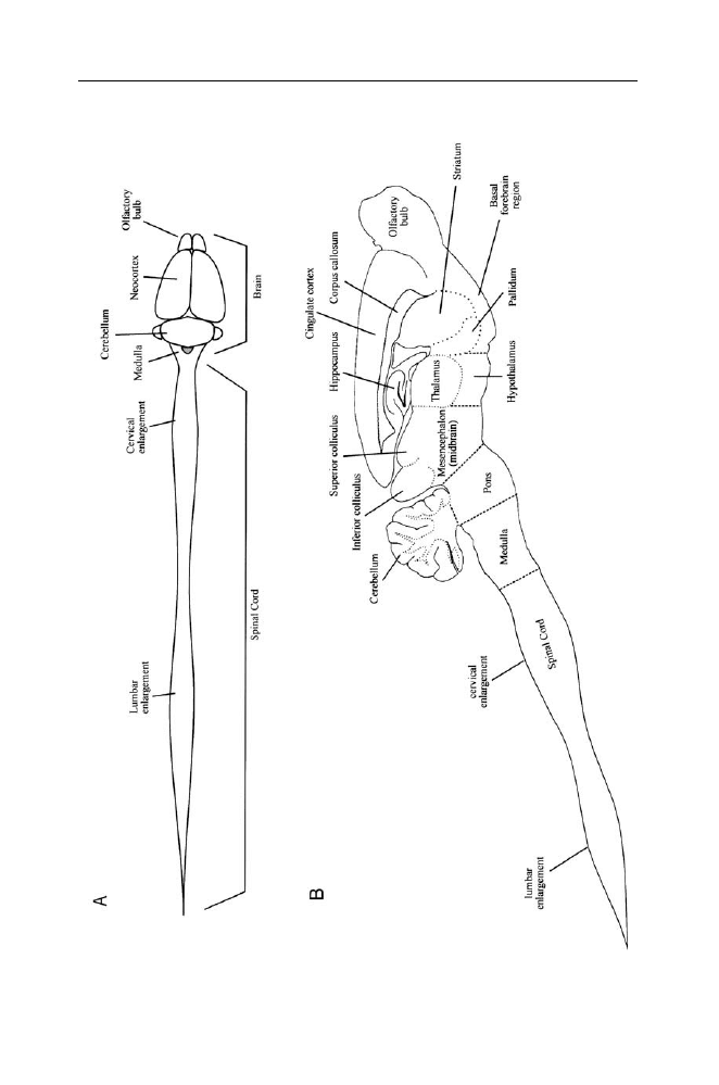
Figure III.1. The central nervous system of the rat.
Top: dorsal view of the brain and
spinal cord. Drawn from a photograph in: Vanderwolf, C.H., and Cooley, R.K. (1990).
The sheep brain: A photographic series, London, Ontario: A.J. Kirby Co. Bottom: A
longitudinal section through the central nervous system has divided it into right and
left parts (parasaggital plane, the cut is somewhat to one side of the midline). Major
subdivisions are outlined by dotted lines. The medulla, pons, and midbrain together
constitute the brain stem; the thalamus and hypothalamus together constitute the
diencephalon; the neocortex, cingulate cortex, hippocampus and pyriform cortex (on
the ventral surface of the brain, not shown here) together constitute the cerebral cortex;
the cerebral cortex, striatum, pallidum, basal forebrain region and diencephalon are
included in the forebrain. The striatum (which includes the caudate nucleus and the
putamen) and the pallidum (also known as the globus pallidus) plus the substantia nigra
(located in the ventral midbrain) are often referred to collectively as the basal ganglia.
The forebrain and brainstem are connected to 12 pairs of cranial nerves which have
various sensory and/or motor functions. The spinal cord is connected to 31 pairs of
spinal nerves; each nerve is attached by a dorsal root containing sensory fibers and a
ventral root containing mainly motor fibers which innervate the muscles. The cervical
enlargement governs the functions of the forelimbs, the lumbar enlargement governs
functions of the hindlimbs, and the narrow intermediate part governs functions of the
thorax. A surgical transection through the thoracic or low cervical levels of the cord
disconnects the lower part from the remainder of the central nervous system, permitting
a study of its behavioral capacities in isolation (spinal animal). A similar transection
dividing the midbrain from the forebrain permits study of the behavioral capacities of
the spinal cord, brain stem and cerebellum in isolation (high decerebrate or midbrain
animal).

22
C. H. Vanderwolf
probably function as components of locomotor behavior in a normal intact
animal. Gentle manipulation of the genitalia readily elicits penile erection and
a forward thrusting of the pelvic region in male spinal dogs and cats. Gentle
mechanical stimulation of the clitoris and the walls of the vaginal orifice in
spinal female dogs and cats elicits contractions of the uterus.
Similar reflex phenomena occur in humans who have had the misfortune of
having the spinal cord severed by an injury (from a bullet wound, for example).
Spinal humans display a flexion reflex and crossed extensor reflex. Penile
erection is readily obtained in most chronic spinal men by gentle rubbing of
the glans and frenulum of the penis. Ejaculation can also be elicited and there
are said to be a number of cases of fatherhood in spinal men. Therefore, some
of the basic reactions involved in defensive, locomotor, body grooming, and
reproductive behavior are organized by neural circuits located in the spinal
cord.
2
When one considers that the mass of the spinal cord constitutes about 14
percent of the central nervous system (brain plus spinal cord) in a dog and only
about 2 percent of the central nervous system in humans,
3
these observations
seem remarkable. How can we interpret the fact that a great deal of behavior is
based on spinal reflexes?
It is essential to consider what the isolated spinal cord cannot do as well
as what it can do. For example, although reflex stepping and other locomotor
reactions can be elicited in chronic spinal animals, true locomotion is not
possible and there is no possibility of complex spontaneous behavior of any
kind. The spinal cord cannot maintain an erect posture of the body (i.e.,
equilibrium cannot be maintained) and the tonus or sustained contractile power
of the anti-gravity muscles may not be sufficient to prevent the body from
sagging slowly to the ground.
Much more complex forms of behavior are possible if the brain stem and
cerebellum are allowed to collaborate with the spinal cord. Thus, if the brain
stem is transected along the line dividing the midbrain from the diencephalon
(thalamus and hypothalamus, Figure III.1), the resulting high decerebrate
animal displays a great variety of reflexive and spontaneous behaviors.
4
It can
move the head about spontaneously or turn the head in response to a sound, and
can walk about spontaneously. High decerebrate rats will also lick or nibble at
objects which are brought in contact with the lips and teeth. They are also quite
capable of swallowing. Nonetheless, despite having most of the reflexive bits
of behavior necessary for feeding, they make no attempt to feed themselves.
Similarly, other complex behavior patterns such as mating behavior, maternal
care, or avoidance of dangerous situations are not present in an effective form.
For example, high decerebrate animals will walk off the edge of a table without
the slightest hesitation. We can conclude that neural circuits in the spinal
cord, medulla, pons, cerebellum and midbrain in rats are capable of generating

The Evolving Brain
23
rather normal looking upright posture, head movement and locomotion but that
nonetheless the normal behavior patterns of feeding, reproduction, avoidance
of danger, etc., are grossly impaired owing to the absence of the forebrain.
What aspect of behavior is missing in these animals? How is it possible that
an animal which, for example, is perfectly capable of biting and swallowing
is, nonetheless, completely incapable of feeding itself? Consider the behavior
of a food-deprived normal rat offered a piece of rat chow at a little distance.
Olfactory and other sensory inputs activate the cerebral cortex which then, in
turn activates brain stem and cerebellar circuits which, in their turn, activate
spinal circuits generating locomotion and head movements which bring the
rat’s snout in contact with the food. The contact stimuli thus produced trigger
mouth opening and biting reflexes which result in ingestion. We know that
mouth opening and biting really are reflexive because surgical section of
sensory nerves from the snout in an otherwise intact rat prevent biting even
though the animal still approaches food or prey and places its snout in contact
with it.
5
A high decerebrate rat possesses reflexive biting and swallowing behavior
but no longer possesses the cerebral mechanisms of the control of locomotion
and head movement which normally guide its behavior. If locomotion and head
movement no longer fulfil their normal function of placing the snout in contact
with food, eating cannot occur.
The study of neuroanatomy has revealed the basic neural circuitry involved
in these behaviors.
6
The spinal cord contains columns of large motor neurons
which send out axons via the ventral roots to the large muscles of the shoulders,
hips, and trunk (proximal musculature). The activity of these spinal motor
neurons is controlled jointly by: (a) sensory inputs from the skin, muscles,
tendons, joints and visceral structures; and (b) descending projections from the
brain. The descending brain projections include: (a) vestibulospinal projections
originating in the vestibular nuclei in the dorsolateral part of the medulla;
(b) reticulospinal projections originating in the reticular formation located in
the ventral and medial parts of the medulla and pons; and (c) tectospinal
projections originating in the superior colliculus (tectum). A fourth pathway,
containing fewer fibers but functionally related to the first three, arises from
the interstitial nucleus of Cajal, located in the midbrain. These descending
projections play an essential role in gross movements such as locomotion and
head movement which depend on the activity of the proximal musculature. This
is shown by several types of findings: (a) neurons in the reticular formation and
other sites fire at high rates in correlation with gross movements; (b) surgical
destruction of neurons in the reticular formation and vestibular nuclei abolish
gross movements such as assuming an upright posture when placed on the back
or side (righting) and locomotion; and (c) electrical stimulation of most sites

24
C. H. Vanderwolf
in the ventromedial medulla and pons in freely moving animals gives rise to
locomotor or other gross movements.
It appears to be the case then, that descending projections from the brain
stem to the spinal cord are the primary means by which the brain is able to
control behavior. However, the behavior which the brain stem, cerebellum, and
spinal cord can generate when acting in isolation is dreadfully maladaptive
and inadequate. An animal lacking its forebrain could not live long without
extensive nursing care. Therefore, the forebrain must exert a decisive control
over the activity of brain stem, cerebellar and spinal circuitry.
In neural terms, what all this means is that gross movements of the head,
trunk, and limbs are controlled by a large number of specific spinal circuits
which, when activated, produce isolated bits of behavior such as alternate
stepping, scratching the body, forward thrusting of the pelvis, etc. Such circuits
are often referred to as “central pattern generators.” Pattern generators may be
activated by a sensory input (producing a reflex) or by descending projections
from the brain. Descending projections from the brainstem appear to activate
combinations of pattern generators to produce a co-ordinated behavior such as
walking forward, turning the head, rolling over, etc. The different brain stem
circuits that produce such items of behavior are under the control of forebrain
structures, especially the cerebral cortex. Thus, depending on such factors as
current sensory input, hormonal or nutritional state, etc., the cerebral cortex
will activate one or another brainstem circuit to produce turning right, turning
left, standing motionless, etc.
If the cerebral cortex (neocortex, cingulate cortex, hippocampal formation,
pyriform lobe) is surgically removed without extensive direct injury to the
diencephalon (thalamus and hypothalamus) or the striatum, rats and other
laboratory animals have a somewhat greater range of behavior than high
decerebrate or midbrain animals. Such preparations display a grossly normal
sleep-waking cycle, running about actively during the night and spending
much of the day asleep, often in a curled-up nose-to-tail posture. Thus, the
main features of sleep-waking behavior are organized subcortically. Further,
unlike high decerebrate animals, decorticate animals are eventually able to
feed themselves if food is easy to obtain. For example decorticate rats will
eat a highly palatable food (lard mixed with brown sugar) if a large dollop
is placed on a flat piece of metal but they are quite defeated if the lard-sugar
mixture is placed in a flat dish with edges raised approximately one centimeter.
7
Similarly if adequate environmental support is provided, decorticate male rats
may copulate successfully and decorticate female rats may succeed in raising a
litter of young.
8
The behavioral state produced by extensive destruction of the
cerebral cortex is widely known as “dementia”.
If one searches the neuroscientific literature with the aim of discovering the
neural basis of specific behaviors, one discovers a good deal of information

The Evolving Brain
25
but it is generally haphazard and unsystematic. Investigators preoccupied with
the search for the neural basis of some hypothesized mental process have rarely
done a thorough job of describing behavior. Studies on the role of the amygdala,
a large cellular complex underlying part of the pyriform lobe, provide an
example.
The amygdala are often said to be related to fear and anxiety. If this were
really true one might expect that surgical removal of the amygdala would de-
crease fear or fearful behavior. However, although amygdalectomized monkeys
are “fearless” in their tendency to approach and investigate objects avoided
by normal monkeys, they are abnormally submissive and fearful in social
situations with other monkeys.
9
Therefore, it cannot be said that “fear”, in a
general sense, is either increased or decreased by destruction of the amygdala.
It is likely that there are a variety of different instinctive or learned sensori-
motor behavior patterns which are altered or abolished by amygdalectomy.
We cannot assume that the behavioral changes will conform to what might
be expected on the basis of conventional psychological ideas. What is required
is a far more detailed and comprehensive study of the actual behavior of brain
damaged and normal animals than has usually been done in the past.
Role of the cerebral cortex in behavior. According to traditional ideas on
the subject, the mammalian neocortex is subdivided into three broad types: (1)
primary sensory cortex which receives an input from one or another of the
sense organs such as the retina, the cochlea, or tactile receptors in the skin;
(2) association cortex lying adjacent to two or more sensory cortices; and (3)
motor cortex, a localized region responsible for cortical or voluntary control
of movement. It is widely held that a sensory input produces a conscious
sensation in the primary sensory cortex but that further elaborations, dependent
on memory and associations with other stimuli, occur in association cortex. If a
decision is made to make a movement, the association cortex can then activate
the motor cortex.
Is this theoretical scheme, based on a psychological tradition originating
with Aristotle and his predecessors, really a valid description of what happens
in the brain?
First, since there is no known way of objectively determining when con-
sciousness is present and when it is not, we really cannot tell whether or not
conscious sensations occur in primary sensory cortex. Second, there is serious
doubt that association cortex actually exists. According to I.T. Diamond, nearly
all of the neocortex is divided into three great fields: an auditory field; a
visual field; and a somesthetic field (Figure III.2). If one includes olfaction and
visceral sensation as well, then the entire cerebral cortex is a target of sensory
input of some kind. The significance of the “motor cortex” will be discussed in
more detail below.
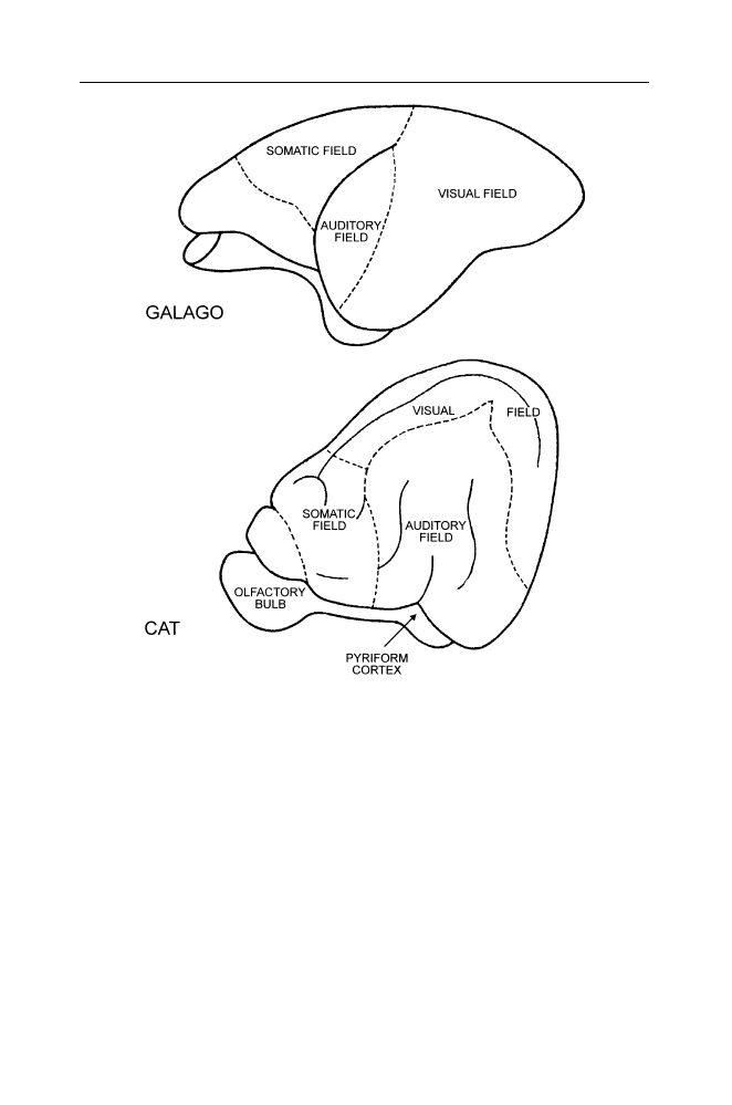
26
C. H. Vanderwolf
Figure III.2. Neocortical sensory fields in the primate and the cat. After Diamond,
I.T. (1985). A history of the study of the cortex: changes in the concept of the
sensory pathway, In: Kimble, G.A., and Schlesinger, K. (editors)
Topics in the history
of psychology. Hillsdale, New Jersey: Lawrence Erlbaum Associates, Inc., pp. 305–387.
The entire neocortex is divided into an auditory field, a somatosensory field and a visual
field. The orbitofrontal area (on the left, above the olfactory bulb), left unlabelled by
Diamond, contains an olfactory field. Olfactory cortex also includes the entire pyriform
lobe at the base of the brain plus the hippocampal formation (shown in Figure III.1).
Since the traditional scheme of the functional organization of the cerebral
cortex is clearly not in agreement with the facts, some other scheme is required.
Where can one begin?
Comparative anatomical and electrophysiological studies have shown that
the cerebral cortex in primitive vertebrate brains is dominated by olfactory
inputs and distributes its outputs to various brain stem structures. In reptiles, for

The Evolving Brain
27
example, although there are many projections from the cerebral cortex to the
brain stem, there appear to be no direct pathways from any part of the cerebral
cortex to the spinal cord.
10
This anatomical arrangement permits the cerebral
cortex to control behavior pattern generators in the brainstem but does not
allow direct cerebral control of circuits in the spinal cord. Thus, when a turtle
or a lizard encounters the odor of food, carrion for example, olfactory inputs
will activate cortical neurons. Descending cortical efferent fibers, directly or
indirectly, then activate brain stem pattern generators for four-legged walking
which, in turn, activate segmental spinal pattern generators to produce the co-
ordinated locomotion needed to bring the reptile to the food. The overall course
and pattern of locomotion will, naturally, be guided not only by olfaction but
also by visual, auditory and tactile stimuli encountered along the route.
In mammals, unlike reptiles, there has been an extensive development of a
new neural structure, the neocortex, which has direct projections to the spinal
cord (corticospinal pathway). This suggests that the mammalian cerebral cortex
can control behavior via two systems of descending projections. An ancient
system, probably present in all vertebrates, is based on direct or indirect cortical
projections to the brainstem. Thus, the hippocampal formation, a prominent
and distinctive part of the mammalian cerebral cortex, has multiple projections
to the diencephalon and the ventral midbrain (tegmentum).
11
The pyriform
cortex, the primary olfactory cortex, also has prominent efferent projections
to the brainstem. Similarly the neocortex has prominent corticoreticular pro-
jections to the brainstem reticular formation, corticotectal projections to the
tectum (superior colliculus) and corticostriatal projections to the striatum
(caudate nucleus and putamen). Further descending fibers from the striatum
can influence tectospinal fibers via a synaptic relay in the substantia nigra in
the midbrain. The corticopontine projection, a massive system of fibers that
includes efferents from all neocortical regions, provides a route by which the
neocortex can influence not only pontine reticulospinal projections but also
the pontocerebellar projections to the cerebellum. These various projections to
the brain stem and cerebellum permit cortical control of pattern generators for
gross motor activities such as locomotion, postural adjustments and orienting
movements of the head.
In addition to this phylogenetically ancient cortical control of gross move-
ment, mammals have evolved an additional system for the performance of dis-
crete movements. This system presumably evolved to allow ancestral mammals
to use the limbs, especially the forelimbs, for non-locomotor uses which require
individual cortical control of a single limb or even of isolated digits. Thus, a
cat can use a forepaw to strike at prey (or bat a ball) while a rodent or a primate
can manipulate small pieces of food, a behavior that is greatly facilitated by
separate control of individual digits. In contrast, reptiles such as the various

28
C. H. Vanderwolf
turtles, lizards, and crocodilians appear to make very little or no use of one
limb or one digit in isolation to manipulate objects.
The anatomical basis of neocortical control of discrete movement consists of
descending projections to ventral horn cells (motor neurons) in the dorsolateral
part of the ventral horn of the spinal cord. These cells activate muscles
controlling the distal parts of the limbs. The descending fibers include: (a)
corticospinal fibers originating in the neocortex; and (b) rubrospinal fibers
originating in the red nucleus in the midbrain. Corticorubral projections permit
cortical control of the rubrospinal pathway. Section of corticospinal fibers in
rhesus monkeys has very little effect on gross motor patterns such as climbing,
walking, running, jumping, turning the head or changing posture. However, in
the immediate post-operative period there is a marked inability to move a limb
in isolation, making it difficult or impossible to reach out, grasp, and lift a piece
of food while sitting immobile. Although there is some recovery of reaching,
grasping, and lifting with the passage of time, independent movements of the
digits never recover. Thus, monkeys sustaining an extensive interruption of
corticospinal fibers can make a grasping movement by flexing all the fingers
together but cannot extend the index finger and thumb to make a precision
grip (for example, to pick up a raisin or peanut) while keeping the other
fingers flexed. It appears that the corticospinal system (probably assisted by the
rubrospinal system) is involved in discrete cortical control of restricted parts of
the body.
Experiments by Leyton and Sherrington early in the twentieth century
showed that in deeply anesthetized chimpanzees, gorillas, and orang-utans,
movements could be elicited by localized electrical stimulation only in re-
stricted regions of the neocortex. Movements of the limbs and body could
be elicited from the precentral gyrus in the frontal lobe but movements of
the eyes or eye-lids could also be elicited from a more rostral region in the
frontal lobe and from a part of the occipital lobe. This experiment, and others
using similar methods, were very influential in establishing the concept that
there is a localized motor cortex in the frontal region of the neocortex. Later
experimenters who used unanesthetized or more lightly anesthetized animals
have found that movements of one kind or another can be elicited by localized
electrical stimulation of virtually all regions of the neocortex.
12
The differing
results are probably due to the use or non-use of anesthetic drugs which have
the effect of depressing synaptic transmission. The precentral gyrus, which has
strong access to spinal motoneurons over a pathway involving only one or two
synapses would be relatively unaffected by anesthetic but other cortical regions,
the occipital lobe, for example, which has access to spinal motor neurons
only over multi-synaptic output pathways, involving the striatum (corticostriate
fibers), the tectum (corticotectal fibers), or the reticular formation (corticoretic-
ular fibers) would be more strongly affected by anesthetics. It appears, therefore

The Evolving Brain
29
that “motor” functions are not restricted to a small precentral cortical region but
are instead located throughout the neocortex. It is helpful to think about this in
anatomical terms. The neocortex consists of several distinctive cell layers. The
more superficial layers which contain large numbers of small pyramidal cells or
granule cells receive the main sensory inputs from the thalamus while the deep
layers contain larger pyramidal cells that send axons outside the neocortex. The
large pyramidal cells of layer V, in particular, appear to be responsible for all
neocortical projections to subcortical structures other than the thalamus. All
neocortical areas, then, have a sensory or input zone, interneurons, and a set of
efferent or motor cells. The “visual” neocortical area is really a “visuomotor”
area, the “auditory” neocortical area is really an “audiomotor” area, and so on.
Similarly, in the hippocampal formation, the dentate granule cells are the main
target of the olfactory inputs to this structure while the large pyramidal cells of
Ammon’s horn provide an efferent output. Therefore, the entire cerebral cortex
can be thought of as having both sensory and motor functions but the traditional
“sensori-motor” areas of the neocortex have a particular role in the performance
of discrete movements.
The differing roles of the different neocortical areas are revealed quite
clearly when they are removed. Experimental removal of the sensori-motor
cortex in the rat has very little effect on gross movements such as walking
or turning the head but produces severe impairments in the ability to reach
for food, pick it up and manipulate it with one paw. On the other hand
large neocortical lesions in any part of the neocortex produce clear deficits in
directed locomotion, as is required for example in such behaviors as hoarding
food, running through a maze or following and mounting a sexual partner.
Although behavioral deficits of this type are often given complex psychological
interpretations, they can be viewed more simply as indicative of a defect in the
cortical control of locomotion which is normally exerted via sensory control of
corticostriate, corticotectal, or corticoreticular projections. For example, cats in
which the auditory cortex has been surgically removed may be quite capable
of turning the head accurately toward a sound (probably a reflexive behavior
involving the midbrain tectum) but cannot walk accurately toward a sound.
13
From a functional point of view, it is interesting that the major source of
corticospinal projections lies in a region of cortex that receives somesthetic and
proprioceptive inputs. There is a very close relation between sensory input and
motor output. A cortical motor neuron which produces adduction of the thumb
when it is electrically stimulated receives an input from skin on the medial
surface of the thumb; a different cortical neuron that produces flexion of the
thumb when it is stimulated receives an input from skin on the palmar surface of
the thumb.
14
Thus, a cortical motor neuron controlling a particular movement
receives input from a skin area which is likely to receive stimulation as a result
of that movement. It is likely that this arrangement evolved because of the

30
C. H. Vanderwolf
advantages of close tactile or proprioceptive control of delicate manipulatory
movements.
Other sense modalities such as vision, audition, or olfaction are not so
closely related to discrete movements probably because these sense modalities
are usually involved in the control of gross motor activities such as approaching
food, fleeing a predator etc. However, mechanisms for close visual control of
discrete motor activity have evolved in some animals, especially primates, but
also in cats which use a forelimb to strike at visually located prey. Tract-cutting
experiments have indicated that this kind of visuomotor control is exerted by
descending pathways from visual cortex to some subcortical structure which
then activates the classical motor areas via a second ascending pathway.
15
The
details of these pathways remain to be discovered.
What does the sensory cortex do? The conventional answer to this question
is that sensory cortex constructs an internal representation of the outside world.
There is, however, a logical difficulty in this. If the eye, for example, forms an
image of the outside world and the visual cortex constructs another image based
on information provided by the eye, what is it that looks at the cortical image?
It is evident that the conventional theory is based on an implicit assumption of
a mind or psyche, distinct from the brain, which can view representations of
the outside world that form in the sensory cortices.
16
This is the theory of the
imprisoned knower whose only contact with the outside world is provided by a
bank of television screens. Is there any conceivable alternative to this theory?
Sensory neocortex receives two general types of input from subcortical
structures. The thalamus provides a massive, finely differentiated input which
permits cortical cells to respond selectively to detailed features of sensory
stimuli. Visual cortical cells may respond selectively to lines or edges or more
complex stimuli such as a human or animal face. Cells in auditory cortex may
respond selectively to pure tones of specific frequencies or to vocalizations
of conspecifics. In addition to these specific inputs, the entire neocortex and
hippocampal formation receive a sparser but very widespread input from cells
in the basal forebrain region and from cells in the brainstem which make use
of the neurotransmitters acetylcholine and serotonin, respectively. If either the
specific or widespread classes of input is blocked, the cortex cannot function
and the experimental animal becomes grossly demented, behaving in much the
same way as animals in which the entire cerebral cortex has been surgically
removed.
17
Thus, both large thalamic lesions and selective blockade of choliner-
gic (acetylcholine-dependent) plus serotonergic neurotransmission produces
a state of general dementia. It is likely that the cholinergic and serotonergic
projections fulfil a general regulatory role with respect to cortical neural
activity while the thalamic projections provide a much more detailed and
specific input.

The Evolving Brain
31
How, in general terms, does the whole thing work? There is reason to think
that the entity or process known as “perception” is fundamentally an activation
of motor programs. Someone looking at a complex figure in which a smaller
figure is concealed, may not detect the hidden figure at first. When the figure
is finally identified, the observer is immediately capable of doing a number of
things that were impossible a moment before. The hidden figure can be pointed
out, described verbally, or illustrated in a sketch.
18
As J.J. Gibson
19
pointed
out, perception of something is knowing what one can do with it or what one
can do with respect to it. Gibson invented the term “affordance” to describe
the situation. Thus, water affords drinking, swimming, or boating, a small rock
affords throwing, hammering, weighing down papers, etc. A thing is perceived
when motor programs relevant to it have been activated. One can imagine that
viewing a complex visual scene sets up patterns of activity in visual cortex
which have an output to such structures as the striatum, the thalamus, the
tectum and the reticular formation. This normally results in behavior which is
adapted to the perceived environment but the details of how it is accomplished
are, as yet, almost completely unknown.
Notes
1. Sherrington, C.S. (1906). The integrative action of the nervous system, New Haven: Yale
University Press.
Forssberg, H. (1979). On the integrative motor functions in the cat’s spinal cord. Acta
Physiologica Scandinavica (Supplementum 474), 1–56.
2. Fulton, J.F. (1949). Physiology of the nervous system, 3
rd
edition, New York: Oxford
University Press.
3. Krompecher, S. and Lipak, J. (1966). A simple method for determining cerebralization brain
weight, and intelligence. Journal of Comparative Neurology, 127: 113–120.
4. Woods, J.W. (1964). Behavior of chronic decerebrate rats. Journal of Neurophysiology, 27:
635–644.
Lovick, T.A. (1972). The behavioural repertoire of precollicular decerebrate rats. Journal of
Physiology (London), 226: 4–6p.
5. Gregoire, S.E. and Smith, D.E. (1975). Mouse-killing in the rat: Effects of sensory deficits
on attack behaviour and stereotyped biting. Animal Behaviour, 23: 186–191.
6. Kuypers, H.G.J.M. (1982). A new look at the organization of the motor system. In: H.G.J.M.
Kuypers and G.F. Martin (eds). Anatomy of descending pathways to the spinal cord.
Progress in Brain Research, 57: 381–403.
7. Personal observations.
8. Whishaw, I.Q. (1990). The decorticate rat. In: Kolb, B., and Tees, R.C. (eds) The cerebral
cortex of the rat, Cambridge, Massachusetts: The MIT Press, pp. 239–267.
9. Horel, J.A., Keating, E.G., and Misantone, L.J. (1975). Partial Klüver-Bucy syndrome
produced by destroying temporal neocortex or amygdala, Brain Research, 94: 347–359.
10. Ten Donkelaar, H.J. (1982). Organization of descending pathways to the spinal cord in
amphibians and reptiles. In: H.G.J.M. Kuypers and G.F. Martin (eds) Anatomy of descending
pathways to the spinal cord. Progress in Brain Research, 57: 25–67.

32
C. H. Vanderwolf
Belekhova, M.G. (1979). Neurophysiology of the forebrain. In: C. Gans, R.G. Northcutt,
and P. Ulinski (eds.) Biology of the reptilia, vol. 10, Neurology B, London: Academic Press,
pp. 287–359.
11. Vanderwolf, C.H. (2001). The hippocampus as an olfacto-motor mechanism: were the
classical anatomists right after all? Behavioural Brain Research, 127: 25–47.
12. Lilly, J.C. (1958). Correlations between neurophysiological activity in the cortex and
short-term behaviour in the monkey. In: Harlow, H.F., and Woolsey, C.N., Biological and
biochemical bases of behaviour, Madison: The University of Wisconsin Press, pp. 83–100.
Neafsey, E.J. (1990). The complete ratunculus: output organization of layer V of the
cerebral cortex. In: Kolb, B., and Tees, R.C. The cerebral cortex of the rat, Cambridge,
Massachusetts: The MIT Press, pp. 197–212.
13. Heffner, H.H., and Masterton, R.B. (1975). Contributions of auditory cortex to sound
localization in the monkey. Journal of Neurophysiology, 38: 1340–1358.
14. Rosen, I., and Asanuma, H. (1972). Peripheral afferent inputs to the forelimb area of the
monkey motor cortex: Input-output relations. Experimental Brain Research, 14: 257-273.
15. Myers, R.E., Sperry, R.W., and McCurdy, N.M. (1962). Neural mechanisms of visual
guidance of limb movement. Archives of Neurology, 7: 195–202.
Penfield, W. (1954). Mechanisms of voluntary movement. Brain, 77: 1–17.
16. Stent, G.S. (1975). Limits to the scientific understanding of man: human sciences face an
impasse since their central concept of the self is transcendental. Science, 187: 1052–1057.
17. Vanderwolf, C.H. (2003). An odyssey through the brain, behavior, and the mind. Boston:
Kluwer Academic Publishers.
18. Sperry, R.W. (1952). Neurology and the mind-brain problem. American Scientist, 40: 291–
312.
19. Gibson, J.J. (1979). The ecological approach to visual perception, Boston: Houghton Mifflin
Company.

IV. Human origins and adaptations
Before one can begin to think seriously about the human brain and behavior
in a general sense one must consider some fundamental questions. What are the
basic characteristics of human behavior and how does human behavior compare
with the behavior of other animals? An intelligent alien from a remote galaxy,
observing modern humans for the first time, might well conclude that these
creatures are, in the main, adapted for a crowded social life in an environment
consisting largely of concrete, asphalt, glass and steel and that they feed
largely on manufactured foods provided in plastic containers. More prolonged
investigation, however would reveal that the urban environment now inhabited
by many people is a very recent development and that throughout more than
ninety-nine percent of its history humankind lived in small social groups of
no more than a few dozen individuals subsisting on plant and animal products
obtained in their natural state. Consequently, the natural condition for human
beings is life as it was during the long paleolithic era prior to the development
of agriculture. This must be the condition to which human behavior and the
function of the human body are most closely adapted.
A rational understanding of the history of humankind has been achieved
only in the last century or so. Traditional Christianity teaches that all of
humanity descended from an original pair, Adam and Eve, specially created
by God only a few thousand years ago. In opposition to this, Charles Darwin
proposed in 1871 that mankind, over an immense period, descended from
ape-like creatures and that our closest living relatives are the chimpanzee and
gorilla. Since chimpanzees and gorillas are native to Africa, this suggested to
Darwin that the human species probably originated there.
1
Although there was
little direct evidence to support this idea in 1871, the subsequent discovery of
numerous fossilized remains of ancient humans and of human-like creatures
has strongly confirmed Darwin’s hypotheses.
2
In 1925 Raymond Dart, a teacher of anatomy, described the fossilized skull
and lower jaw of a child, with characteristics intermediate to those of apes and
humans, which had been found in a limestone mine near Taung in South Africa.
This, and similar fossils subsequently discovered elsewhere in South and East
Africa (Ethiopia, Kenya, Tanzania), have been shown to range from about 2.5–
5.0 million years old. The creatures whose remains had been preserved in this

34
C. H. Vanderwolf
way have become known as austrolopithecines after the name suggested by
Dart for the Taung child (Australopithecus africanus). Most australopithecines
were rather small creatures, probably about 100–150 cm in height and 30–
60 kg in weight. The size of the canines and the prognathism, or forward
projection of the lower face to form a muzzle, were reduced in comparison to
apes or monkeys. The brain volume was about 400–519 cc which is comparable
to present-day gorillas (about 425 cc) and chimpanzees (about 320–336 cc).
Modern human brains usually range from 1300–1460 cc.
3
Despite the rather
small brain of the australopithecines, there is clear evidence from the structure
of the skull, the forelimbs, the pelvis, and the foot that they walked erect
and bipedally, much as we do. This anatomical conjecture was dramatically
confirmed by the discovery of a series of footprints made by three hominids
walking in a fresh ash fall from a nearby volcano (Sadiman, near Laetoli,
in Tanzania) that occurred between 3.49 and 3.76 million years ago.
4
The
footprints closely resemble those one can see in the sand of any present-day
beach where modern humans congregate.
5
This is an important finding because
it demonstrates that obligatory bipedalism, one of the distinctive characteristics
of humankind, developed long before the evolution of a large brain, another
distinctive human characteristic.
Paleoanthropologists have suggested the former existence of a variety
of different species of australopithecines including a rather fine-boned or
gracile type (A. afarensis, A. africanus, A. anamensis) and a heavier “robust’
type (A. boisei, A. robustus, Zinjanthropus boisei, Paranthropus sp., and A.
aethiopicus). It appears to be true that different species of australopithecines
with different habits and different diets were alive at the same time in the same
general region of Africa.
In more recent geological strata, deposited about 2 million years ago,
australopithecine fossils are replaced by hominid fossils presenting evidence
of a larger brain, smaller teeth and a hand that must have been capable of a
good thumb-against-finger tip grip (precision grip). An early group of fossils
of this type, referred to as Homo habilis or Homo rudolfensis was apparently
replaced by taller, bigger brained creatures known as Homo ergaster, Homo
erectus and Homo sapiens, the latter being similar to present day humans.
Some paleoanthropologists consider that Homo habilis and Homo rudolfensis
are more appropriately referred to as Australopithecus habilis and that later
species of Homo should all be referred to as Homo sapiens. The rather confused
state of the terminology in this field will, no doubt, be resolved when a much
larger number of hominid fossils have been discovered. For the moment, it
appears that we have an ancient australopithecine group which was gradually
replaced by a Homo group which was taller, heavier, had smaller teeth and jaws,
relatively longer legs and shorter arms, a flatter face, and a much larger brain.
Homo (or Australopithecus) habilis had a brain volume of about 640 cc. while

The Evolving Brain
35
Homo erectus (also known as early Homo sapiens) had a brain volume of 895–
930 cc.
3
This is at least twice the size of the brain in any living non-human
primate but is still substantially smaller than the brain in modern humans.
Humans with a fully modern anatomy appeared in roughly the last 100,000–
250,000 years.
Hominid fossils begin to appear outside Africa, as far a field as Indonesia,
as early as 1.5–2.0 million years ago. Evidence of the presence of humans does
not appear in Europe north of the Pyrenees earlier than about 700,000 years
ago. Neanderthal man, a unique type with a very robust skeleton, prominent
brow-ridges, a noticeable occipital protuberance or “bun”, and a receding
forehead but with a brain fully as large as modern humans, appears to have
been indigenous to Europe and the Middle East. Australia was colonized as
early as 115,000 years ago but humans seem to have arrived in the Americas
only 10–20,000 years ago.
One of the outstanding characteristics of humans as compared to other
animals is a strong tendency to make and use tools. It is true that a variety
of animals use simple tools but never to the extent that humans do. Wild
chimpanzees, for example, will break nuts by placing them on a stone and
hammering them with another stone. They also strip the leaves from a stem
of grass or a twig to make a tool which can be inserted into ant or termite
nests. When the insects attack this “fishing pole”, the chimpanzee pulls it out
and licks them off, or having stripped them off with one hand, licks them up
from the hand. Chimpanzees and other apes have never been observed to make
tools from stone or other hard materials such as bone, but early mankind did so
regularly.
6
When human ancestors first began to make tools from organic materials
such as sticks or grass is unknown since such artifacts are rarely preserved as
fossils. The earliest known stone tools have been excavated in Olduvai Gorge
in Tanzania in geological strata as much as 2.4 million years old. The artisans
of this Oldowan Industry, as it has become known, made edged tools by flaking
pieces from pebbles or small stones. The edge was produced by hammering
flakes from both sides of a stone so that the two exposed faces met at an acute
angle. Rocks broken by natural processes generally do not have this bifacial
appearance. Furthermore, microscopic study of the sharp edges of Oldowan
tools reveal a pattern of wear that can be duplicated in modern experimentally-
produced stone tools by such activities as scraping wood or skin, cutting meat
and so forth. Different types of use produce recognizably different types of
wear.
Gradually the making of stone tools became more systematic. A type
referred to as Acheulean tools comprise large numbers of bifacially flaked
pear-shaped objects that have a sharp point. These are generally referred to
as “hand-axes”, under the supposition that they were held in the hand, without

36
C. H. Vanderwolf
an attached handle, and used to chop or cut a variety of materials. Although
they were first discovered at Saint-Acheul in France in 1854, their earliest
known appearance in the geological record is in Ethiopia about 1.4 million
years ago. They continued to be used without significant change for a million
years or more. The appearance of Acheulian hand-axes was associated (very
roughly speaking) with other technological advances. Cutting tools began to be
resharpened when they had become dull by use. Specialized tools of various
types began to appear. Evidence that fire was used deliberately by ancient man
appears as early as 1.6 million years ago in Koobi Fora in Kenya.
The human use of tools is often related to the evolutionary development
of the human hand. In the words of two eminent authorities on this topic,
J.R. Napier and P.H. Napier, “Through natural selection, the opposition of the
thumb prompted the adaptation of the upright posture and bipedal walking,
tool-using and tool-making which, in turn, led to an enlargement of the brain”.
7
Is this likely to be true? The question is somewhat more complex than it
first appears. The development of a peripheral anatomy that permits good
opposition of the thumb to the fingers is of little consequence unless there
is a prior or concomitant development of brain circuits permitting individual
control of the thumb and fingers in a variety of different patterns. Which is the
more important and which is likely to have evolved first, the brain circuits or
the peripheral anatomy of the limb?
An answer to this question is suggested by the clinical syndromes of
phocomelia and amelia which became relatively common for a time following
the introduction of the drug thalidomide nearly 50 years ago. Thalidomide
is a hypnotic and sedative drug with an extremely wide safety margin (the
difference between a hypnotic dose and a lethal dose) and very few side effects
in human adults. It was released for unrestricted use in West Germany in 1958
and in other countries soon afterwards. This precipitated an individual, medical,
and social disaster. Many women who took thalidomide during pregnancy
subsequently gave birth to children with no limbs at all (amelia) or deformed
limbs somewhat resembling seal flippers (phocomelia). Cases of particular
interest to students of human evolution are those in which the upper limbs
are grossly deformed or absent while the lower limbs are normal. Other body
structures are generally normal. Phocomelic children with no thumbs or index
fingers at all, and the remaining digits weak, deformed, or partially fused
together, could, nonetheless, use their upper limbs to eat and write and often
did well in school. Children with no upper limbs at all learned to use their feet
to feed themselves using cups, spoons, etc., to button or unbutton clothing,
remove clothing etc. It is apparent that in such cases “functional disability
cannot be accurately deduced from knowledge of the structural defect”.
8
The thalidomide children demonstrate that a modern human central nervous
system can generate a great deal of skilled tool use even when it is housed

The Evolving Brain
37
in a grossly defective body. If a chimera were created with a chimpanzee
body and a human nervous system there can be little doubt that its behavior
would be entirely human. These observations are consistent with the hypothesis
that effective tool use and the brain circuits underlying tool use evolved long
before the appearance of the modern human hand. Natural selection favoring
individuals who were particularly adept in the manufacture and use of tools
would then slowly transform an ape-like hand into a human hand.
Most living animals make little or no use of tools. Why was early man an
exception? A possible answer to this question is suggested by two evolutionary
principles with wide applicability. First, the diet of an animal, the way it makes
its living, has an immense impact on its anatomy, physiology and behavior.
Thus, the obvious differences between a deer and a wolf in terms of teeth,
bones, guts, etc., as well as behavior are, to a great extent, explicable in terms
of the diet to which these animals are adapted. Second, natural selection can
usually modify behavior more rapidly than gross anatomical structures. One
example of this is provided by the behavior of the woodpecker-finch on the
Galapagos Islands west of Ecuador. There are no true woodpeckers on the
Galapagos Islands. However, the woodpecker-finch, a small sparrow-like bird,
lacks the unique anatomical adaptations of woodpeckers, including a long
tongue capable of removing insects from deep narrow cavities in wood or bark.
Consequently, it has acquired the behavior of rooting insects out of holes by
means of a cactus thorn or twig held in the beak.
9
Similarly, the shrikes, song
birds which have secondarily adopted a predatory and carnivorous life style,
have had to make do with a peripheral anatomy that is not well adapted to
the life of a bird of prey. Hawks, eagles and owls have powerful clawed feet
with which they can seize and kill other animals by driving the claws into
the body. The dead prey can then be dismembered by holding it in the feet
and tearing off pieces with the bill. Shrikes, having only the relatively weak
feet of a typical songbird, kill their prey (insects, small reptiles, birds, and
rodents) by pecking at the head. The body of the prey is then pulled apart
with the bill after impaling it on a thorny tree or a barbed wire fence.
10
In these
cases, tool-using behavior seems to have evolved to compensate for a lack of
appropriate peripheral anatomical structures. It is possible that in humans, as
in woodpecker-finches and shrikes, tool use was developed in association with
a change in the diet. Thus, in place of the teeth and claws of typical predators,
humans evolved the brain circuits necessary to produce and use a variety of
tools for the killing, dismemberment, and transport of prey animals.
Primates, for the most part, are herbivorous animals. Some, like the gorilla,
subsist primarily on leaves, shoots, and stems. Chimpanzees eat mainly fruit,
supplemented by leaves and other plant foods. Although chimpanzees are
active hunters (see below), meat constitutes less than 10 percent of their diet.
Humans in traditional hunter-gatherer societies, as well as in modern affluent

38
C. H. Vanderwolf
societies, typically eat far more meat than this. Animal meat and fat are
preferred foods in virtually all human societies.
11
Even when the diet is largely
vegetarian, meat is likely to be eaten on festive occasions such as weddings.
In the most extreme case of human carnivory, the traditional diet of the
Inuit or Eskimo people consisted of nothing other than animal tissue and
water. The effects of such a diet become known to science as a result of the
observations of Vilhjalmur Stefansson, a Canadian arctic explorer in the early
twentieth century.
12
Stefansson lived for some time, in the period 1908–1912,
with the Copper Eskimos, a people living north of Great Bear Lake in the
North-West Territories who had never seen a white man before he arrived.
The Copper Eskimo ate fish and killed caribou, feeding their dogs on the
guts, liver, heart and tenderloin, and reserving most of the fat, marrow, and
other muscle tissue for themselves. They were well aware that eating nothing
but lean meat produces an illness characterized by weakness, head-ache and
diarrhea. This condition, well-known in the Canadian North, was popularly
called “rabbit starvation” because it inevitably put in an appearance if people
were forced to live on a diet of snow-shoe hares (Lepus americanus) which
have extremely little body fat in the winter. Inuit living entirely on a diet
of lean meat and fat maintain a state of vigorous health with no deficiency
symptoms of any kind and no evidence of unusual cardiovascular disease. It
was at first suspected that the Inuit had some sort of special genetic adaptation
to a totally carnivorous diet but this was disproved in an experiment in which
Stefansson and Karsten Andersen, another arctic explorer, spent a year living
under medical supervision on a diet of lean meat (left rare according to Inuit
traditional practice which avoids destruction of vitamin C and other nutrients)
and fat plus water, black coffee and tea (without milk, cream, or sugar). Both
men remained in good health except that Andersen had a bout of pneumonia
from which he recovered successfully. Prior to this test, Stefansson had lived
in good health for a total of about 9 years on a diet of fat meat and water.
It is not widely understood today that a diet consisting of animal tissue and
water provides all the essential nutrients required by humans. The content of
the traditional Eskimo daily diet has been estimated as: carbohydrates, 10 gms;
fat, 185 gms; and protein, 200 gms. In energetic terms, fat provides about 66
percent of the caloric value of such a diet while carbohydrates provide only 2
percent.
To most modern urbanized people, a diet of fat meat and water seems rather
odd but throughout much of human prehistory prior to the development of
agriculture, it would have been common in the temperate, boreal, and arctic
regions of the world simply because there is very little else to eat for much of
the year. Certainly, in early Canada both the aboriginal people and the European
fur traders lived almost entirely on fish and game, either fresh or in the form
of pemmican (the flesh of deer or bison cut in thin strips, dried in the sun,

The Evolving Brain
39
pulverized, thoroughly mixed with fat and stored in a bag made from the dried
skin of the slaughtered animal).
13
It is noteworthy that the Pleistocene epoch (about 2,500,000 to 10,000 years
ago) was characterized by a great abundance of large mammals and a small
widely scattered human population. In contrast to present day hunter-gatherers,
who have in many cases been driven into rather marginal environments, early
humans lived in a hunter’s paradise. Animal tissue, the preferred food of
humans, would have been readily available. There is reason to suspect that the
invention of tool-assisted predatory behavior by humans was overwhelmingly
successful. There appears to be a correlation between the appearance of humans
in various parts of the world and the extinction of many prey species during
the late Pleistocene.
14
Numerous species of large marsupials had become
extinct in Australia by about 30,000 years ago, but in North America, large
land mammals (such as mammoths, ground sloths, native horses and camels,
and giant beavers) became extinct only 8–12,000 years ago. In New Zealand,
large flightless birds such as the moa became extinct only in the last 1,000
years or so. In each case, these extinctions coincide very roughly with the
arrival of Homo sapiens in the region in question. One may wonder whether
the extensive modern human consumption of vegetable products and the
development of agriculture about 10,000 years ago may have occurred in
response to increasingly poor hunting. Subsequently, various religious or quasi-
religious vegetarian doctrines may have arisen in an attempt to present an
unwelcome necessity as a virtue.
No one knows exactly when our ancestors first acquired a strong taste for
meat. Among our closest primate relatives, gorillas, orangutans and pygmy
chimpanzees (bonobos) eat virtually no animal tissues of any kind but the
common chimpanzee does include some meat in its diet. It is possible that
the common ancestor of humans and chimpanzees ate significant amounts of
meat or, alternatively, hunting behavior and meat-eating may have evolved
independently in humans and chimpanzees. What is certain is that humans
have been hunting and/or scavenging and eating meat for a very long time. An
archaeological study of a tool-and-fossil-rich site with an age of 1.76 to 1.86
million years in Olduvai gorge, Tanzania, provides abundant evidence that early
Homo had ready access to meat-rich carcasses of various African antelopes and
other animals.
15
Carnivores such as lions or hyenas remove meat and marrow
from bones by gnawing on them. Their teeth leave U-shaped grooves and round
puncture holes. Early man cut meat from bones with stone tools that left V-
shaped cuts and also smashed bones with hammer stones to extract marrow.
Thus, the feeding activities of humans and conventional carnivores can be
distinguished by careful microscopic study of the bones and bone fragments left
behind. Further, U-shaped grooves superimposed on V-shaped grooves indicate
that humans were often the primary predators in Tanzania nearly 2 million

40
C. H. Vanderwolf
years ago, and that carnivores subsequently scavenged carcasses discarded by
humans.
Similar work carried out in the Middle Awash Valley in Ethiopia indicates
that early human ancestors were cutting flesh from bones and using hammer-
stones to break marrow bones from prey species that included antelopes of
various kinds, pig-like animals, and ancient three-toed horses as early as 2.5
million years ago.
16
It may be that the early development of the use of tools as
an aid to carnivory played a major role in the evolution of the reduced dentition
and elaborate manipulatory abilities which are characteristic of humans.
Since most primates are largely herbivorous, it seems probable that the
ancient common ancestor of humans and the living apes was also herbivo-
rous. Since herbivores and carnivores display characteristic differences in the
structure and function of the gastrointestinal tract, it would seem likely that
the human gut would have gradually evolved from a herbivore-like pattern to
a more carnivore-like pattern. Figure IV.1 illustrates some typical mammalian
anatomical patterns.
17
Digestion in a typical carnivore such as the dog is largely
dependent on enzymes that are released into the stomach and small intestine.
Proteins are decomposed into amino acids, fats are decomposed into fatty acids.
These products are then absorbed, mainly in the small intestine, distributed in
the blood stream and used to provide energy or to synthesize new proteins
and fats in various body tissues. Glucose, the major metabolic fuel of nervous
tissue, is provided by the breakdown of glycogen, a polymer of glucose found
in muscle and liver, and by synthesis (gluconeogenesis) from certain amino
acids (alanine, aspartic acid, glutamic acid).
Digestion in herbivorous animals differs markedly from this pattern. Some
of the major components of plants such as cellulose, hemicelluloses, and pectin
cannot be hydrolyzed by any of the digestive enzymes that mammals can
produce. However since some bacteria are able to produce enzymes that will
decompose these giant organic molecules, herbivorous animals have evolved
a symbiotic relation with them. Ruminant animals such as cattle or sheep
have a greatly enlarged stomach consisting of four chambers: the reticulum,
the rumen, the omasum, and the abomasum. The abomasum corresponds to
the simple stomach of a carnivore: it begins the hydrolysis of protein by the
enzyme pepsin in a strongly acidic environment. The other three chambers
constitute a large fermentation tank in which a culture consisting of masticated
plant material thoroughly mixed with saliva is kept at a high and constant
temperature to favor the growth of many trillions of bacteria plus protozoa
which feed on the bacteria. These micro-organisms synthesize proteins which
can be digested in the abomasum and small intestine. Thus, ruminants actually
live, not on plants themselves, but on a sort of yogurt-like material containing
the bodies and metabolic products of vast numbers of bacteria and protozoa
which they house in their stomach.
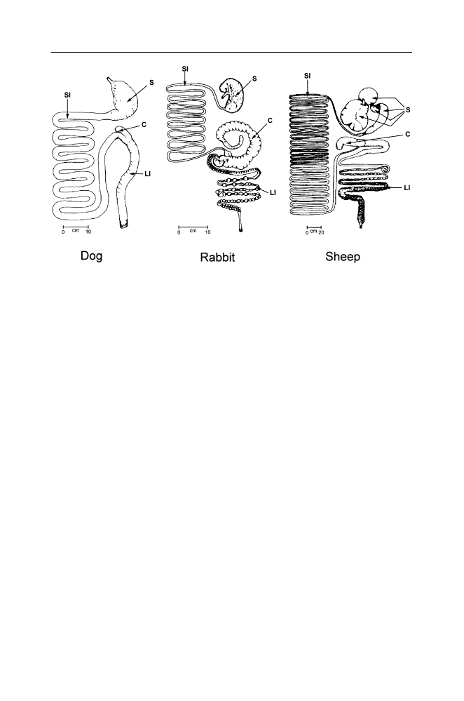
The Evolving Brain
41
Figure IV.1. Sketches of the gross morphology of the gastrointestinal system in the
dog, rabbit and sheep (redrawn from Swenson, M.J. & Reece, W.O. (eds)
Dukes’
physiology of domestic animals, 11th ed., Ithaca, N.Y. Cornell University Press, 1993).
In all mammals the surface area of the small intestine is related closely to body size
regardless of the customary diet.
18
Carnivora, such as dogs, usually have a small
stomach, a small cecum, and small large intestine but in herbivores either the stomach
is greatly enlarged, as in the sheep and other ruminants, or the large intestine and
cecum are greatly enlarged, as in the rabbit. Since fermentation in the large intestine
and cecum is a less efficient means of obtaining nutrition (absorption from the large
intestine is rather poor) rabbits have adopted the habit of eating a part of their own feces
in order to obtain additional nutrients on a second pass through the small intestine. The
fecal pellets which are eaten differ from those that are excreted in the ordinary way
in that they are soft, have a protective membrane, are specially formed in the cecum,
and are eaten as they emerge from the anus. The eating of feces (coprophagia) occurs
in many herbivorous mammals. For example, elephant calves eat large quantities of
fresh adult elephant dung, a behavior that allows them to acquire the proper set of
intestinal bacteria. These facts illustrate an important principle of evolution and natural
history: Mother Nature has no shame. C, cecum; LI, large intestine; S, stomach; SI,
small intestine.
In non-ruminant herbivores such as rabbits, horses, many rodents and some
primates, the cecum and/or large intestine are greatly enlarged to provide a
microbial fermentation chamber while the stomach has remained relatively
small. Colobus monkeys and gorillas are examples of this type. Many primates
eat a good deal of fruit and have a rather unspecialized type of gastrointestinal
tract, but humans and cebus (capuchin) monkeys are unusual since they have a
small stomach, a small cecum and a small large intestine. This is a pattern that
approximates the one observed in most carnivores.
18
Since capuchin monkeys

42
C. H. Vanderwolf
eat a great deal of animal matter (insects, small lizards, birds, etc) they, like
humans, appear to have evolved a carnivore-like gastrointestinal system.
The evolution from a herbivore-type of gut to a more carnivore-type of gut
in humans presumably required a rather long period of time. This agrees with
archaeological evidence indicating that the ancestors of modern humans began
eating significant amounts of meat a very long time ago. There is a third line
of evidence that provides further support for this – the natural history of the
tapeworm.
Tapeworms are parasitic flatworms which, when adult, live in the intestine
of meat-eating mammals (the definitive host). An adult tapeworm has a head or
scolex equipped with hooks and suckers to cling to the mucosa of the intestine.
Behind the head is a neck which buds off a succession of reproductive units or
proglottids. This results in a long segmented ribbon-like body structure which
can grow to as much as several meters in length. Each proglottid contains both
male and female sexual organs so that tapeworms can fertilize either themselves
or a different individual. Ripe proglottids, filled with fertilized eggs, detach
themselves from the tapeworm, are passed out with the feces, and disintegrate,
releasing the eggs on the ground, grass, etc. If the eggs are then swallowed
by a herbivorous animal of an appropriate species (the intermediate host) the
eggs develop into larvae in the host’s intestine. The larvae burrow into blood
or lymph vessels and are carried to the host’s muscles or other tissues where
they form cysts or bladderworms which remain quiescent for long periods. If
the herbivore host is then eaten by a carnivore, the larval tapeworms attach
themselves to the wall of the intestine, grow into adults and repeat the cycle
again.
Different species of tapeworm are adapted to specific predator-prey pairs
and may have difficulty surviving in the tissues of other species. Thus Taenia
solium and Taenia asiatica, the pork tapeworms, are adapted to the pig as
the intermediate host and the human as the definitive host. Similarly, the beef
tapeworm, Taenia saginata, is adapted to cattle as the intermediate host and to
humans as the definitive host. If one could tell how long it took for these distinct
species of human tapeworms to evolve, then one would know approximately
when humans first began to feed regularly on the muscle tissue of pigs, cattle,
or their relatives.
An attempt to do this by studying structural and biochemical differences
in various species of tapeworms suggests that human tapeworms arose from
tapeworms that infest large African cats (cheetahs, lions) hyenas, jackals or
African hunting dogs. Early humans, from 90,000–1,710,000 years ago, or
more, acquired these parasites by eating various prey animals which were also
eaten by the carnivores. Long afterwards, when people domesticated cattle and
pigs they transmitted the tapeworms to these animals.
19

The Evolving Brain
43
During the course of evolutionary history, metabolic processes tend to
become adapted to an animal’s habitual diet. The domestic cat, for example,
has descended from a line of carnivorous felid or cat-like ancestors extending
back at least 35 million years. It appears that during this time a number of
metabolic pathways needed by herbivorous or partly herbivorous animals have
disappeared in the cat.
20
Thus, most adult mammals, including humans, can
survive indefinitely on a diet lacking the amino acid arginine because it can
be synthesized in the body. Cats have lost this ability, presumably because a
meat diet is rich in arginine, making such a synthetic pathway superfluous.
An adult cat fed a single meal of an amino acid diet, complete except for
arginine, rapidly developed high blood levels of ammonia associated with
vomiting, muscle spasms, neurological symptoms and death. Similarly, cats
are absolutely dependent on a dietary source of niacin or nicotinic acid which
is abundantly present in meat, because they have largely lost the ability (present
in humans) to synthesize this substance from the amino acid tryptophan. Cats
have several other metabolic peculiarities. For example, most mammals can
synthesize vitamin A from
β-carotene, a substance occurring in plants. Since
adequate levels of vitamin A are present in meat, cats, having no need of this
particular pathway, no longer possess it. That is to say, in the remote past a
mutant line of cat ancestors that lacked the dioxygenase enzyme responsible
for converting
β-carotene to retinal suffered no selective disadvantage and
may even have had a slight advantage because they did not waste metabolic
resources on an unnecessary bit of biochemistry.
It appears that domestic cats (and perhaps, other cat-like animals) are
obligatory carnivores. It is really not possible for the lion to lie down with
the lamb. Although humans are certainly less committed to an all-meat diet
than cats, our species is not well adapted to a completely vegetarian diet. Plant
proteins are less readily digested than animal proteins and are usually deficient
in one or another amino acid. Thus, gliadin and gluten, the principal proteins
in wheat, are deficient in lysine: zein, the principal protein in corn is deficient
in tryptophan. Since we have little capacity to store amino acids, several of
such deficient proteins must be eaten at each meal if no animal protein is eaten.
We cannot avoid lysine and tryptophan deficiencies by eating wheat one day
and corn the next day. Deficiency diseases such as kwashiorkor or pellagra are
common in parts of the world where, for economic or religious reasons, people
subsist on totally vegetarian diets. Such a diet also appears to be associated
with an increased incidence of peptic ulcers.
21
Vitamin B
12
is a metabolic necessity which cannot be synthesized by
mammalian tissues. Bacteria in the stomach and/or intestines of herbivorous
animals synthesize B
12
which is absorbed by the intestine and distributed to
various tissues, providing a source of B
12
for carnivores. Since humans lack
sufficient intestinal bacterial fermentation to supply B
12
, they can acquire

44
C. H. Vanderwolf
this vitamin only by eating animal tissues (or by buying a supply from a
modern drugstore). Consequently strict vegetarians and their breast-fed infants
are likely to become deficient in B
12
. In extreme cases, this may produce
megaloblastic anemia, a condition characterized by abnormal red blood cell
formation, plus gastrointestinal and neurological abnormalities.
Further, humans absorb the iron contained in hemoglobin at a higher rate
(25–35%) than iron from other sources (2–20%). Additionally, meat contains
a special “meat factor” that markedly increases iron absorption from non-
hemoglobin sources such as vegetables, fruits and grains
21
. Consequently,
a diet totally lacking in red meat may produce an iron deficiency anemia,
especially in cases where the requirements of hemoglobin synthesis are high
as in menstruating women (who lose about 35 ml of blood every month) or in
pregnant women (who need extra iron to provide for a growing fetus). Since
testosterone produces an increase an the formation of red blood cells, the onset
of puberty in boys is also associated with an increased requirement for iron.
According to Washburn and Lancaster,
22
the fossil evidence indicates that
human ancestors have been hunting and eating meat for at least 2.4 million
years but that eating fish, shellfish and grinding seeds and nuts for food
(many such products are indigestible to man without grinding, cooking, etc.)
appeared only in the past few thousand years. Consequently, there may not
have been sufficient time for the human gastrointestinal system to evolve
effective adaptations to such foods. This may be related to the fact that many
people develop a pathological condition of the mouth and intestine (sprue)
23
in reaction to ingestion of gluten (a protein found in wheat and other grains)
and to the widespread occurrence of severe allergic reactions to fish, shellfish,
peanuts, or tree nuts. Cow’s milk, a food that would have been widely available
only after the development of agriculture, is also a frequent cause of severe
allergic reactions.
24
Similarly, many human adults are intolerant of lactose, the
principal carbohydrate in milk. In a pre-agricultural world, only infants require
intestinal lactase, an enzyme essential to the digestion of lactose.
In conclusion, if one examines the available evidence objectively, it appears
that modern humans are well adapted to a largely carnivorous diet. An interest-
ing perspective on this is provided by a study of the San or Bushmen, a hunting-
gathering group of people living in the Kalahari Desert in South Africa.
25
In
the Kalahari, rain falls in the December to April period and hunting is best
from April to August. Meat accounted for a average caloric intake of 2,260
calories per person in July but during a considerable part of the year, the San
were forced to subsist mainly on vegetable products. The body weight of adults
over 20 years of ago reached an average maximum of 46 kg in June and July
when meat was abundant but dropped to about 43.5 kg in January and February
when mainly vegetable foods were eaten. Further, it appeared that 32 per cent
of all births occurred in March and April, nine months after the time when

The Evolving Brain
45
meat intake and body weight were at their peaks. Good health and high fertility
appear to be associated with meat-eating in the San people.
These observations may help illuminate an interesting aspect of human
evolution. Carnivory provides access to a concentrated food that is easily
digested and absorbed but it also carries a significant risk. Predators are
notoriously susceptible to periods of famine if, for any reason, their habitual
prey becomes scarce. Consequently many “carnivorous” animals such as foxes,
coyotes, martens, etc., are quite willing to supplement their diets with fruit
or corn.
26
Hunting and gathering aboriginal peoples all over the world seem
to have evolved a life style in which men hunt and women gather vegetable
foods.
27
Thus, in good times the family can enjoy steak while in bad times
they can make do with roots and berries. Human ancestors evidently saw the
wisdom of a maxim of modern-day investors: never put all your eggs in one
basket.
It is controversial whether or not the diet of preagricultural man can serve
as a guide to the selection of a healthful diet under modern conditions. There
is much evidence that high blood levels of cholesterol and low density lipopro-
teins favour the development of atherosclerosis, leading to heart attack, stroke,
and other disorders. Multiple factors including a hereditary predisposition,
diabetes, obesity, lack of exercise, diet, the use of tobacco, etc., contribute
to the development of atherosclerosis. Eskimos consuming the traditional fat
meat diet have low levels of blood cholesterol and, although only limited data
are available, no unusual levels of vascular disease. When Eskimos adopt the
mixed diet characteristic of other segments of the North American population
(including a large proportion of carbohydrates) they display increases in
“obesity, cardiovascular disease, hypertension, and tooth decay” (see paper by
H.H. Draper mentioned in Note #12).
A recent study in which obese American subjects were randomly assigned
to either a high-fat, high-protein, low-carbohydrate diet or a low-calorie, high-
carbohydrate, low-fat (conventional) diet concluded that over a six-month
period the high-fat, high-protein, low-carbohydrate diet resulted in more weight
loss than the conventional diet, a greater increase in blood levels of high-density
lipoproteins, and a greater decrease in blood levels of triglycerides.
28
These
effects are believed to decrease the risk of atherosclerosis. However, many
subjects abandoned the carnivorous diet and many of the beneficial effects
had disappeared within 12 months. Clearly, more extensive investigations are
needed of the relative effects of these diets.
Modern humans display many anatomical and functional characteristics
which can be understood as evolved adaptations to a hunting-gathering mode of
life. Agriculture seems to have evolved only in the last 10,000 years or so and
then only in certain parts of the world.
29
Many aboriginal peoples continued to
live in a hunting-gathering mode of life well into the last two centuries. From

46
C. H. Vanderwolf
a behavioral point of view such people differ very little from people living
in technologically advanced societies. There is no doubt that living humans
constitute a single though rather varied species.
30
Consequently, it is reasonable
to assume that we are all children of the long paleolithic era of human life
and that our behavior is fundamentally the behavior of a hunting-gathering
mammal.
A striking characteristic which distinguishes humans from other predatory
animals is that hunting is performed almost exclusively by males. In Murdock’s
survey
31
of 179 different human cultures, hunting was carried out exclusively
by men in 166 cultures or tribes (92.7%) while women took part in hunting
infrequently or in a subordinate capacity in 13 cultures. There were no cases in
which women played the major role in hunting or even a role equal to the role
of men.
It is interesting that in chimpanzees, as in humans, hunting is carried on
primarily or exclusively by males but the meat obtained is shared with females
and juveniles.
32
Since it is somewhat improbable that this unusual pattern of
exclusively male predatory behavior would have evolved independently in two
closely related species, it seems likely that the common ancestor of humans
and chimpanzees was also an active hunter and that its hunting was carried on
primarily by adult males. Similarly, the fact that chimpanzees hunt but do not
scavenge already dead animals can be taken as evidence that scavenging never
played an important role in human evolution.
There are numerous differences between modern men and women that
can be understood as adaptations to a way of life in which men hunted over
wide territories while women remained close to a temporary or permanent
camp caring for the children, foraging nearby for vegetable foods and other
materials, and working in the camp. Murdock reports that in a majority of
human cultures such activities as gathering fruits, berries, nuts, herbs, roots,
seeds, and fuel for fires are performed primarily by women. It is interesting
that the gathering of shellfish, a prey which can neither flee nor defend itself, is
also done primarily by women. An analogous situation occurs in chimpanzees
in which the behavior of “fishing’ for termites with a grass stem or twig occurs
primarily in females.
33
Although modern humans living in an urbanized society may have no
experience whatsoever with hunting or gathering food and other materials in
a natural environment they continue to display behavioral adaptations that
probably evolved in remote prehistory.
34
Men, on average, are better than
women in performing “spatial” tasks such as finding an efficient route from one
place to another. Further, in finding their way about, women tend to rely heavily
on visible landmarks while men rely more on abstract directions (north, east,
south, west). For a hunter traveling long distances over unfamiliar or only partly

The Evolving Brain
47
familiar terrain, landmarks are of limited utility but a knowledge of direction is
essential.
The adaptation of human males to traveling long distances while hunting
may also be revealed in the differing tolerance of modern men and women to
long-continued vigorous exercise such as running. A substantial proportion of
female athletes develop a triad of disorders which includes reproductive dis-
turbances (amenorrhea, failure to ovulate), osteoporosis, and eating disorders
(anorexia, bulimia).
35
Comparable disorders rarely occur in male athletes.
Men are, on average, much more accurate than women in throwing darts or
balls at a target, an ability that probably evolved in relation to our species long
use of spears and other thrown missiles in hunting.
36
Women, on the other hand, are superior to men on tasks involving fine motor
control and accurate discrimination of small nearby objects,
34
abilities that
would be useful in such ancient feminine pursuits as gathering seeds or berries,
making baskets, etc. Women also seem to be better than men in detecting
changes in facial expression, tone and posture that may indicate hostility, sexual
attraction, etc. Such abilities would be of great value in detecting and resolving
conflicts in a close-knit social group.
More generally, the transition from the vegetarian diet characteristic of most
primates to a partially or largely carnivorous diet is likely to have led to the
evolution of extensive changes in the social behavior of ancestral hominids.
37
Among mammals, males are often not well integrated into whatever social
grouping a species may possess. In solitary mammals, such as most cats and
many rodents, the male and female associate only during a brief mating period
and the female raises the young alone. In some other species, such as the North
American wapiti or elk, males avoid females and young during much of the
year but during the annual rut males attempt to gather together and control a
harem of females while aggressively driving off rival males. In such primates
as baboons and macaques a similar pattern of male aggression and dominance
prevails except that males and females remain together throughout the year.
Since high levels of intraspecific aggression will tend to interfere with co-
operative group action, social carnivores such as wolves and African hunting
dogs, which hunt in packs, have evolved various means of living together
peacefully.
38
Pack hunting has several advantages, allowing a group of preda-
tors to attack successfully large prey which could not be captured by a single
predator, and allowing one or more predators to drive prey toward conspecifics
who lie in wait. The selective advantages of pack hunting would tend to
promote effective communication among pack members and the evolution of
mechanisms for reducing intra-group conflict. Thus, social carnivores such
as wolves and African hunting dogs share their food without quarreling and
have reduced conflicts over sexual opportunities. In a wolf pack, mating is
usually confined to a single male and a single female. Other adults in the pack

48
C. H. Vanderwolf
remain celibate and there is little or no sexually-inspired conflict. In gorillas,
a single dominant male controls a harem of females but chimpanzees have a
promiscuous mating system in which several males mate with an estrus female.
There are prominent signs of estrus in female chimpanzees (conspicuous red
swollen vaginal labia and circumanal tissue)
39
and mating does not occur
during non-estrus periods. In humans, visible signs of estrus have disappeared,
mating can occur at any time during the menstrual cycle, and although our
species could fairly be described as somewhat polygynous,
40
there is a far
greater tendency to form long-term monogamous heterosexual pairs then is
the case in our nearest relatives, chimpanzees and gorillas. Concealment of
estrus and an increased tendency toward monogamy (perhaps facilitated by the
liberation of copulation from a purely reproductive function) may have evolved
as a means to greater social harmony and co-operation. Presumably this should
be regarded as an example of convergent evolution in which ancestral hominids
developed social behaviors which are comparable in many respects to those
prevailing among social carnivores such as wolves.
Food-sharing behavior provides another example of human evolution con-
verging with the pattern found in social carnivores. An adult male wolf will
bring meat to its den to supply its pups and the lactating mother. A similar
pattern occurs in the red fox. Among non-human primates the sharing of food
is uncommon or non-existent except for the tendency of male chimpanzees to
share meat with other group members. Although this behavior may possibly
have a common evolutionary origin with food-sharing in humans it is apparent
that it is enormously more developed in humans than in chimpanzees. In
hunting-gathering societies both men and women collect food far in excess of
immediate personal need, transport it to a home base and share it communally
with others. Food-sharing results not only in a more efficient use of resources
but also contributes to the survival of sick or injured individuals who are
temporarily unable to find food for themselves.
In conclusion it appears that during the course of human evolution a
quadrupedal ape-like ancestor became bipedal, developed a large brain, ac-
quired extensive tool-making and tool-using skills, acquired an unparalleled
ability to communicate using gestures and audible speech, became increasingly
dependent on meat as a food, developed a common but by no means universal
tendency to form long-term male-female pairings, and developed an increased
capacity for complex social organization. Some features of human evolution
such as a reduction in the size of the jaws, the canines and other teeth may have
been secondary consequences of the use of tools in fighting or predatory attack
and in the preparation of food by grinding, slicing and cooking. Manipulatory
and constructive abilities would have resulted in a selective advantage, not
only for their use in tool making, hunting and the preparation of food, but
also because they are essential in making shelters and clothing. Without such

The Evolving Brain
49
abilities hairless humans could not have colonized the colder regions of the
world.
The extraordinary exploratory and manipulatory abilities of humans which
led, eventually, to the development of science and technology may also owe
their existence to behaviors that evolved in a primate that became a carnivore.
S.E. Glickman and R.W. Sroges
41
placed various novel objects (lengths of
steel chain, pieces of wood dowel, rubber tubing and a crumpled piece of
white paper, each presented in succession) in the cages of various zoo animals.
In general, they observed that carnivorous mammals (various canids, felids,
mustelids, and procyonids) reacted to these objects by vigorous examination,
touching and grasping them and knocking them about while herbivorous ani-
mals such as various rodents and marsupials reacted minimally or ignored the
objects. Among primates, the entirely herbivorous Colobinae displayed little
reactivity while the more generally omnivorous Cercopithecinae (including
baboons and rhesus monkeys) reacted very vigorously. Thus, what is often
termed the human quest for knowledge may have evolved because exploratory
and manipulatory abilities were advantageous in an animal adopting a tool-
assisted carnivorous life style.
Similarly, it has been pointed out by many observers of wildlife on the
African plains that the different species of herbivore take little interest in one
another. Wildebeest, zebras, and gazelles graze side by side with little or no
interaction. In contrast, carnivorous animals take an acute interest in other
species. It may appear paradoxical, but it is very likely that modern humans’
interest in such pursuits as bird-watching or wildlife photography has its origin
in adaptations to a life of hunting and carnivory.
Although evolutionary hypotheses of this type are easy and pleasant to
imagine, it has been very difficult to identify the selective factors that may have
led to the evolution of different distinctive human characteristics.
42
However,
for many purposes it is sufficient to know what evolved even though we may
not know exactly why it evolved.
Survival in the modern world. If it is really true that humans are best adapted
to a Paleolithic lifestyle of hunting, gathering, and living in small groups, one
can but wonder how we mange to survive in modern urban environments which
are quite unnatural in both physical and social aspects. Experience with keeping
wild animals in zoos has taught us that many species have special requirements
with respect to water, type of earth, type of vegetation, light levels, etc. Such
habitat preferences appear to be at least partly instinctive. It may be that the
persisting appeal, in modern humans, of outdoor activities such as walking in a
park, going on picnics, camping out, etc., is an indication of an innate attraction
to such things as earth, rocks, water, green vegetation, and opportunities to
view wild life (or animals in zoos). The consequences for human behavior of
deprivation of these things can only be guessed at. One obvious consequence is

50
C. H. Vanderwolf
that modern urban people typically have a very poor knowledge of the natural
world, knowing little or nothing about the properties of different rocks or
plants, animal behavior, the seasons, phases of the moon, etc. Whether isolation
from the natural world contributes in any way to behavioral pathology in an
urban environment is quite unknown. It is fairly obvious, however that life in a
modern urban environment contributes to obesity and poor physical condition
because it provides very little opportunity for strenuous physical exercise.
The modern urban world restricts or prevents many activities which are
natural to humans. In a hunter-gatherer society, children of different ages play
and work together, a situation that results in much learning by the younger
children from the examples set by the older ones. It appears that many rhymes,
songs, games, as well as more formal skills and knowledge about life are trans-
mitted from child to child in this way without adult intervention. Large modern
urban schools, designed on an industrial model, interfere with this process by
rigid separation of children into grades based primarily on age. Similarly, the
tendency to hive off elderly people into “retirement communities” deprives
children of the opportunity to see first hand the full cycle of human life, the
inevitability of death and the possibility of a point of view differing from one’s
own. Further, in a traditional society, children can observe directly the various
tasks undertaken by their parents, allowing them excellent opportunities to
acquire the skills and knowledge essential for successful adult life. In modern
urban homes in which the father (and probably the mother as well) is absent
most of the day, there is no opportunity for children to observe and learn from
the work of their parents. It may be that television and computers are less than
adequate as substitute means of teaching because they fail to provide a living
role model with which a child can interact. These factors may increase the
difficulty of making a successful transition from childhood to adult life.
From puberty onwards, young men in traditional societies compete and
“show off” in games, hunting, or fighting to establish themselves in as high
a social rank as possible. This behavior promotes alliances useful in later life
and may provide mating opportunities. Women everywhere are attracted to
successful high status men. (Consider the “groupies” who attach themselves to
rock stars or sports stars.) The modern urban world provides no opportunities
for demonstrating prowess in hunting, and discourages violent competition
between young men, except for those few who have special aptitudes for
professional sports. Paradoxically, this may increase urban violence because
many young men are attracted to youth gangs which engage in high-risk
criminal activity, daring one another to more and more excessive behavior in
competitive attempts to gain social status.
Among people living in small hunter – gatherer groups, the perpetrator of
an anti-social act is readily identified and may be subjected to sanctions or
punishment of some sort. In the vast anonymous crowd in a modern urban

The Evolving Brain
51
centre, the perpetrators of antisocial acts may entirely escape detection, a
fact which may play a role in the appearance of serial killers, serial rapists,
pedophiles who lure young children with candy, etc.
Finally, although it seems clear that the development of civilization in
the past ten thousand years or so has been of enormous benefit, we must
also consider its costs. The scientific and technical knowledge that permits
mechanized agriculture, modern medicine and such amenities as paved roads,
electrical power, jet aircraft, and espresso coffee, also permits aerial bombing,
the machine gun, and chemical and biological warfare. The social organization
and communications that permit modern countries to enjoy unprecedented
levels of peace and prosperity also permit concentration camps, mass torture
and organized genocide. We are leaving our descendents much scope for
improvement.
Notes
1. Darwin, C. (1998). The descent of man, Amherst, New York: Prometheus Books (first
published, 1871).
2. A good comprehensive discussion of ancient hominid fossils and artefacts can be found
in: Wolpoff, M.H. (1999). Paleoanthropology, 2
nd
ed., Boston, MA: McGraw-Hill. An
interesting synopsis of the history of civilization has been provided by: Wright, R. (2004).
A short history of progress, Toronto, Ontario: House of Anansi Press, Inc.
3. Data on brain volumes in apes, modern humans and human ancestors can be found in:
Semendeferi, K., and Damasio, H. (2000). The brain and its main anatomical subdivisions in
living hominids using magnetic resonance imaging. Journal of Human Evolution, 38: 317–
332, and in Tobias, P.V. (1988). The brain of Homo habilis: A new level of organization in
cerebral evolution. Journal of Human Evolution, 16: 741–761.
4. Leakey, M.D., and Hay, R.L. (1979). Pliocene footprints in the Laetoli beds, northern
Tanzania, Nature, London, 278: 317–323.
5. White, T.D., and Suwa, G. (1987). Hominid footprints at Laetoli: facts and interpretation.
American Journal of Physical Anthropology, 72: 485–514.
6. The classic account of the behavior of wild chimpanzees is: Goodall, J. (1986). The
chimpanzees of Gombe: patterns of behavior. Cambridge, MA: Harvard University Press.
7. Napier, J.R. and Napier, P.H. (1985). The natural history of primates. London: British
Museum (Natural History), p. 53.
8. Schmid, H. (1971). Foot skills in children with severe upper limb deficiencies. The American
Journal of Occupational Therapy, 25: 159–163.
Smithells, R.W. (1973). Defects and disabilities of thalidomide children. British Medical
Journal, 1: 269–272. The comment quoted is from Smithells’ paper.
9. Lack, D. (1983). Darwin’s finches. Cambridge, U.K.: Cambridge University Press, pp 58–
59.
10. Smith, S.M. (1972). The ontogeny of impaling behaviour in the loggerhead shrike, Lanius
ludovicianus L. Behaviour, 42: 232–247.
11. Abrams, H.L., Jr. (1987). The preference for animal protein and fat: A cross-cultural survey.
In: M. Harris and E.B. Ross (eds.) Food and evolution, Philadelphia: Temple University
Press, pp. 207–223.

52
C. H. Vanderwolf
12. Draper, H.H. (1977). The aboriginal Eskimo diet in modern perspective. American Anthro-
pologist, 79: 309–316.
Lieb, C.W. (1926). The effects of an exclusive long-continued meat diet, based on the history,
experiences and clinical survey of Vilhjalmur Stefansson, Artic explorer. Journal of the
American Medical Association, 87: 25–26.
Lieb, C.W. (1929). The effects on human beings of a twelve month’s exclusive meat diet.
Journal of the American Medical Association, 93: 20–22. Stefansson, V. (1960). Food and
food habits in Alaska and northern Canada. In: I. Galdston (ed.) Human nutrition: Historic
and scientific, Monograph III. The New York Academy of Medicine, New York: International
Universities Press, pp. 23–60.
13. Innis, H.A. (1962). The fur trade in Canada. Toronto: University of Toronto Press, p 235 ff.
(first published 1930). The manufacture of pemmican is described by: Steele, S.B. (1973).
Forty years in Canada: Reminiscenses of the great north-west, Toronto: Coles Publishing
Company, see pp. 95–96 (first published in 1915).
14. Martin, P.S., and Klein, R.G. (eds, 1984). Quaternary extinctions: a prehistoric revolution.
Tucson, Arizona: The University of Arizona Press.
Although there is a good deal of circumstantial evidence that human hunting was a primary
cause of the extinction of many animals, it has not been possible to prove this in a rigorous
way and show that other factors such as disease or changes in climate were not responsible.
15. Oliver, J.S. (1994). Estimates of hominid and carnivore involvement in the FLK Zinjanthro-
pus fossil assemblage: some socioecological implications. Journal of Human Evolution, 27:
267–294.
16. de Heinzelin, J., Clark, J.D., White, T., Hart, W., Renne, P., Wolde Gabriel, G., Beyene,
Y., and Vrba, E. (1999). Environment and behavior of 2.5-million-year old Bouri hominids.
Science, 284: 625–629.
17. A good source-book for information about digestion and other aspects of physiology in
various domestic animals is: Swenson, M.J., and Reece, W.O. (eds). Dukes’ physiology of
domestic animals 11
th
ed. Ithaca, N.Y.: Cornell University Press, 1993.
18. Martin, R.D., Chivers, D.J., MacLarnon, A.M., and Hladik, C.M. (1985). Gastrointestinal
allometry in primates and other mammals. In: W.L. Jungers (ed.) Size and scaling in primate
biology, New York: Plenum Press, pp. 61–89.
19. Hoberg, E.P., Alkive, N.L., de Queiroz, A., and Jones, A. (2001). Out of Africa: origins
of the Taenia tapeworms in humans. Proceedings of the Royal Society of London B, 268:
781–787.
20. Morris, J.G., and Rogers, Q.R. (1982). Metabolic basis for some of the nutritional peculiar-
ities of the cat. Journal of Small Animal Practice, 23: 599–613.
21. A good discussion of dietary deficiency diseases can be found in: E. Braunwald, S.L.
Hauser, A.S. Fauci, D.L. Longo, D.L. Kasper, and J.L. Jameson (eds.) Harrison’s principles
of internal medicine, 15
th
ed. 2001. New York: McGraw-Hill, pp. 451–469. A more
biochemically oriented discussion is presented in: A. White, P. Handler, E.L. Smith, R.L.
Hill and I.R. Lehman (1978). Principles of biochemistry, New York: McGraw-Hill, pp.
1320–1379. Dietary absorption of iron is discussed by Monsen, E.R., Hallberg, L., Layrisse,
M., Hegsted, D.M., Cook, J.D., Mertz, W., and Finch, C.A. (1978). Estimation of available
dietary iron. American Journal of Clinical Nutrition, 31: 134–141.
22. Washburn, S.L., and Lancaster, C.S. (1968). The evolution of hunting. In: R.B. Lee and I.
DeVore (eds) Man the hunter. Chicago: Aldine-Atherton, pp. 293–303.
23. Sprue is discussed in the references in note #21.
24. Ewan, P.W. (1997). Anaphylaxis. British Medical Journal, 316: 1442–1445.
25. Wilmsen, E.N. (1978). Seasonal effects of dietary intake on Kalahari San. Federation
Proceedings, 37: 65–72.

The Evolving Brain
53
26. A classic discussion of the larger implications of predation can be found in: Errington, P.L.
(1967). Of predation and life. Ames, Iowa: Iowa State University Press.
27. Coon, C.S. (1971). The hunting peoples. Boston: Little, Brown and Company. Also see:
Bicchieri, M.G . (ed., 1972). Hunters and gatherers today. New York: Holt Rinehart and
Winston, Inc.
28. Foster, G.D., Wyatt, H.R., Hill, J.O., McGuckin, B.G., Brill, C., Mohammed, B.S., Szapary,
P.O., Rader, D.J., Edman, J.S., and Klein, S. (2003). A randomized trial of a low-
carbohydrate diet for obesity. The New England Journal of Medicine, 348: 2082–2090.
29. Struever, S. (ed.) (1971). Prehistoric agriculture, Garden City, New York: The American
Museum of Natural History, The Natural History Press.
30. Eibl-Eibesfeldt, I. (1989). Human ethology, New York; Aldine de Gruyter.
31. Murdock, G.P. (1965). Culture and society, Pittsburgh: University of Pittsburgh Press, pp.
308–310.
32. Teleki, G. (1973). The predatory behaviour of wild chimpanzees, Lewisburg: Bucknell
University Press.
33. McGrew, W.C. (1992). Chimpanzee material culture, Cambridge, U.K.: Cambridge Univer-
sity Press.
34. Kimura, D. (2000). Sex and cognition, Cambridge, Massachusetts: MIT Press.
35. Ireland, M.L. and Nattiv, A. (eds. 2002). The female athlete, Philadelphia, Pennsylvania:
Elsevier Science.
36. An excellently preserved spear, 2.3 m in length and made from an individual spruce tree
that had been debarked and shaped like a modern javelin with a heavy front end, a sharp
tip, and a long tapering tail was found in German geological deposits estimated to be about
400,000 years old [Thieme, H. (1997). Lower paleolithic hunting spears from Germany.
Nature, 385: 807–810]. Such a spear, having good aerodynamic properties which would
cause it to travel long distances in a point-first orientation when thrown, suggest that spear-
making was already an ancient and well-understood art nearly half a million years ago.
Associated with this spear were other wooden tools, worked flints, and over 1,000 large
mammal bones, many bearing the marks of butchering.
37. Etkin, W. (1954). Social behaviour and the evolution of man’s mental faculties. The
American Naturalist, 88: 129–142.
38. Schaller, G.B., and Lowther, G.R. (1969). The relevance of carnivore behaviour to the study
of early hominids. Southwestern Journal of Anthropology, 25: 307–341.
39. Short, R.V. (1979). Sexual selection and its component parts, somatic and genital selection,
as illustrated by men and the great apes. Advances in the study of behaviour, 9: 131–158.
40. Daly, M., and Wilson, M. (1978). Sex, evolution and behaviour, Belmont, California:
Wadsworth Publishing Co.
41. Glickman, S.E., and Sroges, R.W. (1966). Curiosity in zoo animals. Behaviour, 26: 151–188.
42. A non-exhaustive list of hypotheses to account for bipedalism, loss of body hair, and various
other characteristics of modern humans can be found in the following papers:
a) Cant, J.G.H. (1981). Hypothesis for the evolution of human breasts and buttocks.
American Naturalist, 117: 199–204.
b) Ebling, J. (1985). The mythological evolution of nudity. Journal of Human Evolution,
14: 33–41.
c) Hunt, K.D. (1994). The evolution of human bipedality: Ecology and functional morphol-
ogy. Journal of Human Evolution, 26: 183–202.
d) Jablonski, N.G., and Chaplin, G. (1993). Origin of habitual terrestrial bipedalism in the
ancestor of the Hominidae. Journal of Human Evolution, 24: 259–280.
e) Kushlan, J.A. (1985). The vestiary hypothesis of human hair reduction. Journal of Human
Evolution, 14: 29–32.

54
C. H. Vanderwolf
f) Rodman, P.S., and McHenry, H.M. (1980). Bioenergetics and the origin of hominid
bipedalism. American Journal of Physical Anthropology, 52: 103–106.
g) Sheets-Johnstone, M. (1989). Hominid bipedality and sexual selection theory. Evolution-
ary Theory, 9: 57–70.
h) Sanford, C.B. (1999). The hunting apes: meat eating and the origins of human behaviour.
Princeton, New Jersey: Princeton University Press.
i) Wheeler, P.E. (1984). The evolution of bipedality and loss of functional body hair in
hominids. Journal of Human Evolution, 13: 91–98.
j) Wheeler, P.E. (1985). The loss of functional body hair in man: the influence of thermal
environment, body form, and bipedality. Journal of Human Evolution, 14: 23–28.
k) Wheeler, P.E. (1993). The influence of stature and body form on hominid energy and
water budgets: A comparison of Australopithecus and early Homo physiques. Journal of
Human Evolution, 24: 13–28.

V. Human instinctive behavior
Anyone who takes even a casual interest in the behavior of wild or domestic
animals cannot fail to notice that different species have their own characteristic
ways of doing things. Gilbert White,
1
an eighteenth century English naturalist,
noted that although field mice, squirrels, and nut-hatches all feed on hazel nuts,
each species has its own special way of opening them. White believed that
many such behaviors are largely instinctive, that is, they are inborn, innate, or
unlearned. He pointed out, for example, that a viper’s characteristic movements
and the tendency to bite are present in fully developed form in young which
have no fangs as yet and which have been surgically removed from their
mother’s abdomen before birth (vipers are ovoviviparous, i.e. the eggs hatch in
the mother’s oviducts and the young are born alive). It may be added that young
vipers, delivered into the world in this way, would have had no opportunity to
learn their characteristic defensive behavior.
It is likely, as was already noted by Gilbert White, that human behavior is
also at least partly instinctive. Much of human behavior is quite independent of
particular cultures or individual experience including: (a) bipedal locomotion;
(b) extensive use of the forelimbs to manipulate objects; (c) some form of
speech; (d) a varied and finely differentiated repertoire of facial movements;
(e) a complex system of social behavior based on individual recognition and
involving (usually) rather marked differences in behavior between the two
sexes; and (f) patterns of reproductive and parental behavior which are unusual,
possibly unique, among primates. Common observation suggests that much of
this is species-specific and instinctive. In many households, kittens, puppies
and children all grow up together, eating the same food in many cases, all
played with and all spoken to by the adult humans who live there. Nonetheless,
the kittens grow up to behave like cats, the puppies grow up to be dogs and the
children are transformed into adult humans. Even children who are horribly
abused usually develop articulate speech and the rudiments of normal human
behavior.
The study of instinctive behavior was transformed from casual observations
and anecdotes to a systematic scientific field primarily by the efforts of Konrad
Lorenz (1903–1989) an Austrian zoologist, and Nikolaas Tinbergen (1907–

56
C. H. Vanderwolf
1988) a Dutch zoologist who moved to a position at Oxford University in
England in 1949.
Among the concepts developed by Lorenz and Tinbergen was the idea
that instinctive behavior, in general, involves the activation of “fixed action
patterns” (characteristic taxon-specific items of behavior) by specific environ-
mental stimuli (termed “releasers” or “releasing stimuli”).
2
For example, a
male stickleback fish (Gasterosteus aculeatus) will, in its spring-time mating
period, attack another stickleback which has a red underside (present only
in males). The red colored patch is essential since a crude wooden cigar-
shaped model will suffice to elicit an attack provided that its underside is
painted red but an accurate fish model lacking the red patch is ignored. Lorenz
and Tinbergen further assumed that once a fixed action pattern (such as the
attack behavior of a stickleback) had been triggered it would carry on to
the completion without feedback from the movements already performed.
Another characteristic of many instinctive behaviors, which was emphasized
by Tinbergen, is the tendency to react in an automatic and unintelligent manner.
For example, the oyster catcher (Haematopus ostralegus) will attempt to
incubate a giant artificial egg in preference to its own eggs when given a choice.
The giant egg is evidently a more effective eliciting stimulus for incubation than
the bird’s own eggs.
One of the intriguing concepts proposed by Lorenz and Tinbergen was
the idea that certain anatomical structures have evolved, together with an
associated behavior, in order to send signals to other animals, especially
those of the same species. The signals, then, elicit some instinctive act in
the recipient.
3
Thus, many species of birds have elongated tail feathers, ruffs,
crests, bright colors, etc., together with specialized movement patterns that
show off the anatomical features in a most impressive manner. The elaborate
tail fanning and strutting courtship displays of male grouse, peacocks and
turkeys provide examples. It is presumed that these anatomical and behavioral
features have evolved because they are effective elicitors of mating behavior in
females.
In humans too, some morphological features and the behaviors that display
them to advantage may have evolved because they elicit instinctive reactions
in other humans.
4
The beard may serve as a sexual signal in human males,
(although the actual function of the beard, if any, remains to be demonstrated)
and the distinctive mobile eyebrows may serve as social signals in both sexes.
Pubertal enlargement of the hips and breasts in girls is a uniquely human
adaptation, an indication of sexual maturity. In other mammals the mammary
glands become enlarged only during lactation, not throughout the year as is the
case in humans, and the hips are generally not enlarged at all. It may be that, in
parallel with the evolution of these morphological features in women, together
with relevant display behaviors (side-to-side movement of the pelvis during

The Evolving Brain
57
walking, the wearing of bustles, tight jeans, padded brassieres, etc.) the male
human brain developed neural circuitry capable of reacting to such signals by
the activation of courtship behavior.
Lorenz’s and Tinbergen’s proposal that behavior can be inherited in much
the same way that anatomical, biochemical, or physiological features can be
inherited aroused strong opposition from people who believed that behavior,
especially in humans, is largely or totally a matter of learning and culture.
Although this debate still continues in some quarters (radical feminists, for
example, insist that any differences that there may be in the behavior of men
and women are due entirely to cultural influences), it has become clear that
instinctive behavioral tendencies interact with the effects of experience in a
complicated manner.
5
This interaction is well illustrated by a classic study on
the development of predatory behavior in cats.
Most people who keep domestic cats as pets will have noted that a mother
cat will carry home mice, chipmunks, sparrows, or other small prey, crippled
but still alive, which are offered to the kittens to play with. The mother lies
nearby, watching attentively as the kittens chase, catch, strike and bite the prey.
If the prey animal attempts to escape or fights back, attempting to bite a kitten
for example, the mother attacks it immediately, biting it severely enough to
cripple it further but not enough to kill it. The mother will also bring home
prey which she eats as the kittens watch. Similar behaviors occur in larger cats
such as tigers and cheetahs.
6
It would appear that mother cats engage in a form of active teaching of their
kittens. Is the predatory behavior of cats then truly instinctive or is it dependent
on learning and the transmission of a hunting culture?
Zing Yang Kuo, in a classic study published in 1930,
7
raised groups of
kittens under varying conditions prior to exposing them to a rat or a mouse in
repeated 30 min tests. Nine kittens in a group of 20 raised in isolation (separated
from the mother at weaning, no experience with rats or mice whatever) made
one or more kills before the age of 4 months. In contrast, 18 kittens in a group
of 21 who had witnessed their mother killing a rat or a mouse in an adjacent
cage, made kills before the age of 4 months. In a group of 18 kittens raised
alone in a cage with a rat or a mouse companion from the age of 6–8 days
(the mother was removed from the cage every day when the rat or mouse was
present) only 3 ever made a kill in Kuo’s tests.
Kuo’s experiments indicate that killing mice and rats is partially instinctive
in cats. Nearly half (9/20) of a group of kittens that had never witnessed a
predatory attack and had no experience with rats or mice made one or more
kills. Three kittens in a group of 18 that had been habituated to rats and mice
from an early age similarly killed at least one rat or mouse.
It is relevant to note that if Kuo had used non-carnivorous animals such as
guinea pigs or goats as experimental subjects he would have observed no rat or

58
C. H. Vanderwolf
mouse killing whatsoever. Cats, unlike guinea pigs or goats, have an inherited
disposition to attack, kill, and eat small mammals. This disposition, however,
becomes fully developed only as a result of experience. Most of Kuo’s kittens
(18/21) that had watched their mother killing a rat or mouse in an adjacent cage
became successful predators themselves. However, some kittens (3/21) did not
become successful predators even after watching a demonstration of predation
from a distance. It is possible that these three kittens would also have become
successful predators if they had received the complete natural training course,
which includes playing with crippled prey, but Kuo did not test this.
There are data suggesting that the role of experience in instinctive behavior
is, at least in part, a matter of determining which stimuli will become effective
elicitors of the behavior and that the instinctive behavioral performance itself
develops in its natural form even in the absence of relevant experience. This
is shown, for example, by studies of the effect of stimulation of the lateral
hypothalamus in cats.
8
Electrical stimulation of a limited region of the lateral
hypothalamus elicits what has been termed a “quiet biting attack” in which the
cat approaches a stimulus rat with the body lowered and the neck extended.
The forepaws are used to seize and hold the rat which is then quickly killed
by biting. Such stimulus objects as a plastic sponge or a furry toy dog are
less effective in eliciting attack during hypothalamic stimulation than a live
rat. Presumably the visual and other stimuli afforded by the prey objects
summate with the hypothalamic stimulus to activate the brain circuitry involved
in predatory attack. Live rats are especially effective stimuli. Cats that had been
reared in social isolation from the age of 5 days reacted to lateral hypothalamic
stimulation in much the same way as normally reared cats in such tests except
for a tendency to attack the inanimate stimulus objects more readily than
normally reared cats would do. Since we know from Kuo’s results that most
cats reared in isolation will not spontaneously attack rats and mice, we can
conclude that the predatory behavior pattern of cats develops normally in the
absence of relevant experience but that isolation rearing makes this behavior
less readily elicitable by natural stimuli.
Many human behaviors which have a characteristic taxon-specific form
or pattern, nonetheless display great variability with respect to the effective
eliciting stimuli. Children who are born blind and deaf have a much reduced
opportunity of mimicking the behavior of other humans but they, nevertheless,
display essentially normal patterns of smiling, laughing, pouting, crying and
temper tantrums.
9
On the other hand, as one might expect, blind children
cannot voluntarily pose happy or sad faces as well as normal children.
10
In
normal humans, as everyone knows, there is considerable variation around the
world in the stimuli that will elicit “genuine” (instinctive) smiling, laughing,
pouting, crying and temper tantrums or the voluntary (posed) forms of such
behaviors.

The Evolving Brain
59
The conclusions suggested by these and other studies of instinctive behavior
in mammals are that: (1) many distinctive behavior patterns develop sponta-
neously without much influence from special training or culture; (2) experience
has a major effect in determining which stimuli will be effective in eliciting a
given behavior pattern; and (3) complex behavior patterns may be refined and
further developed in various ways as a result of experience.
Neuroanatomical observations on the effect of environmental experience on
the brain are broadly consistent with this pattern. The brains of rats, cats, guinea
pigs, goats, etc., differ, not only in size and shape, but also in the patterns of
internal connectivity of the neurons.
11
These innate differences in development
and neural connectivity are, no doubt, responsible for the differing behaviors
characteristic of these species. Animals that have been raised under different
conditions or exposed to different learning situations show differences in the
microstructure of the brain (the dendritic bush of specific neurons may be
enlarged or reduced; patterns of synaptic connectivity may be altered) but the
large scale features of the brain are little affected. Rats reared in a complex
environment permitting many different experiences, have a cerebral cortex
that is about 4% heavier than rats reared in a small cage in an unchanging
environment. Thus, environmental stimulation and opportunity to learn has
an effect on brain development but the effect is rather small.
12
No amount of
training will make a cat brain look like a rat or rabbit brain just as no amount
of training will make a cat behave like a rat or a rabbit.
Human speech provides a clear example of the cooperative influence of
hereditary and experiential factors in the development of behavior. Speech is
clearly a species-specific or instinctive characteristic of humans.
13
No other
animal spontaneously arranges a limited repertoire of sounds and/or gestures
into an almost infinite number of sequential patterns which convey differing
propositions. Nonetheless, it is also clear that speech is a learned behavior.
Children acquire the speech of the adults among whom they grow up and the
mutually incomprehensible languages spoken around the world number in the
thousands.
There is clear evidence that a distinct neural apparatus for the control of
vocalization and gesture has evolved in the human brain. Long experience
with the effects of localized ischemic brain damage (stroke) in humans has
shown that the ability to speak can be disturbed or abolished by injury to
several different regions of the neocortex, usually on the left side of the brain.
14
Comparable lesions in the monkey have no effect whatever on the various
vocalizations of these animals.
15
The conclusions suggested by the effects of
brain lesions are confirmed by the effects of localized electrical stimulation
of the intact brain. In conscious humans, stimulation of the wide regions of
the neocortex elicits either or both: (1) a sustained vocalization (usually a
prolonged vowel sound); or (2) interference or blockade of normal speech.
16

60
C. H. Vanderwolf
In contrast, electrical stimulation of the neocortex in monkeys or cats gener-
ally produces no vocalization at all although vocalization is readily elicited
by stimulation of a variety of subcortical structures such as the amygdala,
hypothalamus and central grey.
17
Therefore, it appears to be the case that the evolution of language in humans
was associated with the appearance of a neocortical control of vocalization
which is not found in non-human mammals. Presumably this cortical control
apparatus, established initially by developmental processes under the control
of the genome, can be developed and modified by experience thereby making
possible the great diversity of human languages which occur around the world.
Human speech appears to be a specialized instinctive behavior which is
relatively independent of brain size or overall intellectual development. This
may be illustrated by the syndrome of microcephaly, a congenital failure of
the brain to develop normally.
18
Adult microcephalic patients may have a brain
weight of 300 grams or even less (normal human brain weight is about 1400
grams), which is roughly equivalent to the brain weight in chimpanzees. Apart
from small size, the microcephalic brain often has a normal or nearly normal
appearance. If the brain weight is below about 500 grams, the patients are
invariable idiots but, nonetheless, they display many normal human behaviors.
They walk bipedally, use the hands to manipulate objects, laugh, cry, have
temper tantrums, display many normal facial expressions, may dance or play
music, and can speak in simple sentences, although they are usually incapable
of carrying on a conversation. The fact that human microcephalic patients
walk bipedally and speak in a limited fashion but chimpanzees and gorillas do
not indicates that speech and bipedal locomotion are dependent on particular
patterns of neural connections in the brain rather than on brain size.
Human speech, unlike many of the vocalizations of other mammals, is
an operant or voluntary behavior controlled by rewards and punishments.
19
Attempts to train non-human primates to utter various vocalizations to obtain
food, for example, have generally failed
20
but young children readily acquire
the sequences of sounds that induce their parents, or other adults, to offer
food, pick them up, etc. In the past half century or so, there has been much
acrimonious and pointless debate about whether the rules of language are
learned or innately specified. The truth is that all behavior, including speech,
depends on neural circuits that develop under the guidance of the genome.
This basic circuitry can then be refined and further developed to some extent
by experience. In the case of speech, an important aspect of this experience
is that words have social consequences. People are usually helpful if spoken
to politely in a language they are familiar with. This encourages speakers to
conform to the conventional norms of the local dialect.
One may assume that, since exposure to Russian leads to an ability to speak
Russian, while exposure to English leads to an ability to speak English, that

The Evolving Brain
61
these differing experiences result in the establishment of different patterns of
connectivity among cortical neurons. If our knowledge of cortical microstruc-
ture were sufficiently detailed, it should be possible, in principle at least, to
determine which languages were spoken during life by study of cortical tissue
removed soon after death.
It is clear that not only specific sensori-motor reactions, but also the overall
organization of behavior in different species is controlled mainly by hereditary
factors. Consider the organization of social and reproductive behavior in
different mammals. In North American elk or wapiti (Cervus canadensis), for
example, adult males (bulls) live alone or in company with other bulls, avoiding
all contact with adult females (cows) and young (calves) during most of the
year. In summer and autumn, in response to a light-controlled hypertrophy of
the testis and the release of high levels of testosterone, the bulls develop bony
antlers, enlarged muscles, gradually become very intolerant of other bulls, and,
in autumn, attempt to control and mate with harems of cows. At this time there
is much threatening and fighting between rival bulls.
21
Bull elk, in common with many male mammals live rather solitary lives.
The male lynx (Lynx canadensis), to take another example, lives alone in vast
boreal forests all his adult life, engaged in an unending pursuit of snow shoe
hares and other prey, and having very little contact with other lynx except with
females during a brief annual rut. The female lynx raises her family alone.
22
In humans, in contrast, there is no marked seasonal hypertrophy of the
gonads: mating occurs throughout the year rather than in a brief period of
rut.
23
Men, women and children commonly live in close association throughout
the year. It is apparent that if the organization of reproductive processes in
humans resembled that of elk or lynx, complex human society could not have
developed. Men would have no interest in women except during the annual
rut, no interest in children at any time, and a limited willingness to tolerate the
presence of other men. There could be no family life and little or no cooperative
effort by groups of men.
It is interesting to imagine what animals like elk or lynx would be like
if they had evolved larger brains and high intelligence but retained the same
reproductive and social organization as they have at present. It seems unlikely
that such animals would ever evolve language since they would have little to
say to one another. Ethics and morality would, no doubt, be quite different from
the pattern observed in humans. Prohibitions against murder or theft would
be quite incomprehensible among animals who have not the slightest interest
in maintaining a cohesive social group. Adultery would be a meaningless
concept in the absence of long-term pair-bonding or marriage. The idea of
honoring one’s father would also appear very strange among animals who do
not know who their father is and who could not have a concept approximating
the human idea of fatherhood. One comes to the conclusion that human ethics

62
C. H. Vanderwolf
and morality arise directly from the structure of our instinctive tendencies to
form social groups. To a large extent, human nature determines human culture.
It is, perhaps, worth adding that if the brain circuits that give rise to ethical
behavior are developed under genetic control, their development would be
expected to vary from one individual to another. Common observation supports
this idea. Certain individuals (psychopaths, sociopaths) seem to have little or no
appreciation of normal ethical and moral standards. An intelligent lynx might
well consider a male human psychopath to be much saner and more reasonable
than the general run of humanity.
It has been proposed that human social organization evolved as a result of
the selective advantage of group hunting of large prey animals that could not
be attacked successfully by a single hunter. This is essentially a theory that
human social organization arose in response to the same selection pressures that
induced wolves to live in packs.
24
It is also possible that co-operation among
male humans evolved to promote success in wars between rival human groups.
Organized intergroup conflict has a very long history in our species and also
occurs in chimpanzees and in wolves.
25
Human groups generally have a leader, a chief, chairman, general, president,
king, etc., who possesses to an unusual degree the social skills required to make
others obey him. It may be assumed that what we may call followership evolved
when those who followed an effective leader experienced greater reproductive
success than those who did not. The tendency to follow a charismatic leader
is perhaps, frequently beneficial, but as in the case of the oystercatcher tricked
into incubating a large artificial egg, it is an instinctive tendency which can
lead to maladaptive consequences. Consider the millions of soldiers who
enthusiastically obeyed the behests of such leaders as Napoleon, Hitler, or
Stalin even though it led them to their deaths.
The tendency of humans to invent and follow religious doctrines may be
related to the tendency to follow a charismatic leader. Such major religions
as Buddhism, Christianity and Islam owe their origin to the remarkable
leadership qualities of individual men (Siddhartha Gautama, Jesus Christ, and
Muhammad). Present day religious cults generally appear to be dominated
by a single charismatic leader, usually male.
26
Religion is a phenomenon
that appears to be specific to the human species: religious doctrines would
appear to be impossible in animals lacking propositional language. All known
human societies, however, from stone-age hunter-gatherers to the inhabitants
of wealthy modern nations, have religious practices of some sort. Estimates
of the total number of distinct religions throughout history are as high as
100,000.
27
An accurate estimate is impossible to achieve owing to the difficulty
of defining religion. The belief systems associated with Nazism or Communism
resembled religion in many ways, including adherence to dogmas that cannot

The Evolving Brain
63
be questioned, a worshipful attitude towards the leadership, plus a high degree
of intolerance and self-righteousness.
Whether the tendency toward religion is beneficial to mankind or whether
it is a harmful side effect of the evolution of human social behavior is open to
debate. On the positive side, there is no doubt that membership in a religious
community brings security and comfort to many people. Further, Christianity,
for example, has inspired much beautiful music and art. On the negative side,
there is a long history of human sacrifice, religious wars, crusades, jihads,
genocides, and witch burnings inspired by religion plus the increased danger
of religious fanaticism in a world armed with modern weapons.
28
It is sobering
to consider that the organized genocides occurring in Nazi Germany, Eastern
Europe, the former Yugoslavia, Rwanda, and Cambodia in the twentieth
century could not have occurred in any species other than our own.
The long sad history of torture, murder and war have suggested a long-
standing question: are humans good by nature or are they evil? William
Shakespeare
29
tells us through the words of Hamlet, “What a piece of work
is man! How noble in reason, how infinite in faculty, in form and moving
how express and admirable, in action how like an angel, in apprehension
how like a god – the beauty of the world, the paragon of animals!” Charles
Darwin, living during the Victorian period, a time of relative peace, prosperity
and continual improvement in many aspects of life, thought that “of all the
differences between man and the lower animals, the moral sense or conscience
is by far the most important.”
30
Those now living, who have witnessed (or
at least learned of) the horrific genocidal wars, the torture, murder, and rape
occurring in Europe, Africa, South America, and Asia during the past 100
years, may possibly be excused for suspecting that Shakespeare and Darwin,
despite their genius, may have been misled by the optimism of the historical
periods in which they lived. Many today would be more inclined to agree
with Jonathan Swift
31
who described mankind as Yahoos, animals of a nature
cunning, malicious, treacherous, cowardly, insolent, libidinous, abject and
cruel. Perhaps Karl Linnaeus
32
misjudged the case when he conferred the Latin
name Homo sapiens (wise man) on humankind. It could be argued that man
possesses not wisdom but only a kind of low cunning. Perhaps a better name
than Homo sapiens would be Homo vafer (sly, cunning, or crafty man).
From the point of view of evolutionary biology it makes little sense to
view actions as moral or immoral. The important question is: are the actions
adaptive? Do they contribute to survival and reproductive success? Humans
are animals adapted to live by hunting and gathering in small family groups.
Although conflicts do inevitably occur in such groups, people tend to be kind
toward relatives and friends but, as recent history teaches, are easily aroused
to display the utmost cruelty toward humans outside what is perceived as the
family group. From this perspective, humans cannot be described as good or

64
C. H. Vanderwolf
evil: they are simply animals attempting to live their lives in a world which
differs greatly from the world to which they were originally adapted.
Notes
1. White, G. (1994). The natural history and antiquities of Selborne, London: The Folio
Society (first published in 1788).
2. Lorenz, K.Z. (1981). The foundations of ethology. (Translated from German by K.Z. Lorenz
and R.W. Kickert) New York: Springer-Verlag. Tinbergen, N. (1951). The study of instinct.
New York: Oxford University Press.
Tinbergen, N. (1953). The herring gull’s world. London: Collins. The term “releasing
stimulus” seems to have been adopted as a result of the observation that even a weak sensory
input may elicit a strong motor reaction, together with the theory that motor reactions were
actively inhibited prior to their “release”. In general, a historically-minded neuroscientist
will note that some of Lorenz and Tinbergen’s central concepts are merely a restatement of
the concepts of Sherringtonian reflexology. Thus, the terms “eliciting stimulus” or “adequate
stimulus” seem preferable to “releasing stimulus” since: (a) they have historical priority; and
(b) do not refer to an unsupported theory. Further, the lack of dependence of rather complex
behavior patterns on sensory feedback was first demonstrated by Sherrington [Sherrington,
C.S. (1906). Observations on the scratch-reflex in the spinal dog. Journal of Physiology, 34:
1–50] who noted that rhythmical scratching movements in a spinal dog are not impaired by
surgical section of the dorsal root (sensory) fibers supplying the active limb. This constitutes
an early demonstration of a spinal pattern generator.
3. Although these ideas appear to be very plausible, it is not easy to demonstrate rigorously that
they are correct. A review by U.M. Savalli (The evolution of bird coloration and plumage
elaboration: A review of hypotheses, in: Power, D.M. (ed.) Current ornithology, 1995,
12: 141–190) discusses various theories offered to account for the varieties of color and
appearance in different species of birds.
4. Beautifully illustrated introductions to the relations between behavior and various human
anatomical specializations have been published by: Morris, D. (1977). Man watching: a field
guide to human behavior, London: Jonathan Cape; and Morris, D. (1985). Body watching:
a field guide to the human species, London: Jonathan Cape.
5. Lehrman, D.S. (1970). Semantic and conceptual issues in the nature-nurture problem. Pp.
17–52 in: L.R. Aronson, E. Tobach, D.S. Lehrman, and J. Rosenblatt (eds.) Development and
evolution of behavior: Essays in memory of T.C. Schneirla, San Francisco: W.H. Freeman.
6. Caro, T.M., and Hauser, M.D. (1992). Is there teaching in nonhuman animals? Quarterly
Review of Biology, 67: 151–174.
7. Kuo, Z.Y. (1930). The genesis of the cat’s response to the rat. Journal of Comparative
Psychology, 11: 1–35.
8. Roberts, W.W. (1970). Hypothalamic mechanisms for motivational and species-typical
behavior. In: Whalen, R.E., Thompson, R.F., Verzeano, M. and Weinberger, N. The neural
control of behavior. New York: Academic Press, pp. 175–206.
9. Goodenough, F.L. (1932). Expression of the emotions in a blind-deaf child. Journal of
Abnormal and Social Psychology, 27: 328–333.
Thompson, J. (1941). Development of facial expression in blind and seeing children.
Archives of Psychology, #264: 1–47.
10. Fulcher, J.S. (1942). “Voluntary” facial expression in blind and seeing children. Archives of
Psychology, #272: 1–49.

The Evolving Brain
65
11. The most comprehensive textbook on comparative neuroanatomy in English, though now
badly out of date, is: Kappers, C.U., Huber, G.C., and Crosby, E.C. (1965). The comparative
anatomy of the nervous system of vertebrates, including man, vols 1–3, New York: Hafner
Publishing Co. (first published 1936). A more recent textbook is: Butler, A.B., and Hodos,
W. (1996). Comparative vertebrate neuroanatomy: Evolution and adaptation, New York:
Wiley-Liss. An excellent textbook focussed mainly on the human brain but containing some
data on other species is: Brodal, A. (1981). Neurological anatomy in relation to clinical
medicine, 3
rd
ed., New York: Oxford University Press.
12. Diamond, M.C. (1988). Enriching heredity: the impact of the environment on the anatomy
of the brain. New York: The Free Press.
Kolb, B. (1995). Brain plasticity and behavior. Mahwah, New Jersey: Lawrence Erlbaum
Associates.
13. Anderson, S.R., and Lightfoot, D.W. (1999). The human language faculty as an organ.
Annual Review of Physiology, 62: 697–722.
Pinker, S. (1994). The language instinct. New York: William Morrow and Company.
14. Damasio, H. (1991). Neuroanatomical correlates of the aphasias. In: Sarno, M.T. (ed)
Acquired aphasia, 2
nd
ed. San Diego: Academic Press, pp. 45–71.
15. Myers, R.E. (1976). Comparative neurology of vocalization and speech: proof of a di-
chotomy. Annals of the New York Academy of Sciences, 280: 745–757.
16. Penfield, W., and Roberts, L. (1966). Speech and brain mechanisms, New York: Atheneum.
17. Jurgens, U., and Ploog, D. (1970). Cerebral representation of vocalization in the squirrel
monkey. Experimental Brain Research, 10: 532–554.
Robinson, B.W. (1967). Vocalization evoked from forebrain in Macaca mulatta. Physiology
and Behavior, 2: 345–354.
18. Jensen-Jazbutis, G.T. (1971). Clinical-anatomical study of microcephalia vera (a micro-
cephalic brother and sister with atrophy of the left mammillary body) Zeitschrift für
Hirnforschung, 12: 287–305.
Ross, J.J., and Frias, J.L. (1977). Microcephaly. In: P.J. Vinken and G.W. Bruyn (eds) in
collaboration with N.C. Myrianthopoulos, Congenital malformations of the brain and skull,
Part I, volume 30, pp. 507–524, Handbook of clinical neurology, Amsterdam: North-Holland
Publishing Company. A classical description of the behavior of a microcephalic woman can
be found in: Korsakov, S.S. (1956). On the psychology of microcephalics. American Journal
of Mental Deficiency, 62: 108–121 (first published 1894).
19. Skinner, B.F. (1974). About behaviorism, New York: Alfred A. Knopf.
20. Breland, K., and Breland, M. (1966). Animal behavior, New York: Macmillan.
21. Geist, V. (1982). Adaptive behavioral strategies. In: Thomas, J.W., and Toweil, D.E. (eds)
Elk of North America: ecology and management, Harrisburg, Pennsylvania: Stackpole
Books and the United States Department of Agriculture, Forest Service, pp. 219–277.
22. Ewer, R.F. (1973). The carnivores, Ithaca, New York: Cornell University Press.
23. It is interesting that although humans cannot generally be regarded as seasonal breeders there
are seasonal variations in gonadal hormone levels and a statistical tendency for more births
to occur in the spring than in other seasons in northern countries such as Sweden and Finland
[Lam, D.A., and Miron, J.A. (1994). Global patterns of seasonal variation in human fertility,
Annals of the New York Academy of Sciences, 709: 9–28]. One may, perhaps, regard this
as a vestigial piece of physiology and behavior dating back to a time when human primate
ancestors gave birth in spring and the young matured sufficiently in a single summer to better
withstand the trials of winter.

66
C. H. Vanderwolf
24. Sanford, C.B. (1999). The hunting apes: meat eating and the origins of human behavior.
Princeton, New Jersey: Princeton University Press. Schaller, G.B., and Lowther, G.R.
(1969). The relevance of carnivore behavior to the study of early hominids. Southwestern
Journal of Anthropology, 25: 307–341.
25. Dyer, G. (2004). War, Canada: Random House.
Goodall, J. (1986). The chimpanzees of Gombe: patterns of behavior. Cambridge: Harvard
University Press.
26. Barrett, D.V. (2001). The new believers: a survey of sects, cults, and alternative religions,
London: Cassell and Co.
27. Wilson, E.D. (1978). On human nature, Cambridge, Mass: Harvard University Press, p. 169.
28. Harris, S. (2004). The end of faith: religion, terror and the future of reason, New York: W.W.
Norton and Company.
Russell, B. (1961). The basic writings of Bertrand Russell. By R.E. Egner and L.E. Dononn
(eds.), London: George Allen and Unwin, Ltd. For Russell’s comments on religion see pp.
73–99 and 565–604.
29. Greenblatt, S. (ed., 1997). The Norton Shakespeare, New York: W.W. Norton and Co. The
quotation is from Hamlet, Act 2, Scene 2.
30. Darwin, C. (1998). The descent of man, New York: Prometheus Books (first published in
New York in 1874). The quotation is taken from p. 100.
31. Swift, J. (1970). Gulliver’s travels, 2
nd
ed. (first published in 1727). See Part IV: A voyage
to the country of the Houyhnhnms.
32. Singer, C. (1931). A history of biology, 3
rd
ed., London: Abelard-Schuman.

VI. Memory and experience-dependent behavior
If one considers critically the hypothesis that the brain is organized in terms
of conventional psychological processes, the concept of memory provides a
major test case.
1
Our language attaches great importance to memory. We
commonly say that people may have a good memory or a bad memory, that
someone forgot (failed to remember) when they neglected to do something
that had been expected of them, that a senile individual has lost his memory,
etc. Furthermore, there has been an enormous amount of psychological and
neuroscientific research on memory.
The assumption that memory has a distinct localization in the brain has a
very long history. Nemesius, Bishop of Emesa in Turkey, proposed, about 390
AD, that the lateral ventricles of the brain received sensations and generated
imagination, that the third ventricle housed cognition and reason, and that
the fourth ventricle housed memory.
2
These concepts concerning the brain
persisted throughout the medieval period and into Renaissance times, but in-
creasing knowledge of the brain eventually led to the belief that neural tissues,
rather than the fluid-filled ventricles, were the essential substrates of function.
The psychological component of these concepts remained essentially unaltered
however, and present-day ideas that “memory” is located in the hippocampus,
the thalamus, the frontal lobe, etc., are clearly a modern reworking of 1600-year
old ideas.
What are we really talking about when we use such words as “memory” and
“learning?” These words can be defined in at least three different ways. (1) In
psychological terms, memory is a mental process distinct from other mental
processes such as sensation, perception, attention, motivation, or emotion.
“Memory” in this sense usually refers to a conscious experience, a recollection
of some event or scene; “learning” is the process of establishing such a memory.
(2) In behavioral terms, learning and memory refer to the establishment
and maintainance of long-lasting experience-dependent adaptive changes in
behavior occurring within the lifetime of one individual. (3) In neuroscientific
terms, learning and memory refer to the establishment and maintainance of
experience-dependent changes in synaptic transmission in the nervous system.
Much confusion has been generated in the study of memory by a failure to
make clear distinctions between these different types of definition.

68
C. H. Vanderwolf
A further point of major importance is that much of the basic brain circuitry
underlying behavior is established by developmental processes which are little
affected by varying individual experience. In the laboratory rat, for example,
it is generally understood that the patterns of locomotion, feeding behavior,
grooming behavior, mating behavior, maternal behavior, etc., are instinctive in
this sense.
There is much evidence that learned behavior is a refinement and further
elaboration of instinctive behavior. For this reason, animals have great difficulty
acquiring behaviors for which they have no instinctive predisposition. For
example, the instinctive pack hunting of dogs permits easy co-operation with
humans in joint attacks on prey but the instinctive solitary hunting of cats does
not permit this. Dogs, but not cats, will point out game to a hunter, retrieve shot
small game, and co-operate with humans in the pursuit of larger game such
as deer, bear, wild boar, large cats, etc. Similarly, humans normally learn to
speak but other animals do not because they lack the basic circuitry underlying
linguistic abilities which develops spontaneously in the normal human brain
(see Chapter V).
It appears that instinctive and learned (individually acquired) behaviors
share a common neural basis. For example, in the laboratory rat, destruction of
areas of the neocortex or hippocampus that impair learned behavior, e.g. run-
ning through a maze without making errors, will also impair the performance
of instinctive behaviors, e.g. maternal behavior, sexual behavior, or hoarding
food. Similarly, in the intact brain, the large-scale patterns of electrical activity
of the hippocampus and neocortex during learned behavior are essentially the
same as the large-scale patterns of electrical activity of these structures during
instinctive behavior.
3
These facts are consistent with behavioral evidence
suggesting that learned behavior is a refinement and extension of instinctive
behavior. Therefore, in an adult laboratory animal or in a human, performance
of a learned behavior will inevitably be associated with the activation of neural
circuitry that evolved to control instinctive behavior.
Bearing these points in mind, let us consider how the three types of
definition of memory, the psychological, the behavioral, and the neural, relate
to neuroscientific data. A simple behavioral definition of memory does not
appear to be adequate for the analytic work required in neuroscientific studies.
Consider, for example, the definition of learning offered in a popular textbook
of animal behavior “the durable modification of behavior in response to in-
formation acquired from specific experiences”. If an experimentally produced
brain lesion, for example, were found to abolish a learned visual discrimination,
we might doubt that the effect was due to a loss of “memory”. It might
be attributable to an interruption of visual input to the brain, to impaired
motoric abilities, or other factors. If good performance in a test of learning
and memory is really dependent on the innate brain circuitry involved in

The Evolving Brain
69
instinctive behavior, then a brain lesion that removes such circuitry will impair
performance even though “memory” was not directly affected. We recognize
intuitively that correct performance of a learned behavior depends upon the
normal functioning of a multiplicity of systems and that a “memory”, an
“engram”, or “neuroplasticity” will be only one component among many. In
a normal working brain in an animal engaged in some behavior or other, there
will always be a wide array of different processes all operating at the same
time. Some of these processes will be part of the innate sensori-motor circuitry
involved in performing the learned behavior but there will be others that have
only an incidental relation to the acquired performance. A definition of memory
that is to be useful in neuroscience must take this into account and devise
adequate means of distinguishing between “memory” and the other activities
that are simultaneously in progress with it during the course of behavior.
It is surprising that these basic points have often been neglected in experi-
ments in which measures of the electrical or chemical activity of the brain have
been studied in relation to performance in tests of learning. The behavioral tests
of “learning and memory” used in such work have varied widely, ranging form
eyelid conditioning through training in various types of mazes, shock avoidance
tests, and delayed response or delayed matching tests, all in laboratory animals,
to memory for stories, nonsense syllables, etc., in humans. Although the details
of these tests are largely irrelevant to the point to be made here, it is of great
importance that recording only some arbitrarily defined aspect of behavior as
“the measure of learning” ignores much of what is actually going on during a
behavioral test. Rats in a maze or a Skinner box (a box in which a rat can press
a lever to obtain food according to some more or less complicated schedule)
do not make only the responses being recorded (errors and correct choices in
the maze, lever presses in the Skinner box). They walk or run about at varying
speeds, sniff at the floor, rear, pause to face-wash or scratch themselves, or
stand stock-still for varying periods. In correlation with these varying behaviors
there will be changes in respiration, heart rate, blood pressure, and in core
temperature. Not only are all these activities likely to change in a systematic
way during the course of training or retention performance in the learning task,
but they are also associated with distinctive patterns of brain activity. Some
of these patterns are antecedent to specific behaviors and may play a role in
causing them; others are consequent to specific behaviors because they result
from behavior-dependent sensory feedback.
Therefore, if one finds systematic changes in some type of brain activity
during the course of training in a behavioral task, at least two types of
conclusions are possible. (a) The brain activity in question is directly related
to learning and memory processes. (b) The brain activity in question is related
in some way to behavior or physiological processes which are changing during
the course of the experiment but may not be recorded systematically by the

70
C. H. Vanderwolf
experimenters and may have nothing directly to do with “memory”. The history
of electrophysiological studies of brain activity during tests of learning and
memory contains many instances in which a pattern of electrical activity
(spontaneous field potentials, artificially evoked potentials, spontaneous or
evoked unit discharges) which was at first heralded as a sign of memory was
subsequently found to be related to gross motor activity, olfactory input elicited
by sniffing, or changes in core temperature. Motor activity, time spent sniffing,
and core temperature all change during the course of a behavioral learning
experiment and brain activity will change in correlation with these phenomena.
However, since identical phenomena occur during spontaneous behavior when
no training is involved, there is no justification for assuming that any of them
have anything directly to do with “memory”.
Similar considerations apply to studies in which brain imaging techniques
are used in conjunction with an experiment on “memory” in humans. During,
the course of a memory experiment there are likely to be systematic changes
in muscle tone, minor movements (fidgeting), respiration, etc. Any changes in
brain activity which are detected in different phases of the experiment may be
related to such factors rather than to memory itself. Such possibilities must be
systematically investigated.
A popular method of studying the neural basis of “memory” has consisted
of examining the effect of localized destruction of various parts of the brain
on an animal’s ability to perform in a behavioral test. The method is important
because it offers a means of achieving a better understanding of the effects
of brain injury by disease or trauma in humans but there has been very little
tendency to make a rigorous inquiry into the meaning of the results obtained.
It is very common to see experimental papers with titles of the general form
“Contributions of structure X to learning and memory.” The contents of the
paper reveal that destruction of structure X (e.g. hippocampus, various parts
of the neocortex, thalamus, basal ganglia, etc) impairs an animal’s ability to
run a maze or perform some other more or less complicated task. What does
this really mean? No one supposes that behavioral testing of an animal with a
hole somewhere in the brain will tell us anything specific about the details of
synaptic change that may result from training. The details of synaptic function
and their alteration by individual experience clearly require a very detailed
approach at a cellular and physicochemical level. The value of the brain lesion-
behavioral-memory-testing method is evidently presumed to lie in its supposed
ability to tell us something about the neural basis of memory considered as a
psychological category. There are several difficulties with this. First, lesions
in all the major brain regions (cerebral cortex, subcortical white matter, basal
ganglia, basal forebrain, diencephalon, brainstem, cerebellum) have been found
to impair some form or other of individually acquired behavior. Similarly,
it is generally accepted that damage to the temporal lobes, frontal lobes,

The Evolving Brain
71
thalamus, basal forebrain and subcortical white matter can all produce losses of
“memory” in humans. Consequently “memory”, considered as a psychological
process, does not have a discrete localization in the brain. The ancient theory of
Nemesius that there is a distinct faculty of memory with a discrete localization
in the brain is clearly wrong and should be abandoned.
A second problem with attempts to identify a brain location for memory
as a psychological process is that although learning and memory have always
been thought of as something quite distinct from “instinct”, the evidence from
research on animals makes it quite clear that learned behaviors are a secondary
modification of instinctive behaviors and, further, that the neural modifications
produced by individual experience occur within the neural circuitry that forms
the basis of instinctive behavior.
The idea that individual experience can modify synaptic connectivity within
specific pre-existing sensori-motor circuits is supported by a great deal of
neuroscientific evidence.
4
Visual experiences can alter the physical appearance
and connections of neurons in the visual cortex and can radically alter the
responsivity of such neurons to visual stimuli. Somesthetic experiences have
similar effects on neurons in somatosensory cortex. Acrobatic training alters
the connectivity of neurons in the cerebellum. The neural changes that are
responsible for behaviors acquired as a result of individual experience do not
depend on a specialized “memory system” with a circumscribed location in the
brain. It is more likely that plastic changes occur concurrently at many loci
within the sensori-motor systems activated by the environmental situation in
which the learning occurs.
The preoccupation with a circumscribed “memory system” located some-
where in the brain has prevented people from appreciating the widespread
anatomical and physiological effects of individual experience. Learning and
memory are a part of the broad range of processes referred to as “adaptation ”.
Long practice in riding a bicycle or paddling a canoe produces not only a steady
improvement in skill but also a multiplicity of changes throughout the body.
5
The development of calluses on the soles or palms are obvious to everyone.
The increased size of muscle cells, the changes in muscle protein levels, the
increased thickness of cartilage on the surfaces of joints, the increased size
and strength of tendons, and changes in cardiovascular activity, such as a fall
in resting heart rate, are less obvious but can all be readily demonstrated by
appropriate procedures.
The very structure of the bones of the skeleton is determined to some extent
by the forces developed during habitual activities. New bone is laid down along
the lines of maximum compression or tension in long bones such as the femur.
The upper end of the tibia has a distinct groove caused by pressure from the
patellar ligament in people who habitually sit in a squatting position but non-
squatting people have no trace of such a groove.
6
Similarly, one must expect

72
C. H. Vanderwolf
that there will be many adjustments made throughout the body of a habitual
scholar who spends much time sitting quietly at a desk. The changes occurring
in the central nervous system as a result of experience should be viewed as
components in the overall process of adaptation.
A further implication of the findings indicating that learning is a sec-
ondary modification of brain circuits involved in instinctive behavior is related
to conventional classifications of “memory”. It has long been thought that
conditioning and learning includes a number of distinct categories including
habituation, classical or Pavlovian conditioning, and operant conditioning, plus
numerous categories of “memory” including; imaginal memory, associative
memory, episodic memory, semantic memory, procedural memory, declara-
tive memory, iconic memory, short-term memory, long-term memory, work-
ing memory, reference memory, verbal memory, spatial memory, evaluative
memory, autobiographical memory, automatic memory, effortful memory, etc.
Apart from the fact that these terms seem to spring up in an undisciplined
manner, like weeds in an untended garden, it is apparent that they refer to
an overall behavioral performance involving complex sensori-motor circuitry.
The performance of intact or brain-injured humans or laboratory animals on
a complex behavioral test cannot provide specific information concerning any
neuroplastic elements that may be involved. The behavioral performance is due
to the output of a complex neural system including both plastic and non-plastic
elements. It is conceivable that the plastic changes are actually quite similar
in all cases and that it is the properties of the non-plastic neural elements
involved in the performances that have given rise to many of the distinctions
noted above.
One can conclude that the conventional category of “memory” as a psy-
chological process is not a useful concept in neuroscientific studies. In order
to make advances in our understanding of the neural basis of experience
dependent changes in behavior, we must adopt a neuroscientific definition of
learning and memory. Although there is no doubt that our everyday language
will continue to speak of “pleasant memories”, “unhappy memories”, “losing
one’s memory”, etc., we must remember that the behavioral performances
referred to by such terms are dependent on neural circuitry laid down primarily
by developmental processes as well as neural circuitry developed as a result of
individual experience. A victim of Alzheimer’s disease, for example, loses not
only learned behavior but instinctive behavior as well.
This does not necessarily mean that the term “memory” should be aban-
doned. Many outdated concepts survive harmlessly in everyday English and
even in scientific terminology. The element “oxygen” is so named in English
because it was once believed, erroneously, that it was responsible for acidity
(Greek: oxys, sour; plus gennan, to produce). “Sauerstoff”, the German word
for oxygen, has a similar origin. No one now is troubled or confused by this. We

The Evolving Brain
73
recognise that our language, illogical though it often is, is a cherished product
of a long history but, nonetheless, we must take care that it does not interfere
with our ability to understand the natural world.
Notes
1. Most of the ideas discussed in this chapter are presented in a more detailed and technical
form in: Vanderwolf, C.H. (2001). The hippocampus as an olfacto-motor mechanism: were
the classical anatomists right after all? Behavioural Brain Research, 127: 25–47; and in
Vanderwolf, C.H., and Cain, D.P. (1994). The behavioral neurobiology of learning and
memory: a conceptual reorientation. Brain Research, 19: 264–297.
2. Marshall, L.H., and Magoun, H.W. (1998). Discoveries in the human brain. Totowa, New
Jersey: Humana Press.
3. Vanderwolf, C.H. (2003). An odyssey through the brain, behavior, and the mind. Boston:
Kluwer Academic Publishers.
4. Bailey, C.H., and Kandel, E.R. (1993). Structural changes accompanying memory storage.
Annual Review of Physiology, 55: 397–426.
Buonomano, D.V., and Merzenich, M.M. (1998). Cortical plasticity: from synapses to maps.
Annual Review of Neuroscience, 21: 149–186.
5. Astrand, P.-D., and Rodahl, K. (1970). Textbook of work physiology, New York: McGraw-Hill
Book Company.
Harries, M., Williams, C., Stanish, W.D., and Micheli, L.J. (1998). Oxford textbook of sports
medicine, 2
nd
edition, Oxford: Oxford University Press, see pp. 301–320, pp. 379–388, and
pp. 389–404.
6. Kate, B.R., and Robert, S.L. (1965). Some observations on the upper end of the tibia in
squatters, Journal of Anatomy, 99: 137–141.

VII. Neural mechanisms of locomotion in humans
It is instructive to consider human locomotion from an evolutionary
perspective.
1
The earliest land vertebrates, amphibians and reptiles, arose from
fish, animals which swim by making side to side movements of the trunk and
tail. This pattern of movement is preserved in four legged reptiles which walk
and run by moving diagonal pairs of limbs together (the left foreleg moves
with the right hind leg; the right foreleg moves with the left hind leg) assisted
by lateral undulations of the trunk and tail. This pattern can be observed in the
locomotion of crocodiles, alligators and quadrupedal lizards. Some reptiles
(e.g. the basilisk, a type of tropical American water lizard) run bipedally
using alternating movements of the hind legs. Mammals evolved an entirely
new form of locomotion, the gallop, in which the trunk is flexed in a dorso-
ventral direction, rather than laterally, and the hind limbs are moved forward
in synchrony (approximately) and in alternation with the forelegs which are
also moved forward in approximate synchrony. Fast bipedal locomotion in
mammals other than humans seems to consist of hopping forward on the hind
legs, a pattern that can be thought of as a bipedal form of the gallop. This type
of locomotion is observed in the various types of kangaroos of Australasia,
jerboas (desert rodents of Africa and Asia), the kangaroo rats of the Western
hemisphere, the springhaas of South Africa, and the elephant shrew of Africa.
All these animals have long hind legs, a long tail, and a hopping or bouncing
type of locomotion. The long tail is not essential however since the arctic hare
can hop rapidly on its long hind legs, with its body erect, even though it has
only a very short tail.
Humans locomote almost exclusively by means of alternating movements
of the hind limbs. Hopping forward, with both legs moving forward together, is
for us a slow, unnatural, and effortful means of progression. This is consistent
with the idea that humans evolved from a line of arboreal climbing mammals
rather than terrestrial galloping mammals.
During walking or running, humans move the arms together with the legs
in a pattern that preserves the ancestral diagonal pattern of locomotion; the left
arm swings forward with the right leg and the right arm swings forward with
the left leg. The arm movements are produced by active muscular contractions
which persist even when the arms are tied to the trunk to prevent swinging.
2

76
C. H. Vanderwolf
This suggests that the arm and leg movements are co-ordinated by a pattern
generator in the central nervous system.
The anatomical locomotion of the pattern generators for human locomotion
is not well understood. Unlike the situation in spinal dogs and cats, clear
instances of stepping have seldom been observed in spinal humans, suggesting
that the evolution of the unique form of locomotion observed in humans was
associated with a reduction or loss of the ancestral spinal pattern generators
for stepping. Spinal monkeys also do not display stepping under the same
conditions in which stepping is easily elicited in spinal cats or dogs.
3
Furthermore, although high decerebrate monkeys display typical decere-
brate rigidity, a posture in which all four limbs are extended, locomotor patterns
have not been observed. If only the neocortex is removed in an experimental
animal (decorticated preparation) locomotion on a level surface is excellent
in rats or cats, as we have seen (see Chapter III) and is also present (though
rather feeble) in monkeys if care is taken to prevent the development of muscle
contractures.
4
If the forebrain is extensively damaged in humans as a result of accidental
mechanical trauma or the growth of large intracranial tumors, a decerebrate
posture is assumed in which the legs are stiffly extended. The arms may also
be stiffly extended, resembling the situation in laboratory animals, but in some
cases the arms are held in a semi-flexed posture. These different postures may
be due to differences in the parts of the brain that are damaged.
5
In macaque
monkeys a transection through the lower midbrain results in decerebrate
rigidity with extension of all four limbs while removal of the cerebral cortex
alone tends to produce, at times, a posture with extension of the hind limbs
and flexion of the upper limbs. Despite this, decorticate monkeys are able to
walk, as already noted. In human cases of decortication or decerebration (the
extent of damage is often difficult to ascertain with precision), the ability to
walk appears to be lost. Passive rotation of the head to the right usually elicits
extension in the right arm and increased flexion in the left arm; passive rotation
of the head to the left usually elicits extension of the left arm and flexion of
the right arm. These reactions are brainstem reflexes closely resembling those
elicitable in decerebrate cats or monkeys.
A related posture often occurs in hemiplegia, a disorder usually occurring
in humans as a result of a hemorrhage or plugging (due to an embolus or
to atherosclerotic narrowing) of the middle cerebral artery or its branches.
This may result in the destruction of a large area of the neocortex or of its
descending connections in the internal capsule on one side of the brain, leading
to a flaccid paralysis of the leg and arm on the side opposite (contralateral) to
the brain injury (see Chapter VIII, The neural control of voluntary movement in
humans). With the passage of time the tonus of the muscles in the affected limbs
increases, resulting in a characteristic standing posture in which the leg is stiffly

The Evolving Brain
77
extended while the arm is flexed against the chest. This posture is comparable
to the extension in all four limbs observed in decerebrate quadrupeds if one
considers that the anti-gravity muscles in an erect human include the extensors
in the legs and the flexors in the arms.
6
The paralyzed arm and hand will assume an extensor posture in some
hemiplegic patients if they are placed in a quadrupedal attitude, standing on the
floor with the hands resting on the seat of a chair. The fingers are held in a flexed
posture with the weight of the body supported by the knuckles, a posture which
closely resembles the normal standing posture of chimpanzees and gorillas.
These observations suggest that pattern generators for knuckle standing were
present in early human ancestors and further, that they still persist in the modern
human brain but are normally suppressed by more recently acquired neural
circuitry. After a cerebral lesion the ancient coordination pattern may reveal its
existence.
7
Locomotion is very much impaired in hemiplegia, partly because the patient
has little or no voluntary control of the leg, especially in the early flaccid
paralysis stage of hemiplegia. Laboratory animals, such as rats, do not display
flaccid paralysis after removal of neocortex and can walk, climb and support
their weight by holding on to the edge of a vertical board within a few hours
after their surgery. In the later stages of human hemiplegia, locomotion is still
impaired because the development of strong extensor muscle tone makes it
difficult to flex the knee and hip. The patient may be able to walk by swinging
the stiffly extended leg out in a wide arc, scraping the toe and medial side of
the foot on the ground as he does so. When one compares this situation to the
excellent locomotor abilities of decorticate or high decerebrate rats and cats,
and also to the presence of quadrupedal locomotion in decorticate monkeys,
one is led to the conclusion that the evolution of bipedal locomotion in man may
have been associated with a transfer of the pattern generators for locomotion
from spinal and brain stem mechanisms to mechanisms located in the neocortex
and structures associated with it.
The evolutionary transition from quadrupedal to bipedal locomotion re-
quired the solution of several complex physical problems. A walking quad-
rupedal ape has a low center of gravity and the body is always supported by
at least two legs on opposite sides of the body. In contrast, an erect human has
a high center of gravity and must support the weight of the body on only one
leg whenever a step is taken. This position is mechanically unstable. A normal
human standing erect on two legs has a center of gravity located above a point
midway between the two feet. Before one foot can be lifted to take a step, the
center of gravity must be shifted to a point over the other foot. Consequently,
the upper body must be rocked to the right in order to take a step with the left
foot and it must be rocked to the left in order to take a step with the right foot.
These lateral rocking movements can be seen with especial clarity in a young

78
C. H. Vanderwolf
child walking or running directly toward or away from the observer because
children tend to place their feet rather far apart to improve postural stability.
In addition to rocking from side to side, bipedal forward locomotion
requires that the body be inclined forward so that the center of gravity is located
over a point anterior to the toes. During a step, the body begins to fall diagonally
forward and to the side of the lifted leg. If a human is filmed while walking past
a wall inscribed with a grid of lines, it can be seen that the head bobs down and
up again with each step. Turning is accomplished by allowing the body to fall
slightly toward the desired side. Walking backwards is accomplished by leaning
backwards slightly, the reverse of walking forward.
Since people lean forward at a slight angle whenever they are walking
forward, the center of gravity, even at its highest, is lower than it is during
quiet standing. It has been pointed out that if a human were to walk through
a tunnel with a height exactly equal to the height during immobile standing,
there would be a centimetre or more of clearance between the top of the head
and the tunnel during walking.
Our understanding of the relation between brain activity and the physical re-
quirements of bipedal locomotion was greatly improved by observations made
by J.P. Martin in a group of patients who had developed Parkinson’s disease
following inflammation of the brain (encephalitis) brought on by influenza
during the great world-wide influenza epidemic of 1918–1920.
8
Martin showed
that many of the difficulties in locomotion that are characteristic of Parkinson’s
disease are due to a loss of the lateral rocking and forward leaning postural
reactions which are essential for human locomotion. Some patients who could
stand erect quite well were unable to walk, the feet appearing to “stick to the
ground”. However, since normal stepping could be elicited if someone else,
walking behind, rocked their upper body from side to side, it appears that the
stepping mechanism was quite normal. What was lacking was the ability to
rock the body from side to side. Other patients, lacking the ability to initiate
and maintain forward leaning of the body, could walk only if someone else
held their upper body in a forward leaning posture. Some of the patients with
this disability had discovered for themselves that they could walk if they held
a weight, such as a chair, before them to force the body to lean forward. Still
others were able to initiate walking but could not stop because they were unable
to straighten up from a forward leaning position. Since the leaning forward
posture in such patients typically increased progressively once it was initiated,
the patients were forced to walk more and more rapidly, then run uncontrollably
forward until they fell or collided with some object. Uncontrollable forward
locomotion (festination), a rather common symptom in Parkinson’s disease,
therefore, appears to be due to a lack of control over the posture of leaning
forward. Since individual patients could lose either the rocking or forward

The Evolving Brain
79
leaning abilities, or both together, the two seem to be dependent on separable
mechanisms.
Patients who have lost their locomotor abilities as a result of Parkinson’s
disease appear to have returned to a condition similar to that present in very
young infants. If a new-born child is held gently around the trunk with the feet
in contact with a horizontal surface, stepping movements can usually be elicited
by: (a) holding the body in a forward leaning position; (b) moving the body
forward while (c) rocking the upper part of the body from side to side.
9
This
reflexive stepping, which can be elicited on a vertical surface or even on the
ceiling, normally disappears before genuine spontaneous locomotion occurs.
If we could determine the precise brain structures which, when damaged in
Parkinson’s disease, give rise to the peculiar locomotor disorder characteristic
of this condition, our understanding of human locomotion would be greatly
increased. Parkinson’s disease is generally regarded as a disease of the basal
ganglia, a term which refers primarily to the substantia nigra in the midbrain,
the striatum (including the caudate nucleus and the putamen) and the pallidum
(often known as the globus pallidus). Cases of post-encephalitic Parkinson’s
disease studied by J.P. Martin had suffered an extensive loss of neurons in both
the striatum and pallidum. However, many of the symptoms of Parkinson’s dis-
ease may also occur after isolated destruction of a class of dopamine-containing
neurons in the substantia nigra which project to the striatum.
10
It is often the
case that destruction of a variety of different components of a complex working
mechanism give rise to essentially similar symptoms. The basal ganglia have
complex anatomical connections with the neocortex, parts of the thalamus
(the lateral and anterior parts of the ventral thalamic nuclei) and with the
tectum (superior colliculus). These structures undoubtedly also interact with
the cerebellum via corticopontocerebellar pathways. The cerebellum projects
to: the reticular formation and vestibular nuclei (influencing reticulospinal and
vestibulospinal pathways; the red nucleus (influencing rubrospinal pathways);
to the lateral and anterior parts of the ventral thalamic nuclei which project to
the sensori-motor areas of the neocortex; and to the intralaminar nuclei of the
thalamus which project to the caudate nucleus and the putamen. Exactly how
this complex neural circuitry controls locomotion has yet to be determined.
Notes
1. Smith, J.M. (1966). The theory of evolution. Harmondsworth, Middlesex, England: Penguin
Books Ltd.
2. Fernandez Ballesteros, M.L., Buchthal, F., and Rosenfalck, P. (1965). The pattern of
muscular activity during the arm swing of natural walking. Acta Physiologica Scandinavica,
63: 296–310.

80
C. H. Vanderwolf
A diagonal pattern of limb movement is characteristic of primates in general. See: Larson,
S.G. (1998). Unique aspects of quadrupedal locomotion in nonhuman primates, In: Strasser,
E., Fleagle, J., Rosenberger, A., and McHenry, H. (editors) Primate locomotion: Recent
advances, New York: Plenum Press, pp. 157–173.
3. Eidelberg, E. (1981). Locomotor control in macaque monkeys. Brain, 104: 647–663.
4. Denny-Brown, D. (1966). The cerebral control of movement. Springfield, Illinois: Charles
C. Thomas, see p. 90.
Travis, A.M., and Woolsey, C.N. (1956). Motor performance of monkeys after bilateral
partial or total cerebral decortications. American Journal of Physical Medicine, 35: 273–
310.
5. Fulton, J.F. (1949). Physiology of the nervous system, 3
rd
ed. New York; Oxford University
Press.
6. In South American sloths, animals which spend their lives clinging to the underside of
branches, the antigravity muscles of both the limbs and the trunk are flexors. Consequently
decerebrate rigidity in the sloth consists of a posture in which the neck and trunk are
ventroflexed and the limbs are flexed against the body. This indicates that in decerebrate
rigidity antigravity muscles are strongly contracted regardless of whether they are flexors or
extensors. See: Richter, C.P., and Bartemeier, L.H. (1926). Decerebrate rigidity of the sloth.
Brain, 49: 207–225.
7. Brain, W.R. (1927). On the significance of the flexor posture of the upper limb in hemiplegia,
with an account of a quadrupedal extensor reflex. Brain: 50: 113–137.
8. Martin, J.P. (1967). The basal ganglia and posture. London: Pitman Medical Publishing
Company Limited.
9. Peiper, A. (1963). Cerebral function in infancy and childhood. New York: Consultants
Bureau, The International Behavioral Sciences Series.
10. Schultz, W. (1982). Depletion of dopamine in the striatum as an experimental model of
Parkinsonism: direct effects and adaptive mechanisms. Progress in Neurobiology, 18: 121–
166.

VIII. The neural control of voluntary movement
in humans
Many of our current ideas about the cerebral control of movement are based
on observations of the loss of voluntary movement in the unfortunate human
victims of stroke. Before discussing this topic in more detail it is important to
consider a fundamental question: what is a voluntary movement?
In everyday speech and according to the definitions supplied by dictionaries,
a voluntary action is one produced by an act of the will. Such a definition is not
very helpful however, since it merely replaces the original question by one even
more difficult. What is the will?
Voluntary control of movement is associated, for many people, with the
concept of free will. What is free will? We are accustomed to say that people
are free to act as they will if their action is not constrained by external
forces or threats. This commonsense idea is partly similar to the scientific
concept of spontaneous activity. An isolated heart placed in a warm solution
containing certain amounts of the chlorides of sodium, potassium, and calcium,
plus glucose and oxygen, will beat rhythmically for an indefinite period. This
activity is due to the properties of special pacemaker cells, located in the sino-
atrial node, which permit rhythmical fluxes of ions through the cell membrane.
Pacemaker cells also exist in the nervous system. Humans normally wake
up and go to sleep at fairly regular intervals, even when they are living in
deep caves in an environment of utter darkness with no perceptible daily
variations in temperature, humidity, etc. It is likely that such a circadian
rhythm, as it is known, is due to the activity of special pacemaker cells
located in the suprachiasmatic nucleus in the hypothalamus.
1
Spontaneous
actions, then, can be regarded as originating from causes entirely within an
animal, especially within the nervous system, as opposed to those that originate
from external causes such as sensory stimuli. Under normal circumstances,
of course, behavior is always a joint result of the spontaneous activity of the
nervous system interacting with the effects of sensory inputs.
2
A commonsense view of free will may also include the idea that our action
is not determined: we have the feeling that in any situation we could have
done something different from what we actually did. It is, however, unlikely
that any behavior is truly undetermined, i.e. random. Anyone whose behavior

82
C. H. Vanderwolf
was truly random and completely unpredictable would be astonishing. Most
human behavior is readily predictable, as pointed out many years ago by David
Hume
3
in some famous passages. “There is a general course of nature in human
actions, as well as in the operations of the sun and climate.” “Are the changes
of our body from infancy to old age more regular and certain then those of
our mind and conduct?” “Is it more certain that two flat pieces of marble will
unite together than that two young savages of different sexes will copulate?
Do the children arrive from this copulation more uniformly than does the
parent’s care for their safety and preservation?” Hence, it may be said that
although human behavior may, in principle, be completely predictable it is
only partially predictable in practical terms because it is impossible to have
a complete knowledge of all the relevant causal factors.
It is conventional to contrast voluntary actions with reflex actions which
occur promptly and automatically in response to an adequate stimulus. For
example, such actions as sneezing, coughing, shivering, vomiting, or a startle
response to a sudden loud sound are elicited by definite stimuli and are difficult
or impossible to suppress by voluntary effort. Actions of this type are also
difficult or impossible to perform upon request although this is, of course, very
easy to do in the case of fully voluntary acts. Yawning, laughing, and weeping
are behavior patterns which are not reflexive in any simple sense but are also not
fully voluntary since they may be difficult to suppress in socially inconvenient
circumstances and difficult (for most people) to produce upon request.
The concept of operant behavior developed by E.L. Thorndike and B.F. Skin-
ner corresponds in many ways to the common sense idea of a voluntary action.
4
An operant behavior is one whose future probability of occurrence is modifi-
able by its consequences. Thus, in the terminology of operant conditioning, an
action which is followed by the occurrence of a positive reinforcement, such
as the delivery of food for a hungry animal, is likely to be repeated, while an
action which is followed by the occurrence of a negative reinforcement, such
as the delivery of a noxious stimulus (e.g. an electric shock) is less likely to
be repeated. It is worth considering the methods that have been developed.
Training a hungry rat or a dog for example, is begun by presenting an effective
exteroceptive stimulus such as a loud click immediately before the presentation
of a small piece of food. After a number of such pairings the auditory stimulus
acquires two properties: (a) it serves as a signal to the animal to go to the
place where food is presented; and (b) it becomes a secondary or conditioned
reinforcer capable of increasing the probability of occurrence of preceding
behavior. This conditioned reinforcer can then be used to establish a desired
behavior by a series of successive approximations (“shaping” behavior). This
means that initially, spontaneous behaviors of approximately the desired type
are reinforced but as training proceeds, the requirements for reinforcement
are made more and more stringent. By using such methods, many different

The Evolving Brain
83
species have been taught to perform varied and complex actions involving head
movement, locomotion, and the manipulation of objects. Pigeons can be taught
to play ping-pong; sea lions can be taught to play baseball, and so forth.
Some behavior patterns, however, are not amendable to operant condi-
tioning. That is, the frequency of their occurrence in the future cannot be
altered systematically be making the delivery of reinforcement contingent on
their occurrence.
5
Making the delivery of a conditioned reinforcer plus food
contingent on the occurrence of face-washing does not increase the probability
of face-washing in rats. Other behaviours such as yawning or the pelvic
thrusting movements of copulation are also not available as operants in a variety
of mammals. Presumably, such movement patterns are similar to involuntary
movement patterns in humans.
It appears that some behaviors, the various forms of locomotion for ex-
ample, can be readily brought into the service of diverse functions including
feeding, predator avoidance, reproduction, and temperature regulation while
other behavior patterns may be closely linked to a single functional system
making their occurrence unlikely when the relevant functional system is not
activated. Face-washing in a rat for example, seems to be largely dedicated to
care of the skin, shivering is largely restricted to situations in which the body
temperature has declined appreciably. Smiling and laughing seem to occur in
humans largely or entirely in specific social situations. People seldom laugh
when they are alone. In contrast, such movement patterns as turning the head,
locomotion, and the manipulation of objects can occur at virtually any time and
can be used to obtain food, put on a sweater on a cold day, write a letter, make
a phone call to a friend, etc. Such behaviors can be thought of as voluntary or
operant.
Certain reflexive behavior patterns are initiated by a voluntary act. In the
initial phase of swallowing, for example, a voluntary movement of the tongue
moves material from the mouth into the pharynx. This triggers a reflex response
activating some twenty muscles in overlapping sequence over a period of about
500 milliseconds. A pattern generator co-ordinating all this activity appears to
be located in the reticular formation in the medulla.
6
The reflex character of the
entire response is revealed by the fact that swallowing is impossible if the initial
afferent input is blocked by painting the pharyngeal surface with cocaine.
7
A
similar conclusion is suggested by the common observation that swallowing is
difficult or impossible if the mouth is dry and empty; in this case there is no
stimulus to elicit reflex swallowing.
In the foregoing example, a voluntary act sets the stage for the activation of
a reflex response which is in itself not under direct voluntary control. This type
of organization of motor acts is probably quite common. One can imagine that
the onset of locomotion involves creating a situation in which spinal locomotor
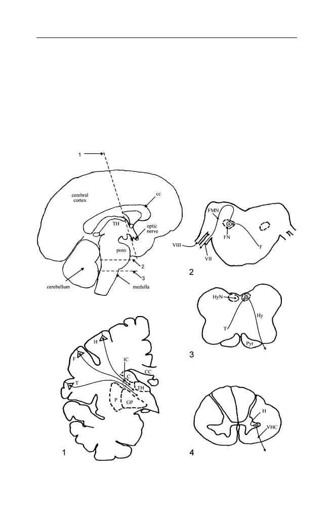
84
C. H. Vanderwolf
reflexes are activated. Similarly, voluntary sexual mounting may set the stage
for the activation of spinal copulatory reflexes.
The conclusion that some behaviors are not fully voluntary or are not readily
available as operants may appear to be contradicted by the common observation
that good actors can produce convincing displays of laughter, weeping, etc.,
upon request. However, there is good evidence from human stroke patients
that voluntary smiling, laughing, weeping, etc., depend on neural mechanisms
different from those involved in the unfeigned forms of these behaviors.

The Evolving Brain
85
It is unfortunately an all too common occurrence for the blood supply
to the internal capsule and part of the basal ganglia to be interrupted by
atherosclerotic narrowing and plugging of the middle cerebral artery or some
of its branches in the human brain. The blood supply may also be interrupted
by an embolus (for example, a blood clot originating in some distant site) or by
hemorrhage following the rupture of an artery. In all such cases, the end result
is that a region of nervous tissue dies, often producing a behavioral syndrome
known as stroke.
8
If a brain lesion interrupts large numbers of axons in the
internal capsule, a common occurrence in stroke, the ability of the neocortex to
control motor activity is diminished or lost (Figure VIII.1).
A severe stroke typically results in a complete paralysis (hemiplegia) and a
reduction or disappearance of muscle tonus on the contralateral (opposite) side
of the body. The affected limbs hang limply and cannot be moved voluntarily by
the patient (e.g. when requested to do so). In the weeks and months following a
stroke, muscle tonus gradually recovers and may become greater than normal.
Reflexes are at first reduced or abolished, but they recover after a few days and
eventually tend to become more vigorous on the paralyzed side of the body
than on the normal side. Thus, stretch reflexes
9
such as the knee jerk reflex are
likely to be abnormally vigorous in a hemiplegic limb. High muscle tone in
combination with exaggerated stretch reflexes is known as spasticity.
Figure VIII.1. Some descending motor pathways from the human neocortex.
Top left:
A crude sketch of a human brain divided along the anatomical midline. CC, corpus
callosum, a major fibre tract connecting the right and left neocortex; TH thalamus. The
dotted lines numbered 1–3 refer to the cross sections shown in the remainder of the
figure. 1. A cross section through one half of a human forebrain. C, caudate nucleus; CC,
corpus callosum; F, layer V pyramidal cell in the face area; GP, globus pallidus; H, layer
V pyramidal cell in the hand area; IC, internal capsule, a large fiber pathway including
a wide variety of ascending sensory and descending motor pathways; P, putamen; T,
layer V pyramidal cell in the tongue area; TH, thalamus. 2. A cross section through the
pons in the human brain. F, descending axon from pyramidal cell F; FN, facial nucleus;
FMN, facial motor neuron, VII, facial (seventh) cranial nerve; VIII, statoacoustic (eighth)
cranial nerve, carrying auditory and vestibular inputs. 3. A cross section through the
medulla in a human brain. Hy, hypoglossal motor neuron; HyN, hypoglossal nucleus;
Pyr, pyramidal tract carrying descending corticospinal fibers; T, descending axon from
pyramidal cell T. 4. A cross section through the cervical part of a human spinal cord.
H, descending axon from pyramidal cell H which has followed a pathway through the
pyramidal tract and the lateral spinal column. Note that the axons of pyramidal cells F,
H, and T cross (decussate) to the opposite side of the brain to influence motor neurons
connected to muscles on the opposite side of the body. As a result, destruction of cortex
or of descending fibers in the internal capsule will produce a loss of voluntary control of
body parts contralateral to the lesion but reflex functions may survive. Thus, a patient
who cannot stick his tongue out voluntarily may retain a normal ability to lick water from
the lips after taking a drink.

86
C. H. Vanderwolf
In intact humans, a tactile stimulus moving along the outer margin of the
sole of the foot in heel-to-toe direction elicits curling of the toes (ventroflexion
or plantar flexion reflex) but in a hemiplegic limb the toes fan out and the big toe
extends (dorsiflexion). This reaction, widely known as a Babinski reflex, has
long been regarded as indicative of destruction of descending cortical pathways
for voluntary movement.
The symptoms of hemiplegia are reduced somewhat with the passage of
time. The degree of recovery of function varies with the size and location of the
lesion and the amount of training and exercise undertaken by the patient. It is
presumed that intact motor pathways can, to some extent, take over the function
of those that were lost. There may even be an apparent complete recovery
after a small lesion but deficits tend to persist indefinitely with large lesions.
Among the earliest movements to recover are co-ordinated flexor synergies of
an entire limb so that flexion of the fingers, for example, occurs in association
with flexion of the wrist, elbow and shoulder.
10
Somewhat later, an extensor
synergy may appear as a co-ordinated extension of the digits, the wrist, the
elbow and the shoulder. Isolated movements of a single joint or a single digit
recover much later, if at all. The gross flexor and extensor synergies are of little
practical use to the patient. It might be thought that a global extensor synergy
might be used to reach out for a glass of water which could then be raised to
the mouth, or at least near it, by means of a flexor synergy. The flexor synergy,
however, seems to be inevitably associated with pronation of the hand (turning
the palm downward) so that the glass is emptied before it nears the mouth.
11
It is apparent that destruction of the neocortical motor areas or their efferent
pathways in the internal capsule has a much more severe effect in humans than
in laboratory animals such as rats or cats (see Chapter III). This suggest that the
evolution in humans of bipedal locomotion and use of the forelimbs primarily
for the manipulation and carrying of objects, involved a reorganization of
brain motor patterns, giving the neocortex a primary role in controlling the
new behavior patterns. However, severely spastic and hemiplegic limbs are
not always totally immobilized. Numerous “associated movements” may occur
after a period of recovery. Some of these are clearly components of the
brainstem reflexes, observed in decerebrate states. Thus, turning the head
strongly toward the paralyzed limb may cause it to become extended: turning
the head strongly toward the sound limb may cause the paralyzed limb to
flex. Clenching the fist of the sound upper limb may produce movements in
a paralyzed upper limb but the actual pattern of such movement tends to vary
from one patient to another. Spontaneous yawning and stretching movements
are usually associated with extension of a paralyzed upper limb.
12
Some movements retain a normal pattern of co-ordinated activity on the
hemiplegic side of the body and the amplitude and rate of the movements
may be normal or even greater than normal. A hemiplegic patient may display

The Evolving Brain
87
a reduction in the movement of the chest wall contralateral to the brain
lesion when asked to draw a deep breath but a normal or even greater than
normal movement of the chest wall contralateral to the brain lesion when
breathing spontaneously. This demonstrates a reduction in voluntary control
of the respiratory muscles coupled with a normal or an exaggerated automatic
or reflexive control of the same muscles.
13
Some patients may be unable to
draw a breath or hold their breath voluntarily when asked to do so even though
their spontaneous breathing is quite normal, and it is clear that they understood
what was asked.
Much the same situation can be demonstrated in the control of facial
expression. A hemiplegic patient may have an obvious impairment in the ability
to show the teeth voluntarily on the side opposite to the brain lesion when
requested to do so, together with an exaggerated display of the teeth on that
side when smiling or laughing occur naturally in a social situation.
14
In cases
in which the cortical motor pathways are damaged bilaterally, vigorous laughter
or weeping with abundant facial movement may occur spontaneously or in
response to slight stimuli, even though voluntary movement of the face and
limbs is virtually abolished.
15
Similarly, voluntary swallowing may be lost in a patient whose reflexive
swallowing is quite normal. Perhaps what is lost in the latter case is primarily
voluntary control of the tongue. Some patients who cannot stick the tongue out
voluntarily may, nonetheless, be capable of licking water from their lips in a
perfectly normal manner after drinking.
If the body temperature is reduced to elicit shivering, the resulting tremors
are of normal amplitude or of greater than normal amplitude in a hemiplegic
limb as compared to a contralateral sound limb.
16
One may conclude that stroke-induced hemiplegia is associated with a
reduction in purely voluntary control of the contralateral limbs together with
normal or even exaggerated contralateral movement in the case of motor
patterns that are not fully voluntary in an intact human. This category includes
breathing, smiling, laughing, weeping, yawning, stretching, shivering, swal-
lowing, associated postural reactions, and perhaps other behaviors that have
not yet been adequately studied.
Many patients with Parkinson’s disease display a behavioral syndrome
which is, in certain respects, the opposite of the syndrome observed in hemi-
plegic patients. Spontaneous laughter, smiling, etc., may be absent giving the
patients a poker-faced appearance even though voluntary control of the facial
muscles appears to be unimpaired. Similarly, the ability to hold the head erect,
an automatic behavior in normal people when they are awake, may disappear in
Parkinson’s disease, allowing the head to slump forward on to the chest when
the patients are sitting quietly awake. Despite this, such patients may be able

88
C. H. Vanderwolf
to extend the head forcefully against an examiner’s hand when requested to do
so.
17
The foregoing clinical neurological observations indicate that phasic volun-
tary movements are dependent on neural circuits different from those involved
in more automatic or reflexive behaviors including those concerned with
involuntary postural reactions. Consequently, an actor who can feign genuine
laughter, weeping, etc., is making use of brain systems different from those
involved in the natural occurrence of these behaviors.
Clinical observations of this type have traditionally been interpreted as
indicating that motor control in humans is dependent on two distinct systems.
(1) Pyramidal tract projections consisting of corticospinal fibers originating
primarily in the sensori-motor cortical areas have been supposed to be respon-
sible for all voluntary movement. (2) An extrapyramidal system consisting
of the basal ganglia and brain stem projections to the spinal cord have been
supposed to be responsible for various non-voluntary and postural reactions.
More recently this concept has lost favor, partly because section of the
pyramidal tract results in a loss of discrete movements of the extremities
rather than a loss of voluntary movement as a whole. Furthermore, lesions
apparently restricted to the caudate nucleus and putamen (with secondary
degenerative changes in other closely connected structures) have been reported
to produce a loss of voluntary control of the fingers and toes without affecting
voluntary control of proximal joints (shoulder, hip) in a human patient.
18
This
is similar to the deficits reported following section of corticospinal fibers. These
clinical observations have been supported by animal experiments showing
that neuronal destruction restricted to either the caudate nucleus and putamen
or the ascending dopaminergic projections from the substantia nigra to the
caudate nucleus and putamen produce impairments in rats using a forelimb
to reach for food.
19
These impairments are of the same general type as those
produced by destruction of the sensori-motor cortex or section of the pyramidal
tract. One can only conclude, as in the case of the problem of the neural
central of locomotion, that discrete movements of the limbs are produced
by complex neural circuits involving many brain structures. The “associated
movements” and other automatic movements observed in hemiplegic patients
are presumably the result of activity in surviving neocortex, the basal ganglia,
the brainstem, and the cerebellum both ipsilateral and contralateral to the brain
lesion.
A traditional view of the sensory control of voluntary movement held
that conscious sensations occurred in the primary sensory areas (such as the
striate area in the occipital lobe in the case of vision, or Heschl’s gyrus in the
temporal lobe in the case of audition). Impulses transmitted transcortically to
the adjoining association cortex then led to the elaboration of perceptions and
ideas which in turn might activate the motor areas (primarily the precentral

The Evolving Brain
89
gyrus) transcortically to produce a voluntary movement. However, this simple
view is unlikely to be correct. W. Penfield, who performed surgical removals
of various neocortical areas for the treatment of epilepsy, showed that removal
of the gyri surrounding the precentral motor region did not prevent dextrous
voluntary movement.
20
Therefore, the transcortical excitation theory of motor
control must be incorrect. Penfield suggested that the motor cortex must
normally be activated and controlled by ascending impulses from systems in
the brainstem and diencephalon. Experiments on animals indicate a similar
conclusion, as discussed in a previous essay (Chapter III).
In attempting to understand neural control of movement, it is important
to be aware that even the simplest of movements involve a complex array of
motor activities. For example, if a motionless human, standing erect, reaches
out with one hand to take a book from a shelf, the change in weight distribution
would cause the body to fall forward were it not for a counteracting backward
movement of the upper part of the body. W.R. Hess
21
distinguished such
general postural adjustments, which he termed the ereismatic phase of support,
from the final directed or telokinetic phase of the total movement (the reaching
movement of the arm and hand). When one considers the behavioral complex-
ity of even a rather simple movement, it is not surprising that many different
brain structures collaborate in the performance of any natural behavior. In order
to understand how the brain controls any such natural behavior, it is clear that
very detailed analytical studies of the behavior are essential.
Notes
1. Moore-Ede, M.C., Sulzman, F.M., and Fuller, C.A. (1982). The clocks that time us,
Cambridge, Massachusetts: Harvard University Press.
2. In the nineteenth and early twentieth centuries, it was assumed by many scientists that
behavior was entirely reflexive, i.e. determined by sensory inputs. The conditioned reflex
theory of I.P. Pavlov was firmly based on this concept [Pavlov, I.P. (1960). Conditioned
reflexes, New York: Dover Publications, Inc. (first published, 1927)]. The discovery that
the brain displays unceasing activity, even under conditions of minimal sensory input, was
revolutionary. However, some students of behavior were already well aware that much
normal behavior is spontaneous and independent of any specific sensory input. Thus, the
concept of operant conditioning assumes that animals generate behavior spontaneously and
that the environmental effects of a behavior (beneficial or noxious) will then influence the
probability of its future occurrence (see text).
3. Hume, D. (1978). A treatise of human nature, 2
nd
edition (edited by L.A. Selby-Bigge and
P.H. Nidditch), Oxford: Clarendon Press (first published 1739–40). Pp. 401–402.
4. A good recent textbook on operant conditioning and learning in general is: Domjan, M.
(1998). The principles of learning and behaviour, 4
th
edition. Pacific Grove, California:
Brooks/Cole Publishing Company.
A simple and practical discussion on the training of animals is provided by: Skinner, B.F.
(1951). How to teach animals. Scientific American, 185: 26–29.

90
C. H. Vanderwolf
5. Annable, A. and Weardon, J.H. (1979). Grooming movements as operants in rats. Journal
of Experimental Analysis of Behavior, 32: 297–304.
Breland, K., and Breland, M. (1966). Animal behavior. New York: The Macmillan Company.
Shettleworth, S.J. (1975). Reinforcement and the organization of behavior in golden ham-
sters: hunger, environment and food reinforcement. Journal of Experimental Psychology:
Animal Behavior Processes, 1: 56–87.
6. Doty, R.W., Richmond, W.H., and Sorey, A.T. (1967). Effects of medullary lesions on
coordination of deglutition. Experimental Neurology, 17: 91–106.
7. Pommerenke, W.T. (1928). A study of the sensory areas eliciting the swallowing reflex.
American Journal of Physiology, 84: 36–41.
8. A good general textbook on hemiplegia and other neurological conditions is: Walton, J.
(1993). Brain’s diseases of the nervous system, 10
th
edition. Oxford: Oxford University
Press.
9. Sudden stretching of a muscle stimulates specialized intramuscular receptor organs (muscle
spindles) which activate sensory fibers projecting to the spinal cord. These fibers in turn
excite motor neurons that project to the stretched muscle causing it to contract briefly. A
well-known stretch reflex is the knee-jerk elicited by tapping on the patellar tendon (just
below the knee cap) while the lower leg hangs freely in a sitting subject. The blow on the
tendon causes a sudden stretching of a large muscle (quadriceps) on the anterior surface of
the thigh, thereby eliciting a quick reflex contraction of that muscle.
10. Twitchell, T.E. (1951). The restoration of motor function following hemiplegia in man.
Brain, 74: 443–480.
11. Brodal, A. (1973). Self-observations and neuroanatomical considerations after a stroke.
Brain, 96: 675–694.
12. Walshe, F.M.R. (1923). On certain tonic or postural reflexes in hemiplegia with special
reference to the so-called “associated movements”. Brain, 46: 1–37.
13. Jackson, J.H. (1899). Case of left hemiplegia with turning of the eyes to the right-slightly
greater amplitude of the left side of the chest in inspiration proper and slightly less amplitude
of movement of that side in voluntary expansion of the chest. Lancet, XIX: 1659–1660.
Cohen, E., Mier, A., Heywood, P., Murphy, K., Boultbee, J., and Guz, A. (1994). Diaphrag-
matic movement in hemiplegic patients measured by ultrasonography, Thorax, 49: 890–895.
Prezedborski, S., Brunko, E., Hubert, M., Mavroudakis, N., and Zegers de Beyl, D. (1988).
The effect of acute hemiplegia on intercostal muscle activity, Neurology, 38: 1882–1884.
Simon, R.P. (2001). Breathing and the nervous system. In: Aminoff, M.J. (editor) Neurology
and general medicine, New York: Churchill Livingstone, pp. 1–21.
14. Monrad-Krohn, G.H. (1939). On facial dissociation. Acta Psychiatrica et Neurologica, 14:
557–566.
15. Ironside, R. (1956). Disorders of laughter due to brain lesions. Brain, 79: 589–609.
16. Uprus, V., Gaylor, G.B., and Carmichael, E.A. (1935). Shivering: a clinical study with
especial references to the afferent and efferent pathways. Brain, 58: 220–232.
17. Martin, J.P. (1967). The basal ganglia and posture. London: Pitman Medical Publishing Co.
18. Oppenheimer, D.R. (1967). A case of striatal hemiplegia. Journal of Neurology Neuro-
surgery and Psychiatry, 30: 134–139.
19. Whishaw, I.Q., O’Connor, W.T., and Dunnett, S.B. (1986). The contributions of motor
cortex, nigrostriatal dopamine and caudate-putamen to skilled forelimb use in the rat. Brain,
109: 805–843.
20. Penfield, W. (1954). Mechanisms of voluntary movement. Brain, 77: 1–17.
21. Hess, W.R. (1954). Diencephalon: autonomic and extrapyramidal functions. New York:
Grune and Stratton. (see p. 31).

IX. About hunting
According to Gilbert White, an eighteenth century English clergyman and
naturalist, “there is such an inherent spirit for hunting in human nature, as
scarce any inhibitions can restrain.”
1
The view that hunting and meat eating
are at least partially instinctive in humans is consistent with archaeological
evidence that human ancestors have been hunters for at least 2.5 million years
(see Chapter IV). Clearly, hunting is not simply an arbitrary custom dependent
on the existence of a particular culture. Nonetheless, many people today regard
hunting as a cruel, barbaric form of behavior which is inconsistent with the
ethics of modern civilization.
2
What do we know about hunting and how should
we regard it?
Predation in the natural world. Walking alone one evening on a quiet gravel
road near where I live in southern Ontario, I heard the repeated cries of a bird
originating high up in a large maple tree. Although the sun was setting, the
light was sufficient for a clear view of a raccoon standing on a branch directly
in front of a large hole in the tree trunk. I could see the raccoon’s head and
jaws moving and I could hear the sounds of its munching interspersed with the
diminishing cries of the dying bird on which it was evidently feeding.
3
I have witnessed scenes of predation involving various species on a number
of occasions in my life. They were always disturbing. One wishes to punish,
kill, or at least drive off the predator. However, on longer reflection, one cannot
avoid the fact that predation occurs everywhere in the natural world, not only
among mammals and birds, but everywhere throughout the animal kingdom.
Predation appears to play an essential role in regulating the abundance of many
species. Many features of animals that we consider beautiful such as the slim
legs and elegant bounding gait of white tailed deer, the thunderous take-off of
ruffed grouse, the heavy protective shell of turtles, the quills of porcupines, as
well as such qualities as the prodigious reproductive capacity of rats and mice,
probably owe their very existence to the selective effects of predation. Animals
that were able to avoid predators by high speed flight or by means of protective
defences, as well as those that were able to offset the losses due to predation by
high fertility, have been more likely to leave surviving descendants than those
who lacked these features. It is clear that predation has played a major role in
shaping the natural world.

92
C. H. Vanderwolf
It is also clear that the natural world was not designed according to
the moral and ethical principles by which humans, sometimes successfully,
attempt to regulate their interactions with one another. One can understand
the conclusions of some religious groups (Gnostics, Manicheans, Zoroastrians)
that the world we see around us could not possibly be the work of a kind,
beneficent god and must, therefore, have been constructed by an evil being,
a devil who, perhaps, accomplished his wicked design in a moment when the
kind god was napping.
Hunting ethics. Hunting and the eating of meat require the killing of
animals. This fact has troubled thoughtful people, not only in our own time,
but also in earlier periods. The historian John Cummins
4
tells us that some
medieval ecclesiastics, living in a time when hunting was highly esteemed,
nonetheless believed that hunters were wicked. Jean-Jacques Rousseau, an
eighteenth century French philosopher, published a long diatribe against the
eating of meat.
5
Nonetheless, both hunting and meat-eating have survived,
sometimes even in countries in which the official religion forbids the taking
of life.
6
Hunters themselves everywhere seem to regard hunting, not as an evil,
but as a joyous activity. Edward of Norwich, who wrote the oldest English book
on hunting between 1406 and 1413,
7
began a section on the hunting life with
the statement “Now shall I prove how hunters live in this world more joyfully
than any other men.”
It is probable that a very ancient form of justification for hunting was one
of necessity. In the words of an Abenaki hunter, presumably addressing the
spirit of a newly slain animal, “I have killed you because I need your skin for
my coat and your flesh for my food. I have nothing else to live on.”
8
Well-
fed contemporary hunters living in wealthy western countries, who can make
no such claims, sometimes say that their actions are an attempt to restore the
balance of nature. There is truth in this. For example, it is generally believed
that white-tailed deer populations in North America, no longer controlled by
non-human predators, would increase to unsustainable levels if human hunting
were to cease.
9
However, it must be acknowledged that men do not take up
hunting because they wish to perform a public service. Men hunt because
they derive a deep satisfaction and pleasure from the entire process, from
the initial pursuit of game to the final sitting down with one’s family to an
excellent dinner. The fact that many people love to hunt but that there is
very little interest in the job of executioner in a slaughterhouse demonstrates
that love of hunting is not simply due to “bloodlust” as the critics of hunting
have sometimes contended. Human enjoyment of a complex activity cannot be
reduced to any single factor. Our enjoyment of a gourmet meal, for example,
is due not only to the food or the wine, but also to the pleasure of conversation
with our companions, the furnishings of the dining room, etc. Similarly, hunters
enjoy being in the woods , enjoy carrying and using a treasured weapon, enjoy

The Evolving Brain
93
the craft of outwitting a wary game animal, enjoy the excitement of the kill and
are delighted when they have been successful.
Concern by hunters for the animals they hunt has led to the development
of a hunting ethic among hunting peoples everywhere in the world. Although
modern hunting organizations have sometimes published ethical guidelines for
hunting,
10
these tend to be of a rather generalized nature, avoiding detailed
statements as to exactly what a hunter should or should not do. However, such
ethical guidelines may encourage: making quick humane kills; making every
reasonable effort to follow up and kill any animals that are unintentionally
wounded; avoiding the wastage of meat or useful skins; continuing efforts by
hunters to increase their knowledge and skill in woodcraft and marksmanship;
conservation of game and preservation of natural habitats; and respect for game
laws and the rights of private landowners.
It is important to realize that the hunting ethics of traditional hunter-
gatherer peoples may have differed substantially from those of modern sport
hunters. For example a modern sportsman would not think of killing a female
duck accompanied by a family of downy ducklings but an aboriginal hunter
who has hungry mouths to feed may not be able to afford such scruples.
Aboriginal hunters everywhere have made use of various types of traps (pit-
traps, deadfalls, snares, etc.) to secure game but modern sportsmen avoid such
methods.
11
In many jurisdictions, in fact, the use of such methods is illegal
except in the case of the trapping of fur-bearing animals by a licensed trapper.
Hunters as individuals tend to have a deep affection for the woods and
wild creatures in their neighbourhood. One of the essential qualifications of
a successful hunter is a practical knowledge of animal ecology and behavior, a
knowledge of where various species are to be found and the time of day when
they are likely to be active. It is also essential for a hunter to be a keen observer,
able to detect well-camouflaged animals hidden in the vegetation. Thus, hunters
tend to be practical naturalists who take a naturalist’s delight in wildlife and
wild places. This often develops into a desire to promote the conservation of
wildlife and wildlife habitats. Hunter organizations such as Ducks Unlimited,
the Ruffed Grouse Society, and the Ontario Federation of Anglers and Hunters,
for example, have been in the forefront of the conservation movement for
decades and have been very effective in encouraging the preservation or
restoration of wildlife habitats and in the reintroduction of native species in
areas in which they had been nearly or completely exterminated. The successful
restoration of wild turkeys and Canada geese in Ontario provide examples
of this.
It is unclear whether concern for conservation of wildlife is a modern
development or whether it is a feature of the hunting ethic in hunter-gatherer
societies. For example, although it is currently fashionable to believe that
aboriginal North Americans traditionally followed wise conservation practices,

94
C. H. Vanderwolf
there is very little evidence to support this belief. According to Ian McTaggart-
Cowan, “There is no evidence that the native people had any concept of
numbers applied to their food animals. They took what they could without
concern for replacement rate or overkill.”
12
However, there is the intriguing
observation that Paleolithic reindeer hunters, 50,000 years ago, and even
earlier, killed mainly adult male deer.
13
Although it is, of course, impossible
to know what the intent of these hunters was, the effect of their behavior would
be to preserve the maximum number of breeding female deer. It is conceivable
that some conservation practices are very ancient indeed.
A discussion of hunting ethics and conservation practices does not address
the question which is central for many people. Is the killing of animals for any
human purpose ever morally justifiable?
First, if one accepts that predation is an intrinsic feature of the natural world
and that humans are a part of the fauna of our planet, then there appears to be
no obvious reason why humans should not be predators. If one believes that
the human killing of other animals is unethical, there seems to be no way of
avoiding the conclusion that the killing of animals by other non-human animals
is also unethical, or at least intolerable and should be stopped. It could, of
course, be maintained that non-human predators do not understand that what
they are doing is wrong and they cannot, therefore, be held responsible for their
actions. A mentally incompetent human who kills another human is also judged
not to be criminally responsible but is incarcerated anyway (in a hospital for the
criminally insane rather than a prison). Thus, a logical consequence of the idea
that killing animals is unethical is that, as ethical beings, we should intervene
massively in the natural world, attempting to separate predators from their prey.
Some philosophers and animal activists have, in fact, advocated taking this
step.
14
Apart from the fact that a program to prevent predation in the natural
world is technically and economically impossible and would have a great many
undesirable consequences, it rests on a philosophical fallacy. Human ethical
principles are our own invention, an outgrowth of the evolution of human
social behavior. They are not a part of the natural non-human world and it is
presumptuous in the extreme to suppose that the whole world should dance to
music of our making. We, however, can choose the ethical principles by which
we ourselves wish to live. Consequently, ethical and moral standards change
somewhat from one historical period to another. Killing other people, ordinarily
considered the most heinous of all crimes, is not only not condemned, but
actively encouraged during wartime. The sexual mores of ordinary people in
Western countries have changed dramatically in the past fifty years. Similarly,
hunting and the killing of other animals for our own purposes will continue
as long as large numbers of people support such practices. Ultimately, public
opinion is the final arbiter of ethical questions.

The Evolving Brain
95
Neural basis of hunting behavior. In considering this topic, it is essential
to be clear about what it is that we hope to explain in neuroscientific terms.
If two human hunters, walking together through woods and open areas, detect
several deer a long way off, what do they do? They immediately stop walking
and are likely to crouch down, concealing themselves behind rocks, bushes, etc.
Conversation is reduced to tense whispers as they consider if any of the deer
are suitable prey, and if so, how, considering the terrain, wind direction, and
the behavior of the deer, they might be able to approach near enough to make
a kill. Their behavior does not in the least resemble the red-faced shouting and
threats typically seen in two men in a quarrel. Hunting is not like intraspecific
aggression and fighting.
15
Neuroscientific studies confirm that predatory behavior in laboratory ani-
mals is distinct from aggressive or defensive displays. Electrical stimulation of
a medial and ventromedial region of the hypothalamus in cats elicits a threat or
defensive response pattern which includes opening of the mouth, retraction of
the lips, hissing, growling, flattening of the ears, piloerection, and striking with
a forepaw plus biting directed towards a nearby rat or an inanimate model.
16
The overall pattern is similar or identical to the behavior displayed by a cat
toward a rival cat or towards an aggressive dog.
17
A similar behavior pattern
can be elicited from the same ventromedial hypothalamic zone in the marsupial
opossum, suggesting that it has a long phylogenetic history.
A predatory type of attack which includes approaching the prey object
with the body lowered and the neck extended, followed by the use of the
forepaws to catch and hold the prey and by severe biting, can also be elicited
by localized electrical stimulation of the hypothalamus. Piloerection does not
occur. The predatory attack pattern of behavior closely resembles naturally
occurring predatory behavior and is more likely to kill or severely wound the
prey than the threat-defensive pattern. Predatory attack can be elicited from
a hypothalamic zone lateral or dorsolateral to the threat-defensive zone in
both cats and opossums. The predatory attack zone extends rostrally into the
preoptic area but, according to W.W. Roberts,
16
the defensive threat zone does
not. Therefore, different systems of hypothalamic neurons are involved in the
two patterns of behavior.
There is evidence that the amygdaloid nuclei, large cellular complexes
located in the temporal region, modulate both defensive-threat behavior and
predatory attack by means of neural projections to the hypothalamus. Elec-
trical stimulation of the amygdala can elicit defensive-threat behavior and
stimulation of different regions of the amygdala may either suppress or
facilitate predatory attack elicited by concurrent hypothalamic stimulation.
16
Large bilateral lesions of the amygdala in cats abolish predatory attack on
a bird or a mouse.
18
Extensive removal of the neocortex would, no doubt,
also abolish effective predatory behavior (although this point has never been

96
C. H. Vanderwolf
specifically tested as far as I know) but this effect would be part of a generalized
impairment of behavior (dementia) rather than a specific (or somewhat specific)
loss of predatory behavior. Animals with amygdaloid lesions are able to feed
themselves and do not suffer from a global impairment of behavior. Large
lateral hypothalamic lesions would also abolish predatory behavior, one can
assume, because such lesions abolish most forms of purposive behavior.
19
It appears that predatory attack behavior in laboratory animals depends on
specific neural systems located in the hypothalamus and amygdaloid nuclei
but also on more widespread systems including the neocortex. The complete
behavioral performance would also, of course, be dependent on other brain
structures including the brain stem, cerebellum, and the spinal cord.
There appears to be no systematic evidence on the neural basis of hunting
behavior in humans. Although it is certainly possible that the neural systems for
predatory behavior in laboratory animals are similarly active in the human case,
one cannot be confident about this. Typical quadrupedal predatory behavior
involves seizing prey and biting, often directed toward the head and neck, but
human predatory attacks ordinarily involve the use of tools (ranging from clubs
and rocks to a modern rifle) and do not involve biting. Consequently, it may be
that human predatory behavior is not fully homologous with predatory behavior
in conventional predators and that it has a unique neural basis.
Notes
1. White, G. (1994). The natural history and antiquities of Selborne, London: The Folio
Society (first published in 1788), see p. 18.
2. The current popular literature attacking or defending hunting is enormous. Two books
written from the point of view of thoughtful hunters are: (1) Petersen, D. (editor). A hunter’s
heart: honest essays on the blood sport, New York: Henry Holt and Company, 1997; and (2)
Swan, J.A. (1996). In defense of hunting, New York: Harper Collins, paperback edition.
3. Many predators kill their prey by eating it. Durward Allen reports the following incident:
“On a flight late in the afternoon, Don and Chief Ranger Ben Zerbey saw the moose (this
animal had been under observation by wolf investigators using an aircraft) down and the
wolf feeding on its rump. The moose lay quietly with its head up watching the wolf,” p. 129
in: Allen, D.L. (1979). Wolves of Minong, Boston: Houghton Mifflin Company.
4. Cummins, J. (1988). The art of medieval hunting: the hound and the hawk, Edison, New
Jersey: Castle Books, p. 10.
5. Rousseau, J.J. (1911). Emile, London: J.M. Dent and Sons, Ltd. (Translated from French by
B. Foxley; first published 1762).
6. Harrar, H. (1953). Seven years in Tibet. Leicester: Ulverscroft, (translated from the German
by R. Graves). The dominant religion in Tibet, a form of Buddhism, forbids the taking of life
even in the case of flies or biting insects. Further, animals may not be deprived of their food.
Nonetheless, meat, fish and honey were widely eaten in traditional Tibetan society, and fur
garments were worn in winter. Tibetans managed to combine these practices with a stated
adherence to their ethical principles by making social outcasts of people who performed,

The Evolving Brain
97
for pay, the tasks of slaughtering animals, preparing skins for clothing and the collection of
honey. Similar hypocrises are not unknown in contemporary Western society.
7. Edward of Norwich (1974). The master of game, New York: AMS Press, from an original
manuscript dating about 1420 (see p. 8). The greater part of this book is a translation of
Gaston Foix’s (also known as Gaston Phebus) Livre de chasse, an illuminated manuscript
available today as: Bise, G. (1978). Medieval hunting scenes, Fribourg: Production Liber
SA (first published about 1390, translated into modern English by J.P. Tallon). For a
philosophical discussion of the place of hunting in human life throughout history and its
contribution to human happiness, see: Ortega y Gasset, J. (1972). Meditations on hunting,
New York: Charles Scribner’s sons, (Translation from the Spanish by H.B. Wescott).
8. Cassell, J. and Fiduccia, P. (editors) The quotable hunter. New York: The Lyon’s Press, 1999,
p. 5.
9. A particularly clear illustration of the growth in a population of predator-free white tail deer
is provided by the George Reserve, a 464 hectare (1,146 acre) plot of Michigan woodland
enclosed by an 11.5 foot deer-proof fence. Six deer released in the reserve in 1928 had
increased to possibly 220 deer by 1933. Since the vegetation was being severely damaged by
so many deer, their numbers were controlled by hunting until 1966 when the population size
was gradually and systematically reduced until only about 10 deer remained in 1975. At this
point hunting was stopped. Within five years the population had increased to 212 deer. Since
then (1980), the population has again been controlled by hunting. [See: McCullough, D.R.
(1984). Lessons from the George Reserve, Michigan, In: Halls, L.K. (editor) White-tailed
deer: ecology and management, Harrisburg, Pennsylvania: Stackpole Books, pp. 211–242.]
10. A magnificently illustrated book [Blüchel, K.G. (2000). Game and hunting, Cologne:
Könemann Verlagsgesellschaft mbH] on the history and present practices of hunting in
Europe contains a statement of the hunting ethics adopted by the Conseil International de la
Chasse et de la Conservation du Gibier (CIC; The International Council of Hunting and the
Conservation of Game, see pp. 646–647).
11. An excellent summary of aboriginal methods of trapping game is provided by: Coon, C.S.
(1971). The hunting peoples, Boston: Little, Brown and Company. Although such methods
as snaring game may be illegal in Western countries, they continue to be used by poachers.
See, for example: Benson, R. (1985). Ragnar’s ten best traps and a few others that are damn
good too, Boulder, Colorado: Paladin Press. The US army survival manual, FM 21-76, New
York: Dorset Press, 1994, also gives detailed instructions on primitive methods of securing
fish and game and preparing it for food.
12. McTaggart-Cowan, I. (1989). Room at the top? In: Hummel, M. (editor) Endangered spaces:
the future for Canada’s wilderness, Toronto: Key Porter Books Ltd., pp. 249–266. Also see:
Krech, S. III (1999). The ecological Indian: myth and history. New York: W.W. Norton and
Company.
13. Gaudzinski, S., and Roebroeks, W. (2000). Adults only. Reindeer hunting at the Middle
Palaeolithic site Salzgitter Lebenstedt, Northern Germany. Journal of Human Evolution, 38:
497–521.
14. See: Causey, A.S. (1996). Is hunting ethical? In: Petersen, D. (editor) A hunter’s heart:
honest essays on blood sport. New York: Henry Holt and Company, pp. 80–89.
15. Any simple dichotomy between intraspecific aggression and predatory behavior in humans
is complicated by the observation that military snipers and serial killers appear to display
an essentially predatory pattern of behavior toward other humans. There appears to be little
scientific information available on any of this, however.
16. A general list of references on central elicitation of defensive and predatory behavior
includes the following: Hess, W.R. (1957). The functional organization of the diencephalon,
New York: Grune and Stratton.

98
C. H. Vanderwolf
Egger, M.D., and Flynn, J.P. (1967). Further studies on the effects of amygdaloid stimulation
and ablation on hypothalamically elicited attack behavior in cats, In: W.R. Adey and
T. Tokizane (editors) Structure and function of the limbic system, Progress in Brain
Research, 27: 165–182. Hunsperger, R.W., and Bucher, V.M. (1967). Affective behavior
produced by electrical stimulation in the forebrain and brain stem of the cat. In: W.R. Adey
and T. Tokizane (editors) Structure and function of the limbic system, Progress in Brain
Research, 27: 103–127.
Roberts, W.W. (1970). Hypothalamic mechanisms for motivational and species-typical
behavior. In: R.E. Whalen, R.F. Thompson, M. Verzeano, and N.M. Weinberger (editors)
The neural control of behavior, New York: Academic Press, pp. 175–206.
Siegel, A., Roeling, T.A.P., Gregg, T.R., and Kruk, M.R. (1999). Neuropharmacology of
brain-stimulation-evoked aggression. Neuroscience and Biobehavioral Reviews, 23: 359–
389.
17. See: Leyhausen, P. (1979). Cat behavior: The predatory and social behavior of domestic and
wild cats, New York: Garland STPM Press (translated from the German by B.A. Tonkin).
18. Cherkes, V.A. (1967–1968). Instinctive and conditioned reactions in cats after removal of
amygdaloid nuclei, Neuroscience Translations, 4: 418–424 (Published by the Federation of
American Societies for Experimental Biology for the National Institutes of Mental Health).
19. Levitt, D.R., and Teitelbaum, P. (1975). Somnolence, akinesia, and sensory activation of
motivated behavior in the lateral hypothalamic syndrome, Proceedings of the National
Academy of Sciences of the U.S.A., 72: 2819–2823.
Robinson, T.E., and Whishaw, I.Q. (1974). Effects of posterior hypothalamic lesions
on voluntary behaviour and hippocampal electroencephalograms in the rat. Journal of
Comparative and Physiological Psychology, 86: 768–786.

Index
Abrams, H.L., Jr., 51
acetylcholine, 30
Adam, 33
adaptation, 71, 72
Adey, W.R., 98
alchemy, 13, 15
Alkive, N.L., 52
Allen, D.L., 96
Aminoff, M.J., 90
amygdala, 25, 95, 96
Andersen, K., 38
Anderson, S.R., 65
Annable, A., 90
Aristotle, 2, 3, 6, 10, 13, 16, 19, 25
Aronson, L.R., 64
Asanuma, H., 32
Aston-Jones, G., 12
Astrand, P.-D., 73
attention, 7, 11, 12
australopithecine, 33, 34
Bailey, C.H., 73
Baker, G.B., 18
Barnes, J., 10
Barrett, D.V., 66
Bartemeier, L.H., 80
basal ganglia, 21, 88
behavior, definition of, 14
Belekhova, M.G., 32
Bennett, T.L., 11
Benson, R., 97
Beyene, Y., 52
Bicchieri, M.G., 53
Bise, G., 97
blindsight, 9
Bloom, F.E., 12
Blüchel, K.G., 97
Bonke, B., 12
Boring, E.G., 10
Boultbee, J., 90
Boulton, A.A., 18
brain imaging, 7, 70
Brain, W.R., 80
brainstem, 22–24, 27
Braunwald, S.L., 52
Breland, K., 65, 90
Breland, M., 65, 90
Brill, C., 53
Brodal, A., 65
Brooks, C.M., 10
Brunko, E., 90
Bruyn, G.W., 65
Bucher, V.M., 98
Buchthal, F., 79
Buonomano, D.V., 73
Butler, A.B., 65
Cain, D.P., 11, 18, 73
Cant, J.G.H., 42
Carmichael, E.A., 90
Caro, T.M., 64
Cassell, J., 97
Causey, A.S., 97
Chaplin, G., 53
Cherkes, V.A., 98
Chivers, D.J., 52
Christ, Jesus, 62
Cichetti, W.M., 11
Clark, J.D., 52
Cohen, E., 90
Colby, C.L., 12
Cook, J.D., 52
Cooley, R.K., 21
Coon, C.S., 53, 97
Cranefield, P.F., 10
Crosby, E.C., 65
Cummins, J., 96

100
C. H. Vanderwolf
Daly, M., 53
Damasio, H., 51, 65
Danziger, K., 6, 11
Dart, R., 33
Darwin, C., 33, 51, 63, 66
Davis, F., 11
decerebrate animal, 22, 23, 76
decorticate animal, 24, 76
de Heinzelin, J., 52
Demos, R., 10
Denny-Brown, D., 17
de Queiroz, A., 52
Descartes, R., 2, 4, 7, 8, 10
DeVore, I., 52
Diamond, I.T., 26
Diamond, M.C., 65
digestive system, 40–42
Domjan, M., 89
Dononn, L.E., 66
Doty, R.W., 90
Draper, H.H., 52
Drickamer, L.C., 17
Dunnett, S.B., 90
Dyer, G., 66
Ebling, J., 53
Eccles, J.C., 9, 12
Edman, J.S., 53
Edward of Norwich, 92, 97
Egger, M.D., 98
Egner, R.E., 66
Eibl-Eibesfeldt, I., 53
Eidelberg, E., 80
Errington, P.L., 26
ethics, biological basis of, 61, 62
Etkin, W., 53
Eve, 33
Everitt, B.J., 12
Ewan, P.W., 52
Ewer, R.F., 65
Fauci, D.L., 52
Fernandez-Ballesteros, M.L., 79
Fiduccia, P., 97
Field, J., 11
Finch, C.A., 52
fixed action patterns, 56
Fleagle, J., 80
Flynn, J.P., 98
Foix, G., 97
Forssberg, H., 31
Foster, G.D., 53
Freeman, W.J., 12
free will, 81, 82
Freud, S., 1, 10
Frias, J.L., 65
Fukuda, T., 17
Fulcher, J.S., 64
Fuller, C.A., 89
Fulton, J.F., 17, 31, 80
Gaffan, D., 11
Galdston, I., 52
Galileo, 2
Gans, C., 32
Gaudzinski, S., 97
Gautama, S., 62
Gaylor, G.B., 90
Geist, V., 65
Gibson, J.J., 31, 32
Glickman, S.E., 49, 53
Gloor, P., 12
Goldberg, M.E., 12
Goldberg, S.R., 11
Goodale, M., 9, 12
Goodall, J., 51, 66
Goodenough, F.L., 64
Greenblatt, S., 66
Gregg, T.R., 98
Gregoire, S.E., 31
Griffin, D.R., 12
Grove, W.M., 11
Grube, G.M.A., 10
Guz, A., 90
Haldane, E.S., 10
Hall, V.E., 11
Hallberg, L., 52
Halls, L.K., 97
Handler, P., 52
Harlow, H.F., 32
Harrar, H., 96
Harries, M., 73
Harris, M., 51
Harris, S., 66
Hart, W., 52
Harvey, W., 2
Hauser, M.D., 64
Hauser, S.L., 52
Hay, R.L., 51
Hebb, D.O., 10

The Evolving Brain
101
Heffner, H.H., 32
Hegsted, D.M., 52
Heinroth, O., 14
hemiplegia, 76, 77, 85–88
Henningfield, J.E., 11
Hernández-Peón, R., 11
Hess, W.R., 89, 90, 97
Heywood, P., 90
Hill, J.O., 53
Hill, R.L., 52
hippocampus, 6, 20, 21, 27, 67, 68, 70, 73
Hitler, A., 62
Hladik, C.M., 52
Hoberg, E.P., 52
Hodos, W., 65
Holmyard, E.J., 17
hominids, 34, 35
Horel, J.A., 11, 31
Houck, L.D., 17
Huber, C.G., 65
Hubert, M., 90
Hume, D., 1, 10, 82, 89
Hummell, M., 97
Humphrey, G., 10
Hunsperger, R.W., 98
Hunt, K.D., 53
hunting, 39, 44–49, 91–96
Huxley, T.H., 10
hypothalamus, 20, 21, 58, 95, 96
Innis, H.A., 52
introspection, 4–6
Ireland, M.L., 53
Ironside, R., 90
Isaacson, R.L., 11
Jablonski, N.G., 53
Jackson, J.H., 90
James, W., 1, 4, 10
Jameson, J.L., 52
Jasper, H.H., 11
Jennings, H.S., 14
Jensen-Jazbutis, G.T., 65
Jones, A., 52
Jouvet, M., 11
Jungers, W.L., 52
Jurgens, U., 65
Kaada, B.R., 11
Kandel, E.R., 73
Kappers, C.U., 65
Kasper, D.L., 52
Kastner, S., 12
Kate, B.R., 73
Katz, J.L., 11
Keating, E.G., 31
Kickert, R.W., 64
Kimble, G.A., 26
Kimura, D., 12, 53
Klein, R.G., 52
Klein, S., 53
Koch, S., 17
Kolb, B., 12, 17, 31, 65
Korsakov, S.S., 65
Krech, S., III, 97
Krompecher, S., 31
Kruk, M.R., 98
Kuo, Z.Y., 57, 58, 64
Kushlan, J.A., 53
Kuypers, H.G.J.M., 31
Lack, D., 51
Lam, D.A., 65
Lamb, R.J., 11
Lancaster, C.S., 52
Larson, S.G., 80
Lavoisier, A., 2
Layrisse, M., 52
Leakey, M.D., 51
Lee, R.B., 52
Lehman, I.R., 52
Lehrman, D.S., 64
Leung, L.-W.S., 18
Levitt, D.R., 98
Leyhausen, P., 98
Leyton, A.S.F., 28
Lieb, C.W., 52
Lightfoot, D.W., 65
Lilly, J.C., 32
limbic system, 3
Lindsley, D.B., 11
Linnaeus, K., 63
Lipak, J., 31
locked-in syndrome, 8
Longo, D.L., 52
Lorenz, K.Z., 14, 17, 55–57, 64
Lovick, T.A., 31
Lowther, G.R., 53
Lykken, D.T., 11
Lyons, W., 10

102
C. H. Vanderwolf
MacLarnon, A.M., 52
Magnus, R., 14
Magoun, H.W., 10, 11, 73
Margulis, L., 12
Marshall, L.H., 73
Martin, G.F., 31
Martin, J.P., 78–80
Martin, P.S., 52
Martin, R.D., 52
Masterton, R.B., 32
Matzel, L.D., 12
Mavroudakis, N., 90
McCullough, D.R., 97
McCurdy, N.M., 32
McGaughy, J., 12
McGrew, W.C., 53
McGuckin, B.G., 53
McHenry, H., 80
McHenry, H.M., 54
McTaggart-Cowan, I., 97
meat-eating, 37–40
Meisch, R.A., 11
memory, definition of, 67
Mertz, W., 52
Merzenich, M.M., 73
Metabolic adaptation to carnivory, 40–42
Micheli, L.J., 73
microcephaly, 60
Mier, A., 90
Milgram, S., 4, 5, 11
Milner, D., 9, 12
Miron, J.A., 65
Misantone, L.J., 31
Mohammed, B.S., 53
Monrad-Krohn, G.H., 90
Monsen, E.R., 52
Moore-Ede, M.C., 89
Morris, D., 64
Morris, J.G., 52
Muhammad, 62
Murdock, G.P., 53
Murphy, K., 90
Myers, R.E., 32, 65
Myrianthopoulos, N.C., 65
Napier, J.R., 36, 51
Napier, P.H., 36, 51
Napoleon, 62
Nattiv, A., 53
Neafsey, E.J., 32
Nemesius, 67, 71
neocortex, 24, 31, 68
Newton, I., 2
Nidditch, P.H., 10, 89
Nisbett, R.E., 11
Northcutt, R.G., 32
O’Connor, C.M., 10
O’Connor, W.T., 90
Oliver, J.S., 52
operant behavior, 82, 83
Oppenheimer, D.R., 90
Ortega y Gasset, J., 97
Ostrum, A.E., 12
Parkinson’s disease, 78, 79
pattern generator, 24, 64, 83
Pavlov, I.P., 14, 89
Peiper, A., 80
Penfield, W., 15, 65, 89, 90
Petersen, D., 96, 97
Pfluger, E., 8, 12
phocomelia, 36
Pinker, S., 65
Plato, 2–4, 13
Ploog, D., 65
Pommerenke, W.T., 90
Popper, K.R., 9, 12
Power, D.M., 64
predatory behavior, 57, 58, 91, 94–96
Preston, K.L., 11
Prezedborski, S., 90
Pribram, K.H., 11
Rader, D.J., 53
Ratliff, F., 17
Reece, W.O., 41, 52
reinforcing effect, 5, 82, 83
releasers, 56
religions, 62, 63
Renne, P., 52
Richmond, W.H., 90
Richter, C.P., 80
Robbins, T.W., 12
Robert, S.L., 73
Roberts, L., 65
Roberts, W.W., 64, 95, 98
Robinson, B.W., 65
Robinson, T.E., 98
Rodahl, K., 73
Rodman, P.S., 54

The Evolving Brain
103
Roebroeks, W., 97
Roeling, T.A.P., 98
Rogers, Q.R., 52
Romijn, H., 12
Rosen, I., 32
Rosenberger, A., 80
Rosenblatt, J., 64
Rosenfalck, P., 79
Rosenfield, L.C., 12
Ross, E.B., 51
Ross, G.R.T., 10
Ross, J.J., 65
Rousseau, J.J., 92, 96
Russell, B., 10, 66
Sagan, D., 12
Sanford, C.B., 66
Sarno, M.T., 65
Sarter, M., 12
Savalli, U.M., 64
Schaller, G.B., 53
Scherrer, H., 11
Schindler, C.W., 11
Schlesinger, K., 26
Schmid, H., 51
Schmitt, F.O., 12
Schultz, W., 80
Sebel, P.S., 12
Sechenov, M., 14
Selby-Bigge, L.A., 10, 89
Semendeferi, K., 51
serotonin, 30
Shakespeare, W., 63
Sheets-Johnstone, M., 54
Sherrington, C.S., 14, 17, 28, 31, 64
Shettleworth, S.J., 90
Shors, T.J., 12
Short, R.V., 53
Siegel, A., 98
Simon, R.P., 90
Singer, C., 66
Skarda, C.A., 12
Skinner, B.F., 14, 65, 82, 89
Smith, D.E., 31
Smith, E.L., 52
Smith, H.W., 10
Smith, J.M., 79
Smith, S.M., 51
Smithells, R.W., 51
Sorey, A.T., 90
speech, neural basis of, 59–61
Sperry, R.W., 8, 12, 32
spinal animal, 19–22, 76
Sroges, R.W., 49, 53
Staddon, J., 17
Stalin, J., 62
Stanish, W.D., 73
Steele, S.B., 52
Stefansson, V., 38, 52
Stent, G.S., 32
Strasser, E., 80
Struever, S., 53
Sulzman, F.M., 89
Suwa, G., 51
Swan, J.A., 96
Swenson, M.J., 41, 52
Swift, J., 66
Szapary, P.O., 53
Tallon, J.P., 97
tapeworm, 42
Tees, R.C., 31
Teitelbaum, P., 98
Teleki, G., 53
Ten Donkelaar, H.J., 10
thalamus, 20, 21, 30, 79
thalidomide, 36
Thieme, H., 53
Thomas, J.W., 65
Thompson, J., 64
Thompson, R.F., 64
Thorndike, E.L., 82
Tinbergen, N., 15, 17, 55–57, 64
Tobach, E., 64
Tobias, P.V., 51
Tokizane, T., 98
Tonkin, B.A., 98
tool making, 35–37
Toweil, D.E., 65
tracts
corticopontine, 27
corticoreticular, 27
corticorubral, 28
corticospinal, 28, 88
corticostriatal, 27
corticotectal, 27
pyramidal, 88
reticulospinal, 23
rubrospinal, 28

104
C. H. Vanderwolf
tectospinal, 23
vestibulospinal, 23
Travis, A.M., 80
Twitchell, T.E., 90
Ulinski, P., 32
Ungerleider, L.G., 90
Uprus, V., 90
US army, 97
Vanderwolf, C.H., 11, 18, 21, 32, 73
Verzeano, M., 64, 98
Vidyasagar, T.R., 5
Vinken, P.J., 65
Vrba, E., 52
Walshe, F.M.R., 90
Walton, J., 90
Washburn, S.L., 52
Watson, J., 14
Weardon, J.H., 90
Weinberger, N.M., 64, 98
Weiskrantz, L., 12
Whalen, R.E., 64, 98
Wheeler, P.E., 54
Whishaw, I.Q., 12, 17, 31, 90, 98
White, A., 52
White, G., 55, 64, 91, 96
White, T., 52
White, T.D., 51
Williams, C., 73
Wilmsen, E.N., 52
Wilson, E.D., 66
Wilson, M., 53
Wilson, T.D., 11
Winogrod, E., 12
Wolde, G., 52
Wolpoff, M.H., 51
Wolstenholme, G.E.W., 10
Woods, J.W., 31
Woolsey, C.N., 32, 80
Worden, F.G., 12
Wright, R., 51
Wurtz, R.H., 12
Wyatt, H.R., 53
Zegers de Beyl, D., 90
Zerbey, B., 96
Printed in the United States of America.
Wyszukiwarka
Podobne podstrony:
Asimov, Isaac The Human Brain(1)
Eric Racine Pragmatic Neuroethics Improving Treatment and Understanding of the Mind Brain Basic Bioe
The Evolving Virus Threat
The Holographic Brain; An Interview With Karl Pribram
eason, adam the happy brain manual
Dariel Fitzkee The Trick Brain
Herbert, Frank The Green Brain
MR IMAGING OF THE NEONATAL BRAIN AT 3 TESLA
Frank Herbert The Green Brain
Educational Creative Puzzles Boost Creativity, Word Power And Lateral Thinking Skills In A Fun Way,
THE SMARTERS brain twister Konkurs Angielski Klasa 3 sp 2010
Frank Herbert The Green Brain
The Evolving Appreciation of Food Safety
THE SMARTERS brain twister Konkurs Angielski Klasa 3 sp
Neither Brain nor Ghost A Nondualist Alternative to the Mind Brain Identity Theory Aug 2005
Child?use on the Brain
The Brain Our Universe Within Review Essay
Brain Sync The Secret Cover
więcej podobnych podstron