
WHO/CDS/CSR/EPH/2002.12
Prevention of hospital-acquired infections
A practical guide
2nd edition
World Health Organization
Department of Communicable Disease,
Surveillance and Response
This document has been downloaded from the WHO/CSR Web site. The original cover
pages and lists of participants are not included. See
http://www.who.int/emc
for more
information.

© World Health Organization
This document is not a formal publication of the World Health Organization (WHO), and
all rights are reserved by the Organization. The document may, however, be freely
reviewed, abstracted, reproduced and translated, in part or in whole, but not for sale nor
for use in conjunction with commercial purposes.
The views expressed in documents by named authors are solely the responsibility of
those authors. The mention of specific companies or specific manufacturers' products
does no imply that they are endorsed or recommended by the World Health Organization
in preference to others of a similar nature that are not mentioned.

Prevention of
hospital-acquired
infections
A PRACTICAL GUIDE
2nd edition
Editors
G. Ducel, Fondation Hygie, Geneva, Switzerland
J. Fabry, Université Claude-Bernard, Lyon, France
L. Nicolle, University of Manitoba, Winnipeg, Canada
Contributors
R. Girard, Centre Hospitalier Lyon-Sud, Lyon, France
M. Perraud, Hôpital Edouard Herriot, Lyon, France
A. Prüss, World Health Organization, Geneva, Switzerland
A. Savey, Centre Hospitalier Lyon-Sud, Lyon, France
E. Tikhomirov, World Health Organization, Geneva, Switzerland
M. Thuriaux, World Health Organization, Geneva, Switzerland
P. Vanhems, Université Claude Bernard, Lyon, France
WHO/CDS/CSR/EPH/2002.12
DISTR: GENERAL
ORIGINAL: ENGLISH
WORLD HEALTH
ORGANIZATION

Acknowledgements
The World Health Organization (WHO) wishes to acknowledge the significant support for this work from the
United States Agency for International Development (USAID).
This document was developed following informal meetings of the editorial working group in Lyon and Ge-
neva from 1997 to 2001.
The editors wish to acknowledge colleagues whose suggestions and remarks have been greatly appreciated:
Professor Franz Daschner (Institute of Environmental Medicine and Hospital Epidemiology, Freiburg, Ger-
many), Dr Scott Fridkin (Centers for Disease Control and Prevention, Atlanta, USA), Dr Bernardus Ganter
(WHO Regional Office for Europe, Copenhagen, Denmark), Dr Yvan Hutin (Blood Safety and Clinical Technol-
ogy, WHO, Geneva, Switzerland), Dr Sudarshan Kumari (WHO Regional Office for South-East Asia, New Delhi,
India), Dr Lionel Pineau (Laboratoire Biotech-Germande, Marseille, France).
The editors would like to thank Brenda Desrosiers, Georges-Pierre Ducel and Penny Ward for their help in
manuscript preparation.
© World Health Organization 2002
This document is not a formal publication of the World Health Organization (WHO), and all rights are reserved by the
Organization. The document may, however, be freely reviewed, abstracted, reproduced and translated, in part or in whole,
but not for sale or for use in conjunction with commercial purposes.
The views expressed in documents by named authors are solely the responsibility of those authors.
The designations employed and the presentation of the material in this document, including tables and maps, do not imply
the expression of any opinion whatsoever on the part of the secretariat of the World Health Organization concerning the
legal status of any country, territory, city or area or of its authorities, or concerning the delimitation of its frontiers or
boundaries. Dotted lines on maps represent approximate border lines for which there may not yet be full agreement.
The mention of specific companies or of certain manufacturers’ products does not imply that they are endorsed or recom-
mended by WHO in preference to others of a similar nature that are not mentioned. Errors and omissions excepted, the
names of proprietary products are distinguished by initial capital letters.
Designed by minimum graphics
Printed in Malta

Contents
iii
Introduction
1
Chapter I. Epidemiology of nosocomial infections
4
1.1 Definitions of nosocomial infections
4
1.2 Nosocomial infection sites
5
1.2.1
Urinary infections
5
1.2.2
Surgical site infections
5
1.2.3
Nosocomial pneumonia
5
1.2.4
Nosocomial bacteraemia
6
1.2.5
Other nosocomial infections
6
1.3 Microorganisms
6
1.3.1
Bacteria
6
1.3.2
Viruses
6
1.3.3
Parasites and fungi
7
1.4 Reservoirs and transmission
7
Chapter II. Infection control programmes
9
2.1 National or regional programmes
9
2.2 Hospital programmes
9
2.2.1
Infection Control Committee
9
2.2.2
Infection control professionals (infection control team)
10
2.2.3
Infection control manual
10
2.3 Infection control responsibility
10
2.3.1
Role of hospital management
10
2.3.2
Role of the physician
10
2.3.3
Role of the microbiologist
11
2.3.4
Role of the hospital pharmacist
11
2.3.5
Role of the nursing staff
12
2.3.6
Role of the central sterilization service
12
2.3.7
Role of the food service
13
2.3.8
Role of the laundry service
13
2.3.9
Role of the housekeeping service
13
2.3.10 Role of maintenance
14
2.3.11 Role of the infection control team (hospital hygiene service)
14

Chapter III. Nosocomial infection surveillance
16
3.1 Objectives
16
3.2 Strategy
16
3.2.1
Implementation at the hospital level
17
3.2.2
Implementation at the network (regional or national) level
17
3.3 Methods
17
3.3.1
Prevalence study
18
3.3.2
Incidence study
18
3.3.3
Calculating rates
19
3.4 Organization for efficient surveillance
19
3.4.1
Data collection and analysis
20
3.4.2
Feedback/dissemination
23
3.4.3
Prevention and evaluation
23
3.5 Evaluation of the surveillance system
23
3.5.1
Evaluation of the surveillance strategy
23
3.5.2
Feedback evaluation
24
3.5.3
Validity/data quality
24
Chapter IV. Dealing with outbreaks
26
4.1 Identifying an outbreak
26
4.2 Investigating an outbreak
26
4.2.1
Planning the investigation
26
4.2.2
Case definition
26
4.2.3
Describing the outbreak
27
4.2.4
Suggesting and testing a hypothesis
27
4.2.5
Control measures and follow-up
28
4.2.6
Communication
28
Chapter V. Prevention of nosocomial infection
30
5.1 Risk stratification
30
5.2 Reducing person-to-person transmission
30
5.2.1
Hand decontamination
30
5.2.2
Personal hygiene
32
5.2.3
Clothing
32
5.2.4
Masks
33
5.2.5
Gloves
33
5.2.6
Safe injection practices
33
5.3 Preventing transmission from the environment
33
5.3.1
Cleaning of the hospital environment
33
5.3.2
Use of hot/superheated water
34
5.3.3
Disinfection of patient equipment
34
5.3.4
Sterilization
34
Chapter VI. Prevention of common endemic nosocomial infections
38
6.1 Urinary tract infections (UTI)
38
6.2 Surgical wound infections (surgical site infections)
39
iv
PREVENTION OF HOSPITAL-ACQUIRED INFECTIONS: A PRACTICAL GUIDE — WHO/CDS/CSR/EPH/2002.12

6.2.1
Operating room environment
40
6.2.2
Operating room staff
40
6.2.3
Pre-intervention preparation of the patient
40
6.2.4
Antimicrobial prophylaxis
41
6.2.5
Surgical wound surveillance
41
6.3 Nosocomial respiratory infections
41
6.3.1
Ventilator-associated pneumonia in the intensive care unit
41
6.3.2
Medical units
41
6.3.3
Surgical units
41
6.3.4
Neurological patients with tracheostomy
41
6.4 Infections associated with intravascular lines
41
6.4.1
Peripheral vascular catheters
42
6.4.2
Central vascular catheters
42
6.4.3
Central vascular totally implanted catheters
42
Chapter VII. Infection control precautions in patient care
44
7.1 Practical aspects
44
7.1.1
Standard (routine) precautions
44
7.1.2
Additional precautions for specific modes of transmission
44
7.2 Antimicrobial-resistant microorganisms
45
Chapter VIII. Environment
47
8.1 Buildings
47
8.1.1
Planning for construction or renovation
47
8.1.2
Architectural segregation
47
8.1.3
Traffic flow
47
8.1.4
Materials
48
8.2 Air
48
8.2.1
Airborne contamination and transmission
48
8.2.2
Ventilation
48
8.2.3
Operating theatres
49
8.2.4
Ultra-clean air
49
8.3 Water
50
8.3.1
Drinking-water
50
8.3.2
Baths
50
8.3.3
Pharmaceutical (medical) water
51
8.3.4
Microbiological monitoring
51
8.4 Food
51
8.4.1
Agents of food poisoning and foodborne infections
52
8.4.2
Factors contributing to food poisoning
52
8.4.3
Prevention of food poisoning
52
8.5 Waste
53
8.5.1
Definition and classification
53
8.5.2
Handling, storage and transportation of health care waste
54
v
CONTENTS

Chapter lX. Antimicrobial use and antimicrobial resistance
56
9.1 Appropriate antimicrobial use
57
9.1.1
Therapy
57
9.1.2
Chemoprophylaxis
57
9.2 Antimicrobial resistance
57
9.2.1
MRSA (methicillin-resistant Staphylococcus aureus)
58
9.2.2
Enterococci
59
9.3 Antibiotic control policy
59
9.3.1
Antimicrobial Use Committee
59
9.3.2
Role of the microbiology laboratory
59
9.3.3
Monitoring antimicrobial use
60
Chapter X. Preventing infections of staff
61
10.1 Exposure to human immunodeficiency virus (HIV)
61
10.2 Exposure to hepatitis B virus
62
10.3 Exposure to hepatitis C virus
62
10.4 Neisseria meningitidis infection
62
10.5 Mycobacterium tuberculosis
62
10.6 Other infections
62
Annex 1. Suggested further reading
63
Annex 2. Internet resources
64
vi
PREVENTION OF HOSPITAL-ACQUIRED INFECTIONS: A PRACTICAL GUIDE — WHO/CDS/CSR/EPH/2002.12

1
A
nosocomial infection — also called “hospital-
acquired infection” can be defined as:
An infection acquired in hospital by a patient who was
admitted for a reason other than that infection (1). An in-
fection occurring in a patient in a hospital or other health
care facility in whom the infection was not present or incu-
bating at the time of admission. This includes infections
acquired in the hospital but appearing after discharge, and
also occupational infections among staff of the facility (2).
Patient care is provided in facilities which range from
highly equipped clinics and technologically ad-
vanced university hospitals to front-line units with
only basic facilities. Despite progress in public health
and hospital care, infections continue to develop in
hospitalized patients, and may also affect hospital
staff. Many factors promote infection among hospi-
talized patients: decreased immunity among patients;
the increasing variety of medical procedures and
invasive techniques creating potential routes of
infection; and the transmission of drug-resistant
bacteria among crowded hospital populations, where
poor infection control practices may facilitate trans-
mission.
Frequency of infection
Nosocomial infections occur worldwide and affect
both developed and resource-poor countries. Infec-
tions acquired in health care settings are among the
major causes of death and increased morbidity
among hospitalized patients. They are a significant
burden both for the patient and for public health. A
prevalence survey conducted under the auspices of
WHO in 55 hospitals of 14 countries representing
4 WHO Regions (Europe, Eastern Mediterranean,
South-East Asia and Western Pacific) showed an
average of 8.7% of hospital patients had nosocomial
infections. At any time, over 1.4 million people world-
wide suffer from infectious complications acquired
in hospital (3). The highest frequencies of nosoco-
mial infections were reported from hospitals in the
Eastern Mediterranean and South-East Asia Regions
(11.8 and 10.0% respectively), with a prevalence of
7.7 and 9.0% respectively in the European and West-
ern Pacific Regions (4).
The most frequent nosocomial infections are infec-
tions of surgical wounds, urinary tract infections and
lower respiratory tract infections. The WHO study,
and others, have also shown that the highest preva-
lence of nosocomial infections occurs in intensive
care units and in acute surgical and orthopaedic
wards. Infection rates are higher among patients with
increased susceptibility because of old age, under-
lying disease, or chemotherapy.
Impact of nosocomial infections
Hospital-acquired infections add to functional dis-
ability and emotional stress of the patient and may,
in some cases, lead to disabling conditions that re-
duce the quality of life. Nosocomial infections are
also one of the leading causes of death (5). The eco-
nomic costs are considerable (6,7). The increased
length of stay for infected patients is the greatest
contributor to cost (8,9,10). One study (11) showed
that the overall increase in the duration of hospi-
talization for patients with surgical wound infections
was 8.2 days, ranging from 3 days for gynaecology
to 9.9 for general surgery and 19.8 for orthopaedic
surgery. Prolonged stay not only increases direct costs
to patients or payers but also indirect costs due to
lost work. The increased use of drugs, the need for
isolation, and the use of additional laboratory and
other diagnostic studies also contribute to costs.
Hospital-acquired infections add to the imbalance
between resource allocation for primary and sec-
ondary health care by diverting scarce funds to the
management of potentially preventable conditions.
The advancing age of patients admitted to health
care settings, the greater prevalence of chronic dis-
eases among admitted patients, and the increased
use of diagnostic and therapeutic procedures which
Introduction

PREVENTION OF HOSPITAL-ACQUIRED INFECTIONS: A PRACTICAL GUIDE — WHO/CDS/CSR/EPH/2002.12
2
affect the host defences will provide continuing
pressure on nosocomial infections in the future.
Organisms causing nosocomial infections can be
transmitted to the community through discharged
patients, staff, and visitors. If organisms are multire-
sistant, they may cause significant disease in the
community.
Factors influencing the development of
nosocomial infections
The microbial agent
The patient is exposed to a variety of microorgan-
isms during hospitalization. Contact between the
patient and a microorganism does not by itself nec-
essarily result in the development of clinical disease
— other factors influence the nature and frequency
of nosocomial infections. The likelihood of expo-
sure leading to infection depends partly on the char-
acteristics of the microorganisms, including resistance
to antimicrobial agents, intrinsic virulence, and
amount (inoculum) of infective material.
Many different bacteria, viruses, fungi and parasites
may cause nosocomial infections. Infections may be
caused by a microorganism acquired from another
person in the hospital (cross-infection) or may be
caused by the patient’s own flora (endogenous in-
fection). Some organisms may be acquired from an
inanimate object or substances recently contami-
nated from another human source (environmental
infection).
Before the introduction of basic hygienic practices
and antibiotics into medical practice, most hospital
infections were due to pathogens of external origin
(foodborne and airborne diseases, gas gangrene, teta-
nus, etc.) or were caused by microorganisms not
present in the normal flora of the patients (e.g. diph-
theria, tuberculosis). Progress in the antibiotic treat-
ment of bacterial infections has considerably reduced
mortality from many infectious diseases. Most in-
fections acquired in hospital today are caused by
microorganisms which are common in the general
population, in whom they cause no or milder dis-
ease than among hospital patients (Staphylococcus
aureus, coagulase-negative staphylococci, enterococci,
Enterobacteriaceae).
Patient susceptibility
Important patient factors influencing acquisition of
infection include age, immune status, underlying
disease, and diagnostic and therapeutic interventions.
The extremes of life — infancy and old age — are as-
sociated with a decreased resistance to infection.
Patients with chronic disease such as malignant tu-
mours, leukaemia, diabetes mellitus, renal failure,
or the acquired immunodeficiency syndrome (AIDS)
have an increased susceptibility to infections with
opportunistic pathogens. The latter are infections
with organism(s) that are normally innocuous, e.g.
part of the normal bacterial flora in the human, but
may become pathogenic when the body’s immuno-
logical defences are compromised. Immunosuppres-
sive drugs or irradiation may lower resistance to
infection. Injuries to skin or mucous membranes
bypass natural defence mechanisms. Malnutrition is
also a risk. Many modern diagnostic and therapeu-
tic procedures, such as biopsies, endoscopic exami-
nations, catheterization, intubation/ventilation and
suction and surgical procedures increase the risk of
infection. Contaminated objects or substances may
be introduced directly into tissues or normally ster-
ile sites such as the urinary tract and the lower res-
piratory tract.
Environmental factors
Health care settings are an environment where both
infected persons and persons at increased risk of
infection congregate. Patients with infections or car-
riers of pathogenic microorganisms admitted to
hospital are potential sources of infection for pa-
tients and staff. Patients who become infected in the
hospital are a further source of infection. Crowded
conditions within the hospital, frequent transfers of
patients from one unit to another, and concentra-
tion of patients highly susceptible to infection in one
area (e.g. newborn infants, burn patients, intensive
care ) all contribute to the development of nosoco-
mial infections. Microbial flora may contaminate
objects, devices, and materials which subsequently
contact susceptible body sites of patients. In addi-
tion, new infections associated with bacteria such as
waterborne bacteria (atypical mycobacteria) and/or
viruses and parasites continue to be identified.
Bacterial resistance
Many patients receive antimicrobial drugs. Through
selection and exchange of genetic resistance elements,
antibiotics promote the emergence of multidrug-
resistant strains of bacteria; microorganisms in the
normal human flora sensitive to the given drug are

3
suppressed, while resistant strains persist and may
become endemic in the hospital. The widespread use
of antimicrobials for therapy or prophylaxis (includ-
ing topical) is the major determinant of resistance.
Antimicrobial agents are, in some cases, becoming
less effective because of resistance. As an antimicro-
bial agent becomes widely used, bacteria resistant
to this drug eventually emerge and may spread in
the health care setting. Many strains of pneumo-
cocci, staphylococci, enterococci, and tuberculosis are
currently resistant to most or all antimicrobials which
were once effective. Multiresistant Klebsiella and Pseu-
domonas aeruginosa are prevalent in many hospitals.
This problem is particularly critical in developing
countries where more expensive second-line anti-
biotics may not be available or affordable (12).
Nosocomial infections are widespread. They are im-
portant contributors to morbidity and mortality. They
will become even more important as a public health
problem with increasing economic and human impact
because of:
●
Increasing numbers and crowding of people.
●
More frequent impaired immunity (age, illness,
treatments).
●
New microorganisms.
●
Increasing bacterial resistance to antibiotics (13).
Purpose of this manual
This manual has been developed to be a practical,
basic, resource which may be used by individuals
with an interest in nosocomial infections and their
control, as well as those who work in nosocomial
infection control in health care facilities. It is appli-
cable to all facilities, but attempts to provide rational
and attainable recommendations for facilities with
relatively limited resources. The information should
assist administrators, infection control personnel, and
patient care workers in such facilities in the initial
development of a nosocomial infection control pro-
gramme, including specific components of such pro-
grammes. Additional reading in specific areas is
provided in the list of WHO relevant documents and
infection control texts at the end of the manual (An-
nex 1), as well as relevant references in each chapter.
References
1. Ducel G et al. Guide pratique pour la lutte contre
l’infection hospitalière. WHO/BAC/79.1.
2. Benenson AS. Control of communicable diseases
manual, 16th edition. Washington, American Pub-
lic Health Association, 1995.
3. Tikhomirov E. WHO Programme for the Control
of Hospital Infections. Chemiotherapia, 1987, 3:148–
151.
4. Mayon-White RT et al. An international survey
of the prevalence of hospital-acquired infection.
J Hosp Infect, 1988, 11 (Supplement A):43–48.
5. Ponce-de-Leon S. The needs of developing coun-
tries and the resources required. J Hosp Infect, 1991,
18 (Supplement):376–381.
6. Plowman R et al. The socio-economic burden of hospi-
tal-acquired infection. London, Public Health Labo-
ratory Service and the London School of Hygiene
and Tropical Medicine, 1999.
7. Wenzel RP. The economics of nosocomial infec-
tions. J Hosp Infect 1995, 31:79–87.
8. Pittet D, Taraara D, Wenzel RP. Nosocomial blood-
stream infections in critically ill patients. Excess
length of stay, extra costs, and attributable mor-
tality. JAMA, 1994, 271:1598–1601.
9. Kirkland KB et al. The impact of surgical-site in-
fections in the 1990’s: attributable mortality, ex-
cess length of hospitalization and extra costs. Infect
Contr Hosp Epidemiol, 1999, 20:725–730.
10. Wakefield DS et al. Cost of nosocomial infection:
relative contributions of laboratory, antibiotic,
and per diem cost in serious Staphylococcus aureus
infections. Amer J Infect Control, 1988, 16:185–192.
11. Coella R et al. The cost of infection in surgical
patients: a case study. J Hosp Infect, 1993, 25:239–
250.
12. Resources. In: Proceedings of the 3rd Decennial Inter-
national Conference on Nosocomial Infections, Preventing
Nosocomial Infections. Progress in the 80’s. Plans for the
90’s, Atlanta, Georgia, July 31–August 3, 1990:30
(abstract 63).
13. Ducel G. Les nouveaux risques infectieux.
Futuribles, 1995, 203:5–32.
INTRODUCTION

PREVENTION OF HOSPITAL-ACQUIRED INFECTIONS: A PRACTICAL GUIDE — WHO/CDS/CSR/EPH/2002.12
4
CHAPTER I
Epidemiology of
nosocomial infections
Changes in health care delivery have resulted in
shorter hospital stays and increased outpatient care.
It has been suggested the term nosocomial infec-
tions should encompass infections occurring in
patients receiving treatment in any health care set-
ting. Infections acquired by staff or visitors to the
hospital or other health care setting may also be
considered nosocomial infections.
Simplified definitions may be helpful for some
facilities without access to full diagnostic techniques
(17). The following table (Table 1) provides defini-
tions for common infections that could be used for
surveys in facilities with limited access to sophisti-
cated diagnostic techniques.
TABLE 1.
Simplified criteria for surveillance of
nosocomial infections
Type of nosocomial
Simplified criteria
infection
Surgical site infection
Any purulent discharge, abscess, or
spreading cellulitis at the surgical
site during the month after the
operation
Urinary infection
Positive urine culture
(1 or 2 species) with at least
10
5
bacteria/ml, with or without
clinical symptoms
Respiratory infection
Respiratory symptoms with at
least two of the following signs
appearing during hospitalization:
— cough
— purulent sputum
— new infiltrate on chest
radiograph consistent with
infection
Vascular catheter
Inflammation, lymphangitis or
infection
purulent discharge at the insertion
site of the catheter
Septicaemia
Fever or rigours and at least one
positive blood culture
S
tudies throughout the world document that
nosocomial infections are a major cause of
morbidity and mortality (1–13). A high frequency of
nosocomial infections is evidence of a poor quality
of health service delivery, and leads to avoidable
costs. Many factors contribute to the frequency of
nosocomial infections: hospitalized patients are
often immunocompromised, they undergo invasive
examinations and treatments, and patient care prac-
tices and the hospital environment may facilitate the
transmission of microorganisms among patients. The
selective pressure of intense antibiotic use promotes
antibiotic resistance. While progress in the preven-
tion of nosocomial infections has been made, changes
in medical practice continually present new oppor-
tunities for development of infection. This chapter
summarizes the main characteristics of nosocomial
infections, based on our current understanding.
1.1 Definitions of nosocomial infections
Nosocomial infections, also called “hospital-acquired
infections”, are infections acquired during hospital
care which are not present or incubating at admis-
sion. Infections occurring more than 48 hours after
admission are usually considered nosocomial. Defi-
nitions to identify nosocomial infections have been
developed for specific infection sites (e.g. urinary,
pulmonary). These are derived from those published
by the Centers for Diseases Control and Prevention
(CDC) in the United States of America (14,15) or dur-
ing international conferences (16) and are used for
surveillance of nosocomial infections. They are based
on clinical and biological criteria, and include ap-
proximately 50 potential infection sites.
Nosocomial infections may also be considered either
endemic or epidemic. Endemic infections are most
common. Epidemic infections occur during out-
breaks, defined as an unusual increase above the
baseline of a specific infection or infecting organ-
ism.

5
1.2 Nosocomial infection sites
An example of the distribution of sites of nosoco-
mial infections is shown in Figure 1.
FIGURE 1.
Sites of the most comon nosocomial
infections: distribution according to the
French national prevalence survey (1996)*
organ spaces are identified separately. The infection
is usually acquired during the operation itself;
either exogenously (e.g. from the air, medical equip-
ment, surgeons and other staff), endogenously from
the flora on the skin or in the operative site or, rarely,
from blood used in surgery. The infecting microor-
ganisms are variable, depending on the type and
location of surgery, and antimicrobials received by
the patient. The main risk factor is the extent of
contamination during the procedure (clean, clean-
contaminated, contaminated, dirty), which is to a
large part dependent on the length of the operation,
and the patient’s general condition (25). Other fac-
tors include the quality of surgical technique, the
presence of foreign bodies including drains, the viru-
lence of the microorganisms, concomitant infection
at other sites, the use of preoperative shaving, and
the experience of the surgical team.
1.2.3 Nosocomial pneumonia
Nosocomial pneumonia occurs in several different
patient groups. The most important are patients on
ventilators in intensive care units, where the rate
of pneumonia is 3% per day. There is a high case-
fatality rate associated with ventilator-associated
pneumonia, although the attributable risk is diffi-
cult to determine because patient comorbidity is so
high. Microorganisms colonize the stomach, upper
airway and bronchi, and cause infection in the lungs
(pneumonia): they are often endogenous (digestive
system or nose and throat), but may be exogenous,
often from contaminated respiratory equipment.
The definition of pneumonia may be based on clini-
cal and radiological criteria which are readily avail-
able but non-specific: recent and progressive
radiological opacities of the pulmonary parenchyma,
purulent sputum, and recent onset of fever. Diagno-
sis is more specific when quantitative microbiologi-
cal samples are obtained using specialized protected
bronchoscopy methods. Known risk factors for
infection include the type and duration of ventila-
tion, the quality of respiratory care, severity of the
patient’s condition (organ failure), and previous use
of antibiotics.
Apart from ventilator-associated pneumonia,
patients with seizures or decreased level of con-
sciousness are at risk for nosocomial infection, even
if not intubated. Viral bronchiolitis (respiratory syn-
cytial virus, RSV) is common in children’s units, and
influenza and secondary bacterial pneumonia may
occur in institutions for the elderly. With highly
CHAPTER I. EPIDEMIOLOGY OF NOSOCOMIAL INFECTIONS
* Adapted fom Enquête nationale de prévalence des infections nosocomiales,
1996. BEH, 1997, 36:161–163.
1.2.1 Urinary infections
This is the most common nosocomial infection; 80%
of infections are associated with the use of an ind-
welling bladder catheter (1,2,3). Urinary infections
are associated with less morbidity than other noso-
comial infections, but can occasionally lead to bacter-
aemia and death. Infections are usually defined by
microbiological criteria: positive quantitative urine
culture (
≥
10
5
microorganisms/ml, with a maximum
of 2 isolated microbial species). The bacteria respon-
sible arise from the gut flora, either normal (Escherichia
coli) or acquired in hospital (multiresistant Klebsiella).
1.2.2 Surgical site infections
Surgical site infections are also frequent: the inci-
dence varies from 0.5 to 15% depending on the type
of operation and underlying patient status (18,19,20).
These are a significant problem which limit the po-
tential benefits of surgical interventions. The impact
on hospital costs and postoperative length of stay
(between 3 and 20 additional days) (21,22,23,24) is
considerable.
The definition is mainly clinical: purulent discharge
around the wound or the insertion site of the drain,
or spreading cellulitis from the wound. Infections of
the surgical wound (whether above or below the
aponeurosis), and deep infections of organs or
Urinary tract U
Lower respiratory
tract R1
Surgical
site S
Skin and
soft tissue SST
Respiratory tract
(other) R2
Bacteraemia B
ENT/Eye E/E
Catheter site C
Other
sites O
U
RI
S
SST
R2
B
E/E
O
C

PREVENTION OF HOSPITAL-ACQUIRED INFECTIONS: A PRACTICAL GUIDE — WHO/CDS/CSR/EPH/2002.12
6
immunocompromised patients, Legionella spp. and
Aspergillus pneumonia may occur. In countries with
a high prevalence of tuberculosis, particularly
multiresistant strains, transmission in health care
settings may be an important problem.
1.2.4 Nosocomial bacteraemia
These infections represent a small proportion of
nosocomial infections (approximately 5%) but case-
fatality rates are high — more than 50% for some
microorganisms. The incidence is increasing, particu-
larly for certain organisms such as multiresistant
coagulase-negative Staphylococcus and Candida spp.
Infection may occur at the skin entry site of the
intravascular device, or in the subcutaneous path of
the catheter (tunnel infection). Organisms coloniz-
ing the catheter within the vessel may produce
bacteraemia without visible external infection. The
resident or transient cutaneous flora is the source of
infection. The main risk factors are the length of
catheterization, level of asepsis at insertion, and
continuing catheter care.
1.2.5 Other nosocomial infections
These are the four most frequent and important
nosocomial infections, but there are many other
potential sites of infection. For example:
●
Skin and soft tissue infections: open sores (ulcers,
burns and bedsores) encourage bacterial coloni-
zation and may lead to systemic infection.
●
Gastroenteritis is the most common nosocomial
infection in children, where rotavirus is a chief
pathogen: Clostridium difficile is the major cause of
nosocomial gastroenteritis in adults in developed
countries.
●
Sinusitis and other enteric infections, infections
of the eye and conjunctiva.
●
Endometritis and other infections of the repro-
ductive organs following childbirth.
1.3 Microorganisms
Many different pathogens may cause nosocomial
infections. The infecting organisms vary among dif-
ferent patient populations, different health care set-
tings, different facilities, and different countries.
1.3.1 Bacteria
These are the most common nosocomial pathogens.
A distinction may be made between:
●
Commensal bacteria found in normal flora of
healthy humans. These have a significant protec-
tive role by preventing colonization by patho-
genic microorganisms. Some commensal bacteria
may cause infection if the natural host is com-
promised. For example, cutaneous coagulase-
negative staphylococci cause intravascular line
infection and intestinal Escherichia coli are the most
common cause of urinary infection.
●
Pathogenic bacteria have greater virulence, and
cause infections (sporadic or epidemic) regardless
of host status. For example:
— Anaerobic Gram-positive rods (e.g. Clostridium)
cause gangrene.
— Gram-positive bacteria: Staphylococcus aureus
(cutaneous bacteria that colonize the skin and
nose of both hospital staff and patients) cause
a wide variety of lung, bone, heart and blood-
stream infections and are frequently resistant
to antibiotics; beta-haemolytic streptococci are
also important.
— Gram-negative bacteria: Enterobacteriacae (e.g.
Escherichia coli, Proteus, Klebsiella, Enterobacter,
Serratia marcescens), may colonize sites when the
host defences are compromised (catheter in-
sertion, bladder catheter, cannula insertion)
and cause serious infections (surgical site, lung,
bacteraemia, peritoneum infection). They may
also be highly resistant.
— Gram-negative organisms such as Pseudomonas
spp. are often isolated in water and damp
areas. They may colonize the digestive tract of
hospitalized patients.
— Selected other bacteria are a unique risk in
hospitals. For instance, Legionella species may
cause pneumonia (sporadic or endemic)
through inhalation of aerosols containing con-
taminated water (air conditioning, showers,
therapeutic aerosols).
1.3.2 Viruses
There is the possibility of nosocomial transmission
of many viruses, including the hepatitis B and C
viruses (transfusions, dialysis, injections, endoscopy),
respiratory syncytial virus (RSV), rotavirus, and

7
enteroviruses (transmitted by hand-to-mouth con-
tact and via the faecal-oral route). Other viruses such
as cytomegalovirus, HIV, Ebola, influenza viruses,
herpes simplex virus, and varicella-zoster virus, may
also be transmitted.
1.3.3 Parasites and fungi
Some parasites (e.g. Giardia lamblia) are transmitted
easily among adults or children. Many fungi and
other parasites are opportunistic organisms and
cause infections during extended antibiotic treatment
and severe immunosuppression (Candida albicans,
Aspergillus spp., Cryptococcus neoformans, Cryptosporidium).
These are a major cause of systemic infections among
immunocompromised patients. Environmental con-
tamination by airborne organisms such as Aspergil-
lus spp. which originate in dust and soil is also a
concern, especially during hospital construction.
Sarcoptes scabies (scabies) is an ectoparasite which has
repeatedly caused outbreaks in health care facilities.
1.4 Reservoirs and transmission
Bacteria that cause nosocomial infections can be
acquired in several ways:
1. The permanent or transient flora of the patient
(endogenous infection). Bacteria present in the nor-
mal flora cause infection because of transmission
to sites outside the natural habitat (urinary tract),
damage to tissue (wound) or inappropriate anti-
biotic therapy that allows overgrowth (C. difficile,
yeast spp.). For example, Gram-negative bacteria
in the digestive tract frequently cause surgical site
infections after abdominal surgery or urinary tract
infection in catheterized patients.
2. Flora from another patient or member of staff
(exogenous cross-infection). Bacteria are transmitted
between patients: (a) through direct contact be-
tween patients (hands, saliva droplets or other
body fluids), (b) in the air (droplets or dust con-
taminated by a patient’s bacteria), (c) via staff
contaminated through patient care (hands, clothes,
nose and throat) who become transient or per-
manent carriers, subsequently transmitting bac-
teria to other patients by direct contact during
care, (d) via objects contaminated by the patient
(including equipment), the staff’s hands, visitors
or other environmental sources (e.g. water, other
fluids, food).
3. Flora from the health care environment (endemic
or epidemic exogenous environmental infections). Several
types of microorganisms survive well in the hos-
pital environment:
— in water, damp areas, and occasionally in sterile
products or disinfectants (Pseudomonas,
Acinetobacter, Mycobacterium)
— in items such as linen, equipment and sup-
plies used in care; appropriate housekeeping
normally limits the risk of bacteria surviving
as most microorganisms require humid or hot
conditions and nutrients to survive
— in food
— in fine dust and droplet nuclei generated by
coughing or speaking (bacteria smaller than
10
µ
m in diameter remain in the air for sev-
eral hours and can be inhaled in the same way
as fine dust).
People are at the centre of the phenomenon:
●
as main reservoir and source of microorganisms
●
as main transmitter, notably during treatment
●
as receptor for microorganisms, thus becoming a
new reservoir.
References
1. Mayon-White R et al. An international survey of
the prevalence of hospital-acquired infection.
J Hosp Infect, 1988, 11 (suppl A):43–48.
2. Emmerson AM et al. The second national preva-
lence survey of infection in hospitals — overview
of the results. J Hosp Infect, 1996, 32:175–190.
3. Enquête nationale de prévalence des infections
nosocomiales. Mai–Juin 1996. Comité technique
national des infections nosocomiales. Bulletin
Èpidémiologique Hebdomadaire, 1997, No 36.
4. Gastmeier P et al. Prevalence of nosocomial in-
fections in representative German hospitals. J Hosp
Infect, 1998, 38:37–49.
5. Vasque J, Rossello J, Arribas JL. Prevalence of
nosocomial infections in Spain: EPINE study
1990–1997. EPINE Working Group. J Hosp Infect,
1999, 43 Suppl:S105–S111.
CHAPTER I. EPIDEMIOLOGY OF NOSOCOMIAL INFECTIONS

PREVENTION OF HOSPITAL-ACQUIRED INFECTIONS: A PRACTICAL GUIDE — WHO/CDS/CSR/EPH/2002.12
8
6. Danchaivijitr S, Tangtrakool T, Chokloikaew S. The
second Thai national prevalence study on noso-
comial infections 1992. J Med Assoc Thai, 1995, 78
Suppl 2:S67–S72.
7. Kim JM et al. Multicentre surveillance study for
nosocomial infections in major hospitals in
Korea. Am J Infect Control, 2000, 28:454–458.
8. Raymond J, Aujard Y, European Study Group.
Nosocomial Infections in Pediatric Patients: A
European, Multicenter Prospective Study. Infect
Control Hosp Epidemiol, 2000, 21:260–263.
9. Pittet D et al. Prevalence and risk factors for no-
socomial infections in four university hospitals
in Switzerland. Infect Control Hosp Epidemiol, 1999,
20:37–42.
10. Gikas A et al. Repeated multi-centre prevalence
surveys of hospital-acquired infection in Greek
hospitals. J Hosp Infect, 1999, 41:11–18.
11. Scheel O, Stormark M. National prevalence sur-
vey in hospital infections in Norway. J Hosp Infect,
1999, 41:331–335.
12. Valinteliene R, Jurkuvenas V, Jepsen OB. Preva-
lence of hospital-acquired infection in a Lithua-
nian hospital. J Hosp Infect, 1996, 34:321–329.
13. Orrett FA, Brooks PJ, Richardson EG. Nosocomial
infections in a rural regional hospital in a devel-
oping country: infection rates by site, service, cost,
and infection control practices. Infect Control Hosp
Epidemiol, 1998, 19:136–140.
14. Garner JS et al. CDC definitions for nosocomial
infections, 1988. Am J Infect Control, 1988, 16:128–
140.
15. Horan TC et al. CDC definitions of nosocomial
surgical site infections, 1992: a modification of
CDC definition of surgical wound infections. Am
J Infect Control, 1992, 13:606–608.
16. McGeer A et al. Definitions of infection for sur-
veillance in long-term care facilities. Am J Infect
Control, 1991, 19:1–7.
17. Girard R. Guide technique d’hygiène hospitalière. Alger,
Institut de la Santé publique et Lyon, Fondation
Marace Mérieux, 1990.
18. Cruse PJE, Ford R. The epidemiology of wound
infection. A 10 year prospective study of 62,939
wounds. Surg Clin North Am, 1980, 60:27–40.
19. Horan TC et al. Nosocomial infections in surgical
patients in the United States, 1986–1992 (NNIS).
Infect Control Hosp Epidemiol, 1993, 14:73–80.
20. Hajjar J et al. Réseau ISO Sud-Est: un an de sur-
veillance des infections du site opératoire. Bulle-
tin Èpidémiologique Hebdomadaire, 1996, No 42.
21. Brachman PS et al. Nosocomial surgical infec-
tions: incidence and cost. Surg Clin North Am, 1980,
60:15–25.
22. Fabry J et al. Cost of nosocomial infections: analy-
sis of 512 digestive surgery patients. World J Surg,
1982, 6:362–365.
23. Prabhakar P et al. Nosocomial surgical infections:
incidence and cost in a developing country. Am J
Infect Control, 1983, 11:51–56.
24. Kirkland KB et al. The impact of surgical-site in-
fections in the 1990’s: attributable mortality, ex-
cess length of hospitalization and extra costs. Infect
Control Hosp Epidemiol, 1999, 20:725–730.
25. Nosocomial infections rates for interhospital com-
parison: limitations and possible solutions — A
report from NNIS System. Infect Control Hosp
Epidemiol, 1991, 12:609–621.

9
CHAPTER II
Infection control programmes
Professional and academic organizations must also
be involved in this programme.
2.2 Hospital programmes
The major preventive effort should be focused in
hospitals and other health care facilities (2). Risk pre-
vention for patients and staff is a concern of every-
one in the facility, and must be supported at the
level of senior administration. A yearly work plan to
assess and promote good health care, appropriate
isolation, sterilization, and other practices, staff train-
ing, and epidemiological surveillance should be de-
veloped. Hospitals must provide sufficient resources
to support this programme.
2.2.1 Infection Control Committee
An Infection Control Committee provides a forum
for multidisciplinary input and cooperation, and
information sharing. This committee should include
wide representation from relevant programmes: e.g.
management, physicians, other health care workers,
clinical microbiology, pharmacy, central supply,
maintenance, housekeeping, training services. The
committee must have a reporting relationship
directly to either administration or the medical staff
to promote programme visibility and effectiveness.
In an emergency (such as an outbreak), this com-
mittee must be able to meet promptly. It has the
following tasks:
●
to review and approve a yearly programme of
activity for surveillance and prevention
●
to review epidemiological surveillance data and
identify areas for intervention
●
to assess and promote improved practice at all
levels of the health facility
●
to ensure appropriate staff training in infection
control and safety
P
revention of nosocomial infections is the respon-
sibility of all individuals and services providing
health care. Everyone must work cooperatively to
reduce the risk of infection for patients and staff.
This includes personnel providing direct patient care,
management, physical plant, provision of materials
and products, and training of health workers. Infec-
tion control programmes (1) are effective provided
they are comprehensive and include surveillance and
prevention activities, as well as staff training. There
must also be effective support at the national and
regional levels.
2.1 National or regional programmes
The responsible health authority should develop a
national (or regional) programme to support hospi-
tals in reducing the risk of nosocomial infections.
Such programmes must:
●
set relevant national objectives consistent with
other national health care objectives
●
develop and continually update guidelines for
recommended health care surveillance, preven-
tion, and practice
●
develop a national system to monitor selected
infections and assess the effectiveness of inter-
ventions
●
harmonize initial and continuing training pro-
grammes for health care professionals
●
facilitate access to materials and products essen-
tial for hygiene and safety
●
encourage health care establishments to monitor
nosocomial infections, with feedback to the pro-
fessionals concerned.
The health authority should designate an agency to
oversee the programme (a ministerial department,
institution or other body), and plan national activi-
ties with the help of a national expert committee.

PREVENTION OF HOSPITAL-ACQUIRED INFECTIONS: A PRACTICAL GUIDE — WHO/CDS/CSR/EPH/2002.12
10
●
to review risks associated with new technologies,
and monitor infectious risks of new devices and
products, prior to their approval for use
●
to review and provide input into investigation of
epidemics
●
to communicate and cooperate with other com-
mittees of the hospital with common interests such
as Pharmacy and Therapeutics or Antimicrobial
Use Committee, Biosafety or Health and Safety
Committees, and Blood Transfusion Committee.
2.2.2 Infection control professionals (infection
control team)
Health care establishments must have access to spe-
cialists in infection control, epidemiology, and
infectious disease including infection control physi-
cians and infection control practitioners (usually
nurses) (2). In some countries, these professionals are
specialized teams working for a hospital or a group
of health care establishments; they may be admin-
istratively part of another unit, (e.g. microbiology
laboratory, medical or nursing administration, pub-
lic health services). The optimal structure will vary
with the type, needs, and resources of the facility.
The reporting structure must, however, ensure the
infection control team has appropriate authority to
manage an effective infection control programme.
In large facilities, this will usually mean a direct re-
porting relationship with senior administration.
The infection control team or individual is respon-
sible for the day-to-day functions of infection con-
trol, as well as preparing the yearly work plan for
review by the infection control committee and ad-
ministration. These individuals have a scientific and
technical support role: e.g. surveillance and research,
developing and assessing policies and practical
supervision, evaluation of material and products,
control of sterilization and disinfection, implemen-
tation of training programmes. They should also
support and participate in research and assessment
programmes at the national and international
levels.
2.2.3 Infection control manual
A nosocomial infection prevention manual (3), com-
piling recommended instructions and practices for
patient care, is an important tool. The manual should
be developed and updated by the infection control
team, with review and approval by the committee.
It must be made readily available for patient care
staff, and updated in a timely fashion.
2.3 Infection control responsibility
2.3.1 Role of hospital management
The administration and/or medical management of
the hospital must provide leadership by supporting
the hospital infection programme. They are respon-
sible for:
●
establishing a multidisciplinary Infection Control
Committee
●
identifying appropriate resources for a programme
to monitor infections and apply the most appro-
priate methods for preventing infection
●
ensuring education and training of all staff
through support of programmes on the preven-
tion of infection in disinfection and sterilization
techniques
●
delegating technical aspects of hospital hygiene
to appropriate staff, such as:
— nursing
— housekeeping
— maintenance
— clinical microbiology laboratory
●
periodically reviewing the status of nosocomial
infections and effectiveness of interventions to
contain them
●
reviewing, approving, and implementing policies
approved by the Infection Control Committee
●
ensuring the infection control team has authority
to facilitate appropriate programme function
●
participating in outbreak investigation.
2.3.2 Role of the physician
Physicians have unique responsibilities for the pre-
vention and control of hospital infections:
●
by providing direct patient care using practices
which minimize infection
●
by following appropriate practice of hygiene
(e.g. handwashing, isolation)
●
serving on the Infection Control Committee
●
supporting the infection control team.

11
Specifically, physicians are responsible for:
●
protecting their own patients from other infected
patients and from hospital staff who may be in-
fected
●
complying with the practices approved by the
Infection Control Committee
●
obtaining appropriate microbiological specimens
when an infection is present or suspected
●
notifying cases of hospital-acquired infection to
the team, as well as the admission of infected pa-
tients
●
complying with the recommendations of the An-
timicrobial Use Committee regarding the use of
antibiotics
●
advising patients, visitors and staff on techniques
to prevent the transmission of infection
●
instituting appropriate treatment for any infec-
tions they themselves have, and taking steps to
prevent such infections being transmitted to other
individuals, especially patients.
2.3.3 Role of the microbiologist
(4)
The microbiologist is responsible for:
●
handling patient and staff specimens to maximize
the likelihood of a microbiological diagnosis
●
developing guidelines for appropriate collection,
transport, and handling of specimens
●
ensuring laboratory practices meet appropriate
standards
●
ensuring safe laboratory practice to prevent in-
fections in staff
●
performing antimicrobial susceptibility testing
following internationally recognized methods, and
providing summary reports of prevalence of re-
sistance
●
monitoring sterilization, disinfection and the
environment where necessary
●
timely communication of results to the Infection
Control Committee or the hygiene officer
●
epidemiological typing of hospital microorgan-
isms where necessary.
2.3.4 Role of the hospital pharmacist
(5)
The hospital pharmacist is responsible for:
●
obtaining, storing and distributing pharmaceuti-
cal preparations using practices which limit
potential transmission of infectious agents to
patients
●
dispensing anti-infectious drugs and maintain-
ing relevant records (potency, incompatibility,
conditions of storage and deterioration)
●
obtaining and storing vaccines or sera, and mak-
ing them available as appropriate
●
maintaining records of antibiotics distributed to
the medical departments
●
providing the Antimicrobial Use Committee and
Infection Control Committee with summary re-
ports and trends of antimicrobial use
●
having available the following information on
disinfectants, antiseptics and other anti-infectious
agents:
— active properties in relation to concentration,
temperature, length of action, antibiotic spec-
trum
— toxic properties including sensitization or
irritation of the skin and mucosa
— substances that are incompatible with anti-
biotics or reduce their potency
— physical conditions which unfavourably affect
potency during storage: temperature, light,
humidity
— harmful effects on materials.
The hospital pharmacist may also participate in the
hospital sterilization and disinfection practices
through:
●
participation in development of guidelines for
antiseptics, disinfectants, and products used for
washing and disinfecting the hands
●
participation in guideline development for reuse
of equipment and patient materials
●
participation in quality control of techniques used
to sterilize equipment in the hospital including
selection of sterilization equipment (type of
appliances) and monitoring.
CHAPTER II. INFECTION CONTROL PROGRAMMES

PREVENTION OF HOSPITAL-ACQUIRED INFECTIONS: A PRACTICAL GUIDE — WHO/CDS/CSR/EPH/2002.12
12
2.3.5 Role of the nursing staff
Implementation of patient care practices for infec-
tion control is the role of the nursing staff. Nurses
should be familiar with practices to prevent the
occurrence and spread of infection, and maintain
appropriate practices for all patients throughout the
duration of their hospital stay.
The senior nursing administrator is responsible for:
●
participating in the Infection Control Committee
●
promoting the development and improvement of
nursing techniques, and ongoing review of asep-
tic nursing policies, with approval by the Infec-
tion Control Committee
●
developing training programmes for members of
the nursing staff
●
supervising the implementation of techniques for
the prevention of infections in specialized areas
such as the operating suite, the intensive care unit,
the maternity unit and newborns
●
monitoring of nursing adherence to policies.
The nurse in charge of a ward is responsible for:
●
maintaining hygiene, consistent with hospital
policies and good nursing practice on the ward
●
monitoring aseptic techniques, including hand-
washing and use of isolation
●
reporting promptly to the attending physician any
evidence of infection in patients under the nurse’s
care
●
initiating patient isolation and ordering culture
specimens from any patient showing signs of a
communicable disease, when the physician is not
immediately available
●
limiting patient exposure to infections from visi-
tors, hospital staff, other patients, or equipment
used for diagnosis or treatment
●
maintaining a safe and adequate supply of ward
equipment, drugs and patient care supplies.
The nurse in charge of infection control is a member of the
infection control team and responsible for :
●
identifying nosocomial infections
●
investigation of the type of infection and infect-
ing organism
●
participating in training of personnel
●
surveillance of hospital infections
●
participating in outbreak investigation
●
development of infection control policy and
review and approval of patient care policies
relevant to infection control
●
ensuring compliance with local and national regu-
lations
●
liaison with public health and with other facili-
ties where appropriate
●
providing expert consultative advice to staff health
and other appropriate hospital programmes in
matters relating to transmission of infections.
2.3.6 Role of the central sterilization service
A central sterilization department serves all hospital
areas, including the operating suite. An appropri-
ately qualified individual must be responsible for
management of the programme. Responsibility for
day-to-day management may be delegated to a nurse
or other individual with appropriate qualifications,
experience, and knowledge of medical devices.
The responsibilities of the central sterilization service are
to clean, decontaminate, test, prepare for use, steri-
lize, and store aseptically all sterile hospital equip-
ment. It works in collaboration with the Infection
Control Committee and other hospital programmes
to develop and monitor policies on cleaning and
decontamination of:
●
reusable equipment
●
contaminated equipment
including
— wrapping procedures, according to the type
of sterilization
— sterilization methods, according to the type of
equipment
— sterilization conditions (e.g. temperature, du-
ration, pressure, humidity) (see Chapter V).
The director of this service must:
●
oversee the use of different methods — physical,
chemical, and bacteriological — to monitor the
sterilization process
●
ensure technical maintenance of the equipment
according to national standards and manufactur-
ers’ recommendations
●
report any defect to administration, maintenance,
infection control and other appropriate personnel

13
●
maintain complete records of each autoclave run,
and ensure long-term availability of records
●
collect or have collected, at regular intervals, all
outdated sterile units
●
communicate, as needed, with the Infection
Control Committee, the nursing service, the op-
erating suite, the hospital transport service,
pharmacy service, maintenance, and other appro-
priate services.
2.3.7 Role of the food service (see Chapter VIII)
The director of food services must be knowledgeable in
food safety, staff training, storage and preparation
of foodstuffs, job analysis, and use of equipment.
The head of catering services is responsible for:
●
defining the criteria for the purchase of foodstuffs,
equipment use, and cleaning procedures to main-
tain a high level of food safety
●
ensuring that the equipment used and all work-
ing and storage areas are kept clean
●
issuing written policies and instructions for
handwashing, clothing, staff responsibilities and
daily disinfection duties
●
ensuring that the methods used for storing, pre-
paring and distributing food will avoid contami-
nation by microorganisms
●
issuing written instructions for the cleaning of
dishes after use, including special considerations
for infected or isolated patients where appropri-
ate
●
ensuring appropriate handling and disposal of
wastes
●
establishing programmes for training staff in food
preparation, cleanliness, and food safety
●
establishing a Hazard Analysis of Critical Control
Points (HACCP) programme, if required.
2.3.8 Role of the laundry service (see Chapter VIII)
The laundry is responsible for:
●
selecting fabrics for use in different hospital
areas, developing policies for working clothes in
each area and group of staff, and maintaining
appropriate supplies
●
distribution of working clothes and, if necessary,
managing changing rooms
●
developing policies for the collection and trans-
port of dirty linen
●
defining, where necessary, the method for disin-
fecting infected linen, either before it is taken to
the laundry or in the laundry itself
●
developing policies for the protection of clean
linen from contamination during transport from
the laundry to the area of use
●
developing criteria for selection of site of laundry
services:
— ensuring appropriate flow of linen, separation
of “clean” and “dirty” areas
— recommending washing conditions (e.g. tem-
perature, duration)
— ensuring safety of laundry staff through
prevention of exposure to sharps or laundry
contaminated with potential pathogens.
2.3.9 Role of the housekeeping service (see 5.3)
The housekeeping service is responsible for the regu-
lar and routine cleaning of all surfaces and main-
taining a high level of hygiene in the facility. In
collaboration with the Infection Control Committee
it is responsible for :
●
classifying the different hospital areas by varying
need for cleaning
●
developing policies for appropriate cleaning tech-
niques
— procedure, frequency, agents used, etc., for each
type of room, from highly contaminated to
the most clean, and ensuring that these prac-
tices are followed
●
developing policies for collection, transport and
disposal of different types of waste (e.g. contain-
ers, frequency)
●
ensuring that liquid soap and paper towel dis-
pensers are replenished regularly
●
informing the maintenance service of any build-
ing problems requiring repair: cracks, defects in
the sanitary or electrical equipment, etc.
●
caring for flowers and plants in public areas
●
pest control (insects, rodents)
CHAPTER II. INFECTION CONTROL PROGRAMMES

PREVENTION OF HOSPITAL-ACQUIRED INFECTIONS: A PRACTICAL GUIDE — WHO/CDS/CSR/EPH/2002.12
14
●
providing appropriate training for all new staff
members and, periodically, for other employees,
and specific training when a new technique is
introduced
●
establishing methods for the cleaning and disin-
fection of bedding (e.g. mattresses, pillows)
●
determining the frequency for the washing of
curtains, screening curtains between beds, etc.
●
reviewing plans for renovations or new furniture,
including special patient beds, to determine fea-
sibility of cleaning.
There should be a continuing programme for staff
training.This programme should stress personal
hygiene, the importance of frequent and careful
washing of hands, and cleaning methods (e.g.
sequence of rooms, correct use of equipment, dilu-
tion of cleaning agents, etc.). Staff must also under-
stand causes of contamination of premises, and how
to limit this, including the method of action of dis-
infectants. Cleaning staff must know to contact staff
health if they have a personal infection, especially
infections of the skin, digestive tract and respiratory
tract.
2.3.10 Role of maintenance
Maintenance is responsible for:
●
collaborating with housekeeping, nursing staff or
other appropriate groups in selecting equipment
and ensuring early identification and prompt cor-
rection of any defect
●
inspections and regular maintenance of the
plumbing, heating, and refrigeration equipment,
and electrical fittings and air conditioning; records
should be kept of this activity
●
developing procedures for emergency repairs in
essential departments
●
ensuring environmental safety outside the hos-
pital, e.g. waste disposal, water sources.
Additional special duties include:
— participation in the choice of equipment if
maintenance of the equipment requires tech-
nical assistance
— inspection, cleaning and regular replacement
of the filters of all appliances for ventilation
and humidifiers
— testing autoclaves (temperature, pressure,
vacuum, recording mechanism) and regular
maintenance (cleaning the inner chamber,
emptying the tubes)
— monitoring the recording thermometers of
refrigerators in pharmacy stores, laboratories,
the blood bank and kitchens
— regularly inspecting all surfaces — walls, floors,
ceilings — to ensure they are kept smooth and
washable
— repairing any opening or crack in partition
walls or window frames
— maintaining hydrotherapy appliances
— notifying infection control of any anticipated
interruption of services such as plumbing or
air conditioning.
2.3.11 Role of the infection control team
(hospital hygiene service)
The infection control programme is responsible for
oversight and coordination of all infection control
activities to ensure an effective programme.
The hospital hygiene service is responsible for:
●
organizing an epidemiological surveillance pro-
gramme for nosocomial infections
●
participating with pharmacy in developing a pro-
gramme for supervising the use of anti-infective
drugs
●
ensuring patient care practices are appropriate to
the level of patient risk
●
checking the efficacy of the methods of disinfec-
tion and sterilization and the efficacy of systems
developed to improve hospital cleanliness
●
participating in development and provision of
teaching programmes for the medical, nursing,
and allied health personnel, as well as all other
categories of staff
●
providing expert advice, analysis, and leadership
in outbreak investigation and control
●
participating in the development and operation
of regional and national infection control initia-
tives
●
the hospital hygiene service may also provide
assistance for smaller institutions, and undertake
research in hospital hygiene and infection con-

15
trol at the facility, local, national, or international
level.
References
1. Haley RW et al. The efficacy of infection surveil-
lance and control programs in preventing noso-
comial infections in US hospitals. Am J. Epidem,
1985, 121:182–205.
2. Schechler WE et al. Requirements for infrastruc-
ture and essential activities of infection control
and epidemiology in hospitals: a consensus panel
report. Society of Healthcare Epidemiology of
America. Infect Control Hosp Epidemiol, 1998, 19:114–
124.
3. Savey A, Troadec M. Le Manuel du CLIN, un outil
pour une demande de qualité — Coordination
C.CLIN Sud-Est. Hygiènes, 2001, IX:73–162.
4. Emory TG, Gaynes RP. An overview of nosoco-
mial infections including the role of the micro-
biology laboratory. Clin Microbiol Rev, 1993,
6:428–442.
5. American Society of Health System Pharmacists.
ASHP statement on the pharmacist’s role in
infection control. Am J Hosp Pharm, 1986, 43:2006–
2008.
CHAPTER II. INFECTION CONTROL PROGRAMMES

PREVENTION OF HOSPITAL-ACQUIRED INFECTIONS: A PRACTICAL GUIDE — WHO/CDS/CSR/EPH/2002.12
16
CHAPTER III
Nosocomial infection surveillance
●
to identify the need for new or intensified pre-
vention programmes, and evaluate the impact of
prevention measures
●
to identify possible areas for improvement in
patient care, and for further epidemiological stud-
ies (i.e. risk factor analysis).
3.2 Strategy
A surveillance system must meet the following
criteria (Table 1):
●
simplicity, to minimize costs and workload, and
promote unit participation by timely feedback
●
flexibility, to allow changes when appropriate
●
acceptability (e.g. evaluated by the level of par-
ticipation, data quality)
●
consistency (use standardized definitions, meth-
odology)
●
sensitivity, although a case-finding method with
low sensitivity can be valid in following trends,
as long as sensitivity remains consistent over time
and cases identified are representative
●
specificity, requiring precise definitions and
trained investigators.
T
he nosocomial infection rate in patients in a
facility is an indicator of quality and safety of
care. The development of a surveillance process to
monitor this rate is an essential first step to identify
local problems and priorities, and evaluate the ef-
fectiveness of infection control activity. Surveillance,
by itself, is an effective process to decrease the fre-
quency of hospital-acquired infections (1,2,3).
●
improvements in health care with increased
quality and safety
but
●
changes in care with new techniques, new
pathogens or changes in resistance, increased
patient acuity, ageing population, etc.
=
●
need for active surveillance to monitor changing
infectious risks
and
●
identify needs for changes in control measures.
3.1 Objectives
The ultimate aim is the reduction of nosoco-
mial infections, and their costs.
The specific objectives of a surveillance programme
include:
●
to improve awareness of clinical staff and other
hospital workers (including administrators) about
nosocomial infections and antimicrobial resist-
ance, so they appreciate the need for preventive
action
●
to monitor trends: incidence and distribution of
nosocomial infections, prevalence and, where
possible, risk-adjusted incidence for intra- and
inter-hospital comparisons
TABLE 1.
Desired characteristics of a nosocomial
infection surveillance system*
Characteristics of the system:
• timeliness, simplicity, flexibility
• acceptability, reasonable cost
• representativeness (or exhaustiveness)
Quality of the data provided:
• sensitivity, specificity
• predictive value (positive and negative)
• usefulness, in relation to the goals of the surveillance
(quality indicators)
* Adapted from Thacker SB, 1988 (4).

17
CHAPTER III. NOSOCOMIAL INFECTION SURVEILLANCE
The extent to which these characteristics are met will
vary among different institutions.
3.2.1 Implementation at the hospital level
Ensuring a valid surveillance system is an impor-
tant hospital function. There must be specific objec-
tives (for units, services, patients, specific care areas)
and defined time periods of surveillance for all
partners: e.g. clinical units and laboratory staff,
infection control practitioner (ICP)/nurse, and direc-
tor, administration.
Initially, discussion should identify the information
needs, and the potential for the chosen indicators to
support implementation of corrective measures (what
or who is going to be influenced by the data). This
discussion will include:
●
the patients and units to be monitored (defined
population)
●
the type of infections and relevant information
to be collected for each case (with precise defini-
tions)
●
the frequency and duration of monitoring
●
methods for data collection
●
methods for data analysis, feedback, and dissemi-
nation
●
confidentiality and anonymity.
The surveillance programme must report to hospi-
tal administration, usually through the Infection
Control Committee (ICC), and must have a dedicated
budget to support its operation.
3.2.2 Implementation at the network (regional
or national) level
Hospitals should share nosocomial infection data,
on a confidential basis, with a network of similar
facilities to support standards development for in-
ter-facility comparisons (5), and to detect trends.
Local, regional, national or international networks
may be developed. The advantages include:
●
technical and methodological assistance
●
reinforcing compliance to existing guidelines and
clinical practices
●
evaluating the importance of surveillance (more
legitimacy) to encourage participation
●
facilitating the exchange of experiences and
solutions
●
promoting epidemiological research, including
analysis of the impact of interventions
●
assisting nation/states in scope and magnitude
estimates to help with resource allocation nation-
ally and internationally
●
the key advantage: possibility of developing valid
inter-hospital comparisons using standardized
methods and adjusted rates.
3.3 Methods
Simply counting infected patients (numerator) pro-
vides only limited information which may be diffi-
cult to interpret. Further data are necessary to fully
describe the problem on a population basis, to quan-
tify its importance, to interpret variations, and to
permit comparisons. Risk factor analysis requires
information for both infected and non-infected
patients. Infection rates, as well as risk-adjusted rates,
can then be calculated.
“Passive surveillance” with reporting by individuals
outside the infection control team (laboratory-based
surveillance, extraction from medical records post-
discharge, infection notification by physicians or
nurses) is of low sensitivity. Therefore some form of
active surveillance for infections (prevalence or
incidence studies) is recommended (Table 2).
FIGURE 1.
“Surveillance is a circular process”
3.
Prevention: decisions and
corrective actions
2.
Feedback and
dissemination: data
analysis,
interpretation,
comparisons,
discussion
4.
Evaluation of the
impact on
nosocomial
infections by
surveillance (trends)
or other studies
1.
Implementation of surveillance:
goals definition, surveillance
protocol data collection
The optimal method (Figure 1) is dependent on hos-
pital characteristics, the desired objectives, resources
available (computers, investigators) and the level of
support of the hospital staff (both administrative and
clinical).

PREVENTION OF HOSPITAL-ACQUIRED INFECTIONS: A PRACTICAL GUIDE — WHO/CDS/CSR/EPH/2002.12
18
3.3.1 Prevalence study (cross-sectional/
transverse)
Infections in all patients hospitalized at a given point
in time are identified (point prevalence) in the en-
tire hospital, or on selected units. Typically, a team
of trained investigators visits every patient of the
hospital on a single day, reviewing medical and nurs-
ing charts, interviewing the clinical staff to identify
infected patients, and collecting risk factor data. The
outcome measure is a prevalence rate.
Prevalence rates are influenced by duration of the
patient’s stay (infected patients stay longer, leading
to an overestimation of patient’s risk of acquiring
an infection) and duration of infections.
Another problem is determining whether an infec-
tion is still “active” on the day of the study.
In small hospitals, or small units, the number of
patients may be too few to develop reliable rates, or
to allow comparisons with statistical significance.
A prevalence study is simple, fast, and relatively in-
expensive. The hospital-wide activity increases
awareness of nosocomial infection problems among
clinical staff, and increases the visibility of the in-
fection control team. It is useful when initiating a
surveillance programme to assess current issues for
all units, for all kinds of infections, and in all pa-
tients, before proceeding to a more focused continu-
ing active surveillance programme. Repeated
prevalence surveys can be useful to monitor trends
by comparing rates in a unit, or in a hospital, over
time.
3.3.2 Incidence study (continuous/longitudinal)
Prospective identification of new infections (incidence
surveillance) requires monitoring of all patients
within a defined population for a specified time pe-
riod. Patients are followed throughout their stay, and
sometimes after discharge (e.g. post-discharge sur-
veillance for surgical site infections). This type of
surveillance provides attack rates, infection ratio and
incidence rates (Table 3). It is more effective in
detecting differences in infection rates, to follow
trends, to link infections to risk factors, and for
inter-hospital and inter-unit comparisons (6).
This surveillance is more labour-intensive than a
prevalence survey, more time-consuming, and costly.
Therefore, it is usually undertaken only for selected
high-risk units on an ongoing basis (i.e. in intensive
care units), or for a limited period, focusing on
selected infections and specialties (i.e. 3 months in
surgery) (7,8,9,10).
Recent trends in “targeted surveillance” include:
●
Site-oriented surveillance: priorities will be to
monitor frequent infections with significant im-
pact in mortality, morbidity, costs (e.g. extra-
hospital days, treatment costs), and which may
be avoidable.
Common priority areas are:
— ventilator-associated pneumonia (a high mor-
tality rate)
— surgical site infections (first for extra-hospital
days and cost)
— primary (intravascular line) bloodstream in-
fections (high mortality)
— multiple-drug resistant bacteria (e.g. methicil-
lin-resistant Staphylococcus aureus, Klebsiella spp.
with extended-spectrum beta-lactamase).
This surveillance is primarily laboratory-based.
The laboratory also provides units with regular
reports on distribution of microorganisms isolated,
and antibiotic susceptibility profiles for the most
frequent pathogens.
●
Unit-oriented surveillance: efforts can focus on
high-risk units such as intensive care units, sur-
gical units, oncology/haematology, burn units,
neonatalogy, etc.
●
Priority-oriented surveillance: surveillance un-
dertaken for a specific issue of concern to the
facility (i.e. urinary tract infections in patients with
urinary catheters in long-term care facilities).
While surveillance is focused in high-risk sectors,
some surveillance activity should occur for the
rest of the hospital. This may be most efficiently
performed on a rotating basis (laboratory-based
or repeated prevalence studies).
TABLE 2.
Key points in the process of surveillance
for nosocomial infection rates
• Active surveillance (prevalence and incidence studies)
• Targeted surveillance (site-, unit-, priority-oriented)
• Appropriately trained investigators
• Standardized methodology
• Risk-adjusted rates for comparisons

19
CHAPTER III. NOSOCOMIAL INFECTION SURVEILLANCE
TABLE 3.
Prevalence and incidence rates
(11,12)
Prevalence rate
Examples
Number of infected patients* at the time of study
/
Prevalence (%) of nosocomial infections (NI)
Number of patients observed at the same time
for 100 hospitalized patients
X100
Prevalence (%) of urinary tract infections (UTI)
(*or number of infections)
for 100 hospitalized patients
Number of infected patients at the time of the study
/
Prevalence (%) of UTI for 100 patients with
Number of patients exposed at the same time
a urinary catheter
X100
Attack rate (cumulative incidence rate)
Number of new infections acquired in a period
/
Attack rate (%) of UTI for 100 hospitalized patients
Number of patients observed in the same period
X100
Number of new infections acquired in a period
/
Attack rate (%) of surgical site infections (SSI)
Number of patients exposed in the same period
for 100 operated patients
X100
Incidence rate
Number of new nosocomial infections acquired
Incidence of bloodstream infection (BSI)
in a period
/
for 1000 patient-days
Total of patient-days for the same period
X1000
Number of new device-associated nosocomial
Incidence of ventilator-associated pneumonia
infections in a period
/
for 1000 ventilation-days
Total device-days for the same period
X1000
3.3.3 Calculating rates
Rates are obtained by dividing a numerator (number
of infections or infected patients observed) by a
denominator (population at risk, or number of
patient-days of risk). The frequency of infection can
be estimated by prevalence and incidence indica-
tors (Table 3).
For multiple-drug resistant bacteria surveillance, the
three main indicators used are :
●
percentage of antimicrobial resistant strains within
isolates of a species, e.g. percentage of Staphylococ-
cus aureus resistant to methicillin (MRSA)
●
attack rate (i.e. number of MRSA/100 admissions)
●
incidence rate (MRSA/1000 patient-days).
For both prevalence and incidence rates, either the
global population under surveillance, or only
patients with a specific risk exposure, may be the
denominator.
Attack rates can be estimated by the calculation of a
simplified infection ratio using an estimate of the
denominator for the same period of time (i.e. number
of admissions or discharges, number of surgical pro-
cedures).
Incidence rates are encouraged as they take into ac-
count the length of exposure, or the length of stay
(and/or follow-up) of the patient; this gives a better
reflection of risk and facilitates comparisons. Either
patient-day rates or device-associated rates can be
used.
3.4 Organization for efficient surveillance
Nosocomial infection surveillance includes data col-
lection, analysis and interpretation, feedback lead-
ing to interventions for preventive action, and
evaluation of the impact of these interventions (see
Figure 1 earlier in this chapter). The director (physi-
cian and/or nurse from the infection control team,

PREVENTION OF HOSPITAL-ACQUIRED INFECTIONS: A PRACTICAL GUIDE — WHO/CDS/CSR/EPH/2002.12
20
the unit under surveillance, or from the Infection
Control Committee) must be a trained professional
specifically responsible for surveillance, including
training of personnel for data collection. A written
protocol must describe the methods to be used, the
data to be collected (e.g. patient inclusion criteria,
definitions), the analysis that can be expected, and
preparation and timing of reports (13).
3.4.1 Data collection and analysis
3.4.1.1 Sources
Data collection requires multiple sources of infor-
mation as no method, by itself, is sensitive enough
to ensure data quality. Trained data extractors (train-
ing should be organized by the infection control team
or the supervisor) performing active surveillance will
increase the sensitivity for identifying infections.
Techniques for case-finding include:
●
Ward activity: looking for clues such as:
— the presence of devices or procedures known
to be a risk for infection (indwelling urinary
and intravascular catheters, mechanical ven-
tilation, surgical procedures)
— record of fever or other clinical signs consist-
ent with infection
— antimicrobial therapy
— laboratory tests
— medical and nursing chart review.
●
Laboratory reports: isolation of microorgan-
isms potentially associated with infection, anti-
microbial resistance patterns, serological tests.
Microbiology laboratory reports have low sensi-
tivity because cultures are not obtained for all
infections, specimens may not be appropriate,
some infectious pathogens may not be isolated
(e.g. virus), and the isolation of a potential patho-
gen may represent colonization rather than
infection (e.g. for surgical site infections, pneu-
monia). Laboratory reports are, however, reliable
for urinary tract infection, bloodstream infections,
and multiple-drug resistant bacteria surveillance,
because the definitions for these are essentially
microbiological.
●
Other diagnostic tests: e.g. white blood counts,
diagnostic imaging, autopsy data.
●
Discussion of cases with the clinical staff dur-
ing periodic ward visits.
Continuing collaboration among infection control
staff, the laboratory, and clinical units will facilitate
an exchange of information and improve data qual-
ity (14). The patient is monitored throughout the
hospital stay, and in some cases (e.g. for surgical site
infections), surveillance includes the post-discharge
period (15). The progressive reduction of the aver-
age length of stay with recent changes in health care
delivery increases the importance of identifying post-
discharge infections.
3.4.1.2 Data elements
Some examples of data collection forms for a preva-
lence study and for surgical site infection surveil-
lance are given in Figures 2 and 3. One form is
completed for each patient. Simple, validated, and
standardized definitions (16,17) are essential for cred-
ibility of the surveillance system and to ensure data
quality. A complete guide for data collection should
include:
●
patient inclusion criteria
●
precise definitions for each variable to be collected
(not only definitions for infections)
●
lists of codes for each variable, including specific
codes for missing data.
This data collection guide is also useful in training
data extractors.
The information to be collected should include:
●
administrative data (e.g. hospital number, admis-
sion date)
●
additional information describing demographic
risk factors (e.g. age, gender, severity of underly-
ing illness, primary diagnosis, immunological
status) and interventions (e.g. device exposure,
surgical procedure, treatments) for infected and
for non-infected patients
●
presence or absence of infection: date of onset,
site of infection, microorganisms isolated, and
antimicrobial susceptibility.
Data validation is essential to ensure correct inter-
pretation and meaningful comparisons. Validation
is a continuous process which may incorporate vari-
ous methods:
●
before data input, information validated by a
second extractor
●
if computerized data collection is used, the soft-
ware should include input checks (each variable

21
CHAPTER III. NOSOCOMIAL INFECTION SURVEILLANCE
FIGURE 2.
Example of a minimum data collection form for prevalence study
Date
(dd/mm/yy)
__ __ __ __ __ __
Hospital
__ __
Unit
__ __
Unit specialty
__ __
Patient
Patient identification
__ __ __ __ __
Age
(years)
__ __ __
Gender
■
■
male
■
■
female
__
Date of admission in the hospital
(dd/mm/yy)
__ __ __ __ __ __
Patient exposure
Surgical procedure (during the last month)
■
■
Yes
■
■
No
__
Urinary catheter
■
■
Yes
■
■
No
__
Mechanical ventilation
■
■
Yes
■
■
No
__
Intravascular catheter
■
■
Yes
■
■
No
__
Antibiotic
■
■
Yes
■
■
No
__
If yes, prescription for
■
■
Prophylaxis
■
■
Therapy
■
■
Other/unknown
__
Nosocomial infection
■
■
Yes
■
■
No
__
If yes, fill the following items
Surgical site infection
■
■
Yes
■
■
No
__
Urinary tract infection
■
■
Yes
■
■
No
__
Bloodstream infection
■
■
Yes
■
■
No
__
Pneumonia
■
■
Yes
■
■
No
__
Other respiratory infection
■
■
Yes
■
■
No
__
Line-related infection
■
■
Yes
■
■
No
__
Other nosocomial infection
■
■
Yes
■
■
No
__

PREVENTION OF HOSPITAL-ACQUIRED INFECTIONS: A PRACTICAL GUIDE — WHO/CDS/CSR/EPH/2002.12
22
FIGURE 3.
Example of a data collection form for surgical site infection surveillance
Hospital
__ __
Unit
__ __
Patient
Patient identification
__ __ __
Age
(years)
__ __ __
Gender
■
■
male
■
■
female
__
Date of admission (in the hospital)
(dd/mm/yy)
__ __ __ __ __ __
Date of discharge (from the unit)
(dd/mm/yy)
__ __ __ __ __ __
Operation
Date of operation
(dd/mm/yy)
__ __ __ __ __ __
Main procedure
(code)
Wound class
■
■
Clean
■
■
Contaminated
■
■
Clean-contaminated
■
■
Dirty/infected
__
ASA score
■
■
1
■
■
2
■
■
3
■
■
4
■
■
5
__
Duration of operation
(minutes)
__ __ __
Urgent
■
■
Yes
■
■
No
__
Prosthesis/implant
■
■
Yes
■
■
No
__
Multiple procedures
■
■
Yes
■
■
No
__
Coeliosurgery
■
■
Yes
■
■
No
__
Antibiotics
Antimicrobial prophylaxis
■
■
Yes
■
■
No
__
Starting date
(dd/mm/yy)
__ __ __ __ __ __
Duration
(days)
__ __
Surgical site infection
Surgical site infection
■
■
Yes
■
■
No
__
Date of infection
(dd/mm/yy)
__ __ __ __ __ __
Infection site
■
■
superficial
■
■
deep
■
■
organ/space
__
Microorganism 1
__ __ __
Microorganism 2
__ __ __
Date of last contact
(dd/mm/yy)
__ __ __ __ __ __

23
collected must be coded according to the proto-
col)
●
before analysis, a retrospective data validation
performed to identify missing values, inconsist-
encies, outliers/possible errors, unexpected val-
ues or codes.
3.4.1.3 Analysis
Information should be collected only if it will be
used in the analysis.
Analysis includes the description of the population,
frequency of risk exposure and infections, calcula-
tion of rates, comparisons of patient groups (with
significance testing), comparisons of rates over time,
etc.
For adequate sample size, and monitoring long-term
trends, continuous surveillance or surveillance
undertaken at periodic intervals of sufficient length
is recommended.
Inclusion of risk factors allows stratification of pa-
tients by risk, and risk-adjusted rates for accurate
comparisons. A single overall nosocomial infection
rate is not useful for inter-hospital comparisons.
Adjusted rates will enable the unit or the hospital to
compare its performance over time with its own
previous results, and with other similar units/hos-
pitals, or with populations of patients with similar
risk levels.
Computerization of data collection and analysis
should be considered, if possible, as it will ensure
rapid feedback and better data quality. Low-cost
computers and different types of software are now
widely available to facilitate analysis for the epide-
miologist. Information already collected and acces-
sible through the hospital computer system should
be used, wherever possible. Integration of nosoco-
mial infection surveillance into routine data han-
dling should be encouraged by defining specific
requirements for hospital information systems.
3.4.2 Feedback/dissemination
To be effective, feedback must be prompt, relevant
to the target group, i.e. the people directly involved
in patient care, and with the potential for maximal
influence on infection prevention (i.e. surgeons for
surgical site infection, physicians and nurses in in-
tensive care units). Reporting may include meetings
for sharing of information and discussion, micro-
biological review, and summary or graphic presen-
tations on a notice board in the unit. Dissemination
of information is also organized through the Infec-
tion Control Committee to other units, management,
and laboratories.
Reports should not identify individual patients.
Codes must also be assigned to hospitals, units and
responsible physicians, to ensure anonymity. Reports
must be returned or disposed of confidentially fol-
lowing established procedures.
3.4.3 Prevention and evaluation
An effective surveillance system must identify pri-
orities for preventive interventions and improvement
in quality of care (18).
By providing quality indicators, surveillance enables
the infection control programme, in collaboration
with patient care units, to improve practice, and to
define and monitor new prevention policies. The
final aim of surveillance is to decrease nosocomial
infections and reduce costs.
Surveillance is a continuous process which needs to
evaluate the impact of interventions to validate the
prevention strategy, and determine if initial objec-
tives are attained.
3.5 Evaluation of the surveillance system
A surveillance system must be continuing if it is to
be credible. Periodic contacts with staff will also help
to maintain a high level of compliance. Once the
surveillance system is functioning, a validation of
the surveillance methods and data should be un-
dertaken at regular intervals, considering the
following criteria:
3.5.1 Evaluation of the surveillance strategy
Review whether the surveillance system meets the
required characteristics (19,20):
●
simplicity/flexibility/acceptance
●
timeliness (is the feedback prompt enough to be
useful?)
●
utility (in terms of priorities, impact, etc.)
●
efficacy/efficiency
Evaluation can be undertaken, for example, through
a questionnaire study exploring how feedback is
CHAPTER III. NOSOCOMIAL INFECTION SURVEILLANCE

PREVENTION OF HOSPITAL-ACQUIRED INFECTIONS: A PRACTICAL GUIDE — WHO/CDS/CSR/EPH/2002.12
24
perceived and how results are used by different
groups.
3.5.2 Feedback evaluation
Specific issues which may be addressed are:
●
Confidentiality: is it respected? Is it compatible
with an optimum use of the results for preven-
tion?
●
Exchanges and publication: are the results dis-
cussed adequately in the units and the hospital,
are inter-facility results reviewed in the context
of the relevant literature?
●
Comparability
— representativity: is the population under sur-
veillance representative of the hospital, or of
the specific patient group?
— risk adjustment/stratification: are these appro-
priate?
— sample size: the length of the surveillance pe-
riod may be adjusted to obtain a sufficient
number of patients for valid analysis.
3.5.3 Validity/data quality
A data quality evaluation should be periodically
undertaken, with criteria such as (19):
●
For the denominator:
— exhaustiveness (missing patients)
TABLE 4.
Data quality for the numerator
Condition PRESENT (patient infected)
YES
NO
Detected
YES A (true positive)
B (false positive)
by
surveillance
NO C (false negative)
D (true negative)
Sensitivity
= proportion of patients detected as being infected who
actually are infected (true positive) among infected
patients = (A/A+C)
Specificity
= proportion of patients detected as “non-infected” who
actually are non-infected (true negative) among non-
infected patients = (D/B+D)
Predictive value positive
= proportion of patients detected as being infected who
actually are infected (true positive) among “infected
patients” detected by the surveillance = (A/A+B)
— completeness (missing data)
— correctness (wrong data).
●
For the numerator: see Table 4.
Validation methods used will depend on timeliness,
areas of surveillance, and resources (e.g. parallel
prospective collection with a trained “expert” inves-
tigator for a short period, retrospective validation of
a random sample of registered records by an inves-
tigator considered as a “gold standard”).
The four principal points for nosocomial infec-
tion surveillance:
●
valid quality indicators (risk-adjusted rates, etc.)
●
effective, timely feedback (rapid, useful)
●
appropriate implementation of interventions
●
evaluation of the impact of interventions by con-
tinued surveillance (trends), and other studies
References
1. Gaynes RP. Surveillance of nosocomial infections.
In: Hospital infections, fourth edition. Bennet and
Brachman, eds. Philadelphia, Lippincott-Raven,
1998:65–84.
2. Lee TB et al. Recommended practices for surveil-
lance. Am J Infect Control, 1998, 26:277–288.
3. Pottinger JM, Herwaldt LA, Perl TM. Basics of sur-
veillance — An overview. Infect Control Hosp
Epidemiol, 1997, 18:513–527.
4. Thacker SB et al. A method for evaluation sys-
tems of epidemiogical surveillance. Wld Hlth Statist
Quart, 1988, 41:11–18.
5. NNIS report, Centers for Disease Control, Atlanta.
Nosocomial infection rates for interhospital com-
parison: limitations and possible solutions. Infect
Control Hosp Epidemiol, 1991, 12:609–621.
6. Emory TG et al. National Nosocomial Infections
Surveillance System. Description of surveillance
methods. Am J Infect Control, 1991, 19:19–35.
7. Roy MC. Basics of surgical site infection surveil-
lance. Infect Control Hosp Epidemiol, 1997, 18:659–668.

25
8. Sherertz RJ et al. Consensus paper on the sur-
veillance of surgical wound infections. Am J Infect
Control, 1992, 20:263–270.
9. HELICS report. European recommendations for
nosocomial infection surveillance in intensive
care units. Hygiènes, 1999, 7:127–134.
10. HELICS report. European recommendations for
surgical site infection surveillance. Hygiènes, 1999,
7:51–59.
11. Freeman J. Modern quantitative epidemiology in
the hospital. In: Hospital epidemiology and infection
control. Mayhall CG, ed. Baltimore, Williams &
Wilkins, 1996.
12. National Nosocomial Infections Surveillance
(NNIS) System Report, Data summary from Janu-
ary 1990–May 1999. Issued June 1999. Am J Infect
Control, 1999, 27:520–532.
13. Perl TM. Surveillance, reporting and the use of
computers. In: Prevention and control of nosocomial
infections, third edition. RP Wenzel, ed. Baltimore,
Williams & Wilkins, 1997:127–161.
14. Emory TG, Gaynes RP. An overview of nosoco-
mial infections including the role for the micro-
biology laboratory. Clin Microbiol Rev, 1993,
6:428–442.
15. Glenister H et al. An assessment of selective sur-
veillance methods for detecting hospital-acquired
infection. Am J Med, 1991, 91 (suppl. 3b):121S–124S.
16. Gardner JS et al. CDC definitions for nosocomial
infections, 1988. Am J Infect Control, 1988, 16:128–
140.
17. Horan TC et al. CDC definitions of nosocomial
surgical site infections, 1992: a modification of
CDC definitions of surgical wound infections.
Infect Control Hosp Epidemiol, 1992, 13:606–608.
18. Emmerson AM. The impact of surveys on hospi-
tal infection. J Hosp Infect, 1995, 30:421–440.
19. Centers for Disease Control, Atlanta. Guidelines
for evaluating surveillance systems. MMWR, 1988,
37 (suppl. n
°
S5).
20. Dettenkofer M, Daschner FD. Cost-effectiveness
of surveillance methods. Baillère’s clinical infectious
diseases, July 1996, Vol 3, No. 2. Emmerson and
Ayliffe, eds. London, Baillère Tindall.
CHAPTER III. NOSOCOMIAL INFECTION SURVEILLANCE

PREVENTION OF HOSPITAL-ACQUIRED INFECTIONS: A PRACTICAL GUIDE — WHO/CDS/CSR/EPH/2002.12
26
CHAPTER IV
Dealing with outbreaks
●
Confirm whether there is an outbreak by review-
ing preliminary information on the number of
potential cases, available microbiology, severity
of the problem, and demographic data of
person(s), place and time.
4.2.2 Case definition
A case definition should be developed. It must in-
clude a unit of time and place and specific biologi-
cal and/or clinical criteria. The inclusion and
exclusion criteria for cases must be precisely identi-
fied. A gradient of definition (as definite, probable
or possible case) is often helpful. The definition
should also differentiate between infection or colo-
nization. Specific criteria to identify the index case
may also be developed if relevant information is
available.
Example of case definition: A definite case patient
will be defined as a patient hospitalized in the geriat-
ric ward in January, with diarrhoea, cramps, vomiting
and in whom routine culture of faeces identifies en-
terotoxin-producing staphylococci.
The case definition can change with time as new
information becomes available, or with additional
diagnostic information.
A data collection form for case-finding should be
developed, and include:
●
demographic characteristics (e.g. age, sex, cause
of admission/leading diagnosis, date of admission,
date of any surgery, prior antimicrobials)
●
clinical data (e.g. onset of symptoms and signs,
frequency and duration of clinical features asso-
ciated with the outbreak, treatments, devices)
●
any other potentially relevant data.
A
n outbreak is defined as an unusual or unex-
pected increase of cases of a known nosoco-
mial infection or the emergence of cases of a new
infection. Outbreaks of nosocomial infection should
be identified and promptly investigated because of
their importance in terms of morbidity, costs and
institutional image. Outbreak investigation may also
lead to sustained improvement in patient care prac-
tices.
4.1 Identifying an outbreak
Early identification of an outbreak is important to
limit transmission among patients by health care
workers or through contaminated materials. A po-
tential problem may be initially identified by nurses,
physicians, microbiologists, or any other health care
worker, or through a nosocomial infection surveil-
lance programme. Appropriate investigations are
required to identify the source of the outbreak, and
to implement control measures. The control meas-
ures will vary depending on the agent and mode of
transmission, but may include isolation procedures
or improvements in patient care or environmental
cleaning.
4.2 Investigating an outbreak
Systematic planning and implementation of an out-
break investigation is necessary.
4.2.1 Planning the investigation
●
Notify the appropriate individuals and depart-
ments in the institution of the problem; establish
terms of reference for the investigation. This must
include development of an outbreak team and
clear delineation of authority.
●
Infection control staff must be part of the out-
break team.
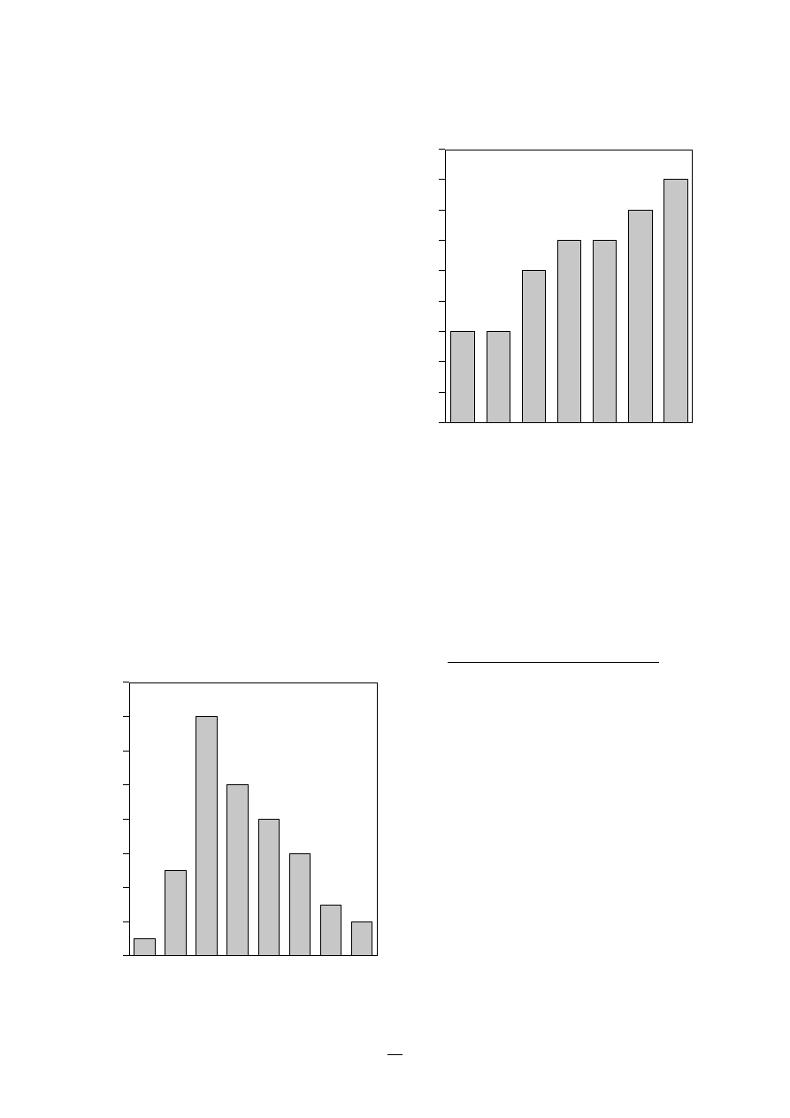
27
The form must be straightforward to use. It is com-
pleted with information extracted from medical
charts, microbiology reports, pharmacy reports and
log books of affected wards. The data collected must
also be checked for validity.
The clinical diagnosis will usually be confirmed
microbiologically. Optimal diagnostic specimens to
be obtained from cases should be described. It may
be appropriate to store selected biological materials
for future analysis in anticipation that new diag-
nostic methods may become available.
To verify the outbreak, the number of cases or iso-
lates observed during the putative outbreak period
is compared with the number of cases (or isolates)
reported during the previous period, or with the
number of cases (or isolates) reported in the same
period of time one month or one year earlier.
4.2.3 Describing the outbreak
The detailed description includes person(s), place, and
time. Cases are also described by other characteris-
tics such as gender, age, date of admission, transfer
from another unit, etc. The graphic representation
of the distribution of cases by time of onset is an
epidemic curve. The epidemic curve should distin-
guish between definite and probable cases. The shape
CHAPTER IV. DEALING WITH OUTBREAKS
FIGURE 1.
Epidemic curve in case of single point
source outbreak*
1–2
5–6
7–8
9–10
11–12 13–14 15–16
0
2
4
6
8
10
12
14
16
Number of cases
Days
3–4
* Adapted from Astagneau P. Duneton P. Management of epidemics of
nosocomial infections. Pathol Biol (Paris) 1998, 46:272–278.
* Adapted from Astagneau P. Duneton P. Management of epidemics of
nosocomial infections. Pathol Biol (Paris) 1998, 46:272–278.
FIGURE 2.
Epidemic curve in case of ongoing
transmission*
Jan
Feb
Mar
Apr
May
Jun
Jul
Months
0
2
4
3
5
6
7
8
9
Number of cases
1
of the epidemic curve may suggest a single point
source (Figure 1), ongoing transmission (Figure 2), or
an intermittent source (Figure 3).
These data allow the calculation of an attack rate,
defined by:
Number of people at risk who are infected
Total number of people at risk
The attack rate can also be calculated stratified by
relevant characteristics such as sex, age, location, or
specific exposure (ventilation, catheterization, oper-
ating rooms, occupational exposure).
At the end of the descriptive analysis, it should be
possible to:
●
formulate a hypothesis on the type of infection
(exogenous, endogenous)
●
tentatively identify the source and route of infec-
tion
●
suggest and implement initial control measures.
4.2.4 Suggesting and testing a hypothesis
This includes identifying a potential exposure (type
and route) for the outbreak and testing this hypoth-
esis using statistical methods. A review of the cur-

PREVENTION OF HOSPITAL-ACQUIRED INFECTIONS: A PRACTICAL GUIDE — WHO/CDS/CSR/EPH/2002.12
28
rent literature may help identify possible routes of
infection for the suspected or known infecting agents.
A case-control study is the most common approach
to hypothesis testing. This compares the frequency
of a risk factor in a group of cases (i.e. individuals
with the nosocomial infection) and in a group of
controls (i.e. individuals without the infection). Con-
trols must be carefully selected to limit bias. Two or
more controls for each case may be necessary to
provide sufficient statistical power. By definition, the
controls are not-cases (individuals without the no-
socomial infection or colonization). Further in-depth
discussion of the selection of controls is described
in several other sources (1,2,3).
The strength of association between exposure and
disease is quantified by the odds ratio in case-
control studies (or the relative risk for cohort stud-
ies), with a 95% confidence interval. The role of
chance, confounding, and bias should be considered
in interpreting results.
TABLE 1.
Immediate control measures for outbreak management
Type of transmission suspected
Suggested action
Cross-transmission (transmission between
Patient isolation and barrier precautions determined by
individuals)
infectious agent(s)
Hand transmission
Improvements in handwashing; cohorting
Airborne agent
Patient isolation with appropriate ventilation
Agent present in water, waterborne agent
Checking of water supply and all liquid containers
Use of disposable devices
Foodborne agent
Elimination of the food at risk
4.2.5 Control measures and follow-up
The aims are:
●
to control the current outbreak by interrupting
the chain of transmission
●
to prevent future occurrence of similar outbreaks.
The selection of control measures (Table 1) is deter-
mined by results of the initial analysis in consulta-
tion with appropriate professionals (infection control
staff, epidemiologist, clinicians, microbiologists, nurs-
ing). This is also an opportunity to initiate or im-
prove a surveillance system to facilitate evaluation
of the efficacy of the control procedures instituted.
Continuous surveillance may be implemented in
high-risk units (see Chapter III).
4.2.6 Communication
During the investigation of an outbreak, timely, up-
to-date information must be communicated to the
1–2
Weeks (i.e. 1–2 : 3 cases between the 1st and the 2nd week)
0
2
4
3
5
6
Number of cases
1
2–3
3–4
4–5
5–6
6–7
7–8
8–9
9–10
10–11
11–12
12–13
13–14
14–15
15–16
16–17
17–18
18–19
19–20
20–21
21–22
22–23
23–24
24–25
FIGURE 3.
Epidemic curve in case of intermittent source*
* adapted from Astagneau P, Duneton P.Management of epidemics of nosocomial infections. Pathol Biol (Paris) 1998, 46:272–278.

29
hospital administration, public health authorities,
and, in some cases, to the public. Information may
be provided to the public and to the media with
agreement of the outbreak team, administration and
local authorities.
A final report on the outbreak investigation should
be prepared. It should describe the outbreak, inter-
ventions, and effectiveness, and summarize the con-
tribution of each team member participating in the
investigation. It should also make recommendations
to prevent future occurrence. This report can be
published in the medical literature, and may be con-
sidered as a legal document.
References
1. Gordis L. Epidemiology. Philadelphia, W.B. Saunders
Company, 1996.
2. Fletcher RH et al. Clinical epidemiology, the essentials.
Baltimore, Williams & Wilkins, 1996.
3. Hennekens CH, Buring JE. Epidemiology in medicine.
Mayrent SL, ed. Boston/Toronto, Little, Brown and
Company, 1987.
CHAPTER IV. DEALING WITH OUTBREAKS

PREVENTION OF HOSPITAL-ACQUIRED INFECTIONS: A PRACTICAL GUIDE — WHO/CDS/CSR/EPH/2002.12
30
CHAPTER V
Prevention of nosocomial infection
P
revention of nosocomial infections requires an
integrated, monitored, programme which in-
cludes the following key components:
●
limiting transmission of organisms between pa-
tients in direct patient care through adequate
handwashing and glove use, and appropriate
aseptic practice , isolation strategies, sterilization
and disinfection practices, and laundry
●
controlling environmental risks for infection
●
protecting patients with appropriate use of pro-
phylactic antimicrobials, nutrition, and vaccina-
tions
●
limiting the risk of endogenous infections by mini-
mizing invasive procedures , and promoting op-
timal antimicrobial use
●
surveillance of infections, identifying and control-
ling outbreaks
●
prevention of infection in staff members
●
enhancing staff patient care practices, and con-
tinuing staff education.
Infection control is the responsibility of all health
care professionals — doctors, nurses, therapists, phar-
macists, engineers and others.
TABLE 1.
Differential nosocomial infection risk by patient and interventions
Risk of infection
Type of patients
Type of procedures
1
Not immunocompromised; no significant
Non-invasive
Minimal
underlying disease
No exposure to biological fluids *
2
Infected patients, or patients with some
Exposure to biological fluids
Medium
risk factors (age, neoplasm)
or
Invasive non-surgical procedure (e.g. peripheral
venous catheter, introduction of urinary catheter)
3
Severely immunocompromised patients,
Surgery
High
(<500 WBC per ml); multiple trauma,
or
severe burns, organ transplant
High-risk invasive procedures (e.g. central venous
catheter, endotracheal intubation)
* Biological fluids include blood, urine, faeces, CSF, fluid from body cavities.
5.1 Risk stratification
(1)
Acquisition of nosocomial infection is determined
by both patient factors, such as degree of immuno-
compromise, and interventions performed which
increase risk. The level of patient care practice may
differ for patient groups at different risk of acquistion
of infection. A risk assessment will be helpful to
categorize patients and plan infection control inter-
ventions.
Tables 1 and 2 provide an example of an approach
which could be customized to a particular facility.
Table 1 stratifies the risk for different patient groups,
and Table 2 provides a hierarchy of patient care prac-
tice for different levels of patient risk.
5.2 Reducing person-to-person transmission
5.2.1 Hand decontamination
The importance of hands in the transmission of hos-
pital infections has been well demonstrated (2), and
can be minimized with appropriate hand hygiene
(3,4,5). Compliance with handwashing, however, is
frequently suboptimal. This is due to a variety of
reasons, including: lack of appropriate accessible
equipment, high staff-to-patient ratios, allergies to

31
handwashing products, insufficient knowledge of
staff about risks and procedures, too long a dura-
tion recommended for washing, and the time
required.
5.2.1.1 Optimal “hand hygiene” requirements
For handwashing:
●
running water: large washbasins which require
little maintenance, with antisplash devices and
hands-free controls
●
products: soap or antiseptic depending on the
procedure
●
facilities for drying without contamination (dis-
posable towels if possible).
For hand disinfection:
●
specific hand disinfectants: alcoholic rubs with
antiseptic and emollient gels which can be ap-
plied to physically clean hands.
5.2.1.2 Procedures
There must be written policies and procedures for
handwashing. Jewellery must be removed before
washing. Simple hygiene procedures may be lim-
ited to hands and wrists; surgical procedures include
the hand and forearm.
Procedures will vary with the patient risk assess-
ment (Table 3):
CHAPTER V. PREVENTION OF NOSOCOMIAL INFECTION
TABLE 2.
Aseptic measures appropriate for different levels of risk of infection
Risk of infection
Asepsis
Antiseptics
Hands
Clothes
Devices*
1
Clean
None
Simple
Street clothes
Clean or
Minimal
handwashing or
disinfected at
hand disinfection
intermediate or
by rubbing
low level
2
Asepsis
Standard
Hygienic
Protection
Disinfected at
Medium
antiseptic
handwashing or
against blood
sterile or high
products
hand disinfection
and biological
level
by rubbing
fluids, as
appropriate
3
Surgical
Specific major
Surgical
Surgical clothes:
Disinfected at
High
asepsis
products
handwashing or
dress, mask, caps, sterile or high
surgical hand
sterile gloves
level
disinfection by
rubbing
* All devices entering sterile body cavities must be sterile.
●
routine care (minimal):
— handwashing with non-antiseptic soap
— or quick hygienic hand disinfection (by rub-
bing) with alcoholic solution
●
antiseptic handcleaning (moderate) — aseptic
care of infected patients:
— hygienic handwashing with antiseptic soap
following manufacturers instructions (e.g. one
minute)
— or quick hygienic hand disinfection: as previ-
ously
●
surgical scrub (surgical care):
— surgical hand and forearm washing with
antiseptic soap and sufficient time and dura-
tion of contact (3–5 minutes)
— or surgical hand and forearm disinfection: sim-
ple handwash and drying followed by two
applications of hand disinfectant, then rub to
dry for the duration of contact defined by the
product.
5.2.1.3 Resource availability
Equipment and products are not equally accessible
in all countries or health care facilities. Flexibility in
products and procedures, and sensitivity to local
needs, will improve compliance. Table 3 provides
suggestions to adapt handwashing for different avail-
ability of resources. In all cases, the maximum pro-
cedure possible should be instituted.

PREVENTION OF HOSPITAL-ACQUIRED INFECTIONS: A PRACTICAL GUIDE — WHO/CDS/CSR/EPH/2002.12
32
5.2.2 Personal hygiene
All staff must maintain good personal hygiene. Nails
must be clean and kept short. False nails should not
be worn. Hair must be worn short or pinned up.
Beard and moustaches must be kept trimmed short
and clean.
5.2.3 Clothing
Working clothes
Staff can normally wear a personal uniform or street
clothes covered by a white coat. In special areas such
as burn or intensive care units, uniform trousers and
a short-sleeved gown are required for men and
TABLE 3.
Hand care and economic constraints
Level
Good resources
Limited resources
Very limited resources
1
Simple handwashing:
Simple handwashing:
Simple handwashing:
Routine
Equipment: large wash-basin,
Equipment: large wash-basin,
Equipment: clean water, locally
(minimal)
water and automatically
water and locally made soap
made soap (dry), towels
distributed washing agent,
(dry), individual towels
washed daily
liquid soap, disposable towels
Hygienic hand disinfection by
Hygienic hand disinfection by
Hygienic hand disinfection by
rubbing:
rubbing:
rubbing:
Specified duration of contact
Specified duration of contact
Specified duration of contact
with hand disinfectant or
with alcohol and rub to dry
between hand and disinfectant,
alcohol, rub to dry
rub to dry
2
Hygienic (or antiseptic)
Hygienic (or antiseptic)
Simple handwashing:
Antiseptic
handwashing:
handwashing:
Equipment: clean water, locally
hand cleaning
Equipment: large wash-basin,
Equipment: large wash-basin,
made soap (dry), towels washed
water and automatically
water and locally made soap
daily
distributed washing agent,
(dry) if antisepsis is undertaken
antiseptic scrub (one-minute
after the washing.
Hygienic hand disinfection
contact), disposable towels
Otherwise: antiseptic scrub
by rubbing:
(1 minute contact), individual
Associated with alcohol
Hygienic hand disinfection
towels
antisepsis, contact and rub to dry
by rubbing:
Specified duration of hand-
Hygienic hand disinfection
disinfectant contact, rub to dry
by rubbing:
Specified duration of contact
with disinfectant or alcohol,
rub to dry
3
Surgical hand-forearm-washing:
Simple hand-forearm-washing:
Simple hand-forearm-washing:
Surgical scrub
Equipment: large wash-basin,
Equipment: large wash-basin,
Equipment: clean water, locally
(maximal)
water and automatically
water and locally made soap
made soap (dry), towels washed
distributed washing agent,
(dry), individual towels
daily
good antiseptic scrub (contact
3 to 5 minutes), sterile
Hygienic hand disinfection
Hygienic hand disinfection
disposable towels
by rubbing:
by rubbing:
Associated with antisepsis:
Associated with alcohol
Surgical hand disinfection
specific hand disinfectant,
antisepsis, repeated twice
by rubbing:
repeated twice
Equipment as for level 2: good
soft soap, specific hand
disinfectant, repeated twice.
women. In other units, women may wear a short-
sleeved dress.
The working outfit must be made of a material easy
to wash and decontaminate. If possible, a clean out-
fit should be worn each day. An outfit must be
changed after exposure to blood or if it becomes
wet through excessive sweating or other fluid expo-
sure.
Shoes
In aseptic units and in operating rooms, staff must
wear dedicated shoes, which must be easy to
clean.

33
Caps
In aseptic units, operating rooms, or performing
selected invasive procedures, staff must wear caps
or hoods which completely cover the hair.
5.2.4 Masks
(6)
Masks of cotton wool, gauze, or paper are ineffec-
tive. Paper masks with synthetic material for filtra-
tion are an effective barrier against microorganisms.
●
Masks are used in various situations; mask re-
quirements differ for different purposes.
●
Patient protection: staff wear masks to work in
the operating room, to care for immuno-compro-
mised patients, to puncture body cavities. A sur-
gical mask is sufficient.
●
Staff protection: staff must wear masks when car-
ing for patients with airborne infections, or when
performing bronchoscopies or similar examina-
tion. A high-efficiency mask is recommended.
●
Patients with infections which may be transmit-
ted by the airborne route must use surgical masks
when outside their isolation room.
5.2.5 Gloves
(6)
Gloves are used for:
●
Patient protection: staff wear sterile gloves for
surgery, care for immunocompromised patients,
invasive procedures which enter body cavities.
●
Non-sterile gloves should be worn for all patient
contacts where hands are likely to be contami-
nated, or for any mucous membrane contact.
●
Staff protection: staff wear non-sterile gloves to
care for patients with communicable disease trans-
mitted by contact, to perform bronchoscopies or
similar examinations.
●
Hands must be washed when gloves are removed
or changed.
●
Disposable gloves should not be reused.
●
Latex or polyvinyl-chloride are the materials most
frequently used for gloves. Quality, i.e. absence of
porosity or holes and duration of use vary con-
siderably from one glove type to another. Sensi-
tivity to latex may occur, and the occupational
health programme must have policies to evalu-
ate and manage this problem.
CHAPTER V. PREVENTION OF NOSOCOMIAL INFECTION
5.2.6 Safe injection practices
To prevent transmission of infections between
patients with injections:
●
eliminate unnecessary injections
●
use sterile needle and syringe
●
use disposable needle and syringes, if possible
●
prevent contamination of medications
●
follow safe sharps disposal practices (Chapter VII,
8.5).
For more information, refer to the WHO guide “Best
infection control practices for skin-piercing intra-
dermal, subcutaneous, and intramuscular needle
injections” (7).
5.3 Preventing transmission from the
environment
To minimize the transmission of microorganisms
from equipment and the environment, adequate
methods for cleaning, disinfecting and sterilizing
must be in place. Written policies and procedures
which are updated on a regular basis must be de-
veloped for each facility.
5.3.1 Cleaning of the hospital environment
(5,6,8)
●
Routine cleaning is necessary to ensure a hospi-
tal environment which is visibly clean, and free
from dust and soil.
●
Ninety per cent of microorganisms are present
within “visible dirt”, and the purpose of routine
cleaning is to eliminate this dirt. Neither soap nor
detergents have antimicrobial activity, and the
cleaning process depends essentially on mechani-
cal action.
●
There must be policies specifying the frequency
of cleaning and cleaning agents used for walls,
floors, windows, beds, curtains, screens, fixtures,
furniture, baths and toilets, and all reused medi-
cal devices.
●
Methods must be appropriate for the likelihood
of contamination, and necessary level of asepsis.
This may be achieved by classifying areas into
one of four hospital zones (8):
— Zone A: no patient contact. Normal domestic
cleaning (e.g. administration, library).

PREVENTION OF HOSPITAL-ACQUIRED INFECTIONS: A PRACTICAL GUIDE — WHO/CDS/CSR/EPH/2002.12
34
— Zone B: care of patients who are not infected,
and not highly susceptible, cleaned by a pro-
cedure that does not raise dust. Dry sweeping
or vacuum cleaners are not recommended. The
use of a detergent solution improves the qual-
ity of cleaning. Disinfect any areas with
visible contamination with blood or body
fluids prior to cleaning.
— Zone C: infected patients (isolation wards).
Clean with a detergent/disinfectant solution,
with separate cleaning equipment for each
room.
— Zone D: highly-susceptible patients (protective
isolation) or protected areas such as operating
suites, delivery rooms, intensive care units,
premature baby units, casualty departments
and haemodialysis units. Clean using a deter-
gent/disinfectant solution and separate clean-
ing equipment.
All horizontal surfaces in zones B, C and D, and all
toilet areas should be cleaned daily.
●
Bacteriological testing of the environment is not
recommended except in selected circumstances
such as:
— epidemic investigation where there is a sus-
pected environmental source
— dialysis water monitoring for bacterial counts,
as required by standards (see Chapter VIII)
— quality control when changing cleaning prac-
tices.
5.3.2 Use of hot/superheated water
An alternative to disinfection for environmental
cleaning for some objects is hot water (Table 4).
TABLE 4.
Disinfection with hot water
Temperature
Duration
1. Sanitary
80
°
C
45–60 seconds
equipment
2. Cooking
80
°
C
1 minute
utensils
3. Linen
70
°
C
25 minutes
95
°
C
10 minutes
5.3.3 Disinfection of patient equipment
Disinfection removes microorganisms without com-
plete sterilization to prevent transmission of organ-
isms between patients. Disinfection procedures must
(5,9,10):
●
meet criteria for killing of organisms
●
have a detergent effect
●
act independently of the number of bacteria
present, the degree of hardness of the water, or
the presence of soap and proteins (that inhibit
some disinfectants).
To be acceptable in the hospital environment, they
must also be:
●
easy to use
●
non-volatile
●
not harmful to equipment, staff or patients
●
free from unpleasant smells
●
effective within a relatively short time.
For further recommendations, see Tables 5 and 6. In
using a disinfectant, manufacturers recommenda-
tions must always be followed. Different products
or processes achieve different levels of disinfection.
These are classified as high-, intermediate- or
low-level disinfection (11); Table 5 provides charac-
teristics of the three levels, and Table 6 makes
recommendations for the level of disinfection for dif-
ferent patient care activity.
High-level disinfection (critical) — this will destroy
all microorganisms, with the exception of heavy con-
tamination by bacterial spores.
Intermediate disinfection (semi-critical) — this
inactivates Mycobacterium tuberculosis, vegetative
bacteria, most viruses and most fungi, but does not
necessarily kill bacterial spores.
Low-level disinfection (non-critical) — this can kill
most bacteria, some viruses and some fungi, but can-
not be relied on for killing more resistant bacteria
such as M. tuberculosis or bacterial spores.
These levels of disinfection are attained by using the
appropriate chemical product in the manner appro-
priate for the desired level of disinfection.
5.3.4 Sterilization
(5–13)
Sterilization is the destruction of all microorganisms.
Operationally this is defined as a decrease in the

35
CHAPTER V. PREVENTION OF NOSOCOMIAL INFECTION
TABLE 5.
Spectrum of activity achieved of the main disinfectants
Level of
Spectrum of
Active ingredients potentially
Factors affecting
disinfection
activity of
capable of satisfying these
the efficacy of
required
desinfectant
spectra of activity
a disinfectant
High
• Sporicidal
• Peracetic acid
• Concentration
• Mycobactericidal
• Chlorine dioxide
• Contact time
• Virucidal
• Formaldehyde
• Temperature
• Fungicidal
• Glutaraldehyde
• Presence of organic matter
• Bactericidal
• Sodium hypochlorite
• pH
• Stabilized hydrogen peroxide
• Presence of calcium or magnesium
• Succinaldehyde (succinic aldehyde)
ions (for example, hardness of the
water used for dilution)
Intermediate
• Tuberculocidal
• Phenol derivatives
• Formulation of the disinfectant
• Virucidal
• Ethyl and isopropyl alcohols
used
• Fungicidal
• Bactericidal
Low
• Bactericidal
• Quaternary ammonium
• Amphiprotic
• Amino acids
TABLE 6.
Level of disinfection for patient equipment in relation with type of care
(11,12)
Devices use
Class
Level of risk
Level of disinfection
Into vascular system, into sterile cavity,
• critical
• high
• sterilization or
into sterile tissues:
high-level disinfection
Surgical instrumentation, e.g. athro-
scopes, biopsies, instrumentation, etc.
Mucous membrane contact,
• semi-critical
• medium
• disinfection of median
non-intact skin:
level
e.g. gastroscopy, etc.
Intact skin or without contact
• non-critical
• low
• disinfection of low level
with patient:
e.g. beds, sink, etc.
microbial load by 10
-6
. Sterilization can be achieved
by either physical or chemical means (Table 7).
●
Sterilization is required for medical devices pen-
etrating sterile body sites, as well as all parenteral
fluids and medications.
●
For reprocessed equipment, sterilization must be
preceded by cleaning to remove visible soil.
●
The object must be wrapped for sterilization. Only
a wrapped sterilized object should be described
as sterile:
Materials for packaging include:
— paper which prevents contamination if intact,
maintains sterility for a long period, can act
as a sterile field, and can also be used to wrap
dirty devices after the procedure
TABLE 7.
Principal sterilization methods
Thermal sterilization
• Wet sterilization: exposure to steam saturated with
water at 121
°
C for 30 minutes, or 134
°
C for 13
minutes in an autoclave; (134
°
C for 18 minutes for
prions).
• Dry sterilization: exposure to 160
°
C for 120 minutes,
or 170
°
C for 60 minutes; this sterilization process is
often considered less reliable than the wet process,
particularly for hollow medical devices.
Chemical sterilization
• Ethylene oxide and formaldehyde for sterilization are
being phased out in many countries because of safety
and greenhouse gas emission concerns.
• Peracetic acid is widely used in the United States and
some other countries in automatic processing
systems.

PREVENTION OF HOSPITAL-ACQUIRED INFECTIONS: A PRACTICAL GUIDE — WHO/CDS/CSR/EPH/2002.12
36
— selected plastics; only polyethylene and poly-
propylene are suitable for sterilization with
ethylene oxide
— non-woven disposable textiles
— containers can be used only if they contain
material intended for a single treatment pro-
cedure for a single patient. They must be pro-
vided with a filter and a valve, which must be
monitored regularly.
●
Packaging systems for sterile items shall meet
local legislation and/or regulations, but must
nevertheless:
— provide adequate seal integrity and be tamper-
proof
— provide an adequate barrier to particulate
matter
— withstand physical conditions of the steriliza-
tion process
— provide an adequate barrier to fluids
— permit adequate air removal
— allow penetration and removal of sterilant
— protect package content from physical dam-
age
— resist tears and punctures
— be free of holes
— be free of toxic ingredients
— have a low lint content
— have a positive cost/benefit ratio
— be used according to the manufacturers’ writ-
ten instructions
— be dated.
●
Proper storage conditions are essential to main-
tain the integrity of sterilized items.
●
The end-user must check the integrity of the pack-
age before use.
●
The sterilization of endoscopes, minimally inva-
sive instruments, and robotic instrumentation is
necessary, but may present a particular challenge
because of the configuration of these instruments.
●
Quality control parameters for the sterilization
process must record information on the steriliza-
tion processing cycle including:
— load number
— load content
— temperature and time exposure record chart
— regular (at least daily) physical/chemical test-
ing
— regular (at least weekly) biological testing
— steam processing (Bacillus stearothermophilus)
— ethylene oxide processing (Bacillus subtilis v.
niger).
●
Regular maintenance must be performed and
documented. The following records must be main-
tained for all sterilization:
— date of service
— model and serial number
— location
— descriptions of replaced parts
— biological testing records
— Bowie-Dick test
— name and signature of controller.
Endoscope reprocessing
Endoscopes are medical devices which may be prob-
lematic to clean and disinfect (long narrow channels,
complex internal design, etc.). Products and/or proc-
esses used (chemical or thermo-chemical disinfection)
may not be as reliable as sterilization methods.
To reduce nosocomial transmission of microorgan-
isms by endoscopy a standard reprocessing proce-
dure must be systematically followed.
1. Immediately after use, the air-water channel should
be cleared with forced air, and tap water or deter-
gent suctioned or pumped through the aspiration/
biopsy channel(s) to remove organic debris.
2. All detachable parts (e.g. hoods and suction valves)
should be removed and soaked in a detergent so-
lution, and the external parts of the endoscopes
gently wiped.
3. All accessible channels should then be irrigated
with tap water or detergent solution, brushed (us-
ing sterile or single use brush) and purged.
4. Before any immersion, the endoscope must be
leak-tested.

37
Endoscope reprocessing continued
After pre-treatment and mechanical cleaning the en-
doscope should be cleaned and disinfected, either
manually or automatically. In both cases, the complete
cycle includes several stages:
5. Cleaning using an approved detergent (this solu-
tion cannot be reused).
6. Rinsing (tap water is sufficient for this in-between
rinsing stage).
7. Disinfection. Using an approved, high level disin-
fectant.
Regarding CJD risk, a disinfectant with protein-
fixative properties (i.e. aldehyde-based products)
should not be used. A non-fixative desinfectant
should be selected.
8. Rinsing: The level of microbial purity of the water
used depends on the further use of the endoscope
(bacteriologically controlled water or sterile
water).
9. Drying: If the endoscope is not stored, this drying
stage includes only air-blowing the channel to re-
move residual water.
Note: new French guidelines regarding variant
Creutzfeldt-Jakob (CJD) risk recommend to clean and
rinse the endoscope twice before disinfection.
References
1. Underwood MA, Pirwitz S. APIC guidelines com-
mittee: using science to guide practice. Am J Infect
Control, 1998, 26:141–144.
2. Larson E. A causelink between handwashing and
risk of infection? Examination of the evidence.
Infect Control Hosp Epidemiol, 1988, 9:28–36.
3. CDC guidelines for handwashing and hospital
environmental control. Amer J Infect Control, 1986,
14:110–129 or Infect Control, 1986, 7:231–242.
4. Larson EL. APIC guideline for handwashing and
hand antisepsis in health care settings. Amer J In-
fect Control, 1995, 23:251–269.
5. Health Canada. Hand washing, cleaning, disin-
fection, and sterilization in health care. Canada
Communicable Disease Report (CCDR), Supplement,
Vol., 24S4, July 1998.
6. Pratt RJ et al. The epic project: Developing na-
tional evidence-based guidelines for preventing
healthcare associated infections. Phase I: Guide-
lines for preventing hospital-acquired infections.
J Hosp Infect, 2001, 47(Supplement):S3–S4.
7. World Health Organization. Best infection control
practices for skin-piercing intradermal, subcutaneous, and
intramuscular needle injections. 2001, WHO/BCT/
DCT/01.02.
8. Ducel G et al. Practical guide to the prevention of hospi-
tal-acquired infections. 1979, WHO/BAC/79.1.
9. Association of Operating Room Nurses. Proposed
recommended practices for chemical disinfection.
AORN J, 1994, 60: 463–466.
10. Rutala WA. APIC guideline for selection and use
of disinfectants. Amer J Infect Control, 1996, 24:313–
342.
11. Alvarado CJ, Reichelderfer M and the 1997, 1998,
1999 APIC Guidelines Committees. APIC guide-
line for infection prevention and control in flex-
ible endoscopy. Amer J Infect Control, 2000,
26:138–155.
12. Galtier F. La stérilisation hospitalière, 2ème édition.
Paris, Maloine, 1998.
13. Medical Devices Agency. Department of Health (UK)
sterilization, disinfection, and cleaning of medical equip-
ment: Guidance on decontamination. London, Depart-
ment of Health, 1996.
CHAPTER V. PREVENTION OF NOSOCOMIAL INFECTION

PREVENTION OF HOSPITAL-ACQUIRED INFECTIONS: A PRACTICAL GUIDE — WHO/CDS/CSR/EPH/2002.12
38
CHAPTER VI
Prevention of common endemic
nosocomial infections
6.1 Urinary tract infections (UTI)
Urinary tract infections are the most frequent noso-
comial infections (1); 80% of these infections are
associated with an indwelling urethral catheter
(Figure 1). Interventions effective in preventing no-
socomial urinary infection include (2,3,4):
●
avoiding urethral catheterization unless there is
a compelling indication
T
he four most common nosocomial infections are
urinary tract infections, surgical wound infec-
tions, pneumonia, and primary bloodstream
infection. Each of these is associated with an inva-
sive medical device or invasive procedure. Specific
policies and practices to minimize these infections
must be established, reviewed and updated regu-
larly, and compliance monitored (Table 1).
TABLE 1.
Measures for prevention of infection
I
nfection
Proven effective
Proven not effective
Urinary tract
Limit duration of catheter
Systemic antibiotic prophylaxis
infections
Aseptic technique at insertion
Bladder irrigation or instillation of normal saline
Maintain closed drainage
antiseptic or antibiotic
Antiseptic added to drainage bag
Antimicrobial-coated catheter
Daily antiseptic perineal cleaning
Surgical site
Surgical technique
Fumigation
infections
Clean operating environment
Preoperative shaving
Staff attire
Limiting preoperative hospital stay
Preoperative shower and local skin
preparation of patient
Optimal antibiotic prophylaxis
Aseptic practice in operating room
Surgical wound surveillance
Pneumonia
Ventilator-associated
Digestive decontamination for all patients
Aseptic intubation and suctioning
Changes of ventilator circuit every 48 or
Limit duration
72 hours
Non-invasive ventilation
Others
Influenza vaccination for staff
Isolation policy
Sterile water for oxygen and aerosol therapy
Prevention of Legionella and Aspergillus
during renovations
Vascular device
All catheters
Antimicrobial creams for skin preparation
infections
Closed system
Limit duration
Local skin preparation
Aseptic technique at insertion
Removal if infection suspected
Central lines
Surgical asepsis for insertion
Limitation of frequency of dressing change
Antibiotic-coated catheter for short term
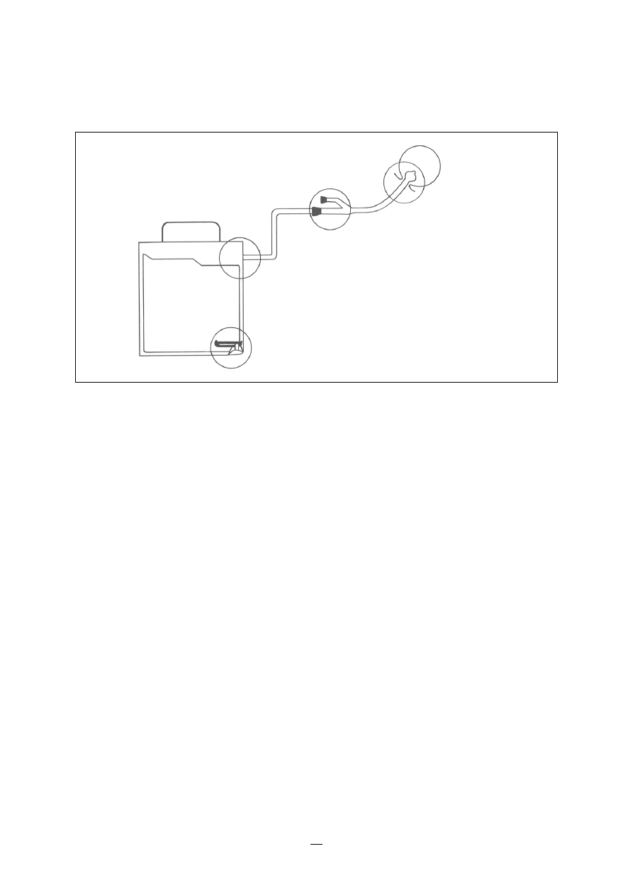
39
●
limiting the duration of drainage, if catheteriza-
tion is necessary
●
maintaining appropriate aseptic practice during
urinary catheter insertion and other invasive
urological procedures (e.g. cystoscopy, urodynamic
testing, cystography)
●
hygienic handwash or rub prior to insertion and
following catheter or drainage bag manipulation
(Chapter V)
●
sterile gloves for insertion
●
perineal cleaning with an antiseptic solution prior
to insertion
●
non-traumatic urethral insertion using an appro-
priate lubricant
●
maintaining a closed drainage system.
Other practices which are recommended, but not
proven to decrease infection include:
●
maintaining good patient hydration
●
appropriate perineal hygiene for patients with
catheters
●
appropriate staff training in catheter insertion and
care
●
maintaining unobstructed drainage of the blad-
der to the collection bag, with the bag below the
level of the bladder.
Generally, the smallest diameter catheter should be
used. Catheter material (latex, silicone) does not in-
fluence infection rates.
For patients with a neurogenic bladder:
●
avoid an indwelling catheter if possible
●
if assisted bladder drainage is necessary, clean
intermittent urinary catheterization should be
used.
6.2 Surgical wound infections (surgical site
infections)
Factors which influence the frequency of surgical
wound infection include (5,6,7,8):
●
surgical technique
●
extent of endogenous contamination of the
wound at surgery (e.g. clean, clean-contaminated)
●
duration of operation
●
underlying patient status
●
operating room environment
●
organisms shed by the operating room team.
A systematic programme for prevention of surgical
wound infections (5) includes the practice of opti-
mal surgical technique, a clean operating room en-
vironment with restricted staff entry and appropriate
CHAPTER VI. PREVENTION OF COMMON ENDEMIC NOSOCOMIAL INFECTIONS
Reproduced by permission of Wiley&Sons, Inc. from Hospital
Infection Control: Principles and Practice, M. Castle, Copyright © 1980
by John Wiley & Sons, Inc.
FIGURE 1.
Portals of entry for microorganisms in urinary drainage systems: the urethral meatus-catheter
junction; the catheter-drainage tubing junction; the drainage tubing-bag junction; and
the outlet that drains urine from the bag
Urethral meatus–
catheter junction
Catheter–drainage
tubing junction
Drainage–tubing
bag junction
Outlet

PREVENTION OF HOSPITAL-ACQUIRED INFECTIONS: A PRACTICAL GUIDE — WHO/CDS/CSR/EPH/2002.12
40
staff attire, sterile equipment, adequate preoperative
preparation of the patient, appropriate use of
preoperative antimicrobial prophylaxis, and a sur-
gical wound surveillance programme. Surgical
wound infection rates are decreased by standard-
ized surveillance for infection with reporting of rates
back to individual surgeons.
6.2.1 Operating room environment
Airborne bacteria must be minimized, and surfaces
kept clean. A recommended schedule for cleaning
and disinfection of the operating theatre is:
●
every morning before any intervention: cleaning of all
horizontal surfaces
●
between procedures: cleaning and disinfection of hori-
zontal surfaces and all surgical items (e.g.
tables, buckets)
●
at the end of the working day: complete cleaning of
the operating theatre using a recommended dis-
infectant cleaner
●
once a week: complete cleaning of the operating
room area, including all annexes such as dress-
ing rooms, technical rooms, cupboards.
All items used within a sterile field must be sterile.
Sterile drapes must be placed on the patient and on
any equipment included in the sterile field; these
drapes must be handled as little as possible. Once a
sterile drape is in position, it must not be moved;
shifting or moving the sterile drape compromises
the sterile field.
For selected high-risk surgery (e.g. orthopaedic pro-
cedures with implants, transplantation) further
specific measures for operating room ventilation may
be considered (Chapter VIII).
6.2.2 Operating room staff
6.2.2.1 Handwashing
A surgical hand disinfection should be performed
by all persons participating in the operative proce-
dure (Chapter V).
6.2.2.2 Operating room attire
Operating staff must wear sterile gloves. The reported
occurrence of glove punctures ranges from 11.5% to
53% of procedures (9), and double gloving is there-
fore advisable for procedures with a high risk of
puncture, such as total joint arthroplasty. Double
gloving is also recommended when operating on
patients known to be infected with bloodborne
pathogens such as the human immunodeficiency
virus (HIV), hepatitis B, or hepatitis C (10). Gloves
should be changed immediately after any acciden-
tal puncture.
All persons entering the surgical theatre must wear
surgical attire restricted to being worn only within
the surgical area. The design and composition of
surgical attire should minimize bacterial shedding
into the environment.
All head and facial hair, including sideburns, and
neckline, must be covered. All personnel entering in
the operating suite must remove any jewellery; nail
polish or artificial nails must not be worn.
Full coverage of the mouth and nose area with a
surgical mask for everyone entering the operating
suite (11).
Sterile surgical gowns must be worn by all persons
participating directly in the operation. Waterproof
gowns or aprons should be worn for procedures at
high risk of blood contamination.
6.2.2.3 Operating room activitiy
●
The number of persons entering the theatre dur-
ing an operation should be minimized.
●
Unnecessary movement or conversation should
be avoided.
6.2.3 Pre-intervention preparation of the
patient
For elective procedures, any existing infections
should be identified and treated before surgery. The
preoperative stay should be minimized. Any mal-
nourished patient should have nutrition improved
before elective surgery.
The patient should normally be bathed or showered
on the evening before the intervention, using an
antimicrobial soap. If hair removal is required, this
should be done by clipping or with a depilatory
rather than by shaving (5,12).
The operative site must be washed with soap and
water, then an antimicrobial preoperative skin prepa-
ration applied from the centre to the periphery.
The area prepared must be large enough to include
the entire incision and adjacent skin sufficient for

41
the surgeon to work without contacting unprepared
skin.
The patient must be covered with sterile drapes; no
part is uncovered except the operating field and
areas needed for the administration and maintenance
of anaesthesia.
6.2.4 Antimicrobial prophylaxis (see Chapter IX)
6.2.5 Surgical wound surveillance (see also
Chapter III)
●
Prospective surgical wound surveillance should
be undertaken for selected procedures.
●
Infection rates should be stratified by the extent
of endogenous bacterial contamination at surgery:
clean, clean-contaminated, or dirty.
●
Surgical wound infection rates may also be strati-
fied by duration of operation and underlying
patient status.
●
Individual surgeons should be provided their own
surgical wound infection rates in a confidential
manner, with a comparator of overall rates for
the facility or region.
6.3 Nosocomial respiratory infections
(13)
Nosocomial respiratory tract infections occur in dif-
ferent patient groups (10). In some cases, the hospi-
tal environment may play a significant role (see
Chapter VIII). Recommendations to prevent these
infections include:
6.3.1 Ventilator-associated pneumonia in the
intensive care unit
●
Appropriate disinfection and in-use care of tub-
ing, respirators, and humidifiers to limit contami-
nation.
●
No routine changes of respirator tubing.
●
Avoid antacids and H2 blockers.
●
Sterile tracheal suctioning.
●
Nurse in head-up position.
6.3.2 Medical units
●
Limit medications which impair consciousness
(sedatives, narcotics).
●
Position comatose patients to limit the potential
for aspiration.
●
Avoid oral feeds in patients with swallowing ab-
normalities.
●
Prevent exposure of neutropenic or transplant
patients to fungal spores during construction or
renovation (Chapter VIII).
6.3.3 Surgical units
●
All invasive devices used during anaesthesia must
be sterile.
●
Anaesthetists must use gloves and mask when
undertaking invasive tracheal or venous or epi-
dural care. Disposable filters (for individual use)
for endotracheal intubation effectively prevent the
transmission of microorganisms among patients
by ventilators.
●
Preoperative physiotherapy prevents postopera-
tive pneumonia in patients with chronic respira-
tory disease.
6.3.4 Neurological patients with tracheostomy
(with or without ventilation)
●
Sterile suctioning at appropriate frequency.
●
Appropriate cleaning and disinfection of respira-
tory machines and other devices.
●
Physiotherapy to assist with drainage of secre-
tions.
6.4 Infections associated with intravascular
lines
(3,14–16)
Local (exit site, tunnel) and systemic infections may
occur (Figure 2). They are most common in inten-
sive care units (14). Key practices for all vascular cath-
eters include:
●
avoiding catheterization unless there is a medical
indication
●
maintaining a high level of asepsis for catheter
insertion and care
●
limiting the use of catheters to as short a dura-
tion as possible
●
preparing fluids aseptically and immediately
before use
●
training of personnel in catheter insertion and
care.
CHAPTER VI. PREVENTION OF COMMON ENDEMIC NOSOCOMIAL INFECTIONS
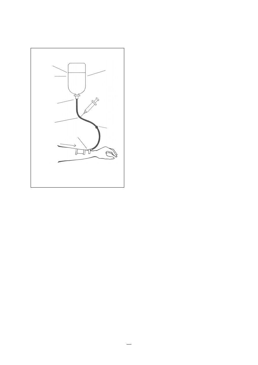
PREVENTION OF HOSPITAL-ACQUIRED INFECTIONS: A PRACTICAL GUIDE — WHO/CDS/CSR/EPH/2002.12
42
6.4.1 Peripheral vascular catheters
●
Hands must be washed before all catheter care,
using hygienic handwash or rub (Chapter V).
●
Wash and disinfect skin at the insertion site with
an antiseptic solution.
●
Intravenous line changes no more frequently than
change of catheters, with the exception of line
changes after the transfusion of blood or
intralipids, and for discontinuous perfusions.
●
A dressing change is not normally necessary.
●
If local infection or phlebitis occurs, the catheter
should be removed immediately.
6.4.2 Central vascular catheters
●
Clean the insertion site with an antiseptic solu-
tion.
●
Do not apply solvents or antimicrobial ointment
to the insertion site.
FIGURE 2.
Portals of entry for microorganisms in
IV systems
Reproduced by permission of Wiley&Sons, Inc. from Hospital Infec-
tion Control: Principles and Practice, M. Castle, Copyright© 1980 by
John Wiley & Sons , Inc.
●
Mask, cap, and sterile gloves and gown must be
worn for insertion.
●
The introduction of the catheter and the subse-
quent catheter dressings require a surgical hand
wash or rub.
●
Follow appropriate aseptic care in accessing the
system, including disinfecting external surfaces of
hub and ports.
●
Change of lines should normally not occur more
often than once every three days. A change of
line is necessary, however, after the transfusion of
blood, blood products, or intralipids, and for dis-
continuous perfusions.
●
Change dressing at the time of the change of lines,
following surgical asepsis.
●
Use a sterile gauze or transparent dressing to cover
the catheter site.
●
Do not replace over a guide wire if infection is
suspected.
●
An increased number of catheter lumens may
increase the risk of infection. A single lumen cath-
eter is preferred wherever possible.
●
Antimicrobial impregnated catheters may decrease
infection in high-risk patients with short-term
(<10 days) catheterization.
●
Use the subclavion site in preference to jugular
or femoral sites.
●
Consider using a peripherally inserted central
catheter, if appropriate.
6.4.3 Central vascular totally implanted
catheters
Implantable vascular access devices should be con-
sidered for patients who require long-term (>30 days)
therapy. Additional preventive practices for these
patients include:
●
a preoperative shower and implantation under
surgical conditions in an operating room
●
local preparation includes washing and antisep-
sis with major antiseptic solution as for other sur-
gical procedures
●
mask, hat, and sterile gloves and gown must be
worn; the introduction of a catheter and the dress-
ing require a surgical handwash or rub
●
maintain a closed system during the use of the
During
manufacture
Additives
Stopcock
Insertion
site
Secondary
infection from
other side
Medication
port
Bottle (bag)–tubing
junction
Hairline cracks
or punctures

43
device; a change of lines should normally occur
every 5 days for continuous use, and at each in-
tervention for intermittent use; a change of line is
necessary after the transfusion of blood, and for
discontinuous perfusions.
References
1. Kunin CM. Urinary tract infection detection, prevention
and management, fifth edition. Baltimore, Williams
& Wilkins, 1997.
2. CDC guideline for the prevention of catheter-
associated urinary tract infections. Am J Infect Con-
trol, 1983,11:28–33.
3. Pratt RJ et al. The epic project: Developing na-
tional evidence-based guidelines for preventing
healthcare associated infections. Phase I: Guide-
lines for preventing hospital-acquired infections.
J Hosp Infect, 2001, 47(Supplement):S3–S4.
4. Falkiner FR. The insertion and management of
indwelling urethral catheter — minimizing the risk
of infection. J Hosp Infect, 1993, 25:79–90.
5. Mangram AJ et al. Guideline for prevention of
surgical site infection. Am J Infect Control, 1999,
27:97–132.
6. Cruse PJE, Ford R. The epidemiology of wound
infections. A 10 year prospective study of 62,939
wounds. Surg Clin North Am, 1980, 60:27–40.
7. Pittet D, Ducel G. Infectious risk factors related to
operating rooms. Infect Control Hosp Epidemiol, 1994,
15:456–462.
CHAPTER VI. PREVENTION OF COMMON ENDEMIC NOSOCOMIAL INFECTIONS
8. Garibaldi R et al. The impact of preoperative skin
disinfection of preventing intraoperative wound
contamination. Infect Control Hosp Epidemiol, 1988,
9:109–113.
9. Dodds RDA et al. Surgical glove perforation. Brit
J Surg, 1988, 75:966–968.
10. Caillot JL et al. Electronic evaluation of the value
of the double gloving. Brit J Surg, 1999, 86:1387–
1390.
11. Caillaud JL, Orr NWM. A mask necessary in the
operating room? Ann R. Coll Surg Engl, 1981, 63:390–
392.
12. Mayhall CG. Surgical infections including burns
in: R. P. Wenzel, ed. Prevention and Control of Nosoco-
mial infections. Baltimore, Williams & Wilkins,
1993:614–644.
13. Tablan OC et al. Guideline for prevention of
nosocomial pneumonia. The Hospital Infection
Control Practices Advisory Committee, Centers
for Disease Control and Prevention. Am J Infect
Control, 1994, 22:247–292.
14. van Wijngaerden E, Bobbaers H. Intravascular
catheter related bloodstream infection: epidemi-
ology, pathogenesis and prevention. Acta Clin Belg,
1997, 52:9–18. Review.
15. Pearson ML. Guideline for prevention of intra-
vascular device-related infections. Hospital In-
fection Control Practices Advisory Committee.
Infect Control Hosp Epidemiol, 1996, 17:438–473.
16. Health Canada. Preventing infections associated
with indwelling intravascular access devices. Can
Commun Dis Rep, 1997, 23 Suppl 8: i–iii, 1–32, i–
iv,1–16.

PREVENTION OF HOSPITAL-ACQUIRED INFECTIONS: A PRACTICAL GUIDE — WHO/CDS/CSR/EPH/2002.12
44
CHAPTER VII
Infection control precautions
in patient care
Standard precautions for all patients
(3,4)
•
Wash hands promptly after contact with infective
material
•
Use no touch technique wherever possible
•
Wear gloves when in contact with blood, body
fluids, secretions, excretions, mucous membranes
and contaminated items
•
Wash hands immediately after removing gloves
•
All sharps should be handled with extreme care
•
Clean up spills of infective material promptly
•
Ensure that patient-care equipment, supplies and
linen contaminated with infective material is
either discarded, or disinfected or sterilized be-
tween each patient use
•
Ensure appropriate waste handling
•
If no washing machine is available for linen soiled
with infective material, the linen can be boiled.
Considerations for protective clothing include:
– gown: should be of washable material, but-
toned or tied at the back and protected, if nec-
essary, by a plastic apron
– gloves: inexpensive plastic gloves are avail-
able and usually sufficient
– mask: surgical masks made of cloth or paper
may be used to protect from splashes.
7.1.2 Additional precautions for specific modes
of transmission
(1,2)
The following precautions are used for selected
patients in addition to those described above:
S
elected patients may require specific precautions
to limit transmission of potential infecting or-
ganisms to other patients.
Recommended isolation precautions depend on the
route of transmission (1). The main routes are:
●
Airborne infection: the infection usually occurs
by the respiratory route, with the agent present
in aerosols (infectious particles <5
µ
m in diam-
eter).
●
Droplet infection: large droplets carry the infec-
tious agent (>5
µ
m in diameter).
●
Infection by direct or indirect contact: infection
occurs through direct contact between the source
of infection and the recipient or indirectly through
contaminated objects.
7.1 Practical aspects
Isolation and other barrier precautions must be
clearly written policies which are standardized, and
adaptable to the infectious agent and the patients.
These include:
– standard or routine precautions to be followed
for all patients
– additional precautions for selected patients.
7.1.1 Standard (routine) precautions
(1,2)
To be applied to the care of all patients. This in-
cludes limiting health care worker contact with all
secretions or biological fluids, skin lesions, mucous
membranes, and blood or body fluids. Health care
workers must wear gloves for each contact which
may lead to contamination, and gowns, mask and
eye protection where contamination of clothes or
the face is anticipated.

45
Airborne precautions (droplet nuclei <5
µ
m) (e.g.
tuberculosis, chickenpox, measles) (5,6)
The following is required:
●
individual room with adequate ventilation; this
includes, where possible, negative pressure; door
closed; at least six air exchanges per hour; ex-
haust to outside away from intake ducts
●
staff wearing high-efficiency masks in room
●
patient to stay in room.
Droplet precautions (droplet nuclei >5
µ
m) (e.g. bac-
terial meningitis, diphtheria, respiratory syncytial
virus)
The following procedures are required:
●
individual room for the patient, if available
●
mask for health care workers
●
restricted circulation for the patient; patient wears
a surgical mask if leaving the room.
Contact precautions
These are required for patients with enteric infec-
tions and diarrhoea which cannot be controlled, or
skin lesions which cannot be contained.
●
individual room for the patient if available;
cohorting of patients if possible
●
staff wear gloves on entering the room; a gown
for patient contact or contact with contaminated
surfaces or material
●
wash hands before and after contact with the
patient, and on leaving the room
●
restrict patient movement outside the room
●
appropriate environmental and equipment clean-
ing, disinfection, and sterilization.
Absolute (strict) isolation (e.g. haemorrhagic fever,
vancomycin-resistant S. aureus) (7,8)
Such isolation is required where there is risk of in-
fection by a highly virulent or other unique agent
of concern where several routes of transmission are
implicated.
●
individual room, in an isolation ward if possible
●
mask, gloves, gowns, cap, eye protection for all
entering the room
●
hygenic handwashing at entry to and exit from
the room
●
incineration of needles, syringes
●
disinfection of medical instruments
●
incineration of excreta, body fluids, nasopharyn-
geal secretions
●
disinfection of linen
●
restrict visitors and staff
●
daily disinfection and terminal disinfection at the
end of the stay
●
use of disposable (single-use) equipment
●
appropriate transport and laboratory manage-
ment of patient specimens.
7.2 Antimicrobial-resistant microorganisms
The increased occurrence of antimicrobial-resistant
microorganisms (i.e. methicillin-resistant S. aureus
(9,10) or vancomycin-resistant enterococci [VRE])
(11,12) is a major medical concern. The spread of
multiresistant strains of S. aureus and VRE is usually
by transient carriage on the hands of health care
workers.
The following precautions are required for the pre-
vention of spread of epidemic MRSA:
●
minimize ward transfers of staff and patients
●
ensure early detection of cases, especially if
admitted from another hospital; screening of high-
risk patients may be considered
●
isolate infected or colonized patients in a single
room, isolation unit or cohorting in a larger ward
●
re-enforce handwashing by staff after contact with
infected or colonized patients; consider using an
antiseptic handwashing agent
●
use gloves for handling MRSA-contaminated
materials, or infected or colonized patients
●
wear gown or apron when handling contaminated
materials or infected or colonized patients
●
consider treating nasal carriers with mupirocin
●
consider antiseptic detergent daily wash or bath
for carriers or infected patients
●
ensure careful handling and disposal of medical
devices, linen, waste, etc.
●
develop guidelines specifying when isolation
measures can be discontinued.
CHAPTER VII. INFECTION CONTROL PRECAUTIONS IN PATIENT CARE

PREVENTION OF HOSPITAL-ACQUIRED INFECTIONS: A PRACTICAL GUIDE — WHO/CDS/CSR/EPH/2002.12
46
References
1. Garner JS. Guideline for isolation precautions in
hospitals. Infect Control Hosp Epidemiol, 1996, 17:54–
65.
2. Health Canada. Routine practices and additional
precautions for preventing transmission of in-
fection in health care. Can Commun Dis Rep, 1999,
25 Suppl 4:1–142.
3. IFIC Newsletter, December 1996, Volume 8, No. 2.
4. Guide to preventing HIV transmission in health facilities.
World Health Organization Global Programme
on AIDS, 1995.
5. CDC/TB www.cdc.gov/ncidod/hip/guide/tuber.
htm
6. Health Canada. Guidelines for preventing the
transmission of tuberculosis in Canadian health
care facilities and other institutional settings. Can
Commun Dis Rep, 1996, 22 S1:i–iv,1–50, i–iv,1–55.
7. CDC. Management of patients with suspected
viral hemorrhagic fever. MMWR, 1998, 37(S–3):
1–6.
8. Health Canada. Canadian contingency plan for
viral haemorrhagic fevers and other related dis-
eases. Can Commun Dis Rep, 1997, 23 S1: i–iii ,1–13,
i–iii, 1–13.
9. Ayliffe GAJ. Recommendations for the control of methi-
cillin-resistant Staphylococcus aureus (MRSA).
WHO/EMC/LTS/96.1.
10. Working party report. Revised guidelines for the
control of methicillin-resistant Staphylococcus aureus
infection in hospitals. J Hosp Infect, 1998, 39:253–
290.
11. CDC recommendations for preventing the spread
of vancomycin-resistance: Recommendations of
the Hospital Infection Control Practices Advisory
Committee (HICPAC). MMWR, 1995, 44(RR–12):
1–12 or Infect Control Hosp Epidemiol, 1995, 16:105–
113.
12. Health Canada. Preventing the spread of vanco-
mycin-resistant enterococci in Canada. Can
Commun Dis Rep, 1997 ,23 S8: i–iv,1–16, i–iv,1–19.

47
CHAPTER VIII
Environment
●
appropriate potable water systems to limit
Legionella spp.
8.1.2 Architectural segregation
It is useful to stratify patient care areas by risk of the
patient population for acquisition of infection. For
some units, including oncology, neonatology, inten-
sive care, and transplant units special ventilation may
be desirable.
Four degrees of risk may be considered:
A – Low-risk areas: e.g. administrative sections
B – Moderate-risk areas: e.g. regular patient units
C – High-risk-areas: e.g. isolation unit, intensive care
units
D – Very-high-risk areas: e.g. operating rooms
Infected patients must be separated from immuno-
compromised patients. Similarly, in a central sterili-
zation unit or in a hospital kitchen, contaminated
areas must not compromise non-contaminated
areas.
8.1.3 Traffic flow
(3)
A room or space, whatever its purpose, is never com-
pletely separate. However, a distinction can be made
between high-traffic and low-traffic areas. One can
consider general services (food and laundry, sterile
equipment, and pharmaceutical distribution), spe-
cialized services (anaesthesiology, medical imaging,
medical or surgical intensive care) and other areas.
A hospital with well-defined areas for specific
activities can be described using flowcharts depict-
ing the flow of in- or outpatients, visitors, staff
(physicians, nurses and paramedics), supplies (ex-
pendable, sterile, catering, clothing, etc.) as well as
T
he discussion of the environment will include
building features, ventilation, water, food and
wastes. Housekeeping and equipment are discussed
in Chapter V.
8.1 Buildings
Health services — including public and private hos-
pital services — must meet quality standards (ISO
9000 and ISO 14000 series) (1). It is recognized that
older facilities, and facilities in developing countries,
may not be able to achieve these standards. How-
ever, the principles underlying these standards
should be kept in mind for local planning and, where
possible, renovations should attempt to achieve
standards.
8.1.1 Planning for construction or renovation
(2,11)
An infection control team member should partici-
pate on the planning team for any new hospital con-
struction or renovation of existing facilities. The role
of infection control in this process is to review and
approve construction plans to ensure they meet
standards for minimizing nosocomial infections.
Considerations will usually include:
●
traffic flow to minimize exposure of high-risk
patients and facilitate patient transport
●
adequate spatial separation of patients
●
adequate number and type of isolation rooms
●
appropriate access to handwashing facilities
●
materials (e.g. carpets, floors) that can be ad-
equately cleaned
●
appropriate ventilation for isolation rooms and
special patient care areas (operating theatres,
transplant units)
●
preventing patient exposure to fungal spores with
renovations

PREVENTION OF HOSPITAL-ACQUIRED INFECTIONS: A PRACTICAL GUIDE — WHO/CDS/CSR/EPH/2002.12
48
the flow of air, liquids and wastes. Other traffic pat-
terns may also be identified. Building or rebuilding
a hospital requires consideration of all physical
movements and communications, and where con-
tamination may occur.
In this context, rather than considering a “clean” and
a “dirty” circuit, consider only circuits where the
different flows can cross without risk provided ma-
terial is properly protected. An elevator can accom-
modate hospital staff, sterile equipment, visitors and
waste, as long as each of these is treated appropri-
ately. Both sterile products and waste must be sealed
in safe containers, and the outside of those contain-
ers must present no risk of biological contamina-
tion.
8.1.4 Materials
The choice of construction materials — especially
those considered in the covering of internal surfaces
— is very important. Floor coverings must be easy to
clean and resistant to disinfection procedures. This
also applies to all items in the patient environment.
All of this calls for:
1. Definition of needs (planning)
2. Definition of the level of risk (segregation)
3.
Description of functional flow patterns (flows and
isolation)
4.
Building or rebuilding (materials)
8.2 Air
8.2.1 Airborne contamination and transmission
Infection may be transmitted over short distances
by large droplets, and at longer distances by droplet
nuclei generated by coughing and sneezing (4). Drop-
let nuclei remain airborne for long periods, may dis-
seminate widely in an environment such as a hospital
ward or an operating room, and can be acquired by
(and infect) patients directly, or indirectly through
contaminated medical devices.
Housekeeping activity such as sweeping, using dry
dust mops or cloths, or shaking out linen, can aero-
solize particles that may contain microorganisms.
Similarly, Legionella pneumophila, the organism respon-
sible for legionellosis (Legionnaires’ disease; Pontiac
fever), can become airborne during the evaporation
of water droplets from air conditioning cooling tow-
ers or with aerosolization in patient showers, and
subsequently may be inhaled by patients at risk of
infection.
The number of organisms present in room air will
depend on the number of people occupying the
room, the amount of activity, and the rate of air ex-
change. Bacteria recovered from air samples usually
consist of Gram-positive cocci originating from the
skin. They can reach large numbers if dispersed from
an infected lesion, particularly an infected exfolia-
tive skin lesion. However, since the contaminated
skin scales are relatively heavy, they do not remain
suspended in the air for long. Gram-negative bacte-
ria are usually found in the air only when associ-
ated with aerosols from contaminated fluids, and
tend to die on drying.
Droplets projected from the infected upper respira-
tory tract may contain a wide variety of
microrganisms, including viruses, and many infec-
tions can be spread by this route (i.e. respiratory vi-
ruses, influenza, measles, chickenpox, tuberculosis).
In most cases, these are spread by large droplets,
and an infective dose will rarely move more than a
few feet from the source patient. Varicella-zoster
(chickenpox), tuberculosis, and a few other agents,
however, may be transmitted over large distances in
droplet nuclei.
8.2.2 Ventilation
Fresh filtered air, appropriately circulated, will
dilute and remove airborne bacterial contamination.
It also eliminates smells. Desirable ventilation rates,
expressed in air changes per hour, vary with the
purpose of a particular area (5). High-risk hospital
areas (operating rooms, nurseries, intensive care
units, oncology, and burn units) should have air with
minimal bacterial contamination.
●
Adequate ventilation systems require proper de-
sign and maintenance to minimize microbial con-
tamination. All outdoor air inlets must be located
as high as possible above ground level; inlets must
be remote from ventilation discharge outlets,
incinerators, or boiler stacks.
●
Within rooms, the location of air inlets and ex-
haust outlets influences the movement of air. High
wall or ceiling inlets and low wall outlets allow
clean air to move downward through the area
toward the contaminated floor where it is removed
through the low exhaust. This pattern is for all

49
areas where high-risk patients receive care, and
in areas subject to heavy contamination.
●
Filters used in the ventilation systems must meet
standards for the patient care activity of the area.
High-efficiency filters must be provided in sys-
tems serving areas where patients are particularly
susceptible to infection (haematology/oncology
units) or where some clinical procedures subject
patients to unusual hazard (for instance surgical
procedure, particularly transplantation).
●
Regular inspection and maintenance of filters,
humidifiers, and grills in the ventilation system
must be performed and documented.
●
Cooling towers and humidifiers should be regu-
larly inspected and cleaned to prevent aerosoli-
zation of Legionella spp.
●
Zoning of air systems may confine the air of a
department to that department alone. A design
that enables air pressure to control air movement
into or out of a specific room or area will control
the spread of contamination. Positive air pressure
is recommended for areas which must be as clean
as possible. It is achieved by supplying more air
into an area than can be removed by the exhaust
ventilation system. This produces an outflow
around doors and other openings, and decreases
entry of air from more contaminated areas. Nega-
tive air pressure is recommended for contami-
nated areas, and is required for isolation of
patients with infections spread by the airborne
route. It is achieved by supplying less air to the
area than can be removed by the ventilation sys-
tem. Negative air pressure produces an inflow
around openings and reduces the movement of
contaminated air out of the area. For effective air
pressurization all doors must be kept closed ex-
cept for essential entrances and exits.
8.2.3 Operating theatres
Modern operating rooms which meet current air
standards are virtually free of particles larger than
0.5
µ
m (including bacteria) when no people are in
the room. Activity of operating room personnel is
the main source of airborne bacteria, which origi-
nate primarily from the skin of individuals in the
room. The number of airborne bacteria depends on
eight factors (Table 1). Conventional operating rooms
are ventilated with 20 to 25 changes per hour of
high-efficiency filtered air delivered in a vertical flow.
High-efficiency particulate air (HEPA) systems re-
move bacteria larger than 0.5 to 5
µ
m in diameter
and are used to obtain downstream bacteria-free air.
The operating room is usually under positive pres-
sure relative to the surrounding corridors, to mini-
mize inflow of air into the room.
TABLE 1.
Factors influencing airborne contamina-
tion in operating theatres
1. Type of surgery
2. Quality of air provided
3. Rate of air exchange
4. Number of persons present in operating theatre
5. Movement of operating room personnel
6. Level of compliance with infection control practices
7. Quality of staff clothing
8. Quality of cleaning process
8.2.4 Ultra-clean air
●
For minimizing airborne particles, air must be cir-
culated into the room with a velocity of at least
0.25 m/sec through a high-efficiency particulate
air (HEPA) filter, which excludes particulate mat-
ter of defined size. If particles 0.3 microns in
diameter and larger are removed, the air entering
the room will be essentially clean and free of bac-
terial contaminants.
●
This principle has been applied to microbiology
laboratories, pharmacies, special intensive care
units, and operating rooms.
Workers in microbiology laboratories use special
unidirectional airflow hoods to handle microbial
cultures. These are particularly useful for certain
highly infectious cultures. Hoods of this type pro-
tect the individual worker as well as the labora-
tory environment from contamination by the
airborne route.
Similar hoods are used in pharmacies to prevent
airborne contamination of sterile fluids when
containers are opened. For example, when add-
ing an antibiotic to a container of sterile glucose
solution for intravenous use, or when preparing
fluids for parenteral hyperalimentation.
In intensive care units, laminar flow units have
been used in the treatment of immunosuppressed
patients.
For operating theatres, a unidirectional clean air-
flow system with a minimum size of 9 m
2
(3 m x
CHAPTER VIII. ENVIRONMENT

PREVENTION OF HOSPITAL-ACQUIRED INFECTIONS: A PRACTICAL GUIDE — WHO/CDS/CSR/EPH/2002.12
50
3 m) and with an air speed of at least 0.25 m/s,
protects the operating field and the instrument
table. This ensures instrument sterility through-
out the procedure. It is possible to reduce the costs
of building and maintaining operating theatres
by positioning such systems in an open space with
several operating teams working together. This is
particularly adapted to high-risk surgery such as
orthopaedics, vascular surgery, or neurosurgery.
Some nosocomial infections are due to airborne mi-
croorganisms.
Appropriate ventilation is necessary, and must be
monitored within risk areas, e.g. orthopaedics, vascu-
lar surgery and neurosurgery.
Unidirectional airflow systems should be incorporated
in appropriate areas in new hospital construction.
8.3 Water
The physical, chemical and bacteriological charac-
teristics of water used in health care institutions must
meet local regulations. The institution is responsi-
ble for the quality of water once it enters the build-
ing. For specific uses, water taken from a public
network must often be treated for medical use (physi-
cal or chemical treatment). Criteria for drinking-
water is usually not adequate for medical uses of
water.
8.3.1 Drinking-water
Drinking-water should be safe for oral ingestion.
National norms and international recommendations
define appropriate criteria for clean drinking-water.
Unless adequate treatment is provided, faecal con-
tamination may be sufficient to cause infection
through food preparation, washing, the general care
of patients, and even through steam or aerosol in-
halation (Legionella pneumophila). Even water that con-
forms to accepted criteria may carry potentially
pathogenic microorganisms. Organisms present in
tap water have frequently been implicated in noso-
comial infections (Table 2). Guidance on drinking-
water quality is provided in WHO guidelines (6).
These microorganisms have caused infection of
wounds (burns, surgical wounds), respiratory tract,
and other sites (semi-critical equipment such as
endoscopes rinsed with tap water after they have
been disinfected).
TABLE 2.
Some microorganisms causing
waterborne nosocomial infections
Gram-negative bacteria:
Pseudomonas aeruginosa
Aeromonas hydrophilia
Burkholderia cepacia
Stenotrophomonas maltophilia
Serratia marcescens
Flavobacterium meningosepticum
Acinetobacter calcoaceticus
Legionella pneumophila and other
Mycobacteria:
Mycobacterium xenopi
Mycobacterium chelonae
Mycobacterium avium-intracellularae
Legionella spp. live in hot water networks where the
temperature promotes their development within
protozoan phagosomes; tap aerators facilitate pro-
liferation of these and other microorganisms, such
as Stenotrophomonas maltophilia. Equipment which uses
tap water may be a risk in health care institutions:
ice machines, dental units, eye- and ear-washing
installations, etc. Water used for flowers and holy
water has also been implicated in nosocomial infec-
tions.
8.3.2 Baths
Baths can be used either for hygiene (patients,
babies) or for specific purposes of care (burns, re-
habilitation in swimming pools, lithotripsy). The
main infectious agent in baths is Pseudomonas
aeruginosa (7). It may cause folliculitis (generally
benign), external otitis, which can become severe
under certain conditions (diabetes, immunosuppres-
sion), and wound infections. Baths can also transmit
other pathogens (Legionella, atypical mycobacteria —
with swimming pool granuloma, enterobacteria such
as Citrobacter freundii).
Viral infections may also be transmitted in commu-
nal baths (Molluscum contagiosum, papillomavirus)
through contact with contaminated surfaces. Para-
sitic infections such as cryptosporidiosis, giardiasis,
and amoebiasis, and mycoses, especially Candida, may
also be transmitted. National regulations for public
swimming pools and baths is a basis for standards
for health care institutions. Protocols for the disin-
fection of equipment and material must be written,

51
and adherence to these practices monitored. Infected
patients should be restricted from using communal
baths. Potential entry points for organisms to cause
infection in patients, such as percutaneous devices,
must be protected with waterproof occlusive dress-
ings.
8.3.3 Pharmaceutical (medical) water
There are physical, chemical, bacteriological, and
biological parameters which must be met for water
used for medical purposes.
Pharmaceutical waters include (8):
●
purified water — sterile water used for the prepa-
ration of drugs that normally do not need to be
sterile, but must be pyrogen-free
●
water used for injectable preparations, which must
be sterile
●
dilution water for haemodyalisis.
In the case of dialysis, contamination may induce
infections (bacteria passing from the dialysate into
the blood) or febrile reactions due to pyrogenic
endotoxins from the degradation of the mem-
branes of Gram-negative bacteria. The CDC rec-
ommends that the water for haemodyalisis
contain:
— less than 200 coliforms/ml for water used for
dilution
— less than 2000 coliforms/ml for dialysate.
The levels of organisms in dialysate should be
monitored once a month. The coliform recom-
mendations may be revised downwards with
improvements in water production, use of dialy-
sis membranes with improved permeability, and
increasing knowledge of the role of bacterial prod-
ucts in the complications of long-term dialysis.
New techniques (haemofiltration, haemodialysis
filtration on line) require stricter guidelines for
water dilution and for haemodialysis solutions
(9).
8.3.4 Microbiological monitoring
Regulations for water analysis (at the national level
for drinking-water, in the Pharmacopoeia for phar-
maceutical waters) define criteria, levels of impuri-
ties, and techniques for monitoring. For water use
for which regulations are not available, parameters
should be appropriate for the planned use and the
requirements of users (including risk factors for
patients).
Methods used for monitoring must suit the use. Bac-
teriological, medical and biochemical methods are
not necessarily adapted to environmental analyses,
and may lead to falsely reassuring conclusions. Two
points which must be considered for water ecosys-
tems are: (1) biofilm, (2) level of stress for the micro-
organism (nutrients, exposure to physical or chemical
antibacterial agents).
Biofilm consists of microorganisms (dead or alive)
and macromolecules of biological origin, and accu-
mulates as a complex gel on the surfaces of con-
duits and reservoirs. It is a dynamic ecosystem with
a wide variety of organisms (bacteria, algae, yeasts,
protozoa, nematodes, insect larvae, molluscs) start-
ing with the biodegradable organic matter of water.
This biofilm is a dynamic reservoir for microorgan-
isms (including pathogenic agents such as Legionella
and Pseudomonas aeruginosa). Individual organisms
may be freed into circulation through shearing at
the surface of the biofilm or through the mechani-
cal impact of vibrations (such as may occur during
construction).
Bacteriological tests may not always give true esti-
mates of contamination because of the presence of
agents such as disinfectants.
Water is used in health care institutions for many
very different uses.
The use determines characteristics needed for the
water. These usually differ from those of tap water.
Infections attributable to water are usually due to
failure to meet water quality standards for the spe-
cific use.
Infection control/hygiene teams must have written,
valid policies for water quality to minimize risk of
adverse outcomes attributable to water in health care
settings.
8.4 Food
Quality and quantity of food are key factors for pa-
tient convalescence. Ensuring safe food is an impor-
tant service delivery in health care.
CHAPTER VIII. ENVIRONMENT

PREVENTION OF HOSPITAL-ACQUIRED INFECTIONS: A PRACTICAL GUIDE — WHO/CDS/CSR/EPH/2002.12
52
8.4.1 Agents of food poisoning and foodborne
infections
Bacterial food poisoning (acute gastroenteritis) is an
infection or intoxication manifested by abdominal
pain and diarrhoea, with or without vomiting or fe-
ver. The onset of symptoms may range from less than
one to more than 48 hours after eating contami-
nated food. Usually, large numbers of organisms
actively growing in food are required to initiate
symptoms of infection or intoxication. Water, milk,
and solid foods are all vehicles for transmission.
Table 3 is a non-exhaustive listing of organisms that
may cause food poisoning.
TABLE 3.
Microbiological agents causing food
poisoning
Bacteria
Salmonella species
Campylobacter jejuni
Staphylococcus aureus
Yersinia enterocolitica
Clostridium perfringens
Vibrio parahaemolyticus
Clostridium botulinum
Vibrio cholerae
Bacillus cereus and other
Aeromonas hydrophilia
aerobic spore-forming
Streptococcus species
bacilli
Listeria monocytogenes
Escherichia coli
Viruses
Parasites
Rotavirus
Giardia lamblia
Caliciviruses
Entamoeba histolytica
8.4.2 Factors contributing to food poisoning
The frequency of foodborne illness is increasing. This
may be due to increasing complexity in modern food
handling, particularly in mass-catering, as well as
increasing importation of potentially contaminated
food products from other countries.
For individuals to develop food poisoning, the
number of organisms in food must be of a sufficient
level. There must also be adequate nutrients, mois-
ture, and warmth for multiplication of organisms,
or toxin production to occur between preparation
and consumption of the food.
Many inappropriate food handling practices permit
contamination, survival and growth of infecting bac-
teria. The most common errors which contribute to
outbreaks include:
— preparing food more than a half day in ad-
vance of needs
— storage at room temperature
— inadequate cooling
— inadequate reheating
— use of contaminated processed food (cooked
meats and poultry, pies and take-away meals)
prepared in premises other than those in which
the food was consumed
— undercooking
— cross-contamination from raw to cooked food
— contamination from food handlers.
Hospital patients may be more susceptible to food-
borne infection, and suffer more serious conse-
quences than healthy people. Thus, high standards
of food hygiene must be maintained. A hospital sur-
veillance system must be able to identify potential
foodborne outbreaks early (Chapter III), and prompt
outbreak investigation and control must be initi-
ated if an outbreak is suspected (Chapter IV).
8.4.3 Prevention of food poisoning
The following food preparation practices must be
hospital policy, and rigorously adhered to:
●
Maintain a clean work area.
●
Separate raw and cooked food to avoid cross-
contamination.
●
Use appropriate cooking techniques and follow
recommendations to prevent growth of micro-
organisms in food.
●
Maintain scrupulous personal hygiene among
food handlers, especially handwashing, as hands
are the main route of contamination (see Chapter
6).
●
Staff should change work clothes at least once a
day, and keep hair covered.
●
Avoid handling food in the presence of an infec-
tious disease (cold, influenza, diarrhoea, vomit-
ing, throat and skin infections), and report all
infections.
Other factors important for quality control are:
●
Purchased food must be of good quality (con-
trolled), and bacteriologically safe.
●
Storage facilities must be adequate, and corre-
spond to requirements for the food type.
●
The quantity of perishable goods should not
exceed an amount corresponding to one day’s
consumption.

53
●
Dry goods, preserves, and canned food should be
stored in dry, well-ventilated storerooms, and
stocks rotated.
●
Frozen food storage and preparation must follow
producers instructions, and be kept at tempera-
tures of at least -18
°
C (-0.4
°
F); do not refreeze.
●
The catering system environment must be washed
often and regularly with tap water and appropri-
ate detergents (and/or disinfectants).
●
Samples of prepared food should be stored for a
specified time period, to allow retrieval for test-
ing should an outbreak occur.
●
Food handlers should receive continuing instruc-
tion in safe practices.
Food poisoning can be avoided by basic principles of
food care:
• Limiting contamination from source, hands, raw
food, and environment
• Purchasing
• Storage
• Refrigeration
• Cooking
• Personal hygiene
• Clean up
•
Pest control
8.5 Waste
Health care waste is a potential reservoir of patho-
genic microorganisms, and requires appropriate han-
dling. The only waste which is clearly a risk for
transmission of infection, however, is sharps con-
taminated with blood. Recommendations for classi-
fication and handling of different types of waste
should be followed (10).
8.5.1 Definition and classification
(10)
Health care waste includes all waste generated by
health care establishments, research facilities, and
laboratories.
Between 75% to 90% of this waste is non-risk or “gen-
eral” health care waste, comparable to domestic
waste. This comes from the administrative and
housekeeping functions of health care facilities. The
remaining 10–25% of health care waste is regarded
as hazardous, and may create some health risks
(Table 4).
Infectious waste is suspected to contain pathogens
(bacteria, viruses, parasites, or fungi) in sufficient
concentrations or quantities to cause disease in sus-
ceptible hosts. This category of waste includes:
●
cultures and stocks of infectious agents from labo-
ratory work
CHAPTER VIII. ENVIRONMENT
TABLE 4.
Categories of health care waste
Waste category
Description and examples
Infectious waste
Waste suspected to contain pathogens, e.g. laboratory cultures; waste from
isolation wards; tissues (swabs), materials, or equipment that have been in
contact with infected patients; excreta
Pathological waste
Human tissues or fluids, e.g. body parts; blood and other body fluids; fetuses
Sharps
Sharp waste, e.g. needles; infusion sets; scalpels; knives; blades; broken glass
Pharmaceutical waste
Waste containing pharmaceuticals, e.g. pharmaceuticals that are expired or
no longer needed; items contaminated by or containing pharmaceuticals
(bottles, boxes)
Cytotoxic waste
Waste containing substances with genotoxic properties, e.g. waste contain-
ing cytostatic drugs (often used in cancer therapy); genotoxic chemicals
Chemical waste
Waste containing chemical substances, e.g. laboratory reagents; film devel-
oper; disinfectants that are expired or no longer needed; solvents
Wastes with high content of heavy metals
Batteries; broken thermometers; blood pressure gauges; etc.
Pressurized containers
Gas cylinders; gas cartridges; aerosol cans
Radioactive waste
Waste containing radioactive substances, e.g. unused liquids from radio-
therapy or laboratory research; contaminated glassware, packages, or
absorbent paper; urine and excreta from patients treated or tested with
unsealed radionucleotides; sealed sources

PREVENTION OF HOSPITAL-ACQUIRED INFECTIONS: A PRACTICAL GUIDE — WHO/CDS/CSR/EPH/2002.12
54
●
waste from surgery and autopsies on patients with
infectious diseases (e.g. tissues, and materials or
equipment that have been in contact with blood
or other body fluids)
●
waste from infected patients in isolation wards
(e.g. excreta, dressings from infected or surgical
wounds, clothes heavily soiled with human blood
or other body fluids)
●
waste that has been in contact with infected
patients undergoing haemodialysis (e.g. dialysis
equipment such as tubing and filters, disposable
towels, gowns, aprons, gloves and laboratory coats)
●
infected animals from laboratories
●
any other instruments or materials that have been
contaminated by infected persons or animals.
8.5.2 Handling, storage and transportation of
health care waste
All waste disposal practices must meet local regula-
tions. The following practices are recommended as a
general guide:
●
For safety and economic reasons, health care in-
stitutions must organize a selective collection of
hospital waste, differentiating between medical
waste, general waste and some specific wastes
(sharp instruments, highly infectious waste, cytoxic
waste).
●
General health care waste may be disposed in the
stream of domestic refuse.
●
Sharps should be collected at source of use in
puncture-proof containers (usually made of metal
or high-density plastic) with fitted covers. Con-
tainers should be rigid, impermeable, and punc-
ture proof. To discourage abuse, containers should
be tamper-proof (difficult to open or break).
Where plastic or metal containers are unavail-
able or too costly, containers made of dense card-
board are recommended — these fold for ease of
transport and may be supplied with a plastic
lining.
●
Bags and other containers used for infectious
waste must be marked with the international in-
fectious substance symbol.
●
Infectious health care waste should be stored in a
secure place with restricted access.
●
Microbiological laboratory waste should be steri-
lized by autoclaving. It must be packaged in bags
compatible with the process: red bags, suitable
for autoclaving, are recommended.
●
Cytotoxic waste, most of which is produced in
major hospital or research facilities, must be col-
lected in strong, leak-proof containers clearly
labelled “Cytotoxic wastes”.
●
Small amounts of chemical or pharmaceutical
waste may be collected together with infectious
waste.
●
Large quantities of obsolete or expired pharma-
ceuticals stored in hospital wards or departments
must be returned to the pharmacy for disposal.
Other pharmaceutical waste generated at the
wards, such as spilled or contaminated drugs, or
packaging containing drug residues must not be
returned because of the risk of contaminating the
pharmacy; it must be deposited in the correct
container at the point of generation.
●
Large quantities of chemical waste must be packed
in chemical-resistant containers and sent to spe-
cialized treatment facilities (if available). The iden-
tity of the chemicals must be clearly marked on
the containers: hazardous chemical wastes of
different types should never be mixed.
●
Waste with a high content of heavy metals (e.g.
cadmium or mercury) must be collected and dis-
posed of separately.
●
Pressurized containers may be collected with gen-
eral health care waste once they are completely
empty, provided that the waste is not destined
for incineration.
●
Low-level radioactive infectious waste (e.g. swabs,
syringes for diagnostic or therapeutic use) may
be collected in yellow bags or containers for
infectious waste if these are destined for incin-
eration.
●
Health care personnel and other hospital work-
ers should be informed about the hazards related
to health care waste and trained in appropriate
waste management practices.
●
Additional information on collection, handling,
storage and disposal of health care wastes, as well
as personal protection and training issues is pro-
vided in a referenced document (10).

55
8. American Society of Hospital Pharmacists. ASHP
technical assistance bulletin on quality assurance
for pharmacy-prepared sterile products. Am J Hosp
Pharm, 1993, 50:2386–98.
9. Ministère français des Affaires sociales et
sanitaires. Circulaire DGS/DH/AFSSAPS No.311
du 7 juin 2000 relative aux spécifications tech-
niques et à la sécurité sanitaire de la pratique de
l’hémofiltration et de l’hémodiafiltration en ligne
dans les établissements de santé. Circulaire DGS/
DH/AFSSAPS No 337 du 20 juin 2000 relative à la
diffusion d’un guide pour la production d’eau
pour l’hémodialyse des patients insuffisants
rénaux.
10. Prüss A, Giroult B, Rushbrook P. Safe management
of wastes from health-care activities. Geneva, WHO,
1999.
11. American Institute of Architects. Guidelines for
design and construction of hospital and health care facili-
ties. Washington, American Institute of Architects
Press, 2001.
CHAPTER VIII. ENVIRONMENT
References
1. ISO — rue de Varembé 1, CH 1200 Geneva.
www.iso.ch
2. Limacher H. Construction hospitalière — Guide de
planification. Département de la Santé publique
du Canton de Zurich.
3. Ducel G. Comment penser une construction ou
une reconstruction hospitalière? Hygiènes, 1993,
1:46–49.
4. Knight MD. Airborne transmission and pulmonary
deposition of respiratory viruses — Airborne transmission
and airborne infection. Enschede, Oosthoek Publish-
ing Company, 1973:175–183.
5. Guide Uniclima — Traitement de l’air en milieu hospitalier.
Paris, Editions SEPAR. ISBN 2.951 117.0.3.
6. World Health Organization. Guidelines for drinking-
water quality, Vol. 1, Recommendations, 2nd edition.
Geneva, WHO, 1993.
7. Pollack M. Pseudomonas aeruginosa in principles and
practices of infectious diseases, 4th ed. New York,
Churchill-Livingstone, 1995, chapter 197.

PREVENTION OF HOSPITAL-ACQUIRED INFECTIONS: A PRACTICAL GUIDE — WHO/CDS/CSR/EPH/2002.12
56
CHAPTER IX
Antimicrobial use and
antimicrobial resistance
F
ollowing the discovery and widespread use of
sulfonamides and penicillin in the mid-20th cen-
tury, the years between 1950 and 1970 saw a “golden
age” of antimicrobial discovery (Table 1) . Many in-
fections that were once serious and potentially fatal
could now be treated and cured. However, these
successes encouraged the overuse and misuse of
antibiotics. Currently many microorganisms have
become resistant to different antimicrobial agents,
and in some cases to nearly all agents. Resistant bac-
teria may cause increased morbidity and death,
particularly among patients with significant under-
lying diseases or who are immunocompromised.
Resistance to antimicrobial agents is a problem in
the community as well as health care facilities, but
in hospitals, transmission of bacteria is amplified be-
cause of the highly susceptible population.
Resistance and its spread among bacteria is gener-
ally the result of selective antibiotic pressure (1,2).
Resistant bacteria are transmitted among patients,
and resistance factors are transferred between bac-
teria, both occurring more frequently in health care
settings. The continuous use of antimicrobial agents
increases selection pressure favouring the emergence,
multiplication, and spread of resistant strains. Inap-
propriate and uncontrolled use of antimicrobial
agents including overprescribing, administration of
suboptimal doses, insufficient duration of treatment,
and misdiagnosis leading to inappropriate choice of
drug, contribute to this. In health care settings, the
spread of resistant organisms is facilitated when
handwashing, barrier precautions, and equipment
cleaning are not optimal. The emergence of resist-
ance is also favoured by underdosing due to short-
age of antibiotics, where lack of microbiological
laboratories results in empiric prescribing, and where
the lack of alternate agents compounds the risk of
therapeutic failure.
TABLE 1.
Commonly used antimicrobials by class
Class
Antibiotics
Aminoglycosides
Streptomycin, kanamycin,
tobramycin, gentamicin,
neomycin, amikacin
Beta-lactams
• Penicillins
Benzylpenicillin (penicillin G),
procaine-benzyl penicillin,
benzathine-benzyl penicillin,
phenoxymethylpenicillin
(penicillin V), ampicillin,
amoxycillin, methicillin,
cloxacillin
• Penicillin/beta-
amoxicillin/clavulanic acid,
lactamase inhibitors
piperacillin/tazobactam
• Cephalosporins
1st generation: cephalexin,
cephalothin
2nd generation: cefuroxime,
cefoxitin, cefaclor
3rd generation: cefotaxime,
ceftriaxone, ceftazidime
Other beta-lactams
Aztreonam,
• Carbapenems
Imipenem, meropenem
• Glycopeptides
Vancomycin, teicoplanin
• Macrolides/azolides
Erythromycin, oleandomycin,
spiramycin, clarithromycin,
azithromycin
• Tetracyclines
Tetracycline, chlortetracycline,
minocycline, doxycycline,
oxytetracycline
• Quinolones
Nalidixic acid, ciprofloxacin,
norfloxacin, pefloxacin,
sparfloxacin, fleroxacin,
ofloxacin, levofloxacin,
gatifloxacin, moxifloxacin
• Oxazolidinone
linezolid
• Streptogramin
Quinupristin/dalfopristin
• Others
Bacitracin, cycloserine,
novobiocin, spectinomycin,
clindamycin, nitrofurantoin
Sulfonamides and
Trimethoprim, trimethoprim/
trimethoprim
sulfamethoxazole

57
9.1 Appropriate antimicrobial use
Each health care facility should have an antimicro-
bial use programme (3,4). The goal is to ensure
effective economical prescribing to minimize the
selection of resistant microorganisms. This policy
must be implemented through the Antimicrobial Use
Committee.
●
Any antibiotic use must be justifiable on the ba-
sis of the clinical diagnosis and known or expected
infecting microorganisms.
●
Appropriate specimens for bacteriological exami-
nation must be obtained before initiating antibi-
otic treatment, to confirm the treatment is
appropriate.
●
The selection of an antibiotic must be based not
only on the nature of the disease and that of the
pathogenic agent(s), but on the sensitivity pat-
tern, patient tolerance, and cost.
●
The physician should receive timely, relevant in-
formation of the prevalence of resistance in the
facility.
●
An agent with as narrow a spectrum as possible
should be used.
●
Antibiotic combinations should be avoided, if
possible.
●
Selected antibiotics may be restricted in use.
●
The correct dose must be used. Low dosages may
be ineffective for treating infection, and encour-
age the development of resistant strains. On the
other hand, excessive doses may have increased
adverse effects, and may not prevent resistance.
Generally speaking, a course of antibiotics should
be of limited duration (5-14 days), depending on the
type of infection. There are selected indications for
longer courses. As a rule, if an antibiotic has not
been effective after three days of therapy, the anti-
biotic should be discontinued and the clinical situ-
ation reassessed.
9.1.1 Therapy
Empirical antimicrobial therapy must be based on
careful clinical evaluation and local epidemiologi-
cal data regarding potential pathogens and antibi-
otic susceptibility. Appropriate specimens for Gram
stain, culture and, if available, sensitivity testing must
be obtained before starting therapy. Therapy selected
should be effective, limit toxicity, and be of the nar-
CHAPTER IX. ANTIMICROBIAL USE AND ANTIMICROBIAL RESISTANCE
rowest spectrum possible. The choice of parenteral,
oral or topical antimicrobial formulations is made
on the basis of clinical presentation (site and sever-
ity of infection). Oral administration is preferred, if
possible. Combinations of antibiotics should be used
selectively and only for specific indications such as
enterococcal endocarditis, tuberculosis, and mixed
infections.
The physician must decide whether antibiotic
therapy is really necessary. In patients with fever,
non-infectious diagnoses must be considered.
The aim of antimicrobial therapy is to choose a drug
that is selectively active against the most likely
pathogen(s) and the least likely to cause adverse
effects or promote resistance.
9.1.2 Chemoprophylaxis
Antibiotic prophylaxis is used only when it has been
documented to have benefits which outweigh risks.
Some accepted indications include:
●
selected surgical prophylaxis (Table 2)
●
endocarditis prophylaxis.
Where chemoprophylaxis is appropriate, antibiotics
must be initiated intravenously within one hour prior
to the intervention. It is often most efficient to order
therapy given at call to the operating room or at the
time of induction of anaesthesia. In most cases,
prophylaxis with a single preoperative dose is suffi-
cient. The regimen selected depends on the prevail-
ing pathogen(s), the pattern of resistance in the
surgical service, the type of surgery, the serum half-
life of the antibiotic, and the cost of the drugs.
Administration of prophylactic antibiotics for a
longer period prior to the operation is counterpro-
ductive, as there will be a risk of infection by a re-
sistant pathogen.
Antibiotic prophylaxis is not a substitute for appro-
priate aseptic surgical practice.
9.2 Antimicrobial resistance
Nosocomial infections are often caused by antibi-
otic-resistant organisms. Where transmission of these
organisms in the health care setting is occurring,
specific control measures are necessary (Table 3,
Table 4). Antimicrobial restriction is also an impor-
tant intervention.

PREVENTION OF HOSPITAL-ACQUIRED INFECTIONS: A PRACTICAL GUIDE — WHO/CDS/CSR/EPH/2002.12
58
TABLE 3.
Infection control measures for
containment of outbreaks with
antimicrobial-resistant organisms
Identify reservoirs
Colonized and infected patients
Environmental contamination
Halt transmission
Improve handwashing and asepsis
Isolate colonized and infected patients
Eliminate any common source; disinfect environment
Separate susceptible from infected and colonized
patients
Close unit to new admissions, if necessary
Modify host risk
Discontinue compromising factors when possible
Control antibiotic use (rotate, restrict, or discon-
tinue)
TABLE 4.
Control of endemic antibiotic resistance
•
Ensure appropriate use of antibiotics (optimal
choice, dosage and duration of antimicrobial
therapy and chemoprophylaxis based on defined
hospital antibiotic policy, monitoring and
antibiotic resistance, and up-to-date antimicro-
bial guidelines).
•
Institute protocol (guidelines) for intensive
infection control procedures and provide
adequate facilities and resources, especially for
handwashing, barrier precautions (isolation), and
environmental control measures.
•
Improve antimicrobial prescribing practices
through educational and administrative methods.
•
Limit use of topical antibiotics.
9.2.1 MRSA (methicillin-resistant
Staphylococcus aureus)
Some strains of methicillin-resistant Staphylococcus
aureus (MRSA) have a particular facility for nosoco-
mial transmission. MRSA strains are often resistant
to several antibiotics in addition to the penicillinase-
resistant penicillins and cephalosporins, and occa-
sionally are sensitive only to vancomycin and
teicoplanin. MRSA infections are similar to those
caused by sensitive strains of S. aureus, e.g. wound
infections, lower respiratory and urinary tract infec-
tions, septicaemia, infections of sites for invasive
devices, pressure sores, burns, and ulcers. Severe
TABLE 2.
Recommendations for antibiotic
prophylaxis in surgery
(5,6,7,8)
Type of surgery
Prophylaxis
Gastrointestinal
Single dose:
Oesophageal,
cephalothin/cefazolin 2 g or
gastric, duodenal
cefuroxime 1.5 g or
piperacillin 4 g or
Biliary tract
above and
doxycycline 200 mg
Pancreatic, intestinal
any of above and
metronidazole 1 g or
tinidazole 800 mg
Urological
Single dose:
Prostatectomy
cefuroxime 1.5 g or
ciprofloxacin 500 mg or
norfloxacin 500 mg or
TMP/SMX* 160/800 mg
Enteric substitutes
same as intestinal
Implanted prosthesis
cefuroxime 1.5 g
Transrectal prostate
ciprofloxacin 500 mg or
biopsy
norfloxacin 400 mg
Gynaecological/
Single dose:
obstetrical
Total hysterectomy
cefuroxime 1.5 g or
cefazolin 2 g or
piperacillin 4 g
Orthopaedic
3–4 doses over 24 hrs
Joint replacement
cloxacillin/nafcillin
Osteosynthes of
1–2 g/dose
trochanteric femur
cephalothin/cefazolin
fractures
1-2 g/dose or
Amputations
clindamycin 600 mg/dose
Vascular
Reconstructive
cefuroxime 1.5 g q8h for
Amputations
24 hours or
Aortic graft stents
ciprofloxacin 750 mg q12h for
24 hours or
**vancomycin 1 g q12h for
24 hours
Thoracic
3–4 doses over 24 hrs
Cardiac
cephalothin/cefazolin 2 g or
Implantation
cloxacillin/nafcillin 2 g or
pacemaker/
clindamycin 600 mg or
defibrillator
**vancomcyin 1 g IV
(2 doses)
Pulmonary
cephalothin/cefazolin 2 g or
cefuroxime 1.5 g or
benzylpenicillin 3 g or
clindamycin 600 mg
* TMP/SMX: Trimethoprim/sulfamethoxazole
** For penicillin-allergic only

59
formulary, prescribing policies, reviews and approves
practice guidelines, audits antibiotic use, oversees
education, and interacts with pharmaceutical repre-
sentatives. The committee must be multidisciplinary,
and should include: infectious disease physicians,
surgeons, infection control nurses, pharmacists,
microbiologists, and administration as well as other
relevant professionals.
Each hospital will develop its own antibiotic policy,
usually including classification of antimicrobial
agents into the following categories:
●
unrestricted (effective, safe and inexpensive, e.g.
benzyl penicillin)
●
restricted or reserved (to be used only in special
situations by selected practitioners with exper-
tise, for severe infection, with particular pattern
of resistance, etc.)
●
excluded (preparations without additional ben-
efit to other, less costly alternatives).
The Antimicrobial Use Committee will usually be a
subcommittee of the Pharmacy and Therapeutics
Committee.
Hospitals should have a simple, flexible and regularly
updated antibiotic-prescribing policy on a disease-
specific basis, relying whenever possible on knowl-
edge of prevailing antibiotic-sensitivity patterns and
controlled use of reserve antibiotics. This should
incorporate local practice guidelines.
9.3.2 Role of the microbiology laboratory
The microbiology laboratory has a major role in
antimicrobial resistance. This includes:
●
perform antibiotic susceptibility testing of appro-
priate microbial isolates consistent with standards
●
determine which antimicrobials are tested and
reported for each organism
●
provide additional antimicrobial testing for se-
lected resistant isolates, as requested
●
participate in activities of the Antimicrobial Use
Committee
●
monitor and report trends in prevalence of bac-
terial resistance to antimicrobial agents
●
provide microbiological support for investigations
of clusters of resistant organisms
CHAPTER IX. ANTIMICROBIAL USE AND ANTIMICROBIAL RESISTANCE
infections are most common in the intensive care
and other high-risk units with highly-susceptible
patients (e.g. burn and cardiothoracic units). Epidemic
spread of MRSA may occur; highly-transmissible
strains tend to spread regionally and nationally to
many hospitals. Factors increasing the likelihood of
acquisition of resistant organisms are shown in the
following box (9).
Patient risk factors for MRSA
•
Possible sites of colonization or infection: nose,
throat, perineum, inguinal folds, less frequently
vagina or rectum; skin of buttocks area in immo-
bile patients (superficial skin lesions, pressure sores,
ulcers, dermatitis); surgical wounds and burns; in-
vasive devices (intravascular and urinary catheters,
stoma tubes, tracheostomy tubes).
•
Prolonged hospital stay.
•
Elderly patients, particularly with reduced mobil-
ity, immunosuppression or previous antibiotic
therapy.
•
Patients in special units, e.g. intensive care unit
(ICU) and burns or referral hospitals.
•
Frequent transfers of patients and staff between
wards or hospitals.
•
Excessive use of antibiotics in unit.
•
Patient overcrowding.
•
Staff shortages.
•
Inadequate facilities for handwashing and appro-
priate isolation.
9.2.2 Enterococci
Some enterococci are now resistant to all antibiotics
except vancomycin (VRE). The combination of peni-
cillin and glycopeptide resistance in Enterococcus
faecium causes infections which cannot be effectively
treated. Fortunately, most VRE cause colonization,
not infection. When infection does occur, it may not
be treatable with antibiotics.
9.3 Antibiotic control policy
9.3.1 Antimicrobial Use Committee
The appropriate use of antimicrobial agents is facili-
tated through the Antimicrobial Use Committee
(3,10). This committee recommends antibiotics for the

PREVENTION OF HOSPITAL-ACQUIRED INFECTIONS: A PRACTICAL GUIDE — WHO/CDS/CSR/EPH/2002.12
60
●
notify infection control promptly of any unusual
antimicrobial resistance patterns in organisms iso-
lated from clinical specimens.
One of the most important functions of the microbi-
ology laboratory is to determine the antibiotic
susceptibility of organisms isolated from infected
patients, in order to assist the physician in the choice
of treatment.
9.3.3 Monitoring antimicrobial use
Antimicrobial use in the facility must be monitored.
This is usually performed by the pharmacy depart-
ment, and should be reported in a timely manner to
the Antimicrobial Use Committee and the Medical
Advisory Committee. Specific elements to be moni-
tored include the amount of different antimicrobials
used during a given period and trends in antimi-
crobial use over time. In addition, the antimicrobial
use in specific patient areas such as the intensive
care units or haematology/oncology units should
be analysed.
In addition to monitoring antimicrobial use, inter-
mittent audits should be undertaken to explore the
appropriateness of antimicrobial use. These audits
should be undertaken under the auspices of the
Antimicrobial Use Committee. The antimicrobial use
to be audited will be based on changes observed in
antimicrobial use, antimicrobial resistance of organ-
isms, or concerns about poor patient outcomes. Phy-
sicians who are caring for patients must participate
in planning the audit and analysis of data. Prior to
undertaking the audit a series of appropriate guide-
lines for antimicrobial use should be developed and
approved by the medical staff. A chart audit to de-
termine to what extent the antimicrobials prescribed
meet these criteria is then performed. If the criteria
have not been met, reasons for inappropriate use
should be identified.
References
1. World Health Organization.WHO Global Strategy for
Containment of Antimicrobial Resistance. WHO/CDS/
CSR/DRS/2001.2.
2. Struelens MJ. The epidemiology of antimicrobial
resistance in hospital-acquired infections: prob-
lems and possible solutions. BMJ, 1998, 317:652–
654.
3. Shlaes DM et al. Society for Healthcare Epidemi-
ology of America and Infectious Diseases Society
of America Joint Committee on the Prevention
of Antimicrobial Resistance: Guidelines for the
prevention of antimicrobial resistance in hospi-
tals. Infect Control Hosp Epidemiol, 1997, 18:275–291.
4. Working Party of the British Society for Antimi-
crobial Chemotherapy. Hospital antibiotic con-
trol measures in the UK. J Antimicrob Chemother,
1994, 34:21–42.
5. Swedish-Norwegian Consensus Group. Antibiotic
prophylaxis in surgery: Summary of a Swedish-
Norwegian consensus conference. Scand J Infect Dis,
1998, 30:547–557.
6. Dellinger EP et al. Quality standard for antimi-
crobial prophylaxis in surgical procedures. Clin
Infect Dis 1994, 18:422–427.
7. Martin C, the French Study Group on Antimicro-
bial Prophylaxis in Surgery, the French Society
of Anesthesia and Intensive Care. Antimicrobial
prophylaxis in surgery: General concepts and
clinical guidelines. Infect Control Hosp Epidemiol,
1994,15:463–471.
8. Page CP et al. Antimicrobial prophylaxis for sur-
gical wounds: Guidelines for clinical care. Arch
Surg 1993, 128:79–88.
9. Ayliffe GAJ. Recommendations for the control of methi-
cillin-resistant Staphylococcus aureus (MRSA).
WHO/EMC/LTS/96.1.
10. Weekes LM, Brooks C. Drugs and therapeutic
committees in Australia: Expected and actual per-
formance. Brit J Clin Pharmacol, 1996, 42:551–557.

61
CHAPTER X
Preventing infections of staff
Factors associated with an increased likelihood of
occupational acquisition of HIV infection following
injury include:
●
deep (intramuscular) injury
●
visible blood on the injuring device
●
injuring device used to enter a blood vessel
●
source patient with high viral load
●
hollow-bore needle
Information on preventive measures must be pro-
vided to all staff with potential exposure to blood
and blood products. Policies must include screening
of patients, disposal of sharps and wastes, protective
clothing, managing inoculation accidents, steriliza-
tion and disinfection.
Hospital policy must include measures to promptly
obtain serological testing of source patients where
necessary. Postexposure prophylaxis should be
started within four hours of exposure. The use of
postexposure antiretroviral drugs is recommended.
The combination of antiretroviral drugs, zidovudine
(AZT), lamivudine (3TC), and indinavir is currently
recommended, but local or national guidelines
should be followed, if available.
A blood sample must be obtained for HIV testing
from the health care worker as soon as possible
after exposure, and at regular intervals to document
a possible seroconversion. Health care workers must
be informed of the clinical presentation of the acute
retroviral syndrome, resembling acute mononucle-
osis, which occurs in 70% to 90% of patients with
acute HIV infection, and immediately report any ill-
ness occurring within 3 months of injury.
An occupational exposure can occur at any time:
counselling, testing and treatment must therefore be
available 24 hours a day. Follow-up of an HIV expo-
sure must be standardized, with repeated serologi-
cal investigations for up to one year.
H
ealth care workers are at risk of acquiring
infection through occupational exposure (1).
Hospital employees can also transmit infections to
patients and other employees. Thus, a programme
must be in place to prevent and manage infections
in hospital staff.
Employees’ health should be reviewed at recruit-
ment, including immunization history and previ-
ous exposures to communicable diseases (e.g.
tuberculosis) and immune status. Some previous
infections (e.g. varicella-zoster virus [VZV]) may be
assessed by serological tests.
Immunizations recommended for staff include: hepa-
titis A and B, yearly influenza, measles, mumps,
rubella, tetanus, diphtheria. Immunization against
varicella may be considered in specific cases. The
Mantoux skin test will document a previous tuber-
culosis infection and must be obtained as a base-
line.
Specific postexposure policies must be developed,
and compliance ensured for: human immunodefi-
ciency virus (HIV), hepatitis A virus, hepatitis B virus,
hepatitis C virus, Neisseria meningitidis, Mycobacterium
tuberculosis, varicella-zoster virus, hepatitis E virus,
Corynebacterium diphtheriae, Bordetella pertussis, and rabies.
10.1 Exposure to human immunodeficiency
virus (HIV)
(2,3,4)
The probability of HIV infection following needlestick
injury from an HIV-positive patient is 0.2% to 0.4%
per injury (1). Risk reduction must be undertaken
for all bloodborne pathogens, including:
●
adherence to standard (routine) precautions with
additional barrier protection as appropriate
●
use of safety devices and a needle disposal sys-
tem to limit sharps exposure
●
continuing training for health care workers in safe
sharps practice.

PREVENTION OF HOSPITAL-ACQUIRED INFECTIONS: A PRACTICAL GUIDE — WHO/CDS/CSR/EPH/2002.12
62
10.2 Exposure to hepatitis B virus
(3,4,5)
Estimates of the probability of HBV infection by
needlestick injury range from 1.9% to 40% per in-
jury. With a sharps injury, the source person must
be tested at the time of exposure to determine
whether he or she is infected. Infection of the health
care worker can occur when detection of hepatitis B
surface antigen (HBsAg) or e antigen (HBeAg) is posi-
tive in the source person.
For previously immunized individuals with an anti-
HBs antibody greater than 10 mlU/ml, no further
treatment is required. For others, prophylaxis con-
sists of the intramuscular injection of hepatitis B
immune globulin, and a complete course of hepati-
tis B vaccine. Hepatitis B immunoglobulin must be
given as soon as possible, preferably within 48 hours,
and not later than a week after exposure. Post-
immunization serology should be obtained to dem-
onstrate an adequate serological response.
Delta hepatitis occurs only in individuals with hepa-
titis B virus infection, and is transmitted by similar
routes. Preventive measures against hepatitis B are
also effective for the delta agent.
10.3 Exposure to hepatitis C virus
(5)
The routes of infection are similar to hepatitis B in-
fection. No postexposure therapy is available for
hepatitis C, but seroconversion (if any) must be docu-
mented. As for hepatitis B viral infection, the source
person must be tested for HCV infection.
For any occupational exposure to bloodborne patho-
gens, counselling and appropriate clinical and sero-
logical follow-up must be provided.
10.4 Neisseria meningitidis infection
N. meningitidis can be transmitted through respira-
tory secretions. Occupational infections are rare, but
the severity of the disease warrants appropriate che-
moprophylaxis for close contact between patients
and health care workers. Close contact is defined as
direct mouth-to-mouth contact as in resuscitation
attempts. Recommended prophylaxis includes one
of: rifampin (600 mg twice a day for two days), a
single dose of ciprofloxacin (500 mg), or a single dose
of ceftriaxone (250 mg) IM.
10.5 Mycobacterium tuberculosis
(6)
Transmission to hospital staff occurs through air-
borne droplet nuclei, usually from patients with
pulmonary tuberculosis. The association of tuber-
culosis with HIV infection and multidrug-resistant
tuberculosis are a current major concern. In the case
of health care exposure, individuals with Mantoux
conversion (
≥
10 mm induration) following exposure
should be considered for isoniazid prophylaxis, de-
pending on local recommendations.
10.6 Other infections (varicella, hepatitis A
and E, influenza, pertussis, diphtheria
and rabies)
(1)
Transmission of these microorganisms may be un-
common, but policies to manage staff exposure
should be developed. Vaccination of hospital staff
against varicella and hepatitis A is recommended.
Influenza vaccination should be given yearly. Ra-
bies vaccination may be appropriate in some facili-
ties in countries where rabies is endemic.
References
1. CDC guidelines for infection control in hospital
personnel. Am J Infect Control, 1998, 26:289–354 or
Infect Control Hosp Epidemiol 1996; 17:438–473.
2. Bouvet E. Risk for health professionals of infec-
tion with human immunodeficiency virus.
Current knowledge and developments in preven-
tive measures. Médecine et Maladies Infectieuses, 1993,
23:28–33.
3. Health Canada. An integrated protocol to man-
age health care workers exposed to bloodborne
pathogens. Can Commun Dis Rep, 1997, 23 Suppl 2:
i–iii, 1–14; i–iii, 1–16.
4. Health Canada. Preventing the transmission of
bloodborne pathogens in health care and public
services. Can Commun Dis Rep, 1997, 23 Suppl 3:
i–vii, 1–43; i–vii, 1–52.
5. AIDS/TB Committee of the Society of Health Care
Epidemiology of America. Management of health
care workers infected with hepatitis B virus, hepa-
titis C virus, human immunodeficiency virus or
other bloodborne pathogens. Infect Control Hosp
Epidemiol, 1997, 18:347–363.

63
ANNEX 1
Suggested further reading
Basic food safety for health workers, Adams M, Motarjemi
M. WHO/SDE/PHE/FOS/99.1. Order No. 1930166.
Safe management of wastes from health-care activities, ed-
ited by Prüss A, Giroult E, Rushbrook P, 1999. ISBN
92 4 15425 9, Order No. 1150453.
Best infection control practices for skin-piercing intradermal,
subcutaneous, and intramuscular needle injection. 2001,
WHO/BCT/DCT/01.02.
Others
Abrutyn E, Goldmann D, Scheckler W, eds. Saunders
infection control reference service (2nd ed). Philadel-
phia, Saunders, 2001.
Bennett JV and Brachman PS, eds. Hospital infections
(4th ed). Philadelphia, Lippincott-Raven, 1998.
Damani NN. Manual of infection control procedures. Lon-
don, Greenwich Medical Media, 1997.
Glynn A et al. Hospital-acquired infection: Surveillance,
policies and practice. London, Public Health Labora-
tory Service, 1997.
Herwaldt LA, Decker MD, eds. A practical handbook for
hospital epidemiologists. Society for Healthcare Epi-
demiology of America (SHEA), 1998.
Lynch P et al. Infection prevention with limited resources (A
handbook for infection committees). Chicago, ETNA
Communications, 1997.
Mayhall C Glen, ed. Hospital epidemiology and infection
control (2nd ed). Philadelphia, Lippincott, Williams
& Wilkins, 1999.
Wenzel RP, ed. Prevention and control of hospital infections
(3rd ed). Philadelphia, Lippincott, Williams &
Wilkins, 1997.
World Health Organization
Indoor air quality: Biological contaminants. European
Series No. 31, 1990. ISBN 92 890 1122 X, Order
No. 1310031.
Hazard Analysis Critical Control Point Evaluation. A guide
to identifying hazards and assessing risks associated with
food preparation and storage, Bryan FL, 1992. ISBN
92 4 154433 3, Order No. 1150370.
The hospital in rural and urban districts. Report of a WHO
Study Group on the functions of hospitals at the first re-
ferral level. WHO Technical Report Series, No. 819,
1992. ISBN 92 4 120819 8, Order No. 1100819.
Basic epidemiology, Beaglehole R, Bonita R, Kjellström
T, 1993. ISBN 92 4 154446 5, Order No. 1150395.
Guidelines for drinking-water quality, Vol. 1, Recommenda-
tions, 2nd edition. WHO, Geneva, 1993.
Guidelines for antimicrobial resistance surveillance. WHO
Regional Publications, Eastern Mediterranean
Series No. 15, 1996. ISBN 92 9021 213 6, Order
No. 14400 15.
Food safety and foodborne disease, World Health Sta-
tistics Quarterly, Vol. 50, No. 1/2, 1997. Order No.
0085012.
Assessment of exposure to indoor air pollutants, edited by
Jantunen M, Jaakkola JJK and Krzyzanowski M.
European Series No. 78, 1997. ISBN 92 890 1342 7,
Order No. 1310078.
Sanitation promotion. WSSCC Working Group on Promotion
of Sanitation, edited by Simpson-Hébert M, Wood
S. WHO/EOS/98.5. Order No. 1930147.
Infection control for viral haemorrhagic fevers in the African
health care setting. WHO/EMC/ESR/98.2.

ANNEX 2
Internet resources
AIRHH: International Association for Research in Hospital Hygiene (Monaco)
http://www.monaco.mc/assoc/airhh/
APIC: Association for Professionals in Infection Control and Epidemiology (USA)
http://www.apic.org/
APSI: Associazione Controllo Infezioni (Italy)
http://www.apsi.it
CDC: Centers for Disease Control and Prevention (USA)
http://www.cdc.gov/cdc.htm
Health Canada: Division of Nosocomial and Occupational Infections
http://www.hc-sc.gc.ca/hpb/lcdc/bid/nosocom/index.html
HELICS: Hospital in Europe Link for Infection Control through Surveillance
http://helics.univ-lyon1.fr
Hospital Infection Society (UK)
http://www.his.org.uk/
Infection Control Nurses Association (UK)
http://www.icna.co.uk
IFIC: International Federation of Infection Control
http://www.ific.narod.ru/
NNIS: National Nosocomial Infections Surveillance System (USA)
http://www.cdc.gov/ncidod/hip/nnis/@nnis.htm
SFHH: Société Française d’Hygiène Hospitalière (France)
http://sfhh.univ-lyon1.fr/
SHEA: Society for Healthcare Epidemiology of America (USA)
http://www.shea-online.org
64
Wyszukiwarka
Podobne podstrony:
The Costs of Hospital Infection
The American Society for the Prevention of Cruelty
Does the number of rescuers affect the survival rate from out-of-hospital cardiac arrests, MEDYCYNA,
ABC Of Sexually Transmitted Infections
69 991 1002 Formation of Alumina Layer on Aluminium Containing Steels for Prevention of
Effect of?renaline on survival in out of hospital?rdiac arrest
Clinical Manifestations of Hepatitis C Virus Infection
Hospital?re?ter resuscitation from out of hospital?rdiac arrest The emperor's new clothes
Epidemiology and Prevention of Viral Hepatitis A to E
Guidance for ambulance personnel on decisions and situations related to out of hospital CPR
Guidance for ambulance personnel on decisions and situations related to out-of-hospital CPR, MEDYCYN
Hospital care after resuscitation from out of hospital cardiac arrest The emperor's new clothes
SKALA wg DUTCH CONSENSUS PREVENTION OF BEDSORES, Studium medyczne
RES , Out of hospital airway management in the United States
The American Society for the Prevention of Cruelty
Does the number of rescuers affect the survival rate from out-of-hospital cardiac arrests, MEDYCYNA,
ABC Of Sexually Transmitted Infections
69 991 1002 Formation of Alumina Layer on Aluminium Containing Steels for Prevention of
więcej podobnych podstron