
Definition:
Shoulder dystocia is an acute obstetric emergency, which
requires immediate, skilled intervention to avoid serious
fetal morbidity or mortality. It occurs when the anterior
shoulder becomes impacted against the symphysis pubis or
the posterior shoulder becomes impacted against the sacral
promontory. Anterior impaction tends to be more common,
but infrequently, both anterior and posterior impaction can
occur. This results in a bony dystocia and any traction that
is applied to the baby will only serve to further impact the
baby’s shoulder(s), impeding efforts to accomplish delivery
(Arulkumaran et al 2003, Coates 2003, Tiran 2003,
RCOG 2005).
Incidence:
True shoulder dystocia (where obstetric manoeuvres are
required to facilitate delivery of the shoulders, rather than
delivery of the body just being delayed) occurs in
approximately 1:200 births (Arulkumaran et al 2003).
There can be high perinatal morbidity and mortality
associated with the complication, even when it is managed
appropriately (Gherman et al 1998). Consequently, the
Royal College of Obstetricians and Gynaecologists (RCOG)
and the Royal College of Midwives (RCM) jointly
recommend annual obstetric skills drills, which include
training in the management of shoulder dystocia (RCOG,
RCM 1999, RCOG 2005).
Causes:
The incidence of shoulder dystocia has reportedly increased
over the past few decades; the reasons for this being linked
to increased fetal size (macrosomia) along with greater
attention to documentation of such occurrences (Leveno
et al 2007). While increased birth weight is the main cause
of shoulder compaction, it is not uncommon in babies of
birth weights < 4000g (Arulkuraman et al 2003, Leveno
et al 2007). While it may be possible to be alert to, or
anticipate, the possibility of shoulder dystocia where a
vaginal birth is planned, management by caesarean section
might be considered appropriate in some women (Leveno
et al 2007). However, diagnosis can only be made at the
point where impaction occurs and then urgent and skilled
management is required to reduce the likelihood of
negative outcomes (Leveno et al 2007).
Risk factors linked with shoulder dystocia:
Antenatal
G
Post-term pregnancy
G
High parity
G
Previous history of shoulder dystocia
G
Previous large babies
Obstetric emergencies
Shoulder Dystocia
Obstetric emergencies / Shoulder Dystocia
01

G
Maternal obesity (weight > 90kgs at delivery)
G
Maternal age over 35 years
G
Maternal diabetes and gestational diabetes
G
Excessive weight gain in pregnancy
G
Clinically large baby/symphysis-fundal height
measurement larger than dates
G
Fetal growth > 90th centile on ultrasound scan
(fetal macrosomia) (Arulkumaran et al 2003,
Coates 2003, CEMACH 2006).
While these factors have been associated with an increased
risk of shoulder dystocia, the poor predictive value of
antenatal risk factors has also been identified
(Gherman 2002).
Intrapartum
G
Birthing in a semi-recumbent position on a bed
can restrict movement of the sacrum and coccyx
(McGeown 2001)
G
Prolonged labour, notably protracted late first
stage (usually between 7–10cm) with a cervix
that is loosely applied to the presenting part
G
Oxytocin augmentation
G
Prolonged second stage
G
Mid-pelvic instrumental delivery
Warning signs that are associated
with impaction:
G
The fetal head may have advanced slowly
G
Difficulty in sweeping the face and chin over
the perineum
G
Once delivered, the head may give the
appearance of trying to return into the vagina
(reverse traction or ‘Turtle neck‘ sign)
G
Once head delivered, baby’s cheeks appear ‘rosy
and fat’, suggesting a large baby (common with
maternal diabetes)
G
Failure of restitution of the fetal head
G
Failure of the presenting shoulder to descend
G
Normal birth manoeuvres fail to accomplish
delivery of the baby (Arulkumaran et al 2003,
Coates 2003, RCOG 2005).
Management:
[See Table 1 - The HELPERR mnemonic]
G
Call for urgent medical assistance – obstetrician,
obstetric anaesthetist, neonatologist, senior
midwife.
G
Keep calm. Try to explain and reassure the
woman and her partner as much as possible, to
ensure full cooperation with the manoeuvres that
may be needed to deliver.
G
Fundal pressure should not be applied, as it is
associated with a high incidence of neonatal
complications and can result in uterine rupture
(RCOG 2005).
G
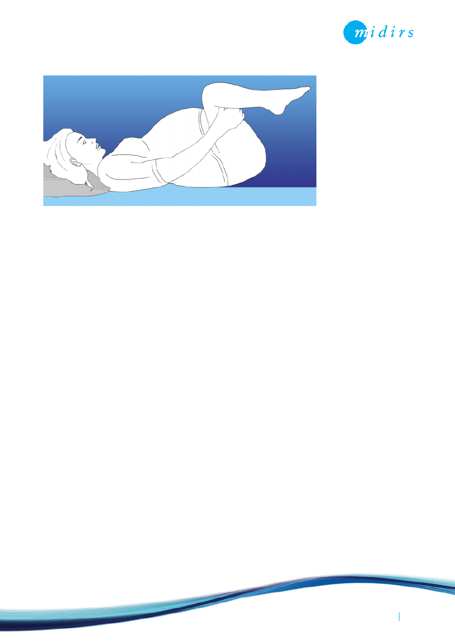
Place the woman in the McRobert’s position, so
that she lies flat with her legs slightly abducted
and hyperflexed at 45
o
to her abdomen– this
position will rotate the angle of the symphysis
pubis superiorly, helps flatten the sacral
promontory, increase the diameter of the pelvic
outlet and release pressure on the anterior
shoulder. The McRobert’s manoeuvre is
associated with the lowest level of morbidity
(Coates 2003) and has a success rate over 40%,
which increases to over 50% when suprapubic
pressure is also applied (Baxley 2003).
G
Apply firm, directed, supra-pubic pressure to the
side of the fetal back, pushing towards the fetal
chest. This reduces the bi-sacromial diameter, and
can help to adduct the shoulders, pushing the
anterior shoulder away from the symphysis pubis.
G
Evaluate the need for an episiotomy, which can
assist manipulations and gain access to the baby
without tearing the perineum and vaginal walls
(RCOG 2005, Leveno et al 2007).
G
Apply gentle traction on the fetal head towards
the longitudinal axis of the fetus, not strong
downward traction which can damage the
cervical spinal cord.
G
The Rubin’s manoeuvre can be used, which
requires the practitioner to identify the posterior
shoulder on vaginal examination. This is then
pushed in the direction of the fetal chest,
Obstetric emergencies / Shoulder Dystocia
02
Obstetric emergencies
Shoulder Dystocia
Obstetric emergencies
Shoulder Dystocia
Obstetric emergencies / Shoulder Dystocia
03
thereby rotating the anterior shoulder away from
the symphysis pubis. This manoeuvre reduces the
12cm bi-sacromial diameter.
G
The Wood’s (screw) manoeuvre can be applied to
rotate the baby’s body so that the posterior
shoulder moves anteriorly. This requires the
practitioner to insert their hand into the woman’s
vagina and identify the fetal chest. By applying
pressure onto the posterior fetal shoulder,
rotation is achieved. The Wood’s manoeuvre will
abduct the shoulders, but enables them to rotate
into a more favourable diameter for delivery.
Delivery on all-fours may make delivery of an
impacted shoulder easier (Arulkumaran
et al 2003).
G
Delivery of the posterior arm and shoulder can be
attempted by inserting the hand into the small
space created by the hollow of the sacrum. This
allows the practitioner to flex the posterior arm
at the elbow and then sweep the forearm over
the baby’s chest. Once the posterior arm has
been brought down, space becomes available
and the anterior shoulder slips behind the
symphysis pubis enabling delivery.
G
Should all of these manoeuvres fail to accomplish
delivery, the obstetrician may consider using the
Zavanelli manoeuvre as an all-out attempt to
deliver a live baby. This manoeuvre requires the
reversal of the mechanisms of delivery so far and
reinsertion of the fetal head into the vagina.
Prompt delivery by caesarean section is then
required; however this manoeuvre has a variable
success rate (Arulkumaran et al 2003, Coates
2003, Tiran 2003).
Where the role of the midwife
is to assist those undertaking
the above manoeuvres, they
should also, where possible,
maintain an accurate and
detailed record of those in
attendance, the manoeuvre(s)
used, the time taken and
force of traction applied, and
the outcome(s) of each
manoeuvre attempted. The
RCOG have suggested a
proforma which can assist
with this (Coates 2003, RCOG 2005). The RCOG suggest
recording the following details:
G
Time of delivery of the head
G
Direction of head after restitution
G
Time of delivery of the body
G
Condition of infant (APGAR, paired cord blood
pH recordings)
G
What time attending staff arrived, including
names and designation
Maternal complications:
G
Postpartum haemorrhage (approximately
two-thirds will have a blood loss >1000 ml)
(Benedetti & Gabbe 1978)
G
Soft tissue trauma
G
Third or fourth degree perineal tears (extension
of episiotomy)
Fetal and neonatal complications:
G
Fetal hypoxia or neonatal asphyxia – potential
for neurological damage
G
Brachial plexus injury – Erb’s Palsy/Klumpke’s
paralysis (Tiran 2003)
G
Fractures to the clavicle or humerus
G
Intrapartum fetal death (Coates 2003).

Obstetric emergencies
Shoulder Dystocia
Post birth:
After the birth, the procedures/manoeuvres used and the
delivery outcome should be explained to both parents,
allowing them time to discuss the birth. Where the likely
cause for the dystocia has been determined, this should
also be explained to the parents along with any potential
risk of its re-occurrence in future pregnancies (Leveno et al
2007). Should there be complications, such as nerve
damage or fetal hypoxia, additional follow-up counselling
and support to the couple should be provided, especially
regarding future pregnancies and the management of the
birth (Arulkumaran et al 2003). Where relevant, there
should be appropriate referral to specialist practitioners in
the multidisciplinary team, including obstetric, neonatology
and physiotherapy services (Department of Health 2004),
as well as specialist family and child support groups, eg The
Erb’s Palsy Group (www.erbspalsygroup.co.uk).
Implications for practice:
The Confidential Enquiry into Stillbirths and Deaths in
Infancy (CESDI) 5
th
annual report recommended ‘a high
level of awareness and training for all birth attendants’
(Maternal and Child Health Research Consortium 1998).
As previously mentioned, the Royal College of Obstetricians
and Gynaecologists (RCOG) and the Royal College of
Midwives (RCM) jointly recommend annual intrapartum
skill drills, which includes shoulder dystocia (RCOG, RCM
1999). Table 1 shows a mnemonic for shoulder dystocia
that is commonly used in such training, which may assist
the midwife in managing this emergency situation.
(Table 1) The HELPERR mnemonic
References:
Arulkumaran S, Symonds IM, Fowlie A eds (2003). Oxford Handbook of Obstetrics
and Gynaecology. Oxford: Oxford University Press: 388-9.
Baxley EG (2003). ALSO : Advanced Life Support in Obstetrics : ALSO course syllabus.
4
th
ed. Leawood Kansas: American Academy of Family Physicians.
Benedetti TJ, Gabbe SG (1978). Shoulder dystocia: a complication of fetal
macrosomia and prolonged second stage of labor with midpelvic delivery. Obstetrics
and Gynecology 52(5):526-29.
Coates T (2003). Shoulder dystocia. In: Fraser DM, Cooper MA eds. Myles Textbook
for Midwives. 14
th
ed. Edinburgh: Churchill Livingstone. 602-7.
Confidential Enquiry into Maternal and Child Health (2006). Perinatal mortality
surveillance 2004: England, Wales and Northern Ireland. London: CEMACH.
Department of Health (2004). National Service Framework for children, young
people and maternity services: Maternity Services. London: Department of Health.
Gherman RB, Ouzounian JG, Goodwin TM (1998). Obstetric maneuvers for shoulder
dystocia and associated fetal morbidity. American Journal of Obstetrics and
Gynecology, 178(6):1126-30.
Gherman RB (2002). Shoulder dystocia: an evidence-based evaluation of the obstetric
nightmare. Clinical Obstetrics and Gynecology. 45(2):345-62.
Leveno KJ, Cunningham FG, Alexander JM eds (2007). Williams manual of obstetrics:
pregnancy complications. 22
nd
ed. London: McGraw-Hill: 513-521
Maternal and Child Health Research Consortium (1998). Confidential Enquiry into
Stillbirths and Deaths in Infancy [CESDI]. 5th annual report. London: Maternal and
Child Health Research Consortium. 73-9.
McGeown P (2001). Practice recommendations for obstetric emergencies. British
Journal of Midwifery. 9(2):71-3.
Royal College of Obstetricians and Gynaecologists (2005). Shoulder dystocia.
Guideline No. 42. London: RCOG.
Royal College of Obstetricians and Gynaecologists, Royal College of Midwives (1999).
Towards safer childbirth: minimum standards for the organisation of labour wards:
Report of a Joint Working Party. London: Royal College of Obstetricians and
Gynaecologists, Royal College of Midwives.
Tiran D (2003). Baillière’s Midwives’ Dictionary. 10
th
ed. Edinburgh: Baillière Tindall.
Obstetric emergencies / Shoulder Dystocia
04
H
E
L
P
E
R
R
Call for help
Evaluate for episiotomy
Legs (the McRobert’s manoeuvre)
Suprapubic pressure
Enter manoeuvres (internal rotation)
Remove the posterior arm
Roll the woman/rotate onto ‘all fours’

Obstetric emergencies
Shoulder Dystocia
Further reading:
Athukorala C, Middleton P, Crowther CA (2006). Intrapartum interventions for
preventing shoulder dystocia. Cochrane Database of Systematic Reviews, issue 4.
Crofts JF, Bartlett C, Ellis D et al (2008). Documentation of simulated shoulder
dystocia: accurate and complete? BJOG: An International Journal of Obstetrics
and Gynaecology 115(10):1303-08.
Crofts JF, Bartlett C, Ellis D et al (2007). Management of shoulder dystocia. Skill
retention 6 and 12 months after training. Obstetrics and Gynecology 110(5):1069-74.
Crofts JF, Fox R, Ellis D et al (2008). Observations from 450 shoulder dystocia
simulations: lessons for skills training. Obstetrics and Gynecology 112(4):906-12.
Crofts JF, Ellis D, James M et al (2007). Pattern and degree of forces applied during
simulation of shoulder dystocia. American Journal of Obstetrics and Gynecology
197(2):156.e1-6
Crofts JF, Attilakos G, Read M et al (2005). Shoulder dystocia training using a new
birth training mannequin. BJOG: An International Journal of Obstetrics and
Gynaecology 112(7): 997-9.
Draycott TJ, Crofts JF, Ash JP et al (2008). Improving neonatal outcome through
practical shoulder dystocia training. Obstetrics and Gynecology 112(1):14-20.
Edwards G ed (2004). Adverse outcomes in maternity care: implications for practice,
applying the recommendations of the Confidential Enquiries. Oxford: Books
for Midwives.
Hope P, Breslin S, Lamont L et al (1998). Fatal shoulder dystocia: a review of 56 cases
reported to the Confidential Enquiry into Stillbirths and Deaths in Infancy. British Journal
of Obstetrics and Gynaecology 105(12):1256-61.
Mahran MA, Sayed AT, Imoh-Ita F (2008). Avoiding over diagnosis of shoulder dystocia.
Journal of Obstetrics and Gynaecology 28(2):173-6.
Miskelly S (2009). Emergencies in labour and birth. In: Chapman V, Charles C eds.
The midwife’s labour and birth handbook. Oxford: Blackwell Publishing.
Obstetric emergencies / Shoulder Dystocia
05
Wyszukiwarka
Podobne podstrony:
więcej podobnych podstron