Title: Spontaneous Integration of Human DNA Fragments into Host Genome
K. Koyama, T. A. Deisher
Sound Choice Pharmaceutical Institute, Seattle, WA
Conclusion
Discussion
A trio of recent publications in the journal NEURON reports the
presence of hundreds of diverse de novo gene mutations indicating that autism
spectrum disorder (ASD) may be a disease of genomic instability, with a
significant environmental component. Altered double strand break formation and
repair pathways (DSB) may be a commonality among the diverse genetic
mutations that have been documented in ASD. US birth year change points in AD
are apparent in 1980, 1988 and 1996, coinciding with the switch to or introduction
of childhood vaccines contaminated with human endogenous retrovirus K
(HERVK) and human fetal DNA fragments (6). We hypothesize that the HERVK
and human fetal DNA contaminants could contribute to the genomic instability of
ASD as demonstrated by de novo mutations.
Cell free DNA can be taken up by healthy cells via receptor mediated
uptake or may spontaneously penetrate cell membranes that have altered
permeability, for instance, during inflammatory reactions. Nuclear uptake of cell
free DNA fragments is thought to provide a source for maintenance of DNA
integrity during rescue of collapsed replication forks or base lesion repair.
Spontaneous extracellular DNA uptake has also been exploited for gene therapy
as well as for cellular gene correction (2,4,5,7,8, and 9). While free DNA uptake
has been used advantageously, the process has also been associated with
generation of mutations and chromosomal aberrations (3).
Vaccines manufactured using human fetal cells contain residual DNA
fragments (50-500 bp) (Table I). It is possible that these contaminating
fragments could be incorporated into a
child’s genome and disrupt normal gene
function, leading to autistic phenotypes. In this study we demonstrate foreign
DNA uptake in human cells and genomic integration by incubating the cells with
Cy3-labeled human Cot1 (placental) DNA fragments which represents
contaminating residual human fetal DNA in vaccines.
Introduction
BE (2)-C (neuroblastoma) cells were grown in the same condition except
medium used was a 1:1mixture of
Eagle’s Minimum Essential Medium (EMEM)
and F12 Medium supplemented with 10%FBS and 1% antibiotic-antimycotic
solution. M059K (Glioblastoma-Double Stranded Break repair proficient) and
M059J (Glioblastoma-Double Stranded Break repair deficient) were also grown
with the same condition except the medium used was a 1:1 mixture of DMEM
and
Ham’s F12 Medium supplemented with 10% FBS, 0.05mM non-essential
amino acids, and 1% antibiotic-antimycotic solution. After cells were cultured in
each condition for 2 to 3 days 500ng Cy3 labeled Human Cot1 DNA was added
and incubated at 37°C under a humidified atmosphere containing 5% CO
2
/95%
air by gently shaking for 24 hours and 48 hours. After incubation nucleus was
stained with Hoechst, German glass cover slips were placed on glass slides,
and cellular and nuclear DNA uptake was analyzed under fluorescent
microscope.
To model inflammation, all adherent cell lines were activated with
lipopolysaccharide (LPS). And, saponin permeabilization was also tested for
HFF1 cells . Three concentrations of LPS, 1ng/10
4
cells, 10ng/10
4
cells, and
100ng/10
4
cells were tested in the wells of each cell line previously mentioned.
Cells were incubated with Cy3 labeled Human Cot1 DNA and LPS at 37°C
under a humidified atmosphere containing 5% CO
2
/95% air by gently shaking
for 24 hours and 48 hours. As well as cells incubated without LPS, these cells
were also stained with Hoechst before cellular and nuclear DNA uptake was
analyzed under fluorescent microscope.
HFF1 cells were incubated with 0.02% saponin , 300ng DAPI, and 500ng Cy3
labeled human Cot 1 DNA for 24 hours, 48 hours, and 72 hours. Cells were
viewed under fluorescent microscope as well.
Results (Table 2):
Spontaneous cellular and nuclear DNA uptake was evident in HFF1,
NCCIT and U937 (Fig1, 3, 7 and 8). DNA uptake in BE (2)-C and M059K was
not measurable because of high auto fluorescence of the cells. No Cy3 signal
was observed in HL-60. With inflammation caused by LPS cellular DNA uptake
was observed in HFF1, NCCIT, M059J, and U937 (Fig 2, 4, 5 and 6).
The amount of labeled Cy3 human Cot1 DNA incorporation in U937
genomic DNA was 0.0111 +/- 0.0034pg (n=12) per cell in 24 hours, which was
approximately 0.167% of total U937 genomic DNA. DNA incorporation in
NCCIT cells was 0.0026pg/cell in 24 hours and 0.04pg/cell in 48 hours which
is 0.6% of total NCCIT genomic DNA.
Acknowledgments: This work was funded by the M.J. Murdock
Charitable Trust and private donations.
1. Golan M, Hizi A, Resau J, Yaa-Hahoshen, Reichman H, Keydar lafa, and Ian Tsarfaty
. “Human
Endogenous Retrovirus (HERV-
K) Reverse Transcriptase as a Breast Cancer Prognostic Marker” NEO
PLASIA, Vol.10, No.6, June 2008, pp. 521-533.
2. Filaci G, Gerloni M, Rizzi M, Castiglion P, Chang H-D, Wheeler MC, Fiocca R, and Zanetti M.
“Spontaneous transgenesis of human B lymphocytes.” Gene therapy, 2004, 11, 42-52.
3. Howe S, Mansour M, Schwarzwaelder K, Bartholomae C, Hubank M, Kempski H, Brugman M, Pike-
Overzet K, Chatters S, Ridder D, Gilmour K, Adams S, Thomhil
S, and other 14. “Insertional mutagenesis
combined with acquired somatic mutations causes leukemogenesis
following gene therapy.” Journal od
Clinical Investigation. Vol 118, No.9, September 2008.
4. Lehmann MJ, and Sczakiel
G. “Spontaneous uptake of biologically active recombinant DNA by
mammalian cells via a selected DNA segment.” Gene Therapu 2005, 12, 446-451.
5. Rogachev V, Likhacheva A, Vratskikh O, Mechetina L, Sebeleva T, Bogachev S, Yakubov L, and
Shurdov
M. “Qualitative and quantitative characteristics of the extracellular DNA delivered to the nucleus of
a living cell” Cancer Cell International, October 2006, 6:23, P1475-2867.
6. Victoria J, Wang C, Jones M, Jaing C, McLoughlin K, Gardner S, and Delwart
E. “Viral Nucleic Acids in
Live-Attenuated Vaccinces
: Detection of Minority Variants and an Adventitious Virus.” Journal of Virology,
June 2010, P.6033-6040.
7. Yakubov L, Deeva E, Zarytova V, Ivanova E, Ryte A, Yurchenko L, and Vlassov
V. “Mechanism of
Oligonucleotide
uptake by cells: , September Involvement of specific receptors?” Proceedings of the
National Academy of Science, Vol. 86, pp 6454-6458, September 1989.
8. Yakubov A, Petrova N, Popova N, Semenov D, Nikolin V, and
Os’kina I. “The Role of Extracellular DNA
in the Stability and Variability of Cell Genomes.” Doklady Biochemistry and Biophysics, Vol. 382, 2002, pp.3
1-34.
9. Yakubov L, Rgachev V, Likhacheva A, Bogachev S, Sebeleva T, Shilov A, Baiborodin S, Petrova N,
Mechetina L, Shurdov M, and Wickstrom
E. “Natural Human Gene Correction by Small Extracellular
Genomic DNA Fragments.” Cell Cycle 6:18, 2293-2301, 15 September 2007.
Table 2: DNA uptake in Various Cell lines
Spontaneous
Cellular
uptake
Spontaneous
Nuclear
uptake
Incorporation
in Genomic
DNA
Cellular /Nuclear
Uptake with LPS or
saponin
HFF1
Yes
Yes
Not Done
Increase/Increase
NCCIT
Yes
Yes
(variable)
0.0026pg per
cell 24 hrs
0.04pg per
cell 48 hrs
Same/Same
BE(2)-C
No
No
Not Done
No/No
M059K
No
No
No
No/No
M059J
No
No
Not Done
Yes/No
U937
Yes
Yes
0.011 +/-
0.003pg per
cell 24 hrs
Same/Same
HL60
No
No
No
No
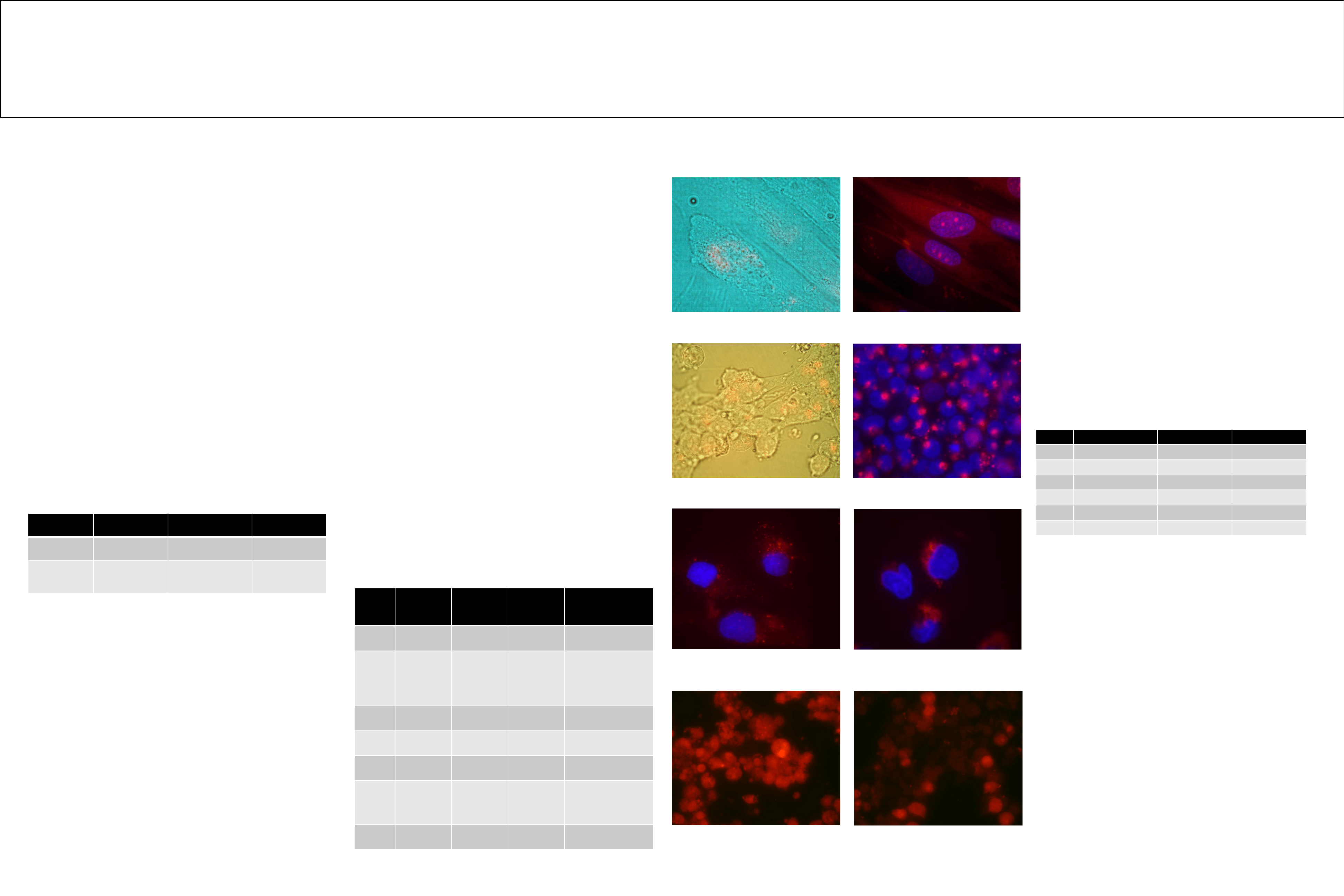
Fig 1. HFF1 spontaneous cellular and nuclear DNA uptake
(bright field & Cy3 red combined).
Fig 2. HFF1 cellular and nuclear DNA uptake after
permeabilization with saponin. (Cy3 red & nucleus blue
combined)
Fig 3. NCCIT spontaneous cellular DNA uptake
(bright field & Cy3 red combined)
Fig 4. NCCIT cellular DNA uptake after lipopolysaccharide
activation (Cy3 red & nucleus blue combined)
Fig 6. M059J cellular DNA uptake after lipopolysaccharide
activation (100ng/10
4
cells).
(Cy3 red & nucleus blue combined).
Fig 7. U937 spontaneous cellular/nuclear DNA uptake
(Cy3 red)
Fig 8. Purified U937 nuclei containing Cy3 labeled DNA
before DNA purification (Cy3 Red)
Vaccine name
Double Stranded
DNA (ng/vial)
Single Stranded
DNA (ng/vial)
Length (bps)
Meruvax II
(Rubella)
142.05
35.00
240
HAVRIX
(Hepatitis A)
276.00
35.74
Not measurable
Table 1. Levels of residual human double stranded DNA (Picogreen
assay) and human single stranded DNA (Oligreen assay ) in Rubella
vaccine (MeruvaxII) and Hepatitis A vaccine (HAVRIX).
Methods and Results
Materials and Methods
: Human Cot1 DNA (Invitrogen) was labeled with
Mirus Label IT Cy
TM
3 Labeling Kit (Mirus).
U937 cells (monocytes) were grown in
Dublecco’s Mofication of
Eagle’s Medium (DMEM) supplemented with 15% fetal bovine serum (FBS)
and 1% antibiotic-antimycotic solution at 37°C under a humidified atmosphere
containing 5% CO
2
/95% air. HL-60 cells (myeloblast) were grown in
Iscove’s
Modified
Dulbecco’s Medium (IMDM) supplemented with 20% FBS and 1%
antibiotic-antimycotic solution at 37°C under the same condition. 750ng of Cy3
labeled Human Cot1 DNA was incubated per1.0×10
7
cells for 24 hours and 48
hours.
Cellular and nuclear DNA uptake was analyzed under fluorescent microscope.
Genomic DNA of U937 cells was purified by ethanol precipitation removing
short fragment of nucleic acids including unincorporated Cy3 labeled Human
Cot1 DNA. The amount of Cy3 labeled human Cot1 DNA incorporated into
U937 chromosomes was calculated with relative fluorescent unit (RFU)
measured by a fluorimeter.
Loosely adherent NCCIT (teratocarcinoma) cells were grown with a
cell density 3×10
4
per well of a 24-well plate which a German glass cover slips
was placed in each well at 37°C under a humidified atmosphere containing 5%
CO
2
/95% air. HFF1 (Human Foreskin Fibroblast 1) cells were grown with the
same condition except DMEM supplemented with 15% fetal bovine serum
(FBS) and 1% antibiotic-antimycotic solution was used as a medium.
Our measured genomic incorporation (0.003 to 0.04 pgs) of
0.2% - 0.6% of the whole genome in 24 to 48 hours seems high at first
glance. However, our numbers are consistent with previous reports
showing that exogenous DNA replaced up to 1% of the whole genome
within 30 minutes (6). Although HL-60 cells did not spontaneously take
up exogenous DNA in our experiments, the cell line has been used in
the past as a model for spontaneous DNA uptake (8).
Cellular and nuclear DNA uptake in human foreskin fibroblast
(HFF1) cells and in NCCIT cells suggests that embryonic and neonatal
cell are more susceptible to DNA uptake than cells from a more mature
source. These results indicate the need for further study of DNA
incorporation from exogenous sources to compare the susceptibility of
infants and toddlers versus teens and adults.
Increased DNA uptake after LPS activation suggests that
systemic inflammation or immune responses could increase
susceptibility for exogenous DNA uptake. Human diploid cell produced
vaccines are contaminated by exogenous DNA fragments and a
retrovirus, and vaccines elicit systemic inflammation and immune
activation. Our future research goals are to localize the sites of DNA
integration, to demonstrate phenotype changes caused by foreign DNA
integration in factor dependent cell lines, and to determine the
biological and/or pathological activities of Human Endogenous
Retrovirus K (HERVK) fragments in vaccines.
Not only damaged human cells, but also healthy human cells can take up
foreign DNA spontaneously. Foreign human DNA taken up by human
cells will be transported into nuclei and be integrated into host genome,
which will cause phenotype change. Hence, residual human fetal DNA
fragments in vaccine can be one of causes of autism spectrum disorder in
children through vaccination. Vaccine must be safe without any human
DNA contaminations or reactivated viruses, and must be produced in
ethically approved manufacturing processes.
Cells
Source
Morphology
Transfection host
U937
Histocytic Lymphoma
Monocyte
Yes
HL60
Leukemia
Myeloblast
Yes
BE(2)C
Neuroblastoma
Neuroblast
No
M059K
Glioblastoma
Fibroblast
No
M059J
Glioblastoma
Fibroblast
No
HFF1
Foreskin
Fibroblast
No
Table3: Cell Description
Fig 5. M059J cellular DNA uptake after lipopolysaccharide
activation (10ng/10
4
cells).
(Cy3 red & nucleus blue combined).
Wyszukiwarka
Podobne podstrony:
No 004 CCS Demonstration Plant fully integrated into new unit 858 MW
Dental DNA fingerprinting in identification of human remains
[17]Chromosomal DNA fragmentation in apoptosis and necrosis induced by oxidative stress
No 004 CCS Demonstration Plant fully integrated into new unit 858 MW
bacterial contamination in drinking water
DIMENSIONS OF INTEGRATION MIGRANT YOUTH IN POLAND
In this picture you?n see two children
ANG ćwicz cough in children
Vaccination in Progress
Immobilization and hypercalciuria in children
New World Orders in Contemporary Children's Literature
In vitro Vaccinum
Patterns of damage in genomic DNA sequences from a Neandertal
children in egipt
więcej podobnych podstron