
26
Am J Psychiatry 157:1, January 2000
Regular Articles
Specific Relationship Between Prefrontal Neuronal
N-Acetylaspartate and Activation of the Working Memory
Cortical Network in Schizophrenia
Alessandro Bertolino, M.D., Giuseppe Esposito, M.D., Joseph H. Callicott, M.D.,
Venkata S. Mattay, M.D., John D. Van Horn, Ph.D., Joseph A. Frank, M.D.,
Karen Faith Berman, M.D., and Daniel R. Weinberger, M.D.
Objective: Abnormal activation of the dorsolateral prefrontal cortex and a related cortical
network during working memory tasks has been demonstrated in patients with schizophre-
nia, but the responsible mechanism has not been identified. The present study was per-
formed to determine whether neuronal pathology of the dorsolateral prefrontal cortex is
linked to the activation of the working memory cortical network in patients with schizophre-
nia. Method: The brains of 13 patients with schizophrenia and 13 comparison subjects
were studied with proton magnetic resonance spectroscopic (
1
H-MRS) imaging (to mea-
sure
N
-acetylaspartate as a marker of neuronal pathology) and with [
15
O]water positron
emission tomography (PET) during performance of the Wisconsin Card Sorting Test (to
measure activation of the working memory cortical network). An independent cohort of pa-
tients (N=7) was also studied in a post hoc experiment with
1
H-MRS imaging and with the
same PET technique during performance of another working memory task (the “N-back”
task). Results: Measures of
N
-acetylaspartate in the dorsolateral prefrontal cortex strongly
correlated with activation of the distributed working memory network, including the dorso-
lateral prefrontal, temporal, and inferior parietal cortices, during both working memory
tasks in the two independent groups of patients with schizophrenia. In contrast,
N
-acetylas-
partate in other cortical regions and in comparison subjects did not show these relation-
ships. Conclusions: These findings directly implicate a population of dorsolateral prefron-
tal cortex neurons as selectively accounting for the activity of the distributed working
memory cortical network in schizophrenia and complement other evidence that dorsolat-
eral prefrontal cortex connectivity is fundamental to the pathophysiology of the disorder.
(Am J Psychiatry 2000; 157:26–33)
W
orking memory is a cognitive construct describ-
ing the ability to hold information transiently in mind
in the service of comprehension, thinking, and plan-
ning (1, 2). Complex cognitive processes such as work-
ing memory are thought to be subserved by the func-
tional integration of interconnected regions forming
large-scale cortical networks (1–5). Data on human
and nonhuman primates show that a key cortical re-
gion for the execution of working memory tasks is the
dorsolateral prefrontal cortex (6–12), which has recip-
rocal anatomical connections with the parietal, tempo-
Received Jan. 8, 1999; revision received June 16, 1999;
accepted July 8, 1999. From the Clinical Brain Disorders Branch,
Intramural Research Programs, NIMH; and the Laboratory of Diag-
nostic Radiology Research, Office of the Director, NIH. Address
reprint requests to Dr. Weinberger, Clinical Brain Disorders
Branch, Intramural Research Programs, NIMH, NIH, Rm. 4S235,
MSC 1379, 10 Center Dr., Bethesda, MD 20892; weinberd@
dirpc.nimh.nih.gov (e-mail).
The authors thank Jozef Duyn, Ph.D., and Chrit Moonen, Ph.D.,
for making the proton magnetic resonance spectroscopic (
1
H-
MRS) imaging pulse sequence available and Alan Barnett, Ph.D.,
for help with processing of the
1
H-MRS imaging data.

Am J Psychiatry 157:1, January 2000
27
BERTOLINO, ESPOSITO, CALLICOTT, ET AL.
ral, and cingulate cortices, which also participate in the
cortical network related to working memory (1–5).
Deficits in working memory have been reported to
be a cardinal feature of the pathophysiology of schizo-
phrenia (13, 14). Attempts to anatomically localize
these deficits with functional neuroimaging studies in
patients performing working memory tasks have often
shown subnormal activation of the dorsolateral pre-
frontal cortex and, to a lesser extent, abnormalities of
other regions in the working memory network (15–
22). There has been considerable debate on issues in-
volving the mechanism of this pattern of hypofunction,
including whether it reflects distributed neuronal pa-
thology, is referable to focal cortical pathology, or is,
perhaps, an artifact of the test procedure (15–22).
Postmortem studies of the brains of patients with
schizophrenia have shown evidence of abnormalities in
a number of cortical areas within the working memory
network, including the dorsolateral prefrontal cortex,
cingulate, and temporal cortices (23–26), although the
most extensive data have implicated the dorsolateral
prefrontal cortex (27–32). Since the overall function of
a cortical network presumably relies on the compe-
tence of both local information processing within spe-
cific local circuits and axonal connections between lo-
cal circuits and distant cortical areas, a deficit of a
single region, for example the dorsolateral prefrontal
cortex, could conceivably have functional reverbera-
tions throughout the working memory network. The
purpose of the present study was to address directly
the question of whether the integrity of a population of
neurons within the dorsolateral prefrontal cortex, as
studied with proton magnetic resonance spectroscopic
(
1
H-MRS) imaging, preferentially accounts for the dis-
tributed pattern of cortical function associated with
working memory in schizophrenia, as studied with
[
15
O]water positron emission tomography (PET) dur-
ing performance of working memory tasks.
1
H-MRS imaging detects signals in multiple brain re-
gions arising from N-acetyl-containing moieties
(mainly N-acetylaspartate, NAA), choline-containing
compounds (CHO), and creatine plus phosphocreatine
(CRE) (33). NAA is an intraneuronal amino acid, the
highest concentrations of which occur in pyramidal
neurons (34). Its biological role has yet to be clearly
defined. However, it acts through the glutamatergic N-
methyl-
D
-aspartic acid (NMDA) receptor to elevate in-
tracellular calcium (35), and its concentrations are re-
duced by pharmacological inhibition of mitochondrial
energy metabolism (36) and by a number of patholog-
ical processes affecting the integrity of neurons (37,
38). It is interesting that a recent study (39) has also
shown increased NAA measures in rats during experi-
mental status epilepticus, suggesting that NAA corre-
lates with the functional status of neurons. Relative
concentrations of NAA have been previously shown to
be lower than normal in the prefrontal cortex of pa-
tients with schizophrenia (40–44).
[
15
O]Water PET identifies changes in regional cere-
bral blood flow (rCBF) associated with neuronal activ-
ity. In the first of two experiments we measured rCBF
during performance of the Wisconsin Card Sorting Test,
an abstract reasoning task involving the use of previ-
ously learned information to formulate a strategy for
present and future actions. To the extent that recent
memory is essential for achieving the correct action, the
test has been considered to involve working memory
and to be sensitive to prefrontal pathology (10, 15, 16,
45, 46). Several earlier studies (15–19) have shown re-
duced rCBF in the dorsolateral prefrontal cortex and
other related cortical areas in patients with schizophre-
nia during performance of the Wisconsin Card Sorting
Test and other tasks involving working memory. We
also performed a second, post hoc experiment to ad-
dress the issue of whether the correlations found in the
patients during the Wisconsin Card Sorting Test are task
specific or related to generic working memory function.
In this second experiment, a separate group of patients
with schizophrenia performed a less complex working
memory task, a version of the “N-back” task (47). This
task has been previously shown to produce activation in
a cortical network including the same regions involved
in the Wisconsin Card Sorting Test and to reveal similar
pathophysiological characteristics in patients with
schizophrenia (22, 48). A “2-back” condition, in which
subjects respond according to a number seen two stim-
uli before, requires continuous updating of the mental
set and the use of working memory (22).
METHOD
Subjects
For the Wisconsin Card Sort study, there were 26 subjects: 13 pa-
tients with a diagnosis of schizophrenia according to DSM-IV crite-
ria (11 men; mean age=35.0 years, SD=8.6) and 13 comparison sub-
jects (eight men; mean age=34.6 years, SD=8.0). Each subject
underwent
1
H-MRS imaging and [
15
O]water PET on two different
days. For the
1
H-MRS imaging scan, five of the patients had been
without drugs for at least 3 weeks, while the others were receiving
neuroleptics. Neuroleptics have been previously shown not to affect
NAA findings (43, 49). On the day of the PET scan, five of the pa-
tients who had been receiving drugs when studied with
1
H-MRS im-
aging were being treated with clozapine, while the others had been
drug free for at least 3 weeks. The patients and comparison subjects
had similar performances on the Wisconsin Card Sorting Test, as ex-
pressed by the average percentage of correct responses (patients,
71%; comparison subjects, 74%). The N-back study involved a dif-
ferent group of seven patients (five men; mean age=31.8 years, SD=
8.7) with a diagnosis of schizophrenia according to DSM-IV. They
had all been without neuroleptics for at least 2 weeks (range=15–30
days) before both the
1
H-MRS imaging and [
15
O]water PET scans.
The average performance of the patients on the 2-back version of the
task was 50%, which is well below that reported for normal subjects
(82%) (22).
After complete description of all studies to the subjects, written
informed consent was obtained from each and every subject.
1
H-MRS Imaging Procedure
The
1
H-MRS imaging studies were performed as previously de-
scribed (33, 41, 43) on a conventional GE Signa 1.5-T nuclear mag-
netic resonance imaging system. The
1
H-MRS imaging pulse se-
quence acquires four spectroscopic slices (TR=2200 msec, TE=272
msec) involving 32
×
32 phase-encoding steps over a 240-mm field
28
Am J Psychiatry 157:1, January 2000
WORKING MEMORY IN SCHIZOPHRENIA
of view for each slice. Each volume element (voxel) has nominal di-
mensions of 7.5 mm
×
7.5 mm
×
15 mm (0.84 ml). Actual volume,
based on full width at half maximum after filtering of k-space, is 1.4
ml. The
1
H-MRS imaging data processing involved locating NAA,
CHO, and CRE in spectra from each voxel and then displaying the
four 32
×
32 arrays showing spatial variation of the magnitude of
each of the signals in each of the slices. Regions of interest were
drawn on coplanar magnetic resonance imaging scans as previously
described (41). The metabolites were studied as ratios of the area un-
der each peak: NAA/CRE, NAA/CHO, CHO/CRE.
[
15
O]Water PET Procedure
For the Wisconsin Card Sorting Test study, each subject underwent
two PET scans during a single session: one scan while performing the
card sorting test and the other one while performing a sensorimotor
control task. The PET data were acquired as described by Berman et
al. (10) on a Scanditronix PC2048-15B PET scanner that simulta-
neously produces 15 contiguous slices in 16 frames over 4 minutes.
An intravenous bolus of approximately 42 mCi of [
15
O]water was
administered before each scan. Arterial input functions were mea-
sured with automated arterial blood sampling, and absolute rCBF
(milliliters of blood per minute per 100 g of tissue) was calculated for
each voxel. For the N-back task study, 60-second PET data were ac-
quired nonquantitatively on a GE Advance PET camera in three-di-
mensional mode after a bolus injection of 10 mCi of [
15
O]water per
scan. Images for each subject were registered by using the Automated
Image Registration (AIR) program and then normalized to the atlas
of Talairach and Tournoux (50) and smoothed with a 15
×
15
×
5 fil-
ter by using the SPM95 package. The PET data were normalized by
expressing each value as a ratio to the global mean. To determine ac-
tivation during the Wisconsin Card Sorting Test, we subtracted the
rCBF during the control condition from that during the Wisconsin
Card Sorting Test. For the N-back study, activation data were derived
by subtracting the average rCBF for seven scans acquired during the
2-back condition from the average for two scans acquired during
rest. Pearson correlations between the
1
H-MRS imaging regional val-
ues and rCBF PET data during the Wisconsin Card Sorting Test were
determined on a voxel-by-voxel basis. The statistical threshold used
was p
<
0.01, corresponding to a Pearson r of 0.683. A cluster thresh-
old of 10 contiguous voxels was applied as well. Because of the
smaller group in the N-back study, we used a Spearman correlation
analysis to avoid a potential outlier effect.
RESULTS
1
H-MRS Imaging Measures and Brain Activation During
Wisconsin Card Sorting Test
In the patients, NAA/CRE in the dorsolateral pre-
frontal cortex was strongly and positively correlated
with activation in the prefrontal cortex (Brodmann’s
areas 9, 10, 44, 45, 46), parietal cortex (Brodmann’s
area 39/40), and temporal association cortex (figure 1
and table 1). NAA/CRE in the dorsolateral prefrontal
cortex also exhibited negative correlations, mostly
with subcortical structures, including the cerebellum
and basal ganglia. On the other hand, NAA/CRE in
the dorsolateral prefrontal cortex of the comparison
subjects showed a different pattern of relationships
with rCBF activation, correlating only in a few scat-
tered voxels in the left inferolateral prefrontal cortex
and not with any other region activated by the Wiscon-
sin Card Sorting Test.
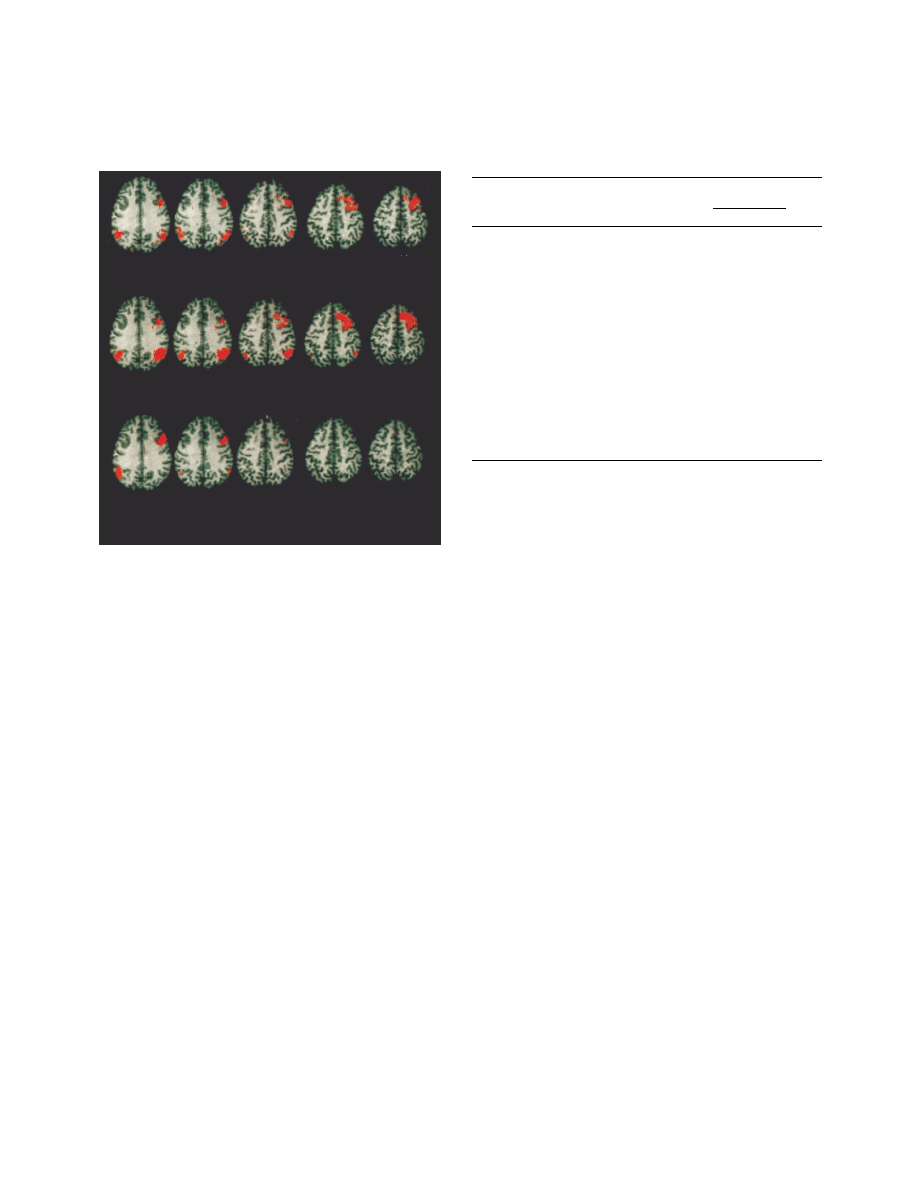
FIGURE 1. Voxel-by-Voxel Correlations Between Metabolite
Ratios for the Dorsolateral Prefrontal Cortex of 13 Schizo-
phrenic Patients and Blood Flow Activation During the Wis-
consin Card Sorting Test
a
a
The slices are identified by position on the z axis (Talairach coordi-
nate). The first row shows, in red, voxels with significant positive
Pearson correlations (r
>
0.68, p
<
0.01) between the ratio of
N
-acety-
laspartate to creatine plus phosphocreatine (NAA/CRE) and acti-
vation of regional cerebral blood flow (rCBF) during the Wisconsin
Card Sorting Test (test condition minus control condition). The sec-
ond row shows areas of significant correlations between the ratio
of
N
-acetylaspartate to choline-containing compounds (NAA/CHO)
and rCBF activation during the Wisconsin Card Sorting Test. The
third row shows significant correlations between NAA/CRE and
rCBF during the Wisconsin Card Sorting Test alone.
+28
+32
+36
+40
+44
NAA/CRE and Wisconsin Card Sorting Test activation
NAA/CHO and Wisconsin Card Sorting Test activation
NAA/CRE and rCBF during Wisconsin Card Sorting Test
TABLE 1. Brain Locations of Maximal Positive Correlations
Between Blood Flow Activation
a
During the Wisconsin Card
Sorting Test and the Ratio of
N
-Acetylaspartate to Creatine
Plus Phosphocreatine in the Dorsolateral Prefrontal Cortex of
13 Schizophrenic Patients
Anatomical Location
Brodmann’s
Areas
Talairach
Coordinates
r
x
y
z
Left
Gyrus frontalis inferior/
gyrus precentralis
44/6
–30
–8 32
0.83
Gyrus frontalis medius
9
40
16 36
0.81
Gyrus frontalis inferior
46
40
42
8
0.78
Gyrus supramarginalis
Site 1
39/40
28 –52 36
0.77
Site 2
39/40
–32 –56 32
0.76
Gyrus occipitalis medius
18
–26 –88 20
0.81
Right
Gyrus frontalis inferior
Site 1
44
46
4 32
0.87
Site 2
45/46
40
40
4
0.74
Gyrus frontalis medius
9
–30
18 36
0.70
Gyrus supramarginalis/
gyrus temporalis superior
39/40
52 –58 32
0.84
Gyrus temporalis superior
39
–46 –58 28
0.81
a
Regional cerebral blood flow (rCBF) during the test minus rCBF
during a control task.
Am J Psychiatry 157:1, January 2000
29
BERTOLINO, ESPOSITO, CALLICOTT, ET AL.
Because it is impossible to perform absolute measure-
ments of metabolites with our
1
H-MRS imaging tech-
nique and because we wished to test whether the rela-
tionships in patients with schizophrenia were
specifically attributable to NAA signals, we also exam-
ined the correlations of two other ratio measures,
NAA/CHO and CHO/CRE, in the dorsolateral prefron-
tal cortex to rCBF activation. While NAA/CHO corre-
lated with activation in exactly the same Brodmann’s
areas as seen with NAA/CRE (figure 1), CHO/CRE
showed only sporadic correlations and none in the ar-
eas associated with working memory. Even though ab-
solute concentrations of the metabolites were not stud-
ied, this consistent pattern of correlations indicates that
they arise from abnormalities in NAA. These results
suggest that the integrity of a population of neurons in
the dorsolateral prefrontal cortex and their connections
predict the activation of the whole working memory
cortical network in patients with schizophrenia.
We also tested whether the correlations between
NAA measures and activation were specific to NAA
measures in the dorsolateral prefrontal cortex. We ex-
amined correlations between NAA/CRE in the hippo-
campal area (also shown to have low NAA measures in
schizophrenia [40, 42, 46, 47, 51, 52]), superior tem-
poral gyrus, anterior cingulate, and occipital cortex
(regions that are activated during working memory
tasks) and the rCBF activation data. Few sporadic cor-
relations emerged, and none of these involved areas of
rCBF activation in the working memory network.
Therefore, these additional data suggest that the corre-
lation between NAA relative measures in the dorsolat-
eral prefrontal cortex and activation in the cortical
working memory network is regionally specific.
We also examined whether the correlations between
NAA measures in the dorsolateral prefrontal cortex
and rCBF activation (obtained by subtraction of blood
flow during the control task from blood flow during
the Wisconsin Card Sorting Test) were related to blood
flow changes during working memory per se, rather
than to blood flow during any volitional task. We per-
formed separate correlations between NAA/CRE in
the dorsolateral prefrontal cortex and rCBF during the
two components of the rCBF activation signal, the
Wisconsin Card Sorting Test and the control task.
While the results of the correlations with rCBF during
the Wisconsin Card Sorting Test showed the same
pattern of correlations as with the activation data
(figure 2), there were few correlations during the con-
trol task and these involved brain areas not activated
by the working memory task. Therefore, the correla-
tions between NAA measures and activation are likely
due to rCBF changes during working memory.
1
H-MRS Imaging Measures and Brain Activation During
N-Back Task
In this post hoc experiment, we addressed several is-
sues, including the reproducibility of the data in an-
other cohort of patients, whether the correlations are
task specific or related to generic working memory
function, and the possible impact of antipsychotic
medication. NAA/CRE in the dorsolateral prefrontal
cortex of these patients was also positively correlated
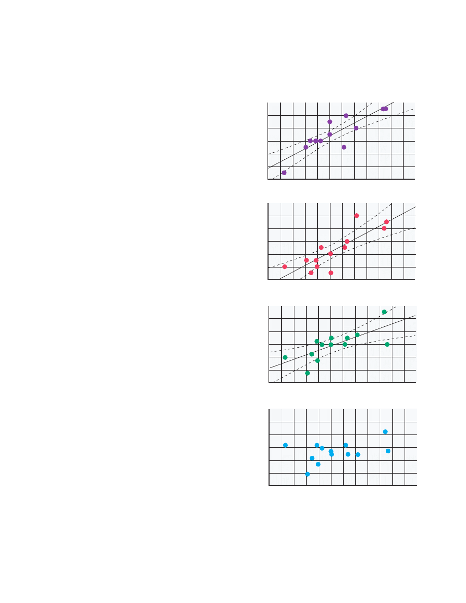
FIGURE 2. Correlations Between the Ratio of
N
-Acetylaspar-
tate to Creatine Plus Phosphocreatine (NAA/CRE) and Re-
gional Cerebral Blood Flow (rCBF) in Areas of the Dorsolateral
Prefrontal Cortex of 13 Patients With Schizophrenia During
the Wisconsin Card Sorting Test and a Control Task
a
rCBF during the test minus rCBF during the control task.
Right Dorsolater
a
l Prefrontal Cor
te
x
Activ
ation
a
of rCBF During
Wisconsin Car
d Sor
ting
T
est (ml/min/100 g)
Right P
a
ri
etal Cor
te
x
1.6
–6
–4
–2
0
2
4
6
2.0
2.4
2.8
3.2
3.6
4.0
1.6
–3
–1
1
3
5
7
9
2.0
2.4
2.8
3.2
3.6
4.0
Wisconsin Card Sor
ting
T
e
st
rCBF in Right Dor
solateral Prefr
ontal Cor
te
x (ml/min/100 g)
1.6
44
48
52
56
60
64
68
2.0
2.4
2.8
3.2
3.6
4.0
NAA/CRE in Right Dorsolateral Prefrontal Cortex
Sensor
imotor Control
T
ask
1.6
44
48
52
56
60
64
68
2.0
2.4
2.8
3.2
3.6
4.0

30
Am J Psychiatry 157:1, January 2000
WORKING MEMORY IN SCHIZOPHRENIA
with activation in the same brain regions (as identified
by the local maxima of the activation) found during
the Wisconsin Card Sorting Test study, including the
prefrontal cortex (r
s
=0.86, N=7, p
<
0.01) and the tem-
poral-parietal cortex (r
s
=0.84, N=7, p
<
0.01). These
data suggest that the correlations are reproducible in
another group of patients with schizophrenia and that
they are typical of tasks engaging the working memory
circuitry, regardless of the specific test used. These ad-
ditional results also indicate that the correlations are
not due to active treatment with antipsychotic drugs,
as this entire cohort was drug free, and not dependent
on task performance per se, as the same relationships
were found during this working memory task, on
which the patients’ performance was abnormal, and
during the Wisconsin Card Sorting Test, on which a
different cohort of patients and comparison subjects
did not differ.
DISCUSSION
Our results show that in schizophrenia the func-
tional integrity of neurons within the dorsolateral pre-
frontal cortex (as represented by NAA measures) has
predictable physiological reverberations throughout
the entire working memory cortical network. NAA
measures in the dorsolateral prefrontal cortex predict
activation of cortical regions involved in the execution
of working memory tasks, including the dorsolateral
prefrontal cortex itself, the parietal cortex, and the
temporal association cortex. Moreover, these relation-
ships are regionally specific, involving only the dorso-
lateral prefrontal cortex as a predictor of network ac-
tivation. The lack of such relationships in healthy
subjects suggests that they emerge in patients because
of disease-associated neuronal pathology in the dorso-
lateral prefrontal cortex. In the present subjects, as in
our previous study groups (41, 43), NAA measures in
the dorsolateral prefrontal cortex of patients (averaged
bilateral NAA/CRE: mean=2.6, SD=0.4) were signifi-
cantly lower than those of normal comparison subjects
(mean=2.9, SD=0.3) (two way ANOVA: F=4.5, df=1,
24, p
<
0.04; no effect of side or side-by-group interac-
tion). To the extent that low NAA measures are a re-
flection of impaired functional integrity of neurons,
this putative impairment constrains in a predictable
way the functional capacity of the distributed working
memory network, as if these dorsolateral prefrontal
cortex neurons by virtue of their projections constitute
a rate-limiting factor for the degree of network recruit-
ment (48). These results are consistent with the ana-
tomical and physiological centrality of the dorsolateral
prefrontal cortex with respect to working memory
function (6–12) and, perhaps, with respect to the
pathophysiology of schizophrenia.
A traditional criticism of functional neuroimaging
studies assessing differences in activation by working
memory tasks between patients with schizophrenia
and healthy subjects has been that patients usually per-
form worse on these tests, thus making the comparison
unfair. Critics of this approach argue that it is impossi-
ble to say whether the abnormal neurobiology causes
deficits in performance or vice versa. To address this
criticism, we selected patients who could perform the
Wisconsin Card Sorting Test well enough to be
matched with comparison subjects. Indeed, previous
studies (53–55) have shown that there is a certain per-
centage of patients with schizophrenia who perform
well on the Wisconsin Card Sorting Test. Moreover, to
further address the issue of performance and the re-
lated neurobiology, we also selected another group of
patients who were not capable of performing a work-
ing memory task as well as the comparison subjects.
The two cohorts of patients allowed us to assess possi-
ble correlations between NAA in the dorsolateral pre-
frontal cortex and activation of the working memory
network in the presence or absence of impaired perfor-
mance. It was interesting that the same pattern of rela-
tionships emerged during both working memory tasks,
irrespective of whether the patients’ performance was
normal. This suggests that the relationships reflect the
capacity of neurons in the dorsolateral prefrontal cor-
tex to recruit the working memory network and that
they are not an epiphenomenon of test score. The fact
that task performance was normal in one group of pa-
tients during the Wisconsin Card Sorting Test but not
in another group during the 2-back test suggests that
network capacity, although constrained by the neu-
ronal integrity of the dorsolateral prefrontal cortex,
was adequate for the demands of the former condition
(the Wisconsin Card Sorting Test) but not for the latter
(the N-back task).
Our findings are consistent with and amplify an
emerging database implicating an abnormality of pre-
frontal cortical connectivity in schizophrenia. While we
have demonstrated this possibility at the level of func-
tional connectivity, others have reported in vivo and
postmortem changes consistent with it. Functional neu-
roimaging studies have suggested that dorsolateral pre-
frontal cortex dysfunction and connectivity may be re-
sponsible for some of the neuropsychological deficits in
schizophrenia (10, 21, 22, 48, 56, 57). Postmortem
studies of the prefrontal cortex in schizophrenia have
shown diminished neuropil (27), a low number of den-
dritic spines on layer III pyramidal neurons (30), small
layer III neurons (32), abnormal levels of developmen-
tal and synaptic proteins such as synaptophysin and
growth-associated protein 43 (28), and selective abnor-
malities in gene expression for glutamate NMDA re-
ceptor subunits (58). The evidence that neuronal con-
nections of layer III neurons may be especially affected
(32, 33) is particularly relevant to our results as these
neurons project to other cortical areas, including those
recruited during working memory (1). Consistent with
our findings and with this body of literature suggesting
abnormal connectivity of the dorsolateral prefrontal
cortex in schizophrenia, we have recently reported that
the same measure of dorsolateral prefrontal cortex neu-
ronal integrity, i.e., NAA-related signals, predicts both

Am J Psychiatry 157:1, January 2000
31
BERTOLINO, ESPOSITO, CALLICOTT, ET AL.
steady-state (59) and amphetamine-induced (60) sub-
cortical dopamine activity in patients with schizophre-
nia. Thus, a population of dorsolateral prefrontal cor-
tex neurons identified by low NAA signals may be
critical effectors of both the cortical pathophysiology
implicated in the cognitive deficits of schizophrenia and
the dopamine-related phenomena implicated in treat-
ment with antipsychotic drugs. We have also shown
(59) that monkeys with developmental prefrontal pa-
thology induced by neonatal lesions of mesial tempo-
ral-limbic structures evince analogous relationships be-
tween prefrontal NAA measures and subcortical
steady-state and stimulus-induced release of dopamine,
further indicating that development of prefrontal neu-
rons and of their connections is a potential mechanism
for the determination of these relationships.
It is obvious, however, that since our results were ob-
tained with statistical correlations, they do not intrin-
sically express a relationship of causality. Therefore,
even though the evidence supporting our interpreta-
tions is robust, the preceding discussion has to be
viewed as conjectural. In fact, another possible inter-
pretation of the present findings is that the NAA mea-
sures in the dorsolateral prefrontal cortex reflect a low
abundance of axon terminals from other regions, e.g.,
the thalamus (as NAA is also found in neuronal pro-
cesses). Indeed, in a previous study of rhesus monkeys
(61) we showed that neonatal mesial-temporal limbic
lesions can induce NAA deficits in the dorsolateral pre-
frontal cortex, perhaps reflecting a loss of inputs from
the lesioned areas. However, by either scenario, i.e.,
low afferent input to the dorsolateral prefrontal cortex
or low efferent activity of the dorsolateral prefrontal
cortex, it is the net effect on the connectivity of dorso-
lateral prefrontal cortex neurons and other cortical ar-
eas that the correlations implicate.
Some further caution in the interpretation of the re-
sults of the present study should be considered. The
presence of a statistical correlation in one group but
not in another could be caused by greater variance in
the former than in the latter. However, this was not the
case in our two groups of subjects in the Wisconsin
Card Sorting Test experiment, who did not have signif-
icant variance differences in either the activation or
NAA data (analyzed with Hartley F-max, Cochran C,
and Bartlett chi-square tests). Moreover, it is conceiv-
able that the correlation between NAA in the dorsolat-
eral prefrontal cortex and activation in the distributed
working memory cortical network in the patients
could simply be an epiphenomenon of the fact that ac-
tivation in all the other regions of the network has a
high degree of covariance with activation in the dorso-
lateral prefrontal cortex. However, if this was the case,
the same correlations between NAA in the dorsolateral
prefrontal cortex and activation in the cortical net-
work would have been evident also in the comparison
group, where we found a similar degree of high covari-
ance between activation in the dorsolateral prefrontal
cortex and the other regions of the network (data not
shown). Since this was not the case, we can assume
that the correlations in the patients are not such an
epiphenomenon. Another line of evidence against the
correlations being an epiphenomenon of the high de-
gree of covariance of the activation of all regions in the
working memory cortical network is the specificity of
correlations to NAA measures in the dorsolateral pre-
frontal cortex. In fact, if the correlations were an
epiphenomenon of intracortical rCBF relationships, it
would be expected that NAA in other cortical regions
of the network would show similar relationships with
activation in the entire working memory cortical net-
work. However, this also was not the case, since NAA
measures in the superior temporal gyrus and anterior
cingulate did not show correlations with activation of
the working memory cortical network at all.
In conclusion, the data of the present study show po-
tentially unique relationships between pathology of
dorsolateral prefrontal cortical neurons and physiolog-
ical activation of the whole working memory network
in patients with schizophrenia. These data are consis-
tent with current speculation focusing on the role
played by development of the dorsolateral prefrontal
cortex and its connections in the pathophysiology of
schizophrenia (31, 62).
REFERENCES
1. Goldman-Rakic PS: Circuitry of primate prefrontal cortex and
regulation of behavior by representational knowledge, in
Handbook of Physiology, Section 1: The Nervous System, vol
V. Edited by Plum F. Bethesda, Md, American Physiological
Society, 1987, pp 373–417
2. Baddeley A: Human Memory: Theory and Practice. Needham
Heights, Mass, Allyn & Bacon, 1990
3. Damasio AR, Tranel D, Damasio H: Face agnosia and the
neural substrates of memory. Annu Rev Neurosci 1990; 13:
89–109
4. Mesulam MM: Large-scale neurocognitive networks and dis-
tributed processing for attention, language, and memory. Ann
Neurol 1990; 5:597–613
5. Fuster JM: Network memory. Trends Neurosci 1997; 20:451–
459
6. Fuster JM, Alexander GE: Neuron activity related to short-
term memory. Science 1971; 173:652–654
7. Kubota K, Niki H: Prefrontal cortical unit activity and delayed
alternation performance in monkeys. J Neurophysiol 1971;
34:337–347
8. Petrides M, Alivisatos B, Meyer E, Evans AC: Functional acti-
vation of the human frontal cortex during the performance of
verbal memory tasks. Proc Natl Acad Sci USA 1993; 90:878–
882
9. Friedman HR, Goldman-Rakic PS: Coactivation of prefrontal
cortex and inferior parietal cortex in working memory tasks re-
vealed by 2DG functional mapping in the rhesus monkey. J
Neurosci 1994; 14:2775–2788
10. Berman KF, Ostrem JL, Randolph C, Gold J, Goldberg TE,
Coppola RC, Carson RE, Herscovitch P, Weinberger DR:
Physiological activation of a cortical network during perfor-
mance of the Wisconsin Card Sorting Test: a positron emis-
sion tomography study. Neuropsychologia 1995; 33:1027–
1046
11. D’Esposito M, Detre JA, Alsop DC, Shin RK, Atlas C, Gross-
man M: The neural basis of the central executive system of
working memory. Nature 1995; 378:279–281
12. Cohen JD, Perlstein WM, Braver TS, Nystrom LE, Noll DC,
Jonides J, Smith EE: Temporal dynamics of brain activation
during a working memory task. Nature 1997; 386:604–608

32
Am J Psychiatry 157:1, January 2000
WORKING MEMORY IN SCHIZOPHRENIA
13. Goldman-Rakic PS: Working memory dysfunction in schizo-
phrenia. J Neuropsychiatry Clin Neurosci 1994; 6:348–357
14. Goldberg TE, Gold JM: Neurocognitive deficits in schizophre-
nia, in Schizophrenia. Edited by Hirsch SR, Weinberger DR.
Oxford, UK, Blackwell Science, 1995, pp 146–162
15. Weinberger DR, Berman KF, Zec RF: Physiological dysfunc-
tion of dorsolateral prefrontal cortex in schizophrenia, I: re-
gional cerebral blood flow (rCBF) evidence. Arch Gen Psychi-
atry 1986; 43:114–125
16. Weinberger DR, Berman KF, Illowsky BP: Physiological dys-
function of dorsolateral prefrontal cortex in schizophrenia, III:
a new cohort and evidence for a monoaminergic mechanism.
Arch Gen Psychiatry 1988; 45:609–615
17. Rubin P, Holm S, Friberg L, Videbech P, Andersen HS, Bend-
sen BB, Stromso N, Larsen JK, Hemmingsen R: Altered mod-
ulation of prefrontal and subcortical brain activity in newly di-
agnosed schizophrenia and schizophreniform disorder: a
regional cerebral blood flow study. Arch Gen Psychiatry 1991;
48:987–995
18. Andreasen NC, Rezai K, Alliger R, Swayze VW II, Flaum M,
Kirchner P, Cohen G, O’Leary DS: Hypofrontality in neurolep-
tic-naive patients and in patients with chronic schizophrenia:
assessment with Xenon 133 single-photon emission com-
puted tomography and the Tower of London. Arch Gen Psy-
chiatry 1992; 49:943–958
19. Catafau AM, Parellada E, Lomena FJ, Bernardo M, Pavia J,
Ros D, Setoain J, Gonzalez-Monclus E: Prefrontal and tempo-
ral blood flow in schizophrenia: resting and activation techne-
tium-99m-HMPAO SPECT patterns in young neuroleptic-na-
ive patients with acute disease. J Nucl Med 1994; 35:935–941
20. Ganguli R, Carter C, Mintun M, Brar J, Becker J, Sarma R,
Nichols T, Bennington E: PET brain mapping study of auditory
verbal supraspan memory versus visual fixation in schizo-
phrenia. Biol Psychiatry 1997; 41:33–42
21. Andreasen NC, Paradiso S, O’Leary DS: “Cognitive dysme-
tria” as an integrative theory of schizophrenia: a dysfunction in
cortical-subcortical-cerebellar circuitry? Schizophr Bull 1998;
24:203–218
22. Callicott JH, Ramsey N, Tallent K, Bertolino A, Knable M,
Coppola R, Goldberg TE, Mattay VS, van Gelderen P, Frank
JA, Moonen CTW, Weinberger DR: Functional magnetic reso-
nance imaging brain mapping in psychiatry: methodological
issues illustrated in a study of working memory in schizophre-
nia. Neuropsychopharmacology 1998; 18:186–196
23. Benes FM, Davidson B, Bird ED: Quantitative cytoarchitec-
tural studies of the cerebral cortex of schizophrenics. Arch
Gen Psychiatry 1986; 43:31–35
24. Falkai P, Bogerts B: Cell loss in the hippocampus of schizo-
phrenics. Eur Arch Psychiatry Neurol Sci 1986; 236:154–161
25. Jakob H, Beckman H: Prenatal developmental disturbances in
the limbic allocortex in schizophrenics. J Neural Transm 1989;
65:303–326
26. Benes FM, McSparren J, Bird ED, SanGiovanni JP, Vincent
SL: Deficits in small interneurons in prefrontal and cingulate
cortices of schizophrenic and schizoaffective patients. Arch
Gen Psychiatry 1991; 48:996–1001
27. Selemon LD, Rajkowska G, Goldman-Rakic PS: Abnormally
high neuronal density in the schizophrenic cortex: a morpho-
metric analysis of prefrontal area 9 and occipital area 17. Arch
Gen Psychiatry 1995; 52:805–818
28. Perrone-Bizzozzero NI, Sower AC, Bird ED, Benowitz LI, Ivins
KJ, Neve RL: Levels of the growth-associated protein GAP-43
are selectively increased in association cortices in schizo-
phrenia. Proc Natl Acad Sci USA 1996; 93:14182–14187
29. Glantz LA, Lewis DA: Reduction of synaptophysin immunore-
activity in the prefrontal cortex of subjects with schizophrenia.
Arch Gen Psychiatry 1997; 54:660–669
30. Hirsch SR, Das I, Garey LJ, de Belleroche J: A pivotal role for
glutamate in the pathogenesis of schizophrenia, and its cogni-
tive dysfunction. Pharmacol Biochem Behav 1997; 56:797–
802
31. Lewis DA: Development of prefrontal cortex during adoles-
cence: insights into vulnerable neural circuits in schizophre-
nia. Neuropsychopharmacology 1997; 16:385–398
32. Rajkowska G, Selemon LD, Goldman-Rakic PS: Neuronal
and glial somal size in the prefrontal cortex. Arch Gen Psychi-
atry 1998; 55:215–224
33. Duyn JH, Gillen J, Sobering G, van Zijl PC, Moonen CTW:
Multisection proton MR spectroscopic imaging of the brain.
Radiology 1993; 188:277–282
34. Moffett JR, Namboodiri MA: Differential distribution of N-
acetylaspartylglutamate and N-acetylaspartate immunoreac-
tivities in rat forebrain. J Neurocytol 1995; 24:409–433
35. Rubin Y, LaPlaca MC, Smith DH, Thibault LE, Lenkinski RE:
The effect of N-acetyl-aspartate on the intracellular calcium
concentration in NTera2-neurons. Neurosci Lett 1995; 198:
209–212
36. Bates TE, Strangward M, Keelan J, Davey GP, Munro PMG,
Clark JB: Inhibition of N-acetylaspartate production: implica-
tions for 1H MRS studies. Neuroreport 1997; 7:1397–1400
37. Vion-Dury J, Salvan AM, Confort-Gouny S, Dhiver C, Coz-
zone P: Reversal of brain metabolic alterations with zidovu-
dine detected by proton localised magnetic resonance spec-
troscopy. Lancet 1995; 345:60–61
38. Hugg JW, Kuzniecky RI, Gilliam FG, Morawetz RB, Faught
RE, Hetherington HP: Normalization of contralateral meta-
bolic function following temporal lobectomy demonstrated by
1H magnetic resonance spectroscopic imaging. Ann Neurol
1996; 40:236–239
39. Najim IM, Wang Y, Shedid D, Luders HO, Ng TC, Comair YG:
MRS metabolic markers of seizures and seizure-induced neu-
ronal damage. Epilepsia 1998; 39:244–250
40. Buckley PF, Moore C, Long H, Larkin C, Thompson P, Mulvany
F, Redmond O, Stack JP, Ennis JT, Waddington JL: 1H Mag-
netic resonance spectroscopy of the left temporal and frontal
lobes in schizophrenia: clinical neurodevelopmental and cog-
nitive correlates. Biol Psychiatry 1994; 36:792–800
41. Bertolino A, Nawroz S, Mattay VS, Barnett AS, Duyn JH,
Moonen CTW, Frank JA, Tedeschi G, Weinberger DR: Re-
gionally specific pattern of neurochemical pathology in schizo-
phrenia as assessed by multislice proton magnetic resonance
spectroscopic imaging. Am J Psychiatry 1996; 153:1554–
1563
42. Deicken RF, Zhou L, Corwin F, Vinogradov S, Weiner MW: De-
creased left frontal lobe
N
-acetylaspartate in schizophrenia.
Am J Psychiatry 1997; 154:688–690
43. Bertolino A, Callicott JH, Elman I, Mattay VS, Tedeschi G,
Frank JA, Breier A, Weinberger DR: Regionally specific neu-
ronal pathology in untreated patients with schizophrenia: a
proton magnetic resonance spectroscopic imaging study. Biol
Psychiatry 1998; 18:1–9
44. Cecil KM, Lenkinski RE, Gur RE, Gur RC: Proton magnetic
resonance spectroscopy in the frontal and temporal lobes of
neuroleptic naive patients with schizophrenia. Neuropsycho-
pharmacology 1998; 20:131–140
45. Milner B: Effects of different brain lesions on card sorting.
Arch Neurol 1963; 9:100–110
46. Konishi S, Nakajima K, Uchida I, Kameyama M, Nakashara K,
Sekihara K, Miyashita Y: Transient activation of inferior pre-
frontal cortex during cognitive set shifting. Nat Neurosci 1998;
1:80–84
47. Gevins A, Smith ME, McEvoy L, Yu D: High resolution EEG
mapping of cortical activation related to working memory: ef-
fects of task difficulty, type of processing and practice. Cereb
Cortex 1997; 7:374–385
48. Callicott JH, Mattay VS, Bertolino A, Santha AKS, Finn K,
Coppola RC, Goldberg TE, Frank JA, Weinberger DR: Capac-
ity constraints in working memory: dissociating cortical activa-
tion from task performance. Cereb Cortex 1999; 9:20–26
49. Renshaw PF, Yurgelun-Todd DA, Tohen M, Gruber S, Cohen
BM: Temporal lobe proton magnetic resonance spectroscopy
of patients with first-episode psychosis. Am J Psychiatry
1995; 152:444–446

Am J Psychiatry 157:1, January 2000
33
BERTOLINO, ESPOSITO, CALLICOTT, ET AL.
50. Talairach J, Tournoux P: Co-Planar Stereotaxic Atlas of the
Human Brain. New York, Thieme Medical, 1988
51. Maier M, Ron MA, Barker GJ, Tofts PS: Proton magnetic res-
onance spectroscopy: an in vivo method of estimating hippo-
campal neuronal depletion in schizophrenia. Psychol Med
1995; 25:1201–1209
52. Nasrallah HA, Skinner TE, Schmalbrock P, Robitaille PM: Pro-
ton magnetic resonance spectroscopy of the hippocampal for-
mation in schizophrenia: a pilot study. Br J Psychiatry 1994;
165:481–485
53. Goldberg TE, Kelsoe JR, Weinberger DR, Pliskin NH, Kirwin
PD, Berman KF: Performance of schizophrenic patients on
putative neuropsychological tests of frontal lobe function. Int J
Neurosci 1988; 42:51–58
54. Braff DL, Heaton R, Kuck J, Cullum M, Moranville J, Grant I,
Zisook S: The generalized pattern of neuropsychological def-
icits in outpatients with chronic schizophrenia with heteroge-
neous Wisconsin Card Sorting Test results. Arch Gen Psychi-
atry 1991; 48:891–898
55. Goldstein G, Beers SR, Shernansky WY: Neuropsychological
differences between schizophrenic patients with heteroge-
neous Wisconsin Card Sorting Test performance. Schizophr
Res 1996; 21:13–18
56. Frith CD, Friston KJ, Herold S, Silbersweig D, Fletcher P, Ca-
hill C, Dolan RJ, Frackowiak RS, Liddle PF: Regional brain ac-
tivity in chronic schizophrenic patients during the performance
of a verbal fluency task. Br J Psychiatry 1995; 167:343–349
57. Wiser AK, Andreasen NC, O’Leary DS, Watkins GL, Boles
Ponto LL, Hichwa RD: Dysfunctional cortico-cerebellar cir-
cuits cause “cognitive dysmetria” in schizophrenia. Neurore-
port 1998; 9:1895–1899
58. Akbarian S, Sucher NJ, Bradley D, Tafazzoli A, Trinh D,
Hetrick WP, Potkin SG, Sandman CA, Bunney WE Jr, Jones
EG: Selective alterations in gene expression for NMDA recep-
tor subunits in prefrontal cortex of schizophrenics. J Neurosci
1996; 16:19–30
59. Bertolino A, Knable M, Saunders R, Callicott JH, Kolachana
B, Mattay VS, Frank JA, Egan M, Weinberger DR: Prefrontal
cortical regulation of subcortical dopamine in patients with
schizophrenia: a 1H-magnetic resonance spectroscopic im-
aging and IBZM-SPECT study. Biol Psychiatry 1999; 45:660–
667
60. Bertolino A, Breier A, Callicott JH, Adler C, Mattay VS, Sha-
piro M, Frank JA, Pickar D, Weinberger DR: The relationship
between dorsolateral prefrontal neuronal N-acetyl aspartate
and evoked release of striatal dopamine in schizophrenia.
Neuropsychopharmacology (in press)
61. Bertolino A, Saunders RC, Mattay VS, Bachevalier J, Frank
JA, Weinberger DR: Altered development of prefrontal neu-
rons in rhesus monkeys with neonatal mesial temporo-limbic
lesions: a proton magnetic resonance spectroscopic imaging
study. Cereb Cortex 1997; 7:740–748
62. Weinberger DR, Lipska BK: Cortical maldevelopment, anti-
psychotic drugs, and schizophrenia: a search for common
ground. Schizophr Res 1995; 16:87–110
Wyszukiwarka
Podobne podstrony:
Selective Relationship Between Prefrontal N Acetylaspartate Measures and Negative Symptoms in Schizo
więcej podobnych podstron