
I
S
F
ACE
R
ECOGNITION
N
OT
S
O
U
NIQUE
A
FTER
A
LL
?
Isabel Gauthier
Department of Diagnostic Radiology, Yale School of Medicine, New Haven, CT, USA
Nikos K. Logothetis
Max Planck Institute for Biological Cybernetics, Tuebingen, Germany
In monkeys, a number of different neocortical as well as limbic structures have cell populations that re
-
spond preferentially to face stimuli. Face selectivity is also differentiated within itself: Cells in the infe
-
rior temporal and prefrontal cortex tend to respond to facial identity, others in the upper bank of the
superior temporal sulcus to gaze directions, and yet another population in the amygdala to facial expres
-
sion. The great majority of these cells are sensitive to the entire configuration of a face. Changing the
spatial arrangement of the facial features greatly diminishes the neurons’ response. It would appear,
then, that an entire neural network for faces exists which contains units highly selective to complex con
-
figurations and that respond to different aspects of the object “face.” Given the vital importance of face
recognition in primates, this may not come as a surprise. But are faces the only objects represented in
this way? Behavioural work in humans suggests that nonface objects may be processed like faces if sub
-
jects are required to discriminate between visually similar exemplars and acquire sufficient expertise in
doing so. Recent neuroimaging studies in humans indicate that level of categorisation and expertise
interact to produce the specialisation for faces in the middle fusiform gyrus. Here we discuss some new
evidence in the monkey suggesting that any arbitrary homogeneous class of artificial objects—which the
animal has to individually learn, remember, and recognise again and again from among a large number
of distractors sharing a number of common features with the target—can induce configurational selec
-
tivity in the response of neurons in the visual system. For all of the animals tested, the neurons from
which we recorded were located in the anterior inferotemporal cortex. However, as we have only re
-
corded from the posterior and anterior ventrolateral temporal lobe, other cells with a similar selectivity
for the same objects may also exist in areas of the medial temporal lobe or in the limbic structures of the
same “expert” monkeys. It seems that the encoding scheme used for faces may also be employed for
other classes with similar properties. Thus, regarding their neural encoding, faces are not “special” but
rather the “default special” class in the primate recognition system.
INTRODUCTION
The current debate on whetherfaces are “special” or
not (Farah, 1996; Tovée, 1998) is firmly rooted in
research on humans. The evidence that face recog
-
nition in humans may be qualitatively different
from the recognition of other objects comes from
brain lesion studies (e.g. Farah, Levinson, & Klein,
1995a; Moscovitch, Winocur, & Behrmann, 1997;
Yin, 1969), behavioural studies (e.g. Farah, Wil
-
son, Drain, & Tanaka, 1998; Young, Hellawell, &
Hay, 1987) and neuroimaging studies (Clark et al.,
COGNITIVE NEUROPSYCHOLOGY, 2000, 17 (1/2/3), 125–142
Ó 2000 Psychology Press Ltd
http://www.tandf.co.uk/journals/pp/02643294.html
125
Requests for reprints should be addressed to Nikos K. Logothetis, Max Planck Institute for Biological Cybernetics, Spemannstr.
38, 72076 Tuebingen, Germany (Tel:
+
49 7071 601
-
650; Fax:
+
49 7071 601
-
660; Email: nikos.logothetis@tuebingen.mpg.de).

1996; Kanwisher, McDermott, & Chun, 1997;
McCarthy, Puce, Gore, & Allison, 1997; Puce,
Allison, Gore, & McCarthy, 1995; Sergent, Ohta,
& MacDonald, 1992; Sergent & Signoret, 1992).
In parallel, we have known of the existence of
“face cells” in the monkey brain for many years
(Gross, Bender, & Rocha
-
Miranda 1969). Mon
-
keys’ face
-
recognition performance is remarkably
similar to that of humans (Bruce, 1982; Hamilton
& Vermeire, 1983; Lutz, Lockard, Gunderson, &
Grant, 1998; Mendelson, Haith, & Goldman
-
Rakic, 1982; Nahm, Perret, Amaral, & Albright
1997; Rosenfield & Van Hoesen, 1979; Wright &
Roberts, 1996). It is not surprising, therefore, that a
great deal of neural tissue is devoted to the process
-
ing of facial information in this species, too. How
-
ever, perhaps because the techniques are so
different, evidence from the animal and human lit
-
eratures is not fully integrated. The physiological
evidence from animal research may considerably
enrich the debate and offer information that is lack
-
ing in humans because of technical and ethical con
-
straints. On the other hand, the monkey and
human work may be difficult to compare because of
large methodological differences. Here we briefly
review the issues that are most debated regarding
the possibility of face
-
specific mechanisms in
humans and we consider relevant evidence from
some recent neurophysiological work in the
monkey.
During the last 15 years, the interpretation of
virtually every piece of evidence for a face
-
specific
system in humans has been contested. Newborns
show a preference for facelike patterns (Johnson &
Morton, 1991; Valenza, Simion, Macchi Cassia, &
Umilta, 1996): However, this preference appears to
depend on a crude subcortical mechanism termed
CONSPEC, whereas cortical circuits specialised
for identifying faces (CONLERN) and responsible
for adult
-
like face recognition are thought to arise
at around 2 months of age, presumably through
repeated exposure to faces (Morton & Johnson,
1991; Simion, Valenza, Umilta, & Dalla Barba,
1998). A stronger inversion effect was found for
faces (i.e., face recognition is more dramatically
impaired by inversion than the recognition of other
objects, Yin, 1969) but this effect was replicated
with dog experts (Diamond & Carey, 1986) and
later on with handwriting experts (Bruyer &
Crispeels, 1992). Faces seemed to be processed in a
more configural (or “holistic”) manner than other
objects (Farah, 1996; Farah et al., 1995b; Young et
al., 1987) but these configural effects have now
been replicated with subjects trained to expertise
with novel objects (Gauthier & Tarr, 1997;
Gauthier, Williams, Tarr, & Tanaka, 1998).
Patients with a selective deficit for faces
(prosopagnosia; Bodamer, 1947) have been
reported (De Renzi, 1986; Farah et al., 1995a), but
recent evidence suggests that past studies have
failed to control adequately for the dramatic
impairment shown by such patients in the discrimi
-
nation of visually similar nonface objects (Gauthier,
Behrmann, & Tarr, 1999b; see also Damasio,
Damasio, & Van Hoesen, 1982). A prosopagnosic
patient was found to be significantly better with
inverted faces than upright faces, contrary to the
inversion effect obtained with normal control sub
-
jects (Farah et al., 1995a). This was interpreted as
evidence for a face
-
specific recognition module
until another prosopagnosic patient (de Gelder,
Bachoud
-
Levi, & Degos, 1998) showed the same
“reversed” inversion effect for…shoes! In neuro
-
imaging, the existence of a cortical area that
responds preferentially to faces in the right fusiform
gyrus has been well established (Kanwisher et al.,
1997; McCarthy et al., 1997; Sergent & Signoret,
1992). Recent studies (Gauthier & Tarr, 1997;
Gauthier et al., this issue) indicate that the same
area can be activated for nonface objects when they
are processed at a specific (or subordinate) level
(e.g. Honda rather than car) and that relatively
short
-
term expertise with novel objects can also
recruit the “face area” (Gauthier, Tarr, Anderson,
Skudlarski, & Gore, 1999a).
The question of a special status for faces is com
-
plicated by the fact that “special” does not mean the
same thing for everybody. Hay and Young (1982)
dissociated two different aspects of this question:
first, the possibility of a specific part of the brain
processing faces (specificity), and second, the issue
of whether or not faces are recognised in a qualita
-
tively different way (uniqueness). We will consider
how neurophysiological evidence in monkeys may
GAUTHIER AND LOGOTHETIS
126
COGNITIVE NEUROPSYCHOLOGY, 2000, 17 (1/2/3)

inform the debate on each of these issues. First,
however, we offer a summary of the anatomy of face
recognition in the monkey and discuss the response
properties of face cells in different cortical areas (for
more details, see Logothetis & Sheinberg, 1996; or
Logothetis, 1998).
THE ANATOMY OF THE FACE
RECOGNITION SYSTEM IN THE
MONKEY
The cortical pathway that originates in the primary
visual cortex and stretches through the extrastriate
areas V2 and V4 to the temporal cortices is known
to be involved in pattern perception and recogni
-
tion. In this pathway, the hierarchically highest
association area that is exclusively visual is the infe
-
rior temporal cortex (IT).
Based on cytoarchitectonic criteria (Von Bonin
& Bailey, 1947) and later also on the deficits that
follow focal lesions (Iwai & Mishkin, 1969), IT
was initially subdivided into a posterior (TEO)
and anterior (TE) part. On the basis of both cyto
-
architectonic and myeloarchitectonic criteria and
of afferent cortical connections, the area TE was
later subdivided further into five more or less paral
-
lel, rostrocaudally oriented cortical sectors termed
areas TE1, TE2, TE3, TEm, and TEa (Seltzer &
Pandya, 1978). Input to the area TE comes pri
-
marily from the area TEO (Desimone, Fleming, &
Gross, 1980; Distler, Boussaoud, Desimone, &
Ungerleider, 1993; Shiwa, 1987; Webster,
Ungerleider, & Bachevalier, 1991), but also
directly from V4 (Shiwa, 1987). Areas TE and
TEO possess many other sparser inputs, send feed
-
back projections to other visual areas and medial
temporal lobe structures, and project to areas in
prefrontal cortex, the limbic system, and to a large
number of subcortical structures (see Logothetis,
1998).
Not surprisingly, many of the TE and TEO sub
-
divisions contain cells that have different physio
-
logical properties. The area TEO has a coarse
visuotopic organisation. Its receptive fields are
larger than those of the neurons in area V4
(Boussaoud, Desimone, & Ungerleider, 1991).
The cells here respond to moderately complex pat
-
terns (K. Tanaka, 1996). The areas TEa, TEm, and
TE1
-
3 are primarily visual and can be activated by
stationary stimuli of various complexity (Baylis,
Rolls, & Leonard, 1987). Areas in the ante
-
rior
-
dorsal part of STS show sensitivity to motion,
whereas cells in the areas TPO, PGa, and IPa are
multimodal.
Face cells were discovered by Charles Gross at
the beginning of the 1970s (Gross et al., 1969;
Gross, Roche
-
Miranda, & Bender, 1972). In their
seminal studies the authors reported a few cells that
responded best to complex shapes, such as hands,
trees, and human and monkey faces, providing the
first evidence for a neurophysiological correlate for
Konorski’s gnostic (Konorski, 1967). A large num
-
ber of investigations confirmed and extended these
initial findings. Face neurons have been found
mainly in the inferotemporal areas TEa and TEm
(lower bank of the STS—within an area also called
IT) as well as in areas TPO1 and TPO2 (upper
bank of the STS—also called superior temporal
sensory area or STP) (Baylis et al., 1987; Desimone,
Albright, Gross, & Bruce, 1984). Face cells tend to
cluster in small patches of 0.5 to 2.5mm across.
Face selective cells were also found outside of the
STS in the amygdala (Rolls, 1992), the ventral
striatum, which receives a projection from the
amygdala (Williams, Rolls, Leonard, & Stern
1993), and the inferior convexity of the prefrontal
cortex (Wilson, Ó Scalaidhe, & Goldman
-
Rakic,
1993; Ó Scalaidhe, Wilson, & Goldman
-
Rakic,
1997).
RELATION TO THE ANATOMY OF
FACE RECOGNITION IN MAN
The presence of face cells in several parts of the
monkey brain may appear inconsistent with the
predominant story in the human of a single “face
area” in the right fusiform gyrus (Kanwisher et al.,
1997; McCarthy et al., 1997). However, cortical
responses to faces in humans are not limited to the
right fusiform gyrus. In PET studies, several
COGNITIVE NEUROPSYCHOLOGY, 2000, 17 (1/2/3)
127
IS FACE RECOGNITION NOT SO UNIQUE?

regions have been implicated in face processing, in
areas of the occipital, temporal, and frontal lobes,
although the control conditions in many of these
studies make it difficult to know whether the
responses are highly selective to faces (see
Ungerleider, 1995, for a review). In fMRI studies
of face recognition, the fusiform “face area” is
often identified using a functional definition
(Gauthier et al., this issue; Kanwisher et al., 1997;
McCarthy et al., 1997). In such a design, a com
-
parison of passive viewing for faces vs. nonface
objects is used by experimenters to define in each
subject the part of the fusiform gyrus that is highly
selective for faces. The strongest activation in this
case is typically an area within the right fusiform
gyrus. However, several other areas are routinely
found to be more activated for faces than objects,
including areas within the left fusiform gyrus,
bilateraly in the anterior fusiform gyrus (Gauthier
et al., 1999a; Sergent & Signoret, 1992), the left
posterior inferior temporal gyrus (Gauthier et al.,
this issue), and in the medial occipital lobe
(Gauthier, personal observation). Recently, Puce,
Allison, Bentin, Gore, and McCarthy (1998) have
identified an area of the human superior temporal
sulcus (STS) that responds to gaze direction and
mouth movements.
The multiplicity of areas that show some
degree of selectivity for faces in both the human
and monkey makes the task of finding homologue
regions particularly difficult. (This is not just a
problem limited to high
-
level visual areas—see
Kaas, 1995.) Because of the unavailability of
cytoarchitectonic and connectivity data in
humans, the evidence is mostly restricted to the
functional properties of different areas. Given this
limited information, we will consider two possible
homologies between the human and monkey face
processing systems. The first is a region in the
STS of both humans and monkeys, which appears
to be important for the processing of eye gaze and
other facial expressions. The second is an area of
the fusiform gyrus in humans and its putative
homologue in areas TEa and TEm, which may be
important for the identification of individual
faces.
FACE CELLS IN THE UPPER BANK
OF STS
In general, cells that respond to facial expressions
and gaze direction are mostly located in the upper
bank and fundus of the STS (Hasselmo et al., 1989;
Perrett, Hietanen, Oram, & Benson, 1992; Perrett
et al., 1991). Most of these face neurons were found
to be 2 to 10 times more sensitive to faces than to
simple geometrical stimuli or three
-
dimensional
objects (Perrett, Oram, Hietanen, & Benson 1994;
Perrett, Rolls, & Caan, 1979, 1982). They show
considerable translation and position invariance,
but their response is affected when a three
-
dimensional head is rotated around the vertical axis
(they are somewhat insensitive to rotations in the
picture plane). A detailed analysis by Perrett and his
colleagues (Perrett et al., 1985, 1994) revealed a
total of five types of cells in STS, each maximally
responsive to one view of the head. The five types of
cells were separately tuned for full face, profile, back
of the head, head up, and head down. In addition,
two subtypes have been discovered that respond
only to left profile or only to right profile, suggest
-
ing that these cells are involved in visual analysis
rather than representing specific behavioural or
emotional responses. The viewpoint selectivity of
these neurons is preserved independently of very
large changes in lighting. For instance, a cell may
respond more to a front view than a profile view
regardless of whether the faces are illuminated from
a front, top, bottom, or side light source (Hietanen,
Perrett, Oram, Benson, & Dittrich, 1992).
Masking out or presenting parts of the face in isola
-
tion revealed that different cells respond to differ
-
ent features or subsets of features. For most cells in
the upper bank of the STS, different faces fail to
elicit differentiated activity of the cells, suggesting
that this cell population was encoding the object
“face” rather than specifying the presence of partic
-
ular faces. However, a small proportion (10%) of
the view
-
selective face cells in this area appear to
show some sensitivity to differences between indi
-
vidual faces (Hietanen et al., 1992).
Lesion experiments in monkeys (Heywood &
Cowey, 1992) first revealed that removal of the cor
-
GAUTHIER AND LOGOTHETIS
128
COGNITIVE NEUROPSYCHOLOGY, 2000, 17 (1/2/3)

tex in the banks and floorof the rostral STS of mon
-
keys results in deficits in the perception of gaze
directions and the facial expression, but not in face
identification. A later study (Eacott, Heywood,
Gross, & Cowey, 1993) found that similar lesions
can result in a marked impairment in learning novel
visual discriminations (rather than for performing
preoperatively learned discriminations as in the
1992 study), but this deficit was not selective for
face or eye gaze discriminations.
Perrett and colleagues (1992) have suggested
that STS face cells may signal “social attention,” or
the direction of another individual’s attention,
information clearly crucial in the social interactions
of primates. A possible human homologue for this
population of face cells has recently been described
by Puce et al. (1998). These authors found that an
area in the human STS (posterior portion of the
straight segment of the STS) is involved in the per
-
ception of gaze direction and mouth movements,
but not the perception of comparable nonfacial
motion. Puce et al. also note that a number of
neuroimaging studies have reported activation in
adjacent areas for the perception of different types
of biological motion (e.g. lip
-
reading or body
movements).
FACE CELLS IN THE LOWER BANK
OF STS
In general, face
-
selective neurons responsive to the
identity of faces are found in a region straddling the
lower lip of the STS, in areas TEa/m (Hasselmo,
Rolls, & Baylis 1989; Young & Yamane, 1992).
These face cells generalise over retinal position but
are sensitive to orientation and size to a larger
extent than cells in the upper bank of the STS. They
show the same type of orientation tuning as Elabo
-
rate cells (K. Tanaka, Saito, Fukada, & Moriya,
1991), which respond to moderately complex fea
-
tures such as a vertically striped triangle. To the
extent that Elaborate cells may be thought of as
shape primitives appropriate to represent nonface
objects, the face cells interspersed among them may
be thought of as features appropriate to the repre
-
sentation of different faces.
Hasselmo et al. (1989) studied face cells with a
set of nine stimuli consisting of three different
monkeys each displaying three different expres
-
sions. Neurons were found to respond to either
dimension independently of the other. Interest
-
ingly, cells responding to expressions clustered in
the STS whereas cells responding to identity clus
-
tered in area TE. Cells in area TEm showed effects
of both dimensions. A quantitative study using cor
-
relation analysis between the quantified facial fea
-
tures and the neurons’ responses showed that
anterior IT face neurons can detect combinations of
the distances between facial parts such as eyes,
mouth, eyebrows, and hair (Young & Yamane,
1992). These cells show a remarkable redundancy
of coding characteristics, as becomes evident from
the fact that two dimensions were already found to
be enough to explain most of the variance in a popu
-
lation of studied neurons. For example, all the
width measurements, such as the width of the eyes
or the mouth, the interocular distance, etc., covary
with the general width of the face. Moreover, the
neurons responsive to faces exhibited graded
responses with respect to the face stimuli, with each
cell appearing to participate in the representation of
many different faces (Young & Yamane, 1992). In
comparison, a population of face neurons in the
upper bank of STS also exhibited a graded repre
-
sentation of the face stimuli but this population
seemed to encode familiarity with the faces (and
possibly some other social properties of the stimuli,
such as dominance) rather than their physical char
-
acteristics. Face
-
selective neurons are remarkably
sensitive to changes in facial configuration, and
their response diminishes significantly if facial fea
-
tures are reduced or their spatial relationship is
changed. Faces are not the only objects that elicit
selective responses in this area. For instance, some
cells in inferotemporal cortex also respond to the
sight of the entire human body or of body parts
(Wachsmuth, Oram, & Perrett, 1994). About 90%
of these neurons responded to the human body with
responses being selective for certain views, whereas
the rest responded equally well to any view of the
COGNITIVE NEUROPSYCHOLOGY, 2000, 17 (1/2/3)
129
IS FACE RECOGNITION NOT SO UNIQUE?

stimulus. An intriguing finding, which may lead
one to question the simplistic view of “social” face
cells in the upper bank of the STS and identity face
cells in the lower bank of the STS, is that of cells in
area TEa that seem to code for actions. These cells
were selectively activated for different instances of
certain actions of the hand (e.g. only for manipu
-
late, pick, or tear), and for many of the cells, the
responses were independent of the object acted
upon (Perrett et al., 1989).
In summary, face cells respond to faces signifi
-
cantly more than to any other visual stimulus (they
respond at least twice as much, and often more, to
faces compared to the best nonface stimuli).
Although they show considerable position and
translation invariance, they also exhibit selectivity
for rotations in depth or in the picture plane.
Most importantly, they appear to encode holistic
information, as the entire configuration of a face is
often critical for the neuron to discharge action
potentials. At this point, population of face cells
in TEa/m (lower bank of STS) represents the
most likely homologue of the human fusiform face
area, since these cell populations are thought to
provide distributed representations about face
identity (Rolls & Tovée, 1995; Young & Yamane,
1992).
METHODOLOGICAL ISSUES
A few technical aspects of single
-
cell recording may
be worth pointing out to some readers who may pri
-
marily be familiar with brain imaging techniques in
humans. A limitation of single
-
cell recording is that
researchers are limited to recording from only a
small part of the brain at any one moment (in con
-
trast to brain imaging techniques with poorer spa
-
tial resolution but a much larger field of view). In
addition, there is no way to record systematically
from a large and representative sample of neurons
of a given brain area: One is more or less dropping a
microphone slowly into a pool of firing cells until a
single voice can be heard and isolated as an individ
-
ual cell. Then, an experiment can begin in which
the response of the cell is examined under a variety
of conditions (for instance, its response to various
visual stimuli). The experiment with this particular
cell can proceed until the cell is lost (usually because
of cell injury), in which case the experimenter can
start looking for another “subject.” These technical
aspects are important because they limit some of the
interpretation of the findings obtained by sin
-
gle
-
cell recording. That is, to characterise the
response of a brain area that would be very homoge
-
neous and would contain cells with identical prop
-
erties, interrogating just a small number of them
would be sufficient. Unfortunately, most brain
areas are not homogeneous: In particular, the
organisation of IT has been shown to be strongly
modular. For instance, the preferred stimuli of dif
-
ferent cells within a small cortical column of cells
tend to be similar and there is a wide range of opti
-
cal stimuli for different cortical columns in the same
area. Even in areas TEa and TEm, only about 20%
of the cells respond to faces. This makes it difficult
to record from a large number of face cells. Given
that faces are only one of the several categories that
an animal may encounter, 20% is a very large repre
-
sentation and this could be due to the particular
importance of faces to primates. The approach
taken in the experiments described later on is to
provide monkeys with extensive training at dis
-
criminating members of a particular object cate
-
gory. As the category gains importance for the
monkey and as an animal becomes capable of very
fine discriminations, this may lead to a more impor
-
tant representation of this category in IT.
Another methodological constraint is that the
measured selectivity of any cell depends directly on
the set of stimuli that it is confronted with. It is pos
-
sible faces are over
-
represented in the sets of stimuli
used in many experiments. As an example, Mikami,
Nakamura, and Kubota (1994) report having used
411 photographs of human faces, 308 photographs
of monkey faces, and 35 nonface objects as stimuli.
They found that 45% of stimulus
-
selective neurons
(responding to less than 20% of the stimuli tested)
responded to human faces, 29% to a monkey face,
7% to food, 9% to a nonfood object, and 10% to
simple geometric shapes. It is difficult to know
what to make of these numbers given the biased
representation of faces in the stimulus set.
GAUTHIER AND LOGOTHETIS
130
COGNITIVE NEUROPSYCHOLOGY, 2000, 17 (1/2/3)

NEURONS SELECTIVE FOR
COMPLEX VIEWS OTHER THAN
FACES
Face cells may be greatly represented within IT
because faces are one of the few categories of visu
-
ally similar objects that a monkey needs to discrimi
-
nate. Consistent with this idea, more face cells in
lab
-
reared monkeys are found to respond to human
faces than monkey faces and cells often show better
responses to familiar than unfamiliar humans
(Mikami et al., 1994). This anecdotal evidence sug
-
gests that experience in discriminating visually sim
-
ilar objects of a novel category could lead to more
neurons being devoted to this category. Logothetis
and Pauls (1995) and Logothetis, Pauls, and
Poggio (1995) addressed this question by generat
-
ing expert monkeys on two different object classes.
They used the same wire
-
like and spheroidal
objects (Fig. 1) that had been studied previously in
human psychophysical experiments (Buelthoff &
Edelman, 1992; Edelman & Buelthoff, 1992).
The animals were trained to recognise novel
objects presented from one view and were then
tested for their ability to generalise recognition to
views generated by rotating the objects mathemati
-
cally around arbitrary axes. More specifically, suc
-
cessful fixation of a central light spot was followed
by the learning phase, during which the monkeys
were allowed to inspect an object, the target, from a
given viewpoint arbitrarily called the zero view of
the target. The learning phase was followed by a
short fixation period, after which the testing phase
started. Each testing phase consisted of up to 10
trials. The beginning of a trial was indicated by a
low
-
pitched tone, immediately followedby the pre
-
sentation of the test stimulus, a shaded, static view
of either the target or a distractor. Target views were
generated by rotating the object around one of four
axes: the vertical, the horizontal, the right oblique,
or the left oblique. Distractors were other objects
from the same or a different class. Two levers were
attached to the front panel of the monkey chair, and
reinforcement was contingent upon pressing the
right lever each time the target was presented.
Pressing the left lever was required upon presenta
-
tion of a distractor.
After the monkeys mastered the task, they were
tested for generalising recognition with a variety of
objects, including pictures of real objects (e.g. cars,
airplanes, fruits), and wire
-
like and spheroid
objects. In contrast to real objects, the recognition
of the novel objects was strictly view
-
dependent.
The monkey could correctly identify the views of
the target around the trained view, whereas its per
-
formance dropped to chance levels for disparities
larger than approximately 40° of rotation in depth.
For many wire
-
like objects the animal’s recognition
was found to exceed criterion performance for
views that resembled “mirror
-
symmetrical,” two
-
dimensional images of each other, due to accidental
lack of self
-
occlusion. Initially, the animal’s gener
-
alisation of recognition was also view
-
dependent
for rotations in the picture plane. However, in the
latter case recognition performance improved, and
in a few sessions it became rotation
-
invariant.
Recording from the anterior inferotemporal
cortex (mostly in the upper bank of the anterior
medial temporal sulcus) during this recognition
task revealed a number of cells that were highly
selective to familiar views of these recently learned
objects (Logothetis & Pauls, 1995; Logothetis et
al., 1995). These cells exhibit a selectivity for
objects and viewpoints that is similar to that found
in face cells. The response of many object
-
selective
neurons was invariant for translations within the
foveal region (centre 5°) and large changes in size
(often by a factor of four in a linear dimension).
To determine the features driving the neural
responses, Jon Pauls developeda method in our lab
-
oratory of eliminating, scrambling, or occluding the
displayed wire segments (Pauls, 1997). By system
-
atically reducing the complexity of the stimulus
with this technique, Pauls found that some cells
were actually selective to a simple feature such as an
angle, rather than to the entire wire configuration.
In sharp contrast to such cells, however, other
wire
-
selective neurons exhibited extreme sensitivity
to alterations of the stimulus configuration. In
other words, reduction of the stimulus was impossi
-
ble without significantly reducing the unit’s
response. Almost all view
-
selective neurons were
recorded around the anterior mediotemporal sulcus
(Fig. 3).
COGNITIVE NEUROPSYCHOLOGY, 2000, 17 (1/2/3)
131
IS FACE RECOGNITION NOT SO UNIQUE?
GAUTHIER AND LOGOTHETIS
132
COGNITIVE NEUROPSYCHOLOGY, 2000, 17 (1/2/3)
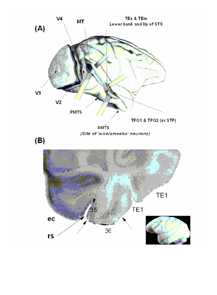
Fig. 1. Lateral view of a monkey’s brain and location of the wire
-
selective neurons. A. Lateral view with the superior temporal sulcus
(STS) opened up to illustrate various visual areas in the temporal pathway. V1, primary (striate) cortex; V2, V4, second and fourth visual
areas; MT (or V5) middle temporal visual area; PMTS, posterior mediotemporal sulcus; AMTS, anterior mediotemporal sulcus; TEa/m
areas within the inferotemporal cortex; TPO1/2 areas within the STS. B. Histological slice showing the anatomical site in which the wire/
amoeba selective neurons were found: ec, entorhinal cortex, 35/36 areas 35 and 46 respectively (perirhinal cortex); rs, rhinal sulcus. The
vertical line depicts the position of the coronal section shown in (B). The arrows depict approximately the borders of the corresponding areas.
COGNITIVE NEUROPSYCHOLOGY, 2000, 17 (1/2/3)
133
IS FACE RECOGNITION NOT SO UNIQUE?
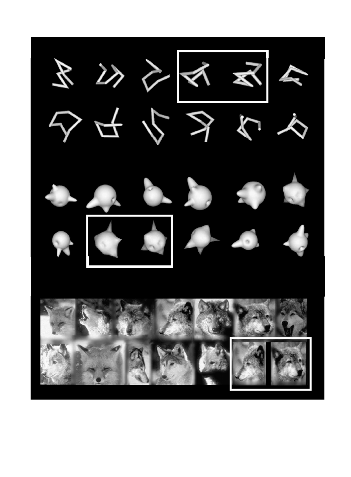
Fig. 2. The wire
-
and amoeba
-
like objects used to study the neural representations that may be employed for recognising objects at the
subordinate level. The exemplars of both classes are different barring the two within each white rectangle, which are two views of the same
objects 90° apart. Recognising individual exemplars of these classes is not unlike recognising individual exemplars of other homogeneous
natural classes. The wolves in the last row are all different barring those within the white rectangle. Again, the latter are two views of the
same animal 90° apart. In each case, identification of a member requires excessive practice.
GAUTHIER AND LOGOTHETIS
134
COGNITIVE NEUROPSYCHOLOGY, 2000, 17 (1/2/3)
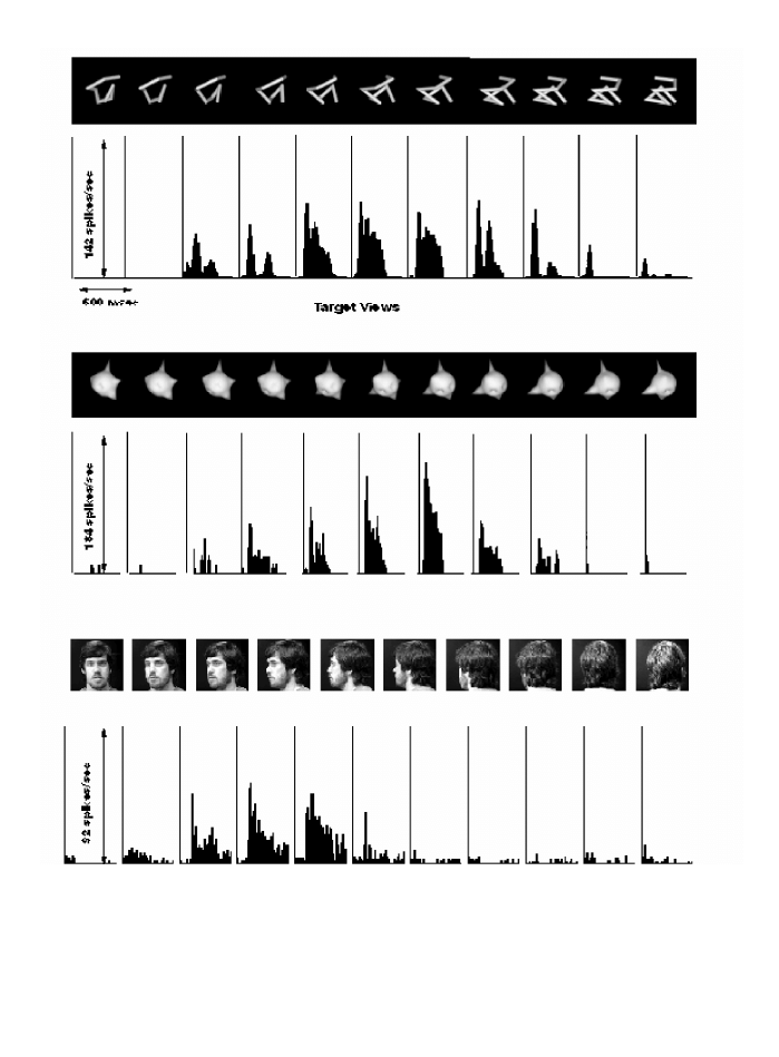
Fig. 3. Responses of single units in the inferior temporal cortex of the monkey. The upper row shows responses to wire
-
like objects and the
middle row to amoeba
-
like objects. The neuron responds best to a recently learned object
-
view and its response diminishes as the object is
rotated in depth. For objects that the monkey could recognise from all vantage points more than one unit was found that responded to
different views of the same object. Systematic decomposition of the wire objects showed that while some neurons could also be activated by
parts of the object (e.g. an angle), others required the entire configuration, strongly diminishing their response even when only a single
wire
-
segment was removed (Pauls, 1997). The bottom row shows responses of a face
-
selective neuron recorded in the upper bank of the STS.
“Wire” and “amoeba” cells display view tuning similar to that of the face cells.

IS FACE PROCESSING UNIQUE?
The finding of “expert” cells in monkeys trained to
discriminate among amoebas and wires suggest
that face recognition may find its homologue in the
brain under the right circumstances. In Hay and
Young’s (1982) framework, one way in which faces
may be special is that they could be represented in a
different manner to nonface objects. In humans,
evidence for unique face processing comes from a
number of behavioural effects that are obtained
with faces but not with nonface control stimuli such
as houses and even inverted faces. Most of these
behavioural effects measure some aspect of what is
called holistic or configural processing. Simply
stated, face recognition is often found to be more
sensitive than nonface recognition to the disruption
of the configuration of features: for instance, mov
-
ing the eyes slightly apart or inverting the entire
face so that relations such as “top of” or “right of”
are changed (for reviews, see Farah, 1996; J.W.
Tanaka & Gauthier, 1997). Evidence against face
processing being unique comes from experiments
where the same configural effects are obtained with
nonface objects when subjects are experts with
these categories (Diamond & Carey, 1986;
Gauthier & Tarr, 1997; J.W. Tanaka & Gauthier,
1997). This suggests that configural sensitivity is
not restricted to faces and that it is the particular
experience with an object category, rather than its
superficial properties, which determines the pro
-
cessing of its exemplars. Here, we consider whether
IT cells may be thought to represent faces in a dif
-
ferent way to other objects.
Face Cells Show a High Degree of
Selectivity to the Face Category
Face cells in anterior IT are sensitive to configura
-
tion of features (Young & Yamane, 1992) and may
be mediating the configural sensitivity that is a hall
-
mark of upright face recognition. In a paper dis
-
cussing face specificity in humans, Farah et al.
(1998) cite the existence of face cells as converging
evidence for faces being represented in a different
fashion, because “the selectivity and strength of
such responses [to nonface objects] are weaker
[than to faces]”. In a recent review article, Tovée
(1998) notes that face cells are resistant to a stimu
-
lus simplification protocol (K. Tanaka, 1997)
whereas the selectivity of most other IT cells can be
reduced to rather simple stimuli. Tovée argues that
“The ‘specialness’ of the face processing system will
rest upon the determination of whether the face
processing cells in IT have no functional equivalent
counterparts for object processing, either in IT or
elsewhere.”
The single
-
cell recording experiments described
in this paper may provide some evidence for
nonface object cells that are the functional equiva
-
lent of face cells. A remarkable similarity exists
between the properties of the face cells and those of
the wire
-
or amoeba
-
selective neurons recorded
from expert monkeys (Logothetis & Pauls, 1995;
Logothetis et al., 1995). The latter type of neurons
show selectivity to complex configurations that
cannot be reduced without diminishing the cells’
response to specific views and to views that appear
to be mirror symmetrical. They also exhibit posi
-
tion and scale invariance, and are clustered in a spe
-
cific brain location. This evidence is consistent with
the possibility that the responses of IT cells are built
from experience and adapted to the interactions of
an animal with objects. In most cases, animals need
to recognise most objects at a categorical level (e.g.
cage, ball, tree) and faces at the exemplar level.
However, if animals need to treat other objects like
faces and discriminate visually similar exemplars, a
number of cells within IT may begin to represent
the features that are best suited to this task.
Face Cells Represent Face Identity in a
Sparse Fashion
Several authors (Rolls & Tovée, 1995; Young &
Yamane, 1992) have suggested that IT face cells
may be representing face identity using sparse cod
-
ing. On a continuum from “grandmother” repre
-
sentations (where a single cell represents a single
object) to highly distributed processing (in which a
very large number of cells contribute to the repre
-
sentation, each one carrying an infinitely small
amount of useful information), sparse coding con
-
stitutes a case where the firing of each neuron
COGNITIVE NEUROPSYCHOLOGY, 2000, 17 (1/2/3)
135
IS FACE RECOGNITION NOT SO UNIQUE?

strongly biases the probability of a response to an
object. Face cell populations are thought to use
sparse rather than distributed coding because each
face cell at least carries a lot of information at the
level of the stimulus class, responding more to any
face than to nonface stimuli. Within the class of
faces, however, the cells respond to many of the
faces in a more distributed fashion. This type of
representation has been suggested to be ideal for the
discrimination of faces (Rolls & Tovée, 1995).
Note that such conclusions are based on what is
called information theoretic analyses, in which
face
-
selective cells are first selected and later shown
to provide more information about faces than about
nonface stimuli. A comparable analysis for nonface
objects would first require the selection of a popula
-
tion of cells that respond best to a certain class of
nonface objects than to other stimuli. As discussed
previously, this may be impractical for nonface cat
-
egories of no particular relevance to an animal but
may be feasible after an animal has been trained to
discriminate among visually similar objects.
Some authors emphasise the similarities
between face cells and other IT cells selective for
elaborate features. For instance, Perrett and Oram
(1993) note that in the anterior temporal cortex,
both face cells and Elaborate cells do not generalise
across orientation and size (whereas face cells in
STP do). In both cases a rotation of 90° in the pic
-
ture plane reduces the response by more than 50%.
However, other authors have contrasted the appar
-
ent sparse coding for faces to the more distributed
coding by which nonface objects appear to be repre
-
sented. K. Tanaka (1997) has suggested that
nonface objects are represented by distributed
coding over a large number of IT columns, each
containing cells selective for moderately complex
features. In this framework, each shape primitive
carries very little information about the identity of
the object and the representation of nonface objects
may be argued to be qualitatively different from
that of faces, in that it would be considerably more
distributed.
Recently, however, Kobatake, Wang, and
Tanaka (1998) have trained monkeys to recognise
28 moderately complex stimuli (mostly combina
-
tions of 2 simple geometric shapes, these stimuli
were less homogeneous than wires or amoebas) and
found a greater proportion of cells responsive to the
trained stimuli in trained than untrained monkeys.
Furthermore, many of these cells responded to
multiple members of the training stimuli, not
unlike face cells. The discriminations learned by the
monkeys may be supported by sparse representa
-
tions and the number of cells that respond to a cer
-
tain object may be partly determined by an animal’s
experience with this category (see also Booth &
Rolls, 1998). However, experience with a visually
homogeneous class of objects (e.g. the wires and
ameobas) may be necessary to build up a population
of cells that will generalise to novel exemplars of the
category. When humans are trained with several
objects of an homogeneous category, their expertise
generalises to novel exemplars (for instance,
configural sensitivity is found for untrained
objects—Gauthier & Tarr, 1997). Given the simi
-
larity of behavioural performance in object recogni
-
tion tasks between man and monkey (Logothetis &
Pauls, 1995), we can hypothesise that expertise in
monkeys would also generalise to novel exemplars
of a trained class. However, such generalisation
could be expected in monkeys trained with ameobas
and wires, but not necessarily for animals trained
with less homogeneous stimulus sets.
IS FACE PROCESSING SPECIFIC?
Even if we found that faces and objects are repre
-
sented by common mechanisms in IT, faces could
still be special in that they could be processed in a
distinct and separate neural system. It may be that
specificity (Hay & Young, 1982) in the location of
cells for any object category is not a sufficient crite
-
rion to designate this category “special” (Tovée,
1998), presumably because specificity would not be
unique to a single category (i.e. if face cells are sepa
-
rated from wire cells, then wire cells are also sepa
-
rated from face cells). However, regardless of the
debate on faces, to consider the spatial organisation
of object
-
selective cells is essential to the under
-
standing of the temporal cortex organisation.
The area where wire and amoeba cells were
found, the AMTS, is anterior to area TE and more
GAUTHIER AND LOGOTHETIS
136
COGNITIVE NEUROPSYCHOLOGY, 2000, 17 (1/2/3)

ventral than areas where face cells are typically
found in other studies. What this means is some
-
what difficult to interpret, given the methodologi
-
cal constraints of single
-
cell recording. As in any
single
-
cell study where there is no prior knowledge
of precisely where selective responses are expected,
Logothetis and colleagues (Logothetis & Pauls,
1995; Logothetis et al., 1995) recorded systemati
-
cally from posterior to anterior areas of the tempo
-
ral lobe, moving to a new area after a week or so
of fruitless explorations. Once a first wire
-
or
amoeba
-
selective cell was found in AMTS, the
researchers kept on recording in this area without
going back to more posterior regions. In addition,
the AMTS was not systematically tested with faces
in this experiment. In other words, the current evi
-
dence suggests that populations of expert object
cells are found in a different area than populations
of face cells with comparable properties, but this
evidence is not as strong as it would be if it came
from a neuroimaging experiment in which all areas
of the visual system had been equally sampled at all
time
-
points.
Evidence that face processing may be segregated
from object processing in the human brain mainly
comes from two different sources. The first is evi
-
dence from patients with selective deficits in face
processing (De Renzi, 1986; Farah et al., 1995a).
The selectivity of face agnosia is controversial, as
many prosopagnosic patients also report difficulties
with other visually similar categories (Bornstein,
Sroka, & Munitz, 1969; Damasio et al., 1982;
Shuttleworth, Syring, & Norman, 1982). Even in
the case of patients who believe that their deficit
applies only to faces, recent work has revealed a
more general impairment for subtle, subordinate
-
level discriminations (Gauthier et al., 1999b).
A second source of evidence comes from neuro
-
imaging studies in which activation in the middle
fusiform gyrus is found when subjects are viewing
faces as opposed to nonface objects (Kanwisher et
al., 1997; McCarthy et al., 1997; Sergent &
Signoret, 1992). To address this evidence and
inspired by the fact that prosopagnosic patients
often have difficulties discriminating objects within
the same category, Gauthier et al. (1998) compared
brain activation when normal subjects verified the
subordinate identity of a picture (e.g. pelican) vs. the
basic level (e.g. bird). They found activation in ven
-
tral temporal areas described as face
-
sensitive in
prior studies. In this issue, a new study (Gauthier et
al., this issue) verified that subordinate
-
level pro
-
cessing of nonface objects activates the small area
that can be defined as face
-
specific in each subject.
Thus, the presentation of faces is not necessary to
engage what is often called the “face area.” This
region can be differentially engaged when the same
nonface object is recognised at the subordinate vs.
the basic level. However, faces appear to activate
only a portion of ventral cortex dedicated to subor
-
dinate
-
level processing. These studies, which sug
-
gest that subordinate level processing accounts for
some of the activation in the face area, are not neces
-
sarily incompatible with other work suggesting that
not all of the activation in the face area can be
accounted for by subordinate
-
level classification
(Kanwisher et al., 1997). What may be happening
is that the former studies focus on the fact that there
is difference between basic level and subordinate
level recognition of nonface objects in the face area,
whereas the latter studies account for a different
part of the data, pointing out that there is still more
evidence for subordinate
-
level recognition of faces
than subordinate
-
level recognition of nonface
objects. A recent fMRI study (Gauthier et al.,
1999a) has revealed that expertise with subordi
-
nate
-
level discrimination of novel objects (similar
training experience as the monkeys in Logothetis &
Pauls, 1995; Logothetis et al., 1995) leads to
increased activation localised in the “face area.”
This suggests that the interaction of two factors,
level of categorisation and expertise, may interact to
produce the specialisation for faces found in the
middle fusiform face area. In the next section, we
consider how what we know of the monkey visual
system can help resolve the role of these two factors.
Level of Categorisation and Expertise
Given the importance of level of categorisation
demonstrated in behavioural (J.W. Tanaka & Tay
-
lor, 1993) and fMRI studies in humans (Gauthier
et al., 1998, this issue), one may ask whether there
is any evidence that this factor is important in
COGNITIVE NEUROPSYCHOLOGY, 2000, 17 (1/2/3)
137
IS FACE RECOGNITION NOT SO UNIQUE?

determining the responses of IT cells. Unfortu
-
nately, no single
-
cell recording study has compared
the responses of cells to the same stimuli when ani
-
mals are requested to recognise it at different levels
of abstraction. However, Logothetis and Pauls
(1995) have trained monkeys to recognise objects
either at the basic level (among distractors differing
largely in shape, such as a wire vs. an amoeba) or at
the subordinate level (for instance, discriminating
between two wires). They found that the animals’
behavioural
performance
was
viewpoint
-
dependent in the case of subordinate
-
level judge
-
ments and viewpoint
-
independent in the case of
basic
-
level judgements. This suggests that level of
categorisation may at least have a similar impor
-
tance for monkey and human visual recognition.
Two recent studies provided monkeys with
experience with certain objects and later found cells
to be responsive to many of these trained objects
(Booth & Rolls, 1998; Kobatake et al., 1998).
However, these studies differ in an important way
from the wire
-
frame and amoeba study by
Logothetis and colleagues: The different objects
did not belong to what would be considered the
same “basic
-
level” category (Rosch, Mervis, Gray,
Johnson, & Boyes
-
Braem, 1976). This is because
they do not share common parts and could be dis
-
criminated by the presence of a single feature (e.g.
the way that the presence of eyes is diagnostic to
detect a face) or simple relationships between parts
(e.g. as for the presence of a nose underneath two
eyes). In comparison, objects from homogeneous
categories share common parts as well as the
first
-
order configuration of these parts (Diamond
& Carey, 1986; Rhodes & McLean, 1990). They
can only be distinguished using subtle differences in
the shape of their parts or subtle differences in the
configuration of their parts (e.g. distances between
different face features). It is expertise discriminat
-
ing between objects of such homogeneous catego
-
ries that is thought to mediate behavioural
configural effects and the increased recruitment of
the fusiform face area (Gauthier & Tarr, 1997;
Gauthier et al., 1999a). Again, there is yet no direct
comparison using physiological measurements of
the difference between basic and subordinate level
processing of objects, but the expertise of monkeys
discriminating between wires and amoebas may be
most relevant to the debate on face recognition in
humans.
In humans, recent fMRI results suggest that
expertise with novel objects (Greebles) can recruit
the middle fusiform face area (Gauthier et al.,
1999a). However, at least one area, in the lateral
occipital gyrus, showed a strong expertise effect,
with more activation for Greeble experts than nov
-
ices, and even more for Greebles than for faces.
This lateral occipital gyrus area did not behave like
the fusiform face area in all conditions: In particu
-
lar, this region responded more to inverted than to
upright faces, whereas the face area responded more
to upright than to inverted faces. Thus, there may
be a complex system of areas within the temporal
lobe that is modified by experience with objects.
This is consistent with the existence of face cells in
many areas of both the human and the monkey
brain. Similarly, AMTS may not be the only area of
expert monkeys where wire and amoeba cells can
be found. At this point, it is likely that further
advances in comparing the man and monkey sys
-
tems will require the addition of novel techniques
such as functional MRI in monkeys (Logothetis,
Guggenberger, Peled, & Pauls, 1999) to those
already available in both species.
CONCLUSIONS
Both humans and monkeys are extremely good at
recognising faces, a fact that is hardly surprising in
view of the vital importance that face recognition
has for the primate. An important neural system
exists in both species for the processing of facial
information. In the human behavioural literature,
starting with Diamond and Carey’s (1986) land
-
mark study of dog expertise, a consensus has grown
that nonface categories of objects can be processed
in the same way as faces given similar task con
-
straints and subject expertise. However, in human
neuropsychological and neuroimaging studies,
there is still an ongoing debate regarding the possi
-
bility that faces may be special.
Interestingly, the single
-
cell recording literature
also converges to suggest that faces are not repre
-
GAUTHIER AND LOGOTHETIS
138
COGNITIVE NEUROPSYCHOLOGY, 2000, 17 (1/2/3)

sented by IT cells in a unique fashion. Several
authors, including C.G. Gross (1992), the pioneer
in the domain of face cells, have suggested that face
cells may appear more specialised than other IT
cells only because face recognition happens to be an
extremely demanding subordinate recognition
task, and for nonhuman primates it may be the only
identification task performed in life. Clearly, such
an hypothesis leads to the prediction that a similar
specialisation may also arise when the identification
of members of other classes becomes the critical
task at hand. This was tested in recent single
-
cell
recording experiments. A remarkable similarity was
found between the properties of the face cells and
those of the wire
-
or amoeba
-
selective neurons
recorded from expert monkeys (Logothetis &
Pauls, 1995; Logothetis et al., 1995). The latter
type of neurons show selectivity to complex config
-
urations that cannot be reduced without diminish
-
ing the cells’ response to specific views and to views
that appear to be mirror
-
symmetrical. They exhibit
position and scale invariance, and are clustered in a
specific brain location. Since recordings have only
been made in the inferotemporal cortex and mostly
in AMTS, it is not currently known whether selec
-
tivity to these objects might not also be found in
other brain structures.
Such results are consistent with behavioural and
fMRI studies in humans showing that novel objects
are processed in a more configural manner with
expertise and can increasingly recruit parts of the
ventral temporal lobe. However, whereas fMRI
results in humans suggests that the very same areas
are recruited for faces and nonface objects, sin
-
gle
-
cell studies in monkeys point to specialisation
of different areas. These techniques are very differ
-
ent and it is important to note that fMRI could pro
-
vide more convincing evidence than single
-
cell data
for a dissociation between the location of face and
object expert processing. On the other hand, the
better spatial resolution of single
-
cell recording
could provide stronger support for an association in
location (e.g. if the very same cells were found to
mediate expert representations of different catego
-
ries). Paradoxically, the current data in fMRI sug
-
gests an association whereas single cell recording
suggests a dissociation, albeit only in the location of
face and wire/amoeba cells within the anterior tem
-
poral lobe. Therefore, for both sources of evidence
the interpretation should be cautious. In any case,
faces are not unique with regard to the type of neu
-
ral activity that can be recorded in a monkey’s brain
when the animal is coping with other classes of
objects in the same manner with which it deals with
faces.
REFERENCES
Baylis, G.C., Rolls, E.T., & Leonard C.M. (1987).
Functional subdivisions of the temporal lobe neocor
-
tex. Journal of Neuroscience, 7, 330–342.
Bodamer, J. (1947). Die Prosopagnosie. Die Agnosie
des Physiognomieerkennes. Arch Psychiatr Nervenkr,
179,
6–54.
Booth, M.C.A., & Rolls, E.T. (1998). View
-
invariant
representations of familiar objects by neurons in the
inferior temporal visual cortex. Cerebral Cortex, 8,
510–523.
Bornstein, B., Sroka, H., & Munitz, H. (1969).
Prosopagnosia with animal face agnosia Cortex, 5,
164–171.
Boussaoud, D., Desimone, R., & Ungerleider, L.G.
(1991). Visual topography of area TEO in the
macaque. Journal of Comparative Neurology, 306,
554–575.
Bruce, C.J. (1982). Face recognition by monkeys:
Absence of an inversion effect. Neuropsychologia, 20,
515–521.
Bruyer, R., & Crispeels, G. (1992). Expertise in person
recognition. Bulletin of the Psychonomic Society, 30,
501–504.
Buelthoff, H.H., & Edelman, S. (1992). Psycho
-
physical support for a two
-
dimensional view inter
-
polation theory of object recognition. Proceedings of
the National Academy of Sciences USA, 89,
60–64.
Clark, V.P., Keil, K., Maisog, J.M., Courtney, S.M.,
Ungerleider, L.G., & Haxby, J.V. (1996). Func
-
tional magnetic resonance imaging of human visual
cortex during face matching: A comparison with posi
-
tron emission tomography. Neuroimage, 4, 1–15.
Damasio, A.R., Damasio, H.C., & Van Hoesen,
G.W. (1982). Prosopagnosia: Anatomic basis and
behavioral mechanisms. Neurology, 32, 331–341.
de Gelder, B., Bachoud
-
Lévi, A.C., & Degos, J.D.
(1998). Inversion superiority in visual agnosia may be
common to a variety of orientation
-
polarised objects
besides faces. Vision Research, 38, 2855–2861.
COGNITIVE NEUROPSYCHOLOGY, 2000, 17 (1/2/3)
139
IS FACE RECOGNITION NOT SO UNIQUE?

De Renzi, E. (1986). Slowly progressive visual agnosia
or apraxia without dementia. Cortex, 22, 171–180.
Desimone, R., Albright, T.D., Gross, C.G., & Bruce,
C.J. (1984). Stimulus
-
selective properties of infe
-
rior temporal neurons in the macaque. Journal of
Neurosciences, 4,
2051–2062.
Desimone, R., Fleming, J.F.R., & Gross, C.G.
(1980). Prestriate afferents to inferior temporal cor
-
tex: An HRP study. Brain Research, 184, 41–55.
Diamond, R., & Carey, S. (1986). Why faces are and
are not special: An effect of expertise, Journal of
Experimental Psychology: General, 115,
107–117.
Distler, C., Boussaoud, D., Desimone, R., & Unger
-
leider, L.G. (1993). Cortical connections of inferior
temporal area TEO in macaque monkeys. Journal of
Comparative Neurology, 334,
125–150.
Eacott, M.J., Heywood, C.A., Gross, C.G., & Cowey,
A. (1993). Visual discrimination impairments fol
-
lowing lesions of the superior temporal sulcus are not
specific for facial stimuli. Neuropsychologia, 31,
609–619.
Edelman, S., & Buelthoff, H.H. (1992). Orientation
dependence in the recognition of familiar and novel
views of 3D objects. Vision Research, 32, 2385–2400.
Farah, M.J. (1996). Is face recognition ‘special’? Evi
-
dence from neuropsychology. Behavioural Brain
Research, 76,
181–189.
Farah, M.J., Levison, K.L., & Klein, K.L. (1995a).
Face perception and within
-
category discrimination
in prosopagnosia. Neuropsychologia, 33, 661–674.
Farah, M.J., McMullen, P.A., & Meyer, M.M. (1991).
Can recognition of living things be selectively
impaired? Neuropsychologia, 29, 185–193.
Farah, M.J., Wilson, K.D., Drain, H.M., & Tanaka,
J.W. (1995b). The inverted face inversion effect in
prosopagnosia: Evidence for mandatory, face
-
specific
perceptual
mechanisms. Vision
Research, 35,
2089–2093.
Farah, M.J., Wilson, K.D., Drain, M., & Tanaka, J.N.
(1998). What is “special” about face perception? Psy
-
chological Review, 105,
482–498.
Gaffan, D., & Heywood, C.A. (1993). A spurious cate
-
gory
-
specific visual agnosia for living things in human
and nonhuman primates. Journal of Cognitive Neuro
-
science, 5,
118–128.
Gauthier, I., Behrmann, M., & Tarr, M.J. (1999). Can
face recognition really be dissociated from object rec
-
ognition? Journal of Cognitive Neuroscience, 11,
349–370.
Gauthier, I., & Tarr, M.J. (1997). Becoming a
“Greeble’’ expert: Exploring the face recognition
mechanism. Vision Research, 37, 1673–1682.
Gauthier, I., Tarr, M.J.,Anderson, A.W., Skudlarski, P.,
& Gore, J.C. (1999a). Activation of the middle
fusiform “face area” increases with experience in rec
-
ognizing novel objects. Nature Neuroscience, 2,
568–573.
Gauthier, I., Tarr, M.J., Moylan, J., Anderson, A.W.,
Skudlarski, P., & Gore, J.C. (this issue). Does
visual subordinate
-
level categorization engage the
functionally
-
defined fusiform face area? Cognitive
Neurospychology, 16,
143–163.
Gauthier, I., Williams, P., Tarr, M.J., & Tanaka,
J.W. (1998). Training “Greeble’’ experts: A frame
-
work for studying expert obect recognition processes.
Vision Research, 38,
2401–2428.
Gross, C. (1992). Representation of visual stimuli in
inferior temporal cortex. Philosophical Transactions of
the Royal Society of London, B, 335,
3–10.
Gross, C.G., Bender, D.B., & Rocha
-
Miranda, C.E.
(1969). Visual receptive fields of neurons in
inferotemporal cortex of the monkey. Science, 166,
1303–1306.
Gross, C.G., Roche
-
Miranda, C.E., & Bender, D.B.
(1972). Visual properties of neurons in infero
-
temporal cortex of the macaque. Journal of Neuro
-
physiology, 35,
96–111.
Hamilton, C.R., & Vermeire, B.A. (1983). Discrimi
-
nation of monkey faces by split
-
brain monkeys.
Behavioural Brain Research, 9,
263–275.
Hasselmo, M.E., Rolls, E.T., & Baylis, G.C.
(1989). The role of expression and identity in the
face
-
selective responses of neurons in the temporal
visual cortex of the monkey. Behavioural Brain
Research, 32,
203–218.
Hay, D.C., & Young, A.W. (1982). The human face.
In A.W. Ellis (Ed.), Normality and pathology in cogni
-
tive function
. London: Academic Press.
Heywood, C.A., & Cowey, A. (1992). The role of the
“face
-
cell” area in the discrimination and recognition
of faces by monkeys. Philosophical Transactions of the
Royal Society of London–Series B: Biological Sciences,
335,
31–37.
Hietanen, J.K.,Perrett, D.I., Oram, M.W., Benson, P.J.,
& Dittrich, W.H. (1992). The effects of lighting
conditions on the responses of cells selective for face
views in the macaque temporal cortex. Experimental
Brain Research, 89,
157–171.
Iwai, E., & Mishkin, M. (1969). Further evidence on
the locus of the visual area in the temporal lobe of the
monkey. Experimental Neurology, 25, 585–594.
Johnson, M.H., & Morton, J. (1991). Biology and cog
-
nitive development: The case of face recognition
. Oxford:
Basil Blackwell.
GAUTHIER AND LOGOTHETIS
140
COGNITIVE NEUROPSYCHOLOGY, 2000, 17 (1/2/3)

Kaas, J.H. (1995). Progress and puzzles. Current Biol
-
ogy, 5,
1126–1128.
Kanwisher, N., McDermott, J., & Chun, M.M.
(1997). The fusiform face area: A module in human
extrastriate cortex specialised for face perception.
Journal of Neuroscience, 17,
4302–4311.
Kobatake, E., Wang, G., & Tanaka, K. (1998). Effects
of shape
-
discrimination training on the selectivity of
inferotemporal cells in adult monkeys. Journal of
Neurophysiology, 80,
324–330.
Konorski, J. (1967). Integrative activity of the brain.
An interdisciplinary approach
. Chicago: University of
Chicago Press.
Logothetis, N.K. (1998). Object vision and visual
awareness. Current Opinion in Neurobiology, 8,
536–544.
Logothetis, N.K., Guggenberger, H., Peled, S., & Pauls,
J. (1999). Functional imaging of the monkey brain.
Nature Neuroscience, 2,
555–562.
Logothetis, N.K., & Pauls, J. (1995). Psychophysical
and physiological evidence for viewer
-
centered object
representations in the primate. Cerebral Cortex, 5,
270–288.
Logothetis, N.K., Pauls, J., Buelthoff, H.H., & Poggio,
T. (1994). View
-
dependent object recognition in
the primate. Current Biology, 4, 401–414.
Logothetis, N.K.,Pauls, J., & Poggio,T. (1995). Shape
representation in the inferior temporal cortex of mon
-
keys. Current Biology, 5, 552–563.
Logothetis, N.K., & Sheinberg, D. (1996). Visual
object recognition, Annual Review of Neuroscience, 19,
577–621.
Lutz, C.K., Lockard, J.S., Gunderson, V.M., & Grant,
K.S. (1998). Infant monkeys’ visual responses to
drawings of normal and distorted faces. American
Journal of Primatology, 44,
169–174.
McCarthy, G., Puce, A., Gore, J.C., & Allison, T.
(1997). Face
-
specific processing in the fusiform
gyrus, Journal of Cognitive Neuroscience, 9, 605–610.
Mendelson, M.J., Haith, M.M., & Goldman
-
Rakic,
P.S. (1982). Face scanning and responsiveness to
social cues in infant rhesus monkeys. Developmental
Psychology, 18,
222–228.
Mikami, A., Nakamura, K., & Kubota, K. (1994).
Neuronal responses to photographs in the superior
temporal sulcus of the rhesus monkey. Behavioural
Brain Research, 60,
1–13.
Morton, J., & Johnson, M.H. (1991). CONSPEC and
CONLERN: A two
-
process theory of infant face rec
-
ognition. Psychological Review, 98, 164–181.
Moscovitch, M., Winocur, G., & Behrmann, M.
(1997). What is special in face recognition? Nineteen
experiments on a person with visual object agnosia
and dyslexia but normal face recognition. Journal of
Cognitive Neuroscience, 9,
555–604.
Nahm, F.D., Perret, A., Amaral, D.G., & Albright,
T.D. (1997). How do monkeys look at faces. Journal
of Cognitive Neuroscience, 9,
611–623.
Ó Scalaidhe, S.P., Wilson, F.A.W., & Goldman
-
Rakic,
P.S. (1997). Areal segregation of face
-
processing
neurons in prefrontal cortex. Science, 278, 1135–1138.
Pauls, J. (1997). The representation of 3
-
dimensional
objects in the primate visual system
. Unpublished dis
-
sertation number 1–162. Baylor College of Medicine.
Perrett, D.I., Harries, M.H., Bevan, R., Thomas, S.,
Benson, P.J., Mistlin, A.J., Chitty, J.K., Hietanen,
J.K., & Ortega, J.E. (1989). Frameworks of analysis
for the neural representation of animate object and
actions. Journal of Experimental Biology, 146, 87–113.
Perrett, D.I., Hietanen, J.K., Oram, M.W., & Benson,
P.J. (1992). Organisation and functions of cells
responsive to faces in the temporal cortex. Philosophi
-
cal Transactions of the Royal Society of London B: Biolog
-
ical Science, 335,
23–30.
Perrett, D.I., & Oram, M.W. (1993). Neurophysiology
of shape processing. Image and Visual Computing, 11,
317–333.
Perrett, D.I., Oram, M.W., Harries, M.H., Bevan, R.,
Hietanen, J.K., Benson, P.J., & Thomas, S. (1991).
Viewer
-
centred and object
-
centred coding of heads in
the macaque temporal cortex. Experimental Brain
Research, 86,
159–173.
Perrett, D.I., Oram, M.W., Hietanen, J.K., & Benson,
P.J. (1994). Issues of representation in object vision.
In M.J. Farah, G. Ratcliff (Eds.), The neuro
-
psychology of high
-
level vision: Collected tutorial
assays, (pp 33–62). Hillsdale NJ: Lawrence Erlbaum
Associates Inc.
Perrett, D.I., Rolls, E.T., & Caan, W. (1979). Tempo
-
ral lobe cells of the monkey with visual responses
selective forfaces. Neuroscience Lettr Suppl, S3, S358.
Perrett, D.I., Rolls, E.T., & Caan, W. (1982). Visual
neurones responsive to faces in the monkey temporal
cortex. Experimental Brain Research, 47, 329–342.
Perrett, D.I., Smith, P.A., Potter, D.D., Mistlin, A.J.,
Head, A.S., Milner, A.D., & Jeeves, M.A. (1985).
Visual cells in the temporal cortex sensitive to face
view and gaze direction. Proceedings of the Royal Society
of London–Series B: Biological Sciences, 223,
293–317.
Puce, A., Allison, T., Bentin, S., Gore, J.C., & McCar
-
thy, G. (1998). Temporal cortex activation in
humans viewing eye and mouth movements. Journal
of Neuroscience, 18,
2188–2199.
Puce, A., Allison, T., Gore, J.C., & McCarthy, G.
(1995). Face
-
sensitive regions in human extrastriate
COGNITIVE NEUROPSYCHOLOGY, 2000, 17 (1/2/3)
141
IS FACE RECOGNITION NOT SO UNIQUE?

cortex studied by functional MRI. Journal of
Neurophysiology, 74
, 1192–1199.
Rhodes, G., & McLean, I.G. (1990). Distinctiveness
and expertise effects with homogeneous stimuli:
Towards a model of configural coding. Perception, 19,
773–794.
Rolls, E.T. (1992). Neurophysiology and functions of
the primate amygdala. In J.P. Aggleton (Ed.),The
amygdala (pp 143–165). New York: Wiley
-
Liss.
Rolls, E.T., & Tovée, M.J. (1995). Sparseness of the
neuronal representation of stimuli in the primate
temporal cortex. Journal of Neurophysiology, 73,
713–726.
Rosch, E., Mervis, C.B., Gray, W.D., Johnson, D.M., &
Boyes
-
Braem, P. (1976). Basic objects in natural
categories. Cognitive Psychology, 8, 382–439.
Rosenfield, S.A., & Van Hoesen, G.W. (1979). Face
recognition in the rhesus monkey. Neuropsychologia,
17,
503–509.
Seltzer, J.B., & Pandya, D.N. (1978). Afferent cortical
connections and architectonics of the superior tempo
-
ral sulcus and surrounding cortex in the rhesus
monkey. Brain Research, 149, 1–24.
Sergent, J., Ohta, S., & MacDonald, B. (1992). Func
-
tional neuroanatomy of face and object processing. A
positron emission tomography study. Brain, 115,
15–36.
Sergent, J., & Signoret, J.L. (1992). Functional and
anatomical decomposition of face processing: Evi
-
dence from prosopagnosia and PET study of normal
subjects. Philosophical Transactions of the Royal Society
of London B, 335,
55–62.
Shiwa, T. (1987). Corticocortical projections to the
monkey temporal lobe with particular reference to the
visual processing pathways. Arch Ital Biol, 125,
139–154.
Shuttleworth, E.C., Syring, V., & Norman, A. (1982).
Further observations on the nature of prosopagnosia.
Brain and Cognition, 1,
307–322.
Simion, F., Valenza, E., Umilta, C., & Dalla Barba, B.
(1998). Preferential orienting to faces in newborns:
A temporal
-
nasal asymmetry. JEP: HPP, 24,
1399–1405.
Tanaka, J.W., & Gauthier, I. (1997). Expertise in
object and face recognition. In R.L. Goldstone, P.G.
Schyns, & D.L. Medin (Eds.), Psychology of learning
and motivation.
San Diego, CA: Academic Press.
Tanaka, J.W., & Taylor, M. (1991). Object categories
and expertise: Is the basic level in the eye of the
beholder? Cognitive Psychology, 23, 457–482.
Tanaka, K. (1996). Inferotemporal cortex and object
vision. Annual Review of Neuroscience, 19, 109–139.
Tanaka, K. (1997). Mechanisms of visual object recog
-
nition: Monkey and human studies. Current Opinion
in Neurobiology, 7,
523–529.
Tanaka, K., Saito, H., Fukada, Y., & Moriya, M.
(1991). Coding visual images of objects in the
inferotemporal cortex of the macaque monkey. Jour
-
nal of Neurophysiology, 66,
170–189.
Toveé, M.J. (1998). Is face processing special? Neuron,
21,
1239–1242.
Ungerleider, L.G. (1995). Functional brain imaging
studies of cortical mechanisms for memory. Science,
270,
769–775.
Valenza, E., Simion, F., Macchi Cassia, V., & Umilta,
C. (1996). Face preference at birth. JEP:HPP, 22,
892–903.
Von Bonin, G., & Bailey, P. (1947). The neocortex of
macaca mulatta
. Urbana, IL: University of Illinois
Press.
Wachsmuth, E., Oram, M.W., & Perrett, D.I. (1994).
Recognition of objects and their component parts:
Responses of single units in the temporal cortex of the
macaque. Cerebral Cortex, 4, 509–522.
Webster, M.J., Ungerleider, L.G., & Bachevalier, J.
(1991). Connections of inferior temporal areas TE
and TEO with medial temporal
-
lobe structures in
infant and adult monkeys. Journal of Neuroscience, 11,
1095–1116.
Williams, G.V., Rolls, E.T., Leonard, C.M., & Stern,
C. (1993). Neuronal responses in the ventral
striatum of the behaving macaque. Behavioural Brain
Research, 55,
243–252.
Wilson, F.A.W., Ó Scalaidhe, S.P., & Goldman
-
Rakic,
P.S. (1993). Dissociation of object and spatial pro
-
cessing domains in primate prefrontal cortex. Science,
260,
1955–1958.
Wright, A.A., & Roberts, W.A. (1996). Monkey and
human face perception: Inversion effects for human
faces but not for monkey faces or scenes. Journal of
Cognitive Neuroscience, 8,
278–290.
Yin, R.K. (1969). Looking at upside
-
down faces, JEP,
81,
141–145.
Young, A.W., Hellawell, D., & Hay, D. (1987).
Configural information in face perception. Perception,
10,
747–759.
Young, M.P., & Yamane, S. (1992). Sparse population
coding of faces in the inferotemporal cortex. Science,
256,
1327–1331.
GAUTHIER AND LOGOTHETIS
142
COGNITIVE NEUROPSYCHOLOGY, 2000, 17 (1/2/3)

Wyszukiwarka
Podobne podstrony:
Why telly is not good for you
N Feature Neural Network Human Face Recognition
Euthanasia is not Murder
WHEN TIME IS NOT ENOUGH Jinko
Young Children Know That Trying Is Not Pretending, Psychologia UJ, Psychologia rozwojowa
Spiral syllabus The name refers not so much to the teaching content which is specified within the sy
Arteriosclerosis is not a modern disease
Writing SF 09 good writing is not enough
Surrender Is Not An Option
THIS LIST IS NOT BEING USED ANYMORE! YOUR EDITS WILL BE LOST NEW LIST UP SOON
EG170, October 2007 A minor dual is not a big deal
The World Is Not Enough
Art of Forgiveness Acceptance is not
2 size 14 is not fat either (ing)
FIDE Trainers Surveys 2014 06 29, Susan Polgar The Game Is Not Over Until It Is Over
Inequality Is Not Inevitable Joseph Stiglitz
A PhD Is Not Enough! A Guide to Surviva Peter J Feibelman
więcej podobnych podstron