
1
H MRSI evidence of metabolic abnormalities in
childhood-onset schizophrenia
Joseph O’Neill,
a,
* Jennifer Levitt,
a
Rochelle Caplan,
a
Robert Asarnow,
a,b
James T. McCracken,
a
Arthur W. Toga,
c
and Jeffry R. Alger
d
a
Division of Child and Adolescent Psychiatry, University of California at Los Angeles, Los Angeles, CA 90095, USA
b
Department of Psychology, University of California at Los Angeles, Los Angeles, CA 90095, USA
c
Laboratory of Neuroimaging, University of California at Los Angeles, Los Angeles, CA 90095, USA
d
Department of Radiology, Brain Research Institute, and Ahmanson-Lovelace Brain Mapping Center, University of California at Los Angeles,
Los Angeles, CA 90095, USA
Received 18 September 2003; revised 12 November 2003; accepted 13 November 2003
In adult schizophrenia, magnetic resonance imaging (MRI) and
magnetic resonance spectroscopy (MRS) have revealed volumetric
and metabolic defects in multiple brain regions, among them the
anterior cingulate, frontal cortex, striatum, thalamus, parietal cortex,
and frontal and parietal white matter. This study used proton magnetic
resonance spectroscopic imaging (
1
H MRSI) to identify potential
metabolic abnormalities in these regions in childhood-onset schizo-
phrenia.
1
H MRSI was acquired at 1.5 T and 272 ms echo time in 11
children and adolescents with schizophrenia (aged 7 – 18 years; seven
boys, four girls; all but two medicated) and 20 age-matched healthy
controls (10 boys, 10 girls). Absolute levels of N-acetyl compounds
(NAA), creatine plus phosphocreatine (Cr), and choline compounds
(Cho) were compared among groups in each region. In schizophrenic
patients relative to controls, Cr was 14.3% higher in superior anterior
cingulate (mean of left and right hemispheres). Cho was higher in
superior anterior cingulate (30.3%), frontal cortex (13.3%), and
caudate head (13.5%). In the thalamus, there was also a diagnosis-
by-gender interaction, whereby NAA was lower in patients for male but
not for female subjects. Elevated Cr suggests abnormal local cell-
energy demand and elevated Cho is consistent with a prior proposal
that patients with early age-of-onset schizophrenia exhibit phospholipid
membrane disturbances. Low NAA may reflect diminished neuronal
integrity.
D 2004 Elsevier Inc. All rights reserved.
Keywords: Anterior cingulate; Frontal cortex; Striatum; Childhood-onset
schizophrenia; Magnetic resonance spectroscopy
Introduction
Noninvasive magnetic resonance techniques reveal effects of
schizophrenia on the living brain. In adult schizophrenia (reviewed
in
Lawrie and Abukmeil, 1998; McCarley et al., 1999; Wright et
al., 2000
), structural magnetic resonance imaging (MRI) has
uncovered volumetric and morphometric abnormalities in multiple
brain regions, including anterior cingulate, frontal cortex, thala-
mus, and striatum; regions also implicated, though less strongly,
include parietal and occipital cortices and frontal and parietal white
matter. Cortical and white matter volumes are often below normal
(Lawrie and Abukmeil, 1998; McCarley et al., 1999; Shapleske et
al., 2002; Wright et al., 2000)
, while subcortical nuclei can be
larger or smaller than normal, depending in part on neuroleptic
treatment
(Keshavan et al., 1998; Lang et al., 2001)
. Proton
magnetic resonance spectroscopy (
1
H MRS) and proton magnetic
resonance spectroscopic imaging (
1
H MRSI) have documented
metabolic abnormalities in many of the same regions (reviewed in
Bertolino and Weinberger, 1999; Deicken et al., 2000b; Delam-
illieure et al., 2000; Kegeles et al., 1998; Keshavan et al., 2000
),
including below-normal levels of N-acetyl compounds (NAA) or
below-normal ratios of NAA to creatine plus phosphocreatine
(NAA/Cr) or to choline compounds (NAA/Cho). Above-normal
Cr has been reported in parietal white matter
while
31
P MRS has measured elevated temporal and parietal
phosphocreatine (
Blu¨ml et al., 1999; Fukuzako et al., 1999
; Volz
et al., 1998). Above-normal Cho or Cho/Cr have also been found
in anterior cingulate
, frontal lobes
al., 2000; Buckley et al., 1994; Cecil et al., 1999)
, thalamus
et al., 2001)
, basal ganglia
(Fujimoto et al., 1996; Shioiri et al.,
1996)
, and parietal white matter
These MRS findings yield insights into possible brain mecha-
nisms of schizophrenia. Low NAA is consistent with diminished
neuronal integrity
(Birken and Oldendorf, 1989; Urenjak et al.,
1992, 1993)
, including possible mitochondrial dysfunction
et al., 2003)
. High Cr may reflect disturbed energy metabolism of
neurons and/or glia, based on the well-known role of creatine and
phosphocreatine in ATP transduction
. Since multiple
choline compounds are involved in neuronal and glial phospho-
lipid metabolism
, elevated Cho may
imply disturbed membrane ‘‘turnover’’
al., 2000; Miller et al., 1996; Speck et al., 1996)
.
1053-8119/$ - see front matter
D 2004 Elsevier Inc. All rights reserved.
doi:10.1016/j.neuroimage.2003.11.005
* Corresponding author. Division of Child and Adolescent Psychiatry,
University of California at Los Angeles, NPI 47-433, 760 Westwood Plaza,
Los Angeles, CA 90024-1759. Fax: +1-310-206-4446.
E-mail address: joneill@mednet.ucla.edu (J. O’Neill).
Available online on ScienceDirect (www.sciencedirect.com.)
www.elsevier.com/locate/ynimg
NeuroImage 21 (2004) 1781 – 1789

have interpreted elevated Cho as supportive of the ‘‘membrane
hypothesis’’ of schizophrenia
(Fenton et al., 2000; Horrobin et al.,
1994)
. They have suggested that earlier onset occurs in patients
with more severe phospholipid disturbances
Childhood-onset schizophrenia is thought of as a more severe
form of schizophrenia
and by
definition emerges relatively early in life. MRI abnormalities have
been found in many of the same brain regions in childhood-onset
schizophrenia as in adult schizophrenia (reviewed in
al., 2000; Mehler and Warnke, 2002; Rapoport et al., 2001; Sowell
et al., 2000
). The proposal of
implies that, of the
three
1
H MRS metabolic defects seen in adult schizophrenia, low
NAA, high Cr, and high Cho, elevated Cho should be especially
prominent in patients with childhood-onset schizophrenia. Some
MRS research
(Bertolino et al., 1998; Brooks et al., 1998)
including work from this laboratory
, sug-
gests anterior cingulate and frontal metabolite abnormalities in
childhood-onset schizophrenia, including below-normal NAA/Cr.
The number of patients with childhood-onset schizophrenia exam-
ined with
1
H MRS to date, however, is small, implying a need for
more investigation. Further, most studies in adult- and childhood-
onset schizophrenia acquired
1
H MRS from one or two isolated
sites. Most reported results as ratios to Cr (an inherently ambiguous
format) rather than as absolute metabolite levels. And few deter-
mined the tissue composition (gray matter, white matter, CSF) of
the
1
H MRS volumes acquired.
We undertook an exploratory
1
H MRSI study on a small
number of children and adolescents with childhood-onset schizo-
phrenia and age-matched healthy controls. Absolute levels of
NAA, Cr, and Cho were measured in anterior cingulate, frontal
cortex, thalamus, and striatum, as well as in parietal and occipital
cortices and frontal and parietal white matter, accounting for
1
H
MRSI voxel tissue composition. Based on the above-cited MRI
and MRS literature and the proposal of
, we
hypothesized below-normal NAA and above-normal Cr and Cho in
each of these regions. Other regions known to show structural and
metabolic abnormalities in schizophrenia, such as the mesial
temporal lobes
(Levitt et al., 2001; Matsumoto et al., 2001a,b)
were outside the scope of this investigation.
Methods
Subjects
The study was conducted under the supervision of the UCLA
Human Subjects Review Board. Informed consent was obtained
from all parents or legal guardians, and written assent was obtained
from all children before participation. Eleven patients with child-
hood-onset schizophrenia (7 – 17.5 years; mean age F SD, 12.3 F
3.8 years; seven boys, four girls) were recruited. Patients had to
have a DSM-IV diagnosis of schizophrenia, absence of neurologic
or other nonpsychiatric illness, and onset of symptoms by age 14 to
be included. Diagnoses were based on a structured interview using
the Kiddie-Schedule for Affective Disorders and Schizophrenia-
Present and Lifetime version (K-SADS-PL;
Current medication and medication history for patients are listed in
. Twenty healthy control children and adolescents (6.8 –
16.3 years; mean age F SD, 11.7 F 2.9 years; 10 boys, 10 girls)
were recruited from public and private schools in the community.
These subjects were screened for psychiatric, neurologic, or
developmental disorders by developmental history and K-SADS-
PL
interviews with parent and child.
Subjects were excluded from the normal sample if they met criteria
for any lifetime significant medical disorder or Axis I mental
disorder. Subject ascertainment and diagnosis are detailed in
. Several patients and no controls had first-
degree relatives with history of schizophrenia or other psychiatric
illness.
Full-scale IQ of 9 of the 11 patients with childhood-onset
schizophrenia was assessed
using the Wechsler Intelli-
gence Scale for Children-Revised (WISCR-R;
) and
averaged 94.4 F 12.6 (mean F SD) across the group. This was
significantly lower ( F = 15.5; df = 1,28; P = 0.001; ANOVA) than
the IQ of the control sample, 118.4 F 16.2 (mean F SD).
MRI/
1
H MRSI acquisition
MR methods were as described in
with
modifications. MRI and
1
H MRSI of the brain were acquired in the
same session lasting 1 – 1.5 h on a 1.5-T GE system (Signa Horizon
5.x) using a standard quadrature head coil. Six of eleven child-
hood-onset schizophrenic patients
and no healthy control
subjects were sedated with intravenous propofol anesthesia at time
of scan. Dose and details of administration were determined by the
staff anesthesiologist presiding. MR sequences were acquired from
each subject in the following order. After initial localizer scout
scan, axial fast spin-echo (FSE) MRI was acquired of the entire
brain [repetition time (TR)/TE = 3000/13 ms; 3-mm contiguous
slices; 0.94 0.94 mm
2
in-plane resolution]. This sequence
Table 1
Age, gender, IQ, concurrent and past medication, and propofol sedation
during MR acquisition for schizophrenic subjects
Age
(years)
Gender
IQ
Medication
History
Sedation
7.0
m
96
risperidone
imipramine,
risperidone,
olanzapine
yes
8.8
m
95
amphetamine
salts, risperidone
none
yes
11.1
m
70
none
none
yes
11.9
m
101
fluoxetine,
risperidone
none
no
15.8
m
–
clozapine,
lithium,
ziprasidone
divalproex,
gabapentine,
lithium,
thiothixene,
olanzapine,
risperidone,
sertraline
yes
16.6
m
99
clonazepam,
risperidone,
trazadone
divalproex,
quetiapine,
ethosuximide,
zonisamide
yes
17.5
m
107
benztropine,
risperidone
none
no
8.6
fm
–
clozapine
none
yes
9.6
fm
87
none
none
no
11.5
fm
84
benztropine,
paroxetine,
risperidone
none
no
16.7
fm
111
clozapine
none
no
J. O’Neill et al. / NeuroImage 21 (2004) 1781–1789
1782

yielded proton-density-weighted images. These images were used
to identify the neuroanatomic structures within which individual
1
H MRSI voxels were selected during post-processing and to
provide the proton-density intensity values to which
1
H MRSI
metabolite resonance intensities were normalized as part of the
process of absolute quantitation of metabolite levels. Next, a
sagittal whole-brain volumetric acquisition was performed using
a spoiled gradient-recalled echo (SPGR) sequence (TR/TE = 24/9
ms; 1.2-mm contiguous partitions; 0.94 0.94 mm
2
in-plane
resolution). This sequence yielded T1-weighted images used for
MRI tissue segmentation. Finally, multislice
1
H MRSI
1993)
was acquired using a 2D inversion-recovery sequence with
CHESS
water-suppression [TR/inversion time
(TI)/TE = 2300/170/272 ms; 1 average; 12-mm slice thickness; 10
10 mm
2
in-plane resolution, nominal voxel volume 1.2 cc] from
three contiguous axial slices
. The first slice centered on the
dorsoventral midplane of the basal ganglia, the second on the
ventricles, and the third on the supraventricular brain. The latter
two slices sampled wide areas of frontal, parietal, and occipital
gray and white matter.
MR image processing
MRI scans were reviewed by staff radiologists to exclude
subjects with structural or clinical abnormalities. MRI (and
1
H
MRSI) post-processing were conducted with operator blinded to
subject diagnosis. Tissue segmentation of T1-weighted MRI has
been described
. Briefly, 20 points each of
representative gray matter, white matter, CSF, and non-brain tissue
were selected manually within each subject’s T1-weighted volume.
An intensity-based algorithm separated the MRI into gray matter,
white matter, CSF, and non-brain component volumes. Interrater
correlation coefficients of 0.94 – 0.98 have been assessed for these
methods
. The gray matter, white matter, and
CSF component volumes were then coregistered
1993)
onto the axial proton-density-weighted MRI volume, which
was already in register with the
1
H MRSI volume.
1
H MRSI post-processing
After Fourier transform, each subject’s
1
H MRSI volume
underwent sine-bell spatial filtering, 2.0-Hz lorenztian temporal-
domain apodization, and automated polynomial baseline fitting
using home-written software in the Interactive Data Language
(IDL).
1
H MRSI voxels with lipid signals exceeding the NAA
signal (i.e., those having a substantial contribution from non-brain
tissue), with NAA signal-to-noise ratio less than 2.0, with line
width greater than 10.0 Hz, or with other detectable artifact (e.g.,
aliased extracranial lipid signals arising from movement), were
rejected manually. Peak intensities were integrated for N-acetyl
compounds (NAA; 2.01 ppm), creatine plus phosphocreatine (Cr;
3.03 ppm), and choline compounds (Cho; 3.23 ppm). Lactate (Lac;
1.36 ppm) was not assayed since it was not always distinguishable
from overlapping lipid resonances.
MRI/
1
H MRSI co-processing
Using the coregistered axial proton-density-weighted MRI to
identify anatomy, an individual
1
H MRSI voxel was selected
within each of the following structures (in left and right cerebral
hemispheres): superior anterior cingulate cortex, inferior anterior
cingulate cortex, frontal cortex (i.e., any frontal cortex outside the
cingulate), parietal cortex, occipital cortex; head of the caudate
nucleus, body of the caudate nucleus, putamen, thalamus, frontal
white matter, and parietal white matter. These structures were sites
of suspected pathology in schizophrenia (see above). Volume
percentages of gray matter, white-matter, and CSF in each selected
1
H MRSI voxel were calculated from the coregistered gray matter,
white matter, and CSF MRI component volumes using home-
written IDL software.
1
H MRSI voxels were sought that contained
z75% gray matter for cortical gray matter sites; z75% white
matter for white matter sites; and z50% gray matter for nuclear
gray matter sites, but some voxels for some subjects fell below
these threshold values. Systematic comparison revealed that there
were no significant between-group differences in gray or white
matter content at any site. Across two independent raters, both
blind to diagnosis, reliability of the voxel-selection procedure was
found to be z95%. Metabolite peak areas were adjusted for
instrumental transmitter and receiver gains, normalized to MRI
proton density intensity, and corrected for voxel CSF content. This
yielded absolute metabolite levels—uncorrected for T1 and T2
relaxation—expressed in Institutional Units (IU).
Statistical analysis
NAA, Cr, and Cho absolute metabolite levels were analyzed
using repeated-measures ANCOVA applied to each left – right
structure pair with hemisphere as within-subjects factor and diag-
nosis as between-subjects factor. Gender and age were used as
covariates to account for slight between-group differences in these
two variables. This statistical model both accounted for the within-
subject character of metabolite comparisons between left- and
right-hemisphere homologous structures and tested explicitly for
possible lateral asymmetries. Where significant interactions involv-
ing diagnosis and hemisphere and/or gender were uncovered,
appropriate post hoc comparisons were undertaken using one-
way ANOVA. Criterion for statistical significance was P < 0.05.
Because this was an exploratory study with a priori hypotheses,
Bonferroni correction for multiple comparisons was not applied.
The childhood-onset schizophrenic group had significantly
lower IQ than the healthy control group. Since low IQ has been
viewed as a cognitive symptom
(Aylward et al., 1984; Frith, 1995)
and a risk factor
(Davidson and Weiser, 2000; Davies et al., 1998;
Fig. 1. Sagittal T1-weighted MRI of brain of a 9.6-year-old schizophrenic
girl showing positioning of three
1
H MRSI acquisition slices.
J. O’Neill et al. / NeuroImage 21 (2004) 1781–1789
1783
Kelly and Murray, 2000)
for childhood- and adult-onset schizo-
phrenia, it was not deemed advisable to remove effects of IQ
statistically. Nine childhood-onset schizophrenic patients and no
healthy controls were taking atypical neuroleptics (and, in some
cases, other agents;
) at time of MRI/
1
H MRSI acquisition.
Therefore, to assess potential effects of neuroleptic medication, for
each significant finding, a one-way ANOVA was performed post
hoc comparing medicated to unmedicated patients. Six childhood-
onset schizophrenic patients
and no healthy controls were
under propofol sedation at time of MRI/
1
H MRSI acquisition.
Therefore, to assess potential effects of propofol sedation, for each
significant finding, an additional post hoc one-way ANOVA was
performed comparing sedated to unsedated patients.
Results
Data quality
At this long TE (272 ms), MR spectra acquired from juvenile
brains were typically of high quality, featuring prominent peaks for
NAA, Cr, and Cho. Lac was generally not evident, but its presence
cannot be excluded with certainty due to the aforementioned
overlap with lipids.
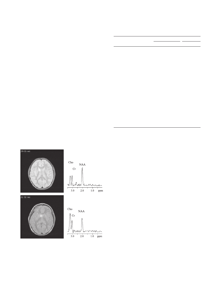
shows a spectrum from a representative
1
H MRSI voxel in the head of the right caudate nucleus of a 9.6-
year-old female patient with schizophrenia compared to an analo-
gous spectrum from a healthy 10.2-year-old girl. Cho and, to a
lesser extent Cr, are visibly elevated, while NAA is lower in the
schizophrenic spectrum. At this site, 8 of 11 subjects with
schizophrenia had a Cho level above the healthy-control mean;
for 5 of 11 it was 1 SD or more above.
Main effects of subject diagnosis on regional neurometabolite
levels
list absolute levels of NAA, Cr, and Cho at all
sites for both subject groups. The following differences (means of
left- and right-hemisphere structures) between the childhood-onset
schizophrenic group and the healthy control group were signifi-
cant (ANCOVA). In superior anterior cingulate, Cr was 14.3%
higher ( F = 5.0; df = 1,21; P = 0.04) in patients than in controls.
Cho was higher in patients than in controls in superior anterior
cingulate (30.3%; F = 9.6; df = 1,21; P = 0.006), frontal cortex
(13.3%; F = 6.3; df = 1,15; P = 0.02), and caudate head (13.5%;
F = 5.2; df = 1,23; P = 0.03). No other main effects of diagnosis
were significant.
Neurometabolite levels: interactions of subject diagnosis with
cerebral hemisphere, gender, and/or age
ANCOVA revealed significant interactions involving diagnosis
for NAA, Cr, and Cho. For NAA, there were several such
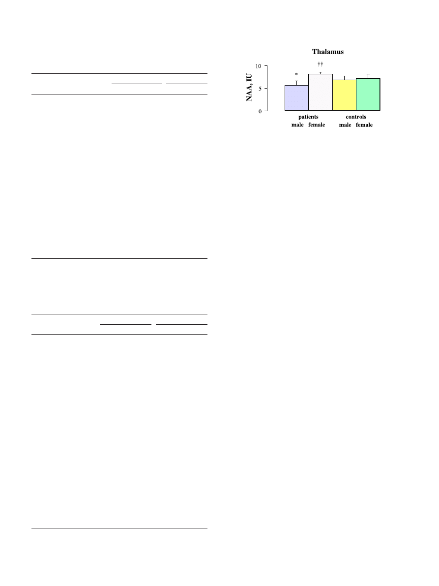
interactions. In the thalamus, there was a significant diagnosis-
by-gender interaction ( F = 6.2; df = 1,22; P = 0.02). In post hoc
ANOVA
, thalamic NAA was significantly lower in male
patients than in female patients ( F = 19.5; df = 1,10; P = 0.002) or
in male controls ( F = 5.8; df = 1,16; P = 0.03). NAA did not differ
significantly between female patients and female controls ( F = 3.4;
df = 1,12; P = ns) or between female controls and male controls
( F = 0.74; df = 1,18; P = ns). In caudate body, there was a
Fig. 2. Axial proton-density-weighted MRI section of brain of healthy 10.2-
year-old girl showing location of single
1
H MRSI voxel sampled in the
head of the right caudate nucleus (top, left).
1
H MR spectrum obtained in
sampled voxel after post-processing, featuring major peaks for NAA, Cr,
and Cho (top, right); same for the 9.6-year-old schizophrenic girl shown in
(bottom). Note elevated Cho and Cr intensities relative to NAA in
patient.
Table 2
1
H MRSI levels of N-acetyl compounds (Institutional Units) at multiple
brain sites
Region
Diagnosis
Mean F SD
ANCOVA
Left
Right
df
F
P
Superior anterior schizophrenia 7.2 F 1.9
6.3 F 1.5 1,21 1.7
ns
cingulate
control
6.1 F 1.6
6.0 F 1.5
Inferior anterior
schizophrenia 5.7 F 2.2
5.7 F 1.7 1,24 0.12
ns
cingulate
control
6.0 F 1.6
5.7 F 1.7
Frontal cortex
schizophrenia 8.0 F 0.5
7.5 F 0.4 1,15 0.74
ns
control
7.8 F 1.6
8.2 F 1.1
Parietal cortex
schizophrenia 7.3 F 0.9* 7.6 F 1.5 1,16 0.39
ns
control
8.2 F 1.0
7.6 F 1.4
Occipital cortex
schizophrenia 7.9 F 2.0
7.6 F 0.9 1,23 0.11
ns
control
7.4 F 1.0
7.3 F 1.1
Caudate head
schizophrenia 4.1 F 1.5
4.5 F 1.3 1,22 0.31
ns
control
4.6 F 1.6
3.8 F 1.4
Caudate body
schizophrenia 6.3 F 1.9
5.1 F 1.5 1,22 0.014 ns
control
6.1 F 1.0
5.0 F 1.5
Putamen
schizophrenia 5.9 F 2.0
5.4 F 1.3 1,24 1.0
ns
control
5.4 F 1.8
5.1 F 1.6
Thalamus
schizophrenia 6.9 F 2.0
6.4 F 1.2 1,22 3.2
ns
control
7.0 F 1.2
7.0 F 1.0
Frontal white
schizophrenia 7.2 F 1.5
6.8 F 2.4 1,22 0.017 ns
matter
control
7.3 F 1.5
6.7 F 1.7
Parietal white
schizophrenia 9.5 F 1.3
8.4 F 1.2 1,21 0.52
ns
matter
control
9.9 F 1.9
8.6 F 2.4
* P < 0.05 vs. controls (left only; ANOVA). ANCOVA is repeated-measures
with between-subjects variable diagnosis, within-subjects variable hemi-
sphere, and covariates age and gender.
J. O’Neill et al. / NeuroImage 21 (2004) 1781–1789
1784
significant three-way diagnosis-by-hemisphere-by-age interaction
( F = 5.4; df = 1,22; P = 0.03). In parietal cortex, there were a
significant diagnosis-by-hemisphere interaction ( F = 7.8; df = 1,16;
P = 0.01) and a significant diagnosis-by-hemisphere-by-gender
interaction ( F = 6.3; df = 1,16; P = 0.02). In post hoc ANOVA,
NAA was significantly lower in patients than in controls in left ( F =
5.1; df = 1,23; P = 0.03), but not in right ( F = 0.004; df = 1,23; P =
ns), parietal cortex. For patients, NAA was lowest in left parietal
cortex of males and highest in left parietal cortex of females; for
controls, NAA was lowest in right parietal cortex of males and
highest in right parietal cortex of females. For Cr in superior
anterior cingulate, there were a significant diagnosis-by-gender
interaction ( F = 5.0; df = 1,21; P = 0.04) and a significant
diagnosis-by-hemisphere-by-age interaction ( F = 5.0; df = 1,21;
P = 0.04). Cr was significantly higher in patients than in controls for
males ( F = 4.6; df = 1,15; P = 0.05), but not for females ( F = 0.67;
df = 1,12; P = ns). For Cho, in superior anterior cingulate, there was
a significant diagnosis-by-gender interaction ( F = 6.2; df = 1,21;
P = 0.02), whereby Cho augmentation was significant for male
patients vs. male controls (35.3%; F = 5.3; df = 1,15; P = 0.005),
but not for female patients vs. female controls (18.2%; F = 0.62;
df = 1,12; P = ns). In frontal cortex, there was also a significant
diagnosis-by-gender interaction ( F = 4.8; df = 1,15; P = 0.04) for
Cho, whereby values were highest for male patients and lowest
for female controls. No other interactions were significant.
Neurometabolite levels: effects of medication and sedation
Patients taking neuroleptic medication at time of study did not
differ significantly from unmedicated patients for any of the above
principal effects of diagnosis (all F < 0.40; df = 1,9; P = ns). Nor
did patients sedated during MR scanning differ significantly from
unsedated patients on these measures (all F < 3.9; df = 1,9; P = ns),
with the exception of Cho in frontal cortex. Frontal cortex Cho was
34.5% higher in sedated than in unsedated patients ( F = 8.2; df =
1,10; P = 0.02).
Discussion
The principal findings of this long-TE
1
H MRSI study were: (1)
above-normal levels of creatine plus phosphocreatine in superior
anterior cingulate and (2) above-normal levels of choline com-
Fig. 3. Absolute levels in Institutional Units (IU; group means F SD) of
NAA in the thalamus (mean left and right) of male and female childhood-
onset schizophrenic patients and male and female age-matched healthy
controls. NAA was 17.6% lower in male patients than in male controls
(*P < 0.05, ANOVA) and 44.6% higher in female than in male patients
(
yy
P < 0.01, ANOVA).
Table 3
1
H MRSI levels of creatine + phosphocreatine (Institutional Units) at
multiple brain sites
Region
Diagnosis
Mean F SD
ANCOVA
Left
Right
df
F
P
Superior anterior schizophrenia 3.3 F 0.9 3.0 F 1.0 1,21 5.0
0.04
cingulate
control
2.7 F 0.6 2.8 F 0.5
Inferior anterior
schizophrenia 3.0 F 1.2 3.2 F 0.9 1,23 0.016 ns
cingulate
control
2.7 F 0.9 2.6 F 0.9
Frontal cortex
schizophrenia 2.9 F 0.7 2.8 F 0.4 1,16 0.027 ns
control
2.6 F 0.8 3.0 F 0.7
Parietal cortex
schizophrenia 2.8 F 0.8 2.9 F 0.9 1,18 2.8
ns
control
2.5 F 0.4 2.6 F 0.8
Occipital cortex
schizophrenia 2.9 F 0.9 2.7 F 0.5 1,25 0.066 ns
control
2.4 F 0.8 2.7 F 0.8
Caudate head
schizophrenia 2.9 F 0.7 2.7 F 0.8 1,22 0.43
ns
control
3.0 F 0.8 2.3 F 0.5
Caudate body
schizophrenia 3.2 F 0.8 2.7 F 0.8 1,25 0.048 ns
control
3.0 F 0.7 2.8 F 0.8
Putamen
schizophrenia 2.8 F 0.8 3.1 F 0.9 1,24 1.0
ns
control
2.6 F 0.8 2.4 F 0.9
Thalamus
schizophrenia 3.0 F 0.8 2.9 F 1.2 1,24 0.11
ns
control
2.6 F 0.6 2.6 F 0.4
Frontal white
schizophrenia 2.4 F 0.9 2.4 F 0.4 1,23 0.12
ns
matter
control
2.4 F 0.8 2.5 F 0.6
Parietal white
schizophrenia 2.8 F 0.7 2.9 F 0.9 1,23 0.041 ns
matter
control
2.4 F 0.8 2.6 F 0.6
ANCOVA is repeated-measures with between-subjects variable diagnosis,
within-subjects variable hemisphere, and covariates age and gender.
Table 4
1
H MRSI levels of choline compounds (Institutional Units) at multiple
brain sites
Region
Diagnosis
Mean F SD
ANCOVA
Left
Right
df
F
P
Superior
schizophrenia 4.0 F 1.3 4.4 F 1.4 1,21
9.6
0.006
anterior
cingulate
control
3.2 F 1.1 3.4 F 1.0
Inferior
schizophrenia 3.7 F 0.9 3.3 F 0.8 1,24
1.1
ns
anterior
cingulate
control
3.5 F 0.8 2.9 F 1.1
Frontal
schizophrenia 3.5 F 0.7 3.4 F 0.9 1,15
6.3
0.02
cortex
control
2.9 F 0.9 3.0 F 0.7
Parietal
schizophrenia 2.7 F 0.8 2.5 F 0.7 1,18
0.07
ns
cortex
control
2.5 F 0.7 2.4 F 0.6
Occipital
schizophrenia 3.2 F 0.9 2.5 F 0.7 1,25
1.2
ns
cortex
control
2.3 F 0.8 2.4 F 1.0
Caudate
schizophrenia 4.2 F 0.8 4.2 F 0.7 1,23
5.2
0.03
head
control
4.0 F 1.5 3.5 F 0.8
Caudate
schizophrenia 3.1 F 1.1 2.9 F 0.6 1,24
0.55
ns
body
control
3.2 F 1.0 2.7 F 1.2
Putamen
schizophrenia 3.4 F 0.6 3.4 F 1.0 1,23
0.072
ns
control
2.4 F 0.9 2.6 F 0.7
Thalamus
schizophrenia 3.9 F 1.3 3.5 F 0.8 1,23
0.38
ns
control
4.0 F 0.9 3.7 F 1.1
Frontal
schizophrenia 4.5 F 1.3 4.1 F 1.2 1,23
0.072
ns
white
matter
control
4.5 F 1.4 4.2 F 1.0
Parietal
schizophrenia 3.4 F 1.1 3.3 F 0.8 1,23 <0.0005 ns
white
matter
control
3.7 F 1.2 3.5 F 1.3
ANCOVA is repeated-measures with between-subjects variable diagnosis,
within-subjects variable hemisphere, and covariates age and gender.
J. O’Neill et al. / NeuroImage 21 (2004) 1781–1789
1785

pounds in superior anterior cingulate, frontal cortex, and caudate
head in child and adolescent patients with childhood-onset schizo-
phrenia. These brain regions exhibit structural
meil, 1998; McCarley et al., 1999; Wright et al., 2000)
and
metabolic
(Bertolino and Weinberger, 1999; Deicken et al.,
2000b; Delamillieure et al., 2000; Kegeles et al., 1998; Keshavan
et al., 2000)
abnormalities in adult schizophrenia. The present
findings suggest that metabolic disturbances exist in these regions
in childhood-onset schizophrenia as well.
The first major finding was above-normal Cr in superior
anterior cingulate. An earlier study from this laboratory
et al., 1998)
acquired single-voxel
1
H MRS from a region labeled
‘‘medial frontal cortex’’ that roughly overlaps with the ‘‘superior
anterior cingulate’’ of the present report. Detailed voluming studies
in progress in our laboratory suggest that both regions actually
contain a mix of anterior cingulate and superior frontal gyral tissue.
The present finding suggests that elevated Cr may have contributed
to the below-normal NAA/Cr seen in patients with childhood-onset
schizophrenia in this region in
(2001)
have suggested that elevated Cr in schizophrenia signals
reduced cellular energy demand and may occur in response to
chronic use of dopaminergic agents. Several patients had been
treated with pharmacologics that influence the dopaminergic sys-
tems of the brain
. Elevated Cr may also reflect patho-
logically altered cellular energetics accompanying putative cell-
membrane disturbances in schizophrenia (see next paragraph).
The second major finding was above-normal Cho at three sites.
This is generally consistent with the notion of
that elevated Cho should be evident in schizophrenic patients with
younger age-of-onset. The Cho signal is thought to rise in tissues
undergoing enhanced throughput of phospholipid membrane con-
stituents, as during times of membrane build-up or degradation
(Gill et al., 1990; Speck et al., 1996)
. In this sense, the present
results support the notion of membrane abnormalities in schizo-
phrenia
(Fenton et al., 2000; Horrobin et al., 1994)
championed by
. Unlike
, however, we
observed above-normal Cho in superior anterior cingulate, frontal
cortex, and caudate head, rather than in left thalamus and left
parietal white matter. A recent report
documents below-normal NAA/Cho and above-normal Cho/Cr
in the anterior cingulate in adult schizophrenia. Above-normal
Cho
or Cho/Cr
and
below-normal NAA/Cho
have been found
previously in the frontal lobes in adult schizophrenia. Two
previous studies in adult-onset schizophrenia
1996; Shioiri et al., 1996)
found above-normal Cho in the basal
ganglia.
found (not significantly) 8 – 10%
above-normal Cho/Cr in putamen in patients with childhood-onset
schizophrenia.
, in contrast, did not find
differences between adults with schizophrenia and healthy con-
trols in Cho/Cr in left frontal lobe. Nor did
find differences between adults with schizophrenia and healthy
controls in Cho in the caudate. These disparate findings exemplify
the difficulties in consistently replicating
1
H MRS Cho findings in
schizophrenia
. Putative brain Cho abnor-
malities in schizophrenia may occur in multiple brain regions and
the site or sites where they are most readily detected may vary
with subject population and/or with MRS technique. The present
long-TE
1
H MRSI study using absolute metabolite quantitation
taking account of voxel tissue content suggests that Cho abnor-
malities do exist in childhood-onset schizophrenia. It is also
noteworthy that the cingulate, frontal cortex, and striatum form
neuronal circuits that participate in the execution of higher
behavioral functions that can be impaired in schizophrenia
and Cummings, 2002)
. Thus, this study is consistent with a
common membrane disturbance besetting all three regions possi-
bly linked to the behavioral symptoms of childhood-onset schizo-
phrenia. At one site, frontal cortex, Cho was significantly higher
in propofol-sedated than in unsedated patients. Since more se-
verely symptomatic patients are more likely to require sedation, it
is thus unclear whether elevated frontal Cho is due to propofol
action or to severity of illness.
Since Cho and Cr are present in higher quantities in glia than in
neurons
(Brand et al., 1993; Urenjak et al., 1993)
, Cr and Cho
levels may index glial density or functional integrity
2000; Miller et al., 1996)
. Alternative explanations of elevated Cr
and/or Cho in cingulate, frontal cortex, and striatum in the present
study may therefore be local glial cell proliferation, glial metabolic
hyperactivity, or abnormal composition of glial population. Prolif-
eration (or loss) of glial cells may in part underlie the gross
volumetric changes observed in striatal nuclei of patients with
schizophrenia with quantitative MRI
et al., 1995; Keshavan et al., 1998; Shihabuddin et al., 2001)
.
Recent pathology studies reveal effects of schizophrenia on astro-
glia or oligodendrocytes in prefrontal cortex or white matter
et al., 2002, 2003; Rajkowska et al., 2002)
and DNA microarray
investigation has found dysregulation of myelination-related genes
in schizophrenia
. Membrane activity, myelino-
genesis (or myelin degradation), and/or other glial activity may be
results of schizophrenia and/or of pharmacologic treatment. The
small number of patients and their heterogeneity with respect to
medication status and history
, however, preclude a
thorough analysis of potential pharmacologic influences on the
present findings.
Of multiple minor findings of the present study, we comment
on only one. This finding was that thalamic NAA was lower in
male patients with childhood-onset schizophrenia than in female
patients or in male controls. Multiple studies have found below-
normal NAA or NAA/Cr in the thalamus of adult patients with
schizophrenia (
Auer et al., 2001; Deicken et al., 2000a; Ende et al.,
2001; Omori et al., 1997, 2000
; but see
These findings imply neuronal dysfunction in this nucleus in
schizophrenia, consistent with volumetric abnormalities in adult
(Ananth et al., 2002; Gilbert et al., 2001; Mehler and Warnke,
2002; Portas et al., 1998; Volz et al., 2000)
and child
2000; Sowell et al., 2000)
patients with schizophrenia. The present
study also supports the notion of low thalamic NAA in schizo-
phrenia, but suggests that gender differences may be important in
child and adolescent patients with this disorder. Note that voxels
were sampled indiscriminately from all parts of the thalamus in the
present study, while recent findings in schizophrenia
2001)
and other pediatric psychiatric conditions (Smith et al., in
press) suggest that neurochemical concentrations vary regionally
within the thalamus. More precise MRI segmentation might allow
1
H MRSI effects in childhood-onset schizophrenia to be ascribed to
particular subnuclei within the thalamus.
This is an exploratory study with a small number of subjects.
Results should be confirmed on larger and more homogeneous
subject populations. There are several further limitations. Pharma-
cologic treatment, sedation during MR acquisition, and low IQ in
the patient, but not the control, group represent confounds in
interpreting the results. Effects ascribed to subject diagnosis may
J. O’Neill et al. / NeuroImage 21 (2004) 1781–1789
1786

in reality have been wholly or partially due to these other factors.
In particular, in frontal cortex, Cho was significantly higher in
sedated than in unsedated patients. Ideally, future studies should
examine drug-naı¨ve patients who do not require sedation and
compare them to lower-IQ healthy controls, although assembling
such populations for this relatively rare disorder would represent a
considerable experimental challenge and might exclude severely
symptomatic patients in need of study.
1
H MR spectra were
acquired at long TE and were not fully relaxed. Subject tolerance
and practical constraints on scanner time, however, did not permit
us to undertake the repeated measurements required to correct
metabolite levels for T1 and T2 effects. Therefore, between-group
differences in absolute metabolite levels may reflect differences in
tissue relaxation properties as well as differences in true metabolite
concentrations. Abnormalities in relaxation properties, if extant,
would represent a different kind of pathology than differences in
concentrations, but would nonetheless be of interest in illuminating
the neural bases of childhood-onset schizophrenia. A further
limitation is that data post-processing did not take account of the
point-spread function of MRSI.
Bearing its limitations in mind, the present study suggests that
cell-membrane and/or cell-energetic metabolism are abnormal in
anterior cingulate, frontal cortex, and striatum of childhood-onset
schizophrenic patients. These results contribute to previously
reported volumetric and metabolic effects in childhood- and
adult-onset schizophrenia. Similarities with findings in adults
may support a common etiology for childhood- and adult-onset
schizophrenia.
Acknowledgments
The authors thank Laura Heinichen, Leah Miner, David Fadale,
and Mimi Lee for assistance with data acquisition and processing.
Special thanks to Katherine Narr, PhD, for reviewing an earlier
version of the manuscript. This research was supported in part by a
Stanley Foundation Grant, by NARSAD grant # 015399 to Dr.
Levitt, and by the Wallis Foundation.
References
Aiken, N.R., Gillies, R.J., 1996. Phosphomonoester metabolism as a func-
tion of cell proliferative status and exogenous precursors. Anticancer
Res. 16, 1393 – 1397.
Ananth, H., Popescu, I., Critchley, H.D., Good, C.D., Frackowiak, R.S.J.,
Dolan, R.J., 2002. Cortical and subcortical gray matter abnormalities in
schizophrenia determined through structural magnetic resonance imag-
ing with optimized voxel-based morphometry. Am. J. Psychiatry 159,
1497 – 1505.
Asarnow, A.R., Asarnow, J.R., 1994. Childhood onset schizophrenia: edi-
tor’s introduction. Schizophr. Bull. 20, 591 – 597.
Asarnow, R.F., Nuechterlein, K.H., Fogelson, D., Subotnik, K.J., Payne,
D.A., Russell, A.T., Asamen, J., Kuppinger, H., Kendler, K.S., 2001.
Schizophrenia and schizophrenia-spectrum personality disorders in the
first-degree relatives of children with schizophrenia: the UCLA family
study. Arch. Gen. Psychiatry 58, 581 – 588.
Auer, D.P., Wilke, M., Grabner, A., Heidenreich, J.O., Bronisch, T., Wetter,
T.C., 2001. Reduced NAA in the thalamus and altered membrane and
glial metabolism in schizophrenic patients detected by
1
H-MRS and
tissue segmentation. Schizophr. Res. 52, 87 – 99.
Aylward, E., Walker, E., Bettes, B., 1984. Intelligence in schizophrenia:
meta-analysis of the research. Schizophr. Bull. 10 (3), 430 – 459.
Bertolino, A., Weinberger, D.R., 1999. Proton magnetic resonance spectro-
scopy in schizophrenia. Eur. J. Radiol. 30, 132 – 141.
Bertolino, A., Kumra, S., Callicott, J.H., Mattay, V.S., Lestz, R.M., Jacob-
sen, L., Barnett, I.S., Duyn, J.H., Frank, J.A., Rapoport, J.L., Weinberg-
er, D.R., 1998. Common pattern of cortical pathology in childhood-
onset and adult-onset schizophrenia as identified by proton magnetic
resonance spectroscopic imaging. Am. J. Psychiatry 155, 1376 – 1383.
Birken, D.L., Oldendorf, W.H., 1989. N-acetyl-
L
-aspartic acid: a literature
review of a compound prominent in
1
H-NMR spectroscopic studies of
brain. Neurosci. Biobehav. Rev. 13 (1), 23 – 31.
Blanton, R.E., Levitt, J.G., Thompson, P.M., Narr, K.L., Capetillo-Cunliffe,
L., Nobel, A., Singerman, J.D., McCracken, J.T., Toga, A.W., 2001.
Mapping cortical asymmetry and complexity patterns in normal chil-
dren. Psychiatry Res.: Neuroimaging 107, 29 – 43.
Block, W., Bayer, T.A., Tepest, R., Tra¨ber, F., Rietschel, M., Mu¨ller, D.J.,
Schulze, T.G., Honer, W.G., Maier, W., Schild, H.H., Falkai, P., 2000.
Decreased frontal lobe ratio of N-acetyl aspartate to choline in familial
schizophrenia: a proton magnetic resonance spectroscopy study. Neuro-
sci. Lett. 289, 147 – 151.
Blu¨ml, S., Tan, J., Harris, K., Aditia, N., Karme, A., Sproull, T., Ross,
B.D., 1999. Quantitative proton-decoupled
31
P MRS of the schizo-
phrenic brain in vivo. J. Comput. Assist. Tomogr. 23, 272 – 275.
Brand, A., Richter-Landsberg, C., Leibfritz, D., 1993. Multinuclear NMR
studies on the energy metabolism of glial and neuronal cells. Dev.
Neurosci. 15, 289 – 298.
Brooks, W.M., Hodde-Vargas, J., Vargas, L.A., Yeo, R.A., Ford, C.C.,
Hendren, R.L., 1998. Frontal lobe of children with schizophrenia spec-
trum disorders: a proton magnetic resonance spectroscopic study. Biol.
Psychiatry 43, 263 – 269.
Buckley, P., Moore, C., Long, H., Larkin, C., Thompson, P., Mulvany, F.,
Redmond, O., Stack, J., Ennis, J.T., Waddington, J.L., 1994.
1
H mag-
netic resonance spectroscopy of the left temporal and frontal lobes in
schizophrenia: clinical, neurodevelopmental, and cognitive correlates.
Biol. Psychiatry 36, 792 – 800.
Bustillo, J.R., Lauriello, J., Rowland, L.M., Jung, R.E., Petropoulos, H.,
Hart, B.L., Blanchard, J., Keith, S.J., Brooks, W.M., 2001. Effects of
chronic haloperidol and clozapine treatments on frontal and caudate
neurochemistry in schizophrenia. Psychiatry Res.: Neuroimaging 107,
135 – 149.
Cecil, K.M., Lenkinski, R.E., Gur, R.E., Gur, R.C., 1999. Proton magnetic
resonance spectroscopy in the frontal and temporal lobes of neuroleptic
naı¨ve patients with schizophrenia. Neuropsychopharmacology 20 (2),
131 – 140.
Corson, P.W., Nopoulos, P., Miller, D.D., Arndt, S., Andreasen, N.C., 1999.
Change in basal ganglia volume over 2 years in patients with schizo-
phrenia: typical versus atypical neuroleptics. Am. J. Psychiatry 156,
1200 – 1204.
Davidson, M., Weiser, M., 2000. Early diagnosis of schizophrenia—The
first step towards secondary prevention. Acta Psychiatr. Scand. Suppl.
400, 7 – 10.
Davies, N., Russell, A., Jones, P., Murray, R.M., 1998. Which character-
istics of schizophrenia predate psychosis? J. Psychiatr. Res. 32 (3-4),
121 – 131.
Deicken, R.F., Johnson, C., Elias, Y., Schuff, N., 2000a. Reduced concen-
trations of thalamic N-acetylaspartate in male patients with schizophre-
nia. Am. J. Psychiatry 157, 644 – 647.
Deicken, R.F., Johnson, C., Pegues, M., 2000b. Proton magnetic resonance
spectroscopy of the human brain in schizophrenia. Rev. Neurosci. 11,
147 – 158.
Delamillieure, P., Constans, J.-M., Fernandez, J., Dollfus, S., 2000. Apport
de la spectroscopie par re´sonance magne´tique dans la schizophre´nie.
L’Ence´phale XXVI, 21 – 31.
Delamillieure, P., Constans, J.-M., Fernandez, J., Brazo, P., Benali, K.,
Courtheoux, P., Thibaut, F., Petit, M., Dollfus, S., 2002. Proton mag-
netic resonance spectroscopy (1H MRS) in schizophrenia: investigation
of the right and left hippocampus, thalamus, and prefrontal cortex.
Schizophr. Bull. 28 (2), 329 – 339.
J. O’Neill et al. / NeuroImage 21 (2004) 1781–1789
1787

Duyn, J.H., Gillen, J., Sobering, G., van Zijl, P.C., Moonen, C.T.W., 1993.
Multisection proton MR spectroscopic imaging of the brain. Radiology
188, 277 – 282.
Ende, G., Braus, D.F., Walter, S., Henn, F.A., 2001. Lower concentration of
thalamic N-acetylaspartate in patients with schizophrenia: a replication
study. Am. J. Psychiatry 158, 1314 – 1316.
Fenton, W.S., Hibbeln, J., Knable, M., 2000. Essential fatty acids, lipid
membrane abnormalities, and the diagnosis and treatment of schizo-
phrenia. Biol. Psychiatry 47, 8 – 21.
Frith, C.D., 1995. The cognitive abnormalities underlying the symptoma-
tology and the disability of patients with schizophrenia. Int. Clin. Psy-
chopharmacol. 10 (Suppl. 3), 87 – 98.
Fujimoto, T., Nakano, T., Takano, T., Takeuchi, K., Yamada, K., Fukuzako,
T., Akimoto, H., 1996. Proton magnetic resonance spectroscopy of
basal ganglia in chronic schizophrenia. Biol. Psychiatry 40, 14 – 18.
Fukuzako, H., Takeuchi, K., Hokazono, Y., Fukuzako, T., Yamada, K.,
Hashiguchi, T., Obo, Y., Ueyama, K., Takigawa, M., Fujimoto, T.,
1995. Proton magnetic resonance spectroscopy of the left medial tem-
poral and frontal lobes in chronic schizophrenia: preliminary report.
Psychiatry Res. 61, 193 – 200.
Fukuzako, H., Fukuzako, T., Hashiguchi, T., Kodama, S., Takigawa, M.,
Fujimoto, T., 1999. Changes in levels of phosphorus metabolites in
temporal lobes of drug-naı¨ve schizophrenic patients. Am. J. Psychiatry
156, 1205 – 1208.
Gilbert, A.R., Rosenberg, D.R., Harenski, K., Spencer, S., Sweeney, J.A.,
Keshavan, M.S., 2001. Thalamic volumes in patients with first-episode
schizophrenia. Am. J. Psychiatry 158, 618 – 624.
Gill, S.S., Thomas, D.G., Van Bruggen, N., Gadian, D.G., Peden, C.J.,
Bell, J.D., Cox, I.J., Menon, D.K., Iles, R.A., Bryant, D.J., 1990.
Proton MR spectroscopy of intracranial tumours: in vivo and in vitro
studies. J. Comput. Assist. Tomogr. 14, 497 – 504.
Gupta, R.K., Cloughesy, T.F., Sinha, U., Garakian, J., Rubino, G., Rubino,
L., Becker, D.P., Vinters, H.V., Alger, J.R., 2000. Relationships between
choline magnetic resonance spectroscopy, apparent diffusion coefficient
and quantitative histopathology in human glioma. J. Neuro-Oncol. 50,
215 – 226.
Haase, A., Frahm, J., Ha¨nicke, W., Matthaei, D., 1985. 1H NMR chemical
shift selective (CHESS) imaging. Phys. Med. Biol. 30 (4), 341 – 344.
Hakak, Y., Walker, J.R., Li, C., Wong, W.H., Davis, K.L., Buxbaum, J.D.,
Haroutunian, V., Fienberg, A.A., 2001. Genome-wide expression anal-
ysis reveals dysregulation of myelination-related genes in chronic schiz-
ophrenia. Proc. Natl. Acad. Sci. 98 (8), 4746 – 4751.
Hendren, R.L., De Backer, I., Pandina, G.J., 2000. Review of neuroimaging
studies of child and adolescent psychiatric disorders from the past 10
years. J. Am. Acad. Child Adolesc. Psych. 39, 815 – 828.
Hof, P.R., Haroutunian, V., Copland, C., Davis, K.L., Buxbaum, J.D., 2002.
Molecular and cellular evidence for an oligodendrocyte abnormality in
schizophrenia. Neurochem. Res. 27 (10), 1193 – 1200.
Hof, P.R., Haroutunian, V., Friedrich Jr., V.L., Byne, W., Buitron, C., Perl,
D.P., Davis, K.L., 2003. Loss and altered spatial distribution of oligo-
dendrocytes in the superior frontal gyrus in schizophrenia. Biol. Psy-
chiatry 53, 1075 – 1085.
Hokama, H., Shenton, M.E., Nestor, P.G., Kikinis, R., Levitt, J.J., Metcalf,
D., Wible, C.G., O’Donnell, B.F., Jolesz, F.A., McCarley, R.W., 1995.
Caudate, putamen, and globus pallidus volume in schizophrenia: a
quantitative MRI study. Psychiatry Res. 61, 209 – 229.
Horrobin, D.F., Glen, M., Vaddadi, K., 1994. The membrane hypothesis of
schizophrenia. Schizophr. Res. 13, 195 – 207.
Kaufman, J., Birmaher, B., Brent, D., Rao, U., Flynn, C., Moreci, P.,
Williamson, D., Ryan, N., 1997. Schedule for affective disorders and
schizophrenia for school age children present and lifetime version (K-
SADS-PL): initial reliability and validity data. J. Am. Acad. Child
Adolesc. Psych. 36 (7), 980 – 988.
Kegeles, L.S., Humaran, T.J., Mann, J.J., 1998. In vivo neurochemistry of
the brain in schizophrenia as revealed by magnetic resonance spectro-
scopy. Biol. Psychiatry 44, 382 – 398.
Kelly, J., Murray, R.M., 2000. What risk factors tell us about the causes
of schizophrenia and related psychoses. Curr. Psychiatry Rep. 2 (5),
378 – 385.
Keshavan, M.S., Rosenberg, D., Sweeney, J.A., Pettegrew, J.W., 1998.
Decreased caudate volume in neuroleptic-naı¨ve schizophrenics. Am.
J. Psychiatry 155, 774 – 778.
Keshavan, M.S., Stanley, J.A., Pettegrew, J.W., 2000. Magnetic resonance
spectroscopy in schizophrenia: methodological issues and findings—
Part II. Biol. Psychiatry 48, 369 – 380.
Kumra, S., Giedd, J.N., Vaituzis, A.C., Jacobsen, L.K., McKenna, K.,
Bedwell, J., Hamburger, S., Nelson, J.E., Lenane, M., Rapoport, J.L.,
2000. Childhood-onset psychotic disorders: magnetic resonance imag-
ing of volumetric differences in brain structure. Am. J. Psychiatry 157,
1467 – 1474.
Lang, D.J., Kopala, L.C., Vandorpe, R.A., Rui, Q., Smith, G.N., Goghari,
V.M., Honer, W.G., 2001. An MRI study of basal ganglia volumes in
first-episode schizophrenia patients treated with risperidone. Am. J.
Psychiatry 158, 625 – 631.
Lawrie, S.M., Abukmeil, S.S., 1998. Brain abnormality in schizophrenia. A
systematic and quantitative review of volumetric magnetic resonance
imaging studies. Br. J. Psychiatry 172, 110 – 120.
Levitt, J.G., Blanton, R.E., Caplan, R., Asarnow, R., Guthrie, D., Toga,
A.W., Capetillo-Cunliffe, L., McCracken, J.T., 2001. Medial temporal
lobe in childhood-onset schizophrenia. Psychiatry Res: Neuroimaging
108, 17 – 27.
Matsumoto, H., Simmons, A., Williams, S., Hadjulis, M., Pipe, R., Murray,
R., Frangou, S., 2001a. Superior temporal gyrus abnormalities in early
onset schizophrenia: similarities and differences with adult-onset schiz-
ophrenia. Am. J. Psychiatry 158, 1299 – 1304.
Matsumoto, H., Simmons, A., Williams, S., Pipe, R., Murray, R., Frangou,
S., 2001b. Structural magnetic imaging of the hippocampus in early
onset schizophrenia. Biol. Psychiatry 49, 824 – 831.
McCarley, R.W., Wible, C.G., Frumin, M., Hirayasu, Y., Levitt, J.J., Fisch-
er, I.A., Shenton, M.E., 1999. MRI anatomy of schizophrenia. Biol.
Psychiatry 45, 1099 – 1119.
Mehler, C., Warnke, A., 2002. Structural brain abnormalities specific to
childhood-onset schizophrenia identified by neuroimaging techniques.
J. Neural Transm. 109 (2), 219 – 234.
Miller, B.L., Chang, L., Booth, R., Ernst, T., Cornford, M., Nikas, D.,
McBride, D., Jenden, D.J., 1996. In vivo 1H MRS choline: correlation
with in vitro chemistry/histology. Life Sci. 58, 1929 – 1935.
Omori, M., Pearce, J., Komoroski, W., Griffin, S.T., Mrak, R.E., Husain,
M.M., Karson, C.N., 1997. In vitro
1
H-magnetic resonance spectro-
scopy of postmortem brains with schizophrenia. Biol. Psychiatry 42,
359 – 366.
Omori, M., Murata, T., Kimura, H., Koshimoto, Y., Kado, H., Ishimori, Y.,
Ito, H., Wada, Y., 2000. Thalamic abnormalities in patients with schiz-
ophrenia revealed by proton magnetic resonance spectroscopy. Biol.
Psychiatry Res. Neuroimaging 98, 155 – 162.
Petroff, O.A.C., Errante, L.D., Kim, J.H., Spencer, D.D., 2003. N-acetyl-
aspartate, total creatine, and myo-inositol in the epileptogenic human
hippocampus. Neurology 60, 1646 – 1651.
Portas, C.M., Goldstein, J.M., Shenton, M.E., Hokama, H.H., Wible, C.G.,
Fischer, I., Kikinis, R., Donnino, R., Jolesz, F.A., McCarley, R.W.,
1998. Volumetric evaluation of the thalamus in schizophrenic male
patients using magnetic resonance imaging. Biol. Psychiatry 43,
649 – 659.
Rajkowska, G., Miguel-Hidalgo, J.J., Makkos, Z., Meltzer, H., Overholser,
J., Stockmeier, C., 2002. Layer-specific reductions in GFAP-reactive
astroglia in the dorsolateral prefrontal cortex in schizophrenia. Schiz-
ophr. Res. 57, 127 – 138.
Rapoport, J.L., Castellanos, F.X., Gogate, N., Janson, K., Kohler, S.,
Nelson, P., 2001. Imaging normal and abnormal brain development:
new perspectives for childhood-onset schizophrenia: progressive
ventricular change during adolescence. Arch. Gen. Psychiatry 54,
897 – 903.
Shapleske, J., Rossell, S.I., Chitnis, X.A., Suckling, J., Simmons, A., Bull-
more, E.T., Woodruff, P.W.R., David, A.S., 2002. A computational
J. O’Neill et al. / NeuroImage 21 (2004) 1781–1789
1788

morphometric MRI study of schizophrenia: effects of hallucinations.
Cereb. Cortex 12, 1331 – 1341.
Shihabuddin, L., Buchsbaum, M.S., Hazlett, E.A., Silverman, J., New, A.,
Brickman, A.M., Mitropoulou, V., Nunn, M., Fleischman, M.B., Tang,
C., Siever, L.J., 2001. Striatal size and glucose metabolic rate in schiz-
otypal personality disorder and schizophrenia. Arch. Gen. Psychiatry
58, 877 – 884.
Shioiri, T., Hamakawa, H., Kato, T., Murashita, J., Fuji, K., Inubushi, T.,
Takahasji, S., 1996. Proton magnetic resonance spectroscopy of the
basal ganglia in patients with schizophrenia: a preliminary report.
Schizophr. Res. 22, 19 – 26.
Siesjo¨, B.K., 1978. Brain Energy Metabolism. Wiley, Chichester.
Sowell, E.R., Thompson, P.M., Holmes, C.J., Batth, R., Jernigan, T.L.,
Toga, A.W., 1999. Localizing age-related changes in brain structure
between childhood and adolescence using statistical parametric map-
ping. NeuroImage 9, 587 – 597.
Sowell, E.R., Toga, A.W., Asarnow, R., 2000. Brain abnormalities ob-
served in childhood-onset schizophrenia: a review of the structural
magnetic resonance imaging literature. Ment. Retard. Dev. Disabil. 6,
180 – 185.
Speck, O., Thiel, T., Hennig, J., 1996. Grading and therapy monitoring of
astrocytomas with 1H-spectroscopy: preliminary study. Anticancer Res.
16, 1581 – 1585.
Tekin, S., Cummings, J.L., 2002. Frontal – subcortical neuronal circuits and
clinical neuropsychiatry: an update. J. Psychosom. Res. 53, 647 – 654.
Thomas, M.A., Ke, Y., Levitt, J., Caplan, R., Curran, J., Asarnow, R.,
McCracken, J., 1998. Preliminary study of frontal lobe
1
H MR spectro-
scopy in childhood-onset schizophrenia. J. Med. Res. Inst. 8, 841 – 846.
Urenjak, J., Williams, S.R., Gadian, D.G., Noble, M., 1992. Specific ex-
pression of N-acetylaspartate in neurons, oligodendrocyte-type-2 astro-
cyte progenitors, and immature oligodendrocytes in vitro. J. Neurochem.
59, 55 – 61.
Urenjak, J., Williams, S.R., Gadian, D.G., Noble, M., 1993. Proton nuclear
magnetic resonance spectroscopy unambiguously identifies different
neural cell types. J. Neurosci. 13 (3), 981 – 989.
Volz, H.-P., Gaser, C., Sauer, H., 2000. Supporting evidence for the model
of cognitive dysmetria in schizophrenia—A structural magnetic reso-
nance imaging study using deformation-based morphometry. Schizophr.
Res. 46, 45 – 56.
Volz, H.P., Rzenny, R., Rosger, G., Hubner, G., Kreitschmann-Audermahr,
I., Kaiser, W.A., Sauer, H., 1998.
31
Phosphorus magnetic resonance
spectroscopy of the dorsolateral prefrontal region in schizophrenics—
a study including 50 pateints and 36 controls. Biol. Psychiatry 44 (6),
399 – 404.
Wechsler, D., 1974. Wechsler Intelligence Scale for Children-Revised
(WISC-R). Psychological Corporation, New York.
Woods, R.P., Mazziotta, J.C., Cherry, S.R., 1993. MRI-PET registration
with automated algorithm. J. Comput. Assist. Tomogr. 17, 536 – 546.
Wright, I.C., Rabe-Hesketh, S., Woodruff, P.W.R., David, A.S., Murray,
R.M., Bullmore, E.T., 2000. Meta-analysis of regional brain volumes in
schizophrenia. Am. J. Psychiatry 157, 16 – 25.
Yamasue, H., Fukui, T., Fukuda, R., Yamada, H., Yamasaki, S., Kuroki,
N., Abe, O., Kasai, K., Tsujii, K., Iwanami, A., Aoki, S., Ohtomo, K.,
Kato, N., Kato, T., 2002.
1
H-MR spectroscopy and gray matter vol-
ume of the anterior cingulate cortex in schizophrenia. NeuroReport 13
(16), 2133 – 2137.
J. O’Neill et al. / NeuroImage 21 (2004) 1781–1789
1789
Document Outline
- 1H MRSI evidence of metabolic abnormalities in childhood-onset schizophrenia
Wyszukiwarka
Podobne podstrony:
więcej podobnych podstron