skanuj0026 (162)


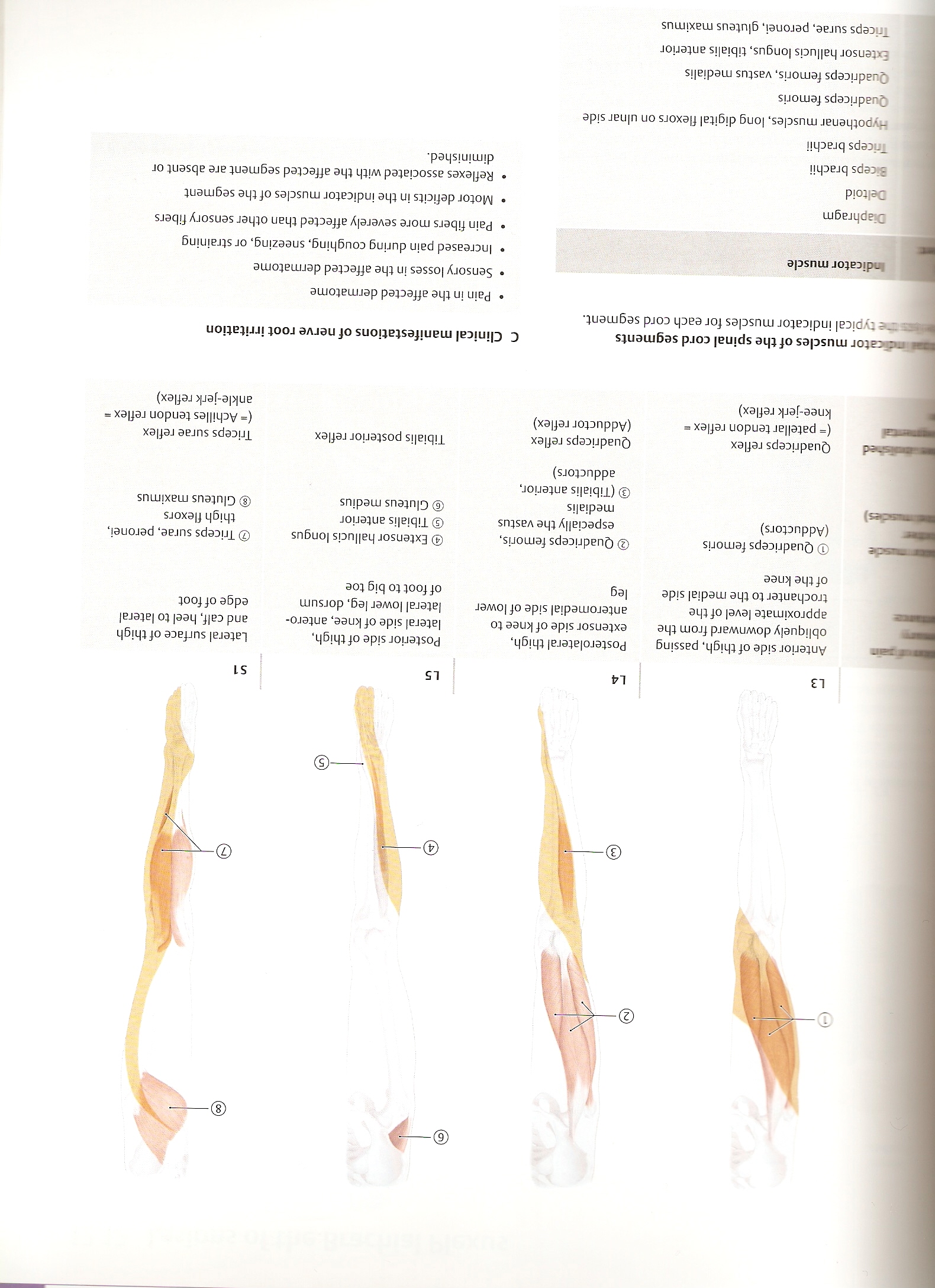
L3
L4
L5
S1
Anterior side of thigh, passing obliquely downward from the approximate level of the trochanter to the medial side ofthe knee
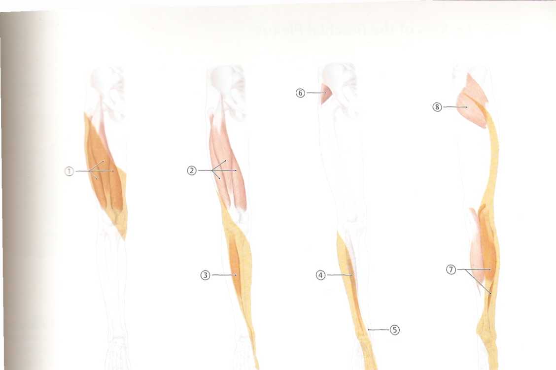
© Quadriceps femoris (Adductors)
Posterolatera! thigh, extensor side of knee to anteromedial side of lower leg
© Quadriceps femoris, especiallythevastus medialis
© (Tibialis anterior, adductors)
Posterior side of thigh, lateral side of knee, antero-lateral lower leg, dorsum offoottobigtoe
© Extensor hallucis longus ©Tibialis anterior © Gluteus medius
Lateral surface of thigh and calf, heel to lateral edgeoffoot
© Triceps surae, peronei, thigh flexors © Gluteus maximus
Quadriceps reflex (■ patellar tendon reflex ■ knee-jerk reflex)
Quadriceps reflex (Adductor reflex)
Tibialis posterior reflex
Triceps surae reflex (* Achilles tendon ref\ex ■ ankle-jerk reflex)
C Clinical manifestations of nerve root irritation
• Pain in the affected dermatome
• Sensory losses in the affected dermatome
• Increased pain during coughing, sneezing, or straining
• Pain fibers morę severely affected than other sensory fibers
• Motor deficits in the indicator musdes of the segment
• Refłexes associated with the affected segment are absent or diminished.
3 tor musdes ofthe spinał cord segments be typical indicator musdes for each cord segment.
Indicator musde
Diaphragm Dełtoid Kceps brachii Triceps brachii
Hypothenar musdes, long digital flexors on ulnar side Quadriceps femoris Quadriceps femoris, vastus medialis Extensor hallucis longus, tibialis anterior Triceps surae, peronei, gluteus maximu$
Wyszukiwarka
Podobne podstrony:
skanuj0020 3 L3 4 - 2l.00m L4 5 >7.00m L5 6 = 57.00m Qlmax = Q()max (Ll 2 + ^2
Po wprowadzeniu dodatkowych zmiennych £3, £4 i £5 oraz pomnożeniu funkcji celu przez —1, otrzymamy p
Wykorzysta 30% 20% 10% 0% L3 L4 L5 L6 L7 Stan bazowy (wyjściowy) .1 .1 5 L39
281 281 L1=6 L2=6.3 L3=7.2 L4=7.6 L5=5.7 L6=4.7 L7=4.3 Fig. 3A (left): ring drilling (vertical
%zadajemy parametry manipulatora Ll=8; L2=8; L3=0; L4=5; L5=4; P5=[L5,0,0,1] ; %pozycja końcówki
skanuj0010 (162) E. Michlowicz: Badania operacyjne i eksploatacyjne - Podstawy Kolejną klasą zadań s
skanuj0012 (162) Ni^i/Siak1 Qx.o CIphiĆt/MCLwmmęhrrf zj WUfc mm co P iiĄpW:rpfl ,4 % efmii /§ MaM sk
skanuj0019 (162) Im .Jach lec a §3 01 silMaa■t * 1 !_& to T r.I -Ć, T® 4& ■e I
skanuj0021 (162) ,
więcej podobnych podstron