skanowanie0008
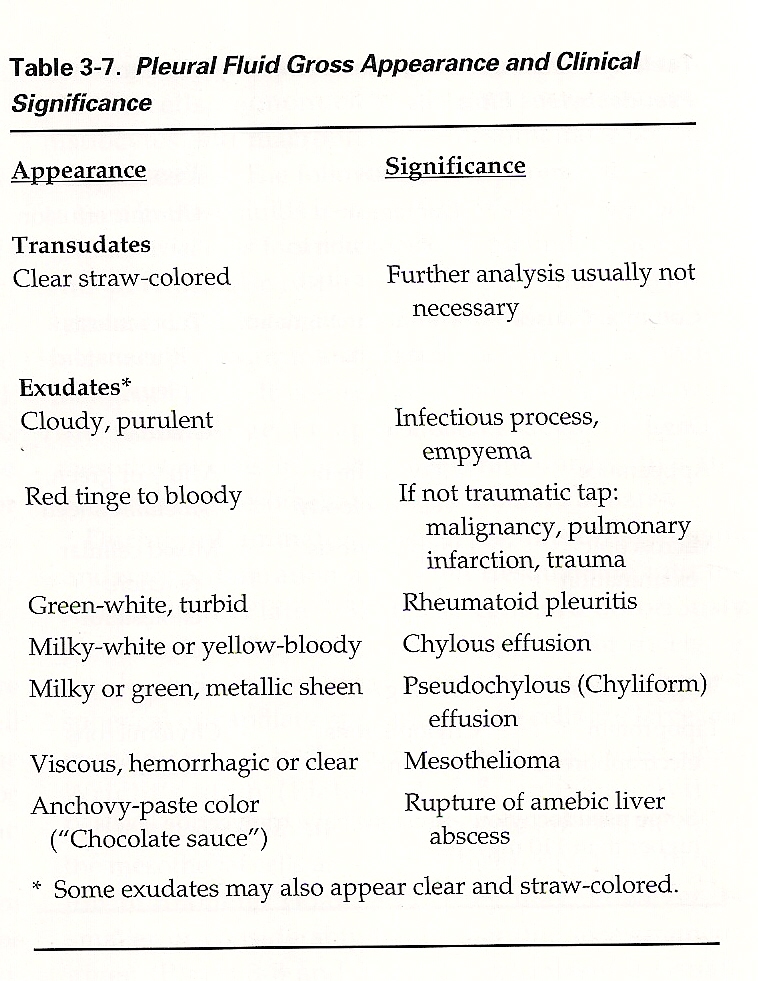
Table 3-7. Pleural Fluid Gross Appearance and Clinical Significance
Appearance
Significance
Transudates
Clear straw-colored Further analysis usually not
necessary
Exudates1
Cloudy, purulent
Red tinge to bloody
Green-white, turbid Milky-white or yellow-bloody Milky or green, metallic sheen
Viscous, hemorrhagic or elear Anchovy-paste color ("Chocolate sauce")
Inf ectious process, empyema If not traumatic tap: malignancy, pulmonary infaretion, trauma Rheumatoid pleuritis Chylous effusion Pseudochylous (Chyliform) effusion Mesothelioma Rupture of amebie liver abscess
Some exudates may also appear elear and straw-colored.
Wyszukiwarka
Podobne podstrony:
skanowanie0010 Platę 3-4. Mesothelial cells with pale gray cytoplasm in pleural fluid.
skanowanie0012 Platę 3-8. Large mesothelial cells in pleural fluid.
skanowanie0013 r i cne o-1 u. mesoineuai ceus wnn permuciear ciear zonę m pleural fluid. Notę also t
skanowanie0013 r i cne o-1 u. mesoineuai ceus wnn permuciear ciear zonę m pleural fluid. Notę also t
skanowanie0011 Platę 3-5. Mesothelial cells with basophilic cytoplasm in pleural fluid.
skanowanie0014 Platę 3-37. Smali macrophages in pleural fluid.
skanowanie0013 r i cne o-1 u. mesoineuai ceus wnn permuciear ciear zonę m pleural fluid. Notę also t
skanowanie0015 Platę 3-41. So-called "signet-ring" macrophages in pleural fluid (arrows).
skanowanie0017 Platę 3-48. Macrophages in pleural fluid containing hematin pigment (yellow crystals)
więcej podobnych podstron