SCAN0045 crop
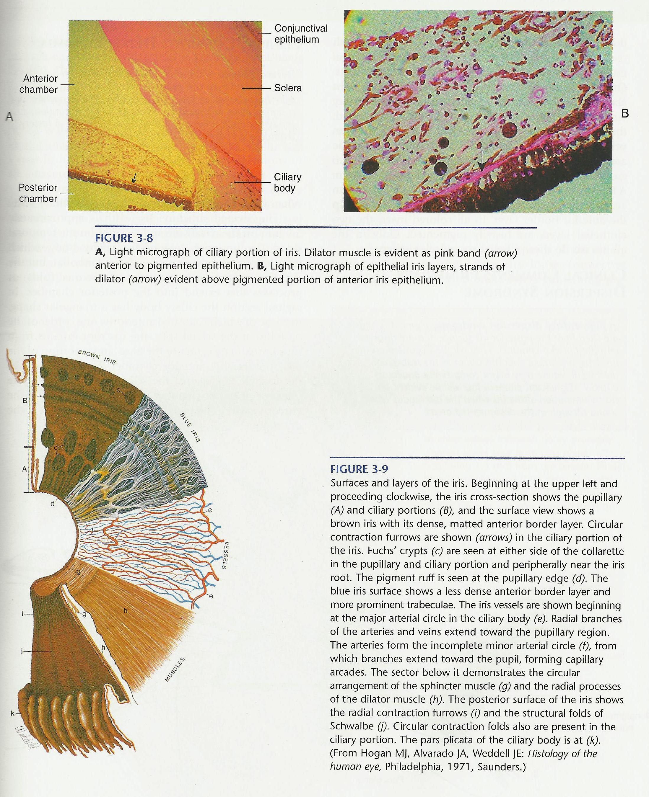
Anterior
chamber
A
Posterior
chamber

Selera
Ciliary
body
Conjunctival
epithelium
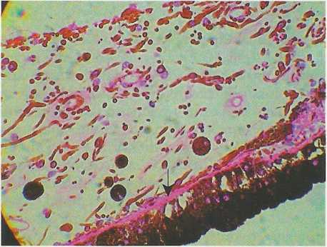
B
FIGURĘ 3-8
A, Light micrograph of ciliary portion of iris. Dilator muscle is evident as pink band (arrow) anterior to pigmented epithelium. B, Light micrograph of epithelial iris layers, strands of dilator (arrow) evident above pigmented portion of anterior iris epithelium.

FIGURĘ 3-9
Surfaces and layers of the iris. Beginning at the upper left and proceeding eloekwise, the iris cross-section shows the pupillary (A) and ciliary portions (B), and the surface view shows a brown iris with its dense, matted anterior border layer. Circular contraction furrows are shown (arrows) in the ciliary portion of the iris. Fuchs' erypts (c) are seen at either side of the collarette in the pupillary and ciliary portion and peripherally near the iris root. The pigment ruff is seen at the pupillary edge (d). The blue iris surface shows a less dense anterior border layer and morę prominent trabeculae. The iris vessels are shown beginning at the major arterial circle in the ciliary body (e). Radial branches of the arteries and veins extend toward the pupillary region. The arteries form the incomplete minor arterial circle (f), from which branches extend toward the pupil, forming capillary arcades. The sector below it demonstrates the circular arrangement of the sphincter muscle (g) and the radial processes of the dilator muscle (h). The posterior surface of the iris shows the radial contraction furrows (i) and the structural folds of Schwalbe (j). Circular contraction folds also are present in the ciliary portion. The pars plicata of the ciliary body is at (k). (From Hogan Mj, Alvarado JA, Weddell JE: Histology of the human eye, Philadelphia, 1971, Saunders.)
Wyszukiwarka
Podobne podstrony:
SCAN0045 Anterior chamber A Posterior chamber Selera Ciliary body Conjunctival epitheliumFIGURĘ
22196 SCAN0049 crop Major circle of the iris Anterior ciliary artery Long posterior ciliary artery M
68839 SCAN0043 crop Cornea Conjunctiva Iris Ciliary body Selera Lens FIGURĘ 3-2 Periphery of anterio
76951 t2 (33) THE EYE: Anterior and Posterior Chambers The Povtrnor Clumbcr I hc Anłmor lluniber S
Anterior Chamber Angle Estimation Card AC = anterior
SCAN0044 crop FIGURĘ 3-6 Pupillary portion of the iris. Dense cellular anterior border layer (a) ter
SCAN0042 crop FIGURĘ 2-1 Corneal dimensions. A, Radius of curvature of cornea and selera. B, View fr
więcej podobnych podstron