22215 skanuj0010 (363)

LI vertebra
Spinał
ganglion

Spinał dura mater
Spinał
arachnoid
Sacral hiatus
T12
Conus medullaris (newborn)
Conus medullaris (adult)

Dural sac (lumbarcistern)
■hewrtebral canal
E anEće and the dorsal surface of the sacrum have ^HLlhe spinał cord in the adult terminates at ap-Ik irst lumbar vertebra (L1). The dorsal and ven-fcoiending from the lower end of the spinał cord KslBOMn collectively as the cauda equina. During Hfetoel. a needle introduced into the subarach-HBtem) normally slips past the spinał nerve roots
BkMarts

Sacral
hiatus
D The spinał cord, dural sac, and vertebral column at different ages
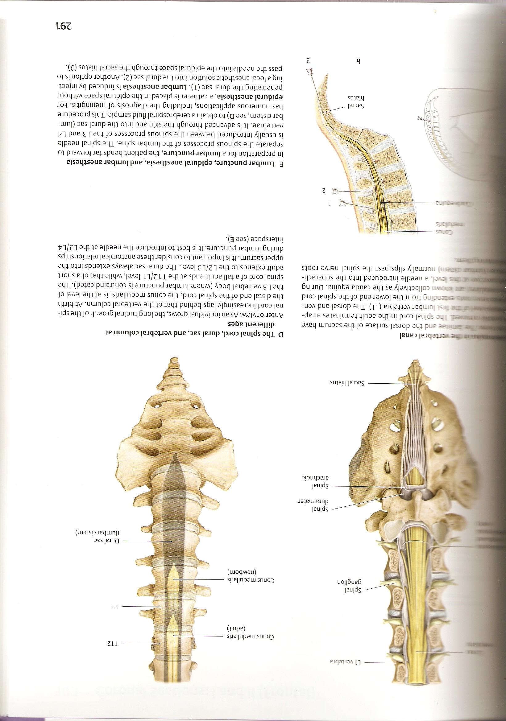
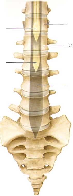
Anterior view. As an individual grows, the longitudinal growth of the spinał cord increasingly lags behind that of the vertebral column. At birth the distal end of the spinał cord, the conus medullaris, is at the level of the L3 vertebral body (where lumbar puncture is contraindicated). The spinał cord of a tali adult ends at the T12/L1 level, while that of a short adult extends to the L2/L3 level. The dural sac always extends into the upper sacrum. It is importantto consider these anatomical relationships during lumbar puncture. It is best to introduce the needle atthe L3/L4 interspace (see E).
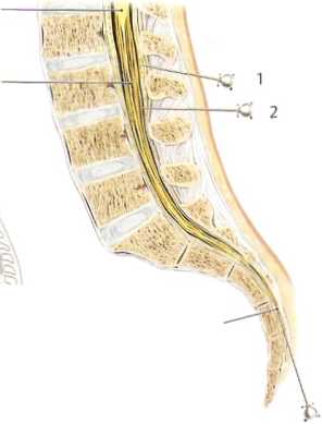
E Lumbar puncture, epidural anesthesia, and lumbar anesthesia In preparation for a lumbar puncture, the patient bends far forward to separate the spinous processes of the lumbar spine. The spinał needle is usually introduced between the spinous processes of the L3 and L4 vertebrae. It is advanced through the skin and into the dural sac (lumbarcistern, see D) to obtain a cerebrospinal fluid sample. This procedurę has numerous applications, induding the diagnosis of meningitis. For epidural anesthesia, a catheter is placed in the epidural space without penetrating the dural sac (1). Lumbar anesthesia is induced by inject-ing a local anesthetic solution into the dural sac (2). Another option is to pass the needle into the epidural space through the sacral hiatus (3).
291
Wyszukiwarka
Podobne podstrony:
skanuj0009 (198) li okres ( Yyx OZU,) jyYjpada na i* rok zyćPa cCr <? (ico uunlićjoij t ~fijriPnL
skanuj0010 (274) -i li~ -«r II i* -iSM W 0 a) O&Sz&uiąz cząsteczkami jakoczasSó M’ Oóćśshąąz
skanuj0013 (175) _■ K? /li tj. U dj)A Bu (Mci * r -------k
skanuj0016 (109) ł ł n-łi.n-r* —! !N 14._J_L }tvdl ^-u^kcjt- -f j ćą. -mają. Ciącp
więcej podobnych podstron