
Evolution in (Brownian) space: a model for the origin of the
bacterial flagellum
Version 1.0 (last updated November 10, 2003)
added September 2006.)
E-mail address: matzke@ATncseweb. (please remove obvious anti-spam modification)
Abstract: The bacterial flagellum is a complex molecular system with multiple
components required for functional motility. Such systems are sometimes proposed
as puzzles for evolutionary theory on the assumption that selection would have no
function to act on until all components are in place. Previous work (Thornhill and
Ussery, 2000, A classification of possible routes of Darwinian evolution. J Theor
Biol. 203 (2), 111-116) has outlined the general pathways by which Darwinian
mechanisms can produce multi-component systems. However, published attempts to
explain flagellar origins suffer from vagueness and are inconsistent with recent
discoveries and the constraints imposed by Brownian motion. A new model is
proposed based on two major arguments. First, analysis of dispersal at low
Reynolds numbers indicates that even very crude motility can be beneficial for large
bacteria. Second, homologies between flagellar and nonflagellar proteins suggest
ancestral systems with functions other than motility. The model consists of six
major stages: export apparatus, secretion system, adhesion system, pilus, undirected
motility, and taxis-enabled motility. The selectability of each stage is documented
using analogies with present-day systems. Conclusions include: (1) There is a strong
possibility, previously unrecognized, of further homologies between the type III
export apparatus and F
1
F
0
-ATP synthetase. (2) Much of the flagellum’s complexity
evolved after crude motility was in place, via internal gene duplications and
subfunctionalization. (3) Only one major system-level change of function, and four
minor shifts of function, need be invoked to explain the origin of the flagellum; this
involves five subsystem-level cooption events. (4) The transition between each stage
is bridgeable by the evolution of a single new binding site, coupling two pre-existing
subsystems, followed by coevolutionary optimization of components. Therefore, like
the eye contemplated by Darwin, careful analysis shows that there are no major
obstacles to gradual evolution of the flagellum.
Contents:

1.
•
•
•
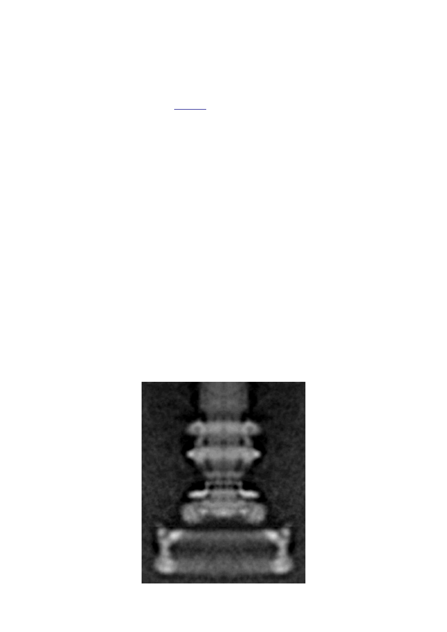
Figure 1: Composite electron micrograph of the flagellum basal
•
1.3. Theory: the evolution of systems with multiple required
•
1.4. Constructing and testing evolutionary models
2.
•
•
Figure 2: Schematic diagram of a typical bacterial flagellum
•
Table 1: Structural components of the
•
•
2.2. Previous attempts to explain flagellar origins
•
•
Table 3: Some microbial motility systems
•
•
•
Figure 3: Rizzotti's (2000) scenario for the origin of a
3.
•
3.1. Phylogenetic context and assumed starting organism
•
3.2. Starting point: protein export system
•
3.2.1. Type III secretion systems
•
Figure 4: Systems with components homologous to
•
Figure 5: Various secretion systems of prokaryotes
•
3.2.2. Are nonflagellar type III secretion systems derived from
•
3.2.3. An ancestral type III secretion system is plausible
•
Table 4: Convergent functions of well-characterized
•
3.2.4. The origin of a primitive type III export system
•
3.2.5. The relationship between type III export and the F
•
Table 5: Similarities between proteins of the F
synthetase and the flagellar type III export apparatus
that may suggest homology
•
3.3. Type III secretion system
•
3.4. Origin of a type III pilus
•
3.4.1. Filament-first hypothesis
•
•
3.4.3. Modified filament-first hypothesis
•
3.4.4. Improvements on the type III pilus
•
3.5. The evolution of flagella
•
3.5.1. The selective advantage of undirected motility
•

function of cell size and absolute swimming velocity
•
•
3.5.3. Loss of outer membrane secretin
•
•
3.5.5. Chemotaxis and switching
•
3.5.6. Hook and additional axial components
•
4.
•
Figure 7: Summary of the evolutionary model for the origin of
the flagellum, showing the six major stages and key
intermediates
•
•
Table 6: Functions and analogs at each stage of the presented
•
4.2. The evolution of other microbial motility systems
•
4.3. The construction of evolutionary models
5.
6.
Update, September 2006
This essay has now been cited in the literature (Pallen et al. 2006, “Evolutionary
links between FliH/YscL-like proteins from bacterial type III secretion systems and
second-stalk components of the FoF1 and vacuolar ATPases.” Protein Science, 15(4),
935-941 -
) and linked from a peer-reviewed article I have just coauthored
(Pallen and Matzke 2006, “From The Origin of Species to the origin of bacterial
flagella.” Nature Reviews Microbiology, 4(10), 784-790. Advanced Online Publication
on September 5, 2006 -
). Therefore, in order to avoid confusion, I will not
update the text of this article at this address. I have, however, made some minor
formatting changes, and updated the
While “Evolution in (Brownian) Space” was admittedly a first attempt, and I was a
dedicated enthusiast rather than a professional, I think the model has stood up
rather well over the last two and a half years. Writing in 2006, I would still agree
with about 90% of the 2003 model. To summarize the major updates I would make:
between the Type 3 Secretion System export
apparatus and the F
1
F
0
-ATPase (and its archaeal and eukaryotic equivalents) has
been dramatically strengthened by the findings of two papers, Lane et al. 2006
(“Molecular basis of the interaction between the flagellar export proteins FliI and
FliH from Helicobacter pylori.” Journal of Biological Chemistry, 281(1), 508-17 -
), and the aforementioned
. As I predicted in 2003, sequence
studies have now confirmed homology between FliH/YscL and F
0
-b (and its
equivalents in other ATPases). They also strongly indicate that F
1
-delta is
homologous to the C-terminal domain of FliH; I did not predict this, but it does

further confirm my more general prediction of “a strong possibility, previously
unrecognized, of further homologies between the type III export apparatus and
F
1
F
0
-ATP synthetase.” However, I would retract some of my more speculative
for ATPase homology to FliJ, FliO, and FliP (FliJ and FliO are
apparently not even universally required in flagella). I am still hopeful regarding
the suggestions for FliQ and FliR.
Secondly, in the 2003 essay I for the most part assumed that the nonflagellar Type 3
Secretion System (NF-T3SS) was derived from the flagellum, rather than being an
outgroup with a sister group relationship. I took this position partially to show that
even under this assumption the evidence for evolution was strong, and partially
because the evidence seemed to lean slightly in that direction. The parsimony
argument of
and various minor points now have me leaning
somewhat towards the view that the flagellar and nonflagellar systems are sister
groups, and the NF-T3SS is therefore an outgroup. However, as we note in
the scientific community is split on this question. There are several
avenues of investigation that might clarify matters, which I will explore in the
future.
Thirdly, the question of which proteins are actually universally “essential” for
flagellum function, and which proteins have homology to other flagellar proteins or
nonflagellar proteins, has been systematically reviewed in
of Pallen and
Matzke (2006). I have reposted the table in
. It is
important to note that this table is much more conservative than the Matzke 2003
homology suggestions, which ranged from well-established to loose speculation. The
homologies in the 2006 table are all well-confirmed by standard BLAST techniques,
except for five proteins where homology is based on structural or other similarities.
Even for these five, two of the flagllar proteins have other known homologies based
on sequence (FliC to FlgL and FliH to YscL), two are not universally essential (FliH
and FliJ), and three of the homologies have been repeatedly put forward in the
literature (FliC to EspA, FliK to YscP, and FliH/YscL to F
0
-b+F
1
-delta and
equivalents). In the entire list, only one required protein has a new proposed
homology that could be considered speculative (FliG, to MgtE).
Many of the homologous and/or inessential proteins found in Table 1 of Pallen and
Matzke 2006 were cited in the 2003 paper, but the 2006 table is an authoritative
update and supercedes what is said here. The important overall point, as discussed
in
, is that of the 42 proteins in Table 1 of Pallen and Matzke, only two
proteins, FliE and FlgD, are both essential and have no identified homologous
proteins. This is substantially more impressive than the situation in 2003, and means
that the evidence for the evolutionary origin of the flagellum by standard gene
duplication and cooption processes is even stronger than in 2003. Important specific
updates include: a homolog of FlgA has been confirmed (along the lines that I
suggested in 2003); FliG has no homolog in NF-T3SS or the Exb/Tol systems, rather
it may be homologous to the magnesium transporter MgtE; and the flagellar
filament protein FliC (and its sister FlgL) is probably homologous to EspA and
other pilus proteins found in NF-T3SS. I still suspect that all of the axial proteins
(including FliE and FlgD) are homologous to each other and therefore to pilus
proteins in NF-T3SS, but only the confirmed homologies are reported in Pallen and
Matzke 2006.

Finally, if I were doing a revision, I would update the terminology along the lines
suggested in
(“Type III secretion: what's in a name?” Trends in
Microbiology 14(4), 157-160, April 2006 -
). As they point out, the terminological
distinction between "flagellum" and "type 3 secretion system" is dubious and
artificial, and it is more true to acknowledge that flagella have a type III secretion
system. Therefore, there are two known groups of type III secretion systems,
flagellar and nonflagellar, abbreviated F-T3SS and NF-T3SS.
There is much more to be said about recent research and its implications for
flagellum evolution. For the near future I intend to post my thoughts on this in the
new
blog.
1. Introduction
1.1. A complex contrivance
The bacterial flagellum is one of the most striking organelles found in biology. In
Escherichia coli the flagellum is about 10 μm long, but the helical filament is only 20
nm wide and the basal body about 45 nm wide. The flagellum is made up of
approximately 20 major protein parts with another 20-30 proteins with roles in
construction and taxis (Berg, 2003; Macnab, 2003). Many but not all of these
proteins are required for assembly and function, with modest variation between
species. Over several decades, thousands of papers have gradually elucidated the
structure, construction, and detailed workings of the flagellum. The conclusions
have often been surprising. Berg and Anderson (1973) made the first convincing
case that the flagellar filament was powered by a rotary motor. This hypothesis was
dramatically confirmed when flagellar filaments were attached to coverslips and the
rotation of cells was directly observed (Silverman and Simon, 1974). The energy
source for the motor is proton motive force rather than ATP (Manson et al., 1977).
The flagellar filament is assembled from the inside out, with flagellin monomers
added at the distal tip after export through a hollow channel inside the flagellar
filament (Emerson et al., 1970). The flagella of E. coli rotate bidirectionally at about
100 Hz, propelling the rod-shaped cell (dimensions 1x2 μm) 10-30 μm/sec. The
flagella of other species, powered by sodium ions rather than hydrogen ions, can
rotate at over 1500 Hz and move cells at speeds of several hundred μm/sec. The
efficiency of energy conversion from ion gradient to rotation may approach 100%
(DeRosier, 1998). The bacterial flagellum is now one of the best understood
molecular complexes, although numerous detailed questions remain concerning the
function of various protein components and the exact mechanism of torque
generation. However, the origins of this remarkable system have hardly been
examined. This article will propose a detailed model for the evolutionary origin of
the bacterial flagellum, along with an assessment of the available evidence and
proposal of further tests. That the time is ripe for a serious consideration of this
question is discussed below.
1.2. An evolutionary puzzle
Biologists find it almost inescapable to compare the bacterial flagellum to human
designs: DeRosier remarks, “More so than other structures, the bacterial flagellum
resembles a human machine” (DeRosier, 1998). The impression is heightened by
electron micrograph images (
) reminiscent of a engine turbine (e.g.,
Whitesides, 2001), and the scientific literature on the flagellum is filled with
analogies to human-designed motors. There is no shortage of authorities willing to
express mystification on the question of the evolutionary origin of flagella. In a
1978 review, Macnab concluded,
As a final comment, one can only marvel at the intricacy, in a simple
bacterium, of the total motor and sensory system which has been the
subject of this review and remark that our concept of evolution by
selective advantage must surely be an oversimplification. What
advantage could derive, for example, from a “preflagellum” (meaning a
subset of its components), and yet what is the probability of
“simultaneous” development of the organelle at a level where it becomes
advantageous?” (Macnab, 1978).
The basic puzzle is that the flagellum is made up of dozens of protein components,
and deletion experiments show that the flagellum will not assemble and/or function
if any one of these components is removed (with some exceptions). How, then, could
this system emerge in a gradual evolutionary fashion, if function is only achieved
when all of the required parts are available?

Figure 1: Composite electron micrograph of the flagellum
basal body and hook, produced by rotational averaging
(Francis et al., 1994). The motor proteins and export
apparatus (included in
) do not survive the
extraction procedure and so are not shown. Image
courtesy of David DeRosier, reproduced with permission.
1.3. Theory: the evolution of systems with multiple required
components
The standard answer to this question was put forward by Darwin. Mivart (1871)
argued that the “incipient stages of useful structures” could not have evolved
gradually by variation and natural selection, because the intermediate stages of
complex systems would have been nonfunctional. Darwin replied in the 6
th
edition
of Origin of Species (Darwin, 1872) by emphasizing the importance of change of
function in evolution. Although Darwin’s most famous discussion of the evolution of
a complex system, the eye, was an example of massive improvement of function
from a rudimentary ancestor (Salvini-Plawen and Mayr, 1977; Nilsson and Pelger,
1994), Darwin gave equal weight to examples of functional shift in evolution. These
included the complex reproductive devices of orchids and barnacles, groups with
which he was particularly familiar (Darwin, 1851, 1854, 1862). Intricate multi-
component systems such as these could not have originated by gradual
improvement of a single function, but if systems and components underwent
functional shift, then selection could have preserved intermediates for a function
different from the final one. The equal importance of improvement of function and
change of function for understanding the evolutionary origin of novel complex
systems has been similarly emphasized by later workers (Maynard Smith, 1975;
Mayr, 1976). Recent studies give cooption of structures a key role in the origin of
feathers (Prum and Brush, 2002), and novel organs (Pellmyr and Krenn, 2002);
Mayr (1976) gives many other examples. Computer simulations also show the
importance of cooption for the origin of complex systems with multiple required
parts (Lenski et al., 2003).
Do these common insights from classical, organismal evolutionary biology help us to
understand the solution to the puzzle Macnab put forward regarding the origin of
flagellum? Cooption at the molecular level is in fact as well-documented at it is at
the macroscopic level (Ganfornina and Sanchez, 1999; Thornhill and Ussery, 2000;

True and Carroll, 2002). It has been implicated in origin of ancient multi-
component molecular systems such as the Krebs cycle (Melendez-Hevia et al., 1996)
as well as the rapid origin of multi-component catabolic pathways for abiotic toxins
that humans have recently introduced into the environment, such as
pentachlorophenol (Anandarajah et al., 2000; Copley, 2000), atrazine (de Souza et
al., 1998; Sadowsky et al., 1998; Seffernick and Wackett, 2001), and 2,4-
dinitrotoluene (Johnson et al., 2002); many other cases of catabolic pathway
evolution exist (Mortlock, 1992). All of these systems absolutely require multiple
protein species for proper function. Even for some molecular systems equaling the
flagellum in complexity, reasonably detailed reconstructions of evolutionary origins
exist. Generally these are available for systems which originated relatively recently
in geological history, which are well-studied due to medical importance, and where
phylogeny is relatively well resolved; examples include the vertebrate blood-clotting
cascade (Doolittle and Feng, 1987; Hanumanthaiah et al., 2002; Jiang and Doolittle,
2003) and the vertebrate immune system (Muller et al., 1999; Pasquier and Litman,
2000).
Thornhill and Ussery (2000) summarized the general pathways by which systems
with multiple required components may evolve. They delineate three gradual routes
to such systems: parallel direct evolution (coevolution of components), elimination
of functional redundancy (“scaffolding,” the loss of once necessary but now
unnecessary components) and adoption from a different function (“cooption,”
functional shift of components); a fourth route, serial direct evolution (change along
a single axis), could not produce multiple-components-required systems. However,
Thornhill and Ussery’s analysis did not distinguish between the various levels of
biological organization at which these pathways might operate. The above-cited
literature on the evolution of complex molecular systems indicates that complex
systems usually originate by a key shift in function of an ancestral system, followed
by an intensive period of improvement of the originally crudely functioning design.
At the level of the system, cooption is usually the key event in the origin of the
modern system with the function of interest. However, a great deal of the
complexity in terms of numbers of parts is added to the system after origination.
These accessory parts get added by duplication and cooption of novel genes (for
reviews of gene duplication in evolution, see Long, 2001; Chothia et al., 2003;
Hooper and Berg, 2003) and/or duplication and subfunctionalization (Force et al.,
1999) of genes already involved in the crudely-functioning system. Cooption of
whole subsystems, linking them to the “core” system, may also occur.
Therefore, improvement of function at the system level might be implemented by
cooption at the level of a protein or subsystem. Change of function at the system
level might occur without any lower level cooption of new components. Thornhill
and Ussery’s four routes can be reduced to the two major pathways proposed by
Darwin: improvement of current function (optimization) and shift of function
(cooption). Cooption remains its own category, while the other three routes (serial
direct evolution, parallel direct evolution, and elimination of functional
redundancy) can be considered as three versions of functional improvement, with
the lower-level components undergoing optimization, coevolutionary optimization,
or loss, respectively. This conceptual framework is basically equivalent to the
patchwork model for the evolution of metabolic pathways (Melendez-Hevia et al.,
1996; Copley, 2000), where components are recruited from diverse sources and
functional improvement or functional shift might occur at any organizational level,

e.g. system, subsystem, protein, or protein domain.
1.4. Constructing and testing evolutionary models
In order to explain the origin of a specific system such as the flagellum, the general
theory discussed above must be combined with the available evidence in order to
produce a detailed, testable model. Detail in evolutionary scenarios makes them
more testable, not less: Cavalier-Smith argues that “Specifying transitional stages in
considerable detail is not unwarranted speculation, but a way of making the ideas
sufficiently explicit to be more easily tested and rigorously evaluated” (Cavalier-
Smith, 2001b). Obviously “detailed” cannot mean that every mutation and
substitution event be recorded – for events that occurred billions of years ago this is
impossible. A detailed evolutionary model should reduce a puzzling event like the
origin of the flagellum into a series of events that occur by well-understood
processes.
In an ideal model, the origin of every protein component will fulfill three criteria.
First, a putative ancestral protein with a different function (a homolog that can
reasonably be suspected to precede the flagellum) should be identified. Second, the
cooption of the protein should occur by a reasonably probable mutation event --
e.g., a mutation produces a single new binding site enabling one protein to act on
another. Initially this new complex functions crudely, but can gradually be
perfected by coevolutionary optimization of the two proteins. Third, the selective
regime favoring retention of the coopted protein should be identified. Each of these
three criteria encourages further testing against new data. Hypothesized homologies
can be assessed by new data, for example by detailed sequence analysis or the
comparison of protein structures. The plausibility of mutational steps can be
investigated by examination of similar mutations observed today; and the selective
forces invoked can be assessed by study of analogies and by mathematical
modeling. Furthermore, an evolutionary model might have testable implications for
other fields: for example, if a biological system is hypothesized to be derived from a
homologous system, similarities in mechanism between the two systems would be
suspected. The fact that we do not have all of the data that we would like, and that
uncertainty is high, are not problems unique to evolutionary models; rather, these
problems are commonplace in any advancing science. For example, many
contradictory models have been published for the mechanism of motor action in the
flagellum, and most (or all) of them must be wrong, but this has not stopped anyone
from proposing new models (Schmitt, 2003). Science is advanced by proposing and
testing hypotheses, not by declaring questions unsolvable.
2. Background
2.1. Modern flagella
The canonical flagellum of E. coli is shown in
. Descriptions of the
structural components are given in
. Cytoplasmic components involved in
regulation and assembly, as well as the chemotaxis components, are listed in
. Excellent overviews of flagellar function and assembly are available elsewhere
(Berg, 2003; Macnab, 2003) and so will not be discussed further here.
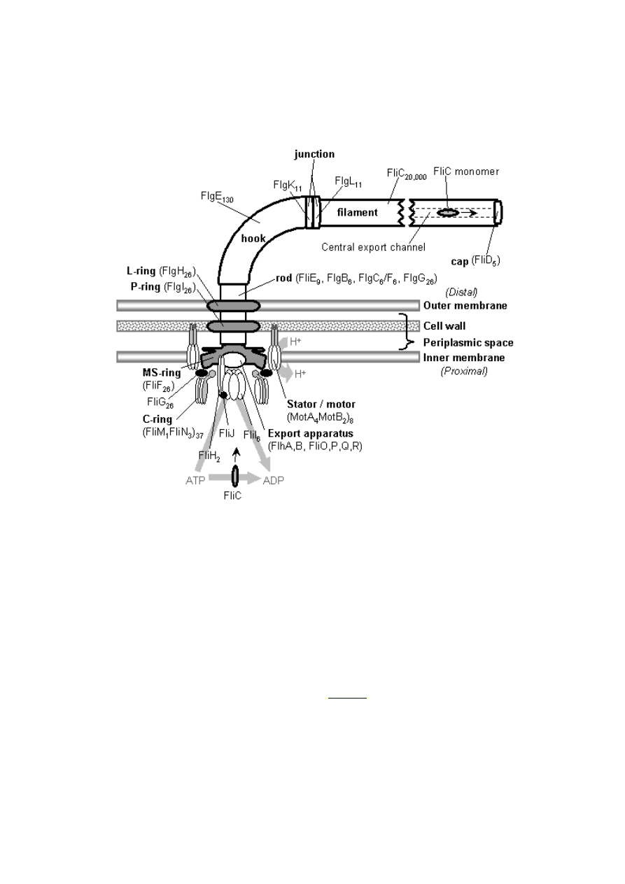
Figure 2: Schematic diagram of a typical bacterial
flagellum, shown in cross-section. The names of
substructures are given in bold, and the names of the
constituent proteins are given in regular type, including
approximate stoichiometry (see
). The depiction of
the flagellar axial protein complex (rod, hook, filament)
and MS-, P-, and L-rings is based on composite electron
micrographs (see DeRosier, 1998). The depictions of the
other proximal components are based on specific
published models: FliM/N C-ring (Mathews et al., 1998),
the position of MotA, MotB, and FliG (Brown et al.,
2002), and the hexameric complex of FliI (Blocker et al.,
2003; Claret et al., 2003). The position of FliJ is a guess
based on its interaction with FliH and FliI (Macnab,
2003). The depiction of FliH is based on studies of its
structure and interaction with FliI (Minamino and
Macnab, 2000; Minamino et al., 2001; Minamino et al.,
2002) and on the homology of FliH to the F
0
-b subunit of
ATP synthetase, postulated in this paper (see text). Apart
from FliH and FliI, the structure and stoichiometry of the
rest of the type III export apparatus are obscure.
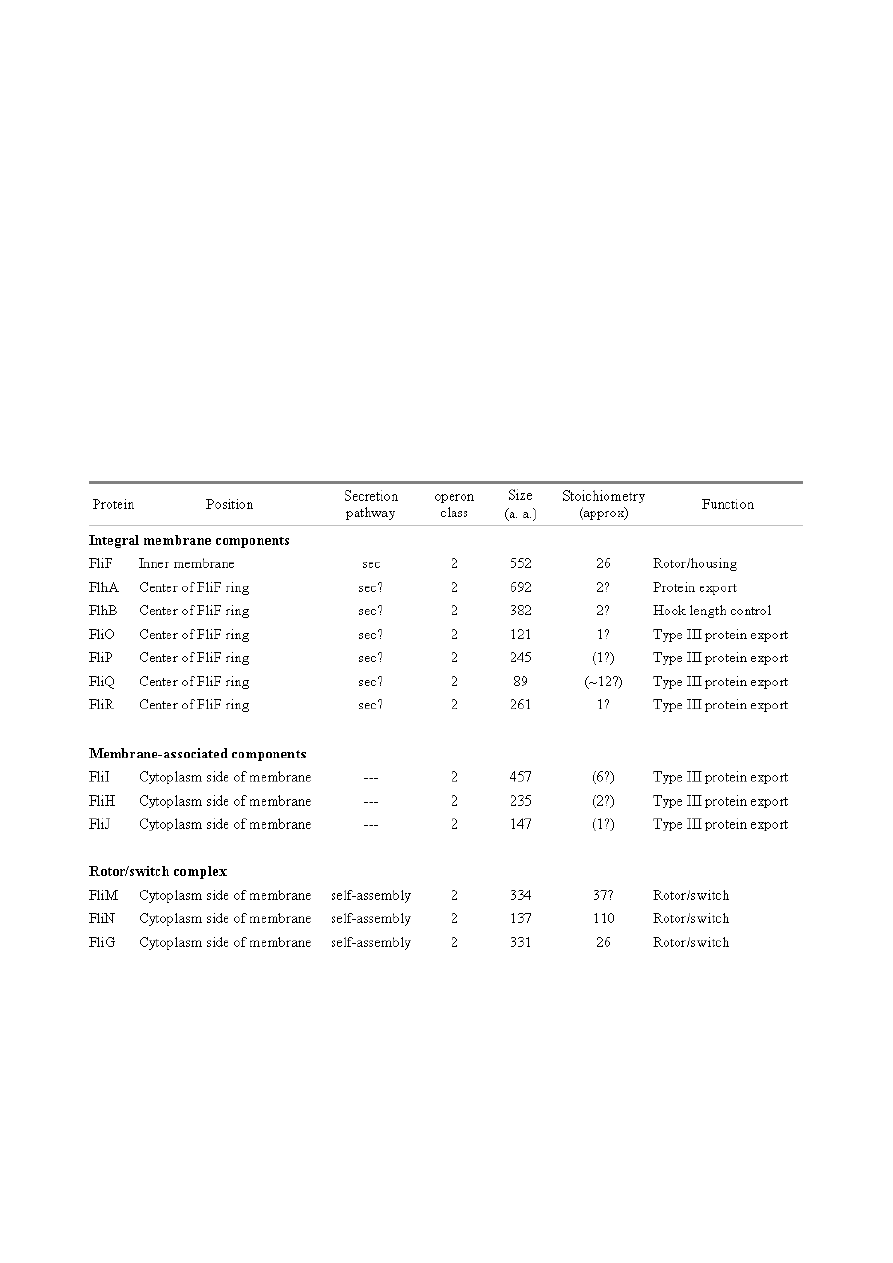
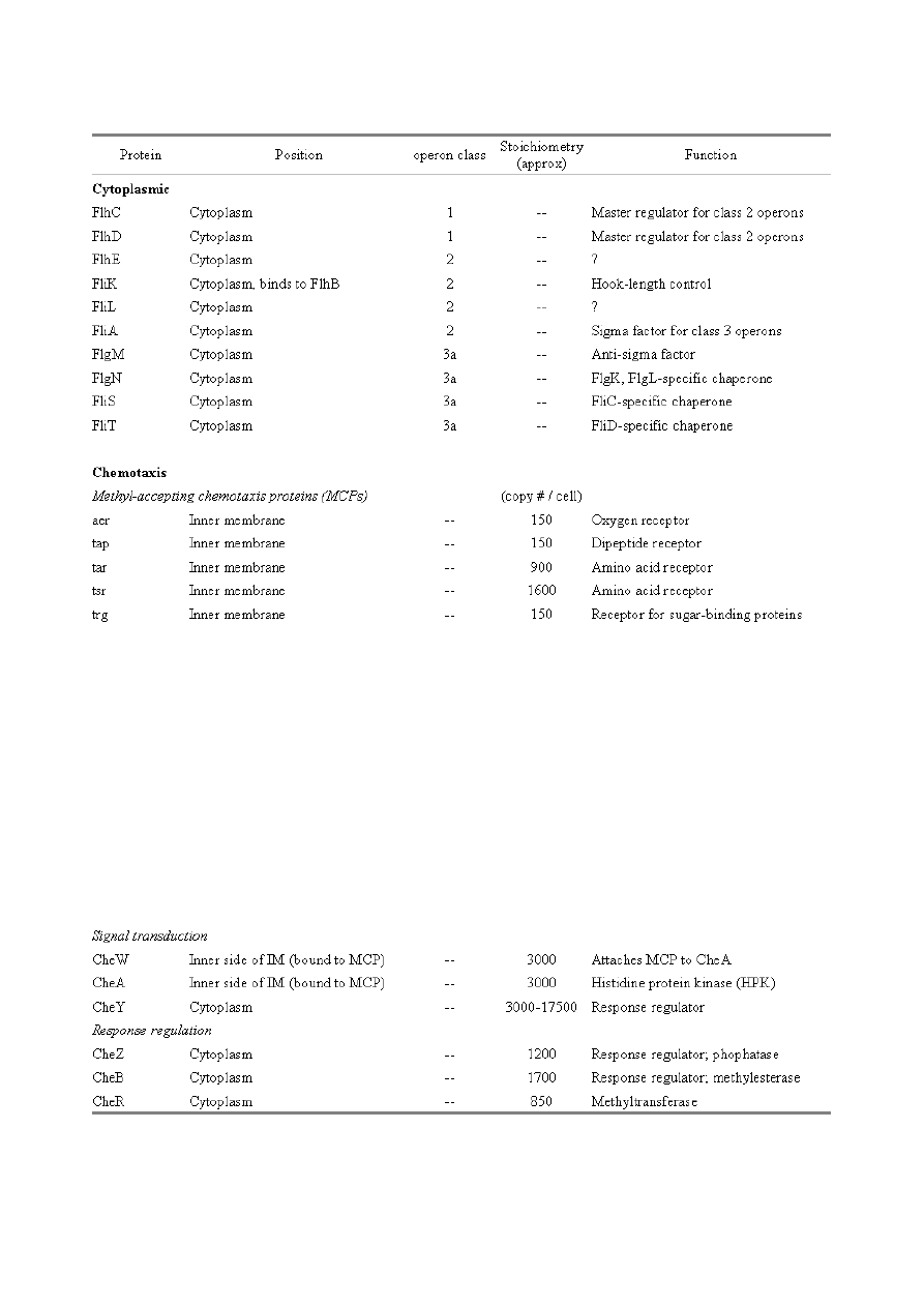
Table 1: Structural components of the E. coli flagellum. Based on recent reviews (Berg, 2003;
Macnab, 2003); figures in parentheses represent suggestions made in this paper. Components with an
asterisk (*) are not included in the final structure.
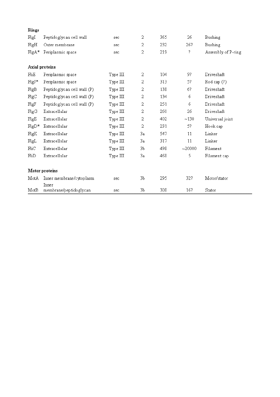
Table 2: Components of the E. coli regulation/assembly and chemotaxis systems. Cytoplasmic
components based on Berg (2003) and Macnab (2003), chemotaxis components based on Eisenbach
(2000).

2.2. Previous attempts to explain flagellar origins
2.2.1. Short discussions
Occasional examples of very general suggestions about the evolutionary origin of flagella can be found
in the literature, for example in discussions of how various aspects of the chemotaxis system are
optimized (Berry, 2000); in the suggestion that prokaryote flagella may have been a relatively late
invention, after biofilms and microbial mats had become well-developed and crowding on surface
habitats became a problem (Stoodley et al., 2002); or in the alleged common ancestry of archaeal and
bacterial flagella (Harshey and Toguchi, 1996). Archaeal and bacterial flagella were indeed once
thought to be homologous (Jones et al., 1987), but they are actually totally distinct motility systems
(Jarrell et al., 1996; Faguy and Jarrell, 1999; Thomas et al., 2001). Although both kinds of flagella
rotate and are superficially similar, archaeal flagella are fundamentally different in many respects
(
). In archaeal flagella, the filaments are thinner, lack a central channel, and subunits are added
from the base rather than the tip. Forward movement is typically attained by clockwise rather than
counterclockwise motion. Additionally, archaeal flagella are probably powered by ATP rather than
protonmotive force (suggested by homologies of FlaI to PilT/U (Jarrell et al., 1999; Thomas et al., 2001;
Merz and Forest, 2002, although the literature is contradictory: Bardy et al. (2003) assert that archaeal
flagella use protonmotive force, but cite no supporting evidence). Finally, the homologies of the two
flagella to nonflagellar secretion systems are different. The bacterial and archaeal flagella are therefore
a classic case of analogy, not homology (Faguy et al., 1994; Jarrell et al., 1996; Bayley and Jarrell, 1998;
Faguy and Jarrell, 1999; Thomas et al., 2001; Thomas et al., 2002; Bardy et al., 2003). However, the
misperception persists in the assumption that the flagella (Harshey and Toguchi, 1996; Campos-Garcia
et al., 2000; Rizzotti, 2000) or their basal bodies (Cavalier-Smith, 2002a, 2002c) are homologous. On the
other hand, the chemotaxis systems are indeed homologous, and are shared with nonflagellar motility
systems as well (Faguy and Jarrell, 1999; Koretke et al., 2000).
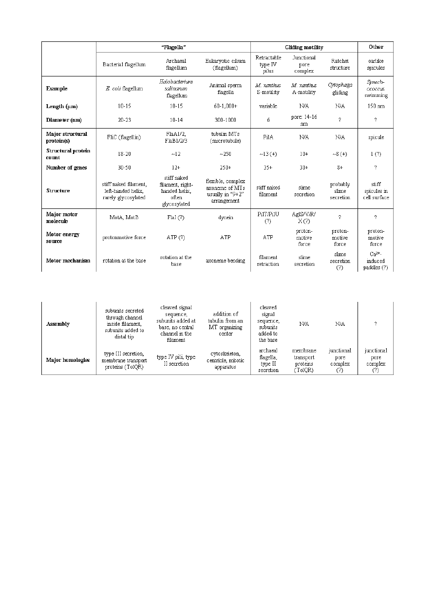
Table 3: Some microbial motility systems. Several more mysterious systems (the perhaps cytoskeleton-
based motilities of Mycoplasma and Spiroplasma; Trachtenberg et al., 2003) have been excluded.
Prokaryotes undoubtedly have additional motility systems that have not yet been discovered. Only
one eukaryote system, the cilium or eukaryotic flagellum, is included in the table, because it is often
confused with the prokaryote systems even though it is totally distinct. Many other eukaryote
motility systems, not relevant here, are not listed. Data gathered from many sources (Young et al.,
1999; Eisenbach, 2000; McBride, 2001; Thomas et al., 2001; Bardy et al., 2003; Youderian et al., 2003).
A slightly more detailed attempt at explaining the origin of the bacterial flagellum
was made by de Duve (1995), who apparently got the bacterial flagellum confused
with the completely different eukaryotic cilium (also known as the eukaryotic
flagellum or undulipodium in an interminable terminological dispute; see Corliss,
1980; Margulis, 1980; Cavalier-Smith, 1982). He suggested that the flagellum,
which he acknowledges is rotary, was somehow descended from a simpler ATP-
powered filament-bending motor. In a more reasonable vein, de Duve then gave a
brief scenario for the gradual origin of chemotactic behavior from random
swimming, but was again puzzling in postulating that essentially fully functional,
bidirectional-switching flagella with specific positioning on the cell surface existed
before the signal transduction system was coupled to the flagellum. What the
purpose of switching would be without a chemotaxis system was not explained. De
Duve furthermore stated that these well-developed but non-chemotactic flagella
gave “little advantage” until they were chemotactically enabled, leaving
unexplained the selective reason for the origin of the whole nearly-complete system

in the first place.
Finally, Goodenough (1998; 2002) offers a short account deriving a flagellum from a
proton-transducing membrane channel. She postulates that a coopted protein
increased the efficiency of proton transport, and rotated the channel as a by-
product. Later binding of a filament to the outside of this rotating channel
produced primitive motility which increased food gathering ability. However, the
original function of proton transport (which, uncoupled to another process, would
simply de-energize the cytoplasmic membrane) is not specified. In her 2002 account
Goodenough suggested that a fibrous protein binding to the F
1
F
0
-ATP synthetase
produced the proto-flagellum. Presumably she meant that the proto-filament would
bind to the distal side of a c-subunit of F
0
. As recent work indicates that F
0
-c and
F
1
-εγ rotate inside the F
0
-ab and F
1
-αβδ complex (Weber and Senior, 2003),
Goodenough’s suggestion is not immediately impossible, but suffers difficulties
similar to those discussed for Rizzotti (2000), below.
2.2.2. Cavalier-Smith (1987)
Cavalier-Smith is one of the few who has proposed detailed hypotheses for the
origin of many fundamental features of eukaryotes and prokaryotes (Cavalier-
Smith, 1987a, 1987b, 2001a, 2002b, 2002a, 2002c). He bases his work on a
refreshingly clearly-stated philosophy for reconstructing the origin of complex
systems, advocating a holistic approach considering environment, organism,
mutation, and selection all together and emphasizing testability (Cavalier-Smith,
2001a). Although Cavalier-Smith has addressed the origin of the eukaryotic cilium
on several occasions (Cavalier-Smith, 1978, 1982, 1987b, 2002b), Cavalier-Smith’s
only treatment of the origin of the bacterial flagellum is found in a 1987 article
(Cavalier-Smith, 1987a). He makes two suggestions: first, that a mutant version of
an outer membrane protein pore formed a tubular polymer extending through the
outer membrane into the extracellular medium. Linking this to proton-conducting
proteins in the cytoplasmic membrane provided the primitive motor. In this
scheme, spirochete axial filaments were derived from regular flagella. His second
suggestion was that flagella evolved from gliding motility systems, which are also
widespread and powered by protonmotive force. Some early models of gliding
motility postulated a spirochete-like mechanism, with rotating filaments in the
periplasmic space, and on this basis spirochetes might represent a transitional
stage. Motility would develop from rotating filaments first used just to stir the fluid
in the periplasmic space and increase diffusion of nutrients. On either scenario, the
rotary mechanism existed from the beginning of the evolutionary sequence, and the
first crude motility function would have been selected for because it increased
random dispersal, useful in overcrowded regions depleted in nutrients. Much of the
complexity could have post-dated the original crudely functioning motility.
Cavalier-Smith was hampered by the relatively primitive state of knowledge at the
time, and he conceded that the actual evolutionary process must have been much
more complicated than his suggestions. The linkage between the filament and
motor is very complex, mediated by about ten proteins, and the filament subunits
are secreted through the base of the flagellum via a type III export pathway, rather
than via a type II pathway as might be expected for a protein derived from an outer
membrane pore; type III virulence systems do utilize an outer membrane secretin
secreted by the type II pathway, and the flagella P- and L-ring proteins FlgI and

FlgH are similarly secreted via the type II pathway (Macnab, 2003). A secretin
might therefore be more likely posited as the source for FlgH; this will be discussed
in more detail below.
Regarding the postulated homology between gliding motility and the axial filaments
of spirochetes, today it is apparent that gliding motility is not a matter of rotating
periplasmic filaments. Two mechanisms for gliding motility have been clearly
identified (Merz and Forest, 2002; Bardy et al., 2003). First, the social gliding of
Myxococcus xanthus occurs via retraction of type IV pili, sometimes also called
twitching motility (Merz and Forest, 2002). Second, the adventurous motility of M.
xanthus is driven by the secretion of a polysaccharide gel (slime) via the junctional
pore complex; a similar complex is found in gliding cyanobacteria. The mechanism
of the gliding motility of Cytophaga and Flavobacterium is still a matter of
speculation (McBride, 2001), but may involve a ratchet structure and slime
secretion (Bardy et al., 2003). These latter forms of gliding motility inspired the
comparison between flagella and gliding motility as they are powered by
protonmotive force, and beads attached to the cell surface of Cytophaga will rotate
(Eisenbach, 2000). Thus, it is occasionally suggested (Cavalier-Smith, 2002a), even
in textbooks (e.g. Campbell, 1993), that flagella and gliding motility are
homologous, and the gliding motility apparatus may be some version of the
flagellum basal body without the flagellar filament. As our understanding of slime-
related gliding motility is still limited (the relevant genes are still being identified,
much less detailed mechanism or structure), the possibility of any connection
between type III protein secretion and polysaccharide secretion is difficult to
evaluate. However, the study of gliding motility bears close watching: the recent
discovery of homology between M. xanthus gliding motility proteins AglS/AglV to
TolR and of AglR/AglX to TolQ (Youderian et al., 2003) which are in turn homologs
of the flagellar motor proteins MotA and MotB (Cascales et al., 2001) suggests that
there may be a common mechanism for coupling proton flow to motility. If the
general similarity between the junctional pore complex and type III secretion
systems (Spormann, 1999; Merz and Forest, 2002) turns out to be more than skin
deep, then the common descent of gliding motility and flagella from an ancestral
motility organelle will have to be seriously considered. Cavalier-Smith’s suggestion
that stirring the periplasmic fluid may have been a precursor to primitive motility is
similar to Rizzotti’s main suggestion and will be discussed in the next section.
2.2.3. Rizzotti (2000)
The only major recent attempt at explaining the origin of the flagellum is that of
Rizzotti (2000), which, like Goodenough, proposes that the flagellum was derived
from the F
1
F
0
ATP synthetase. The initial appeal of this hypothesis derives from the
spate of recent comparisons between the flagellum and ATP synthetase as proton-
driven, rotary motors (Block, 1997; Boyer, 1997; Khan, 1997; Sabbert and Junge,
1997; Berg, 1998; Oplatka, 1998a, 1998b; Berry, 2000; Walz and Caplan, 2002),
sometimes leading to the suggestion of homology (Oster and Wang, 2003). These
comparisons go back at least to Cox et al.’s (1984) proposal that the ATP synthetase
had a rotary mechanism, and continued through the testing and refinement of this
hypothesis (Mitchell, 1985; Sabbert and Junge, 1997; Weber and Senior, 2003),
followed by the conclusive demonstration of rotation by direct observation of an
actin filament tethered to the gamma subunit of F
1
-ATPase (Noji et al., 1997). A
relationship between the F
1
F
0
ATP synthetase and the flagellum is further suggested
by homology between the flagellar ATPase FliI and the β subunit of F
1
-ATPase,
indicated by ~30% sequence similarity (Albertini et al., 1991; Vogler et al., 1991).
The α and β subunit ATP synthetase subunits are themselves paralogous, with only
the β subunit retaining catalytic activity (Gogarten et al., 1989; Gogarten and
Kibak, 1992).
In a creative scenario (
), Rizzotti imagined that an accidental insertion in
the middle of the F
1
-γ subunit created a short filament outside the cytoplasmic
membrane, between the membrane and the cell wall. As the synthetase subunits
rotated, this protofilament served to mix the nearby fluid, increasing the diffusion of
molecules in and out of the cell. This provided sufficient selective benefit to retain
the mutation. Production of a more sophisticated mixing instrument occurred via
duplication and modification of the mutant γ subunit, so that branches of the
filament extended above the cell wall. In the process, the ε and δ subunits were lost,
along with ATPase activity, resulting in a proton-powered stirring mechanism with
incipient motility function. From here, a process of optimization ensued. Selection
first favored random motion of the cell that further improved nearby fluid mixing
and diffusion. More powerful motility followed by extension of the filament and by
duplications of the proton-transmitting proteins of the stator (in this scenario,
derived from the c subunit of the F
0
structure). The F
1
-αβ complex apparently
became the rotor inside the stator ring. Rizzotti concluded by discussing a number
of other steps that must have happened along the way, although the order is not
specified. However, it seems that he considered the origin of the export apparatus a
relatively late event. Rizzotti hypothesized that once the central cavity became large
enough, a secretion complex (presumably a type III export apparatus already
functioning elsewhere) was patched in at the base of the rotor, allowing the secretion
of a more complex filament.
Rizzotti argued that bacteria with a single membrane were simpler and therefore
probably ancestral to gram-negative bacteria with both an inner and outer
membrane. He hypothesized that the outer membrane arose as an alimentary

adaptation from extensions of the inner membrane. The L- and P-rings arose as the
developing outer membrane encroached on the flagellum (gram positive bacteria,
lacking outer membranes, have no requirement for the L- and P-rings and lack
them altogether). Rizzotti discounted the alternative scenario, whereby the
flagellum arose in a bacterium already possessing a double membrane, because he
deemed the simultaneous origin of the rings and filament too difficult.
This scenario is considerably more detailed than any other available, but remains
vague on the specific origin of almost all of the proteins that make up the flagellum.
Although Rizzotti does make use of some interesting similarities between the
flagellum and ATP synthetase, and he is able to come up with a proposal that
includes rotary motion from the beginning, there are major flaws which shall be
discussed shortly. Before the critique, however, it is worth noting that Rizzotti’s
scenario has been cited by Cavalier-Smith (2001a) as well as others (Rosenhouse,
2002), apparently for lack of anything better.
Rizzotti’s suggestion that stirring might be a primitive function of a proto-flagellum
is intuitively appealing, but intuition is a poor guide to life at a low Reynolds
number (Purcell, 1977; Vogel, 1994; Purcell, 1997). Bacteria live in a world
dominated by Brownian motion, where viscous forces overwhelm inertia and small
molecules spread much faster by diffusion than by bulk movement of fluid. The
scale at which moving fluid (stirring) or moving through fluid (swimming) will
increase diffusion into the cell is determined by comparing the time for transport by
diffusion (t
d
) versus the time for transport by bulk flow such as stirring (t
s
) (Purcell,
1977). For diffusion, the average time t
d
for transport of a particle a distance l, with
diffusion coefficient D is (Berg, 1993):
(1)
while the corresponding time for bulk flow transport via stirring (t
s
) is
approximately (Purcell, 1977):
(2)
that is, the distance l divided by the fluid velocity v induced by stirring. Stirring
“works” only if the transport time using stirring is less than the transport time from
simple diffusion:

(3)
(4)
(5)
The ratio in equation (5) gives the Péclet number, Pé, which must be greater than
unity for bulk flow to have substantial impact on diffusion (Vogel, 1994). For a
typical small molecule (e.g. sucrose) in water, D=10
-10
m
2
s
-1
. For a typical-length
bacterium (1 μm) moving fluid past itself with the swimming velocity of a typical
fully functional flagellum (30 μm/s), Pé = 0.06 << 1 (Vogel, 1994). For Rizzotti’s
primitive stirrer, Pé would be even lower. As Purcell (1977) noted, in the world of
low Reynolds number, “stirring isn’t any good”. Bacteria that do induce currents
for their benefit (e.g., Thar and Kuhl, 2002) probably succeed because of the large
number of bacteria cooperating in the effort, in effect increasing body size. Another
postulated function of primitive motility, swimming for the sake of running into
more molecules, also does not work: Purcell calculated that a bacterium would have
to swim 700 μm/sec in order to gather only 10% more food molecules. Thus, if
diffusion of molecules into the cell is the only matter of concern, a bacterium will do
just as well by sitting still as it will by stirring or swimming. The reason bacteria
swim is not to increase diffusion but to find locations with a higher local
concentration of nutrient molecules (Purcell, 1977; Berg, 1993; Vogel, 1994).
Purcell’s argument breaks down in situations where the uptake rate parameter, a,
representing the fraction of available molecules being consumed each second, is
greater than 1 s
-1
. However, a typical value for a is 0.01, where uptake is considered
negligible (Dillon et al., 1995; Mitchell, 2002). Thus, fundamental physical
considerations make the hypothesized stirring filament an unlikely intermediate.
Additional difficulties with Rizzotti’s model exist. While it is unrealistic to expect
sequence similarity to give evidence for the ancestry of every component of the 3+
billion year old flagellum, considering the time lapse and large nature of some of the
changes that must be postulated on any scenario, a scenario certainly should not
contradict those homologies that have been identified. The Rizzotti scenario (
) implies homology between the synthetase F
1
-αβ subunits and FliF/FliG (the
flagellar rotor), but the homology that inspired the scenario is between F
1
-αβ and
FliI (the ATPase that energizes export of rod, hook, and filament). Similarly,
Rizzotti (2000) implies that the F
0
-c subunit is homologous with the flagellar motor
proteins MotAB, but sequence homology has instead been discovered homology
between MotAB and a phylogenetically widespread family of proteins that couple
protonmotive force to diverse membrane transport processes. These homologs,
namely ExbBD (Kojima and Blair, 2001) and TolQR (Cascales et al., 2001), provide

a simpler and much more direct ancestor for MotAB. The homologies could be
explained by invoking additional independent cooption events, but this would
require a rather more complex scenario than that presented by Rizzotti.
As Rizzotti’s scenario fails on the twin tests of homology and a simple model of
stirring at a low Reynolds number, it is now time to see if Rizzotti can be improved
upon. It should be noted that although published proposals about flagellar
evolution are very limited, the topic is a popular one as the flagellum is the icon of
the antievolutionary “Intelligent Design” movement. Therefore several of the ideas
proposed here have been previously raised in informal debates about flagellar
evolution. Miller (2003, 2004) and Musgrave (2004) review this aspect of the debate
in detail, and Musgrave proposes a model that is similar in outline to that presented
here, although his account is more general.
3. The Model
3.1. Phylogenetic context and assumed starting organism
The paradigm for prokaryote phylogeny, if there is one, is the universal rRNA tree.
This shows a number of widely separated bacterial lineages, with archaea and
eukaryotes separated from them all by a very long branch. This tree is unrooted,
and many possible rootings have been proposed in the literature. As these are the
most remote and difficult phylogenetic events it is possible to study, and as there is
by definition no outgroup to life in general, the debate can be expected to continue
for some time. For current purposes the most important point is that flagella are
widespread across the bacterial phylogenetic tree, with losses in various taxa and no
clearly primitive nonflagellate taxa. It is therefore assumed that flagella evolved
near the base of the bacterial tree.
Rizzotti (2000) and others (e.g., Koch, 2003) have suggested that the last common
ancestor of bacteria was gram positive. However, the very general consideration
that most of the bacterial phyla are gram negative, including the many different
taxa that come out as basal on different analyses, weighs against this hypothesis.
Therefore, we shall side with Cavalier-Smith, who argues that the last common
ancestor was gram-negative. He has put forward the most detailed model for the
origin of bacteria and the double membrane (Cavalier-Smith, 2001a, 2002a). The
model thus begins with a generic double-membraned, gram-negative bacterium.
Whether or not archaea are an outgroup to extant bacteria (the most common
opinion), or a relatively late group derived from actinobacteria (high G+C content
gram-positive bacteria), in turn derived from endobacteria (low G+C-content gram-
positives) and cyanobacteria (Cavalier-Smith, 2002a) shall be left unresolved,
although implications of flagellar evolution for Cavalier-Smith’s scheme will be
highlighted. The present model will begin with a reasonably complex bacterium,
already possessing the general secretory pathway and type II secretion system, as
well as signal transduction, a peptidoglycan cell wall, and F
1
F
0
-ATP synthetase. As
these components are ubiquitous, almost certainly predating the cenancestor,
whereas many bacteria (perhaps 50% of species) lack flagella entirely, this seems
plausible. These assumptions are consistent with Cavalier-Smith’s position that the
cenancestor was a bacterium similar in complexity to modern bacteria (Cavalier-
Smith, 2001a, 2002a). Cavalier-Smith (2002a) hypothesizes that chlorobacteria may
be the most basal offshoot of the tree and be primitively nonflagellate.
3.2. Starting point: protein export system
3.2.1. Type III secretion systems
The model begins with a hypothetical primitive type III export apparatus. As
terminology is sometimes inconsistently used, following Hueck (1998), the term
“secretion” is reserved for the transport of proteins from the cytoplasm to the cell
surface or the extracellular medium. “Export” refers to the transport of proteins
from the cytoplasm to the periplasmic space. An export system plus a mechanism to
cross the outer membrane forms a secretion system. Bacteria make use of a number
of distinct secretion systems, reviewed as a group elsewhere (Hueck, 1998; Thanassi
and Hultgren, 2000a; van Wely et al., 2001). Six major well-characterized secretion
systems (
) are reviewed by Thanassi and Hultgren (2000a).
These are: (1) autotransporters (Henderson et al., 1998), (2) the chaperone/usher
pathway (Thanassi et al., 1998), (3) type I secretion or the ATP-binding cassette
(ABC) transporter (Buchanan, 2001), (4) type II secretion or general secretory
pathway (Pugsley, 1993; Sandkvist, 2001; Cao and Saier, 2003), (5) type III secretion
systems of flagellar export and some infectious systems (Hueck, 1998; Cornelis and
Van Gijsegem, 2000), and (6) type IV secretion (Christie and Vogel, 2000; Christie,
2001), homologous to type II secretion, conjugation pili, twitching motility systems,
and archaeal flagella (Jarrell et al., 1996; Bayley and Jarrell, 1998; Sandkvist, 2001;
Peabody et al., 2003). It is likely that systems will be added to the list in time.
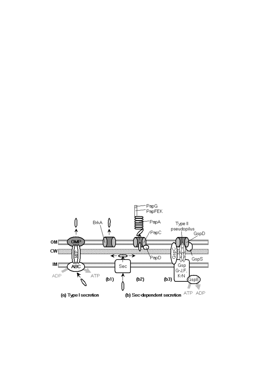
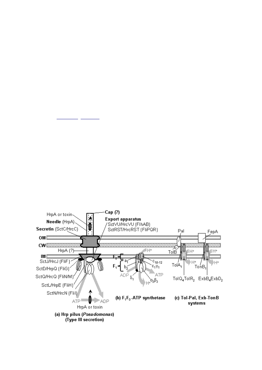
Figure 4: Systems with components homologous to
flagellar components. (a) Hrp pilus of Pseudomonas spp.
For components with well-documented homology to
flagellar components, the name according to the unified
nomenclature for type III secretion systems proposed by
Hueck (1998) is given (Sct: Secretion and Cellular
Translocation) first, followed by the currently accepted
name for the Hrp protein. The name of the flagellar
homolog is shown in brackets. (b) The F
1
F
0
-ATP
synthetase shown to scale, based on Capaldi and Aggeler
(2002). The F
1
-α and β subunits are homologous to each
other and to FliI (Gogarten et al., 1992). Further possible
homologies are discussed in the text. (c) The Tol-Pal
system, similar to the Exb-TonB system. TolA is
homologous to TonB, and TolQR, ExbBD, and MotAB are
homologs (Cascales et al., 2001). The 4:2 stoichiometry
for MotAB is favored in recent models (Schmitt, 2003;
Zhai et al., 2003).
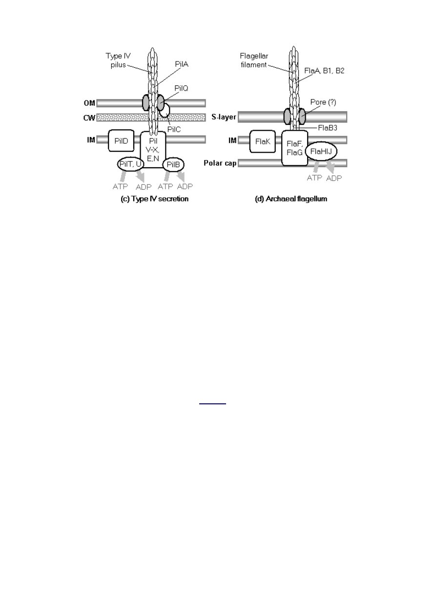
Figure 5: Various secretion systems of prokaryotes. (a)
Type I secretion system, a single-step transporter,
substrates are recognized by an uncleaved C-terminal
sequence. OMP, outer membrane channel-forming
protein; MFP, membrane fusion protein; ABC, ATP-
binding cassette exporter. (b) Three sec-dependent
secretion systems: (b1) Autotransporter. (b2)
Chaperone/usher pathway and P pilus. (b3) Type II
secretion. (c) Type IV secretion, also sec-dependent. (d)
The archaeal flagellum, with several components
homologous to type IV secretion. Based on several
sources (Jarrell et al., 2000; Thanassi and Hultgren,
2000a; Büttner and Bonas, 2002; Thanassi, 2002; Bardy
et al., 2003). Another nucleotide may be substituted for
ATP in some cases. See
for description of the
functions of the systems.
About 10 well-conserved protein species make up the core of the type III export apparatus, which is
used to export the axial components of bacterial flagella (rod, hook, filament, adaptor, and cap
proteins). In 1994 it was discovered that homologs of these proteins are also used to secret virulence
factors in a diverse array of proteobacterial pathogens, such as Yersinia pestis, Salmonella typhimurium,
Pseudomonas aeruginosa and enteropathogenic E. coli (Hueck, 1998). The term “type III secretion
system” is commonly used to refer to the virulence systems, but here it will be used to denote the class
of secretion systems that make use of the type III export pathway. This includes the two currently
known members (virulence and flagellar secretion systems) and any unknown homologs.
The existence of a nonflagellar type III export apparatus falsifies the argument that flagellar
components are useless if they are not part of a fully functioning flagellum. One answer to Macnab’s
(1978) query, “What advantage could derive…from a ‘preflagellum’ (meaning a subset of its
components)” is now obvious: a subset of flagellar components could serve as an export system. Thus,
the model for the origin of flagella begins with the hypothesis of a primitive type III export system.

This hypothesis, however, requires justification on several grounds in order to ameliorate obvious
objections.
3.2.2. Are nonflagellar type III secretion systems derived from flagella?
The fact that known nonflagellar type III secretion systems are restricted to proteobacteria, and that
these systems are mostly virulence systems specializing on eukaryotes (which are probably far younger
than flagella), lead Macnab (1999) as well as others (He, 1998; Kim, 2001; Plano et al., 2001) to
conclude that the flagellar pathway is probably the older one, and that type III virulence systems are
derived from flagella. Although some apparently avirulent type III secretion systems have been
discovered (e.g., in the legume symbiote Rhizobium; see Marie et al., 2001), and the phylogenetic
distribution of type III secretion systems has been widened somewhat by their discovery in
Chlamydiales (Kim, 2001), these data still support the conclusion that type III virulence systems are
derived eukaryote-interaction systems, rather than phylogenetically basal homologs. Phylogenetic
analysis of type III secretion systems seemed to confirm the case (Nguyen et al., 2000). Aizawa (2001)
was one of the few dissenting opinions, arguing that flagella and virulence systems might have diverged
in parallel from a common nonflagellar ancestor, pointing out that there are bacteria that parasitize or
prey on other bacteria, a point with some merit although predatory bacteria are poorly studied
(Guerrero et al., 1987).
Nguyen et al.’s (2000) conclusion has recently been challenged by Gophna et al. (2003), who
demonstrated with phylogenetic trees of FlhA, FliI, FliP, and FliO homologs that type III virulence
system sequences do not nest within flagellar sequences. This supports the view that the two systems
diverged from a common ancestor, which could plausibly have been a type III export system
functioning in a nonflagellar, nonpathogenic context. However, Gophna et al. (2003) are not able to
exclude the possibility that virulence systems evolve more rapidly, or that the frequent lateral transfer
of type III virulence system genes (Nguyen et al., 2000; Gophna et al., 2003) might have increased the
rate of sequence divergence. Gophna et al. also cite for support the progressionist notion that evolution
disfavors events such as the simplification of complex systems like the flagellum, a dubious proposition
in modern evolutionary theory, especially considering the common evolutionary trend of simplification
in pathogens and parasites. As long as known nonflagellar type III secretion systems are
phylogenetically restricted and only function as specialized systems for eukaryote penetration, the
suspicion will remain that they are derived from flagella. For the purposes of the current discussion it
will be assumed that type III virulence systems are derived, although they still give valuable insights
about the possible traits of a hypothetical ancestral type III secretion system.
3.2.3. An ancestral type III secretion system is plausible
If type III virulence systems are derived from flagella, what is the basis for hypothesizing a type III
secretion system ancestral to flagella? The question would be resolved if nonflagellar homologs of the
type III export apparatus were to be discovered in other bacterial phyla, performing functions that
would be useful in a pre-eukaryote world. That such an observation has not yet been made is a valid
point against the present model, but at the same time serves as a prediction: the model will be
considerably strengthened if a such a homolog is discovered. For the moment, it is easy enough to
explain the lack of discovery of such a homolog on the basis of lack of data. Knowledge of microbial
diversity is quite poor (Whitman et al., 1998): far less than 1% of bacteria extant in a particular
environment are readily culturable (Hayward, 2000). Cultivation-independent surveys of prokaryote
diversity based on environmental rRNA sequencing commonly discover deeply-branching microbes
previously unknown to science (DeLong and Pace, 2001), and that certain groups are unexpectedly
ubiquitous (Karner et al., 2001). In addition, only a fraction of cultured microbes have been studied in
any substantial biochemical or genetic detail, and this subsample is heavily skewed towards pathogens
and convenient model organisms. Of the ~112 complete bacterial genomes sequenced as of July 2003
http://www.ncbi.nlm.nih.gov/PMGifs/Genomes/eub_g.html
), at least two-thirds are pathogens,
mutualists, or commensals of multicellular eukaryotes. Many of the free-living bacteria that have been
sequenced are extremophiles or are used in industrial applications.
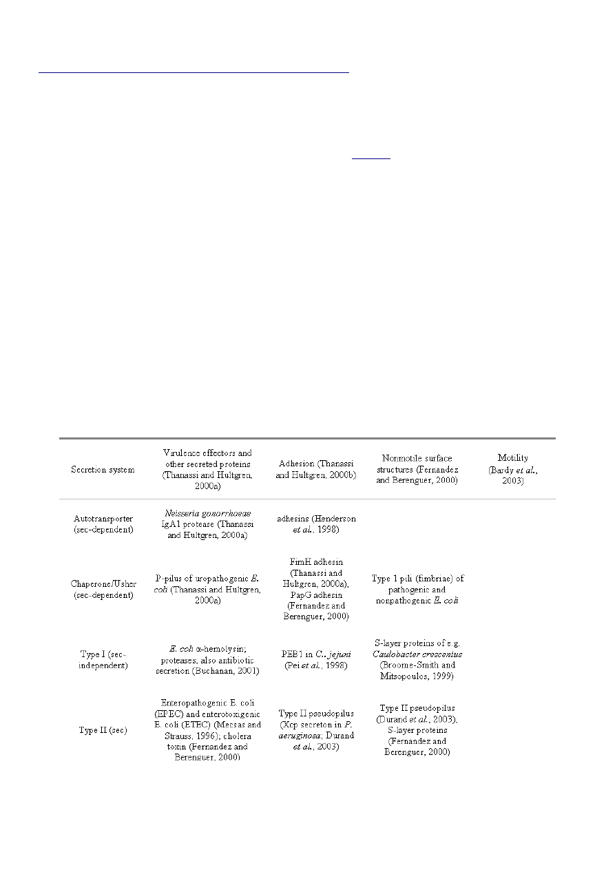
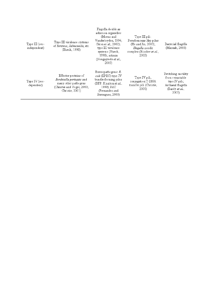
Even with such a skewed dataset, a general argument for the plausibility of a primitive type III export
system can be constructed on the basis of analogy. Each of the six secretion systems described above
has been coopted to serve diverse functions by prokaryotes (
). The thoroughness of some of the
observed convergences is remarkable – notably, all of the systems have been adapted for eukaryotic
virulence, five secrete surface structures, at least four are used for adhesion, three or four form pili, and
two perform motility-related functions. That pili and adhesion often play a role in virulence in well-
studied organisms is not particularly significant, as such functions are useful in free-living contexts as
well (Kennedy, 1987). The overall picture is that any secretion system that exists will sooner or later get
coopted for diverse functions, including virulence, in various lineages. The commonality of the
virulence function in known systems almost certainly reflects human interests rather than the situation
in the wild.
Table 4: Convergent functions of well-characterized prokaryote secretion systems. Other secretion
systems are known to exist: e.g., curli fimbriae based on the extracellular nucleation/precipitation
pathway (Smyth et al., 1996; Wu and Fives-Taylor, 2001; Chapman et al., 2002) and slime secretion
(Merz and Forest, 2002). Others undoubtedly remain to be discovered.
It might be objected that with so many available secretion systems, postulating the existence of an
additional system is superfluous. However, many bacteria have multiple secretion systems. An
illustrative case is Pseudomonas aeruginosa, which has all of the above-listed systems (Bitter, 2003).
Furthermore, many bacteria will have two or more copies of certain types of secretion systems, with
mildly to strongly divergent functions: e.g., E. coli can have both P-pili and type 1 pili (Thanassi and
Hultgren, 2000a); Salmonella and Yersinia have two type III virulence systems each (Cornelis and Van
Gijsegem, 2000); and Pseudomonas aeruginosa has at least two type II secretion systems and probably
two kinds of type IV pili (Bitter, 2003).
3.2.4. The origin of a primitive type III export system
Type III virulence systems have well-conserved homologs of the following flagellar components (Plano
et al., 2001): FliF (the membrane-embedded MS-ring); FlhA, FlhB, FliP, FliQ, FliR (integral membrane
export components inside the MS-ring); FliI and FliH (ATPase and regulator); and FliG and FliM/N
(the switch complex). The primitive type III secretion system would not necessarily have had all of the
components that are conserved in the possibly derived virulence systems. In particular, if the type III
virulence systems are derived, the homologs of the switch complex proteins (FliN/M, FliG) are probably
retained only in order to stabilize/support the coadapted secretion complex and FliF ring, and are
otherwise vestigial.
FliF is fundamentally a membrane pore and so its origin must lie with the origin of transport proteins
in general, a question explored by Saier (2003). FlhA and FlhB are larger than FliOPQR, and have
large cytoplasmic C-terminal domains that appear to bind the export substrates. FlhA interacts with
FliF and the soluble components of the type III secretion system but its exact function is unknown.
FlhB plays a key role in determining whether rod/hook or filament axial proteins are secreted, and
therefore controls the length of the hook by a poorly-understood mechanism (Macnab, 2003).
Substrate switching would not have been a necessary feature of a primitive type III secretion system,
but perhaps the association of proto-FlhA and/or FlhB with the proto-FliF pore turned it from a
somewhat general passive transporter into a substrate-specific passive transporter. One of the
differences between type II and type III secretion systems is that type II systems recognize their
substrates by a N-terminal signal peptide that is removed during transport. The signal sequences for

type III secretion substrates are also in the N-terminal regions but they are not cleaved (Büttner and
Bonas, 2002). Perhaps this difference allowed the primitive type III secretion system to export an
important substrate on a different control circuit independent of the sec pathway, and this finer control
was the selective basis for the retention of the system.
3.2.5. The relationship between type III export and the F
1
F
0
-ATP synthetase
That a phylogenetically basal type III export apparatus must have existed is supported by several
additional facts. As discussed previously, the protein that powers protein export in type III secretion,
FliI, has long been considered homologous to the F
1
subunit of F
1
F
0
-ATP synthetase on the basis of
about 30% amino acid identity to the active F
1
-β subunit (Albertini et al., 1991; Vogler et al., 1991;
Gogarten et al., 1992). The F
1
-αβ ATPase is a heterohexamer made up of alternating α-subunits
(noncatalytic) and β-subunits (catalytic). This pattern is shared by all bacteria and is also found in the
archaeal A-ATP synthase and eukaryote V-ATP synthase, so F
1
-α and F
1
-β are thought to have diverged
before the cenancestor (Gogarten and Kibak, 1992). FliI, on the other hand, probably consists of a
homohexamer of catalytic subunits (FliI’s hexameric nature was only recognized very recently: Blocker
et al., 2003; Claret et al., 2003). It diverges before the F
1
-α and F
1
-β split in sequence similarity trees,
and thus probably also diverged prior to the cenancestor (Gogarten and Kibak, 1992). However, it is
more similar to the F
1
subunits than the more distantly related hexameric ATPases such as the
RNA/DNA helicase termination factor rho (Boyer, 1997), and therefore Gogarten and Kibak (1992)
conclude that the FliI family diverged specifically from a primitive F
1
-ATPase prior to the cenancestor.
There is not similar evidence that flagella specifically evolved before the cenancestor, so this is a point
in favor of the primitive type III export system hypothesis.
In light of the long-established homology between FliI and F
1
-αβ, it is surprising that there have been
few searches for further homologies between the F
1
F
0
-ATP synthetase and type III export system.
Sequence similarity searches do not turn up significant hits, but considering the timespan and
divergence in function this is not necessarily surprising. As discussed above, homology between the
F
1
F
0
-ATP synthetase and flagellum is commonly suggested, but explicit protein-protein homologies are
never proposed, and the assumption that the rotational mechanisms of the two systems are homologous
implies a quite radical transformation of ATP synthetase components. However, several recent
discoveries suggest specific homologies that are much more conservative than those implied by previous
workers. First, FliH forms a (FliH)
2
FliI heterotrimer with FliI (Minamino and Macnab, 2000;
Minamino et al., 2001). FliH has an elongated shape (Minamino et al., 2001), and both FliI and FliH
are soluble cytoplasmic components that associate intrinsically with the membrane and with lipid
vesicles (Auvray et al., 2002). If the FliH
2
homodimer associates with the FliI
6
complex in vivo, all of
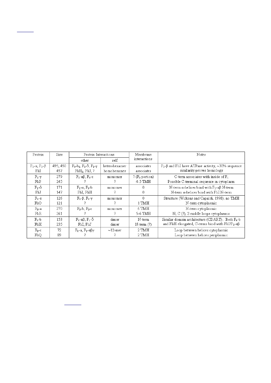
this begins to look suspiciously similar to the association (
) between the F
1
F
0
-ATP synthetase
F
1
-α
3
β
3
and F
0
-b subunits: two elongated F
0
-b subunits form a dimer and interact with F
1
-α
3
β
3
. In F
0
-
b it is the N-terminal region that associates with the membrane, and the C-terminal region with the N-
terminal regions of F
1
-α
3
β
3
(Boyer, 1997; Weber and Senior, 2003). In FliH it is known that the C-
terminal region associates with N-terminal region of FliI (Gonzalez-Pedrajo et al., 2002), but the region
responsible for membrane association is undetermined (Auvray et al., 2002); F
0
-b – FliH homology
would predict that the FliH N-terminus associates with the membrane. Although BLAST searches on
FliH only return F
0
-b as a non-significant hit, a search of NCBI’s CDART (Geer et al., 2002) based on
FliH does retrieve F
0
-b as a result with similar domain architecture (using the default e-value cutoff of
0.01), another point in favor of the hypothesis of homology. Jackson and Plano (2000) report that the

Yersinia pestis FliH homolog YscL (corresponding to SctL/HrpE in
) has low but significant
sequence similarity with the e subunit of the archaeal ATPase of Methanococcus jannaschii and the e
subunit of the vacuolar ATPase of Desulfurococcus spp.; these subunits are the homologs of the b
subunit of the F
1
F
0
-ATP synthetase. Thus the present scenario predicts that careful multiple alignment
of FliH sequences with bacterial F
0
-b and the corresponding archaeal and eukaryotic homologs (all of
which would be equally related to FliH) will confirm homology.
Can further homologies between flagella and the F
1
F
0
-ATP synthetase be discerned? In the F
1
F
0
-ATP
synthetase, an F
1
-δ monomer associates with the proximal end of F
1
-α
3
β
3
and F
0
-b
2
. In the type III
export apparatus, it is FliJ that interacts with FliI and FliH
2
. FliJ seems to be required for the export of
all flagellar components, and so has been interpreted as a general chaperone in the cytoplasm (Macnab,
2003). However, this observation is equally well explained if FliJ is a required part of a FliI
6
FliH
2
complex essential for export. Both FliJ and F
1
-δ have a similar size and N-terminal binding sites to the
N-terminal regions of FliI/F
1
-α. There may also be a structural similarity: FliJ has a high probability
of exhibiting an N-terminal α-helical coiled-coil arrangement (Macnab, 2003), using sequence-based
predictions (Lupas et al., 1991, method implemented at
http://www.ch.embnet.org/software/COILS_form.html
). F
1
-δ has several conserved α-helices at its N-
terminal binding site to F
1
(Weber et al., 2003b). Although predictions do not generally yield a high
probability of coiled-coil structure for F
1
-δ, a cursory non-exhaustive sampling of orthologs shows that
at least one FliJ protein does not show a high probability prediction of coiled-coil structure either
(Buchnera aphidicola, accession no. P57179) while at least one F
1
-δ protein does (Rhodopseudomonas
blastica, accession no. P05437). It appears that the C-terminal region of F
1
-δ associates with the C-
terminal region of F
0
-b
2
, although the details remain to be worked out (Weber and Senior, 2003).
Regarding the FliJ-FliH
2
interaction, Fraser et al. (2003) favor a model where FliJ interacts with the N-
terminal region of FliH
2
, but their data (Gonzalez-Pedrajo et al., 2002) shows that deletions in either
the N-terminus (perhaps the region that associates with the membrane) or middle (dimerization region)
of FliH preclude FliJ binding; thus failure of FliJ binding could be due to general malformation of
FliH
2
due to the failure of FliH to dimerize (middle deletion) or associate with the membrane (N-
terminal deletion). Homology between F
1
-δ and FliJ would predict that FliJ-FliH interaction is
actually mediated through the C-terminal regions of each, but that the association may be rather weak,
as it is between F
0
-b
2
and F
1
-δ (Weber and Senior, 2003).
Similarities in F
1
F
0
-δca, the integral membrane proteins FliPQR of the type III export apparatus, and
the proteins SecFEY of type II secretion proteins were pointed out by Aizawa (2001), who calls these
triplets the “proto-channel” and suggests homology. His evidence is of a general nature (calculated
similarities in molecular size, aliphatic index, instability index, and isoelectric point) and so cannot be
accepted uncritically. In particular, it is no longer thought that F
1
-δ (or its eukaryote homolog OSCP)
is associated with the membrane or ATP synthetase stalk (Weber et al., 2003a), and the evidence
discussed above points to a different homology for F
1
-δ. However, the proposed matches between FliQ--
F
0
-c and FliR--F
0
-a are decent in terms of protein size and also the number of transmembrane helices
of the respective proteins (
). And surprisingly, extrapolating the homology hypothesis to match
the two remaining type III secretion components (FliO and FliP) to the two remaining synthetase
components (F
1
-ε and F
1
-γ, respectively) also seems to provide plausible matches in terms of size.
When the similarities between F
1
F
0
-ATP synthetase and type III export components are tabulated
), it is apparent that that each component of the F
1
F
0
-ATP synthetase can be matched to a
component of the type III export apparatus with a similar size and topology, as far as evidence is
available (the function and structure of the flagellar proteins FliOPQR are poorly understood).
Table 5: Similarities between proteins of the F
1
F
0
-ATP synthetase and the flagellar type III export
apparatus that may suggest homology. Protein size is the length in amino acids for E. coli. TMH =
Transmembrane helices. Little detailed information on FliOPQR is available, the topologies listed are
the predictions of Minamino and Macnab (1999). Data taken from several sources: general ATP
synthase component information (Boyer, 1997, updated by later references); FliI--F
1
-β homology
(Gogarten et al., 1992; N-terminal F
1
-α to N-terminal F
1
-δ interaction (Weber et al., 2003a); FliIHJ
(Minamino and Macnab, 2000; Minamino et al., 2001; Auvray et al., 2002; Minamino et al., 2002;
Macnab, 2003). The membrane-associating region of FliH is not determined (Auvray et al., 2002), but
the C-terminal region interactions appear similar to the C-terminal interactions for F
0
-b (see text), so
an N-terminal association with the membrane seems likely.
Individually, the cited similarities are easily attributable to chance, but together
they are at least suggestive. Although detectable sequence similarity may be too
much to hope for given the already very low similarity between FliI--F
1
-α
3
β
3
and
FliH--F
0
-b, the postulated homologies would be further testable by Aizawa’s
also shows that there are some apparent dissimilarities. Notably,
while both F
0
-c and FliQ have 2 transmembrane helices, the loop between the
helices is exposed to the cytoplasm in F
0
-c (Birkenhager et al., 1999), while the loop
between the helices in FliQ was predicted to be periplasmic (Ohnishi et al., 1997); a
reversal of this finding would support the homology hypothesis. The weakest case
for homology is between F
1
-ε and FliO; FliO is predicted (Ohnishi et al., 1997) to
have a single transmembrane helix, while the structure of F
1
-ε has been solved

(Wilkens and Capaldi, 1998) as a two-domain protein that binds to the stalk.
However, both proteins tolerate substantial variability; F
1
-ε functions with large
deletions (Wilkens and Capaldi, 1998) and clear homologs of FliO have not even
been identified in type III virulence systems (Gophna et al., 2003).
The hypothesis that the entirety of a primitive F
1
F
0
-ATP synthetase may have been
coopted in toto into a primitive gated pore (proto-FliF and proto-FlhA/B) is
certainly provocative; it would explain at a stroke the origin of most of the type III
export apparatus and provide a phylogenetically basal precursor to the flagellum
even though clearly basal type III secretion systems remain undiscovered. The
complex would fit well in the FliF ring; using the stoichiometry of FlhA
2
FlhB
2
proposed by Macnab (2003), and the equivalent stoichiometry of an ATP synthetase
for the other integral membrane components, FliO
1
P
1
Q
~12
R
1
, the total number of
transmembrane helices is 60, well within the approximate MS-ring capacity of
about 70 transmembrane alpha-helices (Fan et al., 1997). Fan et al. estimate <3
copies of FliR per flagellum, which is consistent with the ATP synthase homology
hypothesis, but also estimate 4-5 copies for FliP, which is not, so if the ATP
synthetase hypothesis is true it would be expected that the FliP finding is in error.
Macnab (1999) called the homology between FliI and F
1
-αβ “inexplicabl[e]”.
However, there may be a relatively simple explanation. If the postulated homology
between the ATP synthetase and type III export is correct, then the key event in the
origin of type III export was the association of a primitive F
1
F
0
-ATP synthetase with
a proto-FlhA or FlhB inside the proto-FliF ring, converting it from a passive to
active transporter. Since little is known about the details of the coupling of ATPase
activity to protein export in Type III export, this step remains speculative. Probably
motion in the synthetase was linked to a conformational change in FlhA and/or
FlhB, with the proton pumping function of the synthetase lost soon afterwards.
Currently there are several documented associations between FlhAB and the rest of
the type III export apparatus (Macnab, 2003). These associations include proteins
in both the “F
0
” and “F
1
” regions of the type III export apparatus. FlhA or FlhB
may thus take over some of the linker role that is played by F
0
-b in the ATP
synthetase and (on the homology hypothesis) by FliH in the type III export
apparatus; this would help to explain why FliH is not absolutely required for
successful construction of flagella, and FliH null mutants can be compensated by
mutations in FlhA and FlhB (Minamino et al., 2003).
Other possible hypotheses for the origin of the type III export apparatus are not
currently ruled out, such as the idea that much of apparatus is descended from a
passive channel and that only a portion of the F
1
F
0
-ATP synthetase was coopted to
power transport, or that there is an ancient, obscured homology between the
various secretion systems. Alternatives are currently disfavored because they are
more complex and explain the origin of fewer components. However, even if FliI
remains the only confirmed homolog to the F
1
F
0
-ATP synthetase, general
considerations indicate that the evolution of an export system is not very difficult. A
diversity of export systems of varying complexity exist, and there is a functional
continuum of membrane complexes ranging from single proteins and passive pores
through to active, gated export systems, indicating that there are no major
evolutionary puzzles to solve. The cataloguing and categorizing of transport

proteins is already yielding insights into their origin (Saier, 2003).
The ATP synthetase homology hypothesis has the advantage of numerous testable
implications for the structure and function of FliHIJOPQR. The ATP synthetase is
relatively well-understood; structures have been determined for most of the
components and a number of sophisticated techniques for studying the complex as a
whole have been developed. If the homology hypothesis is correct, then similar
structures would be expected for the corresponding type III export components, and
many of the techniques applied to the ATP synthetase should apply to the export
apparatus. It is worth noting in passing that if a significant portion of the type III
export apparatus is indeed homologous to the ATP synthetase, then it becomes fairly
likely that the rotary flagellum contains within it a second rotary motor powering
protein export. This is a fairly incredible notion, but would merely be the latest in a
long line of surprising discoveries yielded by the study of the flagellum. This
possibility might mean that the proto-flagellar secretion system was rotating from
the start (echoing the rotation-early hypotheses of Cavalier-Smith, Goodenough,
and Rizzotti), although this is not a necessary postulate for the rest of the scenario to
proceed.
3.3. Type III secretion system
For the remainder, the hypothesis of a primitive type III export system will be taken
for granted. This complex would have transported proteins manufactured in the
cytoplasm into the periplasmic space. If secretins were already available from the
type II secretion system, as they probably were given the universal distribution of
type II secretion, then from the start the type III export system would have been a
primitive kind of type III secretion system, as small proteins could diffuse in the
periplasmic space until they found an outer membrane pore and diffused out.
Digestive proteases or antibiotic molecules are likely candidates for the secreted
proteins. Alternatively, the export system could have originally secreted proteins
destined for the periplasm, and later cooption of a secretin converted the export
system into a secretion system.
The association of an outer membrane channel with the type III export apparatus
would improve the efficiency of secretion. This advantage would increase as
exported substrates became larger, because the peptidoglycan cell wall only allows
the diffusion of globular proteins with a size less than about 50 kDa (Young, 2001);
as protein size increased, diffusion would be increasingly impeded. Once a new
single-step secretion channel was available it would be possible to secrete larger
proteins and proteins that would be harmful if left to wander about the periplasmic
space. These are selective forces that would favor the spread and diversification of
the channel, after its origin as an efficiency-improving measure.
Outer membrane secretins have been coopted repeatedly by various versions of the
secretion systems discussed above (Hueck, 1998; Thanassi, 2002; Bitter, 2003); if the
type III virulence system is derived from the flagellum, it probably originated in
part by replacing the flagellar L- and P-ring proteins with a secretin. The first
association of a secretin with a primitive type III export apparatus was probably
mediated by the simultaneous cooption of a secretin and its outer membrane
lipoprotein chaperone (Dailey and Macnab, 2002). Both of these proteins are
secreted by the type II secretion pathway. The channel to the extracellular medium
could be recruited in a single step if a mutation caused the secretin to associate with

the type III export apparatus. The secretin appears to cross both the cell wall and
outer membrane in the Hrp pilus, and to associate with the FliF homolog
(SctJ/HrcJ;
) in the cytoplasmic membrane (Blocker et al., 2003), so
having two ring proteins (the L- and P-rings in flagella) does not appear to be a
prerequisite for secretion. Thus the double ring may have been a later addition to
the system, perhaps even coinciding with the early stages of improvement of the
proto-flagellum and the loss or modification of the secretin (see below).
3.4. Origin of a type III pilus
In the model, flagellin and all of the proteins of the axial structure – FlgBCFG (rod),
FlgE (hook), FlgKL (adaptor), FlgD and FliD (caps), in addition to FliC (flagellin) --
are descended from a common ancestral pilin secreted from the primitive type III
secretion system. All of these proteins are placed in the axial protein family
(Homma et al., 1990a; Hirano et al., 2001). Homma et al. (1990a) put the rod, hook,
and first adaptor (FlgK) proteins into a closely-related subfamily. The divergence of
the axial filament family probably occurred mostly after the origin of a functioning
protoflagellum; this will be discussed in a later section. First, the origin of the pilus
must be considered.
The diversity of surface structures based on secretion systems was documented in
; modern flagella retain many of these functions (Moens and Vanderleyden,
1996). To expand on a likely function of a primitive pilus, successfully adhering to a
surface can be a problem for a floating bacterium: at a low Reynolds number, the
boundary layer near a surface can be a significant barrier (Vogel, 1994). A
bacterium can increase its chances of attachment by secreting adhesins with an
affinity for the desired surface, ensuring successive attachment if it happens to get
near a surface (e.g., the adhesins secreted by autotransporters, independent of pili
(Henderson et al., 1998)). It can increase its chances still further either by putting
the adhesin at the end of a filament (e.g., the PapG adhesin located at the tip of the
P pilus fibrillum (Sauer et al., 2000); the flagellar cap FliD of Pseudomonas
aeruginosa doubles as an adhesin (Scharfman et al., 2001)) and/or by making the
filament adhesive along its whole length, which is a common occurrence in modern
bacterial flagella as well as many other surface structures (Kennedy, 1987; Moens
and Vanderleyden, 1996; Fernandez and Berenguer, 2000; Giron et al., 2002).
Probably any filament, adhesins or no, will have some utility in attaching to
inorganic surfaces, simply by expanding effective size and surface area available for
adhesion. Even in the absence of specific adhesins, charge, hydrophobicity, and/or
van der Waals forces can be exploited for more general surface adhesion (Vogel,
1988), particularly at the small scale of bacteria.
Three hypotheses present themselves as to how the ancestral pilus originated:
filament-first, cap-first, and modified filament-first. The latter hypothesis combines
the best features of the filament-first and cap-first hypotheses.
3.4.1. Filament-first hypothesis
One way that the kinds of pili described above could get their start is by simple
polymerization of a surface adhesin. The adhesin would have inherited from its
ancestor the ability to bind with the outer membrane channel and with the
extracellular substrate; all that would have to be added is self-binding capability.

The plausibility of this step is attested by several facts: first, structures made up of
multiple copies of the same subunit are biochemically ubiquitous, and the evolution
of large multimeric complexes has in many instances been traced back to simpler
ancestors, e.g., AAA ATPases (Mocz and Gibbons, 2001). Second, polymerization
into a filament or tubule via mutation is a quite common event: sickle-cell
hemoglobin, derived by only a single substitution from regular hemoglobin, forms
not only self-assembling polymers but dynamic polymers (Mitchison, 1995). In fact,
Mitchison (1995) argues that evolution can start with just about any protein fold
and produce a self-assembling polymer.
An alternative to polymerizing an adhesin is to postulate that a gene for a pre-
existing filament-forming protein was coopted by transposition of the promoter and
N-terminal signal sequence of an already secreted protein. Support for this second
possibility might be found in homology between flagellin and a modern filament-
forming protein. Homology between flagellin and actin has been proposed. Harris
and Elder (2002) cite an 8/13 amino acid sequence match between flagellin and actin
in the N-terminal region, but this could easily be due to chance. Novikova et al.
(2000) found that flagellar filaments co-precipitate with rabbit skeletal myosin, and
that flagellin and F-actin compete for myosin binding, but this might be explained
by a general similarity of filaments rather than homology. The search for flagellin-
actin homology is somewhat misguided in any case, because actin is a eukaryotic
protein, so ancient prokaryote actin homologs such as FtsA (Mitchison, 1995) would
be more appropriate subjects. Similarly, what should be sought is not the homolog
of flagellin but the homolog of the entire axial protein family. Given the large
divergence of flagellin from the more conserved rod and hook members of the
family (Homma et al., 1990a), any relationships outside of this family are bound to
be difficult to detect.
A simple assumption is that the first filament was a chain of monomers, probably in
an open helix. Longer filaments are presumably better for adherence than short
filaments, and thus selection for adhesion can be expected to favor longer filaments.
Once a polymer filament of reasonable length has been built, however, there may be
a difficulty in extending it. The problem does not arise if filament subunits are
added at the base, as occurs in type IV pili: type IV pili are based on type II
secretion systems, which use a two-step process to transport proteins. First,
proteins are exported into the periplasm, and then they are pushed out through a
secretin, perhaps via a “plunger”-type mechanism involving a pseudopilus (Thomas
et al., 2001). However, the type III secretion system exports proteins from the
cytoplasm in one step. By exporting a number of individual subunits, a short
filament binding to the outer membrane pore can be formed, but its possible length
will be severely limited by the decreasing chances of successfully adding monomers
to the receding distal tip. This problem might be overcome in a gradual manner by
modifications to the open helix, so that it better corralled the monomers as they
exited the secretin. Each mutation that brought the turns of the helix closer together
would decrease the rate of monomer escape, and allow the extension of the
filament. A tube with closed or nearly closed walls would be the optimal solution,
and selection for rigidity (necessary for very long filaments) would also favor the
closed tube.
The result would be something rather like the modern type III virulence pili, which
appear to have far less complex axial structures than flagella. Indeed, despite
several investigations it has yet to be determined that the Hrp pilus has any axial

components (rod-like, hook-like, etc.) apart from the main protein of the pilus,
HrpA (MxiH/PrgI in Shigella/Salmonella; Blocker et al., 2003; Aizawa (2001)
tentatively suggests a few others). The extracellular portion of the filament seems to
extend continuously into the secretion complex (Fernandez and Berenguer, 2000)
whereas in flagella there is a distinction between filament, hook, and rod.
It might be objected at this point that the flagellum requires the cap (FliD) in order
to chaperone the flagellin subunits into place at the elongating tip of the filament;
without it, they diffuse away and are lost (Blocker et al., 2003). The hook has its
own temporary cap (FlgD), and it has been suggested, but not proven (Hirano et al.,
2001; Berg, 2003; Macnab, 2003), that the rod has a cap protein as well (FlgJ).
However, the necessity of the cap for successfully assembling subunits is ambiguous.
Flagellin will self-assemble into filaments in vitro (Hirano et al., 2001). No cap has
been identified in any type III virulence systems (Blocker et al., 2003), and although
PrgJ has been suggested as a possible cap for the Salmonella needle (Sukhan et al.,
2003), the evidence is indeterminate as Sukhan et al. could not detect PrgJ in
sheared-off needles and did not detect it at needle tips using immunoelectron
microscopy (they therefore suggest that PrgJ may be a basal component). The polar
flagellum of Vibrio grows normally without the cap (Bardy et al., 2003), probably
because it is sheathed by an extension of the cell membrane (McCarter, 2001) that
constrains the subunits. Finally, even in the canonical E. coli flagellum the adaptor
proteins FlgK and FlgL are added without any capping structure (Macnab, 2003),
leading Macnab (2003) to argue that “capping structures are perhaps best viewed as
a means of increasing efficiency of addition rather than as an absolute
requirement.” On this view, the cap could be a relatively late evolutionary addition
to the pilus structure, originating by pentamerization of a pilus subunit and initially
improving speed and efficiency of pilus assembly. Later co-adaptation between
filament and cap subunits would make it a more-or-less required feature.
3.4.2. Cap-first hypothesis
An alternative to gradual formation of a hollow pilus would be to start with the
cap. In Pseudomonas aeruginosa, FliD serves not only as a cap protein, but also as
an adhesin. P. aeruginosa infects the human respiratory system by adhering to
mucins. FliD mutants were found to be nonadhesive, which could occur either
because FliD is a necessary adhesin, or because the flagellar filament fails to
assemble without the cap. However, Arora et al. (1998) found that FliC null mutants
retained adhesive ability. This implies that it is FliD specifically which serves as the
adhesin, and not the whole flagellar filament, a conclusion supported by additional
lines of evidence (Arora et al., 1998; Scharfman et al., 2001). On the filament-first
hypothesis, an adhesin attached to the outer membrane secretin mutated to form a
polymeric chain. The fact that caps can be adhesive, however, suggests an
alternative hypothesis. Instead of a mutation forming a polymeric chain, the
mutant adhesin formed an oligomer that associated with the distal rim of the outer
membrane secretin, approximately covered the mouth of the secretin, and allowing
more adhesin monomers to be packed into the available space. In this case, a
pentamer ring was approximately the right size. Once this was established,
however, the utility of the adhesive “secretin cap” would be further improved if it
could be extended away from the surface of the cell. This could occur by mutation of
a duplicate cap protein that formed a slightly wider ring. This ring would associate
with the base of the cap, but the size mismatch would allow the insertion of more
subunits, forming a short pilus in one step. On this view, the primitive pilus would

derive its structure of ~5 subunits per turn from the pentameric cap, rather than the
reverse. The channel inside the pilus would be descended from the hole in expanded
ring.
3.4.3. Modified filament-first hypothesis
The filament-first hypothesis has the disadvantage of explaining the addition of
distal subunits to the filament before a tube structure could evolve. The cap-first
hypothesis has the difficulty that a pentameric proto-cap covering the surface of the
secretin pore might impede the secretion of other substrates. The difficulty is not
insurmountable, as secreted substrates might escape from beneath the sides of the
cap, or alternatively might knock loose the cap, which is then replaced by
continually secreted cap protein (both mechanisms may operate in modern
flagella). However, these problems dissolve if the hypothesized adhesin pentamer
were to initially form a ring atop the secretin, instead of a cap. Secretion of diverse
substrates could then continue unabated without continual secretion of the adhesin
protein. From here, a proto-pilus could easily be formed by a mutant adhesin that
polymerized the ring into a tube. This hypothesis is simpler and more appealing
because it combines the advantages of the two previous scenarios: a pilus initially
assembled by simple mechanisms without a cap, but having a well-formed tube
structure from the start, and allowing the uninterrupted secretion of other type III
secretion system substrates. On this hypothesis, the proto-cap would again be a late
addition (a modified pilus protein) increasing assembly efficiency.
It is interesting to reflect on the surprise many researchers felt when the mechanism
of flagellar filament construction – adding subunits at the tip rather than the base –
was discovered. It has been called “astonishing” and “somewhat bizarre” (Macnab
and DeRosier, 1988). However, with the modified filament-first hypothesis in hand,
the decidedly unintuitive method of filament assembly used by the flagellum can be
seen as a product of the constraint of building a pilus from the starting point of type
III secretion. That a flagellum can also be built perfectly well from the base is
shown by the archaeal flagellum with its type IV secretion-like system (Peabody et
al., 2003). But unlike the type IV secretion system, which has periplasmic ATPases
and other outer membrane-associated active transport components, the type III
system had no mechanism of powered outer membrane transport available to build
on.
3.4.4. Improvements on the type III pilus
Once a primitive pilus has evolved, a number of rapid improvements can be
expected. First would be optimization of the pilin protein for its new role, under
selection for increased strength, minimizing breakage, increased speed of assembly,
etc. Addition of the cap and subsequent coevolution of the pilin and cap subunits
could have occurred fairly early, particularly as the pili became very long and
assembly times became significant. Pilus lengthening would be selected for because
it increases reach and adhesive surface area. That lengthening is a trivial matter of
regulation is shown by various lab-produced mutants that exhibit lengthened hooks,
needles, etc. due to simple mutations. Type III pili might have reached the length of
flagellar filaments (10 μm) long before motility originated; flagella and Hrp pili are
of comparable length (He and Jin, 2003, Figure 1). Soon the type III secretion
system would become a specialized pilus-secretion apparatus, and pili would be
adapted for a variety of more strenuous uses. For example, pili might be used as

stalks to elevate the bacterium above the surface, in order to better access light, a
particular concentration of molecules, or escape competition from bacteria on the
surface, all functions of attachment organelles today (Dyer, 2003). Duplication and
modification of the pilin protein would allow greater functional flexibility, such as
adhesion to different substrates and the production of certain kinds of pili based on
environmental stimuli. One important modification that might have occurred after
these trends were well along would be to strengthen the pilus attachment to the cell
by extending the filament down into the secretion system to attach to the export
apparatus embedded in the cytoplasmic membrane. This would have been effected
by cooption of a duplicate pilin. The core tubule structure of the flagellar rod, hook,
and filament is constructed exclusively by the N- and C-terminal domains of the
axial proteins; the middle domains are placed on the outside of the tubule, and in
flagellin are highly modifiable and often dispensable (Cohen-Krausz and
Trachtenberg, 2003). Therefore, the “proto-rod” probably originated by loss of the
outer domains, assuming that the extracellular pilus had them for adhesion or
structural purposes. This duplication event would create the ancestors of the
rod/hook subfamily and flagellin. Initially, one cap protein could chaperone the
assembly of both structures, but as they diverged, the cap protein would be
duplicated as well to allow specialization on each protein, assuming that modern
flagellar rods have cap proteins, as has been suggested (FlgJ; Berg, 2003; Macnab,
2003).
It is not clear that modern type III virulence pili make use of rodlike proteins at all
(the filament may simply extend all the way from the cytoplasmic export apparatus
out into the extracellular space), so it is also possible that differentiation of filament
and rod proteins occurred later and that attachment to the export apparatus
occurred via modification of the pilin subunit. However, the hypothesis that the
duplication was early helps to explain the high divergence between flagellin and the
rod/hook subfamily, as well as explaining how the filament became attached to the
export apparatus instead of the outer membrane secretin (although some
attachment to the secretin may have remained; see below). Another protein, FliE,
serves as the adaptor protein between FliF (the MS-ring) and FlgB (the proximal
rod protein). FliE homologs have not yet been detected in type III virulence
systems, so the utility of a FliE-like adaptor in nonmotile systems is ambiguous.
Here it will be assumed that it was a relatively late, post-motility addition that
strengthened the attachment between the MS-ring and the rod. Investigation of the
attachment mechanisms of modern type III pili to the secretion system may shed
light on the relative likelihood of these possibilities.
3.5. The evolution of flagella
3.5.1. The selective advantage of undirected motility
Even with a complex pilus in place, the modern flagellum could not have originated
in a single step. It is hypothesized that the first, very crude motility function was
random dispersal. The function was probably not stirring or gathering more food
by more rapid movement, because of the previously-discussed constraints of life at a
low Reynolds number. Dispersal, on the other hand, is both a ubiquitous adaptation
in biology and rather undemanding in terms of motility. Vogel (1994) reports that
passive dispersal (i.e., unpowered dispersal by wind or current) is found in every
phylum of animals and division of plants. For creatures such as bacteria, some
dispersal will occur without any adaptations whatsoever: random physical events
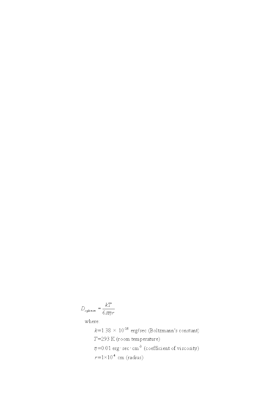
can dislodge them from the substrate, and Brownian motion and larger-scale
turbulence and flow will move them about. Even in agar, nonflagellate bacteria
without other motility systems can spread via “sliding,” motility due to colony
growth requiring only the production of a surfactant to reduce friction (Brown and
Häse, 2001). However, dispersal is not always a good thing for a bacterium. Since a
bacterium existing in a location is most likely descended from a successfully
reproducing bacterium also at that location, and therefore the environment is
conducive to reproduction, it would be expected that the best choice for a bacterium
would be to stay where it is at, rather than gambling everything on a rather literal
leap into the blue. On the other hand, this logic will rapidly break down because
any environment conducive to replication will soon become filled to the brim with
bacteria, at which point competition for nutrients, space, light, etc. will become
severe. In such a situation there are numerous potential responses (spore formation,
killing fellow bacteria by secreting antibiotics, etc.), but one of them is clearly
dispersal (Dusenbery, 1998; Stoodley et al., 2002).
The best general strategy would be for the bacterium to “decide” whether or not to
disperse based on environmental cues: if life is good, stay put, but if resources are
scarce, go somewhere else. In fact, this is basically what regulates the production of
flagella in modern bacteria. In E. coli, the master operon (class 1) for flagella
encodes FlhC and FlhD. These proteins activate the genes for flagellar biosynthesis
in the next operon (class 2), but are repressed in high glucose conditions, where
nutrients are plentiful and movement is pointless. If conditions deteriorate, the
level of cyclic AMP in the cell increases, and the expression of FlhDC is activated by
cAMP and the catabolite repressor/activator protein (CAP) (Berg, 2003). Dispersal
processes and evolutionary stable dispersal strategies are tractable to mathematical
and computer modeling, resulting with a large literature (e.g., Gandon and
Roussett, 1999; Lebreton et al., 2000; Mathias et al., 2001; Poethke and Hovestadt,
2002), although most of it is not aimed specifically at bacteria (for exceptions see
Kreft et al., 2001; Kerr et al., 2002). All that is needed for the argument to proceed
at this point is that dispersal be a widely beneficial behavior. However, the basic
dynamics of random bacterial diffusion are so simply described (Berg, 1993) that a
short investigation of what kind of bacterium might be expected to evolve random
dispersal is irresistible.
Even dead or otherwise nonmotile bacteria have a non-trivial diffusion coefficient: a
sphere with a radius r will have a diffusion coefficient D
sphere
of (Berg, 1993, eqn.
4.13):

For a dead bacterium with a diameter of 2 μm, D
sphere
= 2.1x10
-9
cm
2
/sec. The
average time t it will take to diffuse a distance x is given by (Berg, 1993, eqn. 1.10):
(7)
For the above bacterium, this means that it can be expected to passively diffuse its 2
μm diameter in ~9.3 sec, purely by Brownian motion. However, because Brownian
motion is a random walk, after each ‘step’ the diffusing particle has an equal chance
of going in any direction; the overall drift is zero. A large number of particles
placed at one location will gradually spread out in all directions by diffusion, but the
mean location of the population will not change from the starting point unless
additional forces come into play. The fact that average diffusion time increases with
the square of the distance means that diffusion becomes an increasing poor way to
travel as the dispersal distance increases. The 2 μm nonmotile bacterium, above,
takes an average of 9 seconds to diffuse one body length, but to diffuse 100 body
lengths takes not 100 times as long (15 minutes) but 10,000 times as long (26 hours).
These figures are somewhat misleading as they completely ignore turbulence and
flow (perfume molecules would take a month to diffuse across a room if diffusion
was the only relevant process; Berg, 1993), but beneath the laminar boundary layer,
very near a surface, these forces will be reduced (Vogel, 1994).
Therefore, one very crude way for a bacterium adhered to a surface to disperse is to
detach its adhesins and/or adhesive pili, and let passive dispersal take place. At
least one such dispersal mechanism has been documented (Coutte et al., 2003). If
the bacterium can manage to rise substantially in the boundary layer, flow or
turbulence may carry it some distance, at which point it can re-secrete adhesive pili
and attach to a new surface. It will probably do better if it starts out at the top of
long pilus; similar dispersal-enhancing mechanisms are well known (fruiting bodies
in bacteria such as Myxococcus; Caulobacter stalks).
Under what conditions would it be advantageous to enhance the effects of Brownian
motion by adding crude active motility? Usefully, Dusenbery (1997; 1998) has
derived an equation allowing the calculation of the relative utility of active and
passive dispersal for organisms at a low Reynolds number. Dusenbery assumes that
the organisms are spherical and that they swim at a velocity of 10 body lengths/sec,
a common value at widely varying scales (Dusenbery, 1996). Taking into account
the fact that rotational Brownian motion will keep any cell from swimming in a
straight line, the ratio of the diffusion coefficient with motility (D
m
) to the diffusion

coefficient without it (D
0
) is (Dusenbery, 1997, Table 2):
(8)
where r is again the radius and u is the cell’s swimming velocity in radii/sec (here,
20). Dusenbery calculates that a bacterium would have to have a diameter of at
least 0.64 μm in order to double its diffusion coefficient for the purpose of
undirected dispersal. Small bacteria have a very high diffusion coefficient even
without motility, and for small bacteria Brownian rotation is so severe that
swimming straight for any distance is impossible. Therefore, for very small
bacteria, active swimming with flagella is pointless; random Brownian motion is as
good as it gets (other swimming methods may be successful, e.g. the linear motor of
Spiroplasma; Trachtenberg et al., 2003). This has the obvious implication that
flagella cannot have evolved in very small bacteria; Dusenbery surveyed bacteria
genera and found that the smallest motile genus had a diameter of 0.8 μm. Similar
minimum size constraints were found for motility with chemotaxis, phototaxis, and
thermotaxis (Dusenbery, 1997).
Equation (8) can be modified by converting the relative swimming velocity u (in
radii/sec) into the absolute swimming velocity v (in cm/sec):
(9)
Equation (9) can be used to estimate the minimum swimming velocity required for a
protoflagellum to substantially increase the diffusion coefficient of a cell. D
m
/D
0
was
calculated for cells ranging from 0-8 μm in diameter, with absolute swimming
velocities of 0.1, 1, and 10 μm/sec (see
). Some advantage to diffusion would
result from motility for any values of D
m
/D
0
. However, because the construction
and movement of filaments has some cost, we have followed Dusenbery in setting
the cutoff for “selectable motility function” at doubling the passive diffusion
coefficient (D
m
> 2D
0
, Dusenbery’s result is
approximately reproduced (swimming velocity here is absolute rather than relative
to cell size, so slightly different input values were used): D
m
/D
0
is doubled for a ~0.6
μm bacterium swimming at 10 μm/sec.
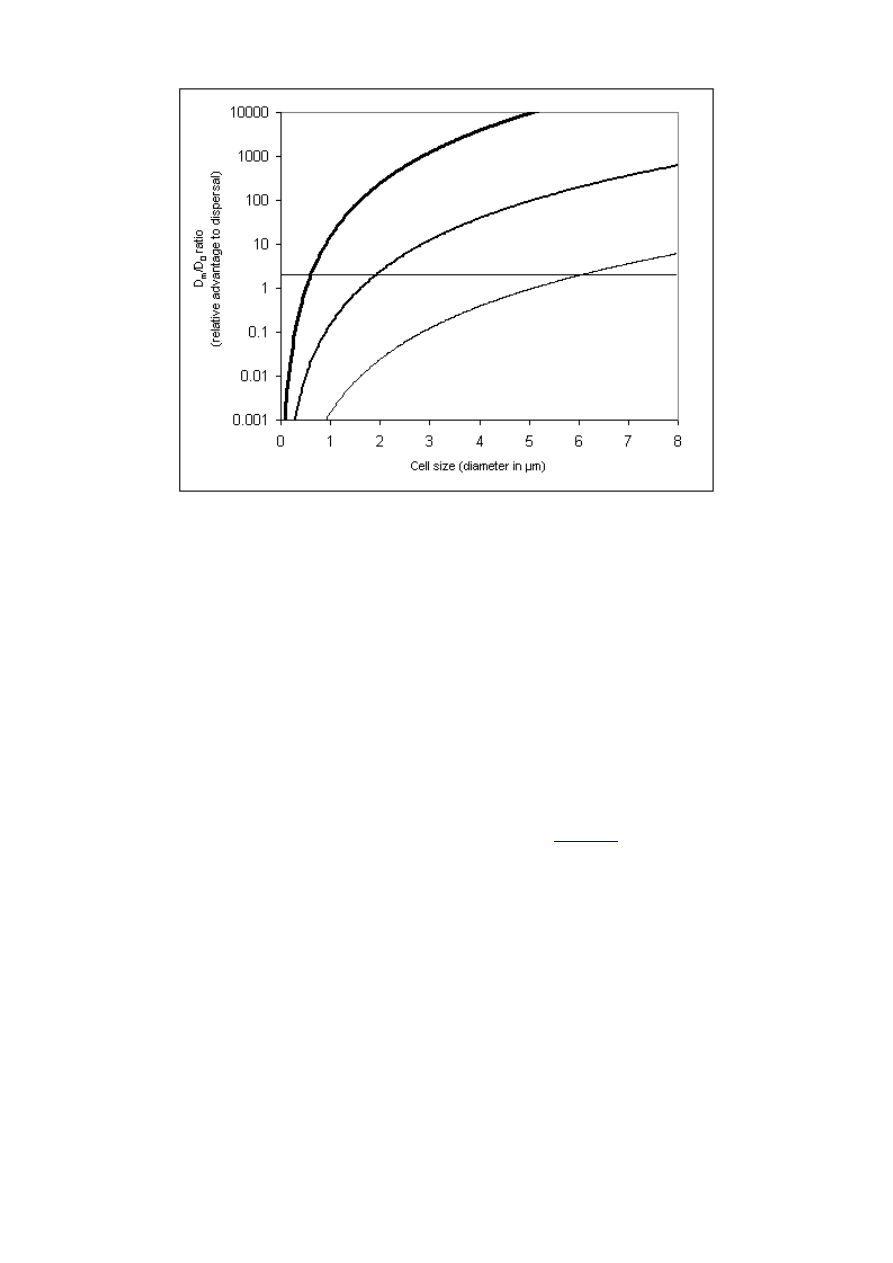
Figure 6: Relative diffusion advantage of motility (D
m
/D
0
)
as a function of cell size and absolute swimming velocity,
plotted on a log scale. Calculations were made for three
swimming velocities. Thin line: 0.1 μm/sec; Medium line:
1 μm/sec. Thick line: 10 μm/sec (typical E. coli swimming
velocity). The horizontal line represents D
m
/D
0
= 2, the
point at which active motility doubles the diffusion
coefficient.
However, a crudely functioning protoflagellum cannot be expected to push a
bacterium at 10 μm/sec. The important result shown in
is that even very
slow absolute swimming velocities can result in a significant improvement in the
diffusion coefficient for large bacteria. Swimming at 1/10 the velocity of E. coli is
advantageous to dispersal in a bacterium of ~2 μm diameter, and swimming at 1/100
the velocity of E. coli is advantageous to the dispersal of a bacterium with a
diameter of ~6 μm. Two factors contribute to this pattern: for larger bacteria,
passive diffusion is slower, increasing the relative advantage of swimming.
Similarly, rotational diffusion is also slower for larger bacteria, but this factor
enhances the efficacy of swimming as swimming runs will take longer to be
randomly reoriented.
There are additional reasons to think that the protoflagellum may have originated
in a large bacterium. Similar beneficial scaling applies if swimming velocity is
considered in terms of body lengths/second: Dusenbery’s 0.64 μm bacterium has to
swim at 10 body lengths/second in order to beat diffusion, but a 6 μm bacterium
need only swim 0.17 body lengths/second in order to achieve a benefit. A
consideration of carbon budgets also points this direction: for a (hypothetical) very

small flagellated bacterium, diameter 0.4 μm, producing 10 peritricious flagella,
each ten times the body length of the cell, would cost 50% of the cell’s carbon
(Mitchell, 2002). However, for a 1 μm cell, the relative cost of 10 flagella of
proportional size is only about ~2% of cell carbon. For the 6 μm diameter cell
discussed above, it is ~0.2%. 6 μm diameter bacteria are well within the usual size
range of bacteria (Dusenbery, 1997). For a moderately large bacterium, the costs of
crude, poorly functioning flagella are trivial, while the relative benefits in terms of
dispersal are substantial. The exponential nature of the relationships is such that
moderate violations of the input assumptions will not greatly change the qualitative
results (Dusenbery, 1997); at some moderately large size the costs of primitive
motility become low and the benefits high.
3.5.2. Primitive flagella
The flagellar motor is made up of two proteins, MotA and MotB. MotB binds to the
peptidoglycan cell wall, allowing the complex to serve as a stator. MotB (and
perhaps MotA) also forms a proton conducting channel. Although the exact
mechanism of motor function is still mysterious, with many proposed models (Berg,
2003; Schmitt, 2003), energy from the translocation of proteins in the vicinity of
MotB is somehow transformed into mechanical energy to move the rotor. Probably
this occurs by conformational change in MotA, which then binds reversibly with the
rotor protein FliG, causing rotation. Speaking very metaphorically, FliG appears to
act like the teeth of a gear, converting (in one model) the power stroke of MotA into
rotary motion. FliG is mounted on the central MS-ring (FliF). Also attached to the
MS-ring (perhaps mostly but not exclusively via FliG) are the switch proteins FliM
and FliN. FliM contains a receptor domain for the phosphorylated chemotaxis
protein CheY-P, and the binding of CheY-P induces some kind of conformational
change in FliM, FliN, and FliG that results in switching the direction of motor
rotation from counterclockwise to clockwise. This in turn results in a short ‘tumble’
which reorients the cell, and then the flagellum returns to counterclockwise
rotation.
Even given the minimal costs and substantial selective benefits of crude motility,
how could the sudden origin of the rotary motor complex be mutationally possible?
The basic answer is that the ancestors of the motor proteins were already fully
formed and serving other functions in the cell. It was recently discovered (Cascales
et al., 2001; Kojima and Blair, 2001) that the flagellar motor proteins MotAB have
nonflagellar homologs: ExbBD and TolQR (
). These proteins share
significant sequence similarity and all form ion channels that energize work at a
distance by a third protein; ExbBD and TolQR energize outer membrane transport
via action on TonB and TolA, respectively, while MotAB energize flagellar motion
via action on FliG. The recently discovered homologs involved in Myxococcus
gliding systems (Youderian et al., 2003) will likely add another instance, although no
detailed studies of their function have been performed. The nonflagellar MotAB
homologs are phylogenetically widely distributed, found in proteobacteria,
cyanobacteria, Aquifex, and even in archaea (Kojima and Blair, 2001). These facts
led Kojima and Blair to note that these proteins “could perform work in contexts
other than (and simpler than) the flagellar motor,” and they conclude that
“ancestral forms of the MotA/MotB complex might have arisen independently of
any part of the rotor.”
In order to form the motor-rotor interface, however, the origin of a third protein,

FliG, must also be accounted for. No nonflagellar homologs of FliG have been
discovered (except in type III virulence systems), perhaps not surprisingly given the
peculiar function of this protein and the radical change it must have undergone,
whatever its ancestral function. The structure of the middle and C-terminal
domains of FliG has been resolved (Brown et al., 2002), and is primarily made of
alpha helices. Alpha helices are ubiquitous in proteins, so FliG is not necessarily
structurally bizarre, despite its unusual function. Three general possibilities present
themselves for the origin of the FliG-MotAB complex. (1) TolQR homologs were
coopted via a mutation that allowed them to bind directly to FliF. FliG was a later
addition that enhanced motility by improving binding between the MS-ring and
MotA, and gradually took over the interface function completely. (2) Proto-FliG
was bound to FliF before the cooption of MotAB for some other reason, perhaps a
stabilization or structural function similar to that served by the FliG homologs in
type III virulence systems. Mutant TolQR homologs then bound to proto-FliG. (3)
FliG was coopted simultaneously with MotAB, because it originated as a fragment
of a TolA homolog that ancestrally interacted with a TolQ homolog. The third
hypothesis is the simplest and most direct pathway; the only novel interaction would
be the binding of the proto-FliG to FliF; binding to the proto-MotA would be
inherited. This is less demanding than postulating the re-engineering of the
interface between a TolQ homolog and its substrate (a feature of both hypotheses #1
and #2), and does not require postulating an independent cooption of FliG from an
unknown source. The hypothesis also has the advantage of being testable via
determination of the structures of TonB or TolA and investigation of their
interactions with ExbBD and TolQR.
On any of the hypotheses, it is not absolutely necessary that crude motility be an
immediate product; a coopted TolQRA-like complex could first have associated with
the type III secretion system to enhance or help to control protein transport.
However, all that would initially be required for the very weak motility postulated
above would be a slow rotation (other things being equal, a swimming velocity of
1/100
th
that of E. coli would imply a rotation rate similarly reduced; E. coli flagella
rotate at 100 Hz, so perhaps the protoflagellum rotated at ~1 Hz). Although the
model does not hypothesize that the ancestral pilus was short, even severely
truncated flagella (0.3-1.2 μm) can support residual motility (Josenhans et al., 1995;
Suerbaum, 1995), lending plausibility to the notion that perfectly formed flagella
are not necessary for crude motility. In addition to the motility advantages
discussed above, wiggling and twisting would probably help to dislodge the
bacterium from the surface and from nearby bacteria; just such a function of
certain type IV bundle-forming pili has been observed in E. coli (Knutton et al.,
1999).
A possible objection here is that the ancestral pilus cannot be expected to have been
freely rotating, preadapted for the addition of a motor complex; the rod might have
been bound to the peptidoglycan cell wall via the P-ring, making motility
impossible. However, this assumes that a P-ring existed at this point, a dubious
assumption if a secretin can bridge both the cell wall and cell membrane.
Additionally, it is actually not clear that pili are commonly rigidly fixed to the cell
wall via a secretin. It would be very interesting to know if type III and other pili,
when attached to a substrate, allow a bacterium to rotate via Brownian motion in a
fashion similar to motor-disabled bacteria with their flagella attached to coverslips.
Similar observations would be useful for membrane-embedded structures such as
outer membrane secretins. One selective reason that might favor freely-rotating pili

prior to the evolution of motility is, again, adhesion. A curved pilus that is allowed to
rotate via Brownian motion can continually explore more area around a cell
(particularly a large, slowly diffusing, slowly rotating cell) than a rigidly attached
pilus, increasing the chances of finding a substrate.
A final possible objection is that if the pilus was perfectly straight, then rotating it
would not produce motion. While this is true, it is irrelevant because modern pili
are not always straight or stiff (Bullitt and Makowski, 1998, Figures 2-4; Honma
and Nakasone, 1990; Yamashiro et al., 1994), the interconversion of pilus shapes is
mutationally trivial, and there are many more ways to build a curved, helical
filament than a straight one from protein subunits. Additionally, for the pilus
rotated by Brownian motion postulated above, some curvature and helical character
would be a requirement in order for the pilus to explore a larger area than a fixed
pilus. The exact shape of the protoflagellum is not crucial in the drag-intensive
world of the low Reynolds number; Purcell (1997) has calculated that any number
of peculiar rotating shapes can swim with varying efficiency (and efficiency is in fact
basically energetically irrelevant at this scale; Purcell, 1977; Berg, 1993). Purcell
(1977) notes that turning anything nonsymmetrical will result in swimming.
3.5.3. Loss of outer membrane secretin
Unlike other secretion systems (type I-IV, including type III virulence systems), the
type III secretion systems of bacterial flagella do not actually have an outer
membrane secretin. The protein that plays the role of an outer membrane pore,
FlgH, is actually a lipoprotein that Aizawa (2001) and Dailey and Macnab (2002)
suggest is homologous to the Salmonella type III secretion system protein InvH.
InvH is a lipoprotein required for the insertion of the secretin InvG (SctC in
Hueck’s (1998) unified nomenclature) into the outer membrane (Crago and
Koronakis, 1998). Such a mechanism is common for outer membrane secretin
assembly; in the type II secretion system of Klebsiella oxytoca, the outer membrane
lipoprotein PulS binds 1:1 with the secretin protein PulD, preventing periplasmic
degradation and helping the localized assembly of the secretin into the outer
membrane. Twelve PulS proteins probably form a ring about the 12-PulD secretin
pore (Bitter, 2003).
These observations suggest a hypothesis for the origin of the flagellar L-ring: it is
not derived from an outer membrane secretin, as would be naively assumed based
on its position in the outer membrane. Rather, the FlgH L-ring may be derived
from the lipoprotein that was the chaperone for the secretin of the primitive type III
secretion system. What happened to the secretin? An obvious possibility is that it
slowed the rotation of the protoflagellum. On the present model, the primitive type
III pilus originally bound to the outer membrane secretin, and later the channel was
extended down to the MS-ring in the cytoplasmic membrane. However, some
association or binding might have remained between the outer membrane secretin
and the filament. If this was the case, it probably would not have mattered for a pili
rotating by Brownian motion or rotating at low Hertz in the protoflagellum. The
outer membrane can tolerate some rotational motion of embedded components, just
as membranes tolerate the lateral diffusion of proteins. Some modern flagella are
even covered by the membrane, such as the polar flagella of Vibrio, although here a
special sheath evidently prevents tearing during rotation at 1500 Hz. However, as
rotation speeds increased, the risk of outer membrane tearing would increase. A
simple solution to this problem could be to delete the portion of the secretin binding

to the filament; the outer lipoprotein ring would take over the role of outer
membrane pore, but would not interact with the filament and would provide a
bearing between the filament and outer membrane. A channel through the cell wall
would be continuously maintained if the secretin became the proto-P-ring. Aizawa
(2001) suggests homology between FlgI and the secretin InvG. The hypothesis would
be strengthened if lipoproteins other than FlgH form rings in the outer membrane
in other secretion systems; thus far, lipoprotein has not been found in isolated
secretin complexes although the ring structure is suspected (Bitter, 2003).
3.5.4. Refinements
For a bacterium, a sphere is the optimal shape for maximizing passive dispersal
(Dusenbery, 1998). It can thus be surmised that the cell evolving the protoflagellum
was a coccus, like some flagellated bacteria today (Zaar et al., 2003). In such a cell,
the surface positioning of a flagellum is irrelevant – one place is as good as another
on a sphere, so no positioning mechanism is required. Since the model postulates
that random dispersal was the original function of flagella, the first, crudely
functioning protoflagellum lacked many parts that are important in modern
flagella. First, chemotaxis and switching are not required for dispersal, so neither
the Che proteins nor the switch complex would have been required. If switching is
not required and the protoflagellar filament is sticking straight out at a random
position on spherical cell surface, then the hook region is similarly dispensible.
Once functional motility was even marginally established, however, there would be
rapid selection for improvements. These might have occurred in more or less any
order, or concurrently, so they will be discussed topically.
3.5.5. Chemotaxis and switching
The “flagellar” chemotaxis genes (Eisenbach, 2000;
, this paper) are in fact
not specific to flagella; the same system is coupled to diverse motility systems,
including archaeal flagella and twitching motility (Faguy and Jarrell, 1999; Bardy
et al., 2003). Furthermore, homologous components are tied to all manner of cellular
responses to the environment; the central two-component signal transduction
system (consisting of the histidine kinase CheA and the response regulator CheY in
flagellar chemotaxis) is ancient, found in all three domains, and used for diverse
functions. Their evolution is discussed by Koretke et al. (2000). Another major set
of chemotaxis components, the membrane bound methyl-accepting chemotaxis
proteins (MCPs) which are the receptors for attractants and repellants, have a
similarly wide set of homologs with diverse functions (Zhulin et al., 2003). More
could be said about details, as there are substantial variations between the
chemotaxis systems of various organisms (Eisenbach, 2000; Kirby et al., 2001), but
for the purposes of this paper it will be assumed that some sort of sensory
transduction system preceded the origin of the flagellum, and that one of the
response regulators was the ancestor of CheY. In modern flagella, a worsening in
conditions results in the increasing phosphorylation of CheY into CheY-P.
In E. coli, switching rotation from counterclockwise (CCW) to clockwise (CW)
causes the cell to tumble, reorienting it to swim in a new direction. The probability
of switching is increased by the binding of CheY-P to FliM. If concentrations of
attractants are increasing during a run, CheY-P decreases and switching is
suppressed, and thus favorable runs tend to last longer. If repellents are increasing,
CheY-P increases, switching is promoted, and thus unfavorable runs are shortened.

The cell uses this method to bias its random walk, imposing an overall drift towards
regions with higher concentrations of attractants (Berg, 1993). However, for
bacteria with a diameter larger than 1.4 μm, run-and-stop or run-and-reverse
strategies are more energetically favored than the run-and-tumble strategy, due to
the larger costs of actively rotating a large cell (Mitchell, 2002). As a result the run-
and-tumble strategy, while common in model organisms, is far from universal.
Rhodobacter sphaeroides swims with a single, stop-start flagellum, with no reversing
(Shah and Sockett, 1995; Shah et al., 2000). Passive rotation via Brownian motion
reorients the stopped cell. This is but one of many variations on switching
(Eisenbach, 2000), but probably resembles the most primitive version.
Explaining the origin of the switch complex, which couples the chemotaxis system to
flagellar rotation, requires an examination of the domain structure and interactions
of the switch proteins. FliN and FliM, which make up the C-ring, are partially
homologous. FliN is homologous to the C-terminal domain of FliM, and as a result
the two proteins probably occupy similar positions in the C-ring, perhaps
alternating in a 3 FliN:1 FliM pattern, which approximately matches their
stoichiometry (Mathews et al., 1998; see also
, this paper). FliM also has a
N-terminal domain with no counterpart in FliN that is the actual CheY-P receptor.
CheY-P binds to the receptor domain, increasing the probability of a switch to CW
rotation (Eisenbach, 2000) via an unknown mechanism involving interactions
between FliM/N and FliG (Mathews et al., 1998). The receptor domain is
homologous to the single-domain chemotaxis protein CheC of Bacillus subtilis
(Kirby et al., 2001). CheC binds reversibly to the Bacillus C-ring, and is released
when it binds to CheY-P. CheC has not been found in E. coli, but homologs are
found in many early-branching bacteria, as well as archaea. A cladogram generated
for CheC and the FliM CheC-like domain shows that CheC is phylogenetically basal
(Kirby et al., 2001).
A pre-existing sensory transduction system could be coupled to flagellar rotation in
a single step on the hypothesis that a FliN-like protein existed for some nonflagellar
cellular response purpose, serving as a receptor for CheC. The exact function of
modern CheC is not known, but it appears to interact with CheA, CheD, and McpB,
which form a receptor complex (Kirby et al., 2001). CheC may also have a FliM-
like function via interaction with the C-ring (Szurmant et al., 2003). The ancestor of
FliN might therefore be found among the other proteins that CheC interacts with.
On the model, a mutation in this FliN-like protein created a proto-FliN that bound
to FliG, slowing or jamming the motor. The reversible binding of CheC to proto-
FliN, however, happened to alleviate this effect by changing the conformation of
proto-FliN. CheY-P binding to CheC would result in the dephosphorylation of
CheY-P and the release of CheC from proto-FliN, resulting in the slowed-rotation
behavior. Chemotactic behavior would thereby originate by a single mutation (all
other interactions would be inherited), which could then be followed by gradual
improvements in the initial crude function. This hypothesis is more economical
than supposing that FliN originated for some role in structural support or
enhancing export, and was later coopted to a switching function via the binding of
CheC, although this remains a possibility as FliN homologs are retained in type III
virulence systems for some purpose. The first hypothesis suggests that the homolog
of FliN will be found within sensory transduction systems as one of the proteins that
CheC or a CheC homolog interacts with; it is difficult to know where to look with
the latter hypothesis. The considerable variations in the C-ring of bacteria may
yield further hints, as major variations on chemotaxis and the switch complex are

known; for example, Aquifex aeolicus lacks the traditional chemotaxis system as
well as FliM; Bacillus spp. have FliY (a FliM-FliN fusion protein; Bischoff and
Ordal, 1992; Celandroni et al., 2000) rather than FliN. In any case, the fusion of
CheC-like and FliN-like proteins would produce the FliM seen in most bacteria.
Hypothesizing a detailed pathway explaining how a stop-start switch complex could
be converted into the other varieties of switching will depend on detailed knowledge
of motor mechanism; many models of the flagellar motor have been proposed and
the question is far from settled (Berg, 2003). However, if proton-induced
conformational changes in MotA induce some kind of power-stroke against FliG,
followed by release of the FliG binding site and a return stroke to the original
position, then perhaps the answer is fairly simple. If the conformational change in
the switch complex shifts the FliG binding site up or down relative to MotA, then
perhaps the difference between “forward” and “reverse” is just the difference
between MotA-FliG binding on the forward power stroke or the return stroke.
Although a detailed analysis will not be performed here, the transition between
random dispersal and dispersal + chemotaxis is quite gradual; adding just a small
amount of directional drift to the random walk of bacteria allows gradual migration
towards nutrient gradients and away from toxins or waste products. The
advantages of directional drift over random diffusion are exponential (Berg, 1993),
and the costs in terms of extra carbon consumption are trivial compared to the
already small costs of building a flagellum in the first place.
3.5.6. Hook and additional axial components
It seems likely that the hook (FlgE) and the four rod proteins (FlgBCFG) are all
duplicates of an ancestral rod protein; their sequence relationships have been
described in detail elsewhere (Homma et al., 1990a; Homma et al., 1990b). Whether
phylogeny can be expected to correlate well with sequence similarity in this case is
somewhat debatable, as adjacent axial components will tend to have relatively
similar structural roles and signal sequences. However, it is apparent that
“adaptor” proteins (FlgK and FlgL between the hook and filament; FlgG between
the hook and the rest of the rod) must have originated after the duplication of the
major components; for example, as the proto-hook and proto-filament proteins
began to specialize in their particular roles, the mismatches between the subunits
would become increasingly troublesome, limiting further divergence. However,
duplication and modification of hook and flagellin proteins to produce adaptor
proteins (a FlgE copy producing FlgK, and a FliC copy producing FlgL) would
allow both tighter binding between hook and filament, and would remove the
constraints on specialization of the major structures. These duplications need not
have happened simultaneously; with a moderate amount of divergence, one adaptor
might do (e.g., FlgG is the adaptor between the rod and hook), with a second being
added as divergence continued. This form of protein subfunctionalization (Force et
al., 1999) can probably explain the rest of the axial proteins as well: the hook (FlgE)
might well have originated as an adaptor between the proto-rod and proto-filament
in the very early flagellum. As FlgE specialized for the hook role, adaptors for the
filament (FlgK) and rod (FlgG) would have been produced from copied hook
proteins. The reason that the flagellum has three proximal rod proteins (FlgBCF) is
not clear, but may have something to do with assembly checkpoints and
coordinating the addition of the P- and L-rings at the appropriate moment. FlgB is
the proximal rod component, interfacing with FliF via FliE (Berg, 2003); the relative

order of FlgC and FlgF has not been determined, but perhaps one assembles while
the P-ring is being assembled around it, and the other assembles coincident with the
L-ring. These components are highly conserved across all known bacterial flagella,
probably because of co-adapted interactions between the components, but their
dispensability for building a basic filament appears to be shown by type III
virulence systems, where no rod homologs have yet been discovered (Blocker et al.,
2003) although the pilus protein shares similarities with axial proteins (Aizawa,
2001; Blocker et al., 2003; Cordes et al., 2003). Thus the coordinated assembly of
rod and the P- and L-rings could have been a relatively late innovation. The origin
of FliE, the adaptor between the basal rod component FlgB and the FliF MS-ring,
was probably a very early event, occuring just after the origin of motility, in order
to strengthen the association between the mismatched symmetries of FliF and the
proto-rod. Since FliE is exported via the type III pathway and is quite small (11
kDa), perhaps it originated as a fragment of the proto-rod protein.
The hook capping protein FlgD probably originated as a duplicate of the putative
rod capping protein (FlgJ), in a manner similar to the divergence of the rod cap and
filament cap discussed previously. However, E. coli FlgJ has a C-terminal
muramidase domain in addition to an N-terminal portion that interacts with the rod
proteins. This domain shows homology to other muramidases and so was probably
coopted in order to speed up flagellar assembly by boring a hole through the cell
wall. This was probably a late addition; even today muramidase activity is not
absolutely required for successful flagellar assembly, probably because the
assembling rod has a chance of finding a suitable gap in the peptidoglycan on its
own (Hirano et al., 2001). Some bacteria lack the muramidase domain
(Rhodobacter) or FlgJ (gram-positive bacteria) entirely (Hirano et al., 2001). An
investigation of the assembly of the SctC ring in type III virulence systems, and
similar structures in other systems, might shed light on just what the muramidase is
for, as its requirement has not yet been reported for other secretion systems.
Perhaps positioning of the primitive type III pilus and protoflagellum was originally
determined by the ability of the secretin to find a sufficient peptidoglycan gap to
insert itself in; association of the secretin with proto-FliF brought the secretory
structure together. Having a dedicated muramidase in the modern flagellar
pathway might simply enable flagella production on demand, at any predetermined
spot, whether or not a sufficient hole in the peptidoglycan is already available.
3.5.7. Modern variations
The model has arrived at something like the common ancestor of all currently
known bacterial flagella. Some of the variant flagella, such as those found in gram-
positive bacteria, spirochetes, Aquifex, or Rhodobacter, might in fact be early
offshoots of flagellar evolution. This is not required on the current model, but an
improved understanding of bacterial phylogeny may change the situation. In any
case, most of these variants are probably derived (Cavalier-Smith, 2002a), as are
many other minor variations that are known (Eisenbach, 2000; Bardy et al., 2003).
4. Conclusions
The detailed evolutionary model described above is summarized in
. The
role that various evolutionary processes played in the model can now be roughly
quantified. Only one major shift of function occurred at the system level, the

transition from a pilus to a protoflagellum. All of the other changes in system
function can be seen as minor modifications of a basic function; if these are
enumerated (export --> secretion --> adhesion --> pilus, and dispersal --> taxis),
then four minor shifts of function occurred. In all cases a “shift” in function is
actually more accurately described as an addition of function at the system level, as
previous functions are maintained. At the level of subsystems (consisting of two or
more proteins), the cooption events can be tabulated: subsystem cooption was
invoked for the origin of the core export apparatus, outer membrane secretin
(proto-FlgI) and lipoprotein chaperone (proto-FlgH), the adhesin ancestral to the
axial protein family, the motor complex, and the chemotaxis/switch complex, for a
total of five subsystem cooption events. In each of these cases, cooption occurred by
the mutation of one protein to link two preexisting systems (
), followed by
the duplication and integration of the new subsystem proteins into the major
system. Except for the major transition between pilus and motility, subsystem
cooption was associated with improvements of system function rather than major
changes in system function. At the gene level, duplication events within the core
system were invoked 11 times for origin of 12 axial proteins from one, and an
additional time for the divergence of FliN and FliM. None of these events requires
postulating functional shift at the subsystem or system levels. Addition of a new
domain with novel functionality was identified twice (FliN+CheC --> FliM, rod
cap+muramidase --> FlgJ), although it probably occurred in additional instances
where homologies are currently more vague. It appears that loss of a component is
only a possibility for the outer membrane secretin of the primitive type III secretion
system, although if this became FlgI then no component loss events are necessary.
This is the case even though some components that are ancient on the model (e.g.,
FliH) are apparently not absolutely required in modern flagella (Minamino et al.,
2003). All other changes at all levels were matters of gradual improvement of
function, i.e. optimization and co-adaptation of components. Even at this early stage
of development, the model gives decent estimate of the relative importance of
various evolutionary processes involved in the origin of complex biochemical
systems.
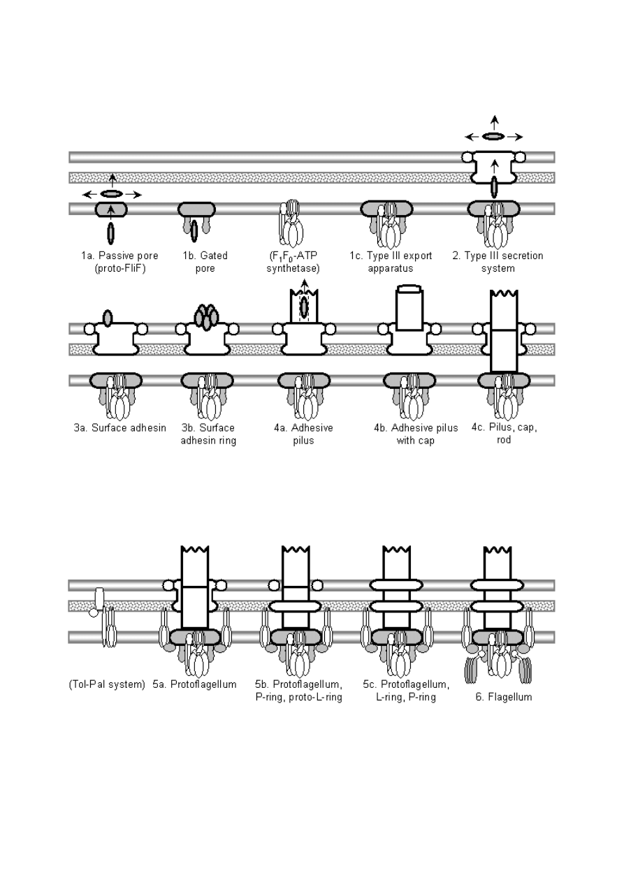
Figure 7: Summary of the evolutionary model for the
origin of the flagellum, showing the six major stages and

key intermediates. White components have identified or
reasonably probable nonflagellar homologs; grey
components have either suggested but unsupported
homologs, or no specific identified homologs, although
ancestral functions can be postulated. The model begins
with a passive, somewhat general inner membrane pore
(1a) that is converted to a more substrate-specific pore
(1b) by binding of proto-FlhA and/or FlhB to FliF.
Interaction of an F
1
F
0
-ATP synthetase with FlhA/B
produces an active transporter, a primitive type III export
apparatus (1c). Addition of a secretin which associates
with the cytoplasmic ring converts this to a type III
secretion system (2). A mutated secretion substrate
becomes a secreted adhesin (or alternatively an adhesin is
coopted by transposition of the secretion recognition
sequence), and a later mutation lets it bind to the outer
side of the secretin (3a). Oligomerization of the adhesin
produces a pentameric ring, allowing more surface
adhesins without blocking other secretion substrates (3b).
Polymerization of this ring produces a tube, a primitive
type III pilus (4a; in the diagram, a white axial structure
is substituted for the individual pilin subunits; all further
axial proteins are descended from this common pilin
ancestor). Oligomerization of a pilin produces the cap,
increasing assembly speed and efficiency (4b). A
duplicate pilin that loses its outer domains becomes the
proto-rod protein, extending down through the secretin
and strengthening pilus attachment by association with
the base (4c). Further duplications of the proto-rod,
filament, and cap proteins, occurring before and after the
origin of the flagellum (6) produce the rest of the axial
proteins; these repeated subfunctionalization events are
not shown here. The protoflagellum (5a) is produced by
cooption of TolQR homologs from a Tol-Pal-like system;
perhaps a portion of a TolA homolog bound to FliF to
produce proto-FliG. In order to improve rotation, the
secretin loses its binding sites to the axial filament,
becoming the proto-P-ring, and the role of outer
membrane pore is taken over by the secretin’s lipoprotein
chaperone ring, which becomes the proto-L-ring (5b).
Perfection of the L-ring and addition of the rod cap FlgJ
muramidase domain (which removes the necessity of
finding a natural gap in the cell wall) results in 5c.
Finally, binding of a mutant proto-FliN (probably a CheC
receptor) to FliG couples the signal transduction system
to the protoflagellum, producing a chemotactic flagellum
(6); fusion of proto-FliN and CheC produces FliM. Each
stage would obviously be followed by gradual
coevolutionary optimization of component interactions.
The origin of the flagellum is thus reduced to a series of
mutationally plausible steps.

Even the present extended treatment has left out detailed discussion of the origin of
the chemotaxis and regulatory proteins listed in
. However, many of these
proteins have homologs functional in different systems, and the chaperones of axial
proteins might have originated by duplication in a fashion similar to the axial
proteins themselves. The evolution of the organization of flagellar genes and
operons also deserves attention, although the precise organization found in modern
bacteria is probably not essential (Kalir et al., 2001).
4.1. Evaluating the model
Biological evidence supporting the model is summarized in
, in terms of
extant analogs to the hypothesized intermediates and nonflagellar homologs of
system components. Of the 30 major structural components listed in
axial proteins and probably share a common (unidentified) ancestor, a hypothetical
type III pilin subunit. Of the remaining 18 components, four (FliI, MotA, MotB,
and FliM) have well-accepted nonflagellar homologs based on significant sequence
similarity. Suggestive evidence of homology exists for eight components,
FliHJOPQR (with components of the ATP synthetase), the P-ring FlgI (with
secretins), and the lipoprotein FlgH (with lipoprotein chaperones of secretins). On
the basis of interactions with other components with identified nonflagellar
homologs, homologies can be postulated, with little current supporting evidence, for
two components, FlgA (with other secretin-associated proteins secreted by the type
II secretion system), and FliG (with a fragment of a TolA homolog). Finally, five
components (FliF, FlhA, FlhB, FliN, and the ancestor of the axial proteins) have no
identified potential homologs, although nonflagellar ancestral functions are not
difficult to postulate. The type III virulence system contains homologs of most of
these proteins (probably including an axial protein; Cordes et al., 2003), but as
discussed previously its phylogenetic position is controversial.
Table 6: Functions and analogs at each stage of the
presented model. See
and text for further
details.
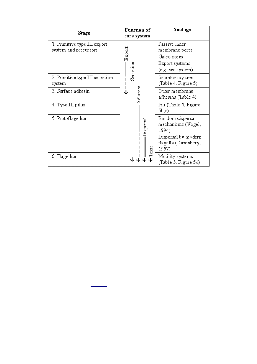
At this early stage of investigation this mixed bag should not be surprising.
Structural information (which is conserved even when sequence similarity is lost) is
not available for most of the proteins, and current sampling of bacterial genomes is
not very balanced. However, the homologies postulated provide opportunities to
test the model with future observations: if the model presented here is correct, then
it is expected that nonflagellar homologs for most flagellar proteins will be found
serving the suggested functions, in the suggested systems. Similarly, the model can
be falsified by discovery of homologies in unexpected locations: for example, if the
proteins of the flagellar basal body are discovered to be homologous to proteins of
the junctional pore of gliding motility rather than a primitive type III secretion
system, then the entire model would be overthrown and replaced by a model
relating these two systems.
The proposed analogies (
) provide another set of tests of the model. Each of
the systems proposed as analogies to stages in flagellar evolution is a piece of
evidence that the selective forces invoked in the model are common; the fact that the
functions of secretion, adhesion, pilus formation, and motility appear to be related
in analogous systems lends support to the model, which postulates transitions
between these functions. The model would be weakened if the proposed analogies,
mostly based on well-studied laboratory organisms, were found to actually be rare
in free-living prokaryotes. On the other hand, the discovery of further similar
analogs will strengthen the model – for example, it is expected that many additional

components of the archaeal flagellum will be determined to be homologous to type
IV secretion (Peabody et al., 2003). The conclusions of the simple cost-benefit model
proposed here can also be tested via analogs. Calculations indicated that the cost-
benefit tradeoff is strongly in favor of motility, even very crude motility, in a
moderately large bacterium. It would therefore be expected that, in an
experimental environment where dispersal is advantageous, selection would favor
the retention of even severely impaired (but still motile) flagella for large bacteria,
while similarly impaired flagella would be selected against in small bacteria.
Similarly, attempts to evolve crude motility in the lab (or re-evolve motility after the
deletion of a crucial component) would only work if large bacteria are used. As
there are experimental conditions where it is selectively advantageous for bacteria
to lose motility (Velicer et al., 2002), a careful consideration of the microbial
environment would be required.
4.2. The evolution of other microbial motility systems
The present model has several implications for the evolution of other prokaryote
motility systems. The conclusions of the cost/benefit analysis, that stirring is an
unlikely intermediate function and that even crude motility is advantageous for
dispersal in large bacteria, will apply to the evolution of any type of flagellar-like
motility in prokaryotes (the tiny Spiroplasma are apparently motile, but use a
radically different system). However, these conclusions cannot be generalized to the
evolution of the eukaryotic cilium, as many eukaryotes have reached the size where
stirring and swimming become useful feeding behaviors (Vogel, 1994). Although
detailed information on mechanism and homologies is not yet available, gliding
motility and archaeal flagella probably both originated via evolutionary processes
analogous to the present model, by cooption of pre-existing secretion systems. This
basic idea has already been proposed for archaeal flagella (Bayley and Jarrell, 1998;
Peabody et al., 2003). The fact that archaeal and bacterial flagella are completed
unrelated appears to weaken Cavalier-Smith’s (2002a) argument that archaea are
derived; however, if type IV secretion systems can be found in gram positive
bacteria then a plausible ancestor for archaeal flagella would exist in Cavalier-
Smith’s scheme. Presumably, standard bacterial flagella could not be adapted to
hyperthermophillic, hyperacidic conditions (Cavalier-Smith, 2002a), and the
archaeal cenancestor was forced to re-evolve a completely new form of flagellum.
4.3. The construction of evolutionary models
It is sometimes alleged that the construction of evolutionary models amounts to
nothing more than the telling of “just-so stories.” However, the putative originators
of this criticism, Gould and Lewontin (1979), only attacked scenarios that were
untestable or untested. They particularly focused their criticism of “adaptive
storytelling” on cases where the adaptive function of the trait in question was highly
dubious, such as human sacrifice (Gould and Lewontin, 1979). Their point was that
some traits might be explained by processes other than selection. They never
argued that systems like the bacterial flagellum, where function, complexity, and
adaptiveness are obvious, might have an explanation not involving the extended
action of natural selection.
A related objection to evolutionary modeling is that it is armchair theorizing,
unrelated to the practical concerns of the present day. However, an examination of

recent discoveries of nonflagellar homologs of flagellar components shows that this
is not the case. The recognition of homology between flagella and type III virulence
systems has contributed greatly to an understanding of the latter, which are
implicated in many diseases of humans, livestock, and crops (Hueck, 1998; Cornelis
and Van Gijsegem, 2000; Büttner and Bonas, 2002; Blocker et al., 2003; He and Jin,
2003). Similarly, the homology between ion channels and flagellar motor proteins
contributes to the understanding of the still-mysterious mechanism of the flagellar
motor (Schmitt, 2003; Zhai et al., 2003). In the case of the present model, the
hypothesis of more extensive homology between the F
1
F
0
-ATP synthetase and the
type III export apparatus, if true, has important implications, as the integral
membrane components are the most poorly understood portion of the flagellum and
type III virulence systems (Macnab, 2003).
A final advantage of constructing an evolutionary model is that it encourages the
synthesis of data, relating the discoveries of specialist subfields in a coherent
framework. Such a framework is a prerequisite for more detailed evolutionary
investigations, providing research questions and hypotheses to test, and challenging
dissenters to come up with better models. Until now a detailed evolutionary model
had never been seriously attempted for the bacterial flagellum, and even this fairly
basic survey has yielded several discoveries that were not obvious at the outset. The
bacterial flagellum (and prokaryote motility systems in general) probably arose in
large, coccus-shaped bacteria that were essentially modern in terms of complexity.
It is not necessary to suppose that the flagellum co-evolved with the cell wall and
membranes before the last common ancestor of life. This would be a much more
difficult event to study in any case. The previously accepted homologies between
flagellar components and nonflagellar systems (such as for FliI and MotAB) are not
the strange anomalies they appear to be when viewed in isolation, rather they fit
well into a gradual model of flagellar evolution, and give clues as to where further
homologies may be discovered. Cooption of preexisting subsystems are the key
events of interest in the model. Gene duplications within the system primarily add
complexity after the origin of the protoflagellum, and other processes, such as
domain swapping and the loss of “scaffolding” components, are relatively minor
players. Finally, in light of the organized complexity and apparent “design” of the
flagellum, the very fact that a step-by-step Darwinian model can be constructed that
is plausible and testable significantly weakens the suggestion that extraordinary
explanations might be required.
5. Acknowledgements
This work could not have been accomplished without help from numerous
individuals, who supplied ideas, references, encouragement, and helpful comments.
First, Ian Musgrave, his work on this topic, and his critique of this paper were
inspirational, and he was a helpful discussant. Pete Dunkelberg, Matt Inlay, Alan
Gishlick, Wesley Elsberry, John Wilkins, Pim van Meurs, and many others gave
help in terms of editing and informal discussions. The initial inspiration was
another paper, on the evolution of biological complexity, written for a course taught
Jim Proctor, although the present paper ended up going far beyond the original
topic.

6. References
Aizawa, S. I., 2001. Bacterial flagella and type III secretion systems. FEMS
Microbiol Lett. 202 (2), 157-164.
Albertini, A. M., Caramori, T., Crabb, W. D., Scoffone, F. and Galizzi, A., 1991. The
flaA locus of Bacillus subtilis is part of a large operon coding for flagellar structures,
motility functions, and an ATPase-like polypeptide. J Bacteriol. 173 (11), 3573-3579.
Anandarajah, K., Kiefer, P. M., Donohoe, B. S. and Copley, S. D., 2000. Recruitment
of a double bond isomerase to serve as a reductive dehalogenase during
biodegradation of pentachlorophenol. Biochemistry. 39 (18), 5303-5311.
Arora, S. K., Ritchings, B. W., Almira, E. C., Lory, S. and Ramphal, R., 1998. The
Pseudomonas aeruginosa flagellar cap protein, FliD, is responsible for mucin
adhesion. Infect Immun. 66 (3), 1000-1007.
Auvray, F., Ozin, A. J., Claret, L. and Hughes, C., 2002. Intrinsic membrane
targeting of the flagellar export ATPase FliI: Interaction with acidic phospholipids
and FliH. Journal of Molecular Biology. 318 (4), 941-950.
Bardy, S. L., Ng, S. Y. and Jarrell, K. F., 2003. Prokaryotic motility structures.
Microbiology. 149 (Pt 2), 295-304.
Bayley, D. P. and Jarrell, K. F., 1998. Further evidence to suggest that archaeal
flagella are related to bacterial type IV pili. J Mol Evol. 46 (3), 370-373.
Berg, H. C., 1993. Random Walks in Biology. Princeton University Press, Princeton.
Berg, H. C., 1998. Keeping up with the F
1
-ATPase. Nature. 394 (6691), 324-325.
Berg, H. C., 2003. The rotary motor of bacterial flagella. Annu Rev Biochem. 72, 19-
54.
Berg, H. C. and Anderson, R. A., 1973. Bacteria swim by rotating their flagellar
filaments. Nature. 245 (5425), 380-382.
Berry, R. M., 2000. Theories of rotary motors. Philos Trans R Soc Lond B Biol Sci.
355 (1396), 503-509.
Birkenhager, R., Greie, J.-C., Altendorf, K. and Deckers-Hebestreit, G., 1999. F
0
complex of the Escherichia coli ATP synthase . Not all monomers of the subunit c
oligomer are involved in F
1
interaction. Eur J Biochem. 264 (2), 385-396.
Bischoff, D. S. and Ordal, G. W., 1992. Identification and characterization of FliY, a
novel component of the Bacillus subtilis flagellar switch complex. Mol Microbiol. 6
(18), 2715-2723.
Bitter, W., 2003. Secretins of Pseudomonas aeruginosa: large holes in the outer

membrane. Arch Microbiology. 179 (5), 307-314.
Block, S. M., 1997. Real engines of creation. Nature. 386 (6622), 217-219.
Blocker, A., Komoriya, K. and Aizawa, S. I., 2003. Type III secretion systems and
bacterial flagella: Insights into their function from structural similarities. Proc Natl
Acad Sci U S A. 100 (6), 3027-3030.
Boyer, P. D., 1997. The ATP synthase - A splendid molecular machine. Annu Rev
Biochem. 66, 717-749.
Broome-Smith, J. K. and Mitsopoulos, C., 1999. Overview: Transport of molecules
across microbial membranes -- a sticky situation to get to grips with, in: Broome-
Smith, J. K., Baumberg, S., Stirling, C. J. and Ward, F. B. (Eds.), Transport of
molecules across microbial membranes, General Society for Microbiology,
Cambridge University Press, Cambridge, UK, pp.1-14.
Brown, I. I. and Häse, C. C., 2001. Flagellum-independent surface migration of
Vibrio cholerae and Escherichia coli. J Bacteriol. 183 (12), 3784-3790.
Brown, P. N., Hill, C. P. and Blair, D. F., 2002. Crystal structure of the middle and
C-terminal domains of the flagellar rotor protein FliG. Embo J. 21 (13), 3225-3234.
Buchanan, S. K., 2001. Type I secretion and multidrug efflux: transport through the
TolC channel-tunnel. Trends in Biochemical Sciences. 26 (1), 3-6.
Bullitt, E. and Makowski, L., 1998. Bacterial adhesion pili are heterologous
assemblies of similar subunits. Biophys J. 74 (1), 623-632.
Büttner, D. and Bonas, U., 2002. Port of entry – the type III secretion translocon.
Trends Microbiol. 10 (4), 186-192.
Campbell, N. A., 1993. Biology. Benjamin/Cummings, Redwood City, CA.
Campos-Garcia, J., Najera, R., Camarena, L. and Soberon-Chavez, G., 2000. The
Pseudomonas aeruginosa motR gene involved in regulation of bacterial motility.
FEMS Microbiol Lett. 184 (1), 57-62.
Cao, T. B. and Saier, M. H., Jr., 2003. The general protein secretory pathway:
phylogenetic analyses leading to evolutionary conclusions. Biochim Biophys Acta.
1609 (1), 115-125.
Capaldi, R. A. and Aggeler, R., 2002. Mechanism of the F
1
-F
0
-type ATP synthase, a
biological rotary motor. Trends in the Biochemical Sciences. 27 (3), 154-160.
Cascales, E., Lloubes, R. and Sturgis, J. N., 2001. The TolQ-TolR proteins energize
TolA and share homologies with the flagellar motor proteins MotA-MotB. Mol
Microbiol. 42 (3), 795-807.

Cavalier-Smith, T., 1978. The evolutionary origin and phylogeny of microtubules,
mitotic spindles and eukaryote flagella. Biosystems. 10 (1-2), 93-114.
Cavalier-Smith, T., 1982. The evolutionary origin and phylogeny of eukaryote
flagella. Symposia of the Society for Experimental Biology. 35 (5896), 465-493.
Cavalier-Smith, T., 1987a. The origin of cells: a symbiosis between genes, catalysts,
and membranes. Cold Spring Harbor Symposia on Quantitative Biology. 52 (6111),
805-824.
Cavalier-Smith, T., 1987b. The origin of eukaryotic and archaebacterial cells.
Annals of the New York Academy of Sciences. 503, 17-54.
Cavalier-Smith, T., 2001a. Obcells as proto-organisms: membrane heredity,
lithophosphorylation, and the origins of the genetic code, the first cells, and
photosynthesis. Journal of Molecular Evolution. 53 (4-5), 555-595.
Cavalier-Smith, T., 2001b. Early evolution: from the appearance of the first cell to
the first modern organisms (review). Quarterly Review of Biology. 76 (2), 233.
Cavalier-Smith, T., 2002a. The neomuran origin of archaebacteria, the negibacterial
root of the universal tree and bacterial megaclassification. Int J Syst Evol Microbiol.
52, 7-76.
Cavalier-Smith, T., 2002b. The phagotrophic origin of eukaryotes and phylogenetic
classification of Protozoa. Int J Syst Evol Microbiol. 52, 297-354.
Cavalier-Smith, T., 2002c. Origins of the machinery of recombination and sex.
Heredity. 88, 125-141.
Celandroni, F., Ghelardi, E., Pastore, M., Lupetti, A., Kolsto, A. B. and Senesi, S.,
2000. Characterization of the chemotaxis fliY and cheA genes in Bacillus cereus.
FEMS Microbiol Lett. 190 (2), 247-253.
Chapman, M. R., Robinson, L. S., Pinkner, J. S., Roth, R., Heuser, J., Hammar, M.,
Normark, S. and Hultgren, S. J., 2002. Role of Escherichia coli curli operons in
directing amyloid fiber formation. Science. 295 (5556), 851-855.
Chothia, C., Gough, J., Vogel, C. and Teichmann, S. A., 2003. Evolution of the
protein repertoire. Science. 300 (5626), 1701-1703.
Christie, P. J., 2001. Type IV secretion: intercellular transfer of macromolecules by
systems ancestrally related to conjugation machines. Mol Microbiol. 40 (2), 294-305.
Christie, P. J. and Vogel, J. P., 2000. Bacterial type IV secretion: conjugation
systems adapted to deliver effector molecules to host cells. Trends Microbiol. 8 (8),
354-360.
Claret, L., Calder, S. R., Higgins, M. and Hughes, C., 2003. Oligomerization and
activation of the FliI ATPase central to bacterial flagellum assembly. Mol Microbiol.
48 (5), 1349-1355.

Cohen-Krausz, S. and Trachtenberg, S., 2003. The structure of the helically
perturbed flagellar filament of Pseudomonas rhodos: implications for the absence of
the outer domain in other complex flagellins and for the flexibility of the radial
spokes. Mol Microbiol. 48 (5), 1305-1316.
Copley, S. D., 2000. Evolution of a metabolic pathway for degradation of a toxic
xenobiotic: the patchwork approach. Trends in Biochemical Sciences. V25 (N6),
261-265.
Cordes, F. S., Komoriya, K., Larquet, E., Yang, S., Egelman, E. H., Blocker, A. and
Lea, S. M., 2003. Helical structure of the needle of the type III secretion system of
Shigella flexneri. J Biol Chem. 278 (19), 17103-17107.
Corliss, J. O., 1980. Objection to "undulipodium" as an inappropriate and
unnecessary term. Biosystems. 12 (1-2), 109-110.
Cornelis, G. R. and Van Gijsegem, F., 2000. Assembly and function of type III
secretory systems. Annu Rev Microbiol. 54, 735-774.
Coutte, L., Alonso, S., Reveneau, N., Willery, E., Quatannens, B., Locht, C. and
Jacob-Dubuisson, F., 2003. Role of adhesin release for mucosal colonization by a
bacterial pathogen. J Exp Med. 197 (6), 735-742.
Cox, G. B., Jans, D. A., Fimmel, A. L., Gibson, F. and Hatch, L., 1984. Hypothesis.
The mechanism of ATP synthase. Conformational change by rotation of the beta-
subunit. Biochim Biophys Acta. 768 (3-4), 201-208.
Crago, A. M. and Koronakis, V., 1998. Salmonella InvG forms a ring-like multimer
that requires the InvH lipoprotein for outer membrane localization. Mol Microbiol.
30 (1), 47-56.
Dailey, F. E. and Macnab, R. M., 2002. Effects of lipoprotein biogenesis mutations
on flagellar assembly in Salmonella. J Bacteriol. 184 (3), 771-776.
Darwin, C., 1851. A Monograph of the Sub-class Cirripedia, with Figures of all the
Species. The Lepadidae; or, Pedunculated Cirripedes. Ray Society, London.
Darwin, C., 1854. A Monograph of the Sub-class Cirripedia, with Figures of all the
Species. The Balanidae (or Sessile Cirripedes); the Verrucidae, etc. Ray Society,
London.
Darwin, C., 1862. On the various contrivances by which orchids are fertilised. J.
Murray, London.
Darwin, C., 1872. The origin of species by means of natural selection. 6th edition.
John Murray, London.
de Duve, C., 1995. Vital Dust: Life as a cosmic imperative. Basic Books, New York.
de Souza, M. L., Seffernick, J., Martinez, B., Sadowsky, M. J. and Wackett, L. P.,
1998. The atrazine catabolism genes atzABC are widespread and highly conserved.

J Bacteriol. 180 (7), 1951-1954.
DeLong, E. F. and Pace, N. R., 2001. Environmental diversity of bacteria and
archaea. Systematic Biology. 50 (4), 470-478.
DeRosier, D. J., 1998. The turn of the screw: the bacterial flagellar motor. Cell. 93
(1), 17-20.
Dillon, R., Fauci, L. and Gaver, D., 3rd, 1995. A microscale model of bacterial
swimming, chemotaxis and substrate transport. J Theor Biol. 177 (4), 325-340.
Doolittle, R. F. and Feng, D. F., 1987. Reconstructing the evolution of vertebrate
blood coagulation from a consideration of the amino acid sequences of clotting
proteins. Cold Spring Harbor Symposia on Quantitative Biology: The Evolution of
Catalytic Function. LII, 869-874.
Durand, E., Bernadac, A., Ball, G., Lazdunski, A., Sturgis, J. N. and Filloux, A.,
2003. Type II protein secretion in Pseudomonas aeruginosa: the pseudopilus is a
multifibrillar and adhesive structure. J Bacteriol. 185 (9), 2749-2758.
Dusenbery, D. B., 1996. Life At Small Scale: The Behavior of Microbes. Scientific
American Library, New York.
Dusenbery, D. B., 1997. Minimum size limit for useful locomotion by free-swimming
microbes. Proc Natl Acad Sci U S A. 94 (20), 10949-10954.
Dusenbery, D. B., 1998. Fitness landscapes for effects of shape on chemotaxis and
other behaviors of bacteria. J Bacteriol. 180 (22), 5978-5983.
Dyer, B. D., 2003. A Field Guide to Bacteria. Cornell University Press, Ithaca.
Eisenbach, M., 2000. Bacterial chemotaxis, Nature Encyclopedia of Life Sciences,
Nature Publishing Group, London.
Emerson, S. U., Tokuyasu, K. and Simon, M. I., 1970. Bacterial flagella: polarity of
elongation. Science. 169 (941), 190-192.
Faguy, D. M. and Jarrell, K. F., 1999. A twisted tale: the origin and evolution of
motility and chemotaxis in prokaryotes. Microbiology. 145 (Pt 2), 279-281.
Faguy, D. M., Jarrell, K. F., Kuzio, J. and Kalmokoff, M. L., 1994. Molecular
analysis of archael flagellins: similarity to the type IV pilin-transport superfamily
widespread in bacteria. Can J Microbiol. 40 (1), 67-71.
Fan, F., Ohnishi, K., Francis, N. R. and Macnab, R. M., 1997. The FliP and FliR
proteins of Salmonella typhimurium, putative components of the type III flagellar
export apparatus, are located in the flagellar basal body. Mol Microbiol. 26 (5),
1035-1046.
Fernandez, L. A. and Berenguer, J., 2000. Secretion and assembly of regular surface
structures in Gram-negative bacteria. FEMS Microbiol Rev. 24 (1), 21-44.

Force, A., Lynch, M., Pickett, F. B., Amores, A., Yan, Y. L. and Postlethwait, J., 1999.
Preservation of duplicate genes by complementary, degenerative mutations.
Genetics. 151 (4), 1531-1545.
Francis, N. R., Sosinsky, G. E., Thomas, D. and DeRosier, D. J., 1994. Isolation,
characterization and structure of bacterial flagellar motors containing the switch
complex. J Mol Biol. 235 (4), 1261-1270.
Fraser, G. M., Gonzalez-Pedrajo, B., Tame, J. R. and Macnab, R. M., 2003.
Interactions of FliJ with the Salmonella Type III Flagellar Export Apparatus. J
Bacteriol. 185 (18), 5546-5554.
Gandon, S. and Roussett, F., 1999. Evolution of stepping-stone dispersal rates. Proc
R Soc Lond B Biol Sci. 266 (1437), 2507-2513.
Ganfornina, M. D. and Sanchez, D., 1999. Generation of evolutionary novelty by
functional shift. Bioessays. V21 (N5), 432-439.
Geer, L. Y., Domrachev, M., Lipman, D. J. and Bryant, S. H., 2002. CDART: protein
homology by domain architecture. Genome Res. 12 (10), 1619-1623.
Giron, J. A., Torres, A. G., Freer, E. and Kaper, J. B., 2002. The flagella of
enteropathogenic Escherichia coli mediate adherence to epithelial cells. Mol
Microbiol. 44 (2), 361-379.
Gogarten, J. P. and Kibak, H., 1992. The bioenergetics of the last common ancestor
and the origin of the eukaryotic endomembrane system. Biosystems. 28 (1-3), 131-
153.
Gogarten, J. P., Kibak, H., Dittrich, P., Taiz, L., Bowman, E. J., Bowman, B. J.,
Manolson, M. F., Poole, R. J., Date, T. and Oshima, T., 1989. Evolution of the
vacuolar H
+
-ATPase: implications for the origin of eukaryotes. Proc Natl Acad Sci
U S A. 86 (17), 6661-6665.
Gogarten, J. P., Starke, T., Kibak, H., Fishman, J. and Taiz, L., 1992. Evolution and
isoforms of V-ATPase subunits. J Exp Biol. 172, 137-147.
Gonzalez-Pedrajo, B., Fraser, G. M., Minamino, T. and Macnab, R. M., 2002.
Molecular dissection of Salmonella FliH, a regulator of the ATPase FliI and the type
III flagellar protein export pathway. Mol Microbiol. 45 (4), 967-982.
Goodenough, U., 1998. The Sacred Depths of Nature. Oxford University Press, New
York.
Goodenough, U., 2002. Of flagella and outboard motors, accessed online. URL:
http://www.metanexus.net/archives/printerfriendly.asp?archiveid=7143
Gophna, U., Ron, E. Z. and Graur, D., 2003. Bacterial type III secretion systems are
ancient and evolved by multiple horizontal-transfer events. Gene. 312, 151-163.
Gould, S. J. and Lewontin, R. C., 1979. The spandrels of San Marco and the

Panglossian paradigm: A critique of the adaptationist programmme. Proceedings of
the Royal Society of London, Series B. 205 (1161), 581-598.
Guerrero, R., Esteve, I., Pedrós-Alió, C. and Gaju, N., 1987. Predatory bacteria in
prokaryotic communities. Annals of the New York Academy of Sciences. 503, 238-
250.
Hanumanthaiah, R., Day, K. and Jagadeeswaran, P., 2002. Comprehensive analysis
of blood coagulation pathways in Teleostei: evolution of coagulation factor genes
and identification of zebrafish factor VIIi. Blood Cells, Molecules, and Diseases. 29
(1), 57-68.
Harris, R. J. and Elder, D., 2002. Actin and flagellin may have an N-terminal
relationship. J Mol Evol. 54 (2), 283-284.
Harshey, R. M. and Toguchi, A., 1996. Spinning tails: homologies among bacterial
flagellar systems. Trends Microbiol. 4 (6), 226-231.
Hayward, C. A., 2000. Microorganisms, Nature Encyclopedia of Life Sciences,
Nature Publishing Group, London.
He, S. Y., 1998. Type III protein secretion in plant and animal pathogenic bacteria.
Annual Reviews in Phytopathology. 36, 363-392.
He, S. Y. and Jin, Q., 2003. The Hrp pilus: learning from flagella. Curr Opin
Microbiol. 6 (1), 15-19.
Henderson, I. R., Navarro-Garcia, F. and Nataro, J. P., 1998. The great escape:
structure and function of the autotransporter proteins. Trends Microbiol. 6 (9), 370-
378.
Hirano, T., Minamino, T. and Macnab, R. M., 2001. The role in flagellar rod
assembly of the N-terminal domain of Salmonella FlgJ, a flagellum-specific
muramidase. J Mol Biol. 312 (2), 359-369.
Homma, M., DeRosier, D. J. and Macnab, R. M., 1990a. Flagellar hook and hook-
associated proteins of Salmonella typhimurium and their relationship to other axial
components of the flagellum. J Mol Biol. 213 (4), 819-832.
Homma, M., Kutsukake, K., Hasebe, M., Iino, T. and Macnab, R. M., 1990b. FlgB,
FlgC, FlgF and FlgG. A family of structurally related proteins in the flagellar basal
body of Salmonella typhimurium. J Mol Biol. 211 (2), 465-477.
Honma, Y. and Nakasone, N., 1990. Pili of Aeromonas hydrophila: purification,
characterization, and biological role. Microbiol Immunol. 34 (2), 83-98.
Hooper, S. D. and Berg, O. G., 2003. On the nature of gene innovation: duplication
patterns in microbial genomes. Mol Biol Evol. 20 (6), 945-954.
Hueck, C. J., 1998. Type III protein secretion systems in bacterial pathogens of
animals and plants. Microbiol Mol Biol Rev. 62 (2), 379-433.

Jackson, M. W. and Plano, G. V., 2000. Interactions between type III secretion
apparatus components from Yersinia pestis detected using the yeast two-hybrid
system. FEMS Microbiol Lett. 186 (1), 85-90.
Jarrell, K. F., Bayley, D. P., Correia, J. D. and Thomas, N. A., 1999. Recent
excitement about the archaea. Bioscience. 49 (7), 530-541.
Jarrell, K. F., Bayley, D. P., Correia, J. D. and Thomas, N. A., 2000. Archaeal
flagella, Nature Encyclopedia of Life Sciences, Nature Publishing Group, London.
Jarrell, K. F., Bayley, D. P. and Kostyukova, A. S., 1996. The archaeal flagellum: a
unique motility structure. J Bacteriol. 178 (17), 5057-5064.
Jiang, Y. and Doolittle, R. F., 2003. The evolution of vertebrate blood coagulation as
viewed from a comparison of puffer fish and sea squirt genomes. Proc Natl Acad Sci
U S A. 100 (13), 7527–7532.
Johnson, G. R., Jain, R. K. and Spain, J. C., 2002. Origins of the 2,4-dinitrotoluene
pathway. Journal of Bacteriology. 184 (15), 4219-4232.
Jones, W. J., Nagle, D. P., Jr. and Whitman, W. B., 1987. Methanogens and the
diversity of archaebacteria. Microbiol Rev. 51 (1), 135-177.
Josenhans, C., Labigne, A. and Suerbaum, S., 1995. Comparative ultrastructural
and functional studies of Helicobacter pylori and Helicobacter mustelae flagellin
mutants: both flagellin subunits, FlaA and FlaB, are necessary for full motility in
Helicobacter species. J Bacteriol. 177 (11), 3010-3020.
Kalir, S., McClure, J., Pabbaraju, K., Southward, C., Ronen, M., Leibler, S.,
Surette, M. G. and Alon, U., 2001. Ordering genes in a flagella pathway by analysis
of expression kinetics from living bacteria. Science. 292 (5524), 2080-2083.
Karner, M. B., DeLong, E. F. and Karl, D. M., 2001. Archaeal dominance in the
mesopelagic zone of the Pacific Ocean. Nature. 409 (6819), 507-510.
Kennedy, M. J., 1987. Role of motility, chemotaxis, and adhesion in microbial
ecology. Annals of the New York Academy of Sciences. 503, 260-273.
Kerr, B., Riley, M. A., Feldman, M. W. and Bohannan, B. J., 2002. Local dispersal
promotes biodiversity in a real-life game of rock-paper-scissors. Nature. 418 (6894),
171-174.
Khan, S., 1997. Rotary chemiosmotic machines. Biochim Biophys Acta. 1322 (2-3),
86-105.
Kim, J. F., 2001. Revisiting the chlamydial type III protein secretion system: clues to
the origin of type III protein secretion. Trends Genet. 17 (2), 65-69.
Kirby, J. R., Kristich, C. J., Saulmon, M. M., Zimmer, M. A., Garrity, L. F., Zhulin,
I. B. and Ordal, G. W., 2001. CheC is related to the family of flagellar switch
proteins and acts independently from CheD to control chemotaxis in Bacillus

subtilis. Mol Microbiol. 42 (3), 573-585.
Knutton, S., Shaw, R. K., Anantha, R. P., Donnenberg, M. S. and Zorgani, A. A.,
1999. The type IV bundle-forming pilus of enteropathogenic Escherichia coli
undergoes dramatic alterations in structure associated with bacterial adherence,
aggregation and dispersal. Mol Microbiol. 33 (3), 499-509.
Koch, A. L., 2003. Were Gram-positive rods the first bacteria? Trends Microbiol. 11
(4), 166-170.
Kojima, S. and Blair, D. F., 2001. Conformational change in the stator of the
bacterial flagellar motor. Biochemistry. 40 (43), 13041-13050.
Koretke, K. K., Lupas, A. N., Warren, P. V., Rosenberg, M. and Brown, J. R., 2000.
Evolution of two-component signal transduction. Mol Biol Evol. 17 (12), 1956-1970.
Kreft, J. U., Picioreanu, C., Wimpenny, J. W. and van Loosdrecht, M. C., 2001.
Individual-based modelling of biofilms. Microbiology. 147 (Pt 11), 2897-2912.
Lebreton, J., Khaladi, M. and Grosbois, V., 2000. An explicit approach to
evolutionarily stable dispersal strategies: no cost of dispersal. Math Biosci. 165 (2),
163-176.
Lenski, R. E., Ofria, C., Pennock, R. T. and Adami, C., 2003. The evolutionary
origin of complex features. Nature. 423 (6936), 139-144.
Long, M., 2001. Evolution of novel genes. Curr Opin Genet Dev. 11 (6), 673-680.
Lupas, A., Van Dyke, M. and Stock, J., 1991. Predicting coiled coils from protein
sequences. Science. 252 (5010), 1162-1164.
Macnab, R. M., 1978. Bacterial motility and chemotaxis: the molecular biology of a
behavioral system. CRC Crit Rev Biochem. 5 (4), 291-341.
Macnab, R. M., 1999. The bacterial flagellum: reversible rotary propellor and type
III export apparatus. J Bacteriol. 181 (23), 7149-7153.
Macnab, R. M., 2003. How bacteria assemble flagella. Annu Rev Microbiol. 57, 77-
100.
Macnab, R. M. and DeRosier, D. J., 1988. Bacterial flagellar structure and function.
Can J Microbiol. 34 (4), 442-451.
Manson, M. D., Tedesco, P., Berg, H. C., Harold, F. M. and Van der Drift, C., 1977.
A protonmotive force drives bacterial flagella. Proc Natl Acad Sci U S A. 74 (7),
3060-3064.
Margulis, L., 1980. Undulipodia, flagella and cilia. Biosystems. 12 (1-2), 105-108.
Marie, C., Broughton, W. J. and Deakin, W. J., 2001. Rhizobium type III secretion
systems: legume charmers or alarmers? Curr Opin Plant Biol. 4 (4), 336-342.

Mathews, M. A., Tang, H. L. and Blair, D. F., 1998. Domain analysis of the FliM
protein of Escherichia coli. J Bacteriol. 180 (21), 5580-5590.
Mathias, A., Kisdi, E. and Olivieri, I., 2001. Divergent evolution of dispersal in a
heterogeneous landscape. Evolution Int J Org Evolution. 55 (2), 246-259.
Maynard Smith, J., 1975. The Theory of Evolution. Cambridge University Press.
Mayr, E., 1976. The emergence of evolutionary novelties, Evolution and the
diversity of life, Harvard University Press, Cambridge, pp. 88-113.
McBride, M. J., 2001. Bacterial gliding motility: multiple mechanisms for cell
movement over surfaces. Annu Rev Microbiol. 55, 49-75.
McCarter, L. L., 2001. Polar flagellar motility of the Vibrionaceae. Microbiol Mol
Biol Rev. 65 (3), 445-462, table of contents.
Mecsas, J. J. and Strauss, E. J., 1996. Molecular mechanisms of bacterial virulence:
type III secretion and pathogenicity islands. Emerg Infect Dis. 2 (4), 270-288.
Melendez-Hevia, E., Waddell, T. G. and Cascante, M., 1996. The puzzle of the Krebs
citric acid cycle: assembling the pieces of chemically feasible reactions, and
opportunism in the design of metabolic pathways during evolution. J Mol Evol. 43
(3), 293-303.
Merz, A. J. and Forest, K. T., 2002. Bacterial surface motility: slime trails, grappling
hooks and nozzles. Curr Biol. 12 (8), R297-303.
Miller, K. R., 2003. Answering the biochemical argument from design, in: Manson,
N. (Ed.), God and design: the teleological argument and modern science, Routledge.
URL:
http://www.millerandlevine.com/km/evol/design1/article.html
Miller, K. R., 2004. The Flagellum Unspun: The Collapse of "Irreducible
Complexity", in: Ruse, M. and Dembski, W. (Eds.), Debating Design: from Darwin
to DNA, Cambridge University Press, Cambridge, MA. URL:
http://www.millerandlevine.com/km/evol/design2/article.html
Minamino, T., Gonzalez-Pedrajo, B., Kihara, M., Namba, K. and Macnab, R. M.,
2003. The ATPase FliI can interact with the type III flagellar protein export
apparatus in the absence of its regulator, FliH. J Bacteriol. 185 (13), 3983-3988.
Minamino, T., Gonzalez-Pedrajo, B., Oosawa, K., Namba, K. and Macnab, R. M.,
2002. Structural properties of FliH, an ATPase regulatory component of the
Salmonella type III flagellar export apparatus. J Mol Biol. 322 (2), 281-290.
Minamino, T. and Macnab, R. M., 1999. Components of the Salmonella flagellar
export apparatus and classification of export substrates. J Bacteriol. 181 (5), 1388-
1394.

Minamino, T. and Macnab, R. M., 2000. FliH, a soluble component of the type III
flagellar export apparatus of Salmonella, forms a complex with FliI and inhibits its
ATPase activity. Mol Microbiol. 37 (6), 1494-1503.
Minamino, T., Tame, J. R., Namba, K. and Macnab, R. M., 2001. Proteolytic
analysis of the FliH/FliI complex, the ATPase component of the type III flagellar
export apparatus of Salmonella. J Mol Biol. 312 (5), 1027-1036.
Mitchell, J. G., 2002. The energetics and scaling of search strategies in bacteria. The
American Naturalist. 160 (6), 727-740.
Mitchell, P., 1985. Molecular mechanics of protonmotive F
0
F
1
ATPases. Rolling well
and turnstile hypothesis. FEBS Lett. 182 (1), 1-7.
Mitchison, T. J., 1995. Evolution of a dynamic cytoskeleton. Philos Trans R Soc
Lond B Biol Sci. 349 (1329), 299-304.
Mivart, S. G., 1871. Genesis of Species. Macmillan, London.
Mocz, G. and Gibbons, I. R., 2001. Model for the motor component of dynein heavy
chain based on homology to the AAA family of oligomeric ATPases. Structure
(Camb). 9 (2), 93-103.
Moens, S. and Vanderleyden, J., 1996. Functions of bacterial flagella. Crit Rev
Microbiol. 22 (2), 67-100.
Mortlock, R. P. (Ed.), 1992. The Evolution of Metabolic Function. CRC Press, Boca
Raton Fla.
Muller, W. E., Blumbach, B. and Muller, I. M., 1999. Evolution of the innate and
adaptive immune systems: relationships between potential immune molecules in the
lowest metazoan phylum (Porifera) and those in vertebrates. Transplantation. 68
(9), 1215-1227.
Musgrave, I., 2004. Evolution of the Bacterial Flagellum, in: Young, M. and Edis, T.
(Eds.), Why Intelligent Design Fails: A Scientific Critique of the Neocreationism,
forthcoming from Rutgers University Press, Piscataway, N. J.
Nguyen, L., Paulsen, I. T., Tchieu, J., Hueck, C. J. and Saier, M. H., Jr., 2000.
Phylogenetic analyses of the constituents of Type III protein secretion systems. J
Mol Microbiol Biotechnol. 2 (2), 125-144.
Nilsson, D.-E. and Pelger, S., 1994. A pessimistic estimate of the time required for an
eye to evolve. Proceedings of the Royal Society of London Series B: Biological
Sciences. 256, 53-58.
Noji, H., Yasuda, R., Yoshida, M. and Kinosita, K., Jr., 1997. Direct observation of
the rotation of F
1
-ATPase. Nature. 386 (6622), 299-302.
Nougayrede, J. P., Fernandes, P. J. and Donnenberg, M. S., 2003. Adhesion of
enteropathogenic Escherichia coli to host cells. Cell Microbiology. 5 (6), 359-372.

Novikova, N. A., Levitsky, D. I., Metlina, A. L. and Poglazov, B. F., 2000. Interaction
of bacterial flagellum filaments with skeletal muscle myosin. IUBMB Life. 50 (6),
385-390.
Ohnishi, K., Fan, F., Schoenhals, G. J., Kihara, M. and Macnab, R. M., 1997. The
FliO, FliP, FliQ, and FliR proteins of Salmonella typhimurium: putative components
for flagellar assembly. J Bacteriol. 179 (19), 6092-6099.
Oplatka, A., 1998a. Are rotors at the heart of all biological motors? Biochem
Biophys Res Commun. 246 (2), 301-306.
Oplatka, A., 1998b. Do the bacterial flagellar motor and ATP synthase operate as
water turbines? Biochem Biophys Res Commun. 249 (3), 573-578.
Oster, G. and Wang, H., 2003. Rotary protein motors. Trends Cell Biol. 13 (3), 114-
121.
Pasquier, L. D. and Litman, G. W. (Eds.), 2000. Origin and Evolution of the
Vertebrate Immune System (Series: Current Topics in Microbiology and
Immunology). Springer, Berlin.
Peabody, C. R., Chung, Y. J., Yen, M. R., Vidal-Ingigliardi, D., Pugsley, A. P. and
Saier, M. H., Jr., 2003. Type II protein secretion and its relationship to bacterial type
IV pili and archaeal flagella. Microbiology. 149 (Pt 11), 3051-3072.
Pei, Z., Burucoa, C., Grignon, B., Baqar, S., Huang, X. Z., Kopecko, D. J.,
Bourgeois, A. L., Fauchere, J. L. and Blaser, M. J., 1998. Mutation in the peb1A
locus of Campylobacter jejuni reduces interactions with epithelial cells and intestinal
colonization of mice. Infect Immun. 66 (3), 938-943.
Pellmyr, O. and Krenn, H. W., 2002. Origin of a complex key innovation in an
obligate insect-plant mutualism. Proc Natl Acad Sci U S A. 99 (8), 5498-5502.
Plano, G. V., Day, J. B. and Ferracci, F., 2001. Type III export: new uses for an old
pathway. Mol Microbiol. 40 (2), 284-293.
Poethke, H. J. and Hovestadt, T., 2002. Evolution of density- and patch-size-
dependent dispersal rates. Proc R Soc Lond B Biol Sci. 269 (1491), 637-645.
Prum, R. O. and Brush, A. H., 2002. The evolutionary origin and diversification of
feathers. Q Rev Biol. 77 (3), 261-295.
Pugsley, A. P., 1993. The complete general secretory pathway in gram-negative
bacteria. Microbiol Rev. 57 (1), 50-108.
Purcell, E. M., 1977. Life at low Reynolds number. American Journal of Physics. 45,
3-11.

Purcell, E. M., 1997. The efficiency of propulsion by a rotating flagellum. Proc Natl
Acad Sci U S A. 94, 11307-11311.
Rizzotti, M., 2000. Early Evolution: From the appearance of the first cell to the first
modern organisms. Birkhäuser Verlag, Boston.
Rosenhouse, J., 2002. Probability, optimization theory, and evolution. Evolution. 56
(8), 1721-1722.
Sabbert, D. and Junge, W., 1997. Stepped versus continuous rotatory motors at the
molecular scale. Proc Natl Acad Sci U S A. 94 (6), 2312-2317.
Sadowsky, M. J., Tong, Z., de Souza, M. and Wackett, L. P., 1998. AtzC is a new
member of the amidohydrolase protein superfamily and is homologous to other
atrazine-metabolizing enzymes. J Bacteriol. 180 (1), 152-158.
Saier, M. H., Jr., 2003. Tracing pathways of transport protein evolution. Mol
Microbiol. 48 (5), 1145-1156.
Salvini-Plawen, S. V. and Mayr, E., 1977. On the evolution of photoreceptors and
eyes. Evolutionary Biology. 10, 207-263.
Sandkvist, M., 2001. Biology of type II secretion. Mol Microbiol. 40 (2), 271-283.
Sauer, F. G., Barnhart, M., Choudhury, D., Knight, S. D., Waksman, G. and
Hultgren, S. J., 2000. Chaperone-assisted pilus assembly and bacterial attachment.
Curr Opin Struct Biol. 10, 548-556.
Scharfman, A., Arora, S. K., Delmotte, P., Van Brussel, E., Mazurier, J., Ramphal,
R. and Roussel, P., 2001. Recognition of Lewis x derivatives present on mucins by
flagellar components of Pseudomonas aeruginosa. Infect Immun. 69 (9), 5243-5248.
Schmitt, R., 2003. Helix rotation model of the flagellar rotary motor. Biophys J. 85
(2), 843-852.
Seffernick, J. L. and Wackett, L. P., 2001. Rapid evolution of bacterial catabolic
enzymes: a case study with atrazine chlorohydrolase. Biochemistry. 40 (43), 12747-
12753.
Shah, D. S., Perehinec, T., Stevens, S. M., Aizawa, S. I. and Sockett, R. E., 2000. The
flagellar filament of Rhodobacter sphaeroides: pH-induced polymorphic transitions
and analysis of the fliC gene. J Bacteriol. 182 (18), 5218-5224.
Shah, D. S. and Sockett, R. E., 1995. Analysis of the motA flagellar motor gene from
Rhodobacter sphaeroides, a bacterium with a unidirectional, stop-start flagellum.
Mol Microbiol. 17 (5), 961-969.
Silverman, M. and Simon, M., 1974. Flagellar rotation and the mechanism of
bacterial motility. Nature. 249 (452), 73-74.

Smyth, C. J., Marron, M. B., Twohig, J. M. and Smith, S. G., 1996. Fimbrial
adhesins: similarities and variations in structure and biogenesis. FEMS Immunol
Med Microbiol. 16 (2), 127-139.
Spormann, A. M., 1999. Gliding motility in bacteria: insights from studies of
Myxococcus xanthus. Microbiol Mol Biol Rev. 63 (3), 621-641.
Stoodley, P., Sauer, K., Davies, D. G. and Costerton, J. W., 2002. Biofilms as complex
differentiated communities. Annu Rev Microbiol. 56, 187-209.
Suerbaum, S., 1995. The complex flagella of gastric Helicobacter species. Trends
Microbiol. 3 (5), 168-171.
Sukhan, A., Kubori, T. and Galán, J. E., 2003. Synthesis and localization of the
Salmonella SPI-1 type III secretion needle complex proteins PrgI and PrgJ. J
Bacteriol. 185 (11), 3480-3483.
Szurmant, H., Bunn, M. W., Cannistraro, V. J. and Ordal, G. W., 2003. Bacillus
subtilis hydrolyzes CheY-P at the location of its action: the flagellar switch. J Biol
Chem. 14, 14.
Thanassi, D. G., 2002. Ushers and secretins: channels for the secretion of folded
proteins across the bacterial outer membrane. J Mol Microbiol Biotechnol. 4 (1), 11-
20.
Thanassi, D. G. and Hultgren, S. J., 2000a. Multiple pathways allow protein
secretion across the bacterial outer membrane. Curr Opin Cell Biol. 12 (4), 420-430.
Thanassi, D. G. and Hultgren, S. J., 2000b. Assembly of complex organelles: pilus
biogenesis in gram-negative bacteria as a model system. Methods. 20 (1), 111-126.
Thanassi, D. G., Saulino, E. T. and Hultgren, S. J., 1998. The chaperone/usher
pathway: a major terminal branch of the general secretory pathway. Curr Opin
Microbiol. 1 (2), 223-231.
Thar, R. and Kuhl, M., 2002. Conspicuous veils formed by vibrioid bacteria on
sulfidic marine sediment. Appl Environ Microbiol. 68 (12), 6310-6320.
Thomas, N. A., Bardy, S. L. and Jarrell, K. F., 2001. The archaeal flagellum: a
different kind of prokaryotic motility structure. FEMS Microbiol Rev. 25 (2), 147-
174.
Thomas, N. A., Mueller, S., Klein, A. and Jarrell, K. F., 2002. Mutants in flaI and
flaJ of the archaeon Methanococcus voltae are deficient in flagellum assembly. Mol
Microbiol. 46 (3), 879-887.
Thornhill, R. H. and Ussery, D. W., 2000. A classification of possible routes of
Darwinian evolution. J Theor Biol. 203 (2), 111-116.
Trachtenberg, S., Gilad, R. and Geffen, N., 2003. The bacterial linear motor of

Spiroplasma melliferum BC3: from single molecules to swimming cells. Mol
Microbiol. 47 (3), 671-697.
True, J. R. and Carroll, S. B., 2002. Gene co-option in physiological and
morphological evolution. Annu Rev Cell Dev Biol. 18, 53-80.
van Wely, K. H., Swaving, J., Freudl, R. and Driessen, A. J., 2001. Translocation of
proteins across the cell envelope of Gram-positive bacteria. FEMS Microbiol Rev.
25 (4), 437-454.
Velicer, G. J., Lenski, R. E. and Kroos, L., 2002. Rescue of social motility lost during
evolution of Myxococcus xanthus in an asocial environment. J Bacteriol. 184 (10),
2719-2727.
Vogel, S., 1988. Life's devices: the physical world of animals and plants. Princeton
University Press, Princeton, NJ.
Vogel, S., 1994. Life in Moving Fluids. Princeton University Press, Princeton, NJ.
Vogler, A. P., Homma, M., Irikura, V. M. and Macnab, R. M., 1991. Salmonella
typhimurium mutants defective in flagellar filament regrowth and sequence
similarity of FliI to F0F1, vacuolar, and archaebacterial ATPase subunits. J
Bacteriol. 173 (11), 3564-3572.
Walz, D. and Caplan, S. R., 2002. Bacterial flagellar motor and H(+)/ATP synthase:
two proton-driven rotary molecular devices with different functions.
Bioelectrochemistry. 55 (1-2), 89-92.
Weber, J., Muharemagic, A., Wilke-Mounts, S. and Senior, A. E., 2003a. F
1
F
0
-ATP
synthase. Binding of delta subunit to a 22-residue peptide mimicking the N-terminal
region of alpha subunit. J Biol Chem. 278 (16), 13623-13626.
Weber, J. and Senior, A. E., 2003. ATP synthesis driven by proton transport in F
1
F
0
-
ATP synthase. FEBS Lett. 545 (1), 61-70.
Weber, J., Wilke-Mounts, S. and Senior, A. E., 2003b. Identification of the F
1
-
binding surface on the delta-subunit of ATP synthase. J Biol Chem. 278 (15), 13409-
13416.
Whitesides, G. M., 2001. The once and future nanomachine. Scientific American.
285 (3), 78-83.
Whitman, W. B., Coleman, D. C. and Wiebe, W. J., 1998. Prokaryotes: The unseen
majority. Proc Natl Acad Sci U S A. 95, 6578-6583.
Wilkens, S. and Capaldi, R. A., 1998. Solution structure of the epsilon subunit of the
F
1
-ATPase from Escherichia coli and interactions of this subunit with beta subunits
in the complex. J Biol Chem. 273 (41), 26645-26651.
Wu, H. and Fives-Taylor, P. M., 2001. Molecular strategies for fimbrial expression

and assembly. Crit Rev Oral Biol Med. 12 (2), 101-115.
Yamashiro, T., Nakasone, N., Honma, Y., Albert, M. J. and Iwanaga, M., 1994.
Purification and characterization of Vibrio cholerae O139 fimbriae. FEMS
Microbiol Lett. 115 (2-3), 247-252.
Youderian, P., Burke, N., White, D. J. and Hartzell, P. L., 2003. Identification of
genes required for adventurous gliding motility in Myxococcus xanthus with the
transposable element mariner. Mol Microbiol. 49 (2), 555-570.
Young, G. M., Schmiel, D. H. and Miller, V. L., 1999. A new pathway for the
secretion of virulence factors by bacteria: the flagellar export apparatus functions
as a protein-secretion system. Proc Natl Acad Sci U S A. 96 (11), 6456-6461.
Young, K. D., 2001. Peptidoglycan, Nature Encyclopedia of Life Sciences, Nature
Publishing Group, London.
Zaar, A., Fuchs, G., Golecki, J. R. and Overmann, J., 2003. A new purple sulfur
bacterium isolated from a littoral microbial mat, Thiorhodococcus drewsii sp. nov.
Arch Microbiology. 179 (3), 174-183.
Zhai, Y. F., Heijne, W. and Saier, M. H., 2003. Molecular modeling of the bacterial
outer membrane receptor energizer, ExbBD/TonB, based on homology with the
flagellar motor, MotAB. Biochim Biophys Acta. 1614 (2), 201-210.
Zhulin, I. B., Nikolskaya, A. N. and Galperin, M. Y., 2003. Common extracellular
sensory domains in transmembrane receptors for diverse signal transduction
pathways in bacteria and archaea. J Bacteriol. 185 (1), 285-294.
Document Outline
- Evolution in (Brownian) space: a model for the origin of the bacterial flagellum
- Update, September 2006
- 1. Introduction
- 2. Background
- 3. The Model
- 4. Conclusions
- 5. Acknowledgements
- 6. References
Wyszukiwarka
Podobne podstrony:
Cities in Flight 02 A Life For The Stars 2
A Performance Prediction Model for the CUDA GPGPU Platform
A Model for Detecting the Existence of Unknown Computer Viruses in Real Time
Comments on a paper by Voas, Payne & Cohen%3A �%80%9CA model for detecting the existence of software
A case for the role emotion in cultural evolution
The Reasons for the?ll of SocialismCommunism in Russia
Legg Perthes disease in three siblings, two heterozygous and one homozygous for the factor V Leiden
Perfect Phrases in Spanish for the Hotel and Restaurant Industries
Gifford, Lazette [Quest for the Dark Staff 08] Hope in Hell [rtf](1)
Smarzewska, Sylwia; Ciesielski, Witold Application of a Graphene Oxide–Carbon Paste Electrode for t
Britain and the Origin of the Vietnam War UK Policy in Indo China, 1943 50
1942 Hope For the Dead For the Survivors In a Righteous World (Nadzieja dla zmarłych, dla ocalałych
Hillary Clinton and the Order of Illuminati in her quest for the Office of the President(updated)
Hunt for the Skinwalker Science Confronts the Unexplained at a Remote Ranch in Utah by Colm Kellehe
The Code of Honor or Rules for the Government of Principals and Seconds in Duelling by John Lyde Wil
Shan A Model of Wavefunction Collapse in Discrete Space Time
Unexplained Fractures in Infants and Child Abuse The Case for Re
Exclusive Hillary Clinton and the Order of Illuminati in her quest for the Office of the President
więcej podobnych podstron