
C
LINICAL
M
ICROBIOLOGY
R
EVIEWS
,
0893-8512/99/$04.00
10
Jan. 1999, p. 80–96
Vol. 12, No. 1
Copyright © 1999, American Society for Microbiology. All Rights Reserved.
Candida glabrata: Review of Epidemiology, Pathogenesis, and
Clinical Disease with Comparison to C. albicans
PAUL L. FIDEL, JR.,
1
* JOSE A. VAZQUEZ,
2
AND
JACK D. SOBEL
2
Department of Microbiology, Immunology, and Parasitology, Louisiana State University Medical Center,
New Orleans, Louisiana,
1
and Division of Infectious Diseases, Wayne State University
School of Medicine, Detroit, Michigan
2
INTRODUCTION .........................................................................................................................................................80
BIOLOGY ......................................................................................................................................................................81
EPIDEMIOLOGY.........................................................................................................................................................82
PATHOGENESIS..........................................................................................................................................................83
Virulence ....................................................................................................................................................................83
Host Defense..............................................................................................................................................................84
Animal Models ..........................................................................................................................................................85
CLINICAL SPECTRUM OF INFECTION ...............................................................................................................87
Superficial Infections................................................................................................................................................87
Oropharyngeal.......................................................................................................................................................88
(i) Clinical manifestations...............................................................................................................................88
(ii) Management ...............................................................................................................................................88
Esophageal .............................................................................................................................................................88
(i) Clinical manifestations...............................................................................................................................88
(ii) Management ...............................................................................................................................................89
Vulvovaginal...........................................................................................................................................................89
(i) Clinical manifestations...............................................................................................................................89
(ii) Management ...............................................................................................................................................89
Urinary tract..........................................................................................................................................................90
(i) Clinical manifestations...............................................................................................................................90
(ii) Management ...............................................................................................................................................90
Systemic Infections ...................................................................................................................................................90
Clinical manifestations ........................................................................................................................................91
Management ..........................................................................................................................................................91
ANTIFUNGAL RESISTANCE ....................................................................................................................................91
Classification .............................................................................................................................................................91
Evidence for Clinical and In Vitro Resistance .....................................................................................................91
Mechanisms of Resistance.......................................................................................................................................92
Clinical Relevance.....................................................................................................................................................92
CONCLUSION..............................................................................................................................................................93
REFERENCES ..............................................................................................................................................................93
INTRODUCTION
Historically, Candida glabrata has been considered a rela-
tively nonpathogenic saprophyte of the normal flora of healthy
individuals, rarely causing serious infection in humans (57,
163). However, following the widespread and increased use of
immunosuppressive therapy together with broad-spectrum an-
timycotic therapy, the frequency of mucosal and systemic in-
fections caused by C. glabrata has increased significantly (65,
86, 90, 120, 143, 166, 179, 184). In fact, depending on the site
of infection, C. glabrata is often the second or third most
common cause of candidiasis after C. albicans. C. glabrata
infections can be mucosal or systemic and are common in
abnormal hosts (e.g., immunocompromised persons or those
with diabetes mellitus) (53, 148, 149, 182). In contrast to other
Candida species, C. glabrata is not dimorphic; consequently, it
is found as blastoconidia both as a commensal and as a patho-
gen. C. glabrata infections are difficult to treat and are often
resistant to many azole antifungal agents, especially flucon-
azole (65, 90, 167, 179). Consequently, C. glabrata infections
have a high mortality rate in compromised, at-risk hospitalized
patients.
Unfortunately, there have been relatively few investigations
of C. glabrata compared to other Candida species. Although
this infection is second or third in frequency after C. albicans,
difficult to treat, and associated with a high mortality rate,
publications to date on C. glabrata account for only a small
percentage of published studies on medically important fungal
infections. Very little is known about the virulence of C. gla-
brata, and virtually nothing is known about the host defenses
directed against the organism. There are only two established
animal models of experimental C. glabrata infections (systemic
and vaginal) (24, 41). Therefore, studies to understand the
pathogenesis of C. glabrata infections are sorely needed. This
review discusses what is currently known about C. glabrata
infections and includes specific comparisons to C. albicans
wherever possible. Specific topics discussed include its biology,
* Corresponding author. Mailing address: Department of Microbi-
ology, Immunology, and Parasitology, Louisiana State University Med-
ical Center, 1901 Perdido St., New Orleans, LA 70112. Phone: (504)
568-4066. Fax: (504) 568-4066. E-mail: pfidel@lsumc.edu.
80
epidemiology, pathogenesis, clinical perspectives, treatment,
and antifungal resistance.
BIOLOGY
C. glabrata, together with other Candida species, belongs to
the class Fungi Imperfecti, the order Moniliales, and the family
Cryptococcaceae (91, 148). C. glabrata is a nondimorphic yeast
that exists as small blastoconidia under all environmental con-
ditions as a pathogen. In fact, C. glabrata is the only Candida
species that does not form pseudohyphae at temperatures
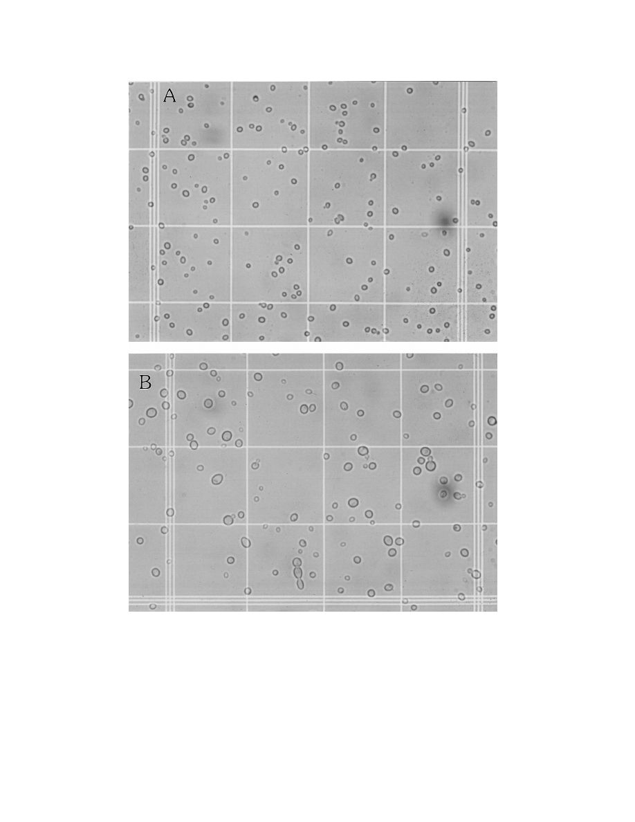
above 37°C. Figure 1 shows wet-mount preparations of C.
glabrata and C. albicans at similar magnifications. It is clear
that C. glabrata blastoconidia (1 to 4
mm) are considerably
smaller than C. albicans blastoconidia (4 to 6
mm). On Sab-
ouraud dextrose agar, C. glabrata forms glistening, smooth,
cream-colored colonies which are relatively indistinguishable
from those of other Candida species except for their relative
size, which is quite small. On Chromagar, a relatively new agar
that distinguishes different Candida species by color as a result
FIG. 1. Size differential of C. glabrata and C. albicans. Shown are wet-mount slide preparations of C. glabrata (A) and C. albicans (B) on a hemocytometer.
Magnification,
3400.
V
OL
. 12, 1999
CANDIDA GLABRATA INFECTIONS
81
of biochemical reactions, C. glabrata colonies appear pink to
purple, in contrast to C. albicans colonies, which appear green
to blue-green. A critical distinguishing characteristic of C. gla-
brata is its haploid genome, in contrast to the diploid genome
of C. albicans and several other non-albicans Candida species
(176). Finally, C. glabrata is distinguishable from C. albicans by
its small-subunit rRNA (4).
Most medically important Candida species can be easily
differentiated from one another by either established commer-
cially available biochemical tests or molecular biology tech-
niques. With the advent of molecular genetics, newer identifi-
cation methods have emerged. These methods use
comparative analysis of chromosomal DNA to identify Can-
dida species from each other and also to delineate different
strains within a species. These newer methods include restric-
tion fragment length polymorphisms, pulsed-field gel electro-
phoresis, randomly amplified polymorphic DNA, and DNA
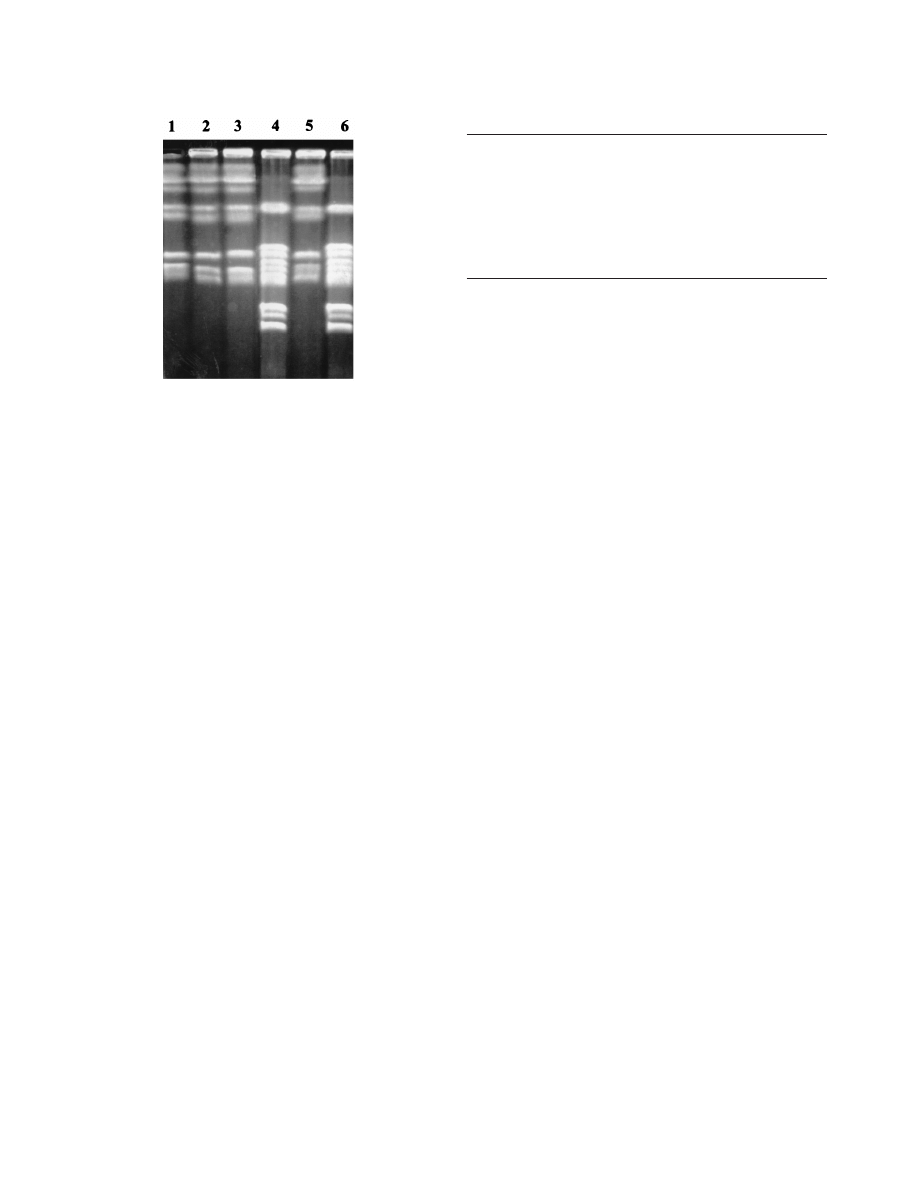
probes (77, 79, 95, 170). By using contour-clamped homoge-
neous electric field gel electrophoresis (CHEF), a form of
pulsed-field gel electrophoresis, chromosomal DNA from C.
glabrata can be separated based on the different chromosomal
molecular weights and thus can be subjected to electrophoretic
karyotyping (EK). The EK pattern of C. glabrata generally
produces 10 to 13 bands (79, 170). Depending on the EK
patterns, C. glabrata can be classified into several different
strain types. To date, 28 strain types have been formally de-
scribed (170), although more than 70 different strains have
been identified (168). In contrast, CHEF usually separates C.
albicans chromosomal DNA into eight chromosomal bands,
with more than 90 different strain types identified to date
(168). Figure 2 shows the CHEF-derived DNA-banding pat-
terns characteristic of C. glabrata and C. albicans.
The biochemical reactions of C. glabrata are also quite dis-
tinct. In contrast to C. albicans, which ferments and/or assim-
ilates a number of sugars, C. glabrata ferments and assimilates
only glucose and trehalose (91). In fact, this repertoire of sugar
utilization is unique compared to the majority of Candida
species and is used by several commercially available kits (API
20C, Uni-Yeast-Tek, and YeastIdent) to identify yeast to the
level of genus and species.
Historically, C. glabrata was classified in the genus Torulopsis
(91). The genus Torulopsis was described in 1894, while the
genus Candida was not named until 1913. C. glabrata was
originally placed in the genus Torulopsis due to its lack of
pseudohypha production. However, in 1978, it was determined
that the ability to produce pseudohyphae was not a reliable
distinguishing factor for members of the genus Candida and it
was proposed that T. glabrata be classified in the genus Can-
dida (91). The incorporation of T. glabrata into the genus
Candida required that the description relative to pseudohy-
phae for the genus Candida be changed from “pseudomycelial”
to “pseudohyphae: absent, rudimentary, or well developed”
(91). This change in nomenclature has taken considerable time
to gain acceptance by the medical mycology community, and
several publications still refer to C. glabrata as T. glabrata.
Wherever possible, efforts should be made to use the contem-
porary nomenclature.
EPIDEMIOLOGY
Candida species are ubiquitous organisms (115). An increas-
ing incidence of fungal infections with Candida species has
been noted in immunocompromised patients such as intensive-
care, postsurgical, and neutropenic patients (7, 11, 14, 67, 90,
175). Candida species are most frequently isolated from the
oral cavity and are detected in approximately 31 to 55% of
healthy individuals (115). Colonization rates increase with se-
verity of illness and duration of hospitalization (115, 170, 175).
Historically, C. albicans accounted for 70 to 80% of the isolates
recovered from infected patients. C. glabrata and C. tropicalis
each accounted for approximately 5 to 8% of isolates, while
other non-albicans Candida species occur only rarely (3, 7).
However, more recent epidemiological data reveal a mycolog-
ical shift from C. albicans to the non-albicans Candida species
such as C. glabrata, C. tropicalis, C. parapsilosis, and C. krusei
(7, 90, 107, 180, 183, 184).
The changing patterns and the increasing incidence of dis-
seminated Candida infection are also evident in a large autopsy
series (11). The high mortality rate associated with bacterial
infections has declined with the early administration of empir-
ical antibiotics, while systemic fungal infections have become
increasingly important in causing morbidity and mortality in
immunocompromised patients. Candida is now the fourth most
common organism recovered from blood cultures in hospital-
ized patients (7). C. glabrata has recently emerged as an im-
portant nosocomial pathogen, yet little is known about its
epidemiology. Although C. albicans is the most common fungal
species isolated from blood, C. glabrata currently ranks fourth
among Candida species (third in patients who have undergone
surgery) and is associated with an equally high mortality rate
(51, 90, 181, 184). C. glabrata is of special importance because
of its innately increased resistance to antifungal agents, specif-
ically the azoles (49, 61, 174, 181, 184). The current epidemi-
ological data for C. glabrata is summarized in Table 1.
A clear understanding of the epidemiology of Candida in-
fection and colonization has been difficult because of a lack of
FIG. 2. CHEF of genomic DNA from representative isolates of C. albicans
and C. glabrata. Lanes 1 to 3 and 5 are similar strains of C. albicans; lanes 4 and
6 are strains of C. glabrata.
TABLE 1. Epidemiology of C. glabrata infection
Predominantly nosocomial (except vaginal)
Immunocompromised or debilitated host
Specific risk factors:
Prolonged hospitalization
Prior antibiotic use
Use of fluconazole
General use in hospital
Patient exposure
Hand carriage by hospital personnel
Often mixed fungal infection
82
FIDEL ET AL.
C
LIN
. M
ICROBIOL
. R
EV
.

reliable typing systems to evaluate strain homology. Previous
typing systems have relied on phenotypic differences within a
Candida species, which may not reflect true strain differences
(26, 71, 106). However, recent advances in the use of molecular
techniques have enabled investigators to develop a typing sys-
tem with greater sensitivity (26, 34, 70, 71, 106, 169, 172).
Molecular typing of Candida by DNA fingerprinting involving
various molecular techniques (restriction fragment length
polymorphism, CHEF, and randomly amplified polymorphic
DNA), has the capability to differentiate closely related strains
which may have phenotypic similarities (26, 70, 79, 161, 169,
172).
Based upon epidemiological studies, it is apparent that hu-
mans are exposed repeatedly to Candida in food and other
sources. However, the natural history of this commensal “nor-
mal” colonization over weeks, months, and years is poorly
understood. Nevertheless, one may reasonably conclude that
Candida colonization is almost universal. A feature common to
colonized individuals is that the most frequent species are still
C. albicans, and so far no unique strains of C. albicans or any
non-albicans Candida species with specific gastrointestinal
tract tropism have been identified. DNA typing of Candida
strains obtained from AIDS patients with oral and esophageal
candidiasis indicate an identical distribution frequency to those
of isolates present in healthy subjects (12). This suggests that
AIDS-associated candidiasis is not caused by unique or partic-
ularly virulent strains but probably results from defects in host
defense mechanisms.
Until recently, most reports describing the epidemiology of
nosocomial C. glabrata have been retrospective, and few stud-
ies have evaluated independent risk factors associated with
nosocomial C. glabrata acquisition and subsequent infection.
Knowledge of the epidemiology of fungal nosocomial coloni-
zation and infection with C. glabrata is, however, essential for
the prevention of further spread as well as of nosocomial
infection. In a recent study by Vazquez and colleagues (170),
multivariate prospective case-control analysis along with mo-
lecular analysis of C. glabrata demonstrated that patients with
new acquisition of C. glabrata had a longer duration of hospi-
talization (18.8 and 7.6 days, respectively; P
, 0.001) and more
frequent prior antimicrobial use (100 and 65%, respectively;
P
, 0.001) compared to patients from whom Candida species
were not recovered during the study. These results are similar
to the findings noted in earlier epidemiological studies of C.
albicans, C. lusitaniae, and C. parapsilosis (138, 139, 172). Little
is known about the hospital reservoirs of C. glabrata, but, as
with C. albicans, probable sources include a complex interac-
tion of environmental and human reservoirs (72, 172). The
unique role of the hospital environment as a potential reservoir
for Candida species is further suggested by findings in a recent
study in which identical strains of C. glabrata were isolated
from the environment before being newly acquired by patients
admitted into a Bone Marrow Transplant Unit (170). Fungal
organisms isolated from the inanimate hospital environment
were previously considered to contribute little to nosocomial
fungal infection. Although infecting strains can be cultured
from environmental surfaces, it is believed that the environ-
ment becomes passively contaminated by organisms from pa-
tients (170, 172). Two studies have implicated carriage on the
hands of hospital personnel as a possible source of an outbreak
(75, 172). Thus, C. glabrata may be similar to C. albicans and
other nosocomial pathogens that are acquired directly or indi-
rectly from contaminated environmental surfaces. Previous un-
derstanding of the pathogenesis of C. glabrata colonization and
infection assumed that the organisms responsible for disease
were endogenously acquired exclusively from the patients’ own
flora.
The role of carriage by personnel in dissemination of C.
glabrata remains to be clarified. Although C. glabrata is not
frequently recovered from the hands of hospital personnel,
transient carriage is suggested by its isolation on environmen-
tal surfaces in contact with hands (170). Perhaps more frequent
culturing of the hands of personnel or the use of liquid media
to recover yeasts may have improved the detection rates of C.
glabrata. Proximity to a patient with infection or colonization
increases the risk of nosocomial acquisition (170). As in earlier
studies (124, 172), the results of longitudinal cultures showed
that 75% of patients generally carried the same strain type of
C. glabrata over time (170), with minimal strain diversity
among individual patients. This finding is significantly different
from the results described for the nosocomial acquisition of C.
albicans, in which there was considerable strain diversity (172).
Moreover, in this study, 71% of patients with positive C. gla-
brata cultures had more than one Candida species isolated.
The most frequent combination was C. glabrata and C. albi-
cans, which was found in approximately 70% of the patients.
This again is in contrast to the findings previously described for
C. albicans, which showed that only 39% of patients with C.
albicans had more than one Candida species identified (175).
Finally, unlike C. albicans, C. glabrata has not been recovered
from the food provided to hospitalized patients, potentially
contributing to the lack of identifiable C. glabrata strain diver-
sity.
In conclusion, these studies suggest that nosocomial acqui-
sition of C. glabrata is not uncommon and may be due to
exogenous acquisition. In addition, two major risk factors as-
sociated with C. glabrata colonization are prolonged duration
of hospitalization and prior antimicrobial use. Further pro-
spective studies are sorely needed to define more clearly the
reservoirs of infection, as well as the mode of transfer and
measures for preventing the spread of infection.
PATHOGENESIS
In this section, although very little has been studied, we
discuss what is currently known about virulence factors of C.
glabrata, host defense against this organism, and established
experimental animal models of C. glabrata infections.
Virulence
The relatively nonpathogenic nature of C. glabrata in animal
models (24, 41, 145) suggests that it has few virulence at-
tributes. However, the high mortality rate and the rapidity of
the spread of disease would argue to the contrary. The fact is
that few studies have been conducted on virulence of C. gla-
brata. In contrast, C. albicans has several known virulence
factors contributing to its pathogenicity that include adherence
to epithelial and endothelial cells, proteinase production (17,
135), hypha and pseudohypha formation (114, 154), pheno-
typic switching (156), phospholipase production (5, 73), and
antigenic modulation as a result of pseudohypha formation
(25). If C. glabrata is low in virulence, the lack of hypha for-
mation may be a contributing factor. Indeed, hypha formation
is a recognized means of increased adherence and tissue inva-
sion by C. albicans as well as a means of increasing proteolytic
enzyme elaboration and antigen modulation (114).
Proteinase production by C. albicans is associated with
pathogenicity (17, 135). For example, virulent C. albicans iso-
lates often produce aspartyl proteinase. These isolates are
more pathogenic in a variety of animal models of experimental
V
OL
. 12, 1999
CANDIDA GLABRATA INFECTIONS
83

Candida infections (17, 23). Although little is known of pro-
teinase production by C. glabrata, a single study has shown that
isolates of C. glabrata are at least capable of proteinase pro-
duction, but the type of proteinase was not specified (19).
Adherence is an extremely important virulence factor, al-
though the actual adherence property may be compounded by
other virulence properties. For example, cell surface hydro-
phobicity (CSH), which is affected by environmental factors,
can affect specific adherence based upon interaction of adhesin
receptors. In a study with limited numbers of C. glabrata iso-
lates tested, C. glabrata was shown to have comparable CSH to
C. albicans (85). Interestingly, however, while the CSH of C.
albicans was extremely sensitive to specific growth conditions,
numerous isolates of C. glabrata were relatively insensitive to
those same growth conditions (60), suggesting that C. glabrata
is not as sensitive or as influenced by environmental factors. In
comparative in vitro assays of adherence to vascular endothe-
lium, while C. albicans was by far the most adherent species, C.
glabrata was the least adherent, alone with C. parapsilosis and
C. kefyr, behind C. tropicalis and C. krusei (84). Moreover,
while C. albicans is recognized avidly by monoclonal antibodies
to
b
2
integrins (adhesin receptors), binding to C. glabrata by
the same antibodies was undetectable, as was binding to C.
parapsilosis and C. krusei. These results suggest that C. glabrata
may not express these specific adhesins and thus would have a
disadvantage in adherence (8). The presence of fibronectin,
and laminin receptors, fibrinogen-binding proteins, and man-
noprotein adhesins are also considered important means of
adhesion to endothelial and/or epithelial cells (reviewed in
reference 69). While extensive work has been performed on
surface ligands of C. albicans, nothing is known about these
receptors and proteins on C. glabrata. It will be important to
reexamine many of the parameters from earlier studies, to-
gether with a number of new parameters, by using current
clinical C. glabrata isolates obtained from patients with fulmi-
nant candidiasis.
Extracellular membrane-damaging phopholipases are con-
sidered virulence factors for C. albicans (5, 73). Although these
enzymes have not been studied extensively, phospholipase A
and B and lysophospholipase-transacylase are produced by
virulent but not avirulent (commensal) strains of C. albicans.
These phospholipase-producing strains also adhered most
strongly to epithelial cells. Furthermore, the production of
these phospholipases by clinical isolates correlated with patho-
genicity and was predictive of mortality in animal models (5,
73). Phospholipase activity has not been studied in C. glabrata.
Another virulence factor of C. albicans is specific phenotypic
instability, which allows strains to switch colony phenotype
without affecting the identifiable genotype; this is termed “phe-
notypic switching” (155, 156). Although phenotypic switching
was studied largely as an in vitro phenomenon, there is some
evidence of in vivo phenotype switching and an association of
switched phenotypes with virulence. Switching of phenotypes
in clinical C. albicans isolates from women with recurrent C.
albicans vaginitis has been reported (158). Recently, it was
determined that phenotype switching does occur in C. glabrata
(157). It is interesting that such a phenomenon would occur in
nondimorphic organisms as well as in haploid organisms. Al-
though the relationship of this C. glabrata phenotype switching
to virulence is unknown, it may enhance virulence and play a
role in causing symptomatic infection.
Host Defense
Little is known about host defense against C. glabrata. In
contrast, considerable work has been described on host de-
fenses against C. albicans. As a result, we now have a fairly
comprehensive understanding of the dominant host defense
and protective mechanisms against invasive C. albicans infec-
tion, both superficial and systemic, but we know little about C.
glabrata infection. With respect to defense against systemic C.
albicans infections, clinical observations and experimental
studies suggest that polymorphonuclear leukocytes are the pre-
dominant cell type that protects against candidemia and sys-
temic candidiasis (32, 35, 66, 114). Clinically, this is supported
by the fact that neutropenic patients are particularly suscepti-
ble to systemic C. albicans infections. In addition, it has been
shown in an animal model that T cells may be of some signif-
icance against systemic C. albicans infections. Specifically,
studies in mice have shown that a Th1-type response charac-
terized by the cytokines interleukin-2 (IL-2), gamma inter-
feron, and IL-12 is associated with protection against systemic
infection whereas Th2-type responses characterized by the cy-
tokines IL-4, IL-5, and IL-10 and antibody production (immu-
noglobulin A [IgA] and IgE) is associated with susceptibility to
systemic infection (134). T cells and cell-mediated immunity
(CMI), on the other hand, form the predominant host defense
mechanism against mucosal C. albicans infection. This comes
from both clinical observations (a high incidence of mucosal
candidiasis in patients with reduced CMI) and clinical and
experimental studies showing the critical role of T cells in
protection against C. albicans mucosal infections (i.e., chronic
mucocutaneous candidiasis and gastrointestinal candidiasis)
(10, 15, 81, 82, 114). Historically, vaginal infections were in-
cluded in the mucosal infections affected by T-cell host defense
mechanisms. However, recent studies suggest that if T cells are
indeed important, it is the local rather than the systemic T-cell
response that is protective against vaginal C. albicans infection.
This conclusion is based in part on studies in an experimental
animal model of vaginitis as well as on clinical studies in
women with recurrent vulvovaginal candidiasis (40, 43–46). In
addition, although controversy abounds, properly controlled
clinical studies suggest that Candida vaginitis is not more com-
mon in human immunodeficiency virus (HIV)-infected women
and, if observed, does not correlate with decreased CD4 cell
counts (20, 74, 131, 177). Recent studies suggest that innate
resistance may also be critical for protection against vaginal C.
albicans infections (160). Although antibodies are readily in-
duced from exposure to C. albicans, it remains unclear if they
play a protective role against C. albicans infections. Although
several authors have concluded that they are nonprotective
(101, 133), there are reports showing that specific antibodies
protect against experimental systemic or vaginal C. albicans
infections (58, 102, 103). Clinical experience, however, shows
that individuals with B-cell deficiencies do not have increased
susceptibility to C. albicans infection (133).
Since C. glabrata is a commensal organism similar to C.
albicans, there are likely to be normal host mechanisms that
effectively control C. glabrata, holding it in check and suppress-
ing the expression of its pathogenic properties, thereby pre-
venting infection. However, the relatively low pathogenicity of
C. glabrata compared to C. albicans in animal models (re-
viewed below) suggests that control of C. glabrata may not
require mechanisms that are as stringent as that required to
hold C. albicans in check. Nevertheless, the increased preva-
lence of C. glabrata infections in immunocompromised indi-
viduals indicates that some level of host defense does indeed
exist. The interaction of Candida species with endothelial and
epithelial cells has recently taken an immunological twist in
addition to a simple adherence phenomenon. We recently
showed that epithelial cells inhibit the growth of C. albicans in
vitro (160), and Filler et al. have shown that endothelial cells
84
FIDEL ET AL.
C
LIN
. M
ICROBIOL
. R
EV
.

phagocytize C. albicans (47). Unfortunately, C. glabrata did not
induce endothelial-cell phagocytosis (47), suggesting that this
endothelial-cell activity may be species specific or restricted to
C. albicans alone. However, it remains possible that both con-
ventional and unconventional immune cells play some role in
innate and/or acquired host defense against C. glabrata infec-
tion.
There has been only one formal clinical study that examined
host defenses in patients with C. glabrata infections (105). In
this German study, humoral and innate cellular defenses were
examined in women with either C. glabrata or C. albicans
vaginitis. A total of 14 women with C. glabrata vaginitis and 20
and 42 women with acute or chronic C. albicans vaginitis,
respectively, were tested. The responses were compared to
those in 77 control women. For each woman, secretory IgA
(sIgA), IgA, and numbers of granulocytes and macrophages in
vaginal secretions and IgA in blood were tested. For each
parameter, few differences were detected with respect to the
controls. In fact, the only difference in the entire study was in
women with C. glabrata vaginitis, who showed a slight, but
significantly lower level of sIgA in vaginal secretions (105).
However, it is unclear what proportion of the IgA measured
was C. glabrata or Candida specific. Also noted in the women
with C. glabrata vaginitis was a lack of inflammation compared
to those with C. albicans vaginitis. While no clear pattern of
local or systemic innate or humoral immune deficiency was
observed in women with C. glabrata vaginitis and although
local or systemic T-cell function in response to C. glabrata was
not tested, it would appear that identification of immunologi-
cal deficiencies and dysfunctions in C. glabrata-infected women
may prove to be as difficult as it has been for those with C.
albicans vaginitis (42, 44, 48, 105).
In the absence of other formal studies, there have been
clinical observations that provide some indication of what may
be important for host defense against mucosal or systemic C.
glabrata infections. The incidence of C. glabrata mucosal or
systemic infections in cancer patients (182), transplant recipi-
ents (184), and AIDS patients (37, 140, 179), in whom T-cell
function is impaired, suggests that T cells may be important for
protection of at least some tissues against C. glabrata infection.
Additionally, histological examination of tissues infected with
C. glabrata has shown relatively mild infiltrates of lymphocytes,
macrophages, and neutrophils (61) compared to that observed
in C. albicans infection. In contrast, there are no known reports
of increased C. glabrata infections in patients with B-cell defi-
ciencies, again suggesting that antibodies are not critical to
protection against C. glabrata infections.
In studies comparing antigens of C. glabrata to those found
in other Candida species, specific antigens appear to be com-
mon across several Candida species (13, 109). Certain antibod-
ies produced against C. albicans recognize C. glabrata as well as
other Candida species. Specifically, antibodies reacting with
antigen 6 of C. albicans serotype A react with C. glabrata as
well, suggesting that antigen 6 is conserved between the two
species (109). Additionally, Cutler and coworkers (13) have
reported an antibody produced against C. glabrata that also
cross-reacts with other Candida species. These results suggest
that protective immunity against Candida species, specifically
C. albicans, may be capable of providing a level of protection
against C. glabrata infections as well. This could potentially
include any form of innate resistance (polymorphonuclear leu-
kocytes, macrophages, and natural killer cells) or acquired
CMI (T cells) in addition to humoral responses (B cells and
antibodies).
Our laboratory has performed a limited number of experi-
ments involving immune system reactivity to C. glabrata. In a
limited number of tests performed with human peripheral
blood lymphocytes, we recently found that human peripheral
blood lymphocytes respond in vitro to heat-killed C. glabrata in
a manner similar (approximately 80 to 85% in magnitude) to
that observed for C. albicans (38). Thus, normal healthy adults
appear to be sensitized to C. glabrata with demonstrable cell-
mediated responsiveness, although we recognize that such re-
sponses may be the result of cross-reactive antigens on C.
glabrata recognized by C. albicans-specific cells. In an animal
model, we found that nonobese diabetic (NOD) mice infected
vaginally with C. glabrata did not respond by developing de-
layed-type hypersensitivity to C. albicans culture filtrate anti-
gen (38) whereas mice used in the experimental C. albicans
vaginitis model (CBA/J mice) readily respond to C. albicans
culture filtrate antigen by developing delayed-type hypersensi-
tivity (39). This data suggests that a vaginal C. glabrata infec-
tion does not induce a systemic CMI response that is cross-
reactive or responsive to C. albicans antigen. However, it is not
known whether this is due to the lack of cross-reactivity be-
tween C. glabrata and C. albicans, the lack of induction of C.
glabrata-specific CMI, or the inability of NOD mice to mount
an effective T-cell response. There have been inconsistent re-
sults with the NOD mice regarding in vitro T-cell reactivity. In
one study, draining lymph node cells from NOD mice infected
vaginally with C. glabrata responded to both heat-killed C.
glabrata and heat-killed C. albicans as detected by lymphocyte
proliferation, whereas in another study, the lymph node cells
did not respond to either particulate antigen (38). Although
additional studies should be performed, if indeed C. glabrata-
infected mice do generate Candida-specific T-cell responses in
the draining lymph nodes, there appears to be some level of
cross-reactivity between the responses to C. glabrata and C.
albicans. However, the critical experiments involving the lymph
node responses to C. glabrata in C. albicans-infected mice have
not been performed. The predominant response of draining
lymph node cells in such infected mice to C. albicans antigen is
a Th1-type response characterized by the production of IL-2
and gamma interferon (110). Finally, understanding the im-
portant host defenses against C. glabrata will require controlled
studies conducted in animal models of systemic and mucosal C.
glabrata infections.
Animal Models
Historically, there has been little interest in developing an-
imal models of C. glabrata infection. Even now, despite the
emergence of both systemic and mucosal C. glabrata infections,
there are still only a few established animals models. The
relative lack of pathogenicity of C. glabrata may have ham-
pered the development of such models, and it continues to do
so. Currently, there are two established murine models of C.
glabrata infections, systemic and vaginal (24, 41). For each
model, steps have had to be taken to either manipulate the
mice or identify a strain of mouse particularly susceptible to
infection. In the systemic model, with several clinical isolates of
C. glabrata, mice had to immunosuppressed with 5-fluorouracil
(150 mg/kg) intravenously or subjected to gamma irradiation
with 450 to 550 rads to achieve a sustained infection for 7 days
(24). The smallest inoculum required to achieve an infection in
these mice was 10
8
blastoconidia. This is approximately 3 to 4
log units higher than that which is lethal for immunocompetent
mice inoculated systemically with C. albicans. In infected mice,
a C. glabrata organ burden was detectable in the kidneys and
spleen 7 days after inoculation. Since the focus of the study was
to test various antimycotic treatment regimens during the
course of a vigorous infection, a kinetic study of the organ
V
OL
. 12, 1999
CANDIDA GLABRATA INFECTIONS
85
fungal burden was not performed although the authors stated
that lethality was not observed. Thus, survival was obviously
not a parameter for consideration in the studies. In any event,
the kidney and spleen fungal burden was quite high in many
animals (10
4
to 10
8
CFU/organ), although the range of CFU
per organ within a group of animals was large. Thus, the organ
burden in C. glabrata-infected mice was comparable to that
detected in C. albicans-infected mice (43); however, one
should recall that the C. glabrata-infected mice were immuno-
suppressed. Moreover, it is notable that experimental C. gla-
brata infections are generally not lethal in animals. From this,
one can appreciate the differences in relative pathogenicity
between C. albicans and C. glabrata. While lack of lethality in
experimental studies does not match the high mortality often
seen in clinical cases of C. glabrata infection, one must recog-
nize that the clinical experience is a reflection of the advanced
state of debilitation of patients who become infected with C.
glabrata. Clearly, more studies of the kinetics of the model
must be performed to better understand the progression of
infection. Although a section in this review is devoted to treat-
ment of C. glabrata infections, the results of this systemic-
infection model are consistent with clinical experience, in that
amphotericin was most efficacious while fluconazole was gen-
erally ineffective. Moreover, a lack of correlation between in
vitro susceptibility tests and in vivo efficacy was often evident
(24, 41).
A recent report describing an increase in C. glabrata vaginal
infections (151) emphasized the need to develop a vaginal
model of C. glabrata infection. In particular, models of C.
glabrata mucosal infections had been difficult to establish. In
one report, an oral C. glabrata infection in rats could not be
achieved (145). Our laboratory attempted to develop an ex-
perimental model of vaginal C. glabrata infection to comple-
ment our model of vaginal C. albicans infection (39). This also
proved difficult. Preliminary experiments with the mouse strain
used for C. albicans vaginal infection (immunocompetent
CBA/J mice) showed no detectable C. glabrata vaginal burden
as early as 6 days following an intravaginal inoculum in spite of
using multiple clinical C. glabrata isolates and pseudoestrus
conditions (required to achieve a vaginal C. albicans infection)
(41). Similarly, a low detectable vaginal fungal burden was
observed in DBA/2 mice, which are highly susceptible to sys-
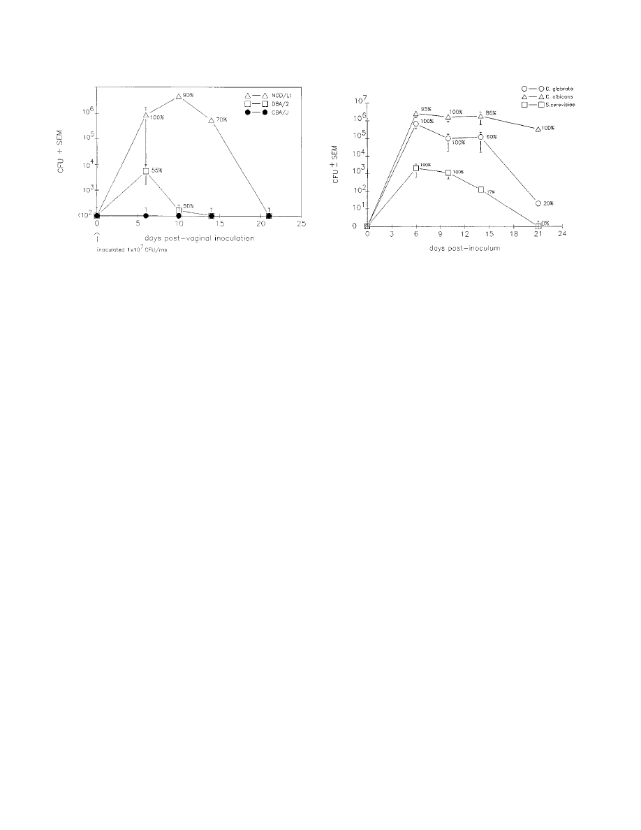
temic C. albicans infection (99). In contrast, nonobese diabetic
(NOD/Lt) mice were susceptible to C. glabrata vaginal infec-
tion (Fig. 3) (41). In comparison to C. albicans infections,
although a higher inoculum of C. glabrata was routinely used
(1
3 10
7
blastoconidia) than that of C. albicans (5
3 10
5
blastoconidia), inocula as low as 5
3 10
5
blastoconidia were
capable of establishing C. glabrata infections. The infection was
sustained for 14 days at high titers and became resolved in
most animals by 21 days. The vaginal titers of C. glabrata at 6
to 14 days postinoculum (
.10
6
CFU) were higher than those
commonly observed in C. albicans-infected mice (10
4
to 10
5
CFU) (39) and persist in pseudoestrus-treated mice for 8
weeks or more (39). We next examined how NOD mice could
support vaginal infections caused by other fungal species,
namely, C. albicans (highly virulent) and Saccharomyces cerevi-
siae (low virulence). Intravaginal inoculation with C. albicans
resulted in extremely high titers of C. albicans (
.10
6
CFU) and
a surprising 20% mortality rate, although no dissemination of
the organism could be detected (kidney dysfunction was sus-
pected as the cause of death). Animals inoculated with S.
cerevisiae had low but detectable titers of vaginal fungal burden
(
,10
3
CFU) early postinoculum (days 6 to 10), with the ma-
jority of animals resolving the infection by 14 days (Fig. 4).
Another interesting feature of the C. glabrata vaginal infection
in NOD mice was the relative lack of a requirement for
pseudoestrus to acquire a sustained vaginal infection with ei-
ther C. glabrata or C. albicans. Although the rates of infection
were generally greater in pseudoestrus-treated mice, the vag-
inal fungal burdens were comparable in pseudoestrus-treated
or and nontreated mice. This observation is in keeping with a
clinical observation of C. glabrata being frequent in postmeno-
pausal women developing Candida vaginitis (150).
Since it is difficult in animal models of vaginitis to determine
whether a state of colonization or infectivity is achieved in the
absence of measurable signs and symptoms of inflammation
(and more difficult for the non-hypha-producing C. glabrata),
there is nevertheless considerable evidence that the C. gla-
brata-inoculated animals were indeed infected. First, NOD
mice had high titers of vaginal fungal burden whereas other
murine strains did not. Second, there was a lymphoid cell-like
FIG. 3. Experimental C. glabrata infections in mice with intermediate
(CBA/J) and high (DBA/2) susceptibilities to C. albicans systemic infection and
in NOD/Lt mice. Data points represent mean CFU
6 standard errors of the
mean (SEM) in animals with positive cultures only (the percentage of animals
with positive cultures is shown). Reprinted from reference 41 with permission of
the publisher.
FIG. 4. Comparative analysis of C. glabrata, C. albicans, and S. cerevisiae
vaginal fungal burden in NOD mice. Data points represent mean CFU
6 SEM
for animals with positive cultures (the percentage of animals with positive cul-
tures is shown) following intravaginal inoculation with 1
3 10
7
blastoconidia of
C. glabrata or S. cerevisiae or 5
3 10
5
blastoconidia of C. albicans. Reprinted from
reference 41 with permission of the publisher.
86
FIDEL ET AL.
C
LIN
. M
ICROBIOL
. R
EV
.
cellular infiltrate in the lavage fluid of C. glabrata-infected
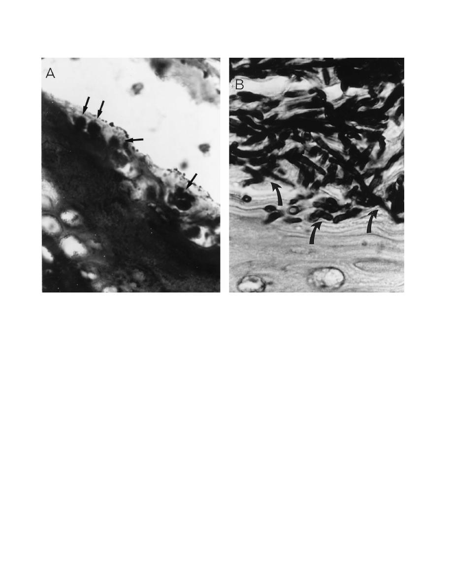
similar to that observed in C. albicans-infected mice. Third,
histopathologic sections of vaginal tissue showed the presence
of C. glabrata blastospores in epithelial vacuole-like vesicles
and not simply lying at the epithelium (Fig. 5). Figure 5 also
shows how C. albicans presents as predominantly hyphae su-
perficially associated with the epithelium during an infection.
Thus, these results show that NOD mice will be useful in
studying the pathogenesis and host response during vaginal C.
glabrata infections, as well as in developing strategies to treat
the infection pharmacologically.
The use of NOD mice as a diabetic model was an interesting
caveat to these studies. The model was originally conceived
based on the high susceptibility of women with diabetes mel-
litus to C. glabrata and C. albicans vaginitis (53, 125, 149, 151).
However, the NOD mice that were susceptible to the vaginal
C. glabrata infection were not yet hyperglycemic. In fact, these
mice do not achieve hyperglycemia until at least 12 weeks of
age (80). The animals used in the named study were 7 to 10
weeks of age, and in our hands the NOD mice did not become
hyperglycemic until 22 weeks of age. This susceptibility of
NOD mice to C. glabrata vaginal infections before the onset of
hyperglycemia prompted us to test the congenic insulitis-resis-
tant strain of mice (NOR/Lt) for susceptibility to vaginal C.
glabrata infection. Interestingly, the NOR mice were resistant
to the vaginal infection (41). These results suggested that NOD
mice may be susceptible to C. glabrata vaginal infection, not by
virtue of a state of hyperglycemia but simply by their genetic
susceptibility to diabetes mellitus. On the other hand, CBA/J
mice made diabetic by exogenous treatment with streptozocin
became susceptible to C. glabrata vaginal infection and NOD
mice similarly treated had higher rates of infectivity (38). Cer-
tainly, more studies are required to better understand the
factors that contribute to the susceptibility to C. glabrata vag-
inal infection. However, this experimental model of C. glabrata
vaginitis provides an opportunity to study the pathogenesis of
C. glabrata vaginal infections, as well as to dissect the genetic
issues related to susceptibility to infection.
CLINICAL SPECTRUM OF INFECTION
Superficial Infections
Symptomatic mucosal candidiasis arises in subjects who are
colonized with Candida and who are predisposed by illness or
have a dysfunction or local reduction in host resistance,
thereby promoting an overgrowth of their own indigenous
yeast flora. The most common mucosal infections include oro-
pharyngeal, esophageal, and vaginal candidiasis. Although C.
albicans remains the species responsible for the overwhelming
majority of infections in HIV-positive and negative patients
(115, 152, 162, 164), there are an increasing number of case
reports describing the recovery of C. glabrata from the mucosal
surfaces of immune compromised patients. The actual rate of
symptomatic oropharyngeal candidiasis (OPC) due to C. gla-
brata is difficult to determine since this species is rarely isolated
alone and is often coisolated with C. albicans. In antifungal
treatment trials involving HIV-positive patients, the recovery
FIG. 5. Histopathology of vaginal tissue from estrogen-treated NOD mice inoculated with C. glabrata (A) and C. albicans (B). Arrows represent blastoconidia or
hyphae. Magnification,
3100. Reprinted from reference 41 with permission of the publisher.
V
OL
. 12, 1999
CANDIDA GLABRATA INFECTIONS
87

of non-albicans Candida species is generally less than 10% of
all isolates recovered, with C. glabrata making up less than 5%
(55, 122, 173). However, subjects in these treatment studies are
often selected, while patients with advanced disease, who are
likely to be infected with resistant strains, are excluded, result-
ing in an underestimation of the frequency of C. glabrata in-
fection. Moreover, in several of the antifungal treatment trials
for fluconazole-refractory OPC in AIDS patients, the inci-
dence of C. glabrata producing OPC was less than 10% (16,
121). In the HIV-seronegative population, the occurrence of
OPC and esophageal candidiasis due to C. glabrata is rare.
Data are still incomplete, because only a few small studies have
attempted to investigate the incidence of non-albicans Candida
species as a cause of OPC and esophageal candidiasis (49,
140). At present, it is unclear why the incidence of mucosal
candidiasis due to C. glabrata is so low. Perhaps studies eval-
uating the virulence factors of C. glabrata involved in the at-
tachment and colonization of mucosal surfaces would shed
some light on this important issue.
There continues to be considerable controversy about
whether C. glabrata, as part of a mixed fungal culture with
coexistent C. albicans, actually contributes to the development
of OPC. Many investigators consider that C. glabrata functions
as an innocent bystander only and that therapy should be based
upon susceptibility of the coexistent C. albicans (140). While C.
albicans is undoubtedly the more virulent, frequent, and dom-
inant pathogen, C. glabrata is occasionally found as the single
and only clinical species isolated in AIDS patients with OPC.
Accordingly, while directing therapy against C. albicans in
mixed infections, especially those not responding to appropri-
ate therapy, it is prudent not to ignore C. glabrata in mixed
infections.
Oropharyngeal. (i) Clinical manifestations.
Several clinical
forms of OPC exist; the most common and widely recognized
is acute pseudomembranous candidiasis, commonly referred to
as thrush. OPC can also occur in an erythematous form that is
often asymptomatic. OPC is often the first manifestation of
HIV infection (21, 56, 147), with approximately 80 to 90% of
patients with AIDS ultimately developing OPC at some stage
during their disease progression (28).
(ii) Management.
Numerous antifungal agents are available
for the treatment of OPC, esophageal and vaginal candidiasis
(Table 2). Since the comparative efficacy of the antifungal
agents has not been established in infections due to C. glabrata,
the choice, dosage, and duration of treatment have not been
well established in patients and remain somewhat controver-
sial. Antimycotic efficacy and response time are inferior in the
HIV-positive population to those in cancer patients. To date,
most of the clinical trials contained few patients with OPC
caused by C. glabrata alone. Thus, the efficacy of these anti-
fungal agents for OPC due to C. glabrata is largely unknown.
Azoles have replaced the topical polyene agents for the treat-
ment of oral candidiasis in most circumstances. Accordingly,
azole therapy for OPC due to C. glabrata is also extrapolated
from the data accumulated from the numerous studies per-
formed on OPC due primarily to C. albicans (Table 2).
The newer triazoles, itraconazole and fluconazole, which
have markedly improved efficacy and safety profiles, have be-
come extremely popular, especially for HIV and AIDS patients
with severe OPC (29, 59, 87, 108, 111). Fluconazole (50 to 100
mg daily) has been studied in several open, placebo-controlled
and double-blinded comparative studies versus clotrimazole or
ketoconazole (59, 87, 108). Studies indicate that while clinical
recovery is achievable in most patients treated, mycological
cure is more difficult to attain. Additionally, most of the iso-
lates recovered from study patients were C. albicans, with only
a few isolates being identified as C. glabrata.
Itraconazole is a newer triazole antifungal with a broad
spectrum of activity. Like the other azoles, it has a similar
mechanism of action, acting by inhibiting the synthesis of fun-
gal ergosterol. However, unlike fluconazole, it has in vitro
activity against many of the non-albicans Candida species, spe-
cifically C. glabrata and C. krusei. In a recently completed
prospective randomized trial involving HIV-positive or AIDS
patients with OPC, itraconazole solution (200 mg/day) was
compared to fluconazole (100 mg/day), both given for 14 days.
The results revealed that the oral solutions of itraconazole and
fluconazole were equivalent for most efficacy parameters. The
clinical response rate was 97% for itraconazole and 87% for
fluconazole, with few adverse events in both groups. Unfortu-
nately, even anecdotally there is little data on OPC due to C.
glabrata as a single pathogen or with C. albicans functioning as
a contributory pathogen in a mixed infection in which specific
anti-C. glabrata therapy was found to be effective when the
anti-C. albicans regimens have failed.
Esophageal. (i) Clinical manifestations.
Candida species are
the most common cause of esophagitis, and after the orophar-
ynx, the esophagus is the most common site of gastrointestinal
candidiasis. The prevalence of Candida esophagitis has in-
creased mainly because of the increased frequency during
AIDS. Approximately 10 to 15% of AIDS patients will suffer
from Candida esophagitis during their disease progression
(28).
Candida is frequently cultured from the esophageal surface
and reaches the esophagus in oral secretions. C. albicans is the
species implicated in the majority of patients with esophagitis;
TABLE 2. Agents available for treatment of OPC and esophageal candidiasis
Drug
Form
Strength
Use
Topical
Nystatin
Vaginal tablet
100,000 U
Dissolve one tablet 3 times daily
Nystatin
Pastille
200,000 U
Dissolve one or two pastilles 4 times daily
Nystatin
Suspension
100,000 U
5-ml swish and swallow 4 times daily
Clotrimazole
Oral troche
10 mg
Dissolve one troche 5 times daily
Amphotericin B
Suspension
1 mg/ml
1-ml swish and swallow 4 times daily
Systemic
Ketoconazole
Tablet
200 mg
Once daily
Fluconazole
Tablet
100 mg
Once daily
Fluconazole
Intravenous
5–10 mg/kg
Once daily
Itraconazole
Capsule
100 mg
200 mg daily
Itraconazole
Solution
10 mg/ml
10–20 ml 2 to 4 times daily
88
FIDEL ET AL.
C
LIN
. M
ICROBIOL
. R
EV
.

rarely is C. glabrata or any other Candida species recovered
from esophageal samples. As with OPC, any C. glabrata strain
recovered from esophageal surfaces is generally coisolated
with C. albicans. However, in contrast to oral candidiasis, even
less is known about host and yeast factors operative in the
pathogenesis of esophageal candidiasis, and experimental
models have not been established. Esophageal candidiasis in
HIV-positive patients may be the first manifestation of frank
AIDS.
(ii) Management.
As stated above, all of the clinical efficacy
studies evaluating antifungal agents for esophageal infection
were performed on C. albicans. Therefore, as with most strat-
egies used to treat infections due to C. glabrata, we tend to
extrapolate the data acquired from studies involving C. albi-
cans.
Oral and intravenous fluconazole treatments have now be-
come an integral part of the management of Candida esoph-
agitis. Oral fluconazole enjoys a superior safety profile com-
pared to ketoconazole and has superior gastric absorption;
when necessary, fluconazole can be given intravenously.
In a recently published trial by Wilcox et al., patients treated
with oral itraconazole solution at a dose of 200 mg/day had a
rate of clinical response comparable to that of patients treated
with 100 mg of fluconazole per day (94 and 91%, respectively)
(178). The mycological cure rates for this study was also sim-
ilar, 92% for itraconazole and 78% for fluconazole.
Although used extensively in the pre-azole era for the more
severe forms of esophagitis, therapy with an intravenous solu-
tion of amphotericin B is now used primarily in the azole-
refractory cases. Low-dose intravenous amphotericin B, using
either 0.15 to 0.3 mg/kg/day or 10 to 20 mg/day for 10 days, is
often sufficient for moderate disease caused by C. albicans (9,
104), but with azole-refractory esophagitis, higher doses (0.5 to
0.7 mg/kg/day) are necessary.
Vulvovaginal. (i) Clinical manifestations.
The majority of
women with Candida vaginitis suffer from uncomplicated vag-
initis characterized by sporadic attacks of mild to moderate
severity due to C. albicans, and these attacks occur in healthy
adult women without any predisposing factors (152). In con-
trast, approximately 10% of women suffer from complicated
Candida vaginitis, in which attacks either are more severe,
occur on a recurrent basis, or are due to non-albicans Candida
species. Patients with complicated Candida vaginitis frequently
have predisposing factors in the form of uncontrolled diabetes
or other immunosuppressive conditions. Accordingly, vaginitis
caused by C. glabrata represents a complicated form of disease.
Most clinical series have found that C. albicans is responsible
for approximately 90% of episodes of Candida vaginitis. In the
last decade, there have been increasing reports of vaginitis due
to non-albicans Candida species. In these patients, C. glabrata
is the most common organism isolated (18, 151). Whether
there is a real, absolute increase in vaginitis episodes caused by
C. glabrata or whether the reported incidents reflect an in-
creased awareness resulting in more frequent cultures taken, as
opposed to routine microscopy, is unclear. Unfortunately, ep-
idemiological studies do not include sentinel screening sites,
but depend on data obtained from tertiary-care centers, which
reflect a major acquisition bias in the overall prevalence and
distribution of Candida species. The apparent increase in vag-
initis caused by non-albicans Candida species is thought to
reflect the increased use of short courses of both topical and
oral azole antimycotic regimens. Other theories include the
widespread use and abuse of topical over-the-counter antifun-
gal agents. Finally, some investigators have postulated that C.
glabrata infections emerge as breakthrough vaginal infections
in women receiving long-term maintenance low-dose flucon-
azole prophylactic regimens. In prospective longitudinal stud-
ies performed by Fidel and coworkers, the emergence of non-
albicans Candida species causing breakthrough Candida
vaginitis in women already receiving maintenance azole ther-
apy was not apparent in studies performed over many years
(97). In contrast, HIV-positive women treated with fluconazole
(200 mg) once weekly as long-term suppressive maintenance
chemoprophylaxis showed a moderate shift in vaginal myco-
flora while demonstrating effective reduction in episodes of
Candida vaginitis (141). The vaginal flora in women receiving
fluconazole shifted to an increase in absolute isolation rates of
C. glabrata, but with a low attack rate of clinical vaginitis.
Although it was postulated that HIV infection would be
associated with an increased prevalence of vaginal non-albi-
cans Candida species in a manner similar to the emergence of
OPC caused by non-albicans Candida species, no such data
have emerged to date in HIV seropositive women. In a wom-
en’s cohort study (142) (HIV Epidemiological Research Study
[HERS]), both baseline and follow-up studies failed to identify
an increased colonization rate as well as vaginitis caused by
non-albicans Candida species in HIV-positive women. Simi-
larly, in contrast to OPC, non-albicans Candida species as well
as C. albicans did not emerge with increased frequency in
women with low CD4 counts (142).
In small clinical studies, a variety of risk factors have
emerged for C. glabrata vaginitis. These include older patients,
underlying medical conditions such as uncontrolled diabetes
mellitus, and douching (53). Given the small number of pa-
tients with C. glabrata vaginitis, no large-scale studies have
described the clinical characteristics of vaginitis caused by C.
glabrata. It is widely assumed that clinical symptoms would be
identical. Geiger et al., however, have reported subtle differ-
ences in the clinical presentation of C. glabrata vaginitis (53).
In a study of 80 patients, an abnormal discharge was less
frequently reported in women with symptomatic vaginitis due
to C. glabrata in comparison to C. albicans. This may reflect the
effects of lack of hypha formation by the C. glabrata blasto-
conidia. In general, vaginitis due to C. glabrata was reported to
be more indolent with reduced inflammation and hence less
dyspareunia. In addition, patients with C. glabrata vaginitis
frequently reported a burning sensation as an alternative to
itch. Clinical findings of the inflammatory reaction in the vulva
and vestibule were similar to those associated with C. albicans.
In contrast, speculum examination of the vagina, although re-
vealing diffuse erythema, rarely revealed a caseous discharge in
the presence of C. glabrata.
Diagnosis of C. glabrata vaginitis is more difficult than that of
typical Candida vaginitis. This is because of the failure of the
C. glabrata organisms to form pseudohyphae and hyphae in
vivo. Accordingly, on saline and KOH microscopy, numerous
budding yeasts are seen but hypha elements are absent. There
is some evidence that vaginitis with C. glabrata often occurs at
a somewhat higher vaginal pH, usually at the upper limit of
normal. Not infrequently, C. glabrata vaginitis coexists with
bacterial vaginosis, and the higher pH of the latter may repre-
sent the link between the two entities.
(ii) Management.
There is scant information on guidelines
for management of vaginitis due to C. glabrata. In virtually all
clinical studies of yeast vaginitis, patients with vaginitis due to
C. glabrata were excluded or the numbers were not large
enough that any variable response rate was detectable, even in
large studies. Accordingly, the clinical response of patients
with C. glabrata vaginitis to conventional topical or oral ther-
apy is largely unknown. Published experience in the manage-
ment of C. glabrata patients reflects a biased view of patients
referred to specialized clinics only after they have failed to
V
OL
. 12, 1999
CANDIDA GLABRATA INFECTIONS
89

respond to a large number of topical and oral azole agents (53,
151, 159). The percentage of patients with C. glabrata vaginitis,
seen by primary care practitioners, who respond to initial
courses of azole therapy is therefore unknown.
In vitro studies reveal that the MICs of all available azoles
for C. glabrata are higher than that for most C. albicans isolates
(96). The increase in MICs varies, however, with the specific
azole. Butoconazole shows excellent in vitro activity, as do
miconazole and clotrimazole. Terconazole, itraconazole, and
ketoconazole show moderate activity. Fluconazole shows rela-
tively poor in vitro activity, and, not infrequently, there is frank
resistance. Published studies, of which there are few, reveal
that in spite of in vitro activity, azole therapy does not predict-
ably eradicate C. glabrata in vivo (125, 151). If an attempt is to
be made to treat C. glabrata with either oral or topical azole
therapy, fluconazole should not be the drug of choice, and all
the other azoles agents should not be prescribed as short
course regimens, i.e., single-dose or 1- to 3-day regimens. Ac-
cordingly, in a previously untreated patient, it is not unreason-
able to use nonfluconazole azoles for 7 to 14 days.
Sobel et al. recently reported on the successful use of boric
acid vaginal capsules in the treatment of C. glabrata vaginitis in
women who had failed several courses of azole therapy (151).
Boric acid, 600 mg in gelatin capsules, was administered intra-
vaginally once a day for 14 days. In uncontrolled studies, the
success rate measured by mycological eradication of the or-
ganism approximated 70%. Approximately 30% of the patients
remained culture positive, and many of these returned within a
short period with recurrence of vulvovaginal symptoms. These
patients were then retreated with boric acid and given a main-
tenance regimen of boric acid prescribed several times a week
for an additional period. However, the safety of the latter
regimen is unknown, and, given the potential systemic toxicity
of boric acid, it should not be undertaken lightly. As an alter-
native to boric acid maintenance therapy, nystatin vaginal sup-
positories (100,000 U daily) can be used as a maintenance
regimen following the initial clinical and mycological successful
therapy with boric acid. For patients who fail to respond to
boric acid or for whom the boric acid or nystatin maintenance
therapy becomes ineffective, topical flucytosine prescribed
once a day for 14 days is generally recommended. A mainte-
nance regimen with flucytosine is not available because of local
toxicity, expense, and the potential for development of resis-
tance. Most patients who receive flucytosine do extremely well,
since C. glabrata is highly sensitive to this drug. For patients
who fail to respond to both boric acid and flucytosine regi-
mens, combination regimens including a topical antifungal
such as boric acid, flucytosine, and nystatin can be coadminis-
tered with oral itraconazole. Although the value of oral itra-
conazole as definitive therapy is largely unknown, itraconazole
demonstrates considerable in vitro activity (96). Based on disk
agar diffusion susceptibility testing, terconazole has been con-
sidered to be highly active against C. glabrata; however, clinical
experience with terconazole does not indicate any advantage
over any of the other topical agents (151).
To date, it is unclear whether recurrent vaginitis due to C.
glabrata is due to the same pathogenic mechanisms as recur-
rent vaginitis due to C. albicans. With C. albicans, a host factor
rather than the lack of susceptibility of a microorganism to
therapy is postulated to be responsible for recurrent disease
(40, 149). In contrast, the additional element contributing to
recurrence of C. glabrata infection is likely to be the resistance
of the organisms to antifungal agents rather than a host factor.
Nevertheless, in some patients, both components may be ac-
tive. The treatment of C. glabrata vaginitis in HIV-positive
women follows the same principles, and there is no evidence of
higher failure rates.
Urinary tract. (i) Clinical manifestations.
Urinary tract in-
fections due to Candida species have markedly increased in the
last two decades (132). Candida species are now responsible
for approximately 10% of urinary tract infections in hospital-
ized patients (185). In contrast to OPC and vaginal candidiasis,
approximately 50% of urinary isolates of Candida are due to
non-albicans Candida species, the most common of which is C.
glabrata. In a recent large multicenter study, C. glabrata was
responsible for 20% of the Candida urinary tract infections
(153). Not infrequently, C. glabrata is part of a polymicrobial
infection, including either bacterial uropathogens or a second
Candida species, usually C. albicans.
No unique epidemiological risk factors for C. glabrata uri-
nary tract infections have been reported, although underlying
diabetes mellitus is by no means an infrequently associated
factor. Similar to C. albicans urinary tract infections, the ma-
jority of C. glabrata urinary tract infections occur in elderly
hospitalized, debilitated, and catheterized patients who have
recently received antibacterial agents.
The clinical spectrum of C. glabrata urinary tract infections
appears identical to that caused by other species of Candida.
The majority of patients are asymptomatic. Rarely do lower
urinary tract symptoms develop, especially in catheterized pa-
tients. The risk of an ascending infection with involvement of
the kidneys is rare and occurs mostly in patients with foreign
bodies or stents and in the presence of obstruction. Rarely
does C. glabrata fungemia complicate ascending Candida pye-
lonephritis. To complete the picture, candiduria caused by C.
glabrata rarely complicates hematogenous candidiasis, in which
renal candidiasis occurs with subsequent seeding of the urine.
The diagnosis of C. glabrata urinary tract infection, although
confirmed on culture, is usually suggested by the presence of
budding yeast without hypha formation on microscopy of urine
samples. The finding of C. glabrata, even in large numbers, in
the urine, while indicative of urinary tract infection, does not
localize the anatomical site of infection, which requires clinical
correlation. Identifying the site of infection forms the basis for
successful management.
(ii) Management.
Asymptomatic candiduria is generally not
treated. The natural history of asymptomatic candiduria is such
that the candiduria often resolves spontaneously, especially
when catheterization is changed or discontinued. Moreover,
ascending infections resulting in sepsis are infrequent. Asymp-
tomatic candiduria should be treated following renal trans-
plantation, in neutropenic patients, and before attempting
elective instrumentation or surgery of the urinary tract.
Symptomatic urinary tract infection caused by C. glabrata,
although often successfully treated with amphotericin B blood
irrigation or washout, may be effectively treated by systemic
therapy with either amphotericin B or fluconazole. In a recent
study of a large number of patients with asymptomatic candi-
duria, C. glabrata urinary tract infection appeared to respond
to fluconazole therapy (200 mg/day) for 14 days at the same
rate as did C. albicans infection. In a logistic regression anal-
ysis, C. glabrata species did not emerge as a factor influencing
the outcome of antifungal therapy (2).
Systemic Infections
Advances in medical technology have had a major effect in
reducing the morbidity and mortality of previously fatal dis-
eases. With these benefits has come an increase in nosocomial
fungal infections, primarily due to Candida species (3, 7, 31).
Candidal infections may involve any anatomical structure and
90
FIDEL ET AL.
C
LIN
. M
ICROBIOL
. R
EV
.

are the cause of more fatalities than are any other systemic
mycosis (115). A myriad of predisposing factors for systemic
candidal infection have been previously identified (14, 90).
Although few studies have evaluated specific risk factors for
systemic C. glabrata infection, the risk factors leading to infec-
tion are similar to those from C. albicans infections. In one
prospective epidemiological study evaluating C. glabrata colo-
nization in medical intensive care units and in bone marrow
transplant patients, the significant risk factors for nosocomial
colonization with C. glabrata were prolonged hospitalization
and prior antimicrobial use (170). A more recent concern,
however, has been the numerous reports describing the in-
creasing incidence of colonization and infection by non-albi-
cans Candida species (specifically C. glabrata and C. krusei) in
immunocompromised hosts (113, 180–182, 184). The increase
in the infections by non-albicans Candida species is postulated
to be associated with the increasing use of antifungal agents.
According to several investigators, the increase in the fre-
quency of C. glabrata infections has paralleled the increase use
of fluconazole in some hospitals (1, 181–184). In a more recent
study, however, investigators described the association be-
tween C. glabrata infection and amphotericin B use rather than
fluconazole (112a). C. glabrata is of special importance because
of its reduced susceptibility to antifungal agents (100, 129).
Clinical manifestations.
Candida may involve any organ sys-
tem, and candidemia has a diverse clinical picture, ranging
from low-grade fever to fulminant septic shock. There are no
characteristic signs and symptoms in disseminated candidiasis.
Similarly, no unique clinical features are associated with C.
glabrata. Often, the only manifestation is persistent fever in a
patient whose condition is deteriorating and who is unrespon-
sive to antimicrobial agents and has negative blood cultures. C.
glabrata fungemia has been associated with a higher mortality
rate than C. albicans. In fact, Komshian et al. reported a 100%
mortality in 12 patients with C. glabrata fungemia (90). The
higher mortality rate described by some investigators may not
signify increased virulence but may reflect the more advanced
state of debilitation in patients who acquire C. glabrata infec-
tion. In a more recent study, Fraser et al. found no difference
in mortality rates between C. albicans and C. glabrata (51).
Management.
Amphotericin B has been the “gold standard”
in systemic fungal infections including candidemia, despite
having a high adverse effect profile. Recently, prospective ran-
domized clinical studies concluded that fluconazole, at a min-
imum dose of 400 mg/day, is as effective as amphotericin B in
the management of candidemia in neutropenic and nonneu-
tropenic patients. In addition, fluconazole is better tolerated
and has fewer adverse effects (2, 130). Unfortunately, as in
previous clinical antifungal trials, the majority of patients
treated in these studies were infected with C. albicans and few
had non-albicans Candida species, including C. glabrata. Ac-
cordingly, a Candida species-specific subanalysis and conclu-
sion was not possible. Physicians are left to extrapolate the
data obtained from the clinical trials treating C. albicans to
managing infections due to C. glabrata. All antifungal agents
have higher MICs for C. glabrata strains than for C. albicans
(129). Thus, until more data are available, many clinicians treat
C. glabrata fungemia with high-dose amphotericin B (0.6 to 1.0
mg/kg/day) or fluconazole (10 to 15 mg/kg/day) until the in
vitro susceptibility data indicate that the clinical isolate is sus-
ceptible to fluconazole. After resolution of fungemia, the treat-
ment course may be completed with oral fluconazole (30).
Amphotericin B is more likely to be chosen to treat the hemo-
dynamically unstable and septic patient.
In the past, many patients with life-threatening candidiasis
died without receiving antifungal therapy. Clinicians are fre-
quently required to act definitively and early on the basis of a
high index of suspicion. To be effective, any therapy must be
given early and, regrettably empirically, in the febrile high-risk
patient. Empirical therapy with amphotericin B is especially
indicated in the granulocytopenic patient with persistent fever
after 3 to 7 days of antibiotic therapy, even in the absence of
microbiological confirmation. Amphotericin B has been the
drug of choice in this setting. This choice is especially justified
since several investigators have documented the increase in the
isolation of C. glabrata and C. krusei in neutropenic patients
(181–184). There are no data on the role of the new lipid
formulation of amphotericin B in treating C. glabrata funge-
mia. Although these new formulations result in higher-dose
amphotericin B administration, superior success rates have not
been determined.
ANTIFUNGAL RESISTANCE
Classification
Antifungal resistance can be divided into two categories:
clinical resistance and in vitro resistance. Clinical resistance
signifies a lack of a clinical response to the antifungal agent
used. More often than not, clinical failure is due to low levels
of the drug in serum and/or tissues for numerous reasons, most
notably noncompliance with the medication regimen. Finally,
one significant reason for clinical failure or resistance in AIDS
patients is the presence of a severely immunosuppressive state,
where the antifungal agents alone, including high-dose fungi-
cidal agents, are unable to eradicate the fungi from the host.
In vitro resistance can be subdivided into primary resistance
and secondary resistance. Primary resistance is also known as
intrinsic or innate resistance and occurs when the organism is
naturally resistant to the antifungal agent (e.g., C. krusei, which
is known to be universally resistant to fluconazole) (183). On
the other hand, secondary or acquired resistance is described
when the isolate producing infection becomes resistant to the
antifungal agent. This form of resistance, which was rare in the
past, is now the most frequently reported form in AIDS pa-
tients who suffer from recurrent azole-resistant oropharyngeal
or esophageal candidiasis (36, 49, 76, 83).
Evidence for Clinical and In Vitro Resistance
Antifungal resistance in Candida species was virtually non-
existent until the arrival of HIV infection. In the past, even
when resistance was described, it was generally associated with
the imidazole class of antifungal agents and was usually dis-
covered in patients with chronic mucocutaneous candidiasis,
who were being given chronic ketoconazole therapy (68). How-
ever, there are now numerous reports of oral thrush and
esophageal candidiasis that are clinically refractory to all azole
and polyene antifungal agents (28, 49, 78, 83, 98, 137). Under
the selective pressure of numerous antifungal agents, popula-
tions of resistant or relatively resistant yeasts have emerged.
There are numerous case reports describing the colonization
and infection of compromised patients taking long-term oral
antifungal agents, from whom C. krusei and C. glabrata with
documented in vitro antifungal resistance have been recovered
(181–184). Even amphotericin B-resistant C. albicans, C. guil-
liermondii, and Cryptococcus neoformans, a rare phenomenon
in the past, have recently been reported (6, 173). These resis-
tant yeasts are capable of producing debilitating and invasive
fungal disease that is more difficult to eradicate (6, 78, 123).
Overall, compared to other Candida species, especially C. al-
bicans, C. glabrata isolates tend to be associated with higher
V
OL
. 12, 1999
CANDIDA GLABRATA INFECTIONS
91

MICs of all azoles and are innately less susceptible to all
antifungal agents including amphotericin B (170, 171). The
frequency or prevalence of azole-resistant C. glabrata is un-
known. Fluconazole-resistant isolates have been found pre-
dominantly in AIDS patients with OPC and esophageal can-
didiasis. In addition, resistant isolates have been found in
fungemic patients and among vaginal isolates. In some cases,
primary in vitro resistance to fluconazole has been reported
(128, 129). By far, however, secondary in vitro resistance is the
most common form of resistance in C. glabrata (181, 182, 184)
and is most often seen for fluconazole. The reason for this
rapid development of secondary antifungal resistance is un-
known, but the haploid state of C. glabrata is thought to be a
contributing factor. In contrast, in vitro resistance of C. gla-
brata and C. albicans to ketoconazole and itraconazole is some-
what less common (
,15%) yet still significant. Several clinical
studies have documented the selection of C. glabrata in pa-
tients treated with fluconazole for prolonged periods (128,
129), whereas C. albicans resistance to fluconazole had been
rare. Accordingly, the use of intermittent versus continuous
long-term azole therapy needs to be compared, as does the
need to establish the minimum effective dose which will not
select for resistant strains of C. albicans or the selection of
more resistant Candida species, including C. glabrata (94).
An emerging dilemma is the development of multi-azole
cross-resistance in Candida isolates recovered from AIDS pa-
tients with fluconazole-refractory OPC (112, 170). In one
study, 45 isolates from 41 patients who failed to respond to at
least 400 mg of fluconazole per day underwent in vitro testing.
Twenty-seven C. albicans isolates had fluconazole MICs of
.20 mg/ml; 41% of these isolates were also cross-resistant to
clotrimazole, while another 11% were cross-resistant to itra-
conazole and ketoconazole (112). In another recent study, the
authors evaluated 25 C. glabrata isolates recovered from pa-
tients with fluconazole-refractory OPC. Of these C. glabrata
isolates, 68% were fluconazole resistant, and of these, 94%
were also cross-resistant to ketoconazole and 88% were also
cross-resistant to clotrimazole (170). In contrast, in the same
study, 60 C. albicans isolates were recovered from similar pa-
tients, and while 78% were resistant to fluconazole, only 7%
were cross-resistant to itraconazole, 11% were cross-resistant
to ketoconazole, and 41% were cross-resistant to clotrimazole.
Fortunately, clinically significant amphotericin B resistance
is still very uncommon among most Candida species except for
C. lusitaniae and C. guilliermondii. Similarly, amphotericin B
resistance has not been described in C. glabrata (52), although
the MICs are higher than those seen for C. albicans.
Flucytosine resistance has been described extensively in C.
albicans. Primary resistance rates vary from 5 to 50% depend-
ing on the species of Candida and the technique used to per-
form the susceptibility studies (54, 144, 173). Flucytosine re-
sistance is also very common in C. tropicalis, C. krusei, and C.
parapsilosis, many isolates of which have greater primary resis-
tance rates than do C. albicans isolates (50, 118, 119, 144). In
contrast, the majority of C. glabrata isolates are exquisitely
susceptible to flucytosine. Flucytosine has not been widely used
in C. glabrata infections but may be useful in the future.
Mechanisms of Resistance
The specific mechanisms of antifungal resistance to the
azole class of antifungal agents are not yet fully understood. It
has been suggested, however, that the sterol composition of the
fungal plasma membrane is altered, thus reducing the uptake
of the antifungal agent into the cell (146). Recent studies with
several different azoles evaluating C. albicans, C. glabrata, and
S. cerevisiae have demonstrated at least three known mecha-
nisms of resistance: (i) changes in the P-450 lanosterol demeth-
ylase enzyme, (ii) changes in
D
5–6
-sterol desaturase, and, more
recently, (iii) an energy-dependent drug efflux mechanism (63,
64, 116, 117). In C. glabrata, several mechanisms of azole re-
sistance have been identified: increased P-450-dependent er-
gosterol synthesis and an energy-dependent efflux pump of
fluconazole, possibly via a multidrug resistance-type trans-
porter (117, 165, 166, 173).
Clinical Relevance
The clinical effects of antifungal resistance in the AIDS
population were recently demonstrated by Koletar et al. (88).
The authors evaluated AIDS patients who failed to respond to
standard antifungal therapy for OPC and reported a median
survival of 184 days after the onset of fluconazole-resistant
thrush and only 83 days after the onset of clinical resistance to
amphotericin B. Although mucosal candidiasis does not result
in death directly, clinical antifungal failure is most probably a
comorbidity factor in the rapid demise of these patients. The
estimated frequency of azole resistance is still unknown; it is
postulated that 4 to 6% of C. albicans isolates recovered from
persons with AIDS are resistant to antifungal agents (27). In
contrast, the frequency of resistance in C. glabrata is relatively
unknown and difficult to predict, since few studies have ad-
dressed the issue. In those that have, few reports have de-
scribed the incidence of azole resistance among any of the
non-albicans Candida species, including C. glabrata (126, 127).
The management of fluconazole-resistant mucosal candidi-
asis is frequently unsatisfactory or short-lived, with periodic
and rapid recurrences. Some patients will respond to a dou-
bling of the dose of fluconazole. For example, if they fail to
respond to 200 mg/day, an increase to 400 mg/day will fre-
quently produce a clinical response for a while. However, the
improvement is generally transient, and the infection recurs
rapidly once this stage of the disease is reached. Several recent
studies of the itraconazole oral solution have demonstrated
promising results in AIDS patients who have not responded to
fluconazole at 200 mg/day (22, 33, 121). These studies have
demonstrated clinical cure and improvement in 55 to 70% of
patients entered into the study. As expected, mycological cure
rates were very low (
,30%) and relapses were rapid (usually
within 14 days) once the itraconazole solution was terminated.
The recent approval of amphotericin B oral suspension is a
new therapeutic option in these patients with azole-unrespon-
sive mucosal candidiasis (113). In several small studies, the
clinical improvement rates varied from 50 to 75%, but as with
all these patients, the relapse rate is high and usually occurred
within 4 weeks (113, 173).
Several new antifungal compounds are currently in various
phases of development, and the results appear encouraging in
early in vitro trials. Two new azoles, voriconazole and SCH
56592, have excellent in vitro activity against fluconazole-resis-
tant C. albicans and C. glabrata isolates (91, 93, 136). In addi-
tion to the azoles, a new group of antifungal agents, the pneu-
mocandins, are being evaluated in clinical trials. MK-911, a
new parenteral pneumocandin, is currently in clinical trials in
the United States. In vitro results with this new antifungal drug
are very promising for many Candida species, including flucon-
azole-resistant C. albicans. In addition, the in vitro activity
against C. glabrata and C. krusei is excellent (173).
92
FIDEL ET AL.
C
LIN
. M
ICROBIOL
. R
EV
.

CONCLUSION
Candida glabrata is emerging as a major pathogen in the
1990s. Previously largely ignored, this organism received little
attention; therefore, not surprisingly, our knowledge of it is not
only incomplete but also significantly lacking. We now have the
molecular tools to study the epidemiology of C. glabrata, and
investigations are needed. With C. glabrata increasingly being
recognized as a problem pathogen in superficial and systemic
candidiasis, host risk factors need additional study, as do im-
portant virulence factors. A major issue is the management of
C. glabrata infections. Symptomatic infection is more difficult
to eradicate with all of the available antifungal drugs. The
azole antifungal agents that have proven so successful against
C. albicans have been woefully inadequate against C. glabrata
vaginitis, although these azoles appear adequate for C. glabrata
fungemia. Understanding the mechanism of innate and ac-
quired resistance may facilitate the development of new targets
for novel antifungal agents. In any event, if C. glabrata infec-
tions are to be adequately controlled in the future, compre-
hensive studies of their epidemiology, pathogenesis, and resis-
tance must be performed.
REFERENCES
1. Abi-Said, D. E., O. Uzon, I. Raad, H. Pinzcowski, and O. Vartivarian. 1997.
The epidemiology of hematogenous candidiasis caused by different Can-
dida species. Clin. Infect. Dis. 24:1122–1128.
2. Anaissie, E. J., R. O. Darouiche, D. Abi-Said, O. Uzum, J. Mera, L. O.
Gentry, T. Williams, D. P. Kontoyiannis, C. L. Karl, and G. P. Bodey.
1996.
Management of invasive candidial infections: result of a prospective, ran-
domized, multicenter study of fluconazole versus amphotericin B and re-
view of the literature. Clin. Infect. Dis. 23:964–972.
3. Banerjee, S. N., T. G. Emori, D. H. Culver, R. P. Gaynes, W. R. Jarvis, and
T. Horan.
1991. Secular trends in nosocomial primary blood stream infec-
tions in the United States. Am. J. Med. 91:86S–89S.
4. Barns, S. M., D. J. Lane, M. L. Sogin, C. Bibeau, and W. G. Weisburg. 1991.
Evolutionary relationships among pathogenic Candida species and rela-
tives. J. Bacteriol. 173:2250–2255.
5. Barret-Bee, K., Y. Hayes, R. G. Wilson, and J. F. Ryley. 1985. A comparison
of phospholipase activity, cellular adherence and pathogenicity of yeasts.
J. Gen. Microbiol. 131:1217–1221.
6. Bastide, M., M. Mallie, B. Montes, and J. M. Bastide. 1989. The epidemi-
ology of hematogenous candidiasis caused by different Candida species.
Pathol. Biol. 37:694–699.
7. Beck-Sague, C. M., and T. R. Jarvis. 1993. Secular trends in the epidemi-
ology of nosocomial fungal infections in the United States. J. Infect. Dis.
167:
1247–1251.
8. Bendel, C. M., and M. K. Hostetter. 1991. Correlation of adhesion and
pathogenic potential in yeast. Pediatr. Res. 29:167A. (Abstract.)
9. Bennett, J. E. 1978. Diagnosis and management of candidiasis in the im-
munocompromised host. Scand. J. Infect. Dis. Suppl. 16:83–86.
10. Bennett, J. E. 1990. Antimicrobial agents: antifungal agents, p. 1165–1181.
A. G. Gilman, T. W. Rall, A. S. Nies, and P. Taylor (ed.), Goodman and
Gilman’s the pharmacological basis of therapeutics. Pergamon Press, Inc.,
Elmsford, N.Y.
11. Bodey, G. P. 1986. Candidiasis in cancer patients. Am. J. Med. 77:13–19.
12. Boerlin, P., F. Boerlin-Petzold, J. Goudet, C. Durussel, J. Pagani, J. Chave,
and J. Bille.
1996. Typing Candida albicans oral isolates from human im-
munodeficiency virus-infected patients by multilocus enzyme electrophore-
sis and DNA fingerprinting. J. Clin. Microbiol. 35:1235–1248.
13. Brawner, D. L., and J. E. Cutler. 1984. Variability in expression of a cell
surface determinant on Candida albicans as evidenced by an agglutinating
monoclonal antibody. Infect. Immun. 43:966.
14. Bross, J., G. H. Talbot, G. Maislin, S. Hurwitz, and B. L. Strom. 1989. Risk
factors for nosocomial candidemia: a case control study in adults without
leukemia. Am. J. Med. 87:614–620.
15. Cantorna, M. T., and E. Balish. 1990. Mucosal and systemic candidiasis in
congenitally immunodeficient mice. Infect. Immun. 58:1093–1100.
16. Cartledge, J. D., J. Midgley, and B. G. Gazzard. 1997. Itraconazole cyclo-
dextrin solution: the role of in vitro susceptibility testing in predicting
successful treatment of HIV-related fluconazole-resistant and fluconazole-
susceptible oral candidosis. AIDS 11:163–168.
17. Cassone, A., F. DeBernardis, F. Mondello, T. Ceddia, and L. Agatensi.
1987. Evidence for a correlation between proteinase secretion and vulvo-
vaginal candidosis. J. Infect. Dis. 156:777–783.
18. Cauwenbergh, G. 1990. Vaginal candidiasis: evolving trends in the inci-
dence and treatment of non-Candida albicans infections. Curr. Probl. Ob-
stet. Gynecol. Fertil. 8:241–245.
19. Chakrabarti, A., N. Nayak, and P. Talwar. 1991. In vitro proteinase pro-
duction by Candida species. Mycopathologia 114:163–168.
20. Clark, R. A., S. A. Blakley, J. Rice, and W. Brandon. 1995. Predictors of
HIV progression in women. J. Acquired Immune Defic. Syndr. Hum. Ret-
rovirol. 9:43–50.
21. Coker, R. J., M. Fisher, and D. R. Tomlinson. 1995. Management of
mycoses associated with HIV disease. Int. J. STD AIDS 6:408–412.
22. Coppola, S., G. Angarano, M. T. Montagna, P. Congedo, L. Monno, A.
Bellisario, and G. Pastore.
1995. Efficacy of itraconazole in treating AIDS-
associated infections due to Candida krusei. Eur. J. Epidemiol. 11:243–244.
23. DeBernardis, F., L. Agatensi, I. K. Ross, G. W. Emerson, R. Lorenzini, P. A.
Sullivan, and A. Cassone.
1990. Evidence for a role for secreted aspartate
proteinase of Candida albicans in vulvovaginal candidiasis. J. Infect. Dis.
161:
1276–1283.
24. DeBernardis, F., P. Chiani, M. Ciccozzi, G. Pellegrini, T. Ceddia, G.
D’Offizzi, I. Quinti, P. Sullivan, and A. Cassone.
1996. Elevated aspartic
proteinase secretion and experimental pathogenicity of Candida albicans
isolates from oral cavities of subjects infected with human immunodefi-
ciency virus. Infect. Immun. 64:466–471.
25. DeBernardis, F., A. Molinari, M. Boccanera, A. Stringaro, R. Robert, J. M.
Senet, G. Arancia, and A. Cassone.
1994. Modulation of cell surface-asso-
ciated mannoprotein antigen expression in experimental candidal vaginitis.
Infect. Immun. 62:509–519.
26. Dembry, L. M., J. A. Vazquez, and M. J. Zervos. 1994. DNA analysis in the
study of the epidemiology of nosocomial candidiasis. Infect. Control. Hosp.
Epidemiol. 5:48–53.
27. Denning, D. W. 1994. Fluconazole resistance in Candida in patients with
AIDS: a therapeutic approach. J. Infect. 26:117–125.
28. Diamond, R. D. 1991. The growing problem of mycoses in patients infected
with the human immunodeficiency virus. Rev. Infect. Dis. 13:480–486.
29. Dupont, B., and C. Drouhet. 1988. Fluconazole in the management of
oropharyngeal candidiasis in a predominantly HIV antibody-positive group
of patients. J. Med. Vet. Mycol. 26:67–71.
30. Edwards, J. E., Jr., G. P. Bodey, R. A. Bowden, T. Buchner, B. E. DePauw,
S. G. Filler, M. A. Ghannoum, M. Glauser, R. Herbrecht, C. A. Kauffman,
S. Kohno, P. Martino, F. Meunier, T. Mori, M. A. Pfaller, J. H. Rex, T. R.
Rogers, R. H. Rubin, J. Solomkin, C. Viscoli, T. J. Walsh, and M. White.
1997. International Conference for the Development of a Consensus on the
Management and Prevention of Severe Candidal Infections. Clin. Infect.
Dis. 25:161–163.
31. Edwards, J. E., Jr. 1991. Invasive Candida infections-evolution of a fungal
pathogen. N. Engl. J. Med. 324:1060–1062.
32. Ehrensaft, D. V., R. B. Epstien, S. Sarpel, and B. R. Andersen. 1979.
Disseminated candidias in leukopenic dogs. Proc. Soc. Exp. Biol. Med.
160:
6–10.
33. Eichel, M., G. Just-Nubling, E. B. Helm, and W. Stille. 1996. Itraconazole
suspension in the treatment of HIV-infected patients suffering from flu-
conazole-resistant oropharyngeal and esophageal candidosis. Mycoses 39:
102–106.
34. Eisenstein, B. I., and N. C. Engleberg. 1986. Applied molecular genetics:
new tools for microbiologists and clinicians. J. Infect. Dis. 153:416–430.
35. Elin, R. J., J. B. Edelin, and S. M. Wolff. 1974. Infection and immunoglob-
ulin concentrations in Che´diak-Higashi mice. Infect. Immun. 10:88–91.
36. Fan-Havard, P., D. Capano, S. M. Smith, A. Mangia, and R. H. Eng. 1991.
Development of resistance in Candida isolates from patients receiving pro-
longed antifungal therapy. Antimicrob. Agents Chemother. 35:2302–2305.
37. Fichtenbaum, C. J., and W. Powderly. 1998. Refractory mucosal candidiasis
in patients with human immunodeficiency virus infection. Clin. Infect. Dis.
26:
556–565.
38. Fidel, P. L., Jr. 1998. Unpublished observations.
39. Fidel, P. L., Jr., M. E. Lynch, and J. D. Sobel. 1993. Candida-specific
cell-mediated immunity is demonstrable in mice with experimental vaginal
candidiasis. Infect. Immun. 61:1990–1995.
40. Fidel, P. L., Jr., and J. D. Sobel. 1996. Immunopathogenesis of recurrent
vulvovaginal candidiasis. Clin. Microbiol. Rev. 9:335–348.
41. Fidel, P. L., Jr., J. L. Cutright, L. Tait, and J. D. Sobel. 1996. A murine
model of Candida glabrata vaginitis. J. Infect. Dis. 173:425–431.
42. Fidel, P. L., Jr., K. A. Ginsburg, J. L. Cutright, N. A. Wolf, D. Leaman, K.
Dunlap, and J. D. Sobel.
1997. Vaginal-associated immunity in women with
recurrent vulvovaginal candidiasis: evidence for vaginal Th1-type responses
following intravaginal challenge with Candida antigen. J. Infect. Dis. 176:
728–739.
43. Fidel, P. L., Jr., M. E. Lynch, D. H. Conaway, L. Tait, and J. D. Sobel. 1995.
Mice immunized by primary vaginal C. albicans infection develop acquired
vaginal mucosal immunity. Infect. Immun. 63:547–553.
44. Fidel, P. L., Jr., M. E. Lynch, V. Redondo-Lopez, J. D. Sobel, and R.
Robinson.
1993. Systemic cell-mediated immune reactivity in women with
recurrent vulvovaginal candidiasis (RVVC). J. Infect. Dis. 168:1458–1465.
45. Fidel, P. L., Jr., M. E. Lynch, and J. D. Sobel. 1994. Effects of pre-induced
Candida-specific systemic cell-mediated immunity on experimental vaginal
V
OL
. 12, 1999
CANDIDA GLABRATA INFECTIONS
93

candidiasis. Infect. Immun. 62:1032–1038.
46. Fidel, P. L., Jr., M. E. Lynch, and J. D. Sobel. 1995. Circulating CD4 and
CD8 T cells have little impact on host defense against experimental vaginal
candidiasis. Infect. Immun. 63:2403–2408.
47. Filler, S. G., J. N. Swerdloff, C. Hobbs, and P. M. Luckett. 1995. Penetration
and damage of endothelial cells by Candida albicans. Infect. Immun. 63:
976–983.
48. Fong, I. W., P. McCleary, and S. Read. 1992. Cellular immunity of patients
with recurrent or refractory vulvovaginal moniliasis. Am. J. Obstet. Gy-
necol. 166:887–890.
49. Fox, R. I., K. R. Neal, C. L. S. Leen, M. E. Ellis, and B. K. Mandal. 1991.
Fluconazole resistant Candida in AIDS. J. Infect. Dis. 22:201–204.
50. Francis, P., and T. J. Walsh. 1992. Evolving role of flucytosine in immu-
nocompromised patients: new insights into safety, pharmacokinetics and
antifungal therapy. Clin. Infect. Dis. 15:1003–1018.
51. Fraser, V. J., J. Dunkel, S. Storfer, G. Medoff, and W. C. Dunagan. 1992.
Candidemia in a tertiary care hospital: epidemiology, risk factors, and
predictors of mortality. Clin. Infect. Dis. 15:414–421.
52. Gallis, H. A., R. H. Drew, and W. W. Pickard. 1990. Amphotericin B: 30
years of clinical experience. Rev. Infect. Dis. 12:308–328.
53. Geiger, A. M., B. Foxman, and J. D. Sobel. 1995. Chronic vulvovaginal
candidiasis: characteristics of women with Candida albicans, Candida gla-
brata, and no Candida. Genitourin. Med. 71:304–307.
54. Ghannoum, M. A., J. R. Rex, and J. N. Galgiani. 1996. Susceptibility testing
of fungi: current status of correlation of in vitro data with clinical outcome.
J. Clin. Microbiol. 34:489–495.
55. Graybill, J. R., J. Vazquez, R. O. Darouiche, R. Morhart, D. Greenspan, C.
Tuazon, L. J. Wheat, J. Carey, I. H. R. G. Leviton, R. R. Macgregor, W.
Valenti, M. Restrepo, and B. L. Moskovitz.
1998. Randomized trial of
itraconazole oral solution for oropharyngeal candidiasis in HIV/AIDS pa-
tients. Am. J. Med. 104:33–39.
56. Greenspan, D. 1994. Treatment of oropharyngeal candidiasis in HIV-pos-
itive patients. J. Am. Acad. Dermatol. 31:S51–S55.
57. Haley, L. D. 1961. Yeasts of medical importance. Am. J. Clin. Pathol.
36:
227–234.
58. Han, Y., and J. E. Cutler. 1995. Antibody response that protects against
disseminated candidiasis. Infect. Immun. 63:2714–2719.
59. Hay, R. J. 1990. Overview of studies of fluconazole in oropharyngeal can-
didiasis. Rev. Infect. Dis. 12:334–337.
60. Hazen, K. C., B. J. Plotkin, and D. M. Klima. 1986. Influence of growth
conditions on cell surface hydrophobicity of Candida albicans and Candida
glabrata. Infect. Immun. 54:269–271.
61. Hickey, W. F., L. H. Sommerville, and F. J. Schoen. 1983. Disseminated
Candida glabrata: report of a uniquely severe infection and a literature
review. Am. J. Clin. Pathol. 80:724–727.
62. Reference deleted.
63. Hitchcock, C. A. 1993. Resistance of Candida albicans to azole antifungal
agents. Biochem. Soc. Trans. 21:1039–1047.
64. Hitchcock, C. A., K. J. Barrett-Bee, and N. J. Russell. 1987. Inhibition of 14
a-sterol demethylase activity in Candida albicans Darlington does not cor-
relate with resistance to azole. J. Med. Vet. Mycol. 25:329–333.
65. Hitchcock, C. A., G. W. Pye, P. F. Troke, E. M. Johnson, and D. W.
Warnock.
1993. Fluconazole resistance in Candida glabrata. Antimicrob.
Agents Chemother. 37:1962–1965.
66. Holm, H. W., and R. M. Marwin. 1967. Effects of surface active agents on
the susceptibility of Swiss mice to Candida albicans. Mycopathol. Mycol.
Appl. 33:186–192.
67. Horn, R., B. Wong, T. E. Kiehn, and D. Armstrong. 1985. Fungemia in a
cancer hospital: changing frequency, earlier onset, and results of therapy.
Rev. Infect. Dis. 7:646–655.
68. Horsburgh, C. R., Jr., and C. H. Kirkpatrick. 1983. Long-term therapy of
chronic mucocutaneous candidiasis with ketoconazole: Experience with 21
patients. Am. J. Med. 74:23–29.
69. Hostetter, M. K. 1994. Adhesins and ligands involved in the interaction of
Candida spp. with epithelial and endothelial surfaces. Clin. Microbiol. Rev.
7:
29–42.
70. Howell, S. A., R. M. Anthony, and E. Power. 1996. Application of RAPD
and restriction enzyme analysis to the study of oral carriage of Candida
albicans. Lett. Appl. Microbiol. 22:125–128.
71. Howell, S. A., and W. C. Noble. 1990. Typing tools for the investigation of
epidemic fungal infection. Epidemiol. Infect. 105:1–9.
72. Hunter, P. R., G. A. J. Harrison, and C. A. M. Fraser. 1990. Cross infection
and diversity of Candida albicans strain carriage in patients and nursing staff
on an intensive care unit. J. Med. Vet. Mycol. 28:317–325.
73. Ibrahim, A. S., F. Mirbod, S. G. Filler, B. Yoshiko, G. Cole, Y. Kitajima,
J. E. Edwards, Jr., Y. Nosawa, and M. A. Ghannoum.
1995. Evidence
implicating phospholipase as a virulence factor of Candida albicans. Infect.
Immun. 63:1993–1998.
74. Imam, N., C. C. J. Carpenter, K. H. Mayer, A. Fisher, M. Stein, and S. B.
Danforth.
1990. Hierarchical pattern of mucosal Candida infections in
HIV-seropositive women. Am. J. Med. 89:142–146.
75. Isenberg, H. D., V. Tucci, F. Cintron, C. Singer, G. S. Weinstein, and D.
Tyras.
1989. Single source outbreak of Candida tropicalis complicating
coronary bypass surgery. J. Clin. Microbiol. 27:2426–2428.
76. Johnson, E. M., D. W. Warnock, J. Luker, S. R. Porter, and C. Scully. 1995.
Emergence of azole drug resistance in Candida species from HIV-infected
patients receiving prolonged fluconazole therapy for oral candidiasis. J.
Antimicrob. Chemother. 35:103–114.
77. Jordan, J. A. 1994. PCR identification of four medically important Candida
species by using a single primer pair. J. Clin. Microbiol. 32:2962–2967.
78. Just, G., D. Steinheimer, M. Schnellbach, C. Bottinger, E. B. Helm, and W.
Stille.
1989. Change of causative organisms under antifungal treatment in
immunosuppressed patients with HIV infection. Mycoses 32:47–51.
79. Khattak, M. N., J. P. Burnie, R. C. Matthews, and B. A. Oppenheim. 1992.
Clamped homogenous electric field gel electrophoresis typing of Torulopsis
glabrata isolates causing nosocomial infections. J. Clin. Microbiol. 30:2211–
2215.
80. Kikutani, H., and S. Makino. 1992. The murine autoimmune diabetes
model: NOD and related strains. Adv. Immunol. 51:285–322.
81. Kirkpatrick, C. H. 1989. Chronic mucocutaneous candidiasis. Eur. J. Clin.
Microbiol. Infect. Dis. 8:448–456.
82. Kirkpatrick, C. H., R. R. Rich, and J. E. Bennett. 1971. Chronic mucocu-
taneous candidiasis: model building in cellular immunity. Ann. Intern. Med.
75:
955–978.
83. Kitchen, V. S., M. Savage, and J. R. Harris. 1995. Candida albicans resis-
tance in AIDS. J. Infect. Dis. 22:204–205.
84. Klotz, S. A., D. Drutz, J. L. Harrison, and M. Huppert. 1983. Adherence
and penetration of vascular endothelium by Candida yeasts. Infect. Immun.
42:
374–384.
85. Klotz, S. A., D. Drutz, and J. E. Zajic. 1985. Factors governing adherence
of Candida species to plastic surfaces. Infect. Immun. 50:97–101.
86. Knoke, M., K. Schulz, and H. Bernhardt. 1997. Dynamics of Candida
isolations from humans from 1992–1995 in Greifswald, Germany. Mycoses
40:
105–110.
87. Koletar, S. L., J. A. Russell, R. J. Fass, and J. F. Plouffe. 1990. Comparison
of oral fluconazole and clotrimazole troches as treatment for oral candidi-
asis in patients infected with human immunodeficiency virus. Antimicrob.
Agents. Chemother. 34:2267–2268.
88. Koletar, S. L., H. G. Raimundo, and M. B. Raimundo. 1994. Thrush unre-
sponsive to treatment with fluconazole in HIV-positive patients, abstr. 35,
p. 61. In Proceedings of the First National Conference on Human Retro-
viruses.
89. Reference deleted.
90. Komshian, S. V., A. K. Uwaydah, and J. D. Sobel. 1989. Fungemia caused
by Candida species and Torulopsis glabrata in the hospitalized patient:
frequency, characteristics, and evaluation of factors influencing outcome.
Rev. Infect. Dis. 11:379–390.
91. Kwon-Chung, K. J., and J. E. Bennett. 1992. Candidiasis (moniliasis,
thrush, Candida paronychia, Candida endocarditis, bronchomycosis, my-
cotic vulvovaginitis, candiosis), p. 280–336. In C. Cann (ed.), Medical my-
cology. Lea & Febiger, Philadelphia, Pa.
92. Reference deleted.
93. Law, D., C. B. Moore, and D. W. Denning. 1997. Activity of SCH 56592
compared with those of fluconazole and itraconazole against Candida spp.
Antimicrob. Agents. Chemother. 41:2310–2311.
94. Leen, C. L. S., E. M. Dunbar, and M. E. Ellis. 1990. Once weekly flucon-
azole to prevent recurrence of oropharyngeal candidiasis in patients with
AIDS and AIDS-related complex: a double-blind placebo-controlled study.
J. Infect. 21:55–60.
95. Lockhart, S. R., S. Joly, C. Pujol, J. D. Sobel, M. A. Pfaller, and D. R. Soll.
1997. Development and verification of fingerprinting probes for Candida
glabrata. Microbiology 143:3733–3746.
96. Lynch, M. E., and J. D. Sobel. 1994. Comparative in vitro activity of
antimycotic agents against pathogenic vaginal yeast isolates. J. Med. Vet.
Mycol. 32:267–274.
97. Lynch, M. E., J. D. Sobel, and P. L. Fidel, Jr. 1996. Role of antifungal drug
resistance in the pathogenesis of recurrent vulvovaginal candidasis. J. Med.
Vet. Mycol. 34:337–339.
98. Maenza, J. R., J. C. Keruly, R. D. Moore, R. E. Chaisson, W. G. Merz, and
J. E. Gallant.
1996. Risk factors for fluconazole-resistant candidiasis in
human immunodeficiency virus-infected patients. J. Infect. Dis. 173:219–
225.
99. Marquis, G., S. Montplaisir, M. Pelletier, S. Moussean, and P. Auger. 1986.
Strain-dependent differences in susceptibility of mice to experimental can-
didosis. J. Infect. Dis. 154:906–909.
100. Marriot, M. S., and K. Richardson. 1987. The discovery and mode of action
of fluconazole. J. R. Prous Science Publishers, Barcelona, Spain.
101. Mathur, S., G. Virella, J. Koistinen, E. O. Horger, T. A. Mahvi, and H. H.
Fudenberg.
1977. Humoral immunity in vaginal candidiasis. Infect. Immun.
15:
287–294.
102. Matthews, R. C., J. P. Burnie, D. Howat, T. Rowland, and F. Walton. 1991.
Autoantibody to heat-shock protein 90 can mediate protection against
systemic candidosis. Immunology 74:20–24.
103. Matthews, R. C., J. P. Burnie, and S. Tabaqchali. 1984. Immunoblot anal-
94
FIDEL ET AL.
C
LIN
. M
ICROBIOL
. R
EV
.

ysis of the serological response in systemic candidosis. Lancet ii:1415–1418.
104. Medoff, G., W. E. Dismukes, R. H. I. Meade, and J. M. Moses. 1972. A new
therapeutic approach to Candida infections, a preliminary report. Arch.
Intern. Med. 130:241–245.
105. Mendling, W., and U. Koldovsky. 1996. Immunological investigations in
vaginal mycoses. Mycoses 39:177–183.
106. Merz, W. G. 1986. Candida albicans strain delineation. Clin. Microbiol.
Rev. 3:321–334.
107. Merz, W. G., J. E. Karp, D. Schron, and R. Saral. 1986. Increased incidence
of fungemia caused by Candida krusei. J. Clin. Microbiol. 24:581–584.
108. Meunier, F. M., M. Paesmans, and P. Autier. 1994. Value of antifungal
prophylaxis with antifungal drugs against oropharyngeal candidiasis in can-
cer patients. Eur. J. Cancer 30B:196–199.
109. Miyakawa, Y., K. Kagaya, Y. Fukazawa, and S. Soe. 1986. Production and
characterization of agglutinating monoclonal antibodies against predomi-
nant antigenic factors for Candida albicans. J. Clin. Microbiol. 23:881.
110. Mosman, T. R., and R. L. Coffman. 1989. Th1 and Th2 cells: different
patterns of lymphokine secretion lead to different functional properties.
Annu. Rev. Immunol. 7:145–173.
111. Murray, P. A., S. L. Koletar, I. Mallegol, J. Wu, and B. L. Moskovitz. 1997.
Itraconazole oral solution versus clotrimazole troches for the treatment of
oropharyngeal candidiasis in immunocompromised patients. Clin. Ther.
19:
1–10.
112. Neuman, S. L., T. P. Flanigan, A. Fisher, M. G. Rinaldi, M. Stein, and K.
Vigilante.
1994. Clinically significant mucosal candidiasis resistant to flu-
conazole treatment in patients with AIDS. Clin. Infect. Dis. 19:684–686.
112a.Nguyen, M. H., J. E. Peacock, A. J. Morris, D. C. Tanner, M. L. Nguyen, D.
R. Snydman, M. M. Wagener, M. G. Rinaldi, and V. L. Yu.
1996. The
changing face of candidemia: emergence of non-Candida albicans species
and antifungal resistance. Am. J. Med. 100:617–623.
113. Nguyen, M. T., P. J. Weiss, and LaBarre. 1996. Orally administered am-
photericin B in the treatment of oral candidiasis in HIV-infected patients
caused by azole-resistant Candida albicans. AIDS 10:1745–1747.
114. Odds, F. C. 1988. Chronic mucocutaneous candidiosis, p. 104–110. In Can-
dida and candidosis. University Park Press, Baltimore, Md.
115. Odds, F. C. 1988. Ecology and epidemiology of candidiasis, p. 89. In Can-
dida and candidosis. University Park Press, Baltimore, Md.
116. Odds, F. C. 1993. Resistance to azole-derivative antifungals. J. Antimicrob.
Chemother. 31:463–471.
117. Parkinson, T., D. J. Falconer, and C. A. Hitchcock. 1995. Fluconazole
resistance due to energy dependent drug efflux in Candida glabrata. Anti-
microb. Agents Chemother. 39:1696–1699.
118. Pawlik, B., and J. Barylak. 1978. Phenotypes of resistance to fluoropyri-
dines and the occurrence of 5-fluorocytosine-resistant mutants in Candida
and Torulopsis yeast. Mykosen 21:377–381.
119. Pawlik, B., Z. Liber, and E. Jurkowska. 1989. Occurrence and the sensitivity
to drugs of fungi other than Candida albicans isolated from the vagina.
Med. Dosw. Mikrobiol. 41:60–66.
120. Pfaller, M. A. 1996. Nosocomial candidiasis: emerging species, reservoirs,
and modes. Clin. Infect. Dis. 22:S89–S94.
121. Phillips, P., J. Zemcov, and W. Mahmood. 1996. Itraconazole cyclodextrin
solution for fluconazole-refractory oropharyngeal candidiasis in AIDS: cor-
relation of clinical response with in vitro susceptibility. AIDS 10:1369–1376.
122. Pons, V., D. Greenspan, and M. Debruin. 1993. Therapy for oropharyngeal
candidiasis in HIV-infected patients: A randomized prospective multi-
center study of oral fluconazole versus clotrimazole troches. J. Acquired
Immune Defic. Syndr. 6:1311–1316.
123. Powderly, W. G., G. S. Kobayashi, G. P. Herzig, and G. Medoff. 1988.
Amphotericin B-resistant yeast infection in severely immunocompromised
patients. Am. J. Med. 84:826–832.
124. Reagan, D. R., M. A. Pfaller, R. J. Hollis, and R. P. Wenzel. 1990. Char-
acterization of the sequence of colonization and nosocomial candidemia
using DNA fingerprinting and a DNA probe. J. Clin. Microbiol. 28:2733–
2738.
125. Redondo-Lopez, V., M. Lynch, C. Schmitt, R. Cook, and J. D. Sobel. 1990.
Torulopsis glabrata vaginitis: clinical aspects and susceptibility to antifungal
agents. Obstet. Gynecol. 76:651–655.
126. Revankar, S. G., O. Dib, W. R. Kirkpatrick, R. K. McAtee, A. W. Fothergill,
M. G. Rinaldi, S. W. Redding, and T. F. Patterson.
1998. Clinical evaluation
and microbiology of oropharyngeal infection due to fluconazole-resistant
Candida in human immunodeficiency virus-infected patients. Clin. Infect.
Dis. 26:960–963.
127. Revankar, S. G., W. R. Kirkpatrick, R. K. McAtee, O. Dib, A. W. Fothergill,
S. W. Redding, M. G. Rinaldi, and T. F. Patterson.
1996. Detection and
significance of fluconazole resistance in oropharyngeal candidiasis in hu-
man immunodeficiency virus-infected patients. J. Infect. Dis. 174:821–827.
128. Rex, J. H., M. A. Pfaller, L. A. Barry, P. W. Nelson, and C. D. Webb. 1995.
Antifungal susceptibility testing of isolates from a randomized, multicenter
trial of fluconazole versus amphotericin B as treatment of nonneutropenic
patients with candidemia. Antimicrob. Agents Chemother. 39:40–44.
129. Rex, J. H., M. G. Rinaldi, and M. A. Pfaller. 1995. Resistance of Candida
species to fluconazole. Antimicrob. Agents Chemother. 39:1–8.
130. Rex, J. H., and A. M. Sugar. 1994. A randomized trial comparing flucon-
azole with amphotericin B for the treatment of candidemia in patients
without neutropenia. N. Engl. J. Med. 331:1325–1330.
131. Rhoads, J. L., D. C. Wright, R. R. Redfield, and D. S. Burke. 1987. Chronic
vaginal candidiasis in women with human immunodeficiency virus infection.
JAMA 257:3105–3107.
132. Rivett, A. G., J. A. Perry, and J. Cohen. 1986. Urinary candidiasis: a
prospective study in hospitalized patients. Urol. Res. 14:183–186.
133. Rogers, T. J., and E. Balish. 1980. Immunity to Candida albicans. Micro-
biol. Rev. 44:660–682.
134. Romani, L., P. Puccetti, and F. Bistoni. 1996. Biological role of Th cell
subsets in candidiasis, p. 114–137. In S. Romagnani (ed.), Th1 and Th2 cells
in health and diseases. Karger, Farmington, Conn.
135. Ross, I. K., F. DeBernardis, G. W. Emerson, A. Cassone, and P. A. Sullivan.
1990. The secreted aspartate proteinase of Candida albicans: physiology of
secretion and virulence of a proteinase-deficient mutant. J. Gen. Microbiol.
136:
687–694.
136. Ruhnke, M., A. Schmidt-Westhausen, and M. Trautmann. 1997. In vitro
activities of voriconazole (UK-109,496) against fluconazole-susceptibility
and resistant Candida albicans isolates from oral cavities of patients with
human immunodeficiency virus infection. Antimicrob. Agents Chemother.
41:
575–577.
137. Ryley, J. F., R. G. Wilson, and K. J. Barrett-Bee. 1984. Azole resistance in
Candida albicans. Sabouraudia 22:53–63.
138. Sanchez, V., J. A. Vazquez, D. Barth-Jones, J. D. Sobel, and M. J. Zervos.
1993. Nosocomial acquisitions of Candida parapsilosis: an epidemiological
study. Am. J. Med. 94:577–582.
139. Sanchez, V. T., J. A. Vazquez, D. Jones, L. M. Dembry, J. D. Sobel, and
M. J. Zervos.
1992. Epidemiology of nosocomial infection with Candida
lusitaniae. J. Clin. Microbiol. 30:3005–3008.
140. Sangeorzan, J. A., S. F. Bradley, X. He, L. T. Zarins, G. L. Ridenour, R. N.
Tiballli, and C. A. Kauffman.
1994. Epidemiology of oral candidiasis in
HIV-infected patients: colonization, infection, treatment, and emergence of
fluconazole resistance. Am. J. Med. 97:339–346.
141. Schuman, P., L. Capps, and G. Peng. 1997. Weekly fluconazole for the
treatment of mucosal candidiasis in women with HIV infection. A random-
ized, double blind, placebo controlled trial. Ann. Intern. Med. 126:689–696.
142. Schuman, P., J. D. Sobel, S. Ohmit, K. H. Mayer, A. Rompalo, A. Duerr,
D. K. Smith, D. Warren, and R. S. Klein.
Mucosal Candida colonization
and candidiasis in women with or without at-risk for HIV infection. Clin.
Infect. Dis., in press.
143. Schwab, U., F. Chernomas, L. Larcom, and J. Weems. 1997. Molecular
typing and fluconazole susceptibility of urinary Candida glabrata isolates
hospitalized patients. Diagn. Microbiol. Infect. Dis. 29:11–17.
144. Shadomy, S., C. B. Kirchoff, and A. Espinel-Ingroff. 1973. In vitro activity
of 5-fluorocytosine against Candida and Torulopsis species. Antimicrob.
Agents Chemother. 3:9–14.
145. Shakir, B. S., M. V. Martin, and C. J. Smith. 1983. Relative effectiveness of
various yeasts, Candida spp. and Torulopsis glabrata, for inducing palatal
infection in the Wistar rat. Arch. Oral Biol. 28:1069–1071.
146. Shepherd, M. G., R. T. M. Poulter, and P. A. Sullivan. 1985. Candida
albicans: biology, genetics, and pathogenicity. Annu. Rev. Microbiol. 39:
579–614.
147. Silverman, S. J., W. Gallo, M. L. McKnight, P. Mayer, S. deSanz, and M.
Tan.
1996. Clinical characteristics and management responses in 85 HIV-
infected patients with oral candidiasis. Oral Surg. Oral Med. Oral Pathol.
Oral Radiol. Endod. 82:402–407.
148. Sinnott, J. T., IV. 1987. Candida (Torulopsis) glabrata. Infect. Control.
8:
334–336.
149. Sobel, J. D. 1988. Pathogenesis and epidemiology of vulvovaginal candidi-
asis. Ann. N. Y. Acad. Sci. 544:547–557.
150. Sobel, J. D. 1998. Unpublished observations.
151. Sobel, J. D., and W. Chaim. 1997. Treatment of Torulopsis glabrata vaginitis:
retrospective review of boric acid therapy. Clin. Infect. Dis. 24:649–652.
152. Sobel, J. D., S. Faro, R. Force, B. Foxman, W. J. Ledger, P. R. Nyirjesy,
B. D. Reed, and P. R. Summers.
1998. Vulvovaginal candidiasis: epidemi-
ologic, diagnostic, and therapeutic considerations. Am. J. Obstet. Gynecol.
178:
203–211.
153. Sobel, J. D., C. A. Kaufman, D. McKinsey, and M. Zervos. Candiduria—a
randomized, double-blind study of treatment with fluconazole. Submitted
for publication.
154. Sobel, J. D., G. Muller, and H. R. Buckley. 1984. Critical role of germ tube
formation in the pathogenesis of candidal vaginitis. Infect. Immun. 44:576–
580.
155. Soll, D. R. 1988. High frequency switching in Candida albicans and its
relations to vaginal candidiasis. Am. J. Obstet. Gynecol. 158:997–1001.
156. Soll, D. R. 1992. High-frequency switching in Candida albicans. Clin. Mi-
crobiol. Rev. 5:183–203.
157. Soll, D. R. 1998. Unpublished observations.
158. Soll, D. R., R. Galask, S. Isley, et al. 1989. Switching of Candida albicans
during successive episodes of recurrent vaginitis. J. Clin. Microbiol. 27:681–
690.
V
OL
. 12, 1999
CANDIDA GLABRATA INFECTIONS
95

159. Spinillo, A., E. Capuzzo, T. Egbe, F. Baltaro, S. Nicola, and G. Piazzi. 1995.
Torulopsis glabrata vaginitis. Obstet. Gynecol. 85:993–998.
160. Steele, C., H. Ozenci, W. Luo, M. Scott, and P. L. Fidel Jr. Growth inhi-
bition of Candida albicans by vaginal cells from native mice. Submitted for
publication.
161. Steffan, P., D. Boikov, C. Xu, J. D. Sobel, R. A. Akins, and J. A. Vazquez.
1997. Identification of Candida species by randomly amplified polymorphic
DNA fingerprinting of colony lysates. J. Clin. Microbiol. 35:2031–2039.
162. Stenderup, A. 1990. Oral mycology. Acta Odontol. Scand. 48:3–10.
163. Stenderup, A., and G. T. Pederson. 1962. Yeasts of human origin. Acta
Pathol. Microbiol. Scand. 54:462–472.
164. Stenderup, A., and H. Schonheyder. 1984. Mycoses complicating AIDS.
Microbiol. Sci. 1:219–223.
165. Vanden-Bossche, H., D. W. Warnock, and B. Dupont. 1994. Mechanisms
and clinical impact of antifungal resistance. J. Med. Vet. Mycol. 32:189–202.
166. Vanden-Bossche, H., P. Marichal, F. C. Odds, L. LeJeune, and M. C.
Coene.
1992. Characterization of an azole-resistant Candida glabrata iso-
late. Antimicrob. Agents Chemother. 36:2602–2610.
167. Reference deleted.
168. Vazquez, J. 1998. Unpublished observations.
169. Vazquez, J. A., A. Beckley, J. D. Sobel, and M. J. Zervos. 1991. Comparison
of restriction enzyme analysis versus pulse-field gradient gel electrophoresis
as a typing system for Candida albicans. J. Clin. Microbiol. 29:962–967.
170. Vazquez, J. A., L. M. Dembry, V. Sanchez, M. A. Vazquez, J. D. Sobel, C.
Dmuchowski, and M. J. Zervos.
1998. Nosocomial Candida glabrata colo-
nization: an epidemiologic study. J. Clin. Microbiol. 36:421–426.
171. Vazquez, J. A., C. Kauffman, J. D. Sobel, M. B. Perri, and M. J. Zervos.
1991. A comparative study of in vitro susceptibility of Candida species, p.
178. In Program and Abstracts of the 31st Interscience Conference on
Antimicrobial Agents and Chemotherapy. American Society for Microbi-
ology, Washington, D.C.
172. Vazquez, J. A., C. Sanchez, C. Dmuchowski, L. Dembry, J. D. Sobel, and
M. J. Zervos.
1993. Nosocomial acquisition of Candida albicans: an epide-
miologic study. J. Infect. Dis. 168:195–201.
173. Vazquez, J. A., and J. D. Sobel. 1997. Epidemiologic overview of resistance
to oral antifungal agents in the immunocompromised host. Excerpta. Med.
Int. Congr. Ser. 1997:1–11.
174. Warnock, D., J. Burke, N. J. Cope, E. Johnson, N. Von Fraunhoffer, and E.
Williams.
1988. Fluconazole resistance in Candida glabrata. Lancet ii:1310.
(Letter.)
175. Wey, S. B., M. Motomi, M. A. Pfaller, R. F. Wolsen, and R. P. Wenzel. 1989.
Risk factors for hospital-acquired candidemia: a matched case control
study. Arch. Intern. Med. 149:2249–2253.
176. Whelan, W. L., S. Simon, E. S. Beneke, and A. L. Rogers. 1984. Auxotrophic
variants of Torulopsis glabrata. FEMS Microbiol. Lett. 24:1–4.
177. White, M. H. 1996. Is vulvovaginal candidiasis an AIDS-related illness. Clin.
Infect. Dis. 22(Suppl. 2):S124–S127.
178. Wilcox, C. M., R. O. Darouiche, L. Laine, B. L. Moskovitz, I. Mallegol, and
J. Wu.
1997. A randomized double-blind comparison of itraconazole oral
solution and fluconazole tablets in the treatment of esophageal candidiasis.
J. Infect. Dis. 176:227–232.
179. Willocks, L., C. L. Leen, R. P. Brettle, D. Urquhart, T. B. Russell, and L. J.
Milne.
1991. Fluconazole resistance in AIDS patients. Antimicrob. Agents
Chemother. 28:939.
180. Wingard, J., W. Merz, and R. Saral. 1979. Candida tropicalis: a major
pathogen in immunocompromised patients. Ann. Intern. Med. 91:529–543.
181. Wingard, J. R. 1994. Infections due to resistant Candida species in patients
with cancer who are receiving chemotherapy. Clin. Infect. Dis. 19:S49–S53.
182. Wingard, J. R. 1995. Importance of Candida species other than C. albicans
as pathogens in oncology patients. Clin. Infect. Dis. 20:115–125.
183. Wingard, J. R., W. G. Merz, M. G. Rinaldi, T. R. Johnson, J. E. Karp, and
R. Saral.
1991. Increase in Candida krusei infection among patients with
bone marrow transplantation and neutropenia treated prophylactically with
fluconazole. N. Engl. J. Med. 18:1274–1277.
184. Wingard, J. R., W. G. Merz, M. G. Rinaldi, C. B. Miller, J. E. Karp, and R.
Saral.
1993. Association of Torulopsis glabrata infections with fluconazole
prophylaxis in neutropenic bone marrow transplant patients. Antimicrob.
Agents Chemother. 37:1847–1849.
185. Wise, G. J., and D. A. Silver. 1993. Fungal infections of the genitourinary
system. J. Urol. 149:1377–1388.
96
FIDEL ET AL.
C
LIN
. M
ICROBIOL
. R
EV
.
Wyszukiwarka
Podobne podstrony:
A Review of Festival and Event Motivation Studies 2006 Li, X , & Petrick, J (2006) A review of festi
A review of the epidemiological evidence on tea, flavanoids, and lung cancer
Comparison of epidemiology, drug resistance machanism and virulence of Candida sp
The Epidemiology and Phenomenology of NSSI Behaviour Among Adolescents A Critical Review of the Lit
Epidemiology and natural history of chronic HCV
A review of molecular techniques to type C glabrata isolates
Epidemiology and Prevention of Viral Hepatitis A to E
Mental Health Issues in Lesbian, Gay, Bisexual, and Transgender Communities Review of Psychiatry
Mental Health Issues in Lesbian, Gay, Bisexual, and Transgender Communities Review of Psychiatry
DDOS Attack and Defense Review of Techniques
Philosophy Of Mind Minds,Machines,And Mathematics A Review Of Shadows Of The Mind By Roger Penrose
Flavonoids a review of propable mechanisms of action and potential aplications
A review of Horizontal Well technology and its domestic application
TeleFOT D4 8 1 Review of Consumer Tests and Standards1
Beard Internet addiction A review of current assessment techniques and potential assessment questio
Review of Existing Methods of Landscape Assessment and Evaluation
Kuss, Griffiths (2012) Internet and gaming addiction A systematic literature review of neuroimaging
An Epidemiological View of Worms and Viruses
więcej podobnych podstron