1999, 181(1):4.
J. Bacteriol.
Hiroshi Nikaido
World
Interaction of Microbes with the External
Microdermatology: Cell Surface in the
http://jb.asm.org/content/181/1/4
Updated information and services can be found at:
These include:
REFERENCES
http://jb.asm.org/content/181/1/4#ref-list-1
at:
This article cites 47 articles, 25 of which can be accessed free
CONTENT ALERTS
articles cite this article),
Receive: RSS Feeds, eTOCs, free email alerts (when new
http://jb.asm.org/site/misc/reprints.xhtml
Information about commercial reprint orders:
http://journals.asm.org/site/subscriptions/
To subscribe to to another ASM Journal go to:
http://jb.asm.org/
Downloaded from

J
OURNAL OF
B
ACTERIOLOGY
,
0021-9193/99/$04.00
10
Jan. 1999, p. 4–8
Vol. 181, No. 1
Copyright © 1999, American Society for Microbiology. All Rights Reserved.
Microdermatology: Cell Surface in the Interaction of Microbes
with the External World†
HIROSHI NIKAIDO*
Department of Molecular and Cell Biology, University of California, Berkeley, California 94720
For many microbiologists, including the present author, the
greatest attraction of the field of microbiology is our ability to
analyze at the molecular level the physiological and ecological
responses of microbes to the environment, thanks to the uni-
cellular nature of most of these organisms. Thus, in microbi-
ology there is hardly the separation seen in zoology and botany
between the molecular disciplines and those that concentrate
on the behavior of whole organisms. Many of these interac-
tions with the external world must take place through the
microbial cell surface.
Modern studies of bacterial cell surfaces began half a cen-
tury ago, with the isolation of cell walls by M. R. J. Salton and
others in 1951, the discovery of what later turned out to be the
precursors of cell wall peptidoglycan by J. T. Park and M. J.
Johnson in 1949, and the isolation of pure lipopolysaccharides
(LPS) by O. Westphal and O. Lu¨deritz in 1952 (see reference
45). All these research areas soon went through explosive de-
velopment, so that by the mid-1960s the major structural fea-
tures of peptidoglycan, teichoic acids, and LPS were already
elucidated. However, these studies may have seemed too “stat-
ic” for a few scientists. S. E. Luria and H. M. Kalckar, who
were then interested in the interactions of bacteria with co-
licins and phages, respectively, thus organized a series of little
meetings beginning in 1961, on a subject that Kalckar would
later call “ektobiology” as a pun of sorts on the talk of exobi-
ology that was fashionable then, during the period of compet-
itive launching of Soviet and American space satellites (20).
Luria called the field “microdermatology,” jokingly claiming
that he wanted to grow hair on bacteria, pointing to his balding
scalp. Obviously, the center of interest was the roles that cell
surface structures played in the “social” behavior of cells, an
interest that was stimulated by the then-emerging notion that
interaction at the cell surface was crucial in controlling the
growth behavior of animal cells (20).
The author was invited to the first of these meetings thanks
to a paper with T. Fukasawa (12) that showed that galE mu-
tants of salmonella, deficient in galactose synthesis, showed
defects in LPS synthesis, a conditional defect that could be
rescued by adding galactose to the growth media. These
changes obviously altered the social behavior of the cells, with
the mutant that was resistant to phage P22 becoming fully
sensitive after growth in galactose-containing media, because
P22 uses the O-side-chain portion of LPS as the receptor. For
a young scientist who was doing these studies without any sense
of perspective, the meeting was an eye opener, and I have
stayed in the area of bacterial cell surfaces ever since, joining
Kalckar’s laboratory at Massachusetts General Hospital in
1962. Kalckar’s hypothesis was that cell surface glycans (often
containing galactose) must be involved in cellular recognition
processes in both microbial and animal cells (20). In a way, our
study of galE mutants was a negative picture of such a phe-
nomenon, because wild-type cells escape nonspecific phagocy-
tosis thanks to the hydrophilic sugar chains of LPS, whereas
the mutants are avirulent because they produce drastically
truncated LPS (12).
Structure and biosynthesis of cell surface glycans.
We can
now see the 1960s as the period in which we acquired much of
our basic knowledge on cell wall peptidoglycan and LPS. Stud-
ies on peptidoglycan biosynthesis, begun in 1949 with the iso-
lation of the “Park nucleotide” from penicillin-treated bacte-
rial cells, were developed beautifully, most prominently by J. L.
Strominger (47), first through the identification of the nucle-
otide as UDP-N-acetylmuramyl-pentapeptide and the realiza-
tion that it represented starting material for peptidoglycan
synthesis and then with the careful characterization of each of
the enzymatic steps. Importantly, these studies were comple-
mented by the structural studies carried out by degrading pep-
tidoglycan with enzymes of different specificities (13).
Similarly, the biosynthetic studies on LPS carried out in the
laboratories of M. J. Osborn (39), P. W. Robbins (44), and
myself (33) were complemented and sometimes even guided by
structural data from the laboratory of Lu¨deritz and Westphal
(27). Furthermore, because the peripheral part of LPS is not
necessary for bacteria growing as pure cultures in the labora-
tory, mutants defective in LPS biosynthesis could be isolated
and were an invaluable help (28).
When these studies were initiated, practically nothing was
known about the biosynthesis of complex polysaccharides. In-
deed, it was even suggested that the structure of the product
may be determined by a template mechanism that utilized the
different nucleotide “handles” for various sugars (CDP-abe-
quose, TDP-rhamnose, GDP-mannose, etc. in the biosynthesis
of Salmonella typhimurium LPS). In comparison with our ig-
norance at the beginning, what we learned in the latter half of
1960s was impressive. It was established that the “core” of LPS
is made by the successive addition of each sugar, the sequence
being determined entirely by the specificities of transferases. In
contrast, the peripheral O side chain, which consists of many
repeats of an oligosaccharide unit, was found to be made first
by assembly of the repeating unit on a lipid carrier (49), which
was then found to become polymerized. (This discovery was
accompanied by the simultaneous observation that a similar
lipid carrier also functions for peptidoglycan synthesis [1]). The
carrier lipid was soon identified as C
55
-undecaprenol (50).
Later studies suggested that the repeating unit is flipped over
to the outer face of the membrane before polymerization (29),
and indeed the recent sequencing of the rfb gene cluster, re-
sponsible for the O-chain synthesis, showed the presence of the
rfbX gene, which codes for a protein with 12 transmembrane
helices that may catalyze this process (18). The polymerized O
chain is finally transferred onto the finished LPS core to com-
plete LPS synthesis.
* Mailing address: Department of Molecular and Cell Biology, 229
Stanley Hall, University of California, Berkeley, CA 94720-3206.
Phone: (510) 642-2027. Fax: (510) 643-9290. E-mail: nhiroshi@uclink4
.berkeley.edu.
† This article is dedicated to the memory of Herman M. Kalckar and
Salvador E. Luria with admiration and gratitude.
4
http://jb.asm.org/
Downloaded from
Structure and functions of the outer membrane.
Electron
microscopy showed that the “cell wall” of gram-negative bac-
teria consists of a trilaminar structure resembling a unit mem-
brane, as well as a more electron-dense peptidoglycan layer
underneath, which in turn is found outside the cytoplasmic or
inner membrane. Accordingly, the term “outer membrane”
(emphasizing the double-membrane construction of the gram-
negative cell envelope) was already in use in 1964 (2). Once we
began to view the LPS-containing structure as a bona fide
membrane, its biological functions suggested themselves, be-
cause the most fundamental function of any biological mem-
brane is to serve as a barrier that separates the inside from the
outside. However, the outer membrane must somehow allow
the passage of nutrients and waste products. Since LPS was
uniquely present in the outer membrane, by making LPS-phos-
pholipid mixed vesicles we tested our simple-minded hypoth-
esis that LPS makes the outer membrane generally leaky, with
completely negative results (37). This led on the one hand to
the finding that S. typhimurium outer membrane acted as a
molecular sieve for hydrophilic solutes, allowing the ready pas-
sage of only these compounds of less than roughly 650 Da (32),
and on the other hand to systematic searches for outer mem-
brane components producing permeability of this type, culmi-
nating in the identification of porins (31).
It was already common knowledge that gram-negative bac-
teria are more resistant to lipophilic dyes, detergents, and most
lipophilic antibiotics than gram-positive bacteria. Now that we
knew that the permeability of the outer membrane to hydro-
philic solutes could be explained by the presence of porins, we
considered the possibility that LPS-containing bilayers were
less permeable (contrary to our earlier assumption) to li-
pophilic compounds than the common phospholipid bilayers.
We have shown that at least in enteric bacteria the outer leaflet
of the outer membrane contains no detectable amounts of
glycerophospholipids (21) and thus by inference contains LPS
only (Fig. 1B). L. Leive showed (25) that EDTA treatment of
whole cells of Escherichia coli removes mostly LPS and at the
same time makes the cells hypersensitive to lipophilic agents. I
further observed that lipophilic probes such as nafcillin appar-
ently failed to enter wild-type S. typhimurium cells (34) and
hastily concluded that the LPS-filled outer monolayer made
the outer membrane practically impermeable to lipophilic sol-
utes. Many years later, P. Ple´siat brought to my laboratory a
clone of Pseudomonas testosteroni sterol dehydrogenase. Incu-
bating various gram-negative cells containing this clone with
steroid hormones allowed us to measure the outer membrane
permeability to these probes, which were immediately oxidized
by the enzyme the moment they crossed the cell envelope.
These experiments showed that the asymmetric, LPS-contain-
ing bilayer was indeed a significant barrier, decreasing the
penetration rates of these lipophilic molecules to about 1/100
of their penetration rates across the usual phospholipid bilayer
membranes, yet the probes did go through the outer mem-
brane with a half-equilibration time of only a few seconds (42).
The reason why nafcillin did not enter S. typhimurium cells was
that it was pumped out by a multidrug efflux pump with an
incredibly wide specificity, AcrAB (35, 36) (Fig. 1B).
In the last example above, we see that what originally ap-
peared to be a static permeability barrier was actually the
result of an active, dynamic transport process. Bacterial cells
can also modulate the seemingly static porin permeability in
surprising ways. Most E. coli strains produce two porin mole-
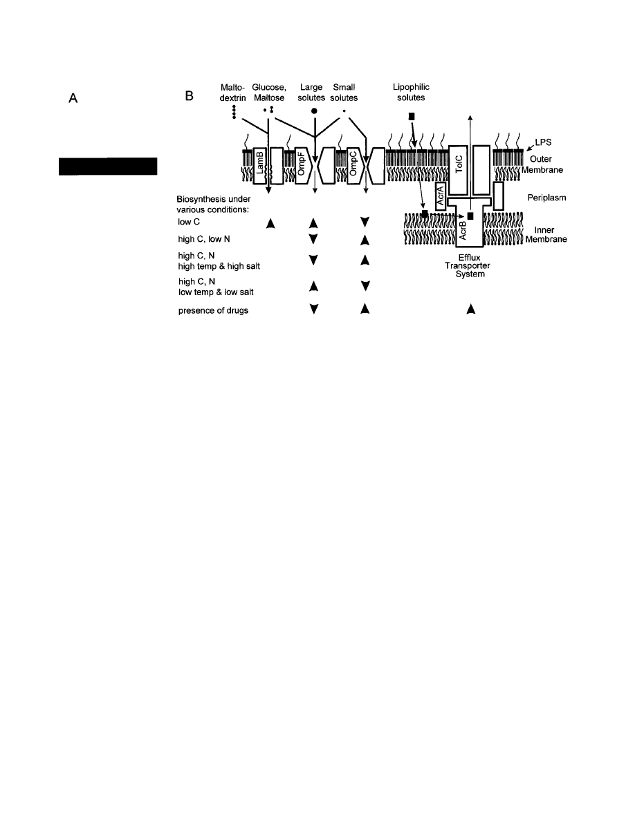
FIG. 1. Structure and barrier functions of the outer membrane of E. coli K-12. (A) A typical representation of the outer membrane [then usually called the
“lipopolysaccharide-(lipo)protein layer”] in microbiology textbooks in the early 1970s. (B) A current model of the outer membrane. Hydrophilic solutes cross this
barrier mainly via channels in nonspecific porins OmpF and OmpC and specific channels such as LamB. Smaller solutes can use both OmpF and OmpC, but larger
solutes (including most inhibitors) can only go through OmpF, with its wider channel. In either case, diffusion is slowed down drastically by the narrow opening in the
channel, shown by the different widths of arrows. Influx of maltose, maltodextrins, and glucose is facilitated by the LamB channel, with its specific sugar-binding site
within the channel. Large, lipophilic solutes traverse the lipid bilayer region of the membrane, but again diffusion is slowed down by the presence of the outer leaflet,
which contains only saturated fatty acid residues of LPS. Such lipophilic solutes tend to be pumped out directly into the medium by the multisubunit efflux transporters,
such as AcrA-AcrB-TolC (shown here) or EmrA-EmrB-TolC. Although the efflux catalyzed by such a pump may be slow, as indicated by the thin arrow, it will still
be effective as it works in synergy with the outer membrane barrier. Regulation of biosynthesis of various transporters under different conditions is shown at the bottom
of the figure. Upward-pointing arrowheads indicate increased synthesis, and downward-pointing ones show decreased synthesis. For details, see text.
V
OL
. 181, 1999
GUEST COMMENTARIES
5
http://jb.asm.org/
Downloaded from

cules, OmpF and OmpC, whose channel diameters are thought
to differ by only about 10% (38) (Fig. 1B). Nevertheless, be-
cause even the larger channel of OmpF is quite narrow (7 by 11
Å [6]), this translates into a very large difference in the diffu-
sion rates of most inhibitors, which are often relatively large
molecules. Thus, for E. coli, the default pattern would be to
express mostly the smaller OmpC channel for self-protection
to prevent the entry of inhibitors, unless other requirements
become more important. In fact, in its normal habitat, which is
rich in inhibitors such as bile salts and free fatty acids, the
synthesis of the larger OmpF channel is downregulated by
utilizing the high temperature (37°C) and high salt concentra-
tion (0.1 to 0.2 M) as signals (43). However, in natural waters,
where both temperature and salt concentration are much
lower, OmpF porin becomes predominant, allowing the cells to
accumulate nutrients more efficiently from the environment
(Fig. 1B). We would also expect that OmpF expression should
increase when the cells are starved for some nutrients, even at
37°C and in high-salt medium. Indeed, starvation for glucose
increases ompF transcription about 20-fold (26) (Fig. 1B). In
contrast, under glucose excess, NH
3
-limited conditions, OmpC
becomes essentially the only porin, a reasonable solution for E.
coli as the small ammonia molecules (or ions) would have no
difficulty in diffusing through the smaller OmpC channel (Fig.
1B). Finally, the presence of certain antibiotics increases the
synthesis of efflux pump AcrAB, at the same time decreasing
the synthesis of large-channel porin OmpF through the action
of the MarA global regulator that was discovered by S. B. Levy;
this tends to increase the resistance to antibiotics more effec-
tively by slowing down their influx and by accelerating their
efflux (reviewed in reference 35) (Fig. 1B).
A few outer membrane porins are specific. LamB (46), the
“phage
l receptor protein,” not only allows the efficient diffu-
sion of maltodextrins, some of which are too large to diffuse
through the regular porin channel, but also facilitates the influx
of maltose and glucose. The synthesis of LamB is induced by
maltose but also by carbon starvation, and under the latter
conditions, LamB becomes the major pathway of glucose entry
(8) (Fig. 1B).
Electrophysiological studies show that porin channels open
and close under various conditions (9), but we do not know the
relevance of these in vitro observations to the physiology of
whole cells. This reminds us that there are many areas still
waiting for exploration, in spite of the fact that we now know
so much about the outer membrane (including the high-reso-
lution structures of pore-forming proteins [6, 46]), in compar-
ison with our ignorance only two decades ago (textbooks in the
early 1970s did not mention the outer membrane, let alone its
functions [Fig. 1A]).
Interestingly, mycobacteria, which belong to the high-GC,
gram-positive bacterial group, were found to have the outer
layer of their rather impermeable cell wall organized essen-
tially as a lipid bilayer, with porin(s) to allow the diffusion of
hydrophilic solutes (17). Although the less-fluid leaflet of this
bilayer is the inner leaflet, in contrast to the gram-negative
bacterial outer membrane, in which the outer leaflet is less
fluid, the similarity in the construction is striking. Interestingly,
these high-GC, gram-positive bacteria appear to be most
closely related to the gram-negative bacteria if the sequences
of several proteins are used as the criterion (15).
Current perspective.
When Luria and Kalckar advocated
studies on bacterial cell surfaces almost 40 year ago, their
major interest was on the roles surface polymers may play in
the cell-to-cell interactions, as mentioned earlier. We have
indeed come a long way in this area. Cell-to-cell interaction
among bacteria obviously may occur in the community of bac-
teria growing as biofilms. It has been known that bacteria in
biofilms behave differently (for example in being much more
resistant to antibiotics) than those in a free-swimming form,
but little attention has been paid so far to the consequences of
interactions between cells. However, the regulation through
the production (and presumably high local concentration) of
autoinducers has been established (7). One would expect an
even larger role in contact-based or short-range interactions in
microorganisms with “social” life styles, such as myxobacteria
or slime molds. C signal in Myxococcus xanthus is indeed
thought to be generated by a surface-located protein and ex-
changed between tightly packed cells (10, 19), and the O chain
of LPS is needed for fruiting body formation (3).
Cell surface glycans obviously play important roles in the
interaction of symbiotic or pathogenic bacteria with their host
cells. In the classical scenario for pathogens, seen for example
with pneumococci, bacterial exopolysaccharides protect patho-
gens against nonspecific phagocytosis. Because recognition by
antibodies will nevertheless result in successful phagocytosis, it
is advantageous for pathogenic bacteria to produce glycans for
which the host will have difficulties in producing antibodies.
These glycans frequently contain unusual components: for ex-
ample, Salmonella LPS often contains rare 3,6-dideoxyhexoses.
Some pathogens even go to the length of producing glycans
that look like the glycans on host cells, a phenomenon called
molecular mimicry (see reference 30). In a remarkable exam-
ple, some human pathogens not only produce LPS whose
structures mimic those of human cell surface glycolipids but
also uses their enzymes and host donor compounds to sialylate
their LPS, presumably so that their cell surface will look even
closer to the host cell surface. With symbionts, in contrast, it
would be more advantageous to have the bacteria recognized
by host cells. When Rhizobium cells interact with the roots of
plants, the early stages are dominated by low-molecular-weight
compounds, nodulation factors. However, when the bacterial
cells reach the epidermis layer through infection threads, then
exopolysaccharides on the surface of Rhizobium (or oligosac-
charides derived from them) become indispensable for bacte-
rial invasion of continually elongating nodules (24). Most in-
terestingly, the invasion defect in exo mutants can be rescued
by the addition of exopolysaccharides from strains that nor-
mally nodulate that particular plant but not those from strains
that nodulate other plant species. Thus, the role of exopolysac-
charides here is specific.
The first step in bacterial pathogenesis in humans and ani-
mals is usually the specific recognition by a bacterial surface
component of a specific component of the host cell surface. In
the evolution of such a specific recognition process, it is easier
to fine-tune the structure of the protein partner than that of
the carbohydrate partner, because the structure of the latter
can be changed only in large increments. It was proposed (4) to
call the active partner (thus usually a protein) a “cognor” and
the passive partner (usually carbohydrate) a “cognon.” Many
gram-positive pathogens recognize and adhere to the compo-
nents of extracellular matrix of the host, and cognor proteins of
bacteria in this case have been called MSCRAMMs (microbial
surface components recognizing adhesive matrix molecules)
(40). Adhesion of E. coli through Pap pili to the cell surface
glycolipids containing
a-galactosyl-(134)-b-galactose struc-
ture, present on the surfaces of cells of some humans (48) is
another classical case of a bacterial cognor recognizing a spe-
cific cognon on animal cells. Most interestingly, it now appears
that this interaction signals changes in the bacterial cell (51), as
well as in the host cell (16). In some cases, however, such
specific recognition is used by host cells to “clear” infecting
organisms: cystic fibrosis transmembrane regulator (CFTR) on
6
GUEST COMMENTARIES
J. B
ACTERIOL
.
http://jb.asm.org/
Downloaded from

airway epithelial cells recognizes LPS on Pseudomonas aerugi-
nosa to initiate clearing, and this explains the common occur-
rence of P. aeruginosa infection in cystic fibrosis patients, who
have defective processing of CFTR (41).
Many bacterial pathogens must invade nonphagocytic host
cells. Paradigms of such interactions involve the invasion by
Shigella and Salmonella cells of a special class of epithelial cells
of the small intestine. This process occurs by the stimulation of
host cells, which are excited to produce spectacular changes in
the local cytoskeleton network and then to engulf bacterial
pathogens in their vacuoles. This stimulation of the host cells
was recently found (see reference 11) to be caused by injection
of a few bacterial proteins into the host cells through the
contact-dependent type III secretion systems, which are dis-
tributed widely, not only among animal pathogens but also
among plant pathogens (5, 23). Secretion machinery of this
type becomes activated by “contact” with the host cell surface,
but the factor that creates specificity in this interaction is still
largely unknown. A fascinating observation was made: Salmo-
nella cell surface assembles, upon contact with epithelial cells,
an appendage (14) which apparently is based on a syringe-like
apparatus reminiscent of a flagellar basal body, marking the
first time the type III secretion apparatus has been visualized
(22). Luria, if he were alive today, would be beside himself
learning that Salmonella cells truly does grow “hair” in a mat-
ter of minutes. This is indeed an exciting period for microbi-
ology and especially for the biology of microbial cell surfaces.
REFERENCES
1. Anderson, J. S., M. Matsuhashi, M. A. Haskin, and J. L. Strominger. 1965.
Lipid-phosphoacetylmuramyl-pentapeptide and lipid-phosphodisaccharide-
peptapeptide: presumed membrane transport intermediates in cell wall syn-
thesis. Proc. Natl. Acad. Sci. USA 53:881–889.
2. Bladen, H. A., and S. E. Mergenhagen. 1964. Ultrastructure of Veillonella
and morphological correlation of an outer membrane with particles associ-
ated with endotoxic activity. J. Bacteriol. 88:1482–1492.
3. Bowden, M. G., and H. B. Kaplan. 1998. The Myxococcus xanthus lipopoly-
saccharide O-antigen is required for social motility and multicellular devel-
opment. Mol. Microbiol. 30:275–284.
4. Burke, D., L. Mendoc¸a-Previato, and C. E. Ballou. 1980. Cell-cell recogni-
tion in yeast: purification of Hansenula wingei 21-cell sexual agglutination
factor and comparison of the factors from three genera. Proc. Natl. Acad.
Sci. USA 77:318–322.
5. Cornelis, G. R. 1998. The Yersinia deadly kiss. J. Bacteriol. 180:5495–5504.
6. Cowan, S. W., T. Schirmer, G. Rummel, M. Steiert, R. Ghosh, R. A. Pauptit,
J. N. Jansonius, and J. P. Rosenbusch.
1992. Crystal structures explain
functional properties of two E. coli porins. Nature 358:727–733.
7. Davies, D. G., M. R. Parsek, J. P. Pearson, B. H. Iglewski, J. W. Costerton,
and E. P. Greenberg.
1998. The involvement of cell-to-cell signals in the
development of a bacterial biofilm. Science 280:295–298.
8. Death, A., L. Notley, and T. Ferenci. 1993. Derepression of LamB protein
facilitates outer membrane permeation of carbohydrates into Escherichia coli
under conditions of nutrient stress. J. Bacteriol. 175:1475–1483.
9. Delcour, A. H. 1997. Function and modulation of bacterial porins: insights
from electrophysiology. FEMS Microbiol. Lett. 151:115–123.
10. Dworkin, M. 1996. Recent advances in the social and developmental biology
of the myxobacteria. Microbiol. Rev. 60:70–102.
11. Finlay, B. B., and S. Falkow. 1997. Common themes in microbial pathoge-
nicity revisited. Microbiol. Mol. Biol. Rev. 61:136–169.
12. Fukasawa, T., and H. Nikaido. 1961. Galactose-sensitive mutants of Salmo-
nella. II. Bacteriolysis induced by galactose. Biochim. Biophys. Acta 48:470–
483.
13. Ghuysen, J.-M. 1968. Use of bacteriolytic enzymes in determination of wall
structure and their role in cell metabolism. Bacteriol. Rev. 32:425–464.
14. Ginocchio, C. C., S. B. Olmsted, C. L. Wells, and J. E. Galan. 1994. Contact
with epithelial cells induces the formation of surface appendages on Salmo-
nella typhimurium. Cell 76:717–724.
15. Gupta, R. S. 1998. What are archaebacteria: life’s third domain or mono-
derm prokaryotes related to Gram-positive bacteria? A new proposal for the
classification of prokaryotic organisms. Mol. Microbiol. 29:695–707.
16. Hedlund, M., M. Svensson, Å. Nilsson, R. D. Duan, and C. Svanborg. 1996.
Role of ceramide-signaling pathway in cytokine responses to P-fimbriated
Escherichia coli. J. Exp. Med. 183:1037–1044.
17. Jarlier, V., and H. Nikaido. 1994. Mycobacterial cell wall: structure and role
in natural resistance to antibiotics. FEMS Microbiol. Lett. 123:11–18.
18. Jiang, K. M., B. Neal, F. Santiago, S. J. Lee, L. K. Romana, and P. R. Reeves.
1991. Structure and sequence of the rfb (O antigen) gene cluster of Salmo-
nella serovar typhimurium (strain LT2). Mol. Microbiol. 5:695–713.
19. Kaiser, D., and R. Losick. 1993. How and why bacteria talk to each other.
Cell 73:873–885.
20. Kalckar, H. M. 1965. Galactose metabolism and cell ’sociology.’ Science
150:
305–313.
21. Kamio, Y., and H. Nikaido. 1976. Outer membrane of Salmonella typhi-
murium: accessibility of phospholipid head groups to phospholipase C and
cyanogen bromide activated dextran in the external medium. Biochemistry
15:
2561–2570.
22. Kubori, T., Y. Matsushima, D. Nakamura, J. Uralil, M. Lara-Tejero, A.
Sukhan, J. E. Galan, and S.-I. Aizawa.
1998. Supramolecular structure of the
Salmonella typhimurium type III protein secretion system. Science 280:602–
605.
23. Lee, C. A. 1997. Type III secretion systems: machines to deliver bacterial
proteins into eukaryotic cells? Trends Microbiol. 5:148–156.
24. Leigh, J. A., and G. C. Walker. 1994. Exopolysaccharides of Rhizobium:
synthesis, regulation and symbiotic function. Trends Genet. 10:63–67.
25. Leive, L. 1965. Release of lipopolysaccharide by EDTA treatment of E. coli.
Biochem. Biophys. Res. Commun. 21:290–296.
26. Liu, X., and T. Ferenci. 1998. Regulation of porin-mediated outer membrane
permeability by nutrient limitation in Escherichia coli. J. Bacteriol. 180:3917–
3922.
27. Lu¨deritz, O., A. M. Staub, and O. Westphal. 1966. Immunochemistry of O
and R antigens of Salmonella and related Enterobacteriaceae. Bacteriol. Rev.
30:
192–255.
28. Ma¨kela¨, P. H., and B. A. D. Stocker. 1984. Genetics of lipopolysaccharide, p.
59–137. In E. T. Rietschel (ed.), Chemistry of endotoxin. Elsevier, Amster-
dam, The Netherlands.
29. McGrath, B. C., and M. J. Osborn. 1991. Localization of the terminal steps
of O-antigen synthesis in Salmonella typhimurium. J. Bacteriol. 173:649–654.
30. Moran, A. P., M. M. Prendergast, and B. J. Appelmelk. 1996. Molecular
mimicry of host structures by bacterial lipopolysaccharides and its contribu-
tion to disease. FEMS Immunol. Med. Microbiol. 16:105–115.
31. Nakae, T. 1976. Outer membrane of Salmonella. Isolation of protein com-
plex that produces transmembrane channels. J. Biol. Chem. 251:2176–2178.
32. Nakae, T., and H. Nikaido. 1975. Outer membrane as a diffusion barrier in
Salmonella typhimurium. Penetration of oligo- and polysaccharides into iso-
lated outer membrane vesicles and cells with degraded peptidoglycan layer.
J. Biol. Chem. 250:7359–7365.
33. Nikaido, H. 1968. Biosynthesis of cell wall lipopolysaccharide in Gram-
negative enteric bacteria. Adv. Enzymol. 31:77–124.
34. Nikaido, H. 1976. Outer membrane of Salmonella typhimurium. Transmem-
brane diffusion of some hydrophobic substances. Biochim. Biophys. Acta
433:
118–132.
35. Nikaido, H. 1996. Multidrug efflux pumps of gram-negative bacteria. J.
Bacteriol. 178:5853–5859.
36. Nikaido, H., M. Basina, V. Nguyen, and E. Y. Rosenberg. 1998. Multidrug
efflux pump AcrAB of Salmonella typhimurium excretes only those
b-lactam
antibiotics containing lipophilic side chains. J. Bacteriol. 180:4686–4692.
37. Nikaido, H., and T. Nakae. 1973. Permeability of model membranes con-
taining phospholipids and lipopolysaccharides: some preliminary results.
J. Infect. Dis. 128:S30–S34.
38. Nikaido, H., and E. Y. Rosenberg. 1983. Porin channels in Escherichia coli:
studies with liposomes reconstituted from purified proteins. J. Bacteriol.
153:
241–252.
39. Osborn, M. J. 1969. Structure and biosynthesis of the bacterial cell wall.
Annu. Rev. Biochem. 38:501–538.
40. Patti, J. M., B. L. Allen, M. J. McGavin, and M. Ho¨o¨k. 1994. MSCRAMM-
mediated adherence of microorganisms to host tissues. Annu. Rev. Micro-
biol. 48:585–617.
41. Pier, G. B., M. Grout, and T. S. Zaidi. 1997. Cystic fibrosis transmembrane
conductance regulator is an epithelial cell receptor for clearance of Pseudo-
monas aeruginosa from the lung. Proc. Natl. Acad. Sci. USA 94:12088–12093.
42. Ple´siat, P., and H. Nikaido. 1992. Outer membranes of Gram-negative bac-
teria are permeable to steroid probes. Mol. Microbiol. 6:1323–1333.
43. Pratt, L. A., H. Hsing, K. E. Gibson, and T. J. Silhavy. 1996. From acids to
osmZ: multiple factors influence synthesis of the OmpF and OmpC porins in
Escherichia coli. Mol. Microbiol. 20:911–917.
44. Robbins, P. W., and A. Wright. 1971. Biosynthesis of O-antigens, p. 351–368.
In G. Weinbaum, S. Kadis, and S. J. Ajl (ed.), Microbial toxins, vol. 4.
Academic Press, New York, N.Y.
45. Salton, M. R. J. 1994. The bacterial cell envelope—a historical perspective,
p. 1–22. In J.-M. Ghuysen and R. Hakenbeck (ed.), Bacterial cell wall,
Elsevier, Amsterdam, The Netherlands.
46. Schirmer, T., T. A. Keller, Y. F. Wang, and J. P. Rosenbusch. 1995. Struc-
tural basis for sugar translocation through maltoporin channels at 3.1 Å.
Science 267:512–514.
47. Strominger, J. L. 1970. Penicillin-sensitive enzymatic reactions in bacterial
V
OL
. 181, 1999
GUEST COMMENTARIES
7
http://jb.asm.org/
Downloaded from

cell wall synthesis. Harvey Lect. 64:179–213.
48. Va¨isa¨nen, V., J. Elo, L. G. Tallgren, A. Shtonen, P. H. Ma¨kela¨, C. Svanborg-
Ede´n, G. Ka¨llenius, S. B. Svenson, H. Hultberg, and T. Korhonen.
1981.
Mannose-resistant haemagglutination and P antigen recognition are charac-
teristic of Escherichia coli causing primary pyelonephritis. Lancet ii:1366–
1369.
49. Wright, A., M. Dankert, and P. W. Robbins. 1965. Evidence for an interme-
diate stage in the biosynthesis of the Salmonella O-antigen. Proc. Natl. Acad.
Sci. USA 54:235–241.
50. Wright, A., M. Dankert, P. Fennessey, and P. W. Robbins. 1967. Character-
ization of a polyisoprenoid compound functional in O-antigen biosynthesis.
Proc. Natl. Acad. Sci. USA 57:1798–1803.
51. Zhang, J. P., and S. Normark. 1996. Induction of gene expression in Esch-
erichia coli after pilus-mediated adherence. Science 273:1234–1236.
8
GUEST COMMENTARIES
J. B
ACTERIOL
.
http://jb.asm.org/
Downloaded from
Wyszukiwarka
Podobne podstrony:
Capability of high pressure cooling in the turning of surface hardened piston rods
736 Home in the woods id 35845 Nieznany (2)
Interaction of fraternal birth order and handedness in the
Interactions between parasites and microbial communities in the human gut
Semi Empirical Method for Estimating the Combustion Wave Transition through the Contact Surface in a
interactive art vs social interactions analysis of interactive art strategies in the light of erving
Dyson, Rebecca M i inni Interactions of the Gasotransmitters Contribute to Microvascular Tone (Dys)
Mettern S P Rome and the Enemy Imperial Strategy in the Principate
Early Variscan magmatism in the Western Carpathians
Applications and opportunities for ultrasound assisted extraction in the food industry — A review
In the end!
Post feeding larval behaviour in the blowfle Calliphora vicinaEffects on post mortem interval estima
Aftershock Protect Yourself and Profit in the Next Global Financial Meltdown
Multiple Intelligences in the Elementary Classroom
Fascia in the Lateral Upper Arm tapeSP
A Guide to the Law and Courts in the Empire
Functional improvements desired by patients before and in the first year after total hip arthroplast
więcej podobnych podstron